Prior heat stress induces moderation of diabetic alterations in glycogen metabolism of rats
11
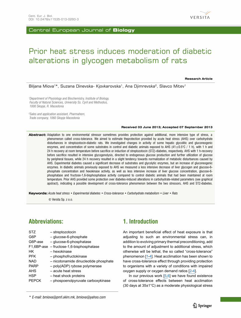
Central European Journal of Biology * E-mail: [email protected], [email protected] Research Article 1 Department of Physiology and Biochemistry, Institute of Biology, Faculty of Natural Sciences, University Ss. Cyril and Methodius, 1000 Skopje, R. Macedonia 2 Sales and application assistant, Pharmahem, Trade company, 1060 Skopje Macedonia Biljana Miova 1 *, Suzana Dinevska- Kjovkarovska 1 , Ana Djimrevska 2 , Slavco Mitev 1 Prior heat stress induces moderation of diabetic alterations in glycogen metabolism of rats Abbreviations: STZ – streptozotocin G6P – glucose-6-phosphate G6P-ase – glucose-6-phosphatase F1,6BP-ase – fructose-1,6-bisphosphatase HK – hexokinase PFK – phosphofructokinase NAD – nicotonamide dinucleotide phosphate PARP – poly(ADP) rybose polymerase AHS – acute heat stress HSP – heat shock proteins PEPCK – phospoenolpyruvate carboxykinase 1. Introduction An important beneficial effect of heat exposure is that adjusting to such an environmental stress can, in addition to evolving primary thermal preconditioning, add to the amount of adjustment to additional stress, which otherwise will be lethal, the so called “cross-tolerance” phenomenon [1-4]. Heat acclimation has been shown to have cross-tolerance effect through providing protection to organisms with a variety of conditions with impaired oxygen supply or oxygen demand ratios [2-4]. In our previous work [5,6] we have found existence of cross-tolerance effects between heat acclimation (30 days at 35±1°C) as a moderate physiological stress Cent. Eur. J. Biol. DOI: 10.2478/s11535-013-0260-3 Received 03 June 2013; Accepted 07 September 2013 Keywords: Acute heat stress • Experimental diabetes • Cross-tolerance • Carbohydrate metabolism • Liver • Rats Abstract: Adaptation to one environmental stressor sometimes provides protection against additional, more intensive type of stress, a phenomenon called cross-tolerance. We aimed to estimate theprotection provided by acute heat stress (AHS) over carbohydrate disturbances in streptozotocin-diabetic rats. We investigated changes in activity of some hepatic glycolitic and gluconeogenic enzymes, and concentration of some substrates in control and diabetic animals exposed to AHS (41±0.5°C / 1 h), with 1 h and 24 h recovery at room temperature before sacrifice or induction of streptozotocin (STZ)-diabetes, respectively. AHS with 1 h-recovery before sacrifice resulted in intensive glycogenolysis, directed to endogenous glucose production and further utilization of glucose by peripheral tissues, while 24 h recovery resulted in a slight tendency towards normalization of metabolic disturbances caused by AHS. Experimental diabetes caused a significant decrease of substrates and glycolytic enzymes, but an increase of gluconeogenic enzymes. In diabetic animals previously exposed to AHS we measured a less intensive decrease of liver glycogen and glucose-6- phosphate concentration and hexokinase activity, as well as less intensive increase of liver glucose concentration, glucose-6- phosphatase and fructose-1,6-bisphosphatase activity compared to control diabetic animals that had been maintained at room temperature. Prior AHS provided some protection over diabetes-induced alterations in carbohydrate-related parameters (see graphical apstract), indicating a possible development of cross-tolerance phenomenon between the two stressors, AHS and STZ-diabetes. © Versita Sp. z o.o.
-
Upload
independent -
Category
Documents
-
view
0 -
download
0
Transcript of Prior heat stress induces moderation of diabetic alterations in glycogen metabolism of rats
Central European Journal of Biology
* E-mail: [email protected], [email protected]
Research Article
1Department of Physiology and Biochemistry, Institute of Biology, Faculty of Natural Sciences, University Ss. Cyril and Methodius, 1000 Skopje, R. Macedonia
2Sales and application assistant, Pharmahem, Trade company, 1060 Skopje Macedonia
Biljana Miova1*, Suzana Dinevska- Kjovkarovska1, Ana Djimrevska2, Slavco Mitev1
Prior heat stress induces moderation of diabetic alterations in glycogen metabolism of rats
Abbreviations:STZ – streptozotocinG6P – glucose-6-phosphateG6P-ase – glucose-6-phosphataseF1,6BP-ase – fructose-1,6-bisphosphataseHK – hexokinasePFK – phosphofructokinaseNAD – nicotonamide dinucleotide phosphatePARP – poly(ADP) rybose polymeraseAHS – acute heat stressHSP – heat shock proteinsPEPCK – phospoenolpyruvate carboxykinase
1. Introduction An important beneficial effect of heat exposure is that adjusting to such an environmental stress can, in addition to evolving primary thermal preconditioning, add to the amount of adjustment to additional stress, which otherwise will be lethal, the so called “cross-tolerance” phenomenon [1-4]. Heat acclimation has been shown to have cross-tolerance effect through providing protection to organisms with a variety of conditions with impaired oxygen supply or oxygen demand ratios [2-4].
In our previous work [5,6] we have found existence of cross-tolerance effects between heat acclimation (30 days at 35±1°C) as a moderate physiological stress
Cent. Eur. J. Biol. DOI: 10.2478/s11535-013-0260-3
Received 03 June 2013; Accepted 07 September 2013
Keywords: Acute heat stress • Experimental diabetes • Cross-tolerance • Carbohydrate metabolism • Liver • Rats
Abstract: Adaptation to one environmental stressor sometimes provides protection against additional, more intensive type of stress, a phenomenon called cross-tolerance. We aimed to estimate theprotection provided by acute heat stress (AHS) over carbohydrate disturbances in streptozotocin-diabetic rats. We investigated changes in activity of some hepatic glycolitic and gluconeogenic enzymes, and concentration of some substrates in control and diabetic animals exposed to AHS (41±0.5°C / 1 h), with 1 h and 24 h recovery at room temperature before sacrifice or induction of streptozotocin (STZ)-diabetes, respectively. AHS with 1 h-recovery before sacrifice resulted in intensive glycogenolysis, directed to endogenous glucose production and further utilization of glucose by peripheral tissues, while 24 h recovery resulted in a slight tendency towards normalization of metabolic disturbances caused by AHS. Experimental diabetes caused a significant decrease of substrates and glycolytic enzymes, but an increase of gluconeogenic enzymes. In diabetic animals previously exposed to AHS we measured a less intensive decrease of liver glycogen and glucose-6-phosphate concentration and hexokinase activity, as well as less intensive increase of liver glucose concentration, glucose-6-phosphatase and fructose-1,6-bisphosphatase activity compared to control diabetic animals that had been maintained at room temperature. Prior AHS provided some protection over diabetes-induced alterations in carbohydrate-related parameters (see graphical apstract), indicating a possible development of cross-tolerance phenomenon between the two stressors, AHS and STZ-diabetes.
© Versita Sp. z o.o.
Prior heat stress and diabetes in rats
and streptozotocin-induced diabetes as an intensive pharmacological stress, observed by moderation of the carbohydrate-related alterations caused by experimental diabetes. Also, it was found that whole body hyperthermia (WBH) may provide a new therapeutic or preventive modality against obesity-related diseases such as type 2 diabetes mellitus and metabolic or insulin resistance syndrome [7]. Concerning cells, heat shock substantially increased the survival of STZ-treated islet cells by reduction of β-cell lysis by approximatwly 80% [8].
Diabetes mellitus, as a complex and heterogenic metabolic syndrome, is characterized by extensive disturbances in different metabolic pathways, with special respect to carbohydrate metabolism [9] and cell vulnerability [10], most obviously because of the down-regulated heat shock proteins (HSP) synthesis [11-13]. Concerning carbohydrate metabolism, it is well known that diabetes mellitus impairs the normal capacity of the liver to synthesize glycogen [14,15], causes suppression of hepatic glycolysis through decreased hexokinase, glucokinase and phosphofructokinase activity) [15,16], increases the activity of gluconeogenic enzymes, i.e. glucose-6-phosphatase, phospoenolpyruvate carboxykinase (PEPCK) [17-19] and fructose-1,6-bisphosphatase [14,18], which finally results in anelevation of blood glucose level.
Considering these findings, we aimed to determine whether animals also can be partially protected from the diabetogenic effects of streptozotocin, particularly the level of carbohydrate-related disturbances, by inducing a previous protective stress response such as a response to acute heat stress (1 hour at 41±0.5°C). Taking into consideration that the protective heat shock response cascade starts within minutes and its full impact is not expressed at the systemic level until 24 hours after a given stress [20,21], we investigated the effect of different recovery times (i.e. 1 h and 24 h) after the heat stress and before induction of experimental diabetes, on liver glycogen levels and glucose metabolism.
We hypothesize that there is cross-tolerance effect between acute hyperthermic stress and streptozotocin-induced diabetes due to effects on the carbohydrate-related enzymes and substrates, which depends on the time of recovery before induction of experimental diabetes.
2. Experimental Procedures2.1 Experimental animals and treatmentThe experiment was performed on adult Wistar rats (4-5 months old; n=48), kept on a standard food and water
regime during the whole experimental procedure and at a 12:12 L:D lighting regime (6 a.m. to 6 p.m. light).
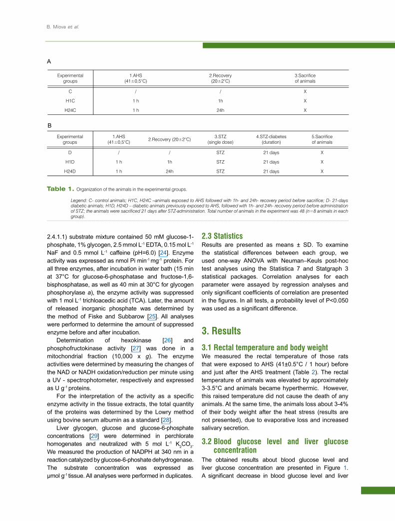
The animals were divided into two main groups: control (C) and diabetic (D). Further, each of the groups was divided into three subgroups, depending on the exposure to heat stress, as well as the period of recovery after the acute hyperthermic stress. Acute hyperthermic stress (AHS) was carried out for 1 hour in a special heat chamber, with a regulated air temperature of 41±0.5°C.
The first of the control subgroups, C was a control group that was maintained at room temperature (20±2°C) for the whole experiment; the second subgroup H1C was exposed to AHS, followed by 1 hour recovery at room temperature before sacrifice; the third subgroup H24C was exposed to AHS, followed by 24 hour recovery at room temperature before sacrifice.
In all diabetic subgroups, the experimental diabetes was induced by a single intraperitoneal injection of streptozotocine (STZ, 55 mg kg-1 body weight) dissolved in 0.1M citrate buffer with a pH of 4.5. For the diabetic animals, the three subgroups were the following: D animals kept at room temperature for the whole experimental period and sacrificed 21 days after STZ-administration; H1D animals that were first exposed to AHS, than allowed to recover for 1 h at room temperature, later injected with STZ and sacrificed 21 days after STZ-administration and; H24D animals that were first exposed to AHS, than allowed to recover for 24 h at room temperature, later injected with STZ and sacrificed 21 days after STZ-administration. The organization of the experimental groups is presented in Table 1a and 1b.
The rectal temperature was measured with a digital rectal thermometer before and immediately after the AHS. Animals were sacrificed after Na-thiopental narcosis (65 mg kg-1 b.w.). After laparatomy, pieces of liver were immediately frozen in liquid nitrogen (-196ºC) and kept in liquid nitrogen store there until analysis.
2.2 Analytical methodsFor determining enzyme activity, crude liver homogenates were prepared in an appropriate medium of homogenization (sucrose for glucose-6-phosphatase, lactic acid for fructose-1,6-bisphosphatase and NaF/glycilglycine for glycogen phosphorylase a). Glucose-6-phosphatase (E.C. 3.1.3.9) was assayed by the method of Hers [22] and the substrate mixture contained G-6-P (100 mmol L-1) and EDTA (2 mmol L-1),pH 6.5. Substrate mixture for fructose-1,6-bisphosphatase (E.C. 3.1.3.11) was prepared from 5 mmol L-1 fructose-1,6-bisphosphate, 2.5 mmol L-1 MgSO4, 5 mmol L-1 MnSO4, 30 mmol L-1 cysteine and 20 mmol L-1 serine [23]. For the activity of liver glycogen phosphorylase a (E.C.
B. Miova et al.
2.4.1.1) substrate mixture contained 50 mM glucose-1-phosphate, 1% glycogen, 2.5 mmol L-1 EDTA, 0.15 mol L-1
NaF and 0.5 mmol L-1 caffeine (pH=6.0) [24]. Enzyme activity was expressed as nmol Pi min-1 mg-1 protein. For all three enzymes, after incubation in water bath (15 min at 37°C for glucose-6-phosphatase and fructose-1,6-bisphosphatase, as well as 40 min at 30°C for glycogen phosphorylase a), the enzyme activity was suppressed with 1 mol L-1 trichloacedic acid (TCA). Later, the amount of released inorganic phosphate was determined by the method of Fiske and Subbarow [25]. All analyses were performed to determine the amount of suppressed enzyme before and after incubation.
Determination of hexokinase [26] and phosphofructokinase activity [27] was done in a mitochondrial fraction (10,000 x g). The enzyme activities were determined by measuring the changes of the NAD or NADH oxidation/reduction per minute using a UV - spectrophotometer, respectively and expressed as U g-1 proteins.
For the interpretation of the activity as a specific enzyme activity in the tissue extracts, the total quantity of the proteins was determined by the Lowry method using bovine serum albumin as a standard [28].
Liver glycogen, glucose and glucose-6-phosphate concentrations [29] were determined in perchlorate homogenates and neutralized with 5 mol L-1 K2CO3. We measured the production of NADPH at 340 nm in a reaction catalyzed by glucose-6-phoshate dehydrogenase. The substrate concentration was expressed as μmol g-1 tissue. All analyses were performed in duplicates.
2.3 StatisticsResults are presented as means ± SD. To examine the statistical differences between each group, we used one-way ANOVA with Neuman–Keuls post-hoc test analyses using the Statistica 7 and Statgraph 3 statistical packages. Correlation analyses for each parameter were assayed by regression analyses and only significant coefficients of correlation are presented in the figures. In all tests, a probability level of P<0.050 was used as a significant difference.
3. Results3.1 Rectal temperature and body weightWe measured the rectal temperature of those rats that were exposed to AHS (41±0.5°C / 1 hour) before and just after the AHS treatment (Table 2). The rectal temperature of animals was elevated by approximately 3-3.5°C and animals became hyperthermic. However, this raised temperature did not cause the death of any animals. At the same time, the animals loss about 3-4% of their body weight after the heat stress (results are not presented), due to evaporative loss and increased salivary secretion.
3.2 Blood glucose level and liver glucose concentration
The obtained results about blood glucose level and liver glucose concentration are presented in Figure 1. A significant decrease in blood glucose level and liver
Experimental groups
1.AHS (41±0,5°C)
2.Recovery (20±2°C)
3.Sacrifice of animals
C / / X
H1C 1 h 1h X
H24C 1 h 24h X
Experimental groups
1.AHS(41±0,5°C) 2.Recovery (20±2°C) 3.STZ
(single dose)4.STZ-diabetes
(duration)5.Sacrifice of animals
D / / STZ 21 days X
H1D 1 h 1h STZ 21 days X
H24D 1 h 24h STZ 21 days X
A
B
Table 1. Organization of the animals in the experimental groups.
�Legend:�C-�control�animals;�H1C,�H24C�–animals�exposed�to�AHS�followed�with�1h-�and�24h-�recovery�period�before�sacrifice;�D-�21-days�diabetic�animals;�H1D,�H24D�–�diabetic�animals�previously�exposed�to�AHS,�followed�with�1h-�and�24h-�recovery�period�before�administration�of�STZ;�the�animals�were�sacrificed�21�days�after�STZ-administration.�Total�number�of�animals�in�the�experiment�was�48�(n=8�animals�in�each�group).
Prior heat stress and diabetes in rats
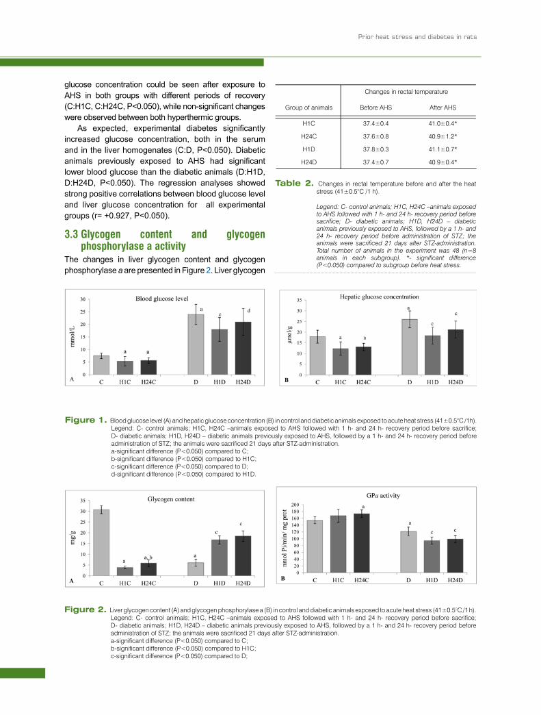
glucose concentration could be seen after exposure to AHS in both groups with different periods of recovery (C:H1C, C:H24C, P<0.050), while non-significant changes were observed between both hyperthermic groups.
As expected, experimental diabetes significantly increased glucose concentration, both in the serum and in the liver homogenates (C:D, P<0.050). Diabetic animals previously exposed to AHS had significant lower blood glucose than the diabetic animals (D:H1D, D:H24D, P<0.050). The regression analyses showed strong positive correlations between blood glucose level and liver glucose concentration for all experimental groups (r= +0.927, P<0.050).
3.3 Glycogen content and glycogen phosphorylase a activity
The changes in liver glycogen content and glycogen phosphorylase a are presented in Figure 2. Liver glycogen
Changes in rectal temperature
Group of animals Before AHS After AHS
H1C 37.4±0.4 41.0±0.4*
H24C 37.6±0.8 40.9±1.2*
H1D 37.8±0.3 41.1±0.7*
H24D 37.4±0.7 40.9±0.4*
Table 2. Changes in rectal temperature before and after the heat stress (41±0.5°C /1 h).
Legend: C- control animals; H1C, H24C –animals exposed to AHS followed with 1 h- and 24 h- recovery period before sacrifice;� D-� diabetic� animals;� H1D,� H24D� –� diabetic�animals�previously�exposed�to�AHS,�followed�by�a�1�h-�and�24 h- recovery period before administration of STZ; the animals�were� sacrificed� 21� days� after� STZ-administration.�Total� number� of� animals� in� the� experiment� was� 48� (n=8�animals� in� each� subgroup).� *-� significant� difference�(P<0.050)�compared�to�subgroup�before�heat�stress.
Figure 1. Blood glucose level (A) and hepatic glucose concentration (B) in control and diabetic animals exposed to acute heat stress (41±0.5°C /1h).Legend: C- control animals; H1C, H24C –animals exposed to AHS followed with 1 h- and 24 h- recovery period before sacrifice; D- diabetic animals; H1D, H24D – diabetic animals previously exposed to AHS, followed by a 1 h- and 24 h- recovery period before administration of STZ; the animals were sacrificed 21 days after STZ-administration.
a-significant difference (P<0.050) compared to C; b-significant difference (P<0.050) compared to H1C; c-significant difference (P<0.050) compared to D; d-significant difference (P<0.050) compared to H1D.
Figure 2. Liver glycogen content (A) and glycogen phosphorylase a (B) in control and diabetic animals exposed to acute heat stress (41±0.5°C /1 h).Legend: C- control animals; H1C, H24C –animals exposed to AHS followed with 1 h- and 24 h- recovery period before sacrifice; D- diabetic animals; H1D, H24D – diabetic animals previously exposed to AHS, followed by a 1 h- and 24 h- recovery period before administration of STZ; the animals were sacrificed 21 days after STZ-administration.
a-significant difference (P<0.050) compared to C; b-significant difference (P<0.050) compared to H1C; c-significant difference (P<0.050) compared to D;
B. Miova et al.
content was significantly and dramatically reduced by approximately 80-90% in AHS treated groups, at both 1 h- and 24 h-recovery subgroups (C:H1C, C:H24C, P<0.050). Nevertheless, the 24 h-recovery caused a rebound of the glycogen content compared to 1 h-recovery subgroup (H1C:H24C, +52.5%, P<0.05).
In the same experimental groups, the obtained results showed that AHS caused a non-significant increase of the GPa activity in the H1C subgroup (8.6%, n.s) and a significant increase in the H24C subgroup (9.8%, P<0.050).
STZ- diabetes significantly decreased liver glycogen content (C:D, P<0.050) and GPa activity (C:D, -22.3, P<0.050). On the other side, in both heat-exposed diabetic animal subgroups (H1D and H24D), the spending of glycogen stores was less evident compared to diabetic unexposed animals (by approximately 2 fold), most obviously as a result of additional decrease of GPa activity (D:H1D and D:H24D, P<0.050). Thus providing the assumption that there is some moderation of the glycogenolytic effect when animals are heat-preconditioned before STZ-administration. It could be that prior heat exposure has some protective effect against the intensive glycogenolytic and hyperglycemic effect of STZ.
3.4 Glucose-6-phosphate concentration, glucose-6-phosphatase and fructose-1,6-bisphosphatase activity
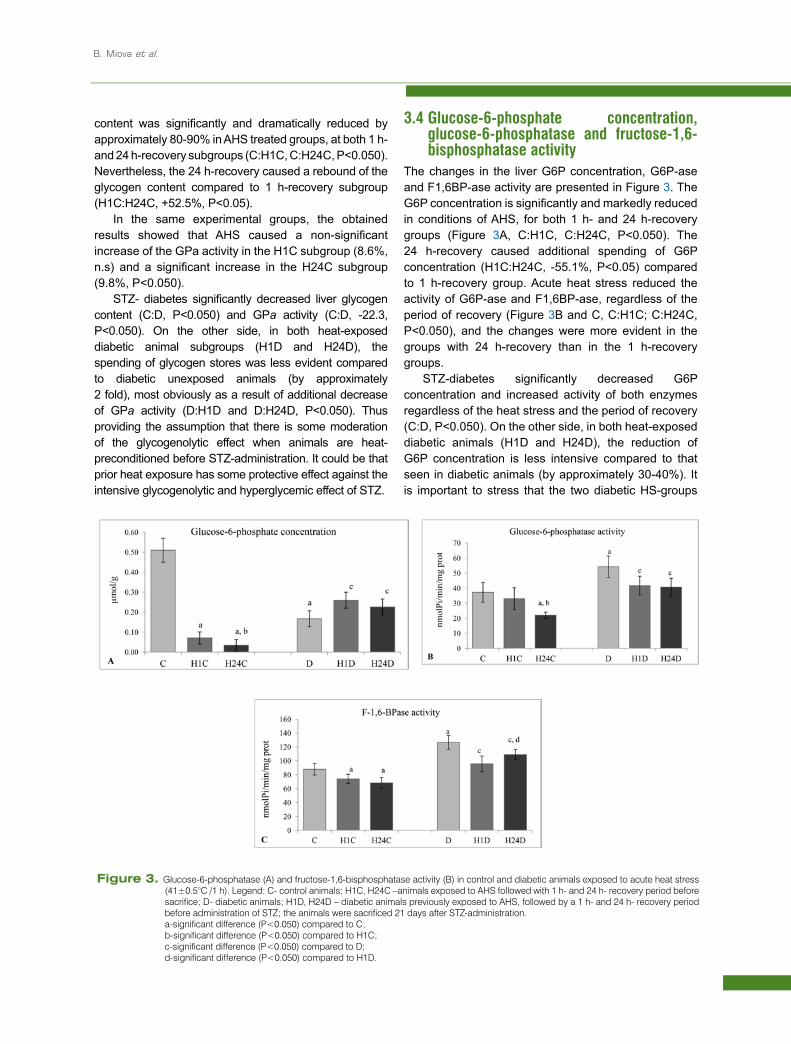
The changes in the liver G6P concentration, G6P-ase and F1,6BP-ase activity are presented in Figure 3. The G6P concentration is significantly and markedly reduced in conditions of AHS, for both 1 h- and 24 h-recovery groups (Figure 3A, C:H1C, C:H24C, P<0.050). The 24 h-recovery caused additional spending of G6P concentration (H1C:H24C, -55.1%, P<0.05) compared to 1 h-recovery group. Acute heat stress reduced the activity of G6P-ase and F1,6BP-ase, regardless of the period of recovery (Figure 3B and C, C:H1C; C:H24C, P<0.050), and the changes were more evident in the groups with 24 h-recovery than in the 1 h-recovery groups.
STZ-diabetes significantly decreased G6P concentration and increased activity of both enzymes regardless of the heat stress and the period of recovery (C:D, P<0.050). On the other side, in both heat-exposed diabetic animals (H1D and H24D), the reduction of G6P concentration is less intensive compared to that seen in diabetic animals (by approximately 30-40%). It is important to stress that the two diabetic HS-groups
Figure 3. Glucose-6-phosphatase (A) and fructose-1,6-bisphosphatase activity (B) in control and diabetic animals exposed to acute heat stress (41±0.5°C /1 h). Legend: C- control animals; H1C, H24C –animals exposed to AHS followed with 1 h- and 24 h- recovery period before sacrifice; D- diabetic animals; H1D, H24D – diabetic animals previously exposed to AHS, followed by a 1 h- and 24 h- recovery period before administration of STZ; the animals were sacrificed 21 days after STZ-administration.
a-significant difference (P<0.050) compared to C; b-significant difference (P<0.050) compared to H1C; c-significant difference (P<0.050) compared to D; d-significant difference (P<0.050) compared to H1D.
Prior heat stress and diabetes in rats
showed lower G6P-ase and F1,6BP-ase activity compared to control diabetic animals (D:H1D, D:H24D, -13.7% up to -25.1%, P<0.05). The results indicated, with some moderation, a diabetes-induced elevation of the gluconeogenetic enzymes caused by prior AHS treatment.
The obtained results show similar trends for both gluconeogenic enzymes for all experimental groups, presented with a moderately strong correlation between G6P-ase and F1,6BP-ase (r = +0.673, P<0.050).
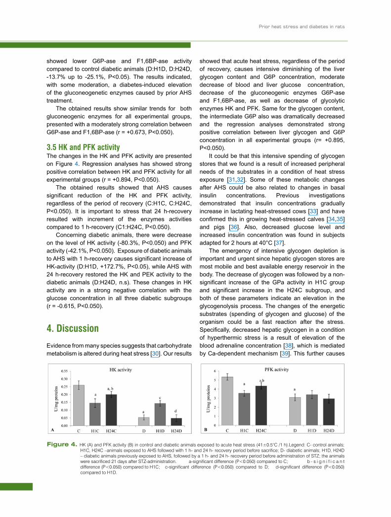
3.5 HK and PFK activityThe changes in the HK and PFK activity are presented on Figure 4. Regression analyses has showed strong positive correlation between HK and PFK activity for all experimental groups (r = +0.894, P<0.050).
The obtained results showed that AHS causes significant reduction of the HK and PFK activity, regardless of the period of recovery (C:H1C, C:H24C, P<0.050). It is important to stress that 24 h-recovery resulted with increment of the enzymes activities compared to 1 h-recovery (C1:H24C, P<0.050).
Concerning diabetic animals, there were decrease on the level of HK activity (-80.3%, P<0.050) and PFK activity (-42.1%, P<0.050). Exposure of diabetic animals to AHS with 1 h-recovery causes significant increase of HK-activity (D:H1D, +172.7%, P<0.05), while AHS with 24 h-recovery restored the HK and PEK activity to the diabetic animals (D:H24D, n.s). These changes in HK activity are in a strong negative correlation with the glucose concentration in all three diabetic subgroups (r = -0.615, P<0.050).
4. DiscussionEvidence from many species suggests that carbohydrate metabolism is altered during heat stress [30]. Our results
showed that acute heat stress, regardless of the period of recovery, causes intensive diminishing of the liver glycogen content and G6P concentration, moderate decrease of blood and liver glucose concentration, decrease of the gluconeogenic enzymes G6P-ase and F1,6BP-ase, as well as decrease of glycolytic enzymes HK and PFK. Same for the glycogen content, the intermediate G6P also was dramatically decreased and the regression analyses demonstrated strong positive correlation between liver glycogen and G6P concentration in all experimental groups (r= +0.895, P<0.050).
It could be that this intensive spending of glycogen stores that we found is a result of increased peripheral needs of the substrates in a condition of heat stress exposure [31,32]. Some of these metabolic changes after AHS could be also related to changes in basal insulin concentrations. Previous investigations demonstrated that insulin concentrations gradually increase in lactating heat-stressed cows [33] and have confirmed this in growing heat-stressed calves [34,35] and pigs [36]. Also, decreased glucose level and increased insulin concentration was found in subjects adapted for 2 hours at 40°C [37].
The emergency of intensive glycogen depletion is important and urgent since hepatic glycogen stores are most mobile and best available energy reservoir in the body. The decrease of glycogen was followed by a non-significant increase of the GPa activity in H1C group and significant increase in the H24C subgroup, and both of these parameters indicate an elevation in the glycogenolysis process. The changes of the energetic substrates (spending of glycogen and glucose) of the organism could be a fast reaction after the stress. Specifically, decreased hepatic glycogen in a condition of hyperthermic stress is a result of elevation of the blood adrenaline concentration [38], which is mediated by Ca-dependent mechanism [39]. This further causes
Figure 4. HK (A) and PFK activity (B) in control and diabetic animals exposed to acute heat stress (41±0.5°C /1 h).Legend: C- control animals; H1C, H24C –animals exposed to AHS followed with 1 h- and 24 h- recovery period before sacrifice; D- diabetic animals; H1D, H24D – diabetic animals previously exposed to AHS, followed by a 1 h- and 24 h- recovery period before administration of STZ; the animals were sacrificed 21 days after STZ-administration. a-significant difference (P<0.050) compared to C; b - s i g n i f i c a n t difference (P<0.050) compared to H1C; c-significant difference (P<0.050) compared to D; d-significant difference (P<0.050) compared to H1D.
B. Miova et al.
increase in activity of Ca-dependent protein kinase and increase of glycogen phosphorylase, as we found in our results.
It could be noticed that in the 1 h-recovery group, the glycogenolisys is directed to endogenous glucose production and its further utilization by the peripheral tissues. Contrastingly, the HK activity in H1C groups was significantly decreased, same for the PFK activity, suggesting decreased hepatic glucose phosphorylation and glycolysis 1 hour after heat stress. Statistical analyses also suggested a strong correlation between HK and PFK activity (r = 0.906, P<0.050) on the level on C, H1C and H24C subgroups.
Concerning 24 h-recovery groups, there is a significant increase in liver glycogen and HK activity compared to H1C subgroups. Since in H24C animals an additional decrease of G6P-ase and G6P concentration is observed compared to H1C animals, it might be that some resynthesis of glycogen is manifested. Also, as there is higher PFK in H24C animals than in the H1C ones, some of the G6P could be directed in glycolysis. Even though the observed changes in the H24C subgroup are slight, they might imply for tendency of normalization of the metabolic disturbances 24 hours after the heat stress.
In both of the different periods of recovery, the gluconeogenesis, manifested by F1,6BP-ase was decreased. It is important to note that the both gluconeogenic enzymes (G6P-ase and F1,6BP-ase) showed parallel manner, with significant positive coefficient of correlation concerning the C, H1C and H24C subgroups (r= 0.517, P<0.050).
As for experimental diabetes, our results showed that besides the well known hyperglycemia, there also was a reduction in hepatic glycogen and G6P concentration, decrease in glycogenolytic/ glycolytic enzymes (GP-ase, HK and PFK) acitivity and an elevation in gluconeogenic enzyme activity (G6P-ase and F1,6BP-ase) in rats. The elevated G6P-ase activity and mRNA level [40] in animals in the diabetic state resulted in increased dephosphorylation of G6P and stimulation of endogenous glucose production [41], which is the main reason for hyperglycemia in all types of diabetes [42]. The increased G6P-ase activity in conditions of experimental diabetes is accompanied with increased activity of F1,6BP-ase. In rats with streptozotocin-induced diabetes there is 2-fold increase in the F1,6BP-ase in hepatocyte suspension [43] and 10-fold increase of F1,6BP-ase mRNA [44].The reduced concentration of G6P in diabetic rats , which is a stimulator of glycogen synthase [45,46], resulted in decreased glycogen production [47]. In our results experimental diabetes caused a decrease of GPa. It was most obvious that lower enzyme activity was caused by a reduction in
the enzyme protein level and was presumably due to a decrease in synthesis [48].
Finally, this research paid special interest to the diabetic subgroups previously exposed to AHS and allowed to recover for 1 h or 24 h before induction of diabetes. In all diabetic groups, STZ-diabetes lasted for 21 days before sacrifice of the animals. Generally, heat preconditioning significantly moderated the diabetes-induced alterations in the observed carbohydrate-related enzymes and substrates.
Specifically, in the diabetic animals previously exposed to heat stress (H1D, H24D) we measured less intensive decrease of liver glycogen content and G6P concentration, as well as less intensive increase of liver glucose concentration than diabetic animals that had no previous heat stress (D). Concerning enzyme activities, we measured higher glucose phosphorylation (higher HK activity) in H1D group, which resulted in a higher G6P concentration and further higher glycogen content than that observed in diabetic animals that had no previous heat stress. Conversely, the glucose output (presented by lower GPa, G6P-ase and F1,6BP-ase) was decreased in the same animals. The situation is similar in H24D groups, except for the less intensive glucose phosphorylation (lower HK activity) and non-significantly lower intracellular G6P compared to the H1D group. If we can take in to consideration that diabetes is manifested with intensive glycogenolysis and gluconeogenesis, obviously prior heat stress causes moderation of both of the enzymatic processes responsible for hepatic glucose output. We can consider that preconditioning of diabetic animals with heat expose provides a protective effect over the metabolic disturbances caused by experimental diabetes.
Our first clarification for the above assumption is reduced loss of viability of pancreatic cells by heat preconditioning. Namely, previous studies have shown that heat shock (43°C for 90 min) reduces cell lysis from streptozotocin by 90% and the depletion of NAD, the major cause of radical induced islet cell death, was suppressed after heat shock [8]. According to Okamoto model for b-cells destruction [49], STZ and other different kind of stimulus (e.g. radiation, viruses) cause DNA damage, which activate the nuclear enzyme poly(ADP-ribose) polymerase (PARP) and cause depletion of the substrate NAD.
Since NAD+ is a key cofactor of glycolysis, the reduction of NAD+ levels is sufficient to impair this process [50], which further diminishes the major source of rapid ATP formation that could be used for NAD+ resynthesis. In addition, the low level of NAD reduces the flow of glucose-derived metabolites into the TCA cycle for oxidation, and later compromises energy
Prior heat stress and diabetes in rats
balance [51]. As heat stress causes preservation and consequently an increase of inracellular NAD [52], we thought that the deleterious effects of PARP and STZ over β-cells should be less evident in heat-exposed organisms. Furthermore, probably there still will be some insulin production, which could be main reason for moderation of the diabetic disturbances in hepatic metabolism.
All of the heat-stressed animals in our investigations become hyperthermic during 1 hour exposure to 41±0.5°C, but it is important to mention that this increment of the rectal temperature was not lethal for the animals . It is well known that the response of the organism to the increased environmental temperature causes induction of heat shock response (HSR) and production of a highly conservative family of proteins, i.e. the heat shock proteins (HSP) [53]. Whole-body hyperthermia is associated with HSP 70 synthesis and induction of thermotolerance [54]. According to some previous investigations [55], whole body hyperthermia also resulted with markedly increased HSP70 levels in the liver and adrenal gland of both control and diabetic rats, but still, the induction of HSP70 was significantly reduced in these tissues of diabetic rats compared to those of controls.
Most of the stress proteins are molecular chaperones [56] and are crucial for the maintenance of cell integrity during normal growth as well as during pathophysiological conditions. Considering this, we propose that HSP synthesis could be another protective mechanism caused by previous heat stress over the diabetogenic effects of STZ. Based on some previous investigations [1-4], we assume that the beneficial effect of heat exposure is that this primary thermal preconditioning adds to the amount of adjustment to additional stress (such as STZ-diabetes), which is previously defined as a “cross-tolerance” phenomenon.
According to Tytell and Hooper [57] preconditioning prior to injury in a period of hours up to 24, is allowed so that HSPs have time to accumulate to higher than normal levels. We assume that a recovery period of 1 h and 24 h (after the applied HS and before STZ induction) will make β-cells and the whole organism more resistant to the DNA-damaging effects of STZ.
Taking in to consideration that diabetes mellitus is associated with impaired HSP response [58,59], it is possible that the increased HSP may protect diabetic organisms from injury changes, since they are efficacious in protecting and repairing damaged proteins, as may occur in protein glycation [60].
These results could be related to the promising effect of sauna therapy for patients with diabetes and other life-style diseases [61], as well as with the helpful hot-tub therapy for diabetic patients [62] and diabetic rats [63]. Moreover, Kokura et al. [7] found that whole body hyperthermia (WBH) may provide a new therapeutic or preventive modality against obesity-related diseases such as type 2 diabetes mellitus and metabolic or insulin resistance syndrome [8].
Considering all above, we conclude that prior heat stress causes moderation of the diabetic alterations in liver carbohydrate metabolism. Finally, we could confirm the hypothesis of the existence of cross-tolerance phenomenon between acute heat stress and STZ-diabetes over moderation of the carbohydrate-related disturbances in diabetic animals.
AcknowledgementsThis study was supported by the Institute of Biology at Faculty of Natural Sciences, University St Cyrilus and Methodius, Skopje.
Conflict of interest: The authors declare that they have no conflict of interest.
[1] Horowitz M., Matching the heart to heat-induced circulatory load: heat acclimatory responses, News Physiol. Sci., 2003,18, 215–221.
[2] Tetievsky A., Cohen O., Eli-Berchoer L., Gerstenblith G., Stern M., Wapinski I., et al., Physiological and molecular evidence of heat acclimation memory: a lesson from thermal responses and ischemic cross-tolerance in the heart, Physiol. Genomics, 2008, 34, 78–87
[3] Eynan M., Knubuvetz T., Meiri U., Navon G., Gerstenblith G., Bromberg Z., et al., Heat
acclimation-induced elevated glycogen, glycolysis, and low thyroxine improve heart ischemic tolerance, J. Appl. Physiol., 2002, 93, 2095–2104
[4] Horowitz M., Eli-Berchoer L., Wapinski I., Friedman N., Kodesh E., Stress-related genomic responses during the course of heat acclimation and its association with ischemic-reperfusion cross-tolerance, J. Appl. Physiol., 2004, 97, 1496–1507
[5] Dinevska-Kjovkarovska S., Miova B., Mitev S., Popeska Z., Some aspects of hepatic carbohydrate
References
B. Miova et al.
metabolism in heat-acclimated diabetic rats, Biol. Maced., 2007, 60. 37-51
[6] Miova B., Acclimation to moderate heat and gene expression of the key carbohydraterelated enzymes in rats with nicotinamide / streptozotocine induced diabetes, Doctoral thesis.2007, Faculty of natural sciences and mathematics, Skopje, R. Macedonia
[7] Kokura S., Adachi S., Manabe E., Mizushima K., Hattori T., Okuda T., et al., Whole body hyperthermia improves obesity-induced insulin resistance in diabetic mice, Int. J. Hyperthermia, 2007, 23, 259-265
[8] Bellmann K., Wenz A., Radons J., Burkart V., Kleemann R., Kolb H., Heat shock induces resistance in rat pancreatic islet cells against nitric oxide, oxygen radicals and streptozotocin toxicity in vitro, J. Clin. Invest., 1995, 95, 2840-2845
[9] Delaney C.A., Dunger A., Di Matteo M., Cunningham J.M., Green M.H., Green I.C., Comparison of inhibition of glucose-stimulated insulin secretion in rat islets of Langerhans by streptozotocin and methyl and ethyl nitrosoureas and methanesulphonates. Lack of correlation with nitric oxide-releasing or 6-alkylating ability, Biochem. Pharmacol., 1993, 50, 2015-2020
[10] Tytell M., Hooper P.L., Heat shock proteins: new keys to the development of cytoprotective therapies, Expert Opin. Ther. Targets, 2001, 5, 2, 267-87
[11] Kurucz I., Morva A., Vaag A., Eriksson K.F., Huang X., Groop Let al., Decreased expression of heat shock protein 72 in skeletal muscle of patients with type 2 diabetes correlates with insulin resistance, Diabetes, 2002, 51, 1102–1109.
[12] Atalay M., Oksala N.K., Laaksonen D.E., Khanna S., Nakao C., Lappalainen J., et al., Exercise training modulates heat shock protein response in diabetic rats, J. Appl. Physiol., 2004, 97, 605–611
[13] Hooper L., Diabetes, Nitric Oxide, and Heat Shock Proteins, Diabetes care, 1993, 26, 3, 951-952
[14] Whitton P.D., Hems D.A., Glycogen synthesis in the perfused liver of streptozotocin-diabetic rats, Biochem. J., 1975, 150. 153-165
[15] Vats V., Yadav S.P., Grover J.K., Effect of T. foenumgraecum on glycogen content of tissues and the key enzymes of carbohydrate metabolism, J. Ethnopharmacol., 2003, 85, 237-242
[16] Pari L., Amarnath-Satheesh M., Effect of pterostilbene on hepatic key enzymes of glucose metabolism in streptozotocin- and nicotinamide-induced diabetic rats, Life Sci., 2006, 79, 641–646
[17] Li Y., Méchin M.C., van de Werve G., Diabetes Affects Similarly the Catalytic Subunit and Putative Glucose-6-phosphate Translocase of Glucose-6-
phosphatase, J. Biol. Chem., 1999, 274, 33866-33878
[18] Mithieux G., Vidal H., Zitoun C., Bruni N., Daniele N., Minassian C., Glucose-6-phosphatase mRNA and activity are increased to the same extent in kidney and liver of diabetic rats, Diabetes, 1996, 45, 891–896
[19] Marzban L., Roshanak R., Brownsey R.W., M’cneill J.H., Mechanisms by which Bis(Maltolato) Oxovanadium(IV) normalizes phosphoenolpyruvate carboxykinase and glucose-6-phosphatase expression in streptozotocin- diabetic rats in vivo, Endocrinology, 2002, 143, 4636–4645
[20] Laszlo A., The thermoresistant state: protection from initial damage or better repair?, Exp. Cell Res., 1992, 202, 519–531
[21] Maloyan A, Palmon A, Horowitz M., Heat acclimation increases basal HSP72 level and alters its production dynamics during heat stress. Am. J. Physiol. Regulatory Integrative Comp. Physiol., 1999, 276, 1506–1515
[22] Hers H.G., Glycogen storage disease. In: Levine R, Luft R, eds. Advances in Metabolic Disorders, New York, London: Academic press, 1959, 36-85
[23] Freedland R.A., Harper A.E., Metabolic adaptations in higher animals. V. The study of metabolic pathways by means of metabolic adaptations, J. Biol. Chem., 1959, 243, 1350-1354
[24] Stalmans W., Wuif H., Hue L., Hers H.G., The sequential inactivation of glycogen phosphorylase and activation of glycogen syntethase after the administration of glucose to mice and rats. The mechanism of the hepatic threshold to glucose, Eur. J. Biochem., 1974, 41, 117-134
[25] Fiske C.A., Subbarow Y., The colorimetric determination of phosphorus, J. Biol. Chem., 1925, 66, 357-400
[26] Bontemps F., Hue L., Hers H.G., Phosphorylation of glucose in isolated hepatocytes, Biochem J., 1978, 774, 603–611
[27] Bergmeyer U., Michal G., Methods of enzymatic analysis, In: Bergmeyer HU (ed) Vol 1., Academic, New York, 1974
[28] Lowry O.H., Rosenbrough J.N., Ffffarr L.A., Rrandall J.R., Protein measurement with the folin phenol reagent, J. Boil. Chem., 1951, 193, 265-275
[29] Keppler D., Decker K., Glycogen determination with amyloglucosidase, In Methods of Enzymatic Analysis, vol. 3 (ed. H.U. Bergmeyer); New York: Academic Press, 1974, 1127-1131
[30] Streffer C., Aspects of metabolic change after hyperthermia, Recent Results Canc. Res., 1998, 107, 7–16
Prior heat stress and diabetes in rats
[31] Kameyama T., Nabeshima T., Nagata H., Hypoglycemia in mice exposed to an environment of high temperature and humidity, Res. Comm. Chem. Pathol. Pharmacol., 181, 32(2), 261-279
[32] Torlinska T., Paluszak J., Waliszevska A., Banach R., Effect of hyperthermia on blood levels of corticosterone and certain metabolites in rats during thiobutabarbital aneasthesia, Acta Physiol. Pol., 1984, 35(3), 243-7
[33] Wheelock J.B., Rhoads R.P., Van Baale M.J., Sanders S.R., Baumgard L.H., Effects of heat stress on energetic metabolism in lactating Holstein cows, J. Dairy Sci., 2010, 93, 644–655
[34] O’Brien M.D., Rhoads R.P., Sanders S.R., Duff G.C., Baumgard L.H., Metabolic adaptations to heat stress in growing cattle, Domest. Anim. Endocrinol., 2010, 38, 86–94
[35] Lance H., Baumgard, Robert P., Rhoads Jr., Effects of Heat Stress on Postabsorptive Metabolism and Energetics, Annu. Rev. Anim. Biosci., 2013, 1, 311–337
[36] Pearce S.C., Upah N.C., Harris A.J., Gabler N.K., Ross J.W., Rhoads, R.P., et al., Effects of heat stress on energetic metabolism in growing pigs, FASEB J., 2011, 25:1052.5 (Abstr.)
[37] Hargreaves M., Angus D., Howlett K., Marmy Conus N., Febbraio M., Effect of heat stress on glucose kinetics during exercise, Appl. Physiol., 1996, 81, 1594-1597
[38] Becker B.A., Kril J.J., Materri R.L., Spiers D.E., Ellersiek M., Misfeldt M.L., Endocrine and thermoregulatory responses to acute thermal exposures to 6-month-old pigs reared to different neonatal environments, J. Thermal. Biology, 1997, 22(2), 87-93
[39] Strickland W.G., Blackmore P.F., Exton J.H., The role of calcium in alphaadrenergic inactivation of glycogen synthase in rat hepatocytes and its inhibition by insulin, Diabetes, 1980, 29, 617-622
[40] Mithieux G., Vidal H., Zitoun C., Bruni N., Daniele N., Minassian C., Glucose-6- phosphatase mRNA and activity are increased to the same extent in kidney and liver of diabetic rats, Diabetes, 1996, 45, 891–896
[41] Henly D.C., Phillips J.W., Berry M.N., Suppressing of glycolysis is associated with an increase in glucose cycling in hepatocytes from diabetic rats, J. Biol. Chem., 1996, 271 (19), 11268-11271
[42] Barzilai N., Rossetti L., Diabetes Affects Similarly the Catalytic Subunit and Putative Glucose-6-phosphate Translocase of Glucose-6-phosphatase, J. Biol. Chem., 1993, 268, 25019–25025.
[43] Golubev M.A., Markova M.S., Stvolinskaia N.S., Level of fructose-2,6-bisphosphate and activity of fructose-1,6-bisphosphatase in hepatocyte suspensions in streptozotocine diabetes, Vopr. Med. Khim., 1993, 39(6), 13-15
[44] El-Maghrabi M.R., Pilkis J., Marker A.J., Colosia A.D., D’Angelo G., Fraser B.A., et al., cDNA sequence of rat liver fructose-1,6-bisphosphatase and evidence for down-regulation of its mRNA by insulin, PNAS., 1988, 85(22), 8430-8434
[45] Cadefau J., Bollen M., Stalmans W., Glucose-induced glycogenesis in the liver involves the G-6-Pate-dependent dephosphorylation of glycogen synthase, Biochem J., 1997, 15(322), 745-750
[46] Villar-Palasi C., Guinovart J.J., The role of glucose-6-phosphate in the control of glycogen synthase, FASEB J., 1997, 11, 544-548
[47] Cline G.W., Petersen K.F., Krssak M., Shen J., Hundal R.S., Trajanovski Z., et al., Impaired glucose transport as a cause of a decreased insulin-stimulated muscle glycogen synthesis in type 2 diabetes, New Engl. J. Med., 1999, 341, 240-246
[48] Rulfs J., Jaspers S.R., Garnache A.K., Miller T.B., Phosphorylase synthesis in diabetic hepatocytes and cardiomyocytes, Am. J. Physiol., 1989, 257, E74–E80
[49] Yamamoto H., Uchigata Y., Okamoto H., Streptozotocin and alloxan induce DNA strand breaks and poly(ADP–ribose) synthetase in pancreatic islets, Nature, 1981, 294, 284–286
[50] Houtkooper R.H., Canto´C., Wanders R.J., Auwerx J., The secret life of NAD+: an old metabolite controlling new metabolic signaling pathways. Endocr. Rev, 2010. 31, 194–223
[51] Bai P., Canto C., The role of PARP-1 and PARP-2 enzymes in metabolic regulation and disease, Cell Metab., 2012, 16, 290-295
[52] Gray C.C., Amrani M., Smolenski T.R., Taylor G.L., Yacoub M.H., Age dependence of heat stress mediated cardioprotection, Ann. Thorac. Surg., 2000, 70, 21–626
[53] Feder M.E. and Hofmann G.E., Heat-shock proteins, molecular chaperones, and the stress response: Evolutionary and ecological physiology, Annu. Rev. Physiol., 1999, 61, 243–282
[54] King Y.T., Lin C.S., Lin J.H., Lee W.C.,Whole-body hyperthermia-induced thermotolerance is associated with the induction of Heat Shock Protein 70 in mice, J. Exp. Biol., 2002, 205, 273–278
[55] Yamagishi N., Nakayama K., Wakatsuki T., Hatayama T., Characteristic changes of stress protein expression in streptozotocin-
B. Miova et al.
induced diabetic rats, Life Sciences, 2001, 69, 2603–2609
[56] Parsell D.A. and Lindquist S., The function of heat shock proteins in stress tolerance: degradation and reactivation of damaged proteins, Annu. Rev. Genet., 1993, 27, 437–496
[57] Tytell M., Hooper L.P., Heat shock proteins: new keys to the development of cytoprotective therapies, Expert. Opin. Ther. Targets, 2001, 5(2), 267-287
[58] Bitar M.S., Farook T., John B., Heat-shock protein 72/73 and impaired wound healing in diabetic and hypercortisolemic states, Surgery, 1999, 125, 594-601
[59] Strokov I.A., Manukhina E.B., Bakhtina L.Y., The function of endogenous protective systems in
patients with insulin- dependent diabetes mellitus and polyneuropathy: effect of antioxidant therapy, Bull. Exp. Biol. Med, 2000, 130, 986-990
[60] Jaattela M., Heat shock proteins as cellular lifeguards, Ann. Med, 1999, 31, 261-271
[61] Biro S., Masuda A., Kihara T., Tei C., Clinical implications of thermal therapy in lifestyle-related diseases, Experimental Biology and Medicine, 2003, 228, 1245-1249
[62] Hooper P., Hot-tub therapy for type 2 diabetes mellitus, New Engl. J. Med., 1999, 341, 924-925
[63] Bathaie S.Z., Jafarnejad A., Hosseinkhani S., Nakhjavani M., The effect of hot-tub therapy on serum Hsp70 level and its benefit on diabetic rats: a preliminary report, 2010, 26(6), 577-585