Multifaceted applications of bile salts in pharmacy - CiteSeerX
Upload
independentCategory
view
1download
0
Carlsberg Res. Commun. Vol. 54, p. 203-229, 1989
GLYCOGEN PHOSPHORYLASE: A MULTIFACETED ENZYME
Presented as the 9th Linderstr0m-Lang Award Lecture at the Carlsberg Laboratory Copenhagen on 29th November, 1989
by
LOUISE N. JOHNSON
Laboratory of Molecular Biophysics, Rex Richards Building, South Parks Road, Oxford OX1 3QU, UK
Keywords: Glycogen phosphorylase, time resolved studies, catalytic mechanism, oligosaccharide recognition, allosteric mechanism, phosphorylation, stereoelectronic effects
1. INTRODUCTION It is a very great honour for me to receive the
Linderstr0m-Lang award and to join your list of distinguished previous recipients. The work of the Carlsberg Laboratory first came to my no- tice in the mid 1960s when I joined Professor EM. RICHARDS'S Laboratory at Yale for a post doctoral year. There I learnt of the influence and inspiration of LINDERSTROM-LANG and the pioneering work that had been achieved on protein folding. In one of the earliest and most remarkable experiments on molecular recogni- tion it was shown that specific cleavage by sub- tilisin of the peptide bond between residues 19 and 20 in ribonuclease yielded ribonuclease S protein and peptide; separately neither was ca- talytically active but on association full activity was regained. Now, the Laboratory is distin- guished in many spheres but especially so in the study of enzymes involved in carbohydrate me- tabolism and it is this area that makes a link with the theme of my lecture, glycogen phosphory- lase. The discussion of our recent results on the X-ray crystallography of this enzyme will centre around the properties associated with protein carbohydrate recognition, the mechanism of ca- talysis with special reference to time resolved
X-ray crystallographic work, and the allosteric mechanism.
Glycogen phosphorylase Glycogen phosphorylase (EC 2.4.1.1) cataly-
ses the degradative phosphorylation of glyco- gen to glucose-l-phosphate, the initial step in the generation of metabolic energy in muscle.
(Glycogen)n + Pi "<= > (Glycogen)n_1 + a-D-Glucose- 1-phosphate
This large enzyme (subunit molecular weight 97,440; functionally active form dimer) is an ar- chetypal control enzyme. It exhibits regulation both by reversible phosphorylation and by ailo- steric effectors and is able to integrate diverse signals associated with ligand binding at 5 spati- ally distinct sites. To a first approximation these effects can be understood in terms of an equili- brium between several conformational states ranging from a low affinity T state to a high affi- nity R state according to the model of MONOD et al. (58) (Fig. 1). In resting muscle, phosphory- lase is in the b form T state and requires AMP for activation to the R state (12,30). The enzyme is inhibited by the T state ligands, glucose-6-P, ATP and ADP and the R state ligand UDPG.
Springer-Verlag 0105-1938/89/0054/0203/$05.40
L.N. JOHNSON: Glycogen phosphorylase
COVALEI~CONTROL )
I phosphocylase kinase i p
~l phosphaCase p
phosphoryIase b phosphorylase a INACTIVE INACTIVE (T s ta te ) (T s t a t e )
AHP ATP Glucose-6-P Glucose Caffeine
Clucose Caffeine
N ~0
N C 0 V A L E N T
C 0 N T R 0 L
phosphoryl ase b phoaphorylase ACTIVE ACTIVE
(R s t a t e ) (R s t a t e )
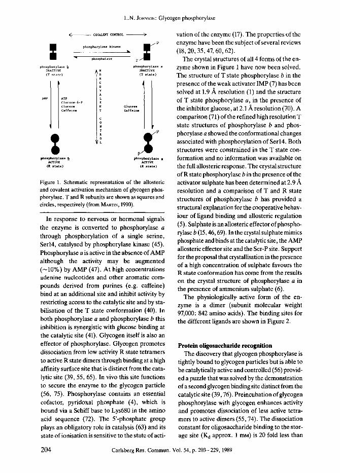
Figure 1. Schematic representation of the allosteric and covalent activation mechanism of glycogen phos- phorylase. T and R subunits are shown as squares and circles, respectively (from MARTXN, 1990).
In response to nervous or hormonal signals the enzyme is converted to phosphorylase a through phosphorylation of a single serine, Serl4, catalysed by phosphorylase kinase (45). Phosphorylase a is active in the absence of AMP although the activity may be augmented ( -10%) by AMP (47). At high concentrations adenine nucleotides and other aromatic com- pounds derived from purines (e.g. caffeine) bind at an additional site and inhibit activity by restricting access to the catalytic site and by sta- bilisation of the T state conformation (40). In both phosphorylase a and phosphorylase b this inhibition is synergistic with glucose binding at the catalytic site (41). Glycogen itself is also an effector of phosphorylase. Glycogen promotes dissociation from low activity R state tetramers to active R state dimers through binding at a high affinity surface site that is distinct from the cata- lytic site (39, 55, 65). In vivo this site functions to secure the enzyme to the glycogen particle (56, 75). Phosphorylase contains an essential cofactor, pyridoxal phosphate (4), which is bound via a Schiff base to Lys680 in the amino acid sequence (72). The 5'-phosphate group plays an obligatory role in catalysis (63) and its state of ionisation is sensitive to the state of acti-
vation of the enzyme (17). The properties of the enzyme have been the subject of several reviews (18, 20, 35, 47, 60, 62).
The crystal structures of all 4 forms of the en- zyme shown in Figure 1 have now been solved. The structure of T state phosphorylase b in the presence of the weak activator IMP (7) has been solved at 1.9/~ resolution (1) and the structure of T state phosphorylase a, in the presence of the inhibitor glucose, at 2.1/~ resolution (70). A comparison (71) of the refined high resolution T state structures of phosphorylase b and phos- phorylase a showed the conformational changes associated with phosphorylation of Serl4. Both structures were constrained in the T state con- formation and no information was available on the full allosteric response. The crystal structure of R state phosphorylase b in the presence of the activator sulphate has been determined at 2.9/~ resolution and a comparison of T and R state structures of phosphorylase b has provided a structural explanation for the cooperative behav- iour of ligand binding and allosteric regulation (5). Sulphate is an allosteric effector of phospho- rylase b (15, 46, 69). In the crystal sulphate mimics phosphate and binds at the catalytic site, the AMP allosteric effector site and the Ser-P site. Support for the proposal that crystallisation in the presence of a high concentration of sulphate favours the R state conformation has come from the results on the crystal structure of phosphorylase a in the presence of ammonium sulphate (6).
The physiologically active form of the en- zyme is a dimer (subunit molecular weight 97,000:842 amino acids). The binding sites for the different ligands are shown in Figure 2.
Protein oligosaccharide recognition The discovery that glycogen phosphorylase is
tightly bound to glycogen particles but is able to be catalytically active and controlled (56) provid- ed a puzzle that was solved by the demonstration of a second glycogen binding site distinct from the catalytic site (39, 76). Preincubation of glycogen phosphorylase with glycogen enhances activity and promotes dissociation of less active tetra- mers to active dimers (55, 74). The dissociation constant for oligosaccharide binding to the stor- age site (K d approx. 1 mM) is 20 fold less than
204 Carlsberg Res. Commun. Vol. 54, p. 203-229, 1989

L.N. JOHNSON: Glycogen phosphorylase
~ , ~ Tower
AIIoster ic effector Site (N)
Contact to allosteric site of other subunit
Glycogen storage Site(G)
a2
20
(~ 19
/
Nucleos |de inhibitor Site
x~_J z /
(I) Catalytic Site (C)
/ /
/ Pyridoxal Phosphate
j/J ~.
ot27
Figure 2. A schematic ribbon diagram of T state phosphorylase b subunit, a helices are shown as cylinders and/3 strands as arrows. The essential cofactor, pyridoxal phosphate, is buried at the centre of the subunit. The catalytic site (C), shown here with glucose-l-phosphate, is close to the cofactor and accessible to the bulk solvent through a channel some 15/~ long. The allosteric effector site (N) is located at the subunit-subunit interface. The glycogen stor- age site (G) is on the surface of the enzyme and removed from the allosteric and catalytic sites. The nucleoside site (I) is situated at the entrance to the catalytic site channel. This site binds purines or nucleosides or nucleotides at high concentrations and occupancy of this site stabilises the T state and inhibits the enzyme.
the K m for oligosaccharide at the catalytic site (39). Studies on the association of oligosacchar-
ides with phosphorylase provide a detailed ex-
ample of protein sugar interactions.
The glycogen storage site is situated on the surface of the molecule and is over 30 ,~ from
the catalytic and allosteric sites (Fig. 2). A ma-
jor and a minor glycogen storage site have been defined from the study of the phosphorylase- maltoheptaose complex which has been refined at 2.5 A resolution to a crystallographic R factor
of 0.146 (34, 53) and from ligand bound com- plex in which oligosaccharide was present (36).
At the major site, b inding of only 5 sugars in
subsites labelled $3-$4-$5-$6-$7 is observed.
The reducing end of the oligosaccharide is in subsite $3 and this site has the lowest crystallo-
graphic z coordinate. Glucosyl residues in these
sites make interactions to residues of the a12 helix and the spur formed by the small antipa- rallel sheet/315-/316 (Fig. 2). All the contacts to the oligosaccharide are included in the stretch
Carlsberg Res. Commun. Vol. 54, p. 203-229, 1989 205

L.N. JoHNSoN:Glycogen phosphorylase
of chain from residues 398-437 (a12-a13-/315- /316). The minor site consists of only 2 sugars and it lies above the non-reducing end of the major site making contacts to the top of the a12 helix, the loop of antiparallel/3 sheet from/38- /39 (the top loop) and the one contact to a resi- due from c~9.
The 5 c~(1-4) linked glucosyl sugars adopt a left-handed amylose like helix such that the 2 ends of the helix curl away from the protein sur- face. The major conformational changes in the protein involve residues Glu433 and Lys437 which move so as to optimise contacts with the sugars in subsites $4 and $5. Tyr404 is in the right orientation to make contact with the oligo- saccharide.
The contacts between the oligosaccharide and protein are shown in Figure 3. The sugar in $5 makes the largest number of contacts to the
protein and is almost inaccessible to solvent. The 02 hydroxyl is hydrogen bonded to Glu433 and Lys437, the 05 to Asn407 and the 06 to Tyr404 main chain oxygen and Asn407 side cha- in. In addition, there are van der Waals contacts to Tyr404, Asn407, Gln408, Glu433 and Lys437. Tyr404 fits into the groove formed by the glyco- sidic linkage between subsites $5 and $6 with the lone pair of electrons on the glycosidic ox- ygen and the ring oxygen directed away from the tyrosine. The hydrogens bonded to C1 and C2 atoms of the sugar in site $6 and to the C4 atom of the sugar in site $5 are directed towards the aromatic ring. Protein-oligosaccharide inter- actions require complementarity both for the polar and for the non-polar components of the sugars. The stacking of some of the non-polar groups against the oligosaccharide are shown in Figure 4. Five non-polar side chains are invol- ved in this surface site and appear to contribute significantly to the binding energy, although the glycogen storage site itself is not significantly more non-polar than other surface regions of the protein (53).
1
Figure 3. The interactions between maltoheptaose and phosphorylase b at the major glycogen storage site. Only 5 subsites labeled $3-$7 are localised (36).
206
. ~ _ ,5
Figure 4. The non-polar residues that contribute to maltoheptaose recognition site (36).
Carlsberg Res. Commun. Vol. 54, p. 203-229, 1989
L.N. JOItNSON: Glycogen phosphorylase
The specificity of the glycogen storage site for different length oligosaccharides ranging from maltose to maltoheptaose and for other com- pounds such as acarbose has been discussed (34), Glucose at 100 mM concentration does not bind to the glycogen storage site in the crystal. Maltose, the smallest compound that has been observed to bind at this site, is located in subsites $4 and $5 and the conformation in these subsites is similar to that shown in Figure 3. The sugar bound in subsite $5 makes numerous specific contacts (6 hydrogen bonds and 28 van der Waals interactions) and the preference of maltose for this site is not surprising. The preference for $4-$5 rather than $5-$6 is interesting. The su- gars in subsites $4 and $6 make 7 and 16 van den Waals interactions, respectively, but the sugar in subsite $4 makes one extra hydrogen bond. The hydrogen bonds with Glu433 and Lys437, which span subsites $4 and $5, appear an import- ant determinant of specificity in directing the second sugar of maltose into subsite $4 despite the fewer van den Waals contacts of this site compared with $6.
Catalysis in the crystal Early kinetic and crystallographic experi-
ments had established that glycogen phosphory- lase b crystals were catalytically active. The kinetic studies (38) showed a decrease in rate of about 30 fold in the crystal compared with solu- tion but little change in Km values for the sub- strates, oligosaccharide and glucose-l-P. The studies also showed a large value for K m for oligosaccharide (about 175 mM), which was si- milar for the enzyme in the crystal and in solution. X-ray experiments on catalysis in the crystal showed that the reaction could be followed either in the direction of oligosaccharide breakdown with the formation of glucose-l-P or in the direc- tion of oligosaccharide synthesis with the libera- tion of inorganic phosphate (23). In these exper- iments oligosaccharide was not observed to bind at the catalytic site although it must have visited the catalytic site in order to achieve the cataly- sis. In the T state access to the catalytic site is re- stricted by a loop the 280s loop (residues 281 to 287) and this observation provides an explana-
tion for the low affinity of the enzyme for oligo- saccharide (Fig. 2). Our most informative stu- dies on catalysis in the crystal have been carried out with the small pseudo substrate heptenitol.
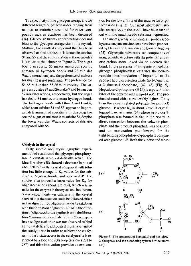
The use of glycosylic substrates to probe carbo- hydrase enzyme mechanisms have been pioneer- ed by HEHRE and LEHMANN and their colleagues (25). Glycosylic substrates are compounds of nonglycosidic structure with the potential anom- eric carbon atom linked via an electron rich bond. In the presence of inorganic phosphate, glycogen phosphorylase catalyses the non-re- versible phosphorylation of heptenitol to the product heptulose-2-phosphate (B-l-C-methyl, a-D-glucose-l-phosphate) (42, 43) (Fig. 5). Heptulose-2-phosphate (H2P) is a potent inhi- bitor of the enzyme with a K i= 14/zM. The pro- duct is bound with a considerably higher affinity than the closely related substrate (or product) glucose 1-P where K m is about 3 mM. In crystal- lographic experiments (54) where heptulose-2- phosphate was formed in situ in the crystal, a direct interaction between the cofactor phos- phate and the product phosphate was observed and an explanation put forward for the tight binding of heptulose-2-phosphate compar- ed with glucose 1-P. Both the kinetic and struc-
C6 06 CH20H
k/C4 Cl ~ - ~ C H ^ I \ / c, -
H OH O2
C6 O6 CH2OH
(b) ~c4 03 c~/'~ HO 0 P = 0 d \o , j 08
. g. o. 07
Figure 5. The structures of heptanitol and heptulose- 2-phosphate and the numbering system for the atoms (36).
Carlsberg Res. Commun. Vol. 54, p. 203-229, 1989 207

L.N. JOHNSON: Glycogen phosphorylase
208 Carlsberg Res. Commun. Vol. 54, p. 203-229, 1989

L.N. JOHNSON: Glycogen phosphorylase
Figure 6. Difference Fourier syntheses in the vicinity of the catalytic site of glycogen phosphorylase b for the hep- tenitol to heptulose-2-phosphate conversion. A single positive contour is shown (300 arbitrary units). Selected amino acids and the pyridoxal phosphate in the native enzyme conformation are shown. Water molecules are in- dicated as crosses. (a) The control experiment: crystal soaked in 100 mM heptenitol. The glucopyranose ring is view- ed almost edge on. His377 is displaced slightly as indicated by additional positive contours. (b) Early stage of the reaction 100 mM phosphate, 50 mM phosphate, 2.5 mM AMP; 10 min soak; 1 h data collection at 13 ~ Additional electron density for the phosphate is apparent. (c) Reaction completed: 100 mM heptenitol, 50 mM phosphate, 2.5 mM AMP, 50 mM maltoheptaose; 50 h soak; 2.5 h data collection. The product heptulose-2-phosphate is apparent from the electron density and there are additional indications for the movement of Arg569 (23).
turai results showed that heptulose-2-phos- phate exhibited some properties characteristic of a transition state analogue.
It is difficult to obtain a value for the turnover number of the enzyme with heptenitol because of strong product inhibition. However, arseno- lysis of heptenitol (a reaction that is closely simi-
lar to phosphorylysis but which yields a product
that decomposes and which does not inhibit the enzyme) can be followed and gives a turnover of about 18 i min- (43). This compares with a value
of about 100 sec -1 for the natural reaction with glycogen. In the crystal the reaction will be slower.
KASVINSKY and MADSEN (38) have demonstrated that the reaction is approximately 30 fold slower
in the crystal than in solution. In the time resolv- ed experiments the control properties of the en- zyme were exploited to further slow down the reaction so that neither AMP or oligosaccharide were effective activators. Under these condi- tions the reaction rate may be reduced by as much as 9000 fold leading to a turnover of about
15% in an experiment with a time course of 70 min (23).
The time resolved experiments at the Syn-
chrotron Radiation Source, Daresbury involv-
ed strenuous X-ray data collection achievable
with superb group work. In the experiments the crystal was mounted in a flow cell (22, 78) and the
reaction initiated by flowing substrate over the
Carlsberg Res. Commun. Vol. 54, p. 203-229, 1989 209

L.N. JOHNSON: Glycogen phosphorylase
crystal. Diffusion and binding times were measur- ed in separate experiments (33) and were found to take 10 to 20 min depending on the size of ligand and crystal size. X-ray data were collect- ed to 2.5 ~ resolution on an Arndt-Wonacott oscillation camera (3) at the synchrotron stations (27, 28) either immediately or after various rest- ing times which allowed the reaction to proceed. The results of the experiment with heptenitol are shown in Figure 6. In Figure 6a, the control experiments, heptenitol is shown bound at the catalytic site close to the essential cofactor pyri- doxal phosphate. In the time resolved experi- ment (Fig. 6b) where measurements were com- pleted within 60 min for a crystal soaked in 100 mM heptenitol, 50 mM phosphate and 2.5mM AMP for 10 min the difference electron den- sity map showed the addition of a phosphate group. A series of other experiments with differ- ent time intervals and conditions were carried out (23) but only the end result (54) is shown in Figure 6c. The product heptulose-2-phosphate has been formed and there is a direct interaction with the product phosphate and the cofactor phosphate.
The phosphorylase-product complex The crystal structure of the phosphorylase-
heptuiose-2-phosphate (36) complex has been refined at 2.9/~ resolution by molecular dyna- mics and crystallographic least squares proce- dures (8). The product is firmly bound at the ca- talytic site and exhibits thermal factors that are comparable to the most well ordered regions of the enzyme. The major conformational change of the enzyme is a movement of an arginine resi- due, Arg569, from a position buried in the pro- tein to a new position in which it can contact the product phosphate. The importance of this resi- due for phosphorylase catalysis and control has been previously recognised from chemical mo- dification experiments (14, 73). The arginine displaces an acidic group, Asp283, from the ca- talytic site and this replacement of an acidic group by a basic group is a key feature of the creation of the phosphate recognition site. Three water molecules are displaced from the catalytic site by heptulose-2-phosphate binding, two by the phosphate group and one by the 03
hydroxyl. The interactions of the pyridoxal phosphate with the enzyme are essentially un- changed from those of the native enzyme (61) apart from the contact to heptulose-2-phosphate.
\ . o / ,Lys C O " Gay677
568 +
G,Y,k, ")%-- -"" 135P I~- 0 / ~ ' 0 "
- ,O" , . o - e . . . . : H N Thr
I"o "o Io,, / __NI-~- :~1- ~ O , l ~ l ~ P l " " 'HN~ 5 NH? ~ '3<" ', ~ v ~ ' . 7 . - "
674 r ' : - " ' , ' ~ u ' ~ ' . H ~" OH- . . . . - o = ~ . / \ ', : ' 0 . - - H" 484 2
s73 GI.~ / C / H i , 672/ 377
Figure 7. Schematic representation of the polar con- tacts between heptulose-2-phosphate and phospbory- lase b (36).
The interactions between the product and the enzyme are summarised in Figure 7. In common with other oligosaccharide binding proteins, all the polar groups of the sugar are observed to form hydrogen bonds with the enzyme. The ex- ception is the ring oxygen, 05, whose separa- tion from the main chain N of Leu136 (3.5/~) is long for a hydrogen bond. Most of the protein groups involved in the hydrogen bonds are from planar groups (main chain N of Gly675 and 677, Asn484, His377, Glu672 and Arg569). The pro- duct phosphate is hydrogen bonded to the co- factor phosphate. The position is stabilised by ionic interactions and hydrogen bonds of the product phosphate to Arg569, Lys574, Tyr573 and the main chain N of Gly135. Contacts be- tween the 2 phosphates are also mediated by a water molecule that bridges the phosphate oxy- gens and links to the 04 hydroxyl of the sugar. Gly135 occurs at the start of a helix. The sequence of residues 132 to 137 is Gly-Asn-Gly-Gly-Leu-
210 Carlsberg Res. Commun. Vol. 54, p. 203-229, 1989

L.N. JOHNSON: Glycogen phosphorylase
Gly and shows similarity to the Gly-X-GIy-X-X- Gly sequence that is important in nucleotide phosphate recognition sites (77).
The conformation of heptulose-2-phosphate bound to the enzyme differs from the conforma- tion observed for glucose-l-phosphate (50) in the torsion angle about the glycosidic bond. In the heptulose-2-phosphate complex the torsion angle O5-C1-O1-P is 224 ~ and the bond angle at O1 is 135 ~ The corresponding values for the glucose-l-phosphate complex are 140 ~ and 120 ~ . The change in torsion angle appears to be direct- ed by the additional methyl group in the beta configuration and there is an internal hydrogen
bond between a phosphate oxygen and the 0 2 hydroxyl. The change in torsion angle decreases the product phosphorus-cofactor phosphorus distance from 6.0 ~ in the glucose-l-phosphate complex to 4.8 A in the heptulose-2-phosphate complex and allows the direct hydrogen bond between the phosphates. This is of importance for proposals for the catalytic mechanism.
Catalytic mechanism The evidence leading to proposals for the ca-
talytic mechanism and the role of the cofactor 5' phosphate group have been reviewed (35, 48,
.o ~ c.2o. c.-o. .o~" \ , ~ c . 3
�9 _A--~f \ ~ �9 HO I HO" ' HO /C H ~ ) ~ ' c H 3
o~" c% -o~. : o
;"~-~o- ",. -CJ~ ~176 0".~. _ O" Off
o: o \p/
~ o / \\o px L.~- 0 / P ~ o PXL/0 PXL /
(,)
.o" \ " ~ o " ~ . o ~"
\ " ~ 0 " "0 ~ Off
o- :-. . . \p / o/~'~o
,x~1O / ~ ,x,-- ~ ~x~ (b)
, HO 0 HO"~H2OH 0
H NOR Ib ~'
5 p ~ O - - - R
O-H" o \ / o-\ : " -
~ / ~o / \o O/~o PXL / PXL~. 0 PXL /
( c )
Figure 8. Proposed catalytic mechanism of phosphorylase for a) phosphorylysis of heptenitol; b) phosphorylysis of oligosaccharides or glycogen; and c) the reverse reaction of oligosaccharide synthesis (36).
Carlsberg Res. Commun. Vol. 54, p. 203-229, 1989 211

L.N. JOHNSON: Glycogen phosphorylase
62). The present results support the proposals that have been put forward on the basis of cry- stallographic (54) and NMR and the kinetic ob- servations (42, 62) and demonstrate the addi- tional contribution of stereoelectronic effects. The structures observed favour a mechanism (Fig. 8a) in which phosphorylysis of heptenitol is catalysed by general acid attack of the sub- strate phosphate promoted by the cofactor phosphate. The proton is donated by the sub- strate phosphate in a concerted reaction in which the substrate phosphate immediately gains a proton from the cofactor phosphate. After protonation of the methylene carbon, the gluco- syl carbonium ion is stabilised by the negatively charged substrate phosphate group. The reac- tion is completed by the nucleophilic attack of the phosphate group on the carbonium ion to give the product heptulose-2-phosphate. The mechanism can be readily extended to the natural reaction as shown in Figure 8b. In the first step the substrate phosphate protonates the glycosidic oxygen resulting in the cleavage of the C1-O1 bond. The glycosyl carbonium ion is stabilised by the phosphate dianion as before and the re- action completed by attack of the phosphate on the carbonium ion to give glucose-l-phosphate. The crystallographic results show that there are no ionisable groups in the vicinity of the C1 atom of the glucopyranose ring that could con- tribute directly to the stabilisation of the transi- tion state carbonium ion nor are there any groups in the vicinity of the O1 oxygen that could protonate the substrate directly. Indeed the presence of the main chain N of Leu136 only 3.5 ,~ from the ring oxygen is likely to discour- age charge delocalisation to an oxonium-carbo- nium ion in the transition state and provides an explanation as to why classical inhibitors of many glucosidases such as norjirimycin are poor inhi- bitors of phosphorylase (2, 35). The mechanism of phosphorylase is different from glycosidases such as lysozyme in the disposition of groups that promote general acid catalysis and which stabilise the transition state. In lysozyme these functions are performed by two acid groups, a glutamic acid (Glu35) and an aspartate (Asp52). In phosphorylase both these functions are per- formed by the substrate phosphate promoted by the cofactor phosphate and the catalysis de-
pends on the direct interaction of the substrate phosphate and the cofactor phosphate.
In the reverse reaction (Fig. 8c), the cofactor phosphate acts as an acid to protonate the phosphate of glucose-l-phosphate and the C!- O1 bond has to be cleaved without direct proto- nation of the glycosidic oxygen. The conforma- tion observed for heptulose-2-phosphate is si- milar to that predicted from stereoelectronic theory to weaken the exo-anomeric effect and promote cleavage of the alpha glycosidic bond (66). The theory predicts that an O-C bond in the grouping O-C-O is strongest (i.e. charge de- localisation is greatest) when the lone pair orbi- tal of the oxygen atom is antiperiplanar (180 ~ to the C-O bond of the other oxygen. For a-glyco- sides (Fig. 9a) the unshared pair of electrons on the ring oxygen are properly disposed (i.e. anti- periplanar to the glycosidic bond) to strengthen the C1-O5 bond (endo-anomeric effect) and to contribute to charge delocalisation so as to as-
l . exo D o ~ R
(~
\ / p -----,o-
Il o
(b)
Figure 9. Diagram showing the contribution of ste- reoelectronic effects for the different conformations of a-glycosides, a) Preferred conformation of glucose-I- phosphate; both C1-O5 (endo) and C1-O1 (exo) bonds are stabilised, b) Conformation observed for heptulose- 2-phosphate; the exo anomeric effect is weakened (36).
212 Carlsberg Res. Commun. Vol. 54, p. 203-229, 1989
L.N, JOHNSON: Glycogen phosphorylase
sist the formation of the oxycarbonium ion with- out distortion of the sugar ring. However, in the preferred conformation of c~-glycosides, such as that observed for the single crystal structures of such compounds, the torsion angle about C1- O1 is such that the unshared pair of electrons on the glycosidic oxygen is antiperiplanar to the C1-O5 bond (Fig. 9a). This will tend to strengthen the glycosidic bond (exo anomeric effect), thus making cleavage of this bond more difficult. Rotation about the C1-O1 bond to the position observed in the heptulose-2-phosphate complex weakens the exo-anomeric effect and results in increasing polarisation of the C1-O1 bond (Fig. 9b).
Thus heptulose-2-phosphate formed in the crystal exhibits a conformation that is anticipat- ed to promote cleavage of the glycosidic bond. The presence of the methyl group in/3 configu- ration prevents access of the oligosaccharide and probably explains why reaction in the direc- tion of glycogen synthesis is not seen with this compound despite its reactive conformation. For glucose-l-phosphate it is anticipated that the presence of oligosaccharide in the catalytic site favours a conformation in which the phos- phate is turned from the conformation observed in the binary phosphorylase-glucose- 1-phosphate complex towards the cofactor phosphate as ob- served in the heptulose-2-phosphate complex so that the cofactor phosphate can function as a ca- talytic group with the assistance of the activated conformation predicted from stereoelectronic theory. The crystallographic results suggest that when the reaction proceeds in the direction of glycogen synthesis the glycosidic bond is weaken- ed by steric factors that facilitate the develop- ment of the carbonium ion and ensure that the phosphate is able to act as a base to abstract a hydrogen from the 04 hydroxyl of the oligosac- charide. When the reaction proceeds in the di- rection of glycogen degradation the glycosidic bond is weakened by direct protonation. In both directions the substrate phosphate plays a crucial role in transition state stabilisation.
Laue diffraction One of the key questions arising from these
proposals for the catalytic mechanism concerns
the position of the phosphate group in the time resolved experiment (Fig. 6b). Does the peak represent the phosphate in the attacking posi- tion as in the ternary enzyme substrate complex or does it represent a small amount of product formed? The separation of the heptulose-2- phosphate peak in Figure 6b is between 0.4 and 1/~ which is just on the edge of significance for data at 2.9 ]k resolution. The question may be answered by recording data at an earlier stage in the reaction. This is made possible by the Laue method.
Laue diffraction refers to the method used by FRIEDRICH, KNIPPING and VON LAUE (19) to re- cord the first X-ray photographs from a crystal of copper sulphate. The method uses the whole polychromatic spectrum of radiation instead of the highly monochromatic beam used in con- ventional crystallography. The method fell into disuse because X-ray sources did not give a sa- tisfactory white spectrum and because of the difficulties in unravelling the complicated dif- fraction patterns obtained. The broad spectral range of synchrotron radiation and the high in- tensity of the beam have been the major factors associated with a revival of this method of record- ing data (26, 57) coupled with the availability of high powered computers and the development of software for indexing and integrating the re- corded intensities (29). With the Laue method (white radiation: stationary crystal) a large number of lattice planes diffract simultaneously as the Bragg condition is satisfied for each of these planes by at least one wavelength of the spectrum. Many reflections can thus be record- ed in a short time with a single exposure. The first Laue photographs were recorded for phos- phorylase in 1984 (Fig. 10). It was found that photographs could be obtained with 1 sec expo- sures using the broad X-ray spectrum emitted by the wiggler beam line (station 9.7) at the SRS, Daresbury in the wavelength range 0.25 to 2.5 /k. Although phosphorylase crystals, like most protein crystals, are sensitive to radiation damage, the crystals grow up to 2 mm in length so that with a fine 0.2 mm colimator several ex- posures can be obtained from a single crystal by translating the crystal after 1 or 2 exposures. This makes it possible to record the native data and to monitor diffusion of ligands and their
Carlsberg Res. Commun. Vol. 54, p. 203-229, 1989 213

\ , e
, \
,i
: / i . .. l
\ / o . - -
\ , ' / \ /
" \ i / . /
f ~~
/
\
, / \ \ \ j
L.N. JOHNSON: Glycogen phosphorylase
Figure 10. Computer simulated Laue diffraction pattern from a phosphorylase b crystal. Reflections generated by different wavelengths are shown in different colours (blue short wavelength and red long wavelength). A typical Laue photograph of phosphorylase crystals contains about 40,000 spots (23).
binding in a series of time resolved experiments on the second time scale.
In 1987 my colleague Dr. J.HAJDU calculated the first Laue difference map (24) which de- monstrated the binding of oligosaccharide to the glycogen storage site ofphosphorylase. Record- ing diffraction data for the native and iiganded structures from the same crystal resulted in more precise measurements than when these data are obtained from different crystals. A unique set of
9029 reflections, comprising about a third of the data to 2.5/~ resolution were obtained from 3 photographs each of which had 1 s exposure. The major loss of data from the films arose not from reflections that exhibited wavelength overlap (which represent only a small fraction of the data (13)) but from reflections that were too close together on the film (i.e. spacing less than 0.2 mm) to be resolved. Since that time there have been improvements in data process-
214 Carlsberg Res. Commun. Vol. 54, p. 203-229, 1989

L.N. JOHNSON: Glycogen phosphorylase
ing programmes that allow deconvolution of spatially overlapped spots (68). These advances in Laue diffraction methodologies (summarised by HAJDU and JOHNSON (21)) have led to the lo- cation of heavy atom binding sites in xylose iso- merase (16), the determination of the calcium site in tomato bushy stunt virus (10), the deter- mination of 2 new protein structures by the Laue method (e.g. turkey egg white lysozyme) (EL. HowELL et al., unpublished results) and glyceraldehyde 3-phosphate dehydrogenase from Trypanosoma brucei (E M.D. VELLIEUX et al., unpublished results) and a study of the ac- tive H-ras P21-GTP complex (67).
In the utilisation of the Laue method for time resolved studies on catalysis in the crystal it is essential that the start of the reaction and the start of data collection are synchronised. It is not possible to tolerate start of the reaction by diffusion of ligands over a 10 min period when the time resolution of data collection is of the order of seconds. Caged compounds (37, 52) offer the most promising approach for the synchronisa- tion of reaction and data collection. The substrate is made biologically inert through the attachment of a photolabile protection group, most com- monly a nitrophenylester. Subsequent illumina- tion by a laser or a xenon flash lamp results in photodissociation of the protecting group and liberation of the substrate. A scheme for the lib- eration of phosphate from caged phosphate is shown in Figure 11. In the recent experiments with the H-ras P21 protein a similar chemical blocking group was used to cage GTP; the caged- GTP-P21 protein complex was crystallised and the cage liberated by photolysis to yield an ac- tive P21-GTP complex from which data were re- corded by the Laue method on a time interval before significant catalysis had occurred (67). For phosphorylase, where very high concentra- tions of phosphate are required to initiate the reaction, compounds such as those shown in Fig- ure 11 have the disadvantage that the liberated cage, the nitrosoketone group, is highly reactive with the thiols of the protein. In most instances this can be ameliorated by inclusion of a reducing agent such as dithiothreitol but phosphorylase crystals do not tolerate such compounds at high concentrations. Recently, in collaboration with Dr. D.R. TRENTHAM, we have explored the use
h~
OH CH 3 I Xc/O-P -O- 6 CAGED
PHOSPHATE ~,~u'~ N ~0
II
O ~max =315nm
CH 3 / / OH " /"#*
+ , . ,
O_
k= 100000 s -1, 20~ pH6.7
c
IL .... +
Figure 1 l. Scheme for the release of phosphate from caged phospate (21, 52).
of 3,5-dinitrophenylphosphate with encouraging results. The cage, 3,5-dinitrophenyl can be lib- erated with high efficiency with 10 flashes from an xenon flash lamp (E. DUKE and J.L. MARTIN, unpublished observations). The liberated cage does not react with the protein, and it exhibits a distinct spectral change at 400 nm so that events in the crystal can be monitored with a specially designed diode array spectrophotometer (J. HAJDU and A. HADFIELD, unpublished results). The way is now open for the crucial time resolved Laue experiment on the phosphorylysis of hep- tenitol in the phosphorylase crystal.
AIIosteric mechanism As indicated in the introduction, glycogen
phosphorylase is an archetypal control protein that exhibits control both by reversible phos- phorylation and by non-covalent association of metabolites. One of the major reasons for the X-ray study of this protein has been to provide a structural explanation for these biological control properties. To a first approximation
Carlsberg Res. Commun. Vol. 54, p. 203-229, 1989 215

L.N. JOHNSON: Glycogen phosphorylase
phosphorylase can be understood in terms of the Monod-Wyman-Changeux hypothesis for allosteric proteins (58). The enzyme exists in 2 (or at least 2) interconvertible states, a T state that exhibits low affinity for substrate and effec- tors and an R state that exhibits a high affinity (Fig. 1). Our present understanding of the allo- steric mechanism, derived from knowledge of the crystal structures of the 4 states of phospho- rylase (Fig. 1), rests on the intimate connection between tertiary structure and quaternary structure and the conservation of symmetry as the structure goes from one state to the other. Changes at the Ser-P and AMP sites directly in- volve subunit contacts so that homotropic and heterotropic interactions between these sites are communicated via conservation of symme- try. The catalytic site is at the centre of the sub- unit and well removed from the subunit interlace. Nevertheless, it is indirectly connected through the 280s loop, the loop of chain that blocks ac- cess to the catalytic site in the T state, so that changes in tertiary structure favour a change in quaternary structure (and vice versa) and allow communication between catalytic sites and allo- steric effector sites.
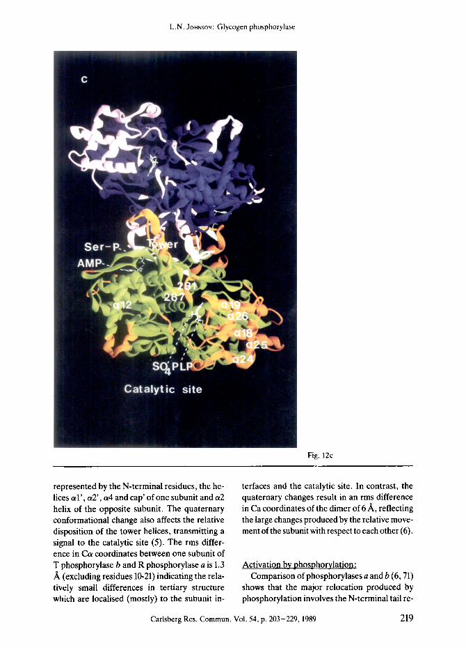
Overall, the tertiary structures of the 4 states of phosphorylase ~hat have been studied crystal- lographically are very similar. There is a core of residues comprising some 66% of the structure that differs by less than 0.5 A between the struc- tures. The major changes in structure take place at the subunit-subunit interface and at the cata- lytic site. Activation of phosphorylase from the T state to the R state leads to a change in asso- ciation of the molecule from dimers to tetra- mers (evidence reviewed by GRAVES and WANG, 1972 (20)). Tetramers exhibit less activity than dimers and under physiological conditions such non-productive association is prevented by the association of the enzyme with glycogen partic- les. In the R state structures there are additional changes in surface residues that flank entrance to the catalytic site and are involved in the asso- ciation of dimers to tetramers. (Details of these changes and their significance for the change in state of aggregation on activation will be de- scribed elsewhere). Two subunits of the func- tional dimer associate at 2 positions located on opposite sides of the enzyme molecule (Fig. 12).
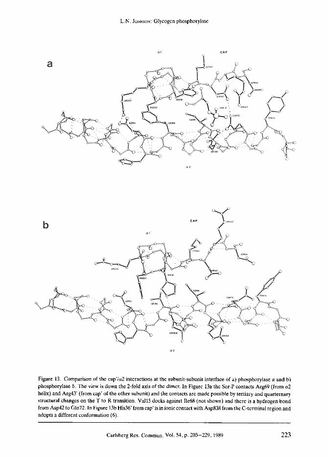
One contact, the cap'/or2 interface, is formed by the association of the cap' (residues 35'-46') with the/37 strand (residues 191 to 193) and a2 helix (residues 47-48) of the other subunit. (Re- sidues from the symmetry related molecule are designated with superscript prime). This inter- face includes the allosteric effector site and Ser- P binding site. An identical interface is produced by the molecular 2-fold symmetry operation. A second subunit-subunit contact involves the tower interface and consists of the anti-parallel asso- ciation of 2 symmetry related helices, ct7 (resi- dues 262-276). The catalytic site is separated from the ailosteric site and Ser-P site by 35 A and 45 A, respectively, and from the symmetry related catalytic site by 70 A. The Ser-P site is located 15 A from the allosteric site and symme- try related allosteric and Ser-P sites are separat- ed by 40 ,~, and 35 A, respectively. Long range interactions therefore operate between different ligand binding sites.
The structures of R state phosphorylases a and b and T state phosphorylase a are similar at the allosteric site interface and the vicinity of the Ser-P site. At the tower-tower interface and the catalytic site the two R state structures are very similar and differ from those of the T state structures. T state phosphorylase a therefore exhibits the interesting properties of a T like structure at the catalytic site (promoted by the binding of the inhibitor glucose) and an R like structure at the aliosteric site (promoted by the Ser-P). The following discussion will focus on the allosteric changes between T state phospho- rylase b and R state phosphorylase a, as these allow the most complete description of the con- formational response. The structure determina- tion and analysis of the R state phosphorylase structures has been led by my colleague Dr. D. BARFORD (5, 6, 32).
The quaternary conformational change can be described as a rotation of one subunit relative to the other by 10 ~ about an axis approximately perpendicular to the molecular 2-fold axis, posi- tioned close to the cap'/a2 interface. This draws the subunits together at the cap'/a2 interface and apart at the tower-tower interface (Figs. 12e and f). The quaternary conformational change is promoted through the concerted but localised conformational change at the cap'/a2 interface
216 Carlsberg Res. Commun. Vol. 54, p. 203-229, 1989

L.N. JOHNSON: Glycogen phosphorylase
Fig. 12a
Figure 12. The dimers of R state phosphorylase a (Figs. a, c, and e) and T state phosphorylase b (Figs. b, d, and f). Colour code: R state (Figs. a, c, and e) subunit 1 green and subunit 2 blue with regions that differ by more than 1 A in Ca positions between R and T state tertiary structures shown in orange and pink for subunits 1 and 2, respec- tively; T state (Figs. b, d, and f) subunit 1 cyan and subunit 2 purple with regions that differ more than 1/~ in Ca positions between R and T states shown in red and yellow. The N-terminal residues (10-23) and the C-terminal re- sidues (837-842) are shown in white. Ligands: pyridoxal phosphate and sulphate (R state) and glucose-l-P (T state) are shown at the catalytic site; AMP at the allosteric effector site; Ser-P at Serl4 (R state GPa only); AMP at the nucleoside inhibitor site (T state only); and maltopentose at the glycogen storage site (T state only), a) and b): View down the 2-fold axis of the dimer with the allosteric sites and Ser-P sites towards the viewer. The catalytic site is at the rear. The view shows the change in conformation of the N-terminal tail from intra to (continued on page 218)
Carlsberg Res. Commun. Vol. 54, p. 203-229, 1989 217

L.N. JOHNSON: Glycogen phosphorylase
Fig. 12b
inter subunit contacts and the shifts of the a l helix-cap-a2 helix and a4-a5 inter-helix loop. c) and d): View down the 2-fold axis of the dimer with the catalytic site and tower helices towards the viewer. The aliosteric site is at the rear. The view illustrates the changes in the tower helices and the 280s loop. The movements in the helix bundle (lower right; orange and red in R and T states) are part of the dimer-dimer contact in the tetramer in which these helices pack against part of the glycogen storage site (left). e) and 0; View normal to the 2-fold axis of the dimer. The view shows the change in quarternary structure in which one subunit (subunit 2; top) rotates 10 ~ with respect to the other subunit (subunit 1 ; bottom) about an axis normal to the 2-fold axis that intercepts the axis at a point near the cap'/t~2 interface. The view shows changes at the subunit-subunit interface and the shifts of the N-terminal and C-terminal residues (from ref. 6). Ribbon diagram by CARSOS and BUG6, 1986 (11).
218 Carlsberg Res. Commun. Vol. 54, p. 203-229, 1989
L.N. JOHNSON: Glycogen phosphorylase
Fig. 12c
represented by the N-terminal residues, the he- lices a l ' , a2', a4 and cap' of one subunit and a2 helix of the opposite subunit. The quaternary conformational change also affects the relative disposition of the tower helices, transmitting a signal to the catalytic site (5). The rms differ- ence in Ca coordinates between one subunit of T phosphorylase b and R phosphorylase a is 1.3 /~ (excluding residues 10-21) indicating the rela- tively small differences in tertiary structure which are localised (mostly) to the subunit in-
terfaces and the catalytic site. In contrast, the quaternary changes result in an rms difference in Ca coordinates of the dimer of 6/~, reflecting the large changes produced by the relative move- ment of the subunit with respect to each other (6).
Activation by phosphorylation: Comparison of phosphorylases a and b (6, 71)
shows that the major relocation produced by phosphorylation involves the N-terminal tail re-
Carlsberg Res. Commun. Vol. 54, p. 203-229, 1989 219

L.N. JOHNSON: Glycogen phosphorylase
Fig. 12d
sidues 10-21. In phosphorylase b these residues are poorly ordered but they have been located (50) in an extended conformation that places Ser-14 close to Glu501 on the helix a16. On phosphorylation the N-terminal residues fold up into an irregular 310 helix and the Ser-P makes contact to 2 arginine residues at the subunit in- terface, Arg69 from its own subunit and Arg43' from the other subunit (Figs. 12a and b). These interactions are made possible by the quater- nary changes that result in the movement of the
a2 helix (that contains Arg69) relative to the cap' (that contains Arg43') and substantial con- formational changes in these residues (Fig. 13). Argl0 shifts 50 A from T state to R state reflect- ing the dramatic restructuring of the N-terminal peptide on phosphorylation. In T state phospho- rylase b there are 11 acidic and 7 basic residues within 15/~ of the Ca atom of Serl4 (excluding residues 10-20). This location of the tail provides a complimentary electrostatic environment to the basic N-terminal tail and one which is inhos-
220 Carlsberg Res. Commun. Vol. 54, p. 203-229, 1989

L.N. JOHNSON: Glycogen phosphorylase
Fig. 12e
pitable to addition of a negatively charged phosphate. In R state phosphorylase a there are 6 acidic and 11 basic residues in the vicinity of Ser-14 and the conformationai change resulting from phosphorylation provides now a stabilis- ing environment for the phosphate.
Activation by AMP: The AMP allosteric site is located on the op-
posite side of the a2 helix to the Ser-P. The T to
R transition results in changes of the cap' resi- dues especially Asp42' and Asn44' that flank Arg43', and to changes of the cap' with respect to the a2 helix that lead to a high affinity site in the R state. The crystallographic study of the binding of AMP to T state phosphorylase (6) showed that the major interactions of the nucle- otide with the enzyme were through the phos- phate group of the nucleotide to Arg309 and Arg310 on the a8 helix (Fig. 14). The base posi- tion was stabilised by long van den Waals inter-
Carlsberg Res. Commun. Vol. 54, p. 203-229, 1989 221

L.N. JO.NSON: Glycogen phosphorylase
Fig. 12f
actions between the adenine and Tyr75 and Va145' and residues of the cap were too far to make significant interactions. The open site cor- relates with the relatively low affinity displayed by T state phosphorylase for AMP (Ko approx. 250-700/zM (51, 59)). The quaternary and tertiary changes on the T to R transition create a site that exhibits a 100 fold increase in affinity (KD = 3/~M) (9). In the R state (6) the phosphate of the nucleotide makes similar contacts to Arg309 and Arg310 but these ionic links are augmented
by a closer contact with Arg242. The most dra- matic changes come from shifts in the cap resi- dues that now allow direct hydrogen bonds be- tween the 02 hydroxyl of the ribose and Asp42' and the N1 of the adenine and Asn44' (Fig. 14). There are improved van der Waals contact be- tween the nucleotide and residues Tyr75, Gin72 and Va145'. The differences can be summarised by noting that there are 4 additional hydrogen bonds made in the R state to AMP compared to the T state and that the nucleotide is some 93%
222 Carlsberg Res. Commun. Vol. 54, p. 203-229, 1989
L.N. JoHNsoN:Glycogen phosphorylase
~ I ' CAP'
a 1 . . . . .
44
. )
Ste -e , A I ~ * 3
�9 C ~ 7 2
i
o 2
CAP ~ A R C 3 b A 4
c v s 4 1 ,
~'~ _ , ILEB8 4~- G1~72 'rfR75
�9 t."US4 , - _ . ,
~2
Figure 13. Comparison of the cap'/u2 interactions at the subunit-subunit interface of a) phosphorylase a and b) phosphorylase b. The view is down the 2-fold axis of the dimer. In Figure 13a the Ser-P contacts Arg69 (from a2 helix) and Arg43' (from cap' of the other subunit) and the contacts are made possible by tertiary and quarternary structural changes on the T to R transition. Vail5 docks against I1e68 (not shown) and there is a hydrogen bond from Asp42 to Gin72. In Figure 13b His36' from cap' is in ionic contact with Asp838 from the C-terminal region and adopts a different conformation (6).
Carlsberg Res. Commun. Vol. 54, p. 203-229, 1989 223

L.N. JOHNSON: Glycogen phosphorylase
O r 2
' ~?~.,~t)As~4 4,
CAP'
; . (~VAL45' i > % ,
a
~ " ' - TYR75
~8
Figure 14. Comparison of AMP binding to a) R state phosphorylase b and b) T state phosphorylase b. In the T state the site is more open and there are few contacts between the enzyme and AMP. In the T to R conversion the AMP shifts further into the site and the site is closed by movements of the cap' relative to the ct2 helix (6).
o buried in the R state compared to 66 Yo buried in the T state.
Communication to the catalytic site:
The changes at the allosteric site and Ser-P site are communicated to the catalytic site
through linked quaternary and localised struc- tural changes that are focussed around the other major subunit interface. In the T state the tower helices of the 2 subunits are arranged in antipa-
rallel constellation with close contacts between Asn270 and Asn274 and their symmetry related
counterparts at the centre of these helices. On the T to R transition the tower helices are pulled
apart by 2 turns of helix and the angle of tilt is changed to -70 ~ (Figs. 12 c-f). This dramatic
change is communicated to the catalytic site
through the 280s loop and indirect contacts to Arg569. The tower helices are anchored at their
base by a short parallel/3 sheet between residues
276-279 and 162-164. From here the chain leads to the loop 281-287 (280s loop) that forms the gate to the catalytic site. A change in the twist of this sheet which appears to be a consequence of the change in the tower helices perturbs resi-
224 Carlsberg Res. Commun. Vol. 54, p. 203-229, 1989

L.N. JOHNSON: Glycogen phosphorylase
C A P '
ARP42 " " ~-z~ ::__
b
a '2
/ !' ' R75
, ( ?
k~
~. ARC309
dues that are in contact with Arg569. In the R state residues 282 to 286 are disordered allowing access to the catalytic site and Arg569 swings down to a conformation similar to that observed in the heptulose-2-phosphate complex, and con- tacts the sulphate molecule that is bound at the catalytic site in the crystal, The sulphate is di- rectly hydrogen bonded to the pyridoxal phos- phate 5'-phosphate group and its position is si- milar to that observed for the phosphate peak in the putative attacking position seen in the time resolved studies (Fig. 6b). The alternative pack- ing adopted by the tower helices provides a par- ticularly suitable method by which tertiary and quaternary structural changes may be linked. The geometry of one helix is constrained by the other helix so that molecular symmetry is con- served and a concerted transition occurs.
Discussion of allosteric mechanism: Comparison of the allosteric mechanism of
phosphorylase with 3 other aUosteric proteins whose structures are known from X-ray evi- dence has shown the common feature of linked tertiary and quaternary structural changes but the changes involve diverse intersubunit inter- actions and diverse mechanisms for communi- cation between sites (32, 64). In glycogen phosphorylase changes at the subunit interface are consistent with both the model proposed by M O N O D , W Y M A N N a n d C H A N G E U X ( 5 8 ) , ( M W C ) ,
and that proposed by KOSHLAND, NEMETHY and FILMER (44)~ (KNF), for enzyme cooperativity and regulation. In both models, the subunit in- terface forms the focus for transmission of infor- mation. The crucial difference between the 2 models is the strength of the constraints at the
Carlsberg Res. Commun. Vol. 54, p. 203-229, 1989 225

L.N. JOHNSON : Glycogen phosphorylase
subunit interface. In the MWC model, effects are modulated by an equilibrium between 2 (or at least 2) symmetrical conformational states that differ in their affinity for ligands. A change in tertiary structure of one subunit must be ac- companied by an equivalent change in the other subunits, facilitated by a quaternary conforma- tional change, in order to conserve symmetry. In the KNF model it was proposed that an induc- ed tertiary conformational response to ligand binding in one subunit leads to changes in the energy of the subunit-subunit interactions that may or may not make it easier for the other sub- unit to respond. A sequential conformational change of subunits is envisaged in which the oli- gomeric assembly need not be symmetrical.
Knowledge of the R and T state structures of the phosphorylase reveals nothing concerning the transient structural events occurring during the T to R transition and does not allow us to un- equivocally distinguish between these 2 models. However, certain predictions regarding the tran- sition can be made. A complete conversion to the R-state structure at one effector site at the sub- unit interface independent of the other allosteric site and independent of a quaternary conforma- tionai change would lead to steric conflict (6). In particular constraints on the geometry of the tow- er helix prevent formation of intermediate struc- tures, ensuring that tertiary structural change of the tower is strictly coupled to the quaternary conformational change. Likewise the creation of the high affinity AMP site that is formed at the cap'/a2 interface on the T to R transition can only be achieved through symmetric conforma- tionai changes that affect both subunits.
Control by phosphorylation is now recognised as a ubiquitous control process by which intra- cellular events can be coupled to extracellular agents such as hormones and growth signals. Glycogen phosphorylase is the first phospho-re- gulated protein whose structure is known. The structural mechanism of activation by phospho- rylation in phosphorylase is similar to that of al- losteric regulation by effectors in which the phosphorylated N-terminal tail acts as an allo- steric effector through binding to its recognition site and favouring changes in conformational state of the enzyme that affect sites remote from the phosphorylation site. It is likely that other
mechanisms will be discovered by which phos- phorylation regulates protein activity. Indeed, in isocitrate dehydrogenase inactivation by phosphorylation appears to be achieved by elec- trostatic repulsion of the substrate (31). The structural studies with phosphorylase have shown how seemingly small modifications to the protein can lead to changes in structure such that binding sites can exhibit at least a 100 fold difference in affinity and allow the enzyme's ac- tivity to be coordinated with metabolic require- ments of the cell.
ACKNOWLEDGEMENTS I wish to thank my collaborators without
whom none of this work would have been poss- ible. In particular Dr. K.R. ACHARYA and Dr. D.I. STUARTwho were responsible for skill and originality in data collection and high resolution refinement of the T state structure and in ligand binding studies; Dr, P.J. McLAUGHLIN who first analysed the formation of heptulose-2-phosphate and the oligosaccharide binding; Dr. J. HAJoU who led the time resolved studies and the Laue diffraction analysis; Dr. N.G. O1KONOMAKOS for work on the pyridoxal phosphate and other ligand binding studies and correlation of crystallogra- phic and kinetic properties; Dr. D. BARFORD out- standing work on R state phosphorylase that led an understanding of the allosteric mechanism; Dr. J. MARTtN who in a iigand binding study first located the N-terminal tail in phosphorylase b; Miss S.-H. Hu for work with R state phosphory- lase a and ligand binding studies. In addition I am grateful to Dr. G.L. TAYLOR for manage- ment of the computer resources at the Laboratory of Molecular Biophysics and to the staff at the SERC's Synchrotron Radiation Source, Dares- bury for the synchrotron facilities. Over the years we have enjoyed fruitful collaboration with Pro- fessor E.J.M. HELMREICH, Dr. D. PALMand Dr. H. KLEIN at Wutzberg and with Professor R.J. FLETrERICK, Professor S.R. SPRANG and Dr. E. GOLDSMITH at San Francisco and Dallas. This work has been supported by the MRC and the SERC. Finally, it is a pleasure to acknowledge the inspiration and encouragement of Sir DAVID PmLUPS throughout nearly 18 years of phospho- rylase crystallography.
226 Carlsberg Res. Commun. Vol. 54, p. 203-229, 1989

L.N. JOHNSON." Glycogen phosphorylase
R E F E R E N C E S 1. AcnArvA, K.R., D.I. Stuart, K.M. VARvtLL &
L.N. JoHnson: Glycogen Phosphorylase b: de- scription of the protein structure. World Scientific Publishers. In press (1990)
2. ArIKI, M. & T. FUKUI: Affinity of glucose anal- ogues for ,~-glucan phosphorylases from rabbit muscle and potato tubers. J. Biochem. 81, 1017- 1024 (1977)
3. ArNot, U.W. & A.J. WOnACOTT: The Rotation Method in Crystallography, North Holland, Am- sterdam (1977)
4. BAgANOWSKI, T., B. ILLINGWORTH, D.H. BROWN & C.F. Corl: The isolation of 5'-pyridoxal phosphate from crystalline muscle phosphorylase. Biochim. Biophys. Acta 25, 16-21 (1957)
5. BARFORD, D. 8/. L.N. JOHNSON: The allosteric transi- tion of glycogen phosphorylase. Nature 340, 609- 616 (1989)
6. BAR~ORD, D., S.-H. Hu & L.N. JOHNSON: The struc- tural mechanism for glycogen phosphorylase con- trol by phosphorylation and AMP. submitted (1990)
7. BLACK, W.J. & J.H. WANG: Studies on the allosteric activation of glycogen phosphorylase b by nucleo- tides. J. Biol. Chem. 243, 5892-5898 (1968)
8. BRUNGER, A.Z., J. KURIYAN ~r M. KARPLUS: Crystal- lographic R factor refinement by molecular dyna- mics. Science 235,458-460 (1987)
9. Buc, H.: On the allosteric interaction between 5'- AMP and orthophosphate in glycogen phosphory- lase b. Biochem. Biophys. Res. Commun. 28, 59- 64 (1967)
10. CAMPBELL, J.W., I.J. CLIFTON, T.J. GREEnOUOH, J. HAJDU, S.C. HARRISON, R.C. LIDDINGTON & A.K. SHRIVE: Calcium binding sites in Tomato Bushy Stunt Virus visualised by Laue crystallography. J. Mol. Biol. 214, 627-632 (1990)
11. CARSON, M. & C.E. BUGG: A ribbon programme for display of protein structures. J. Mot. Graphics 4, 121-122 (1986)
12. Corn, G.T., S.E COLOWtCH & C.F. CORI: The for- mation of glucose-l-phosphoric acid in extracts of mammalian tissues of yeast. J. Biol. Chem. 123, 381-389 (1938)
13. CRUICHSHANK, D.W.J., J.R. HELUWELL & K. MOFFAT: Multiplicity distribution of reflections in Laue dif- fraction. Acta Cryst. A43,656-674 (1987)
14. DREYFUS, M., B. VANDENBUNDER & H. Buc: Mecha- nism of allosteric activation of glycogen phos- phorylase probed by the reactivity of essential argi- nyl residues. Biochemistry 20, 1748-1756 (1980)
15. EnGERS, H.D. & N.B. MADSEN: The effects of anions on the activity of phosphorylase b. Biochem. Bio- phys. Res. Commun. 97, 513-519 (1968)
16. FARBER, G.K., EA. MACHIN, S.c. ALMO, G.A. PErSKO & J. HAJDU: X-ray Laue diffraction from crystals of xylose isomerase. Proc. Natl. Acad. Sci. USA 85, 112-115 (1988)
17. FELDMAN, K. & W.E. HULL: 31p nuclear magnetic resonance studies of glycogen phosphorylase from rabbit muscle: ionisation states of pyridoxal phos- phate. Proc. Natl. Acad. Sci. USA 74, 856-860 (1977)
18. FLETrERICK, R. & S.R. SPRANG: Glycogen phos- phorylase structure and function. Accounts of Chemical Research 15,361-369 (1982)
19. FRIEDERICH, W., l~ KNIPPING & M. VON LAUE; Sitz- ungsberichte der Math. Phys. Klasse, (Kgl.) Baye- rische Akademie der Wissenschaften, Munchen, 303-322 (1912)
20. GRAVES, D.J. & J.H. WANG: a-glucan phosphory- lases- chemical and physical basis of catalysis and regulation. In: The Enzymes (ED. Boyer ed.) 3rd edit. Vol. 7, pp. 435-482, Academic Press, New York (1972)
21. HAJDU, J. & L.N. JOHNSON: Progress with Laue dif- fraction studies on protein and virus crystals. Bio- chemistry 29, 1669q678 (1990)
22. HAJDU, J., P.J. McLAUGHLIN, J.R. HELLIWELL, J. SHELDEN 8s A.W. THOMPSON: A flOW cell for prot rein crystallography. J. Appl. Cryst. 18, 528-532 (1986)
23. HAJDU, J.~ K.R. ACHARYA, D.I. STUART, EJ. MCLAUGHLIN, D. BARFORD, N.G. OWKONOMAKOS, H.W. KLEIN & L.N. JOHNSON: Catalysis in the cry- stal: synchrotron radiation studies with glycogen phosphorylase b. EMBO J. 6, 539-546 (1987)
24. HAJDU, J., EA. MACHIN, J.W. CAMPBELL, T.J. GREENHOUGH, l.J. CLiftON, S. ZUREK, S. GOVER, L.N. JoHnson �9 M, ELDER: Millisecond X-ray dif- fraction and the first electron density map from Laue photographs of a protein crystal. Nature, Lond. 329, 115-116 (1987)
25. HEHRE, E.J., C.E BREWER, T. UCHIYAMA, P. SCHLES- SELMAN & J. LEHMAN: Scope and mechanism of car- bohydrase action. Stereo specific hydration of 2,6- anhydro- l-deoxy-D-gluco-hept- 1-enitol catalysed by ~ and/3 glucosidases and an inverting exo-a-glu- canase. Biochemistry 19, 3557-3564 (1980)
26. HELLIWELL, J.R. : The uses of synchrotron radiation in the crystallography of molecular biology. Rep. Prog. Phys. 47, 1403-1497 (1984)
27. HELEIWELL, J.R., T.J. GREENOUGH, ED. CARR, S.A. RULE, ER. MOORE, A.W. THOMPSON & J.S. WORaAN : A central data collection facility for pro- tein crystallography, small angle diffraction and scattering with the Daresbury Laboratory synchro- tron radiation source. England J. Phys. E 15, 1363- 1372 (1982)
Carlsberg Res. Commun. Vol. 54, p. 203-229, 1989 227
L.N. JOHNSON: Glycogen phosphorylase
28. HELLIWELt., J.R., M.Z. PAPtZ, I.D. GLOVER, J. HABASH, A.W. THOMPSON, P.R. MOORE, N. HARRIS, D. CRov7 & E. PANTOS: The wiggler protein crystallography work station at the Daresbury synchrotron radiation source: progress and results. Nuclear Instrum. Methods Phys. Res. A246, 617- 623 (1986)
29. HELLIWELL, J.R., J. HABASH, D.W.J. CRUICKSHANK, M.M. HARDING, T.J. GREENOUGH, J.W. CAMPBELL, l.J. CLIFTON, M. ELDER, EA. MACHIN, M.Z. PAPIZ • S. ZUREK: The recording and analysis of synchro- tron X-radiation Laue diffraction photographs. J. Appl. Cryst. 22,483-497 (1989)
30. HELMREICH, E. & C.E CORI: The role of adenylic acid in the activity of glycogen phosphorylase. Proc. Natl. Acad. Sci. USA 51,131-138 (1964)
31. HURLEY, J.H,, A.M. DEAN, J.L. SOUL, D.E. KOSHLAND & R.M. STROUO: Regulation of an en- zyme by phosphorylation at the active site. Science 249, 1012-1016 (1990)
32. JOHNSON, L.N. & D. BARFORO: Glycogen phos- phorylase: The structural basis of the allosteric re- sponse and comparison with other allosteric proteins. J. Biol. Chem. 265, 2409-2412 (1990)
33. JoaNsor~, L.N. & J. HAJDU: Synchrotron studies on enzyme catalysis in crystals. In: Biophysics and Synchrotron Radiation (S. Hasnain ed.) Ellis Hor- wood, Chichester pp. 142-155 (1989)
34. JOHNSON, L.N., J. CHEETHAM, P.J. MCLAUGHUN, K.R. ACHARVA, D. BAREORD & D.C. PHILLIPS: Protein oligosaccharide interactions. Current Topics in Microbiol. and lmmunol. 139, 81-134 (1988)
35. JOHNSOr~, L.N., J. HAJDU, K.R. ACHARVA, D.I. STUART, P.J. McLAUGHLIN, N.G. OIKONOMAKOS & D. BARFORD : Glycogen phosphorylase b. In: Allosteric Enzymes 81-127 (G. Herve ed.) CRC Press, Baca Raton, Florida (1989)
36. JOHNSON, L.N., K.R. ACHARYA, M.D. JORDAN& P.J. MCLAUGHLIN: The refined crystal structure of the phosphorylase-heptulose 2-phosphate-oligosac- charide-AMP complex. J. Mol. Biol. 211,645-661 (1990)
37. KAPLAN, J.H., B. FORBUSH & J.E HOFFMAN: Rapid photolytic release of adenosine 5'-triphos- phate analogue: utilisation by the Na:K pump of human red blood cell ghosts. Biochemistry 17, 1929-1935 (1978)
38. KASVlNSKY, P.J. & N.B. MADSEN : Activity of glyco- gen phosphorylase in the crystalline state. J. Biol. Chem. 251, 6852-6859 (1976)
39. KASVlNSKY, P.J., N.B. MADSEN, R.J. FLEI"rERICK & J. SYGUSCH : X-ray crystallographic and kinetic studies of oligosaccharide binding to phosphorylase. J. Biol. Chem. 253, 1290-1296 (1978)
40. KASVINSKY, EJ., N.B. MADSEN, J. SYGUSCH t~ R.J.
FLETI-ERICK : The regulation of glycogen phosphory- lase a by nucleotide derivatives. J. Biol. Chem. 253, 3343-3351 (1978)
41. KASVlNSKY, EJ., S. SHECflOSKY t~ R.J. FLE'rT'E- roCK : Synergistic regulation of phosphorylase a by glucose and caffeine. J. Biol. Chem. 253, 9102-9106 (1978)
42. KLEIN, H.W., M.J. IM, D. PALM & E.J.M. HELM. REICH: Does pyridoxal 5'-phosphate function in glycogen phosphorylase as an electrophile or as a general acid catalyst? Biochemistry 23, 5853-5861 (1984)
43. KLEIN, H.W., M.J. IM & D. PALM; Mechanism of phosphorylase reaction: utilisation of D-gluco- hept-l-enitol in the absence of primer. Eur. J. BiD- chem. 157, 107-114 (1986)
44. KOSHLAND, D.E., G. NEMETHY ~/. D. FILMER: Comparison of experimental binding data and the- oretical models in proteins containing subunits. Biochemistry 5,365-385 (1966)
45. KREBS, E.G. & E.H. FISCHER: The phosphorylase b to a converting enzyme of rabbit skeletal muscle. Biochim. Biophys. Acta 20,150-157 (1956)
46. LEONIDAS, D.D., N.G. OIKONOMAKOS, A.C. PAPA- GEORGIOU, A. XENAKIS, C.Z. CAZIANIS (~ E BEM : The ammonium sulphate activation of phosphorylase b. FEBS Lett. 261,23-27 (1990)
47. MADSEN, N.B.: Glycogen phosphorylase: control by phosphorylation. In: The Enzymes (Boyer, ED. & Krebs, E.G., eds.) 3rd edit. vol. 17, pp. 366-394, Academic Press, New York (1986)
48. MADSEN, N.B. & S.G. WITHERS: Glycogen phos- phorylase and derivatives. In: Coenzymes and Co- factors; Pyridoxal Phosphate and Derivatives (Dolphin, D., Paulson, R. & Avramovic, O., eds.) pp. 1-29, Wiley, New York (1986)
49. MARTIN, J.L. : Molecular interactions involving gly- cogen phosphorylase. D. Phil. Thesis, University of Oxford (1990)
50. MARTIN, J.L., L.N. JOHNSON & S.G. WITHERS: Com- parison of the binding of glucose and glucose-l- phosphate derivatives to T state glyvogen phos- phorylase b. Biochemistry. In press (1990)
51. MATED, P.L., C. BARON, L.M. OBDULIO, J.S. JEMENEZ &. M. CORTIJO: AMP and IMP binding to glycogen phosphorylase b. J. Biol. Chem. 259, 9384-9389 (1984)
52. MCCRAv, J.A. & D.R. TREr~THAM: Properties and uses of photoreactive caged compounds. Ann. Rev. Biophys. Biophys. Chem. 18,239-270 (1989)
53. MCLAUOHLIn, P.J. : Crystallographic studies on gly- cogen phosphorylase b. D.Phil. Thesis, University of Oxford (1985)
54. MCLAUGHLIN, P.J., D.I. STUART, H.W. KLEIN, N.G. OIKONOMAKOS & L.N. JOHNSON: Substrate cofactor
228 Carlsberg Res. Commun. Vol. 54, p. 203-229, 1989

L.N. JoHNSON: Glycogen phosphorylase
interactions for glycogen phosphorylase b. A binding study in the crystal of heptenitol and hep- tulose-2-phosphate. Biochemistry 23, 5862-5873 (1984)
55. METZGER, B.E., E. HELMREICH & L. GLASER: The mechanism of activation of skeletal muscle glyco- gen phosphorylase a by glycogen. Proc. Natl. Acad. Sci. USA 51,131-138 (1967)
56. MEYER, E, L.M.G. HEILMEYER, R.H. HASHKE & E.H. FISCHER : Control of phosphorylase in the gly- cogen particle. I. Isolation and characterisation of the protein glycogen complex. J. Biol. Chem. 245, 6642-6648 (1970)
57. MOFFAT, K., D.M.E. SZEBENYI 8~ D.H. BILDERBACK: X-ray Laue diffraction from protein crystals. Science 223, 1423-1425 (1984)
58. MONOD, J., J. WYMAN (~ J.-E CHANGEUX: On the na- ture of aUosteric transitions: a possible model. J. Mol. Biol. 12, 88-118 (1965)
59. Mort, D.M. & A.L. BIEBER: Structural specificity of the adenosine 5'-phosphate site on glycogen phosphorylase b. J. Biol. Chem. 245, 4058-4066 (1970)
60. NEWGARD, C.B., P.K. HWANG & R.J. FLE'ITERICK: The family of glycogen phosphorylases: structure and function. Crit. Rev. Biochem. Mol. Biol. 24, 69-99 (1989)
61. OIKONOMAKOS, N.G., L.N. JOHNSON, K.R. ACHARYA, D.I. STUART, D. BARFORD, J. HAJDU, K.M. VARVILL, A.E. MELPIDOU, Z. PAPGEORGIOU, D.J. GRAVES & D. PALM: The pyridoxal phosphate site in glycogen phosphorylase b. The structure in the native en- zyme and in 3 derivatives. Biochemistry 26, 8381- 8389 0987)
62. PALM, D., H.W. KLEIN, R. SCHINZEL, M. BUEHNER & E.J.M. HELMREICH: The role of pyridoxal 5'-phos- phate in glycogen phosphorylase catalysis. Bioche- mistry 29, 1099-1107 (1990)
63. PARRtCH, R.E, R.J. UHING & D.J. GRAVES: Effect of phosphate analogues on the activity of pyridoxal reconstituted glycogen phosphorylase. Biochemis- try 16, 4824-4831 (1977)
64. PERUTZ, M.E: Mechanisms of cooperativity and allosteric regulation in proteins. Quart. Rev. Bio- phys. 22, 139-237 (1989)
65. PHILIP. G., G. GRINGEL & D. PALM: Rabbit muscle phosphorylase derivatives with oligosaccharides covalently bound to the glycogen storage site. Bio- chemistry 21, 3042-3050 (1982)
66. PRALV, J.E & R.U. LEMIEUX: The influence of solvent on the magnitude of the anomeric effect. Can. J. Chem. 65,213-223 (1987)
67. SCHLtCmtNG, I., S.C. ALMO, G. RAPP, K.S. WILSON, K. PETRACOS, A. LENTFER, A. WITI-INGHOFER, W. KABSCH, E.E PAl, G.A. PETSKO • R.S. GOODY: Time resolved X-ray crystallographic study of the conformational changes in Ha-Ras p21 protein on GTP hydrolysis. Nature 345,309-315 (1990)
68. SHroVE, A.K., l.J. CLIFU'ON, J. HAJDU & T.J. GREENHOUGH: Laue film integration and decon- volution of overlapping reflections. J. Appl. Cryst. 23,169-174 (1990)
69. SOTIROUDIS, T.G., N.G. OIKONOMAKOS ~,~ A.E. EVAN- GELOPOULOS: Phosphorylase b covalently bound to glycogen. Properties of the complex. Biochem. Biophys. Res. Commun. 90, 234-239 (1978)
70. SPRANG, S.R. & R.J. FLETTERICK: The structure of glycogen phosphorylase a at 2.5 ,~ resolution. J. Mol. Biol. 131,523-551 (1979)
71. SPRANG, S.R., K.R. ACHARYA, E.J. GOLDSMITH, D.I. STUART, K. VARVILL, R.J. FLETTERICK, N.B. MADSEN & L.N. JOHNSON: Protein phosphorylation: struc- tural changes between glycogen phosphorylase b and a. Nature 336,215-221 (1988)
72. TiTANI, K., A. KOiDE, J. HERMANN, L.H. ERICSSON, S. KUMAR, R.D. WADE, K.A. WALSH, H. NEURATH & E.H. FISCHER: Complete amino acid sequence of rabbit muscle glycogen phosphorylase. Proc. Natl. Acad. Sci. USA 74, 4762-4766 (1977)
73. VANDENBUNDER, B. & H. Buc: The reactivitity of arginine residues interacting with glucose-l-phos- phate in glycogen phosphorylase. Eur. J. Biochem. 133,509-513 (1983)
74. WANG, J.H., M. SHONKA t~ D.J. GRAVES: The effect of glucose on the sedimentation and catalytic acti- vity of glycogen phosphorylase. Biochemistry 4, 2296-2301 (1965)
75. WANSOM, J.-C. & EJ. DROCKMANS: Rabbit skeletal muscle glycogen. J. Cell Biol. 38,130-150 (1968)
76. WEBER, I.T., L.N. JOHNSON, K.S. WILSON, D.G.R. YEATES, D. WiLD & J.A. JENKINS: Crystallographic studies on the activity of glycogen phosphorylase b. Nature 274,433-437 (1978) WIERENGA, R.K., M.C.H. MAYER & W.J. Hot: Inter- action of pyrophosphate moities with a helices in dinucleotide binding proteins. Biochemistry 24, 1346-1357 (1985) WYCKOFF, H.W., M. DOSCHER, D. TSERNOGLOU, T. INAGAMI, L.N. JOHNSON, K.D. HARDMAN, N.M. ALLE- WELL, D.M. KELLY & EM. RtCHARDS: The design of a flow cell system for X-ray analysis of crystalline proteins and applications to crystal chemistry. J. Mol. Biol. 27, 563-578 (1967)
77.
78.
Carlsberg Res. Commun. Vol. 54, p. 203-229. 1989 229
Copyright © 2022 FDOKUMEN