Metastasis-associated Differences in Gene Expression in a Murine Model of Osteosarcoma
10
[CANCER RESEARCH 61, 3750 –3759, May 1, 2001] Metastasis-associated Differences in Gene Expression in a Murine Model of Osteosarcoma Chand Khanna, 1 Javed Khan, Phoungmai Nguyen, Jennifer Prehn, Jana Caylor, Choh Yeung, Jane Trepel, Paul Meltzer, and Lee Helman Pediatric Oncology [C. K., J. P., J. C., C. Y., L. H.] and Medicine Branches [P. N., J. T.], National Cancer Institute, and Cancer Genetics Branch, Human Genome Research Institute, NIH [J. K., P. M.], Bethesda, Maryland 20892 ABSTRACT Despite advances in the management of osteosarcoma (OSA) and other solid tumors, the development of metastasis continues to be the most significant problem and cause of death for cancer patients. To define genetic determinants of pulmonary metastasis, we have applied cDNA microarrays to a recently described murine model of OSA that is char- acterized by orthotopic tumor growth, a period of minimal residual disease, spontaneous pulmonary metastasis, and cell line variants that differ in metastatic potential. Microarray analysis defined 53 genes (of 3166 unique cDNAs) that were differentially expressed between the pri- mary tumors of the more aggressive (K7M2) and less aggressive (K12) OSA models. By review of the literature, these differentially expressed genes were assigned to six nonmutually exclusive metastasis-associated categories (proliferation and apoptosis, motility and cytoskeleton, inva- sion, immune surveillance, adherence, and angiogenesis). Functional stud- ies to evaluate K7M2 and K12 for differences in each of these metastasis- associated processes revealed enhanced motility, adherence, and angiogenesis in the more aggressive K7M2 model. For this reason, 10 of the 53 differentially expressed genes that were assigned to the motility and cytoskeleton, adherence, and angiogenesis categories were considered as most likely to define differences in the metastatic behavior of the two models. Ezrin, a gene not described previously in OSA, with functions in motility, invasion, and adherence, was 3-fold overexpressed in K7M2 compared with K12 by microarray. Differential expression for RNA was confirmed by Northern analysis and for protein by immunostaining. Alterations in ezrin protein levels and concomitant cytoskeletal changes in our model confirmed predictions from the arrays. The potential relevance of ezrin in OSA was suggested by its expression in five of five human OSA cell lines. This work represents a rationale approach to the evaluation of microarray data and will be useful to identify genes that may be causally associated with metastasis. INTRODUCTION OSA 2 is the most common primary tumor of bone. Similar to other solid tumors, it is characterized by a high propensity for metastasis (i.e., the lungs, liver, and bone). In OSA, the lung is the most common site for metastasis. In spite of successful control of the primary tumor, death from pulmonary metastases occurs in .30% of patients within 5 years (1). A greater understanding of the biology of pulmonary metastases is needed to improve treatment outcomes and identify patients with the highest risk for disease relapse in OSA and other solid tumors. We have described recently a relevant murine model of OSA characterized by tumor growth at appendicular sites (orthotopic), a period of minimal residual disease, spontaneous pulmonary metas- tasis, and model variants that differ in metastatic potential (2). This model was developed from a cell line (K12) originating from a spontaneous BALB/c OSA (3) and a recently derived, clonally related cell line (K7M2) described by the authors (2). Within the model, the K7M2 cell line is aggressive and highly metastatic, whereas the K12 cell line is less aggressive with infrequent pulmonary metastases (Fig. 1). In K7M2, spontaneous pulmonary metastases from an orthotopic primary tumor develop in .90% of mice compared with K12, where only 33% of mice develop metastases (P , 0.001). The differences in metastatic potential of the clonally related cell lines characterized in this model make it a valuable system for understanding the biology of pulmonary metastases in OSA. Genetic risk factors for pulmonary metastasis have been defined in several solid tumors (4 – 6). The majority of this work has been undertaken in epithelial malignancies. Similar to other solid tumors, OSA metasta- ses are associated with dissemination through the blood stream (hem- atogenous), microscopic metastasis (micrometastases) that exist in the absence of observable metastases, and a high propensity for metasta- ses to the lung (7). Distinctive features of metastasis in OSA include an uncharacteristically long latency between successful control of the primary tumor and the development of pulmonary metastases, a preference for pulmonary metastases compared with other metastatic sites, and relative success associated with surgical removal of pulmo- nary metastases (1). The use of this nonepithelial model provides a unique perspective in the study of metastasis that may uncover novel determinants for metastasis relevant to both mesenchymal and epithe- lial cancers. To define genetic determinants of metastasis in OSA, we have used cDNA microarrays to compare gene expression between the clonally related high metastatic (K7M2) and low metastatic (K12) primary tumors. Array comparisons of K7M2 and K12 have defined 53 of 3166 unique printed cDNA probes (genes) that are differentially expressed. To focus our attention on a smaller group of potentially important genes, we have taken a functional approach to determine the significance and relevance of these differentially expressed genes vis a ` vis pulmonary metastasis. By review of the literature, we have assigned each differentially expressed gene to six nonmutually exclu- sive metastasis-associated categories including proliferation and ap- optosis, motility and cytoskeleton, invasion, immune surveillance, adherence, and angiogenesis (7). K7M2 and K12 were then compared in studies that independently examined each of these metastasis- associated processes. These studies (some presented in the initial description of the model; Ref. 2) demonstrated increased cellular motility and cytoskeletal changes suggestive of motile cells, earlier heterotypic adherence, and enhanced tumor angiogenesis in the more aggressive K7M2 compared with the less aggressive K12 model. On the basis of this functional and metastasis-related characterization, 10 genes that were assigned to the cell motility and cytoskeleton, het- erotypic adherence, and angiogenesis categories were considered to be most likely to describe the aggressive behavior of K7M2 compared with the K12 cells. Ezrin, a member of the ERM gene family was 1 of these 10 genes. Similar to other members of its gene family, ezrin plays a role in linking the actin cytoskeleton to the cell membrane (8, 9). Ezrin has been associated with cell motility, invasion, and adher- ence but has not been described previously in OSA. Differential expression of ezrin between K7M2 and K12 was confirmed at both Received 12/29/00; accepted 3/19/01. The costs of publication of this article were defrayed in part by the payment of page charges. This article must therefore be hereby marked advertisement in accordance with 18 U.S.C. Section 1734 solely to indicate this fact. 1 To whom requests for reprints should be addressed, at Pediatric Oncology Branch, National Cancer Institute, Bethesda, Maryland 20892. 2 The abbreviations used are: OSA, osteosarcoma; ERM, ezrin, radixin, and moesin; EST, expressed sequence tag; MMP, matrix metalloproteinase; scid, severe combined immunodeficient. 3750
-
Upload
independent -
Category
Documents
-
view
2 -
download
0
Transcript of Metastasis-associated Differences in Gene Expression in a Murine Model of Osteosarcoma
[CANCER RESEARCH 61, 3750–3759, May 1, 2001]
Metastasis-associated Differences in Gene Expression in a Murine Modelof Osteosarcoma
Chand Khanna,1 Javed Khan, Phoungmai Nguyen, Jennifer Prehn, Jana Caylor, Choh Yeung, Jane Trepel,Paul Meltzer, and Lee HelmanPediatric Oncology [C. K., J. P., J. C., C. Y., L. H.] and Medicine Branches [P. N., J. T.], National Cancer Institute, and Cancer Genetics Branch, HumanGenome ResearchInstitute, NIH [J. K., P. M.], Bethesda, Maryland 20892
ABSTRACT
Despite advances in the management of osteosarcoma (OSA) and othersolid tumors, the development of metastasis continues to be the mostsignificant problem and cause of death for cancer patients. To definegenetic determinants of pulmonary metastasis, we have applied cDNAmicroarrays to a recently described murine model of OSA that is char-acterized by orthotopic tumor growth, a period of minimal residualdisease, spontaneous pulmonary metastasis, and cell line variants thatdiffer in metastatic potential. Microarray analysis defined 53 genes (of3166 unique cDNAs) that were differentially expressed between the pri-mary tumors of the more aggressive (K7M2) and less aggressive (K12)OSA models. By review of the literature, these differentially expressedgenes were assigned to six nonmutually exclusive metastasis-associatedcategories (proliferation and apoptosis, motility and cytoskeleton, inva-sion, immune surveillance, adherence, and angiogenesis). Functional stud-ies to evaluate K7M2 and K12 for differences in each of these metastasis-associated processes revealed enhanced motility, adherence, andangiogenesis in the more aggressive K7M2 model. For this reason, 10 ofthe 53 differentially expressed genes that were assigned to the motility andcytoskeleton, adherence, and angiogenesis categories were considered asmost likely to define differences in the metastatic behavior of the twomodels.Ezrin, a gene not described previously in OSA, with functions inmotility, invasion, and adherence, was 3-fold overexpressed in K7M2compared with K12 by microarray. Differential expression for RNA wasconfirmed by Northern analysis and for protein by immunostaining.Alterations in ezrin protein levels and concomitant cytoskeletal changes inour model confirmed predictions from the arrays. The potential relevanceof ezrin in OSA was suggested by its expression in five of five human OSAcell lines. This work represents a rationale approach to the evaluation ofmicroarray data and will be useful to identify genes that may be causallyassociated with metastasis.
INTRODUCTION
OSA2 is the most common primary tumor of bone. Similar to othersolid tumors, it is characterized by a high propensity for metastasis(i.e., the lungs, liver, and bone). In OSA, the lung is the most commonsite for metastasis. In spite of successful control of the primary tumor,death from pulmonary metastases occurs in.30% of patients within5 years (1). A greater understanding of the biology of pulmonarymetastases is needed to improve treatment outcomes and identifypatients with the highest risk for disease relapse in OSA and othersolid tumors. We have described recently a relevant murine model ofOSA characterized by tumor growth at appendicular sites (orthotopic),a period of minimal residual disease, spontaneous pulmonary metas-tasis, and model variants that differ in metastatic potential (2). Thismodel was developed from a cell line (K12) originating from aspontaneous BALB/c OSA (3) and a recently derived, clonally related
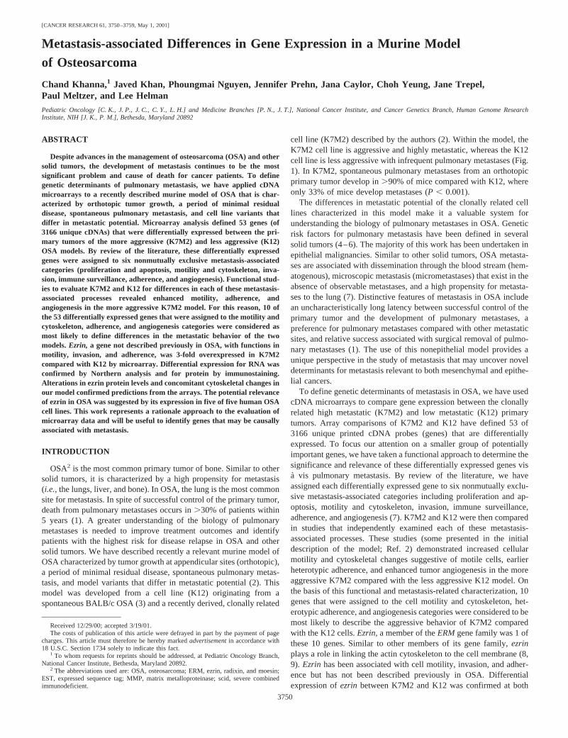
cell line (K7M2) described by the authors (2). Within the model, theK7M2 cell line is aggressive and highly metastatic, whereas the K12cell line is less aggressive with infrequent pulmonary metastases (Fig.1). In K7M2, spontaneous pulmonary metastases from an orthotopicprimary tumor develop in.90% of mice compared with K12, whereonly 33% of mice develop metastases (P , 0.001).
The differences in metastatic potential of the clonally related celllines characterized in this model make it a valuable system forunderstanding the biology of pulmonary metastases in OSA. Geneticrisk factors for pulmonary metastasis have been defined in severalsolid tumors (4–6). The majority of this work has been undertaken inepithelial malignancies. Similar to other solid tumors, OSA metasta-ses are associated with dissemination through the blood stream (hem-atogenous), microscopic metastasis (micrometastases) that exist in theabsence of observable metastases, and a high propensity for metasta-ses to the lung (7). Distinctive features of metastasis in OSA includean uncharacteristically long latency between successful control of theprimary tumor and the development of pulmonary metastases, apreference for pulmonary metastases compared with other metastaticsites, and relative success associated with surgical removal of pulmo-nary metastases (1). The use of this nonepithelial model provides aunique perspective in the study of metastasis that may uncover noveldeterminants for metastasis relevant to both mesenchymal and epithe-lial cancers.
To define genetic determinants of metastasis in OSA, we have usedcDNA microarrays to compare gene expression between the clonallyrelated high metastatic (K7M2) and low metastatic (K12) primarytumors. Array comparisons of K7M2 and K12 have defined 53 of3166 unique printed cDNA probes (genes) that are differentiallyexpressed. To focus our attention on a smaller group of potentiallyimportant genes, we have taken a functional approach to determine thesignificance and relevance of these differentially expressed genes visa vis pulmonary metastasis. By review of the literature, we haveassigned each differentially expressed gene to six nonmutually exclu-sive metastasis-associated categories including proliferation and ap-optosis, motility and cytoskeleton, invasion, immune surveillance,adherence, and angiogenesis (7). K7M2 and K12 were then comparedin studies that independently examined each of these metastasis-associated processes. These studies (some presented in the initialdescription of the model; Ref. 2) demonstrated increased cellularmotility and cytoskeletal changes suggestive of motile cells, earlierheterotypic adherence, and enhanced tumor angiogenesis in the moreaggressive K7M2 compared with the less aggressive K12 model. Onthe basis of this functional and metastasis-related characterization, 10genes that were assigned to the cell motility and cytoskeleton, het-erotypic adherence, and angiogenesis categories were considered to bemost likely to describe the aggressive behavior of K7M2 comparedwith the K12 cells.Ezrin,a member of theERMgene family was 1 ofthese 10 genes. Similar to other members of its gene family,ezrinplays a role in linking the actin cytoskeleton to the cell membrane (8,9). Ezrin has been associated with cell motility, invasion, and adher-ence but has not been described previously in OSA. Differentialexpression ofezrin between K7M2 and K12 was confirmed at both
Received 12/29/00; accepted 3/19/01.The costs of publication of this article were defrayed in part by the payment of page
charges. This article must therefore be hereby markedadvertisementin accordance with18 U.S.C. Section 1734 solely to indicate this fact.
1 To whom requests for reprints should be addressed, at Pediatric Oncology Branch,National Cancer Institute, Bethesda, Maryland 20892.
2 The abbreviations used are: OSA, osteosarcoma; ERM, ezrin, radixin, and moesin;EST, expressed sequence tag; MMP, matrix metalloproteinase; scid, severe combinedimmunodeficient.
3750
mRNA and protein levels.In situ differences in the distribution ofezrin protein suggested a role of ezrin in the motility of K7M2 cells.The relevance of ezrin in human OSA was supported by Northernanalysis that demonstrated its expression in five of five human OSAcell lines.
MATERIALS AND METHODS
Animal Model. The K12 murine OSA cell line was a kind gift of L. C.Gerstenfeld (3, 10). K7M2 was derived within the presented model system(Fig. 1) by two cycles of pulmonary metastasis implantation to the proximaltibia. The model conditions and characterization of the highly metastatic cellline, K7M2, and less metastatic cell line, K12, are summarized in Fig. 1. K7M2and K12 cells were maintainedin vitro using complete culture media [DMEM;Celox Co., Hopkins, MN; 100mg/ml penicillin-streptomycin; and 2 mML-glutamine (Sigma Chemical Co., St. Louis, MO)] with 10% FCS (SigmaChemical Co.) at 37°C at 5% CO2. All cell lines used forin vitro and in vivostudies were from the 3rd to 15th passages. For allin vitro assays, cells wereharvested, using Trypsin/Versene, from near-confluent cultures. Cell viabilitywas assessed using trypan blue, and experiments were not continued if cellviability was ,90%.
RNA Extraction. RNA was extracted fromin vitro tumor cell lines usingthe Qiagen RNeasy Midi kit (Qiagen, Inc., Valencia, CA) according to themanufacturer’s specification. RNA was extracted from grossly dissected pri-mary tumor and from pulmonary metastases using the Trizol (Life Technolo-gies, Inc., Rockville, MD) reagent according to the manufacturer’s specifica-tion. Extracted RNA was quantitated by spectrophotometry and then examinedby 1% agarose gel (Seakem GTG; FMC Bioproducts, Rockland, ME) electro-phoresis. Intact RNA was then reextracted in 1.0 ml of Trizol for use inmicroarray experiments.
Microarrays, Probes, Hybridization, and Scanning. The mouse array iscomposed of 3899 detector elements (probes). Of these, 315 are unclusteredESTs, 630 are clustered ESTs, and 3004 are clustered, named genes. There issignificant redundancy in the named gene portion of the set, with 2221 uniqueclusters (probes) represented. All clones (including clustered and unclusteredESTs) were obtained from Research Genetics (Huntsville, AL); selection ofprobes was based only on availability. Clustering was undertaken by ResearchGenetics. PCR products from these clones were prepared and printed ontoglass slides according to protocols described previously (11, 12). FluorescentcDNA targets (samples) were labeled with either Cy3 or Cy5 (AmershamPharmacia Biotech, Piscataway, NJ) from 100 to 200mg of total RNA by oligo
dT-primed polymerization using SuperScript II reverse transcriptase (LifeTechnologies, Inc., Rockville, MD) as described previously (11, 12). Imagingand image analysis was as described previously (11, 12). Normalization fordifferential efficiencies of labeling and detection was performed using all spots(probes) that performed well as defined by criteria embedded in the DeArraysoftware developed by Chenet al. (13). The normalization constant from thesegenes was then used to calculate the calibrated ratio for every cDNA probewithin the image. Furthermore, the ratio variation of the these genes deter-mined the confidence interval in which ratios are to be considered as nodifference from 1.0. The 99%confidence interval was used throughout theexperiments to test the significance of differentially expressed genes. Additionalinformation on the image analysis as well as the raw data including thehybridization images for each sample is available on the Internet.3 Microarrayoutliers were defined as those genes that were significantly differentiallyexpressed and that had a MaxMean signal intensity (defined as the maximumof the mean intensity of either the red or green channel for each gene).2000in two distinct array experiments (using samples from different mice bearingthe K7M2 and K12 primary tumors). Differential expression of randomlyselected genes was confirmed by Northern analysis. Northern blots consistedof 25 mg of total RNA from K7M2 and K12 cell lines, primary tumors, andpulmonary metastases. Plasmid probes, purified by MaxiPrep (Qiagen, Inc.,Valencia, CA) containing sequence-confirmed cDNAs, were labeled by nicktranslation (Amersham, Life Science) with [a-32P]dCTP. Hybridization ofNorthern blots was carried out using Express Hyb (Clontech, Palo Alto, CA),according to the manufacturer’s recommendations. Equal loading of RNA wasverified by reprobing all Northern blots with an [a-32P]dCTP-labeled murineb-actin plasmid probe. Northern analysis for ezrin in human OSA cell lines(HOS, MG63, U2, G292, and SaOS) was undertaken using a nick-translatedplasmid provided generously by Dr. Richard Lamb (8).
Assignment of Microarray Outliers to Protein Function and Metastasis-related Gene Process Categories.Differentially expressed genes, identifiedfrom cDNA microarray comparisons, were assigned to a modification of theNCBI Clusters of Orthologous Gene classification by searching the OMIM andPubMed databases by gene name.4 Genes were then assigned to six nonmu-tually exclusive metastasis-associated processes (proliferation/apoptosis, mo-tility/cytoskeleton, invasion, adhesion, immune surveillance, and angiogene-sis) using a PubMed database search of the gene name and each of thefollowing terms: cancer, metastasis, proliferation, apoptosis, invasion, motility,immune surveillance, adhesion, and angiogenesis. Each gene was categorizedwith a single and mutually exclusive function, whereas gene assignment tometastasis-associated processes was not mutually exclusive.
Actin Cytoskeleton. Cells were grown on sterile coverslips fixed with3.7% formaldehyde in PBS for 10 min and then extracted with 0.2% TritonX-100 for 10 min at room temperature. After fixation and extraction, one unitof rhodamine phalloidin (Molecular Probes, Eugene, OR) was added to thecoverslip prepared cells, as recommended by the manufacturer, and incubatedat room temperature. After 1 h, the coverslip was washed two times for 2 minwith PBS. Cells were stained with 0.4mg/ml of 49,6-diamidino-2-phenylindole(Sigma Chemical Co.) for 10 min at room temperature. After incubation, thecoverslip was washed with PBS, rinsed quickly with water, air-dried, andmounted onto slides using SlowFade (Molecular Probes). Cells were visualizedon a Zeiss Axiovert microscope using a363 objective, and images werecaptured with an Optronics CCD camera.
Motility and Invasion Assays. Preincubation of 12-mm polycarbonateTranswell (Costar, Cambridge MA) plates for 4 h at37°C was undertaken byadding complete culture medium to the upper chamber and complete culturemedium with 10% FCS to the lower chamber before the addition of cells. Fivethousand K7M2 or K12 cells were added in a volume of 200ml of serum-freecomplete medium to the upper Transwell chamber. The Transwell upperchamber was placed into lower chambers that had been filled with 500ml ofcomplete medium with 10% FCS. For the Matrigel (Collaborative BiomedicalProducts, Bedford. MA) invasion assay, 100ml of a 1:3 dilution of Matrigel inserum-free culture medium was added to the upper chamber of the Transwelland incubated for 2 h at37°C (after the preincubation step). Transwell plates(motility assay) and Transwell plates coated with Matrigel (invasion assay)were incubated for 4, 12, 24, and 48 h at 37°C. At the completion of the
3 Internet address: http://www.nhgri.nih.gov/DIR/LCG/arraydb/.4 Internet address: http://www.ncbi.nlm.nih.gov/cgi-bin/COG/palog?fun5all.
Fig. 1. Orthotopic tumor model used to characterize and derive the clonally relatedaggressive (K7M2) and less aggressive (K12) murine OSA cell lines.A, illustration ofmodel design used to implement the model and to develop the more aggressive K7M2clone from the less metastatic parent cell.B, characterization of differences in metastaticbehavior of the more aggressive K7M2 model and less aggressive K12 model.
3751
EVALUATION OF OSTEOSARCOMA METASTASIS BY cDNA MICROARRAY
incubation period, culture medium was suctioned from upper and lower cham-bers without disturbing cells. The total number of (motile or invasive) cellsmoving through the Transwell membrane was determined in replicates of sixby wiping the apical surface of the Transwell membrane with a cotton swaband then staining the Transwell insert with DiffQuick (American ScientificProducts, McGraw Park, IL). The Transwell membrane was then cut out usinga No. 11 scalpel blade (Becton Dickinson Acute Care, Franklin Lakes, NJ),mounted, and coverslipped on a microscopic slide for cell enumeration. Thetotal number of plated cells was evaluated at each time point (in replicates ofsix) by eliminating the “cotton swab step” and repeating the staining andmembrane preparation procedure described above. The percentage of motilityand percentage of invasion5 (total number of cells on basilar surface ofmembrane)/(total number of cells on both apical and basilar surfaces of the
membrane). Motility and invasion experiments were each repeated three times.Representative results from experimental conditions, repeated in replicates ofsix, are presented.
Gelatinase Zymography. Gelatinase zymography was used to evaluateMMP2 and MMP9 activity in tissue culture supernatant and primary tumor asdescribed previously (14). Serum-free medium was placed on confluent cul-tures of K7M2 and K12 for 24 h. Protein content in culture supernatants wasquantified using the bicinchoninic acid assay (Pierce, Rockford, IL). Up to 25mg of total protein were evaluated by gelatin electrophoresis (Novex Gel;Invitrogen, Carlsbad, CA). Standards for the pro and active forms of MMP2and MMP9 (Calbiochem, La Jolla, CA) allowed identification of MMP forms.
Heterotypic Adherence Assay.K7M2 and K12 cells were added to 96-well, flat-bottomed, substrate-coated plates (collagen type IV precoated plates
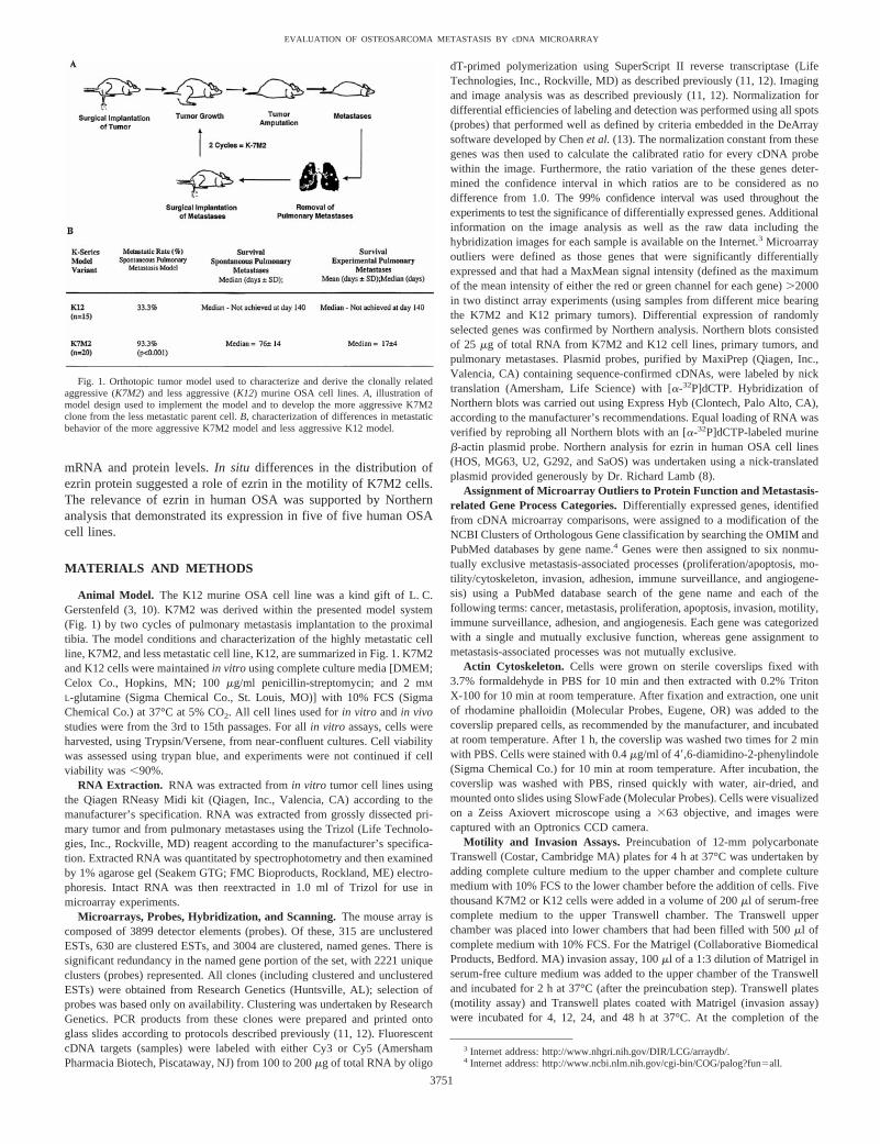
Table 1 Microarray (cDNA) outliers overexpressed in K7M2 primary tumors versus K12 primary tumors
Gene name organized by designated gene function(modification of clusters of orthologous sites)a IMAGE IDb Meanc
Metastasis-associated functional groupsd
I. Information storage and processingTranslation, ribosomal structure and biogenesisProtein kinase, interferon inducible double stranded
RNA dependent597390 3.11
TranscriptionGlucocorticoid-induced leucine zipper (GILZ) 476319 5.82Myeloblastosis oncogene 577875 3.54CCAAT/enhancer binding protein (C/EBP), a 891375 2.84Caudal type homeo box 2 437757 2.70Hepatocyte nuclear factor 3/forkhead homologue 8 537201 2.55Nuclear factor, erythroid derived 2, ubiquitous 576400 2.39E2F-5 585020 2.29DNA replication, recombination, and repairFibroblast inducible secreted protein (connective tissue
growth factor)598684 3.16
II. Cellular processesCell division and chromosome partitioningCyclin D1 419285 5.57Tubulin b-chain 467387 2.60Posttranslational mod., protein turnover, chaperonesProprotein convertase subtilisin/kexin type 3 334336 8.19a-Crystallin A chain-major component 481224 2.60mSTI1 614877 2.25Cell motility/cell and matrix interaction-communicationIntegrin b 4 337221 5.60Integrin a-V (CD51) 960079 3.64Ezrin 635783 2.95Integrin b2 583119 2.84Galectin-3 571813 2.50A disintegrin and metalloprotease domain (ADAM) 8 582054 2.49Inorganic ion transport and metabolismMetallothionein 2 607350 2.48Signal transduction mechanismsATP receptor (P2u) 405832 2.83
III. MetabolismProtein/amino acid transport and metabolismPyrroline-5-carboxylate synthetase short isoform 439868 3.49Asparagine synthetase 605295 3.23Lipid metabolismClusterin 617298 2.59
IV. Cell structure/matrixExtracellular matrixMyelin proteolipid protein 524747 3.05
V. Poorly characterizedA10 533608 7.70Major histocompatibility locus class III regions 420495 2.59
VI. ESTs or Unknown GenesEST, not currently in a UniGene cluster 400844 3.63EST, not currently in a UniGene cluster 420591 3.60EST, not currently in a UniGene cluster 409208 2.49
a Classification, using a modification of the NCBI Clusters of Orthologous Gene Classification, based on the basic function of each outlier gene.b IMAGE clone identification for differentially expressed genes.c Geometric mean of red:green ratios from two-array experiment. Colorimetric representation of geometric mean of red:green ratio with scale available online at www-
dcs.nci.nih.gov/pedonc.d Metastasis-related categories include proliferation and apoptosis, motility and cytoskeleton, invasion, immune surveillance, adherence, and angiogenesis.F, assignment of genes
to metastasis-related categories. Assignment of genes based on PubMed literature search.
3752
EVALUATION OF OSTEOSARCOMA METASTASIS BY cDNA MICROARRAY
and Matrigel precoated plates; Becton Dickinson, Bedford, MA) in quadrupli-cate. Cells were incubated at 37°C in 5% CO2 for 0.5, 1.5, 4, 16, 24, or 48 h.After incubation, medium was carefully suctioned out of each well. Using amultichannel pipette in a controlled manner, each well was washed three timeswith PBS (Biofluids, Inc., Rockville, MD). Between each wash, the plate wasmanually rocked back and forth three times. PBS was carefully suctioned outof each well. After the washes, the 3-(4,5-dimethylthiazol-2-yl)-2,5-diphe-nyltetrazolium bromide assay was used to determine the number of remainingcells (adherent cells; Ref. 15). The percentage of adherence for each timepoint 5 (mean number of cells remaining in a well after washing)/(meannumber of cells in wells that were not washed).
Immune Surveillance. Female beige-scid mice (Charles River Laborato-ries, Wilmington, MA), 4–5 weeks of age, were housed under pathogen-freeconditions with a 12-h light/12-h dark schedule and fed autoclaved standardchow and waterad libitum. In vitro passaged tumor cell lines (K7M2 and K12)were harvested and prepared for injection as described previously (16). Cellswere brought to a final concentration of 13 107 cells/ml in phenol-free HBSSand kept at 4°C. Cells were enumerated, and viability was assessed usingtrypan blue (BioWhittaker, Walkersville, MD) staining. Experiments werecontinued if cell viability was.90%. A volume of 100ml (1 3 106 cells) wasinjected into the lateral tail vein of beige-scid mice (10 mice/cell line). Mice
were monitored at least three times weekly for evidence of morbidity associ-ated with pulmonary metastases. Criteria for morbidity associated with metas-tases in mice included ill thrift, anorexia, dehydration, decreased activity andgrooming behavior, and dyspnea. Sacrifice of mice with presumed pulmonarymetastases was primarily based on the development of dyspnea. All mice thatwere sacrificed because of presumed pulmonary metastases had necropsyconfirmation of diffuse metastases. Statistical comparisons were made usingthe Kruskal-Wallace nonparametrict test calculated with InStat for the MacIn-tosh. Statistical significance was defined asP , 0.05. Animal care and usewere in accordance with guidelines of the NIH Animal Care and Use Com-mittee (17).
Ezrin Immunocytostaining. Coverslip preparations were fixed and ex-tracted as described above. Nonspecific binding sites were blocked by incu-bating the cells with 1% BSA in PBS for 1 h at 4°Cbefore processing forimmunofluorescence labeling. Anti-ezrin antibody (Santa Cruz Biotechnology,Santa Cruz Biotechnology, CA) was added to the coverslip and incubated for1 h at 4°C. After antibody incubation, the coverslip was washed two times for2 min with PBS and incubated at 4°C with Cy3-conjugated rabbit antigoatimmunoglobulin (Jackson Immunoresearch Laboratories, Inc). After 1 h, thecoverslip was washed two times for 2 min with PBS. Cells were stained with0.4mg/ml of 49,6-diamidino-2-phenylindole (Sigma Chemical Co.) for 10 min
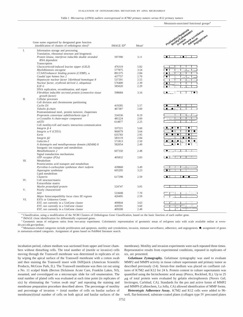
Table 2 Microarray (cDNA) outliers overexpressed in K12 primary tumors versus K7M2 primary tumors
Gene name organized by designated gene function(modification of clusters of orthologous sites)a IMAGE IDb Meanc
Metastasis-associated functional groupsd
I. Information storage and processingTranscriptionStra13 596504 0.23Nephroblastoma overexpressed gene 475200 0.28DNA replication, recombination, and repairBcl-2 a 596444 0.25
II. Cellular processesPosttranslational mod., protein turnover, chaperonesApolipoprotein B editing complex 2 603777 0.33Regulatory protein, T lymphocyte 1 617776 0.37Cell motility/cell and matrix interaction-communicationDecorin 598629 0.25FARP1 403430 0.35Inorganic ion transport and metabolismCeruloplasmin 522108 0.25Signal transduction mechanismsG-protein coupled receptor 574735 0.45
III. MetabolismEnergy production and conversionCarbonic anhydrase 3 618431 0.36Carbohydrate transport and metabolismSkeletal muscle calsequestrin 474650 0.32Lipid metabolismMast cell protease 5 493561 0.36Lipoprotein lipase 475661 0.38
IV. Cell structure/matrixCell membraneMembrane metallo endopeptidase 313540 0.11Potassium voltage gated channel, Shaw-related subfamily, member 1 493098 0.31CD83 574651 0.37CytoskeletonAnkyrin 1, erythroid 635047 0.36Extracellular matrixSecreted phosphoprotein 1 (Osteopontin) 337866 0.45
V. Poorly characterizedSelected mouse cDNA on the Y 598638 0.26Fibrinogen-like protein 2 336457 0.36Histidine-rich calcium-binding protein 475063 0.44
VI. ESTs or unknown genesEST 522100 0.22
a Classification, using a modification of the NCBI Clusters of Orthologous Gene Classification, based on the basic function of each outlier gene.b IMAGE clone identification for differentially expressed genes.c Geometric mean of red:green ratios from two-array experiment. Colorimetric representation of geometric mean of red:green ratio with scale available online at www-
dcs.nci.nih.gov/pedonc.d Metastasis-related categories include proliferation and apoptosis, motility and cytoskeleton, invasion, immune surveillance, adherence, and angiogenesis.F, assignment of genes
to metastasis-related categories. Assignment of genes based on PubMed literature search.
3753
EVALUATION OF OSTEOSARCOMA METASTASIS BY cDNA MICROARRAY

at room temperature. The coverslip was then washed with PBS, rinsed quicklywith water, air-dried, and mounted onto slides using SlowFade (MolecularProbes). Cells were visualized on a Zeiss Axiovert microscope using a363objective, and images were captured with an Optronics CCD camera.
RESULTS
cDNA Microarray Analysis. Microarray experiments examineddifferences in gene expression between the primary tumors of themore aggressive K7M2 and less aggressive K12 models. Fifty-threecDNAs of 3166 were found to be differentially expressed (expressionratios not equal to 1.0 with 99% confidence; MaxMean signal inten-sity, .2000) in replicate microarray experiments using primary tumorsamples from different mice. Variation in patterns of gene expressionwas seen, with;75% of outlier genes found to be common inreplicate experiments. Thirty-one genes common to replicate experi-ments were overexpressed in K7M2 compared with K12 primarytumors, and 22 genes were overexpressed in K12 compared withK7M2 primary tumors (Tables 1 and 2).
Microarray Outlier Classification. To better understand the po-tential importance of each outlier gene and to define genes most likelyto be associated with differences in the metastatic biology of themodels, each outlier gene was assigned to two classification systems.The first classification, using a modification of the National Center forBiotechnology Information Clusters of Orthologous Gene Classifica-tion,4 was based on the basic function of each gene. Assignment of agene to a basic function classification group was mutually exclusive(Tables 1 and 2). The second classification of each gene was to sixnonmutually exclusive metastasis-associated processes (proliferation/apoptosis, motility/cytoskeleton, invasion, adhesion, immune surveil-lance, and angiogenesis). The assignment of each gene into bothclassification systems is presented in Tables 1 and 2. Twenty-nine ofthe 53 differentially expressed genes could be categorized into me-tastasis-related functions. Northern analysis confirmed the pattern ofdifferential gene expression identified by cDNA microarray for se-lected genes (Fig. 2).
Using the information in Tables 1 and 2, we returned to the K7M2and K12 models to uncover specific differences in their biologies foreach of the six metastasis-associated processes (proliferation/apopto-sis, motility/cytoskeleton, invasion, adhesion, immune surveillance,and angiogenesis). The six metastasis-associated processes examinedwere considered to be important steps in the cascade of events thatfollows a tumor cell from its primary site to a distant metastatic site.The metastasis-associated processes of proliferation/apoptosis andangiogenesis were examined in the original report of the K7M2/K12model (2). These studies demonstrated no difference in apoptosis orproliferation between K7M2 and K12 but did demonstrate significantdifferences in tumor angiogenesis favoring K7M2.
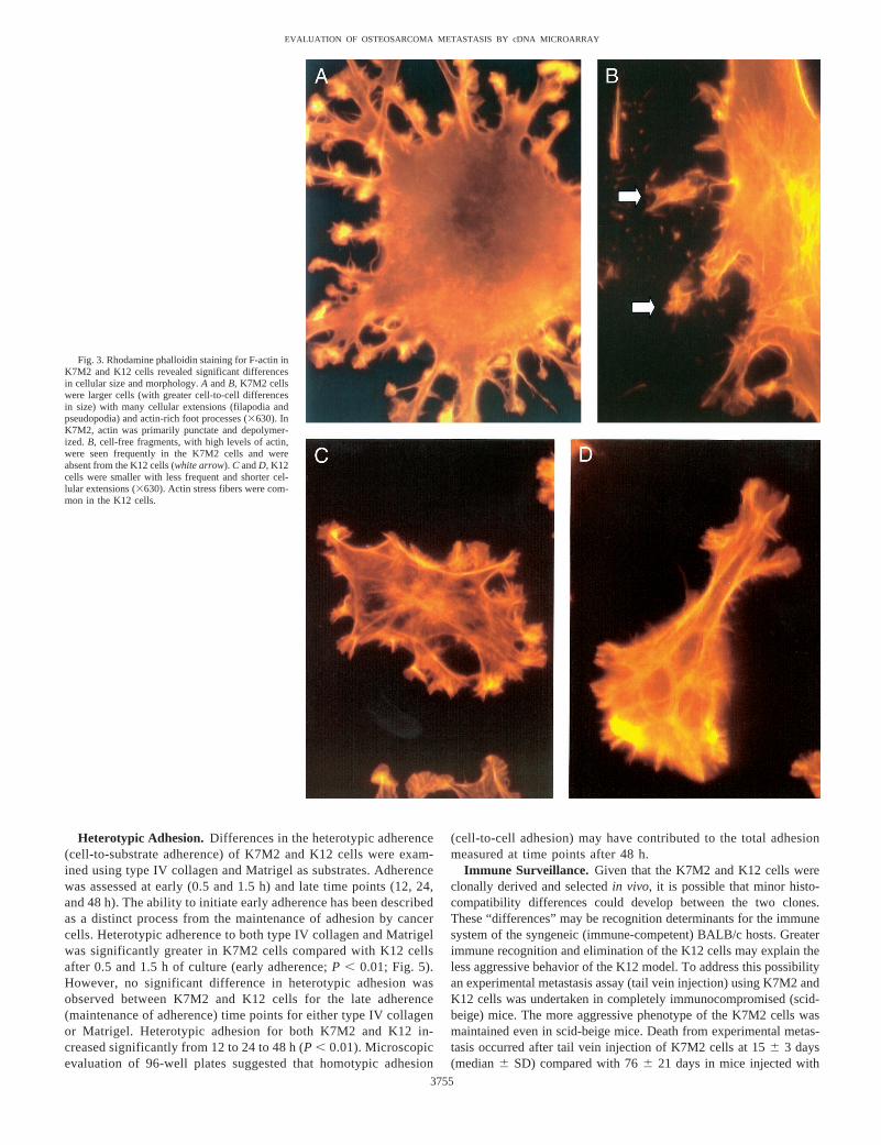
Motility and Cytoskeleton. To define differences in the cytoskel-eton of the OSA cells, we examined their F-actin cytostructuralarchitecture. Significant differences in the size and morphology of theK7M2 and K12 cells was demonstrated by F-actin staining (Fig. 3).The K7M2 cells were larger, had more numerous cellular extensions(filapodia and pseudopodia), greater substrate contact points, andenhanced spread phenotype than the K12 cells. Cellular extensionswere less common and were significantly shorter in the K12 comparedwith the K7M2 cells. K12 cells contained organized actin stressfilaments in the body of the cells, whereas in K7M2 actin stressfilaments were concentrated within cellular extensions. In K7M2cells, cytoplasmic actin was primarily punctate with very little inpolymerized forms. Foot processes from cellular extensions in theK7M2 cells had very high levels of F-actin. Actin-rich fragments, notassociated with the cells, were common in preparations of the K7M2
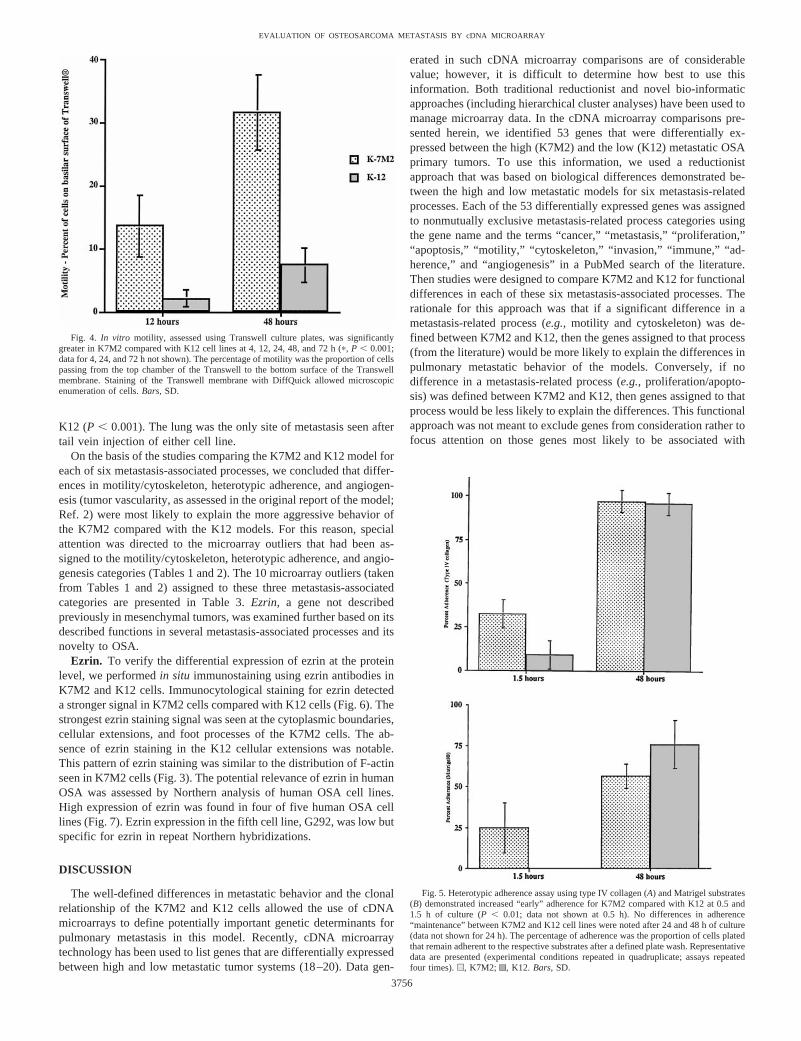
cell line and absent from the K12 cells. These cell-free fragmentsdemonstrated similar patterns of actin expression as the K7M2 footprocesses. F-actin staining seemed to suggest a greater potential formotility in the K7M2 compared with the K12 cells. To examine thispotential difference in cellular motility, we performedin vitro motilityexperiments using Transwell tissue culture plates. In these assays,motility was defined as migration of cells from the apical surface ofthe Transwell membrane through 8-mm pores to the basal surface ofthe membrane. Significantly greaterin vitro motility was demon-strated in the K7M2 cells compared with the K12 cells for all timepoints evaluated (4, 12, 24, 48, and 72 h of culture;P , 0.001; Fig.4). The number of cells leaving the basal surface of the Transwellmembrane into the lower chamber was small (,2% of plated cells)and was similar for K7M2 and K12 cell lines (data not shown).
Invasion. The invasion phenotype of K7M2 and K12 cells wasassessed by comparing their relative abilities to invade tumor extra-cellular matrix (Matrigel) using the Transwell culture system de-scribed above. Both cells were invasive through Matrigel; however,no differences in invasion could be demonstrated between K7M2 andK12 cell lines at any time points examined (data not shown). Tofurther examine the invasion phenotype of the OSA cells, MMPactivity was assessed using gelatinase zymography. MMP-2 andMMP-9 activity was detected in both K7M2 and K12 cell line super-natant and cellular pellets. No significant differences in the precursoror active forms for MMP-2 or MMP-9 were demonstrable betweenK7M2 and K12 (data not shown).
Fig. 2. Analysis of differential gene expression in K7M2 and K12 using Northernanalysis. A, Northern analysis of selected genes (Integrin b4 Subunit, Ezrin, Clusterin,Decorrin, and Ceruloplasmin) found differentially expressed between K7M2 and K12tissues by microarray.Lane 1, K7M2 cell culture RNA;Lane 2, K7M2 primary tumorRNA; Lane 3, K7M2 pulmonary metastasis RNA;Lane 4, K12 cell culture RNA;Lane 5,K12 primary tumor RNA;Lane 6, K12 pulmonary metastasis RNA. For genes overex-pressed in the more aggressive K7M2versusK12 (Integrin b4 subunit, Ezrin,andClusterin), there was a higher level of gene expression in RNA from pulmonary metas-tases compared with primary tumor and cell culture RNA. Equal loading of 25mg of totalRNA was demonstrated by hybridization of representative Northern blot with a murineb-actin cDNA probe.B, concordance of differential gene expression of K7M2versusK12primary tumor using cDNA microarray and Northern. The log meanred:green(R:G) ratiodefined by microarray (Tables 1 and 2) is compared with a Northern densitometricquantification (standardized against theb-actin signal).
3754
EVALUATION OF OSTEOSARCOMA METASTASIS BY cDNA MICROARRAY
Heterotypic Adhesion. Differences in the heterotypic adherence(cell-to-substrate adherence) of K7M2 and K12 cells were exam-ined using type IV collagen and Matrigel as substrates. Adherencewas assessed at early (0.5 and 1.5 h) and late time points (12, 24,and 48 h). The ability to initiate early adherence has been describedas a distinct process from the maintenance of adhesion by cancercells. Heterotypic adherence to both type IV collagen and Matrigelwas significantly greater in K7M2 cells compared with K12 cellsafter 0.5 and 1.5 h of culture (early adherence;P , 0.01; Fig. 5).However, no significant difference in heterotypic adhesion wasobserved between K7M2 and K12 cells for the late adherence(maintenance of adherence) time points for either type IV collagenor Matrigel. Heterotypic adhesion for both K7M2 and K12 in-creased significantly from 12 to 24 to 48 h (P , 0.01). Microscopicevaluation of 96-well plates suggested that homotypic adhesion
(cell-to-cell adhesion) may have contributed to the total adhesionmeasured at time points after 48 h.
Immune Surveillance. Given that the K7M2 and K12 cells wereclonally derived and selectedin vivo, it is possible that minor histo-compatibility differences could develop between the two clones.These “differences” may be recognition determinants for the immunesystem of the syngeneic (immune-competent) BALB/c hosts. Greaterimmune recognition and elimination of the K12 cells may explain theless aggressive behavior of the K12 model. To address this possibilityan experimental metastasis assay (tail vein injection) using K7M2 andK12 cells was undertaken in completely immunocompromised (scid-beige) mice. The more aggressive phenotype of the K7M2 cells wasmaintained even in scid-beige mice. Death from experimental metas-tasis occurred after tail vein injection of K7M2 cells at 156 3 days(median6 SD) compared with 766 21 days in mice injected with
Fig. 3. Rhodamine phalloidin staining for F-actin inK7M2 and K12 cells revealed significant differencesin cellular size and morphology.A andB, K7M2 cellswere larger cells (with greater cell-to-cell differencesin size) with many cellular extensions (filapodia andpseudopodia) and actin-rich foot processes (3630). InK7M2, actin was primarily punctate and depolymer-ized.B, cell-free fragments, with high levels of actin,were seen frequently in the K7M2 cells and wereabsent from the K12 cells (white arrow). C andD, K12cells were smaller with less frequent and shorter cel-lular extensions (3630). Actin stress fibers were com-mon in the K12 cells.
3755
EVALUATION OF OSTEOSARCOMA METASTASIS BY cDNA MICROARRAY
K12 (P , 0.001). The lung was the only site of metastasis seen aftertail vein injection of either cell line.
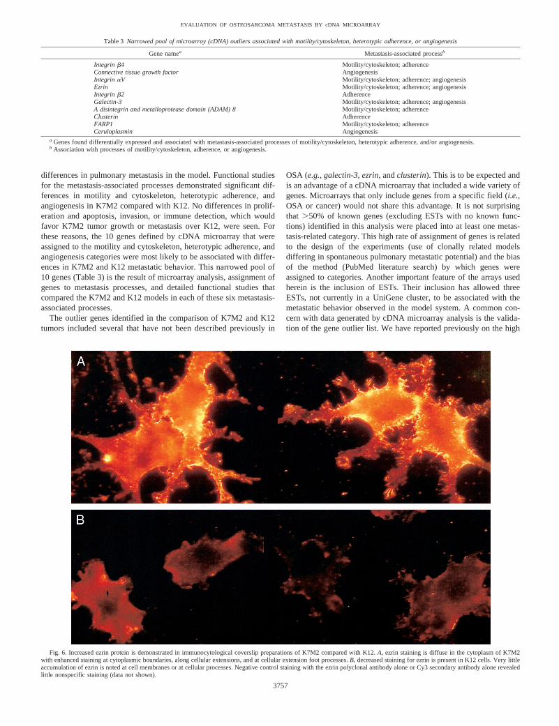
On the basis of the studies comparing the K7M2 and K12 model foreach of six metastasis-associated processes, we concluded that differ-ences in motility/cytoskeleton, heterotypic adherence, and angiogen-esis (tumor vascularity, as assessed in the original report of the model;Ref. 2) were most likely to explain the more aggressive behavior ofthe K7M2 compared with the K12 models. For this reason, specialattention was directed to the microarray outliers that had been as-signed to the motility/cytoskeleton, heterotypic adherence, and angio-genesis categories (Tables 1 and 2). The 10 microarray outliers (takenfrom Tables 1 and 2) assigned to these three metastasis-associatedcategories are presented in Table 3.Ezrin, a gene not describedpreviously in mesenchymal tumors, was examined further based on itsdescribed functions in several metastasis-associated processes and itsnovelty to OSA.
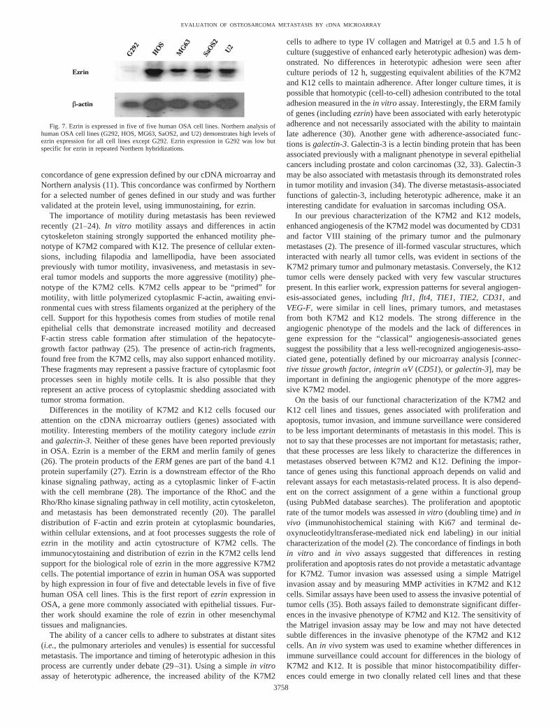
Ezrin. To verify the differential expression of ezrin at the proteinlevel, we performedin situ immunostaining using ezrin antibodies inK7M2 and K12 cells. Immunocytological staining for ezrin detecteda stronger signal in K7M2 cells compared with K12 cells (Fig. 6). Thestrongest ezrin staining signal was seen at the cytoplasmic boundaries,cellular extensions, and foot processes of the K7M2 cells. The ab-sence of ezrin staining in the K12 cellular extensions was notable.This pattern of ezrin staining was similar to the distribution of F-actinseen in K7M2 cells (Fig. 3). The potential relevance of ezrin in humanOSA was assessed by Northern analysis of human OSA cell lines.High expression of ezrin was found in four of five human OSA celllines (Fig. 7). Ezrin expression in the fifth cell line, G292, was low butspecific for ezrin in repeat Northern hybridizations.
DISCUSSION
The well-defined differences in metastatic behavior and the clonalrelationship of the K7M2 and K12 cells allowed the use of cDNAmicroarrays to define potentially important genetic determinants forpulmonary metastasis in this model. Recently, cDNA microarraytechnology has been used to list genes that are differentially expressedbetween high and low metastatic tumor systems (18–20). Data gen-
erated in such cDNA microarray comparisons are of considerablevalue; however, it is difficult to determine how best to use thisinformation. Both traditional reductionist and novel bio-informaticapproaches (including hierarchical cluster analyses) have been used tomanage microarray data. In the cDNA microarray comparisons pre-sented herein, we identified 53 genes that were differentially ex-pressed between the high (K7M2) and the low (K12) metastatic OSAprimary tumors. To use this information, we used a reductionistapproach that was based on biological differences demonstrated be-tween the high and low metastatic models for six metastasis-relatedprocesses. Each of the 53 differentially expressed genes was assignedto nonmutually exclusive metastasis-related process categories usingthe gene name and the terms “cancer,” “metastasis,” “proliferation,”“apoptosis,” “motility,” “cytoskeleton,” “invasion,” “immune,” “ad-herence,” and “angiogenesis” in a PubMed search of the literature.Then studies were designed to compare K7M2 and K12 for functionaldifferences in each of these six metastasis-associated processes. Therationale for this approach was that if a significant difference in ametastasis-related process (e.g., motility and cytoskeleton) was de-fined between K7M2 and K12, then the genes assigned to that process(from the literature) would be more likely to explain the differences inpulmonary metastatic behavior of the models. Conversely, if nodifference in a metastasis-related process (e.g.,proliferation/apopto-sis) was defined between K7M2 and K12, then genes assigned to thatprocess would be less likely to explain the differences. This functionalapproach was not meant to exclude genes from consideration rather tofocus attention on those genes most likely to be associated with
Fig. 5. Heterotypic adherence assay using type IV collagen (A) and Matrigel substrates(B) demonstrated increased “early” adherence for K7M2 compared with K12 at 0.5 and1.5 h of culture (P , 0.01; data not shown at 0.5 h). No differences in adherence“maintenance” between K7M2 and K12 cell lines were noted after 24 and 48 h of culture(data not shown for 24 h). The percentage of adherence was the proportion of cells platedthat remain adherent to the respective substrates after a defined plate wash. Representativedata are presented (experimental conditions repeated in quadruplicate; assays repeatedfour times).u, K7M2; , K12. Bars,SD.
Fig. 4. In vitro motility, assessed using Transwell culture plates, was significantlygreater in K7M2 compared with K12 cell lines at 4, 12, 24, 48, and 72 h (p, P , 0.001;data for 4, 24, and 72 h not shown). The percentage of motility was the proportion of cellspassing from the top chamber of the Transwell to the bottom surface of the Transwellmembrane. Staining of the Transwell membrane with DiffQuick allowed microscopicenumeration of cells.Bars,SD.
3756
EVALUATION OF OSTEOSARCOMA METASTASIS BY cDNA MICROARRAY
differences in pulmonary metastasis in the model. Functional studiesfor the metastasis-associated processes demonstrated significant dif-ferences in motility and cytoskeleton, heterotypic adherence, andangiogenesis in K7M2 compared with K12. No differences in prolif-eration and apoptosis, invasion, or immune detection, which wouldfavor K7M2 tumor growth or metastasis over K12, were seen. Forthese reasons, the 10 genes defined by cDNA microarray that wereassigned to the motility and cytoskeleton, heterotypic adherence, andangiogenesis categories were most likely to be associated with differ-ences in K7M2 and K12 metastatic behavior. This narrowed pool of10 genes (Table 3) is the result of microarray analysis, assignment ofgenes to metastasis processes, and detailed functional studies thatcompared the K7M2 and K12 models in each of these six metastasis-associated processes.
The outlier genes identified in the comparison of K7M2 and K12tumors included several that have not been described previously in
OSA (e.g., galectin-3, ezrin,andclusterin). This is to be expected andis an advantage of a cDNA microarray that included a wide variety ofgenes. Microarrays that only include genes from a specific field (i.e.,OSA or cancer) would not share this advantage. It is not surprisingthat .50% of known genes (excluding ESTs with no known func-tions) identified in this analysis were placed into at least one metas-tasis-related category. This high rate of assignment of genes is relatedto the design of the experiments (use of clonally related modelsdiffering in spontaneous pulmonary metastatic potential) and the biasof the method (PubMed literature search) by which genes wereassigned to categories. Another important feature of the arrays usedherein is the inclusion of ESTs. Their inclusion has allowed threeESTs, not currently in a UniGene cluster, to be associated with themetastatic behavior observed in the model system. A common con-cern with data generated by cDNA microarray analysis is the valida-tion of the gene outlier list. We have reported previously on the high
Table 3 Narrowed pool of microarray (cDNA) outliers associated with motility/cytoskeleton, heterotypic adherence, or angiogenesis
Gene namea Metastasis-associated processb
Integrin b4 Motility/cytoskeleton; adherenceConnective tissue growth factor AngiogenesisIntegrin aV Motility/cytoskeleton; adherence; angiogenesisEzrin Motility/cytoskeleton; adherence; angiogenesisIntegrin b2 AdherenceGalectin-3 Motility/cytoskeleton; adherence; angiogenesisA disintegrin and metalloprotease domain (ADAM) 8 Motility/cytoskeleton; adherenceClusterin AdherenceFARP1 Motility/cytoskeleton; adherenceCeruloplasmin Angiogenesis
a Genes found differentially expressed and associated with metastasis-associated processes of motility/cytoskeleton, heterotypic adherence, and/or angiogenesis.b Association with processes of motility/cytoskeleton, adherence, or angiogenesis.
Fig. 6. Increased ezrin protein is demonstrated in immunocytological coverslip preparations of K7M2 compared with K12.A, ezrin staining is diffuse in the cytoplasm of K7M2with enhanced staining at cytoplasmic boundaries, along cellular extensions, and at cellular extension foot processes.B, decreased staining for ezrin is present in K12 cells. Very littleaccumulation of ezrin is noted at cell membranes or at cellular processes. Negative control staining with the ezrin polyclonal antibody alone or Cy3 secondary antibody alone revealedlittle nonspecific staining (data not shown).
3757
EVALUATION OF OSTEOSARCOMA METASTASIS BY cDNA MICROARRAY
concordance of gene expression defined by our cDNA microarray andNorthern analysis (11). This concordance was confirmed by Northernfor a selected number of genes defined in our study and was furthervalidated at the protein level, using immunostaining, for ezrin.
The importance of motility during metastasis has been reviewedrecently (21–24).In vitro motility assays and differences in actincytoskeleton staining strongly supported the enhanced motility phe-notype of K7M2 compared with K12. The presence of cellular exten-sions, including filapodia and lamellipodia, have been associatedpreviously with tumor motility, invasiveness, and metastasis in sev-eral tumor models and supports the more aggressive (motility) phe-notype of the K7M2 cells. K7M2 cells appear to be “primed” formotility, with little polymerized cytoplasmic F-actin, awaiting envi-ronmental cues with stress filaments organized at the periphery of thecell. Support for this hypothesis comes from studies of motile renalepithelial cells that demonstrate increased motility and decreasedF-actin stress cable formation after stimulation of the hepatocyte-growth factor pathway (25). The presence of actin-rich fragments,found free from the K7M2 cells, may also support enhanced motility.These fragments may represent a passive fracture of cytoplasmic footprocesses seen in highly motile cells. It is also possible that theyrepresent an active process of cytoplasmic shedding associated withtumor stroma formation.
Differences in the motility of K7M2 and K12 cells focused ourattention on the cDNA microarray outliers (genes) associated withmotility. Interesting members of the motility category includeezrinandgalectin-3. Neither of these genes have been reported previouslyin OSA. Ezrin is a member of the ERM and merlin family of genes(26). The protein products of theERMgenes are part of the band 4.1protein superfamily (27). Ezrin is a downstream effector of the Rhokinase signaling pathway, acting as a cytoplasmic linker of F-actinwith the cell membrane (28). The importance of the RhoC and theRho/Rho kinase signaling pathway in cell motility, actin cytoskeleton,and metastasis has been demonstrated recently (20). The paralleldistribution of F-actin and ezrin protein at cytoplasmic boundaries,within cellular extensions, and at foot processes suggests the role ofezrin in the motility and actin cytostructure of K7M2 cells. Theimmunocytostaining and distribution of ezrin in the K7M2 cells lendsupport for the biological role of ezrin in the more aggressive K7M2cells. The potential importance of ezrin in human OSA was supportedby high expression in four of five and detectable levels in five of fivehuman OSA cell lines. This is the first report ofezrin expression inOSA, a gene more commonly associated with epithelial tissues. Fur-ther work should examine the role of ezrin in other mesenchymaltissues and malignancies.
The ability of a cancer cells to adhere to substrates at distant sites(i.e., the pulmonary arterioles and venules) is essential for successfulmetastasis. The importance and timing of heterotypic adhesion in thisprocess are currently under debate (29–31). Using a simplein vitroassay of heterotypic adherence, the increased ability of the K7M2
cells to adhere to type IV collagen and Matrigel at 0.5 and 1.5 h ofculture (suggestive of enhanced early heterotypic adhesion) was dem-onstrated. No differences in heterotypic adhesion were seen afterculture periods of 12 h, suggesting equivalent abilities of the K7M2and K12 cells to maintain adherence. After longer culture times, it ispossible that homotypic (cell-to-cell) adhesion contributed to the totaladhesion measured in thein vitro assay. Interestingly, the ERM familyof genes (includingezrin) have been associated with early heterotypicadherence and not necessarily associated with the ability to maintainlate adherence (30). Another gene with adherence-associated func-tions isgalectin-3. Galectin-3 is a lectin binding protein that has beenassociated previously with a malignant phenotype in several epithelialcancers including prostate and colon carcinomas (32, 33). Galectin-3may be also associated with metastasis through its demonstrated rolesin tumor motility and invasion (34). The diverse metastasis-associatedfunctions of galectin-3, including heterotypic adherence, make it aninteresting candidate for evaluation in sarcomas including OSA.
In our previous characterization of the K7M2 and K12 models,enhanced angiogenesis of the K7M2 model was documented by CD31and factor VIII staining of the primary tumor and the pulmonarymetastases (2). The presence of ill-formed vascular structures, whichinteracted with nearly all tumor cells, was evident in sections of theK7M2 primary tumor and pulmonary metastasis. Conversely, the K12tumor cells were densely packed with very few vascular structurespresent. In this earlier work, expression patterns for several angiogen-esis-associated genes, includingflt1, flt4, TIE1, TIE2, CD31,andVEG-F, were similar in cell lines, primary tumors, and metastasesfrom both K7M2 and K12 models. The strong difference in theangiogenic phenotype of the models and the lack of differences ingene expression for the “classical” angiogenesis-associated genessuggest the possibility that a less well-recognized angiogenesis-asso-ciated gene, potentially defined by our microarray analysis [connec-tive tissue growth factor, integrinaV (CD51), or galectin-3], may beimportant in defining the angiogenic phenotype of the more aggres-sive K7M2 model.
On the basis of our functional characterization of the K7M2 andK12 cell lines and tissues, genes associated with proliferation andapoptosis, tumor invasion, and immune surveillance were consideredto be less important determinants of metastasis in this model. This isnot to say that these processes are not important for metastasis; rather,that these processes are less likely to characterize the differences inmetastases observed between K7M2 and K12. Defining the impor-tance of genes using this functional approach depends on valid andrelevant assays for each metastasis-related process. It is also depend-ent on the correct assignment of a gene within a functional group(using PubMed database searches). The proliferation and apoptoticrate of the tumor models was assessedin vitro (doubling time) andinvivo (immunohistochemical staining with Ki67 and terminal de-oxynucleotidyltransferase-mediated nick end labeling) in our initialcharacterization of the model (2). The concordance of findings in bothin vitro and in vivo assays suggested that differences in restingproliferation and apoptosis rates do not provide a metastatic advantagefor K7M2. Tumor invasion was assessed using a simple Matrigelinvasion assay and by measuring MMP activities in K7M2 and K12cells. Similar assays have been used to assess the invasive potential oftumor cells (35). Both assays failed to demonstrate significant differ-ences in the invasive phenotype of K7M2 and K12. The sensitivity ofthe Matrigel invasion assay may be low and may not have detectedsubtle differences in the invasive phenotype of the K7M2 and K12cells. An in vivo system was used to examine whether differences inimmune surveillance could account for differences in the biology ofK7M2 and K12. It is possible that minor histocompatibility differ-ences could emerge in two clonally related cell lines and that these
Fig. 7. Ezrin is expressed in five of five human OSA cell lines. Northern analysis ofhuman OSA cell lines (G292, HOS, MG63, SaOS2, and U2) demonstrates high levels ofezrin expression for all cell lines except G292. Ezrin expression in G292 was low butspecific for ezrin in repeated Northern hybridizations.
3758
EVALUATION OF OSTEOSARCOMA METASTASIS BY cDNA MICROARRAY
differences could result in the more effective immune recognition anddestruction of one tumor cell line compared with another. Our resultsdemonstrate that the relative metastatic phenotype of the K7M2 andK12 cell lines was maintained in significantly immunocompromisedanimals (beige-scid mice). Therefore, genes associated with the im-mune surveillance and immune rejection of cancer are less likely tocharacterize the differences in the biology of K7M2 and K12.
Using a well-characterized murine model, a microarray that in-cludes a large and diverse number of cDNA probes, and a functionalapproach to analyze microarray outliers, we have defined severalpotentially important genes associated with pulmonary metastasis inOSA. Genes identified in this work include those not describedpreviously in OSA as well as potentially novel metastasis-associatedgenes (ESTs). Functional studies suggest that 10 genes associatedwith motility/cytoskeleton, heterotypic adherence, and angiogenesisare most likely to be associated with differences in the metastaticbehavior of the high and low metastatic OSA model. Genes identifiedin this analysis may have relevance to OSA and other solid tumorswith high rates of pulmonary metastasis. A motility, adherence, andinvasion gene calledezrin was identified using this approach. Thedifferential expression of ezrin protein was confirmed in K7M2 andK12 cells and the potential relevance of ezrin in human OSA sug-gested by finding its expression in five of five human OSA cell lines.This work represents a rationale approach to the evaluation of mi-croarray data and will be useful to identify genes that may be causallyassociated with metastasis.
REFERENCES
1. Bruland, O. S., and Pihl, A. On the current management of osteosarcoma. A criticalevaluation and a proposal for a modified treatment strategy. Eur. J. Cancer,33:1725–1731, 1997.
2. Khanna, C., Prehn, J., Yeung, C., Caylor, J., Tsokos, M., and Helman, L. Anorthotopic murine model of osteosarcoma with clonally related variants differing inpulmonary metastatic potential. Clin. Exp. Metastasis,18: 261–271, 2001.
3. Schmidt, J., Straub, G. P., Schon, A., Luz, A., Murray, B., Melchiori, A., Aresu, O.,and Erfle, V. Establishment and characterization of osteogenic cell lines from aspontaneous murine osteosarcoma. Differentiation,39: 151–160, 1988.
4. Lam, A. K. Molecular biology of esophageal squamous cell carcinoma. Crit. Rev.Oncol. Hematol.,33: 71–90, 2000.
5. Portera, C. A., Berman, R. S., and Ellis, L. M. Molecular determinants of colon cancermetastasis. Surg. Oncol.,7: 183–195, 1998.
6. Bangma, C. H., Nasu, Y., Ren, C., and Thompson, T. C. Metastasis-related genes inprostate cancer. Semin. Oncol.,26: 422–427, 1999.
7. Woodhouse, E. C., Chuaqui, R. F., and Liotta, L. A. General mechanisms ofmetastasis. Cancer (Phila.),80: 1529–1537, 1997.
8. Lamb, R. F., Ozanne, B. W., Roy, C., McGarry, L., Stipp, C., Mangeat, P., and Jay,D. G. Essential functions of ezrin in maintenance of cell shape and lamellipodialextension in normal and transformed fibroblasts. Curr. Biol.,9: 682–688, 1997.
9. Ohtani, K., Sakamoto, H., Rutherford, T., Chen, Z., Satoh, K., and Naftolin, F. Ezrin,a membrane-cytoskeletal linking protein, is involved in the process of invasion ofendometrial cancer cells. Cancer Lett.,147: 31–38, 1999.
10. Gerstenfeld, L. C., Uporova, T., Schmidt, P. G., Straus, P. G., Shih, S. D., Huang,L. F., Gundberg, C., Mizuno, S., and Glowacki, J. Osteogenic potential of murineosteosarcoma cells: comparison of bone-specific gene expressionin vitro andin vivoconditions. Lab. Investig.,74: 895–906, 1996.
11. Khan, J., Bittner, M. L., Saal, L. H., Teichmann, U., Azorsa, D. O., Gooden, G. C.,Pavan, W. J., Trent, J. M., and Meltzer, P. S. cDNA microarrays detect activation ofa myogenic transcription program by the PAX3-FKHR fusion oncogene. Proc. Natl.Acad. Sci. USA,96: 13264–13269, 1999.
12. Khan, J., Simon, R., Bittner, M., Chen, Y., Leighton, S. B., Pohida, T., Smith, P. D.,Jiang, Y., Gooden, G. C., Trent, J. M., and Meltzer, P. S. Gene expression profilingof alveolar rhabdomyosarcoma with cDNA microarrays. Cancer Res.,58: 5009–5013, 1998.
13. Chen, Y., Dougherty, E. R., and Bittner, M. L. Ratio-based decisions and thequantitative analysis of cDNA microarray images. Biomedical Optics,2: 364–374,1997.
14. Lana, S. E., Ogilvie, G. K., Hansen, R. A., Powers, B. E., Dernell, W. S., andWithrow, S. J. Identification of matrix metalloproteinases in canine neoplastic tissue.Am. J. Vet. Res.,61: 111–114, 2000.
15. Eliopoulos, A. G., Kerr, D. J., Herod, J., Hodgkins, L., Krajewski, S., Reed, J. C., andYoung, L. S. The control of apoptosis and drug resistance in ovarian cancer: influenceof p53 and bcl-2. Oncogene,11: 1217–1228, 1995.
16. Anderson, P. M., Katsanis, E., Leonard, A. S., Schow, D., Loeffler, C. M., Goldstein,M. B., and Ochoa, A. C. Increased local antitumor effects of interleukin-2 liposomesin mice with MCA-106 sarcoma pulmonary metastases. Cancer Res.,50: 1853–1856,1990.
17. N. A. R. A. (Committee) Using Animals in Intramural Research. Guidelines forInvestigators and Guidelines for Animal Users. Bethesda, MD: NIH, 1998.
18. Bittner, M., Meltzer, P., Chen, Y., Jiang, Y., Seftor, E., Hendrix, M., Radmacher, M.,Simon, R., Yakhini, Z., Ben-Dor, A., Sampas, N., Dougherty, E., Wang, E.,Marincola, F., Gooden, C., Lueders, J., Glatfelter, A., Pollock, P., Carpten, J.,Gillanders, E., Leja, D., Dietrich, K., Beaudry, C., Berens, M., Alberts, D., Sondak,V., Hayward, N., and Trent, J. Molecular classification of cutaneous malignantmelanoma by gene expression profiling. Nature (Lond.),406: 536–540, 2000.
19. Maniotis, A. J., Folberg, R., Hess, A., Seftor, E. A., Gardner, L. M., Pe’er, J., Trent,J. M., Meltzer, P. S., and Hendrix, M. J. Vascular channel formation by humanmelanoma cellsin vivo and in vitro: vasculogenic mimicry. Am. J. Pathol.,155:739–752, 1999.
20. Clark, E. A., Golub, T. R., Lander, E. S., and Hynes, R. O. Genomic analysis ofmetastasis reveals an essential role for RhoC. Nature (Lond.),406: 532–535, 2000.
21. Banyard, J., and Zetter, B. R. The role of cell motility in prostate cancer. CancerMetastasis Rev.,17: 449–458, 1999.
22. Mahadevan, L., and Matsudaira, P. Motility powered by supramolecular springs andratchets. Science (Wash. DC),288: 95–100, 2000.
23. Machesky, L. M., and Hall, A. Role of actin polymerization and adhesion toextracellular matrix in Rac- and Rho-induced cytoskeletal reorganization. J. CellBiol., 138: 913–926, 1997.
24. Borisy, G. G., and Svitkina, T. M. Actin machinery: pushing the envelope. Curr.Opin. Cell Biol.,12: 104–112, 2000.
25. Crepaldi, T., Gautreau, A., Comoglio, P. M., Louvard, D., and Arpin, M. Ezrin is aneffector of hepatocyte growth factor-mediated migration and morphogenesis in epi-thelial cells. J. Cell Biol.,138: 423–434, 1997.
26. Vaheri, A., Carpen, O., Heiska, L., Helander, T. S., Jaaskelainen, J., Majander-Nordenswan, P., Sainio, M., Timonen, T., and Turunen, O. The ezrin protein family:membrane-cytoskeleton interactions and disease associations. Curr. Opin. Cell Biol.,9: 659–666, 1997.
27. Mangeat, P., Roy, C., and Martin, M. ERM proteins in cell adhesion and membranedynamics. Trends Cell Biol.,9: 187–192, 1999.
28. Gautreau, A., Louvard, D., and Arpin, M. Morphogenic effects of ezrin require aphosphorylation-induced transition from oligomers to monomers at the plasma mem-brane. J. Cell Biol.,150: 193–203, 2000.
29. Luca, M., Hunt, B., Bucana, C. D., Johnson, J. P., Fidler, I. J., and Bar-Eli, M. Directcorrelation between MUC18 expression and metastatic potential of human melanomacells. Melanoma Res.,3: 35–41, 1993.
30. Gronholm, M., Sainio, M., Zhao, F., Heiska, L., Vaheri, A., and Carpen, O. Homo-typic and heterotypic interaction of the neurofibromatosis 2 tumor suppressor proteinmerlin and the ERM protein ezrin. J. Cell Sci.,112: 895–904, 1999.
31. Chambers, A. F., MacDonald, I. C., Schmidt, E. E., Koop, S., Morris, V. L., Khokha,R., and Groom, A. C. Steps in tumor metastasis: new concepts from intravitalvideomicroscopy. Cancer Metastasis Rev.,14: 279–301, 1995.
32. Perillo, N. L., Marcus, M. E., and Baum, L. G. Galectins: versatile modulators of celladhesion, cell proliferation, and cell death. J. Mol. Med.,76: 402–412, 1998.
33. Nakamura, M., Inufusa, H., Adachi, T., Aga, M., Kurimoto, M., Nakatani, Y.,Wakano, T., Nakajima, A., Hida, J. I., Miyake, M., Shindo, K., and Yasutomi, M.Involvement of galectin-3 expression in colorectal cancer progression and metastasis.Int. J. Oncol.,15: 143–148, 1999.
34. Bresalier, R. S., Mazurek, N, Sternberg, L. R., Byrd, J. C., Yunker, C. K., Nagia-Makker, P., Raz, A. Metastasis of human color cancer is altered by modifyingexpression of beta-galactoside binding protein. Gastroenterology,115: 287–297,1998.
35. Bjornland, K., Winberg, J. O., Odegaard, O. T., Hovig, E., Loennechen, T., Aasen,A. O., Fodstad, O., and Maelandsmo, G. M. S100A4 involvement in metastasis:deregulation of matrix metalloproteinases and tissue inhibitors of matrix metallopro-teinases in osteosarcoma cells transfected with an anti-S100A4 ribozyme. CancerRes.,59: 4702–4708, 1999.
3759
EVALUATION OF OSTEOSARCOMA METASTASIS BY cDNA MICROARRAY