
Ecological Requirements for Pallid Sturgeon Reproduction ...
Upload
khangminh22Category
view
1download
0
Medically assisted reproductionin patients with a viral infectionor disease
ESHRE guideline group for MAR in patients with viral infection/diseaseJULY 2021
ww
w.e
shre
.eu/
guid
elin
esEu
rope
an S
ocie
ty o
f Hum
an
Rep
rodu
ctio
n an
d Em
bryo
logy
Follo
w u
s!

2
DISCLAIMER
The European Society of Human Reproduction and Embryology (hereinafter referred to as 'ESHRE') developed the current clinical practice guideline, to provide clinical recommendations to improve the quality of healthcare delivery within the European field of human reproduction and embryology. This guideline represents the views of ESHRE, which were achieved after careful consideration of the scientific evidence available at the time of preparation. In the absence of scientific evidence on certain aspects, a consensus between the relevant ESHRE stakeholders has been obtained.
The aim of clinical practice guidelines is to aid healthcare professionals in everyday clinical decisions about appropriate and effective care of their patients.
However, adherence to these clinical practice guidelines does not guarantee a successful or specific outcome, nor does it establish a standard of care. Clinical practice guidelines do not override the healthcare professional's clinical judgment in diagnosis and treatment of particular patients. Ultimately, healthcare professionals must make their own clinical decisions on a case-by-case basis, using their clinical judgment, knowledge, and expertise, and taking into account the condition, circumstances, and wishes of the individual patient, in consultation with that patient and/or the guardian or carer.
ESHRE makes no warranty, express or implied, regarding the clinical practice guidelines and specifically excludes any warranties of merchantability and fitness for a particular use or purpose. ESHRE shall not be liable for direct, indirect, special, incidental, or consequential damages related to the use of the information contained herein. While ESHRE makes every effort to compile accurate information and to keep it up-to-date, it cannot, however, guarantee the correctness, completeness, and accuracy of the guideline in every respect. In any event, these clinical practice guidelines do not necessarily represent the views of all clinicians that are member of ESHRE.
The information provided in this document does not constitute business, medical or other professional advice, and is subject to change

3
Contents Disclaimer 2
CONTENTS 3
INTRODUCTION TO THE GUIDELINE 8
LIST OF ALL RECOMMENDATIONS 10
PART A: HEPATITIS B VIRUS 25 A1. Prevalence and testing 25
Narrative Question: What is the prevalence of Hepatitis B Virus? 25 Narrative Question: How should testing of Hepatitis B status prior to medically assisted reproduction be performed? 26 REFERENCES 27
A2. Prevention of transmission before medically assisted reproduction 28 PICO Question: What are the risks of Hepatitis B Virus transmission through vaginal/anal intercourse? 28 PICO Question: Is there a threshold below which transmission of Hepatitis B Virus is unlikely? 29 REFERENCES 31
A3. Assisted reproduction techniques and impact on outcomes 32 PICO Question: Should IUI, IVF or ICSI be preferentially used for MAR in Hepatitis B infected couples? 32 PICO Question: Can Hepatitis B Virus DNA be detected in oocytes/ sperm/ placenta? 33 PICO Question: Does Hepatitis B Virus and/or treatment of Hepatitis B Virus before MAR impact the outcome of MAR? 34 REFERENCES 36
A4. Prevention/ reduction of transmission during assisted reproduction 37 PICO Question: Which techniques can be used to prevent/ reduce Hepatitis B transmission during MAR? 37 PICO Question: Does the plasma viral load correlate with Hepatitis B Virus detection in semen? 38 REFERENCES 38
A5. Reducing/ avoiding vertical transmission 40 PICO Question: Which interventions can be used to reduce/avoid vertical transmission of Hepatitis B Virus to the newborn? 40 REFERENCES 45
Summary 47
PART B: HEPATITIS C VIRUS 48 B1. Prevalence and testing 48
Narrative Question: What is the prevalence of Hepatitis C Virus? 48 Narrative Question: How should testing of Hepatitis C status prior to medically assisted reproduction be performed? 48 REFERENCES 49
B2. Prevention of transmission before medically assisted reproduction 52 PICO Question: What are the risks of Hepatitis C Virus transmission through vaginal/anal intercourse? 52

4
PICO Question: Is there a pre-treatment (before MAR) threshold below which transmission of Hepatitis C Virus is unlikely? 54 REFERENCES 55
B3. Assisted reproduction techniques and impact on outcomes 56 PICO Question: Should IUI, IVF or ICSI be preferentially used for MAR in Hepatitis C infected couples? 56 PICO Question: Can Hepatitis C viral RNA be detected in oocytes/ sperm/ placenta? 57 PICO Question: Does Hepatitis C Virus and/or treatment of Hepatitis C Virus before MAR impact the outcome of MAR? 57 REFERENCES 59
B4. Prevention/ reduction of transmission during assisted reproduction 61 PICO Question: Which techniques can be used to prevent/ reduce Hepatitis C Virus transmission during MAR? 61
B5. Semen processing 62 PICO Question: What is the best technique for semen processing to reduce Hepatitis C viral load? 62 PICO Question: Is there a need for PCR testing of post-washed sperm? 63 PICO Question: Is there a need for semen processing when both the male and female are infected? 65 PICO Question: Does the plasma viral load correlate with Hepatitis C Virus detection in semen? 65 REFERENCES 66
B6. Reducing/ avoiding vertical transmission 67 PICO Question: Which interventions can be used to reduce/avoid vertical transmission of Hepatitis C Virus to the newborn? 67 REFERENCES 68
Summary 69
PART C: HUMAN IMMUNODEFICIENCY VIRUS 70 C1. Prevalence and testing 70
Narrative Question: What is the prevalence of Human Immunodeficiency Virus? 70 Narrative Question: How should testing of Human Immunodeficiency Virus status prior to medically assisted reproduction be performed? 71 REFERENCES 72
C2. Prevention of transmission before medically assisted reproduction 73 PICO Question: What are the risks of Human Immunodeficiency Virus transmission through vaginal/anal intercourse? 73 PICO Question: Is there a threshold below which transmission of Human Immunodeficiency Virus is unlikely? 75 REFERENCES 76
C3. Assisted reproduction techniques and impact on outcomes 78 PICO Question: Should IUI, IVF or ICSI be preferentially used for MAR in Human Immunodeficiency Virus infected couples? 78 PICO Question: Can Human Immunodeficiency Virus DNA be detected in oocytes/ sperm/ placenta? 79 PICO Question: Does Human Immunodeficiency Virus and/or treatment of Human Immunodeficiency Virus before MAR impact the outcome of MAR? 82 REFERENCES 84
C4. Prevention/ reduction of transmission during assisted reproduction 86 PICO Question: Which techniques can be used to prevent/ reduce Human Immunodeficiency Virus transmission during MAR? 86

5
C5. Semen processing 87 PICO Question: What is the best technique for semen processing to reduce Human Immunodeficiency Virus viral load? 87 PICO Question: Is there a need for PCR testing of post-washed sperm? 90 PICO Question: Is there a need for semen processing when both the male and female are infected? 91 PICO Question: Does the plasma viral load correlate with Human Immunodeficiency Virus detection in semen? 92 REFERENCES 94
C6. Reducing/ avoiding vertical transmission 97 PICO Question: Which interventions can be used to reduce/avoid vertical transmission of Human Immunodeficiency Virus to the newborn? 97 REFERENCES 103
Summary 105
PART D: HUMAN PAPILLOMA VIRUS 106 D1. Prevalence and testing 106
Narrative Question: What is the prevalence of Human Papilloma Virus? 106 Narrative Question: How should testing of Human Papilloma Virus status prior to medically assisted reproduction be performed? 107 REFERENCES 108
D2. Prevention of transmission before medically assisted reproduction 110 PICO Question: What are the risks of Human papilloma Virus transmission through vaginal/anal intercourse? 110 PICO Question: Is there a threshold below which transmission of Human Papilloma Virus is unlikely? 111 REFERENCES 112
D3. Assisted reproduction techniques and impact on outcomes 113 PICO Question: Should IUI, IVF or ICSI be preferentially used for MAR in Human Papilloma Virus infected couples? 113 PICO Question: Can Human Papilloma Virus DNA be detected in oocytes/ sperm/ placenta? 113 PICO Question: Does Human Papilloma Virus impact the outcome of MAR? 114 REFERENCES 116
D4. Prevention/ reduction of transmission during assisted reproduction 118 PICO Question: Which techniques can be used to prevent/ reduce Human Papilloma Virus transmission during MAR? 118 PICO Question: Does the plasma viral load correlate with Human Papilloma Virus detection in semen? 120 REFERENCES 120
D5. Reducing/ avoiding vertical transmission 121 PICO Question: Which interventions can be used to reduce/avoid vertical transmission of Human Papilloma Virus to the newborn? 121 REFERENCES 122
Summary 124
PART E: HUMAN T-LYMPHOTROPIC VIRUS I/II 125 E1. Prevalence and testing 125
Narrative Question: What is the prevalence of Human T-lymphotropic Virus I/II? 125 Narrative Question: How should testing of Human T-lymphotropic Virus I/II status prior to medically assisted reproduction be performed? 126

6
REFERENCES 127
E2. Prevention of transmission before medically assisted reproduction 131 PICO Question: What are the risks of Human T-lymphotropic Virus I/II transmission through vaginal/anal intercourse? 131 PICO Question: Is there a threshold below which transmission of Human T-lymphotropic Virus I/II is unlikely? 131 REFERENCES 132
E3. Assisted reproduction techniques and impact on outcomes 133 PICO Question: Should IUI, IVF or ICSI be preferentially used for MAR in Human T-lymphotropic Virus I/II infected couples? 133 PICO Question: Can Human T-lymphotropic Virus I/II virus DNA be detected in oocytes/ sperm/ placenta? 133 PICO Question: Does Human T-lymphotropic Virus I/II and/or treatment of HTLV I/II before MAR impact the outcome of MAR? 134 REFERENCES 134
E4. Prevention/ reduction of transmission during assisted reproduction 135 PICO Question: Which techniques can be used to prevent/ reduce Human T-lymphotropic Virus I/II transmission during MAR? 135 PICO Question: Does the plasma viral load correlate with Human T-lymphotropic Virus I/II detection in semen? 135
E5. Reducing/ avoiding vertical transmission 136 PICO Question: Which interventions can be used to reduce/avoid vertical transmission of Human T-lymphotropic Virus I/II to the newborn? 136 REFERENCES 137
Summary 139
PART F: ZIKA VIRUS 140 F1. Prevalence and testing 140
Narrative Question: What is the prevalence of Zika virus? 140 Narrative Question: How should testing of Zika virus status prior to medically assisted reproduction be performed? 141 References 142
F2. Prevention of transmission before medically assisted reproduction 143 PICO Question: What are the risks of Zika virus transmission through vaginal/anal intercourse? 143 PICO Question: Is there a threshold below which transmission of Zika virus is unlikely? 144 REFERENCES 144
F3. Assisted reproduction techniques and impact on outcomes 145 PICO Question: Should IUI, IVF or ICSI be preferentially used for MAR in Zika virus infected couples? 145 PICO Question: Can Zika virus RNA be detected in oocytes/ sperm/ placenta? 145 PICO Question: Does Zika virus impact the outcome of MAR? 147 REFERENCES 147
F4. Prevention/ reduction of transmission during assisted reproduction 149 PICO Question: Which techniques can be used to prevent/ reduce Zika virus transmission during MAR? 149 PICO Question: Does the plasma viral load correlate with ZIKA virus detection in semen? 149 REFERENCES 150
F5. Reducing/ avoiding vertical transmission 152
7
PICO Question: Which interventions can be used to reduce/avoid vertical transmission of Zika virus to the newborn? 152 REFERENCES 152
Summary 154
PART G: SARS-COV-2 155
PART H: LABORATORY SAFETY 156 PICO Question: Can separate cryo tank storage prevent cross contamination of stored material? 156 PICO Question: Can the type of cryostorage environment (liquid versus vapour/open versus closed systems) prevent cross contamination of stored material? 157 PICO Question: Can the type of vials prevent cross-contamination of stored material? 159 PICO Question: Can high security straws prevent cross contamination of stored material? 159 PICO Question: Can the use of separate labs prevent cross contamination? 160 REFERENCES 161
ANNEXES 163
Annex 1: Guideline development group 164 Annex 2: Abbreviations 166 Annex 3: Recommendations for research on MAR in couples with a viral infection/disease 168 Annex 4: Methodology 169 Annex 5: Stakeholder consultation 173 Annex 6: Literature study: flowcharts, list of excluded studies Separate document Annex 7: Evidence tables Separate document

8
Introduction to the guideline
The guideline was developed according to a well-documented methodology, universal to ESHRE guidelines and described in the Manual for ESHRE guideline development (www.eshre.eu/guidelines). Details on the methodology of the current guideline are outlined in Annex 4.
The guideline development group (GDG) was composed of (previous) members of the SIG Safety and Quality in ART, Ethics and Law and the former task force on Viral Diseases, with addition of experts in the field that replied on a call for experts to the ESHRE audience. The members of the guideline development group are listed in Annex 1.
GUIDELINE SCOPE The aim of this guideline is to provide professionals with evidence-based information on the different options for medically assisted reproduction in couples with a viral infection/disease (HBV, HCV, HIV, HPV, HTLV I/II, Zika, SARS-CoV-2). Techniques for medically assisted reproduction in this guideline refer to IUI, IVF and ICSI (Zegers-Hochschild et al., 2017).
The following issues were outside the scope of the current document: natural conception and other viral infections. Due to the complex and unique pathology of patients infected with more than one virus (such as HIV-HCV coinfection), the guideline group decided not to include coinfected patient populations in the guideline, with the exception of the question on semen processing.
TARGET USERS OF THE GUIDELINE Treatment of active and chronic viral infections has evolved significantly over the last decade, resulting in improvements in mortality (life expectancy) and quality of life. For couples at a reproductive age, this may involve starting or growing a family. Therefore, this guideline aims to provide guidance for clinical and laboratory professionals on conditions that warrant medically assisted reproduction (MAR) and how to manage MAR in these couples. The target users include, but are not limited to, reproductive medicine specialists, obstetricians and gynaecologists, embryologists and andrologists, policy makers and regulators.
TERMINOLOGY The current guideline applies the terms and definitions as described in the international glossary on Infertility and Fertility Care (Zegers-Hochschild, et al., 2017). Specifically, the term MAR refers to IUI, IVF and ICSI. A list of further abbreviations can be found in Annex 2.
Outcomes for this guideline Outcomes for this guideline include:
- Safety: o risk of horizontal transmission to partner/family/healthcare providers o risk of vertical transmission to the infant
- Efficacy: implantation rates, pregnancy rates, live birth rates, miscarriage rates

9
REFERENCES Zegers-Hochschild F, Adamson GD, Dyer S, Racowsky C, de Mouzon J, Sokol R, Rienzi L, Sunde A, Schmidt L, Cooke ID et al. The International Glossary on Infertility and Fertility Care, 2017. Human reproduction (Oxford, England) 2017;32: 1786-1801.

10
List of all recommendations
Chap
ter No.
Recommendation Strength Quality of evidence Justification Remarks
Hepatitis B virus
Prevention of transmission before medically assisted reproduction
A2 1 Partners of Hepatitis B virus (HBV)-positive individuals should be vaccinated.
Strong ⊕ The availability of highly effective vaccines outside and during pregnancy allows prevention of horizontal and vertical transmission.
A2 2 Barrier contraception should be used until the completion of the HBV vaccination protocol.
Strong ⊕⊕ Providing a successful vaccination course, the risk of HBV horizontal transmission is eliminated during unprotected intercourse for spontaneous conception.
A2 3 Medically Assisted Reproduction (MAR) services staff should be vaccinated against HBV.
GPP Staff working in general healthcare are required to have HBV vaccination and to have a completed HBV vaccination schedule.
A2 4 All patients with an active or chronic HBV-infection must be reviewed by an infection disease/ liver specialist before initiating any MAR treatment.
Strong ⊕
It has been reported that there is a direct correlation between maternal viral load and the risk of viral vertical transmission
A2 5
Commencing with MAR treatments in patients positive for HBV should be a joint decision between the patient, their partner, the fertility doctor and the infectious disease/liver specialist.
Strong ⊕
A2 6
In the case of the female testing positive for HBV, the possibility of viral vertical transmission, the availability of vaccination during pregnancy and newborn prophylaxis should all be discussed.
GPP
11
Assisted reproduction techniques and impact on outcomes
A3 7 The cause of infertility should dictate the specific technique (IUI/IVF/ICSI) used for MAR in couples where one or both partners test positive for HBV.
Strong ⊕
From the perspective of horizontal and vertical transmission, there is currently not enough evidence to recommend one technique (IUI/IVF/ICSI) over another in patients infected with Hepatitis B.
A3 8 Women testing positive for HBV should be informed that MAR does not eliminate the risk of vertical transmission.
GPP
A3 HBV can be detected in sperm cells, oocytes, granulosa cells and embryos. This equates with a theoretical risk of vertical HBV transmission that remains to be proven.
Conclusion
A3
Existing evidence cannot clarify if the presence of HBV-infection in the male impacts the outcomes of MAR. Multiple studies showed no differences in reproductive outcomes following MAR when comparing seronegative with HBV-seropositive women.
Conclusion
Prevention/reduction of transmission during assisted reproduction
A4 9 Men testing positive for HBV should be informed that no current semen preparation technique can select HBV DNA-free spermatozoa for use in MAR.
GPP
A4 10 Routine semen processing according to the ESHRE guideline on good practice in the IVF laboratory should be used when performing MAR in men testing positive for HBV.
GPP
A4 11 Based on the current evidence, HBV DNA testing on seminal fluid or sperm is not recommended.
Strong ⊕ Considering that we have recommended before that HBV negative women should be vaccinated, the measurement of HBV DNA in semen is not necessary.
Reducing/avoiding vertical transmission
A5 12 Caesarean delivery is not recommended on the basis of maternal HBV-positivity alone.
Strong ⊕⊕ There is no evidence that the risk of HBV transmission from mother to child after caesarean section is lower compared to that after vaginal delivery.
A5 13 Breastfeeding is probably not contra-indicated in women testing positive for HBV.
Conditional ⊕⊕ There is no association between breastfeeding and the risk of HBV transmission from mother to child. Breastfeeding has significant health benefits.

12
A5 14 All neonates born to HBV-positive couples should be vaccinated.
Strong ⊕⊕⊕ Current evidence shows that perinatal transmission of HBV, which is responsible for the majority of cases of chronic HBV infection, can be prevented by vaccination.
A5 15 Administration of Hepatitis B immunoglobulin (HBIG) in addition to vaccination is recommended for children born to mothers testing positive for HBV.
Strong ⊕⊕
A5 16 HBIG administration should follow local or national guidelines.
GPP
Hepatitis C virus
Prevention of transmission before medically assisted reproduction
B2 17
In a monogamous heterosexual relationship of more than 12 months, there is no indication for the use of barrier contraceptives to reduce the risk of Hepatitis C virus (HCV) transmission in a serodiscordant infected couple.
Conditional ⊕⊕
The large prospective studies show a very low transmission between sexual partners. The major transmission route of HCV is parenteral transmission and not sexual intercourse in the absence of STDs or medical comorbidities such as HIV or liver pathology.
B2 18
All patients with an active or chronic HCV-infection must be reviewed by an infectious disease/ liver specialist before initiating any medically assisted reproduction treatment (MAR).
GPP
B2 19
Commencing with MAR treatments in patients positive for HCV should be a joint decision between the patient, their partner, the fertility doctor and the infectious disease/ liver specialist.
Strong ⊕ Vertical transmission has gained importance as the primary HCV transmission route among children once the blood products screening has been implemented
B2 20 In the case of the female testing positive for HCV, the possibility of viral vertical transmission should be discussed prior to MAR treatment.
GPP
Assisted reproduction techniques and impact on outcomes
B3 21 The cause of infertility should dictate the specific technique (IUI/IVF/ICSI) used for MAR in couples where one or both partners test positive for HCV.
Strong ⊕
From the perspective of horizontal and vertical transmission, there is currently not enough evidence to recommend one technique (IUI/IVF/ICSI) over another in patients infected with Hepatitis C.

13
B3 22 Women testing positive for HCV should be informed that MAR does not eliminate the risk of vertical transmission.
GPP
B3 The possibility of HCV viral RNA presence in oocytes cannot be excluded. However, the risk of HCV transmission through the use of reproductive material remains to be proven.
Conclusion
B3
There are contradictory results evaluating effects of male HCV-infection on infertility treatments outcomes. Although the fertilization rate has been reported significantly lower in couples with HCV-RNA-positive men, other studies report that HCV-infection does not affect the IVF-ICSI cycle outcomes in these couples.
Conclusion
B3
There are contradictory results evaluating effects of female HCV infection on infertility treatments outcomes. Although some studies report significantly reduced implantation rates, higher cycle cancellations, and higher FSH use, in HCV positive women, other report no significant differences.
Conclusion
Prevention/reduction of transmission during assisted reproduction
B4
There are no data regarding antiviral therapy in men or women with HCV without co-infections requiring MAR in order to reduce the risk of HCV transmission. None of the currently available HCV antiviral drugs are licensed for use in pregnancy.
Conclusion
Semen processing
B5 23 A discontinuous gradient centrifugation followed by swim-up and washing is recommended for semen processing in patients testing positive for HCV.
Strong ⊕
Current evidence shows that semen can test positive for HCV after single continuous density centrifugation or after discontinuous density centrifugation without wash steps.
B5 24 After advanced semen processing, PCR testing for HCV is not necessary.
Strong ⊕ All available evidence show that semen sample test PCR negative after advanced semen processing. In addition, the viral load in semen is low.
B5 25 Good laboratory practice regarding semen processing should be applied irrespective of whether only the male or both partners are testing positive for HCV.
GPP
14
B5
High plasma HCV viral load is likely to be predictive of the presence of HCV RNA in semen. Strong evidence for the correlation of HCV viral load between serum and semen is currently lacking.
Conclusion
Reducing/avoiding vertical transmission
B6 26 Caesarean delivery is not recommended on the basis of maternal HCV-positivity alone.
Strong ⊕ There is no evidence that the risk of HCV transmission from mother to child after caesarean section is lower compared to that after vaginal delivery.
B6 27 Breastfeeding is not contra-indicated in women testing positive for HCV.
Strong ⊕⊕ There is no association between breastfeeding and the risk of HCV transmission from mother to child. Breastfeeding has significant health benefits.
Human Immunodeficiency Virus
Prevention of transmission before medically assisted reproduction
C2 28
Human immunodeficiency virus (HIV)-1-serodiscordant couples should be informed that there is a risk of sexual transmission of the virus to the unaffected partner. To reduce this risk, couples must be advised to use barrier contraception and seek active therapy to reduce viral load.
Strong ⊕⊕ The viral presence of HIV-1 cannot be eliminated to date, however, anti-retroviral therapy can reduce the HIV-1 viral load to undetectable levels, thereby eliminating the risk of horizontal transmission. Patients newly diagnosed with HIV infection, should get advice from an infectious disease specialist to discuss treatment options and start treatment to reduce viral load.
C2 29
Individuals testing positive for HIV-1, antiretroviral therapy can suppress viral replication. These patients should remain on antiretroviral therapy and providing undetectable viral loads in serum can be achieved and sustained, the risk of horizontal transmission through unprotected intercourse is minimal in the absence of other sexually transmitted diseases.
Strong ⊕⊕
C2 30
Commencing with medically assisted reproduction (MAR) treatments in patients testing positive for HIV-1 or 2 should be a joint decision between the patient, their partner, the fertility doctor and the infectious disease specialist.
Strong ⊕ The decision to commence MAR, including the medications to be used, should be a joint decision by the fertility specialist and the infectious disease specialist.
15
C2 31
All patients testing positive for HIV, wishing to have a child should be counselled about the risk of horizontal and vertical transmission. In the case of the male testing positive for HIV, antiretroviral therapy can reduce the viral load in blood and semen to undetectable levels, allowing the possibility of natural conception. Reproductive counselling should include fertility and antiretroviral covariates.
GPP
C2 32
In the case of the female testing positive for HIV-1 or 2, and even with undetectable viremia, the possibility of viral vertical transmission should be discussed prior to MAR treatment.
GPP
Assisted reproduction techniques and impact on outcomes
C3 33 HIV infection status is not a reason to deny MAR treatment. Strong ⊕ There are no studies published comparing different MAR techniques in similar patient populations testing positive for HIV-1. The current evidence shows that safety is equal with all MAR techniques after specific semen processing.
C3 34 The cause of infertility should dictate the specific technique (IUI/IVF/ICSI) used for MAR in couples where one or both partners test positive for HIV.
Strong ⊕⊕
C3 35 Advanced semen processing should be used for male patients testing positive for HIV-1 to reduce the likelihood of viral presence.
Strong ⊕
Viral DNA and RNA can be detected in semen and spermatozoa of males testing positive for HIV, also co-incubation experiments show the presence of HIV viral-like particles in spermatozoa.
C3 36 No special laboratory techniques are needed for processing of oocytes from female patients testing positive for HIV.
Strong ⊕ Viral DNA and RNA cannot be detected in oocytes when co-incubation experiments with HIV are performed. It is unlikely that HIV-1 will bind to and infect oocytes.
C3 37 Serodiscordant couples with a male partner testing positive for HIV-1 should be informed that the efficacy of MAR is not impacted compared to HIV-seronegative couples.
Strong ⊕
MAR efficacy in HIV serodiscordant couples are not negatively impacted by the HIV-1 infection of the male partner. Therefore, couples requiring MAR may achieve comparable results as HIV seronegative couples.
C3 38
Serodiscordant couples with a female partner testing positive for HIV should be informed that the efficacy of IVF/ICSI could be reduced compared to HIV-seronegative couples.
Conditional ⊕ MAR efficacy in HIV serodiscordant couples is conflicted by the HIV infection of the female partner.
16
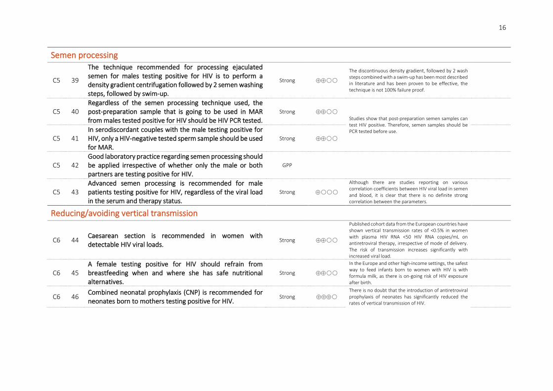
Semen processing
C5 39
The technique recommended for processing ejaculated semen for males testing positive for HIV is to perform a density gradient centrifugation followed by 2 semen washing steps, followed by swim-up.
Strong ⊕⊕
The discontinuous density gradient, followed by 2 wash steps combined with a swim-up has been most described in literature and has been proven to be effective, the technique is not 100% failure proof.
C5 40 Regardless of the semen processing technique used, the post-preparation sample that is going to be used in MAR from males tested positive for HIV should be HIV PCR tested.
Strong ⊕⊕ Studies show that post-preparation semen samples can test HIV positive. Therefore, semen samples should be PCR tested before use.
C5 41 In serodiscordant couples with the male testing positive for HIV, only a HIV-negative tested sperm sample should be used for MAR.
Strong ⊕⊕
C5 42 Good laboratory practice regarding semen processing should be applied irrespective of whether only the male or both partners are testing positive for HIV.
GPP
C5 43 Advanced semen processing is recommended for male patients testing positive for HIV, regardless of the viral load in the serum and therapy status.
Strong ⊕
Although there are studies reporting on various correlation coefficients between HIV viral load in semen and blood, it is clear that there is no definite strong correlation between the parameters.
Reducing/avoiding vertical transmission
C6 44 Caesarean section is recommended in women with detectable HIV viral loads.
Strong ⊕⊕
Published cohort data from the European countries have shown vertical transmission rates of <0.5% in women with plasma HIV RNA <50 HIV RNA copies/mL on antiretroviral therapy, irrespective of mode of delivery. The risk of transmission increases significantly with increased viral load.
C6 45 A female testing positive for HIV should refrain from breastfeeding when and where she has safe nutritional alternatives.
Strong ⊕⊕
In the Europe and other high-income settings, the safest way to feed infants born to women with HIV is with formula milk, as there is on-going risk of HIV exposure after birth.
C6 46 Combined neonatal prophylaxis (CNP) is recommended for neonates born to mothers testing positive for HIV.
Strong ⊕⊕⊕ There is no doubt that the introduction of antiretroviral prophylaxis of neonates has significantly reduced the rates of vertical transmission of HIV.

17
Human Papilloma Virus
Prevention of transmission before medically assisted reproduction
D2 47 The use of barrier contraception during sexual intercourse is advised to lower the risk of Human Papilloma virus (HPV) transmission.
GPP
D2 48 All women starting medically assisted reproduction (MAR) should undergo testing to detect HPV-related cervical lesions.
GPP
D2 There is no evidence that there is a specific HPV DNA copy number threshold below which (horizontal or vertical) transmission is unlikely.
Conclusion
Assisted reproduction techniques and impact on outcomes
D3 49 The cause of infertility should dictate the specific technique (IUI/IVF/ICSI) used for MAR in couples where one or both partners test positive for HPV.
Strong ⊕
From the perspective of horizontal and vertical transmission, there is currently not enough evidence to recommend one technique (IUI/IVF/ICSI) over another in patients infected with HPV.
D3 50 Women infected with HPV should be informed that MAR does not eliminate the risk of vertical transmission.
GPP
D3 51 The possibility of HPV testing could be discussed with couples undergoing IUI.
Research only
D3 52
Couples with a known positive HPV test should be advised that HPV is a transient infection, and postponing MAR treatment is an option depending on the individual circumstances.
GPP
Prevention/reduction of transmission during assisted reproduction
D4
There is weak evidence that therapeutic HPV vaccination in HPV-positive men may increase pregnancy rates in natural conception and reduce miscarriage rates. However, more studies are necessary.
Conclusion

18
D4 53 HPV-positive males should be informed that no current semen preparation technique can eliminate the virus from the infected semen sample.
GPP
Reducing/avoiding vertical transmission
D5 54 Caesarean delivery is not recommended on the basis of maternal HPV-positivity alone.
Strong ⊕⊕ Current evidence does not support the use of caesarean section to lower the risk or prevent mother-to-infant transmission of HPV.
D5 55 Breastfeeding is probably not contra-indicated in HPV-positive women.
Conditional ⊕ Transmission of HPV to the offspring by breastfeeding is very rare. To date there is no evidence of harm to the newborn by vertical transmission of HPV.
Human T-cell lymphotrophic virus I/II
Prevention of transmission before medically assisted reproduction
E2 56
It is suggested to inform Human T-cell lymphotrophic virus (HTLV) I/II-serodiscordant couples that there is a risk of sexual transmission of the virus to the unaffected partner. To reduce this risk, couples could be advised to use barrier contraception and receive reproductive counselling if they want to conceive.
Conditional ⊕ There is a risk of sexual transmission of HTLV I/II. The risk appears to be higher from male to female.
E2 Based on current evidence, we cannot define a threshold of HTLV I/II viral load below which horizontal or vertical transmission of HTLV I/II is not occurring.
Conclusion
Assisted reproduction techniques and impact on outcomes
E3 57
The cause of infertility should dictate the specific technique (IUI/IVF/ICSI) used for medically assisted reproduction (MAR) in couples where one or both partners test positive for HTLV I/II.
Strong ⊕
From the perspective of horizontal and vertical transmission, there is currently not enough evidence to recommend one technique (IUI/IVF/ICSI) over another in patients infected with HTLV I/II.
E3 58 Women testing positive for HTLV I/II should be informed that MAR does not eliminate the risk of vertical transmission.
GPP
19
E3
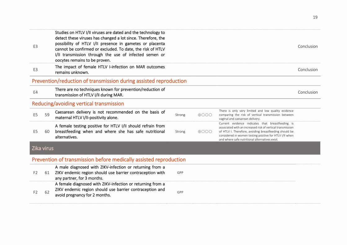
Studies on HTLV I/II viruses are dated and the technology to detect these viruses has changed a lot since. Therefore, the possibility of HTLV I/II presence in gametes or placenta cannot be confirmed or excluded. To date, the risk of HTLV I/II transmission through the use of infected semen or oocytes remains to be proven.
Conclusion
E3 The impact of female HTLV I-infection on MAR outcomes remains unknown.
Conclusion
Prevention/reduction of transmission during assisted reproduction
E4 There are no techniques known for prevention/reduction of transmission of HTLV I/II during MAR.
Conclusion
Reducing/avoiding vertical transmission
E5 59 Caesarean delivery is not recommended on the basis of maternal HTLV I/II-positivity alone.
Strong ⊕ There is only very limited and low quality evidence comparing the risk of vertical transmission between vaginal and caesarean delivery.
E5 60 A female testing positive for HTLV I/II should refrain from breastfeeding when and where she has safe nutritional alternatives.
Strong ⊕
Current evidence indicates that breastfeeding is associated with an increased risk of vertical transmission of HTLV I. Therefore, avoiding breastfeeding should be considered in women testing positive for HTLV I/II when and where safe nutritional alternatives exist.
Zika virus
Prevention of transmission before medically assisted reproduction
F2 61 A male diagnosed with ZIKV-infection or returning from a ZIKV endemic region should use barrier contraception with any partner, for 3 months.
GPP
F2 62
A female diagnosed with ZIKV-infection or returning from a ZIKV endemic region should use barrier contraception and avoid pregnancy for 2 months.
GPP

20
F2
There is no agreed threshold described in the literature below which transmission is unlikely. We advocate the use of barrier contraception to prevent horizontal transmission and avoiding pregnancy for 3 months after diagnosis or return from a ZIKV endemic area to reduce vertical transmission.
Conclusion
Assisted reproduction techniques and impact on outcomes
F3 63
If a patient or partner has been diagnosed with ZIKV-infection or returning from a ZIKV endemic region in the last 3 months, medically assisted reproduction (MAR) treatment should be postponed.
GPP
F3 64 In case of fertility preservation, the approach should be tailored to the individual situation.
GPP
F3 65
In the case of fertility preservation, there is insufficient data on the risk of viral transmission using gametes potentially infected with ZIKV. An individual risk assessment is advised before using these gametes.
GPP
There is insufficient evidence on the association between Zika infection and gametes or potential of transmission to offspring in the absence of maternal infection
F3 66 If ZIKV-infection is diagnosed in male or female during MAR treatment, the cycle should be stopped, and the couple should be advised to use barrier contraception for 3 months.
GPP
Prevention/reduction of transmission during assisted reproduction
F4 There are currently no semen processing techniques available that can completely remove ZIKV from semen.
Conclusion
F4 67 MAR is not advised even if male serum is free of ZIKV because of poor correlation between serum and semen viral load.
Strong ⊕
All infected patients, regardless of viral load, may be infectious through semen. The clearance of Zika virus is slower from semen compared to blood. Therefore, a negative test in plasma/serum does not offer 100% reassurance.
21
Reducing/avoiding vertical transmission
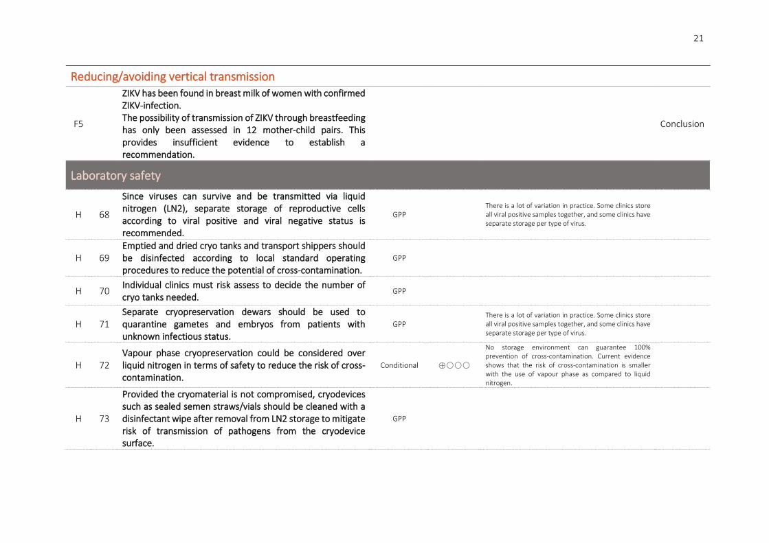
F5
ZIKV has been found in breast milk of women with confirmed ZIKV-infection. The possibility of transmission of ZIKV through breastfeeding has only been assessed in 12 mother-child pairs. This provides insufficient evidence to establish a recommendation.
Conclusion
Laboratory safety
H 68
Since viruses can survive and be transmitted via liquid nitrogen (LN2), separate storage of reproductive cells according to viral positive and viral negative status is recommended.
GPP There is a lot of variation in practice. Some clinics store all viral positive samples together, and some clinics have separate storage per type of virus.
H 69 Emptied and dried cryo tanks and transport shippers should be disinfected according to local standard operating procedures to reduce the potential of cross-contamination.
GPP
H 70 Individual clinics must risk assess to decide the number of cryo tanks needed.
GPP
H 71 Separate cryopreservation dewars should be used to quarantine gametes and embryos from patients with unknown infectious status.
GPP There is a lot of variation in practice. Some clinics store all viral positive samples together, and some clinics have separate storage per type of virus.
H 72 Vapour phase cryopreservation could be considered over liquid nitrogen in terms of safety to reduce the risk of cross-contamination.
Conditional ⊕
No storage environment can guarantee 100% prevention of cross-contamination. Current evidence shows that the risk of cross-contamination is smaller with the use of vapour phase as compared to liquid nitrogen.
H 73
Provided the cryomaterial is not compromised, cryodevices such as sealed semen straws/vials should be cleaned with a disinfectant wipe after removal from LN2 storage to mitigate risk of transmission of pathogens from the cryodevice surface.
GPP
22
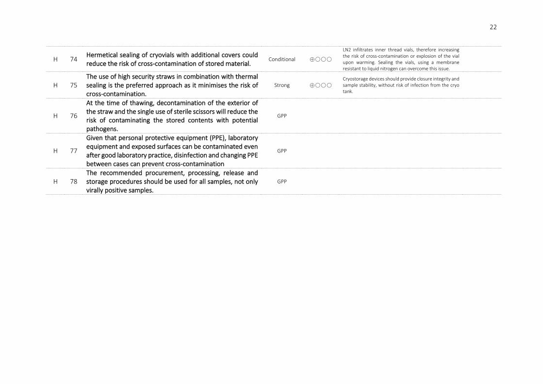
H 74 Hermetical sealing of cryovials with additional covers could reduce the risk of cross-contamination of stored material.
Conditional ⊕
LN2 infiltrates inner thread vials, therefore increasing the risk of cross-contamination or explosion of the vial upon warming. Sealing the vials, using a membrane resistant to liquid nitrogen can overcome this issue.
H 75 The use of high security straws in combination with thermal sealing is the preferred approach as it minimises the risk of cross-contamination.
Strong ⊕ Cryostorage devices should provide closure integrity and sample stability, without risk of infection from the cryo tank.
H 76
At the time of thawing, decontamination of the exterior of the straw and the single use of sterile scissors will reduce the risk of contaminating the stored contents with potential pathogens.
GPP
H 77
Given that personal protective equipment (PPE), laboratory equipment and exposed surfaces can be contaminated even after good laboratory practice, disinfection and changing PPE between cases can prevent cross-contamination
GPP
H 78 The recommended procurement, processing, release and storage procedures should be used for all samples, not only virally positive samples.
GPP

23
Table 1: Summary of the available evidence on the topics included in the guideline.

24
Table 1 Continued.
* only viral-like particles the size of HIV have been detected in spermatozoa.
25
PART A: Hepatitis B virus
A1. Prevalence and testing
NARRATIVE QUESTION: WHAT IS THE PREVALENCE OF HEPATITIS B VIRUS?
Hepatitis B virus (HBV) is an enveloped DNA virus, and a member of the family Hepadnaviridae hepatotropic DNA viruses. Hepatitis B virus causes both acute and chronic infection that can range from asymptomatic infection or mild disease to severe or fulminant hepatitis. In 2015, the global prevalence of HBV infection in the general population was 3.5%. Prevalence was the highest in the African (6.1%), Western Pacific regions (6.2%) and Eastern Mediterranean region (3.3%). In Europe, 30 member’ states reported 26,907 cases of HBV infections for 2017, corresponding to a crude rate of 6.7 cases per 100 000 population. Twenty countries reported HBV chronic infections, leading to an overall notification rate of 7.2 cases per 100,000 population. The United Kingdom reported 62% of all chronic cases in 2017 (ECDC, 2017). Overall, about 257 million persons were living with HBV infection. However, rates of chronic Hepatitis B cases are dependent on local notification requirements. Many infected people remain undiagnosed. Among those born before the Hepatitis B vaccine became available, the proportion of persons living with chronic HBV infection remains high. Approximately 15–40 % of chronically infected patients will develop liver cirrhosis, liver failure, or hepatocellular carcinoma and 15–25 % will ultimately die as a result of their HBV infection. Mortality due to HBV infection is increasing and expected to further increase.
In 2016, the World Health Assembly adopted the Global Health Sector Strategy (GHSS) on viral hepatitis, committing to eliminating viral hepatitis as a public health threat by 2030. The synergistic interventions for prevention, testing and treatment are at the core of an effective hepatitis response and are promoted through the GHSS on viral hepatitis. Assuming that women of reproductive age constitute 25.3% of the world’s population (United Nations data), adults chronically infected may include 65 million women of childbearing age who can potentially transmit HBV to their babies (Lavanchy and Kane, 2016). Most of the burden of disease from HBV infection comes from infections acquired before the age of 5 years (ECDC, 2017). Therefore, prevention of HBV infection focuses on children under 5 years of age. In the 1990s, the World Health Assembly had already asked for the inclusion of Hepatitis B vaccine in routine infant immunization schedule. The low incidence of chronic HBV infection in children under 5 years of age at present can be attributed to the widespread use of Hepatitis B vaccine. Worldwide, in 2015, the estimated prevalence of HBV infection in this age group was about 1.3%, compared with about 4.7% in the pre-vaccination era (which, according to the year of introduction can range from the 1980s to the early 2000s). So, most of the people currently living with HBV infection are persons born before Hepatitis B vaccine was widely available and used in infancy. However, there are regional differences in coverage. The African, Eastern Mediterranean and European regions remain below the global average (WHO, 2017).

26
NARRATIVE QUESTION: HOW SHOULD TESTING OF HEPATITIS B STATUS PRIOR TO MEDICALLY ASSISTED REPRODUCTION BE PERFORMED?
Serological assays are typically used as the first line of the testing strategy to screen for exposure to a virus because of their relatively low cost (compared to nucleic acid testing (NAT)). Serological tests for the detection of Hepatitis B (HB) e-antigen and anti-HBe antibody may also aid in the management of the patient and are widely available. For the diagnosis of chronic HBV infection in adults, adolescents and children (>12 months of age), a serological assay (in either rapid diagnostic test (RDT) or laboratory-based immunoassay format (enzyme immunoassay or chemiluminiscence immunoassay) that meets minimum quality, safety and performance standards (with regard to both analytical and clinical sensitivity and specificity) is recommended to detect Hepatitis B surface antigen (HBsAg). In settings where existing laboratory testing is already available and accessible, laboratory-based enzyme immunoassays (EIAs) are recommended as the preferred assay format. A cut-off value, usually determined by the manufacturer of the assay, specifies the point at which the results are considered to be reactive, and therefore, EIA results are generally reported as optical density divided by the assay cut-off (OD/CO) values. These types of assays are best suited for and most cost–effective to perform in settings with a high throughput of specimens (in excess of 40 per day). In settings where there is limited access to laboratory testing and/or in populations where access to rapid testing would facilitate linkage to care and treatment, use of RDTs is recommended to improve access.
- In settings or populations with an HBsAg seroprevalence of ≥0.4%, a single serological assay for detection of HBsAg is recommended, prior to further evaluation for HBV DNA and staging of liver disease.
- In settings or populations with a low HBsAg seroprevalence of <0.4%, confirmation of HBsAg positivity on the same immunoassay with a neutralization step or a second different RDT assay for detection of HBsAg may be considered. Conditional recommendation, low quality of evidence
Directly following a positive HBsAg serological test, the use of quantitative or qualitative NAT for detection of HBV DNA is recommended as the preferred strategy and to guide who to treat or not treat if there is no evidence of cirrhosis, and to monitor for treatment response, based on existing recommendations from the 2015 WHO HBV management guidelines (WHO, 2015). These assays detect the presence of viral nucleic acid – DNA through targeting a specific segment of the virus, which is then amplified. The amplification step enables the detection of low levels of the virus in the original specimen, which might not otherwise have been detectable. Serum HBV DNA is measured in international units (IU)/mL as the recognized international standard or copies/ml by NAT. HB core antibody (HBcAb) is a marker of past HBV exposure, the guideline review did not look for evidence on this specific antibody.
Conclusion
HBV testing is mandatory according to the European Tissues and Cells Directive as a preventative measure to reduce the risks of transmission to partners and offspring.

27
REFERENCES ECDC. Hepatitis B. . ECDC Annual epidemiological report for 2017 2017. Lavanchy D, Kane M. Global Epidemiology of Hepatitis B Virus Infection. In Liaw Y-F and Zoulim F (eds) Hepatitis B Virus in Human Diseases. 2016. Springer International Publishing, Cham, pp. 187-203. WHO. Guidelines for the prevention, care and treatment of persons with chronic hepatitis B infection. 2015. WHO. Global hepatitis report, 2017. 2017.

28
A2. Prevention of transmission before medically assisted reproduction
PICO QUESTION: WHAT ARE THE RISKS OF HEPATITIS B VIRUS TRANSMISSION THROUGH VAGINAL/ANAL INTERCOURSE?
Evidence A cross-sectional study including 203 participants testing positive for Hepatitis B virus (HBV) and their 138 sexual partners tested for HBV antigen (Ag) and antibody (Ab) to determine current and past HBV infections in sexual partners of HBV index cases. Of the 138 sexual partners, 28 (20.3%) were vaccinated for HBV, 20 (14.5%) tested positive for HBsAg and 36 (26.1%) had evidence of past and current HBV infection. Female sexual partners were significantly more likely to get infected compared to male partners (crude OR 2.31, 95% CI 1.01 to 5.29). Furthermore, partners who were cohabiting were more likely to get infected (OR 3.95, 95% CI 1.73 to 9.04) compared to sexual partners who were not cohabiting (Tufon et al., 2019).
A large cross-sectional study including 2590 individuals testing positive for HBsAg and their 1454 spouses (1003 females and 451 males) measured HBsAg on serum samples of all partners and reported the rate of HBV-exposure (HB core antigen (HBcAb) positivity) to be 48% (n=480) in female spouses, 62.9% (n=281) in male spouses. However, HBsAg was positive in only 2.3% (n=33) of the spouses (4.2% in husbands and 1.4% in wives) (Katoonizadeh et al., 2018).
A small cohort study included 5 index cases testing positive for HBV and their partners (married or engaged) and investigated the HBV sequence homology between spouses. For all five couples, the HBV-infected index subject and the spouse shared a 100% sequence homology for the cloned region (Huo et al., 1998).
An old study including 83 patients with acute HBV infection, investigated the sexual transmission in these patients and found that 18/24 sex partners of included patients tested positive for HBsAg and HBV DNA (Hou et al., 1993).
A cross-sectional study including 1368 females reported that heterosexual transmission was the only risk factor for disease acquisition in 27% of females with a positive HBV test. Furthermore, having anal intercourse and failure to use barrier contraceptives may facilitate transmission of HBV infection to women (Rosenblum et al., 1992).
A prospective cohort study, including 68 pregnant women testing positive for HBsAg and their husbands, investigated the sexual transmission between partners and reported that 30/68 husbands were positive (44.2%), 11.8% were Ag positive and 32.4% were Ab positive. Furthermore, they concluded that transmission occurs particularly if sexual contact takes place during or immediately after menstruation (Inaba et al., 1979).
29
Recommendation
Partners of Hepatitis B virus (HBV)-positive individuals should be vaccinated.
Strong ⊕
Barrier contraception should be used until the completion of the HBV vaccination protocol.
Strong ⊕⊕
Medically assisted reproduction (MAR) services staff should be vaccinated against HBV.
GPP
Justification Multiple studies have shown that sexual transmission of HBV may double the risk of horizontal and vertical transmission. The availability of highly effective vaccines outside and during pregnancy allows prevention of horizontal and vertical transmission.
Providing a successful vaccination course, the risk of HBV horizontal transmission is eliminated during unprotected intercourse for spontaneous conception.
Staff working in general healthcare are required to have HBV vaccination and to have a completed HBV vaccination schedule. For this reason, staff employed by MAR services should follow the same occupational health protocols.
PICO QUESTION: IS THERE A THRESHOLD BELOW WHICH TRANSMISSION OF HEPATITIS B VIRUS IS UNLIKELY?
Evidence Horizontal transmission
No studies could be identified reporting a serum Hepatitis B virus (HBV) DNA load threshold below which horizontal transmission does not occur.
Mother-to-child transmission (MTCT)
No publications could be identified where maternal viral load was determined before pregnancy.
Father-to-child transmission (FTCT)
Only a single retrospective study including 398 couples (spontaneous pregnancy, no semen processing) of males testing positive for HBV with uninfected female partners. There was decreased HBV vertical transmission from father to infant with lower HBV DNA in paternal serum, whatever HBsAb levels in mother. HBV DNA was not detected when paternal serum HBV DNA was <10² IU/ml. However, tests

30
have been performed on blood cord and not after immunoprohylaxis and vaccination in infant (Cao et al., 2016).
There was no evidence of HBV vertical transmission through MAR in the circumstances of a HBV negative woman and a HBV positive male partner.
Conclusion
Based on the current evidence, in the circumstances of medically assisted reproduction, it is not possible to identify a pre-treatment HBV DNA load threshold below which vertical transmission is very unlikely.
Recommendation
All patients with an active or chronic HBV-infection must be reviewed by an infectious disease/ liver specialist before initiating any MAR treatment.
Strong ⊕
Commencing with MAR treatments in patients positive for HBV should be a joint decision between the patient, their partner, the fertility doctor and the infectious disease/ liver specialist.
Strong ⊕
In the case of the female testing positive for HBV, the possibility of viral vertical transmission, the availability of vaccination during pregnancy and newborn prophylaxis should all be discussed.
GPP
Justification Consulting a hepatologist is an important step before considering a pregnancy as it allows an assessment of both partners and prophylactic vaccination of the uninfected partner.
Most studies investigating the threshold of HBV transmission analysed viral load during pregnancy or at delivery as opposed to before pregnancy (pre-treatment). Any HBV-infected mother carries the risk of vertically transmitting the virus to the newborn (Liu et al., 2015, Lu et al., 2017, Sellier et al., 2015, Wiseman et al., 2009).
Therefore, it should be noted that maternal vaccination and neonate immunoprophylaxis significantly reduces but does not eliminate the risk of vertical HBV transmission.

31
REFERENCES Cao L, Li Y, Sun S, Zhao P, Wang S, Duan Y, Liu Z, Xu D. Correlation between paternal serum hepatitis B virus DNA levels and vertical transmission from father to infant. International journal of clinical and experimental medicine 2016;9: 22134-22140. Hou MC, Wu JC, Kuo BI, Sheng WY, Chen TZ, Lee SD, Lo KJ. Heterosexual transmission as the most common route of acute hepatitis B virus infection among adults in Taiwan--the importance of extending vaccination to susceptible adults. The Journal of infectious diseases 1993;167: 938-941. Huo TI, Wu JC, Huang YH, Yang UC, Sheen IJ, Chang FY, Lee SD. Evidence of transmission of hepatitis B virus to spouses from sequence analysis of the viral genome. Journal of gastroenterology and hepatology 1998;13: 1138-1142. Inaba N, Ohkawa R, Matsuura A, Kudoh J, Takamizawa H. Sexual transmission of hepatitis B surface antigen. Infection of husbands by HBsAg carrier-state wives. The British journal of venereal diseases 1979;55: 366-368. Katoonizadeh A, Motamed-Gorji N, Sharafkhah M, Ostovaneh M, Esmaili S, Eslami L, Gharravi A, Khoshnia M, Shayanrad A, Katouli FS et al. Intra-familial transmission of chronic hepatitis B infection: A large population-based cohort study in northern Iran. Archives of Iranian medicine 2018;21: 436-442. Liu CP, Zeng YL, Zhou M, Chen LL, Hu R, Wang L, Tang H. Factors associated with mother-to-child transmission of hepatitis B virus despite immunoprophylaxis. Internal medicine (Tokyo, Japan) 2015;54: 711-716. Lu Y, Zhu FC, Liu JX, Zhai XJ, Chang ZJ, Yan L, Wei KP, Zhang X, Zhuang H, Li J. The maternal viral threshold for antiviral prophylaxis of perinatal hepatitis B virus transmission in settings with limited resources: A large prospective cohort study in China. Vaccine 2017;35: 6627-6633. Rosenblum L, Darrow W, Witte J, Cohen J, French J, Gill PS, Potterat J, Sikes K, Reich R, Hadler S. Sexual practices in the transmission of hepatitis B virus and prevalence of hepatitis delta virus infection in female prostitutes in the United States. JAMA : the journal of the American Medical Association 1992;267: 2477-2481. Sellier P, Maylin S, Amarsy R, Mazeron MC, Larrouy L, Haim-Boukobza S, Lopes A, Moreno MD, Ricbourg A, Simoneau G et al. Untreated highly viraemic pregnant women from Asia or sub-Saharan Africa often transmit hepatitis B virus despite serovaccination to newborns. Liver international : official journal of the International Association for the Study of the Liver 2015;35: 409-416. Tufon KA, Meriki HD, Kwenti TE, Tony NJ, Malika E, Bolimo AF, Kouanou YS, Nkuo-Akenji T, Anong DN. HBV Transmission Risk Assessment in Healthcare Workers, Household and Sexual Contacts of HBV Infected Patients in the Southwest Region of Cameroon. Oman medical journal 2019;34: 313-321. Wiseman E, Fraser MA, Holden S, Glass A, Kidson BL, Heron LG, Maley MW, Ayres A, Locarnini SA, Levy MT. Perinatal transmission of hepatitis B virus: an Australian experience. The Medical journal of Australia 2009;190: 489-492.

32
A3. Assisted reproduction techniques and impact on outcomes
PICO QUESTION: SHOULD IUI, IVF OR ICSI BE PREFERENTIALLY USED FOR MAR IN HEPATITIS B INFECTED COUPLES?
No studies could be found that compare the efficacy in terms of pregnancy rate and safety in terms of risk of vertical transmission between different medically assisted reproductive techniques.
Evidence A prospective cohort study compared IVF with ICSI in 125 women testing positive for Hepatitis B virus (HBV). In total, 176 children were born in the assisted reproduction group, 145 by IVF and 31 by ICSI. When twins were considered as one, the rate of positive HBsAg in IVF children, 5.9% (6/102), was lower than that in ICSI children, 13% (3/23), although the difference was not statistically significant. When twins were considered as two, no difference was found in the rate of HBsAg-positive IVF children as compared with ICSI children (4.8% (7/145) vs. 12.9% (4/31)). All HBsAg-positive children received Hepatitis B Immune globulin (HBIG) treatment and seroconverted to negative at 9-15 months of age (Nie et al., 2019).
Conclusion
From the perspective of horizontal and vertical transmission, there is currently not enough evidence to recommend one technique (IUI/IVF/ICSI) over another in patients infected with Hepatitis B.
Recommendation
The cause of infertility should dictate the specific technique (IUI/IVF/ICSI) used for MAR in couples where one or both partners test positive for HBV.
Strong ⊕
Women testing positive for HBV should be informed that MAR does not eliminate the risk of vertical transmission.
GPP
Justification Only vaccination against HBV and maintenance of measurable antibody levels can protect a woman from HBV infection and her child from vertical transmission. In women testing positive for HBV, the HBV viral DNA level dictates the risk of vertical transmission. Even in the circumstances of infant prophylaxis, the risk of vertical transmission is not zero, yet very low (Boucheron et al., 2021).

33
PICO QUESTION: CAN HEPATITIS B VIRUS DNA BE DETECTED IN OOCYTES/ SPERM/ PLACENTA?
Evidence DNA integration in sperm
In an experimental study, 233 sperm metaphase spreads from 9 males testing positive for HBV were analysed for DNA integration of HBV. Only one patient had HBV DNA integration in the sperm genetic material (Huang et al., 2003).
DNA integration in oocytes/embryos
A prospective cohort study including 72 HBV serodiscordant couples (31 male HBsAg positive, and 41 female HBsAg positive) investigated the relation between HBsAg positivity of oocytes and embryos and the risk of vertical transmission. Twelve infants were born to couples with HBV positive oocytes/embryos: 2 with HBV DNA positive oocytes/embryos, 7 with HBV RNA positive oocytes/embryos and 3 with HBsAg positive oocytes/embryos. Twenty children tested anti-HBs positive. At 6 months, only 1 infant was seropositive for anti-HBs, anti-HBc, and anti-HBe, however, this child had seroconverted by 9 months (Jin et al., 2016).
In the study by Kong et al. ovarian tissues from 50 patients with gynaecological disease and HBV positivity were used to investigate HBV expression and replication in ovum. HBcAg was detected in 12% of ovarian tissue samples (6/50). HBV DNA was detected in the interstitial cells, granulosa cells, and ova in ovarian tissues at a positive rate of 14% (7/50). Three samples were positive for HBV mRNA (3%). Positive signal of HBV mRNA was mainly distributed in the cytoplasm of the ova and the granulosa cells. Patients with detectable HBV markers in ovaries had a higher level of serum HBV DNA (Kong et al., 2016).
In the study by Hu et al. 250 oocytes and 578 embryos that were not used for IVF-ICSI from HBV positive couples were analysed for presence and expression of HBV. HBV DNA was found in 9.6% of oocytes (24/250) and in 14.4% of embryos (83/578). A significant increase in viral positivity in oocytes and embryos was found in women with a high serum HBV DNA level (Hu et al., 2011).
In a report by Quint et al., culture medium for embryo culture in IVF was contaminated with HBV infected serum. HBV DNA could not be demonstrated by PCR in any of the children of mothers exposed to HBV during IVF (Quint et al., 1994).
Placenta
Wei et al. investigated 155 placentae from women testing positive for HBsAg and reported that the total rate of placentae testing positive for HBsAg was 37.42% (58/155) by immunohistochemistry. Furthermore, the placental positivity for HBsAg was higher in mothers testing positive for HBeAg (OR 2.00; 95% CI 1.02 to 3.95). The risk of an HBsAg-positive placenta was higher with increasing maternal blood HBV DNA levels (the relative risk estimate OR was 3.24 to 3.85) (Wei et al., 2015).
Chen et al. investigated the role of placental HBV infection in vertical transmission of HBV to the newborn. Hereto, they collected 157 placental tissue samples from 171 pregnant women testing positive for HBV. The rate of placental HBsAg-positivity by immunohistochemistry (IHC) was 36.9% (58/157) and the rate of HBcAg was 31.8% (50/157). HBV DNA was detected in 42.7% of cases (67/157) by RT-PCR and in situ hybridisation (ISH) showed that the HBV infection rate was 55.4% (87/157) in
34
decidual cells, 51.0% (80/157) in trophoblastic cells,46.5% (73/157) in villous mesenchymal cells, and 29.9% (47/157) in villous capillary endothelial cells (Chen et al., 2013).
In a case-control study, placental tissue from 101 women testing positive for HBsAg was investigated. IHC and ISH showed that HBsAg was present in 33.7% of placental samples (34/101), HBxAg in 37.6% (38/101), HBcAg in 20.8% (21/101) and HBV DNA in 44.6% of placental samples (45/101). Furthermore, the HBV infection rates decreased gradually from the maternal side to the fetal side (Xu et al., 2002).
Conclusion
HBV can be detected in sperm cells, oocytes, granulosa cells and embryos. This equates with a theoretical risk of vertical HBV transmission that remains to be proven.
PICO QUESTION: DOES HEPATITIS B VIRUS AND/OR TREATMENT OF HEPATITIS B VIRUS BEFORE MAR IMPACT THE OUTCOME OF MAR?
MALE INFECTED
Evidence Male infected
A retrospective cohort study, including 66 Hepatitis B (HBV)-serodiscordant couples and 68 controls, compared IVF-ICSI cycles outcomes and reported no significant differences between HBV serodiscordant couples and controls for implantation rate (34.5% (20/58) vs. 25.3 (25/99)), pregnancy rate per cycle (25.8% (17/66) vs. 30.9% (21/68)), miscarriage rate per cycle (17.6% (3/17) vs. 33.3% (7/21)) or live birth rate per cycle (21.2% (14/66) vs. 19.1% (13/68)) (Cito et al., 2019).
A retrospective cohort study included 92 serodiscordant couples with active HBV infection (HBV DNA+), 125 serodiscordant couples with convalescent infection (HBsAg+, HBeAb+, HBcAb+, HBV DNA negative) and 121 seronegative controls. In the couples where ejaculated sperm was used, there was no significant difference between couples with active or convalescent HBV infection or controls for implantation rate (28.3% (45/159) vs. 32.8% (39/122) vs. 23.0% (38/165)), clinical pregnancy rate (44.2% (34/77) vs. 50.8% (30/59) vs. 38.5% (30/78)), early miscarriage rate (8.8% (3/34) vs. 0% (0/30) vs. 6.7% (2/30)) or live birth rate (36.4% (28/77) vs. 49.2% (29/59) vs. 35.9% (28/78)). In the couples where surgically retrieved sperm was used, the early miscarriage rate was significantly higher in couples with convalescent HBV infection as compared to active HBV infection and controls (0% vs. 23.1% (3/13) vs. 5.0% (1/20)). However, there were no significant differences between active and convalescent HBV infection and control couples for implantation rate (31.1% (28/90) vs. 26.1% (18/69) vs. 25.3% (24/95)), clinical pregnancy rate (50% (22/44) vs. 39.4% (13/33) vs. 42.6% (20/47)) or live birth rate (50% (22/44) vs. 27.3% (9/33) vs. 36.2% (17/47)) (Zheng et al., 2016).
A retrospective cohort study including 136 males testing positive for HBsAg and 426 HBV-seronegative controls reported no significant differences for implantation rate (38.5% (104/270) vs. 37.7% (206/547)) or clinical pregnancy rate (58.1% (79/136) vs. 53.7% (146/272)) between groups (Shi et al., 2014).
35
A matched case-control study including 32 males testing positive for HBsAg and 64 controls reported no difference for implantation rate (13.5% vs. 20.0%), clinical pregnancy rate per cycle (18.8% (6/32) vs. 31.3% (20/64)) or live birth rate per cycle (15.6% (5/32) vs. 23.4% (15/64)) between groups (Oger et al., 2011).
A retrospective cohort study including 916 patients testing positive for HBV (457 HBsAg and 459 HBsAg negative) compared the reproductive outcomes of 1824 IVF-ICSI cycles and reported no significant differences in reproductive outcomes after IVF between HBV positive and negative men for implantation rate (24.9% (284/1140) vs. 26.7% (296/1108)) or clinical pregnancy rate (40.5% (217/535) vs. 40.3% (210/521)). However, ICSI in HBV positive males resulted in a significantly lower implantation rate (18.3% (126/688) vs. 24.2% (159/657)) and clinical pregnancy rate (31.2% (96/308) vs. 39.3% (118/300)) as compared to controls (Zhou et al., 2011).
A retrospective cohort study analysing IVF-ICSI reproductive outcomes in HBV-serodiscordant couples (n=161) reported no significant difference between seropositive and seronegative men in ongoing pregnancy rates per started cycle (30.4% vs. 29.9%) (Lee et al., 2010).
Female infected
A retrospective cohort study investigated IVF-ICSI reproductive outcomes in HBsAg+/HBeAg+ women (n=180) with HBsAg+/HBeAg- women (n=714) and seronegative controls (7565). The implantation rate was significantly lower in HBsAg+/HBeAg- women as compared to controls (35.7% (607/1701) vs. 38.7% (6950/17939)), but not in HBsAg+/HBeAg+ women as compared to controls (39.6% (158/399) vs. 38.7% (6950/17939)). There was no significant difference between HBsAg+/HBeAg+ women, HBsAg+/HBeAg- women and controls for clinical pregnancy rate (61.7% (111/180) vs. 57.6% (411/714) vs. 60.4% (4628/7656)), miscarriage rate (11.7% (13/111) vs. 10.0 (41/411) vs. 11.7% (541/4628)) or live birth rate (53.1% (93/175) vs. 51.1% (360/704) vs. 52.3% (3911/7480)) (Wang et al., 2019).
A retrospective case-control study comparing IVF-ICSI cycle outcomes from chronic HBV-serodiscordant couples (n=123 cycles) with matched HBV-negative couples (246 cycles) reported no significant differences between chronic HBV-infected couples and matched HBV-negative controls for implantation rate (30.52% (76/249) vs. 28.34% (142/501)), clinical pregnancy rate (44.72% (55/123) vs. 43.09% (106/246)) or live birth rate (42.28% (52/123) vs. 40.65% (100/246)) (Chen et al., 2014).
A retrospective cohort study including 136 males testing positive for HBsAg and 426 HBV seronegative controls reported no significant differences for implantation rate (36.0% (54/150) vs. 38.5% (117/304)) or clinical pregnancy rate (48.1% (37/77) vs. 50.6% (78/154)) between groups (Shi, et al., 2014).
A retrospective cohort study analysing IVF-ICSI reproductive outcomes in HBV-serodiscordant (n=131) couples reported no significant difference between seropositive and seronegative women in ongoing pregnancy rates per started cycle (26.7% versus 30.2%) (Lee, et al., 2010).
Conclusion
Existing evidence cannot clarify if the presence of HBV infection in the male impacts the outcomes of MAR. Multiple studies showed no differences in reproductive outcomes following MAR when comparing seronegative with HBV seropositive women.

36
REFERENCES Boucheron P, Lu Y, Yoshida K, Zhao T, Funk AL, Lunel-Fabiani F, Guingané A, Tuaillon E, van Holten J, Chou R et al. Accuracy of HBeAg to identify pregnant women at risk of transmitting hepatitis B virus to their neonates: a systematic review and meta-analysis. The Lancet Infectious diseases 2021;21: 85-96. Chen H, Ge HS, Lv JQ, Wu XM, Xi HT, Huang JY, Zhu CF. Chronic hepatitis B virus infection in women is not associated with IVF/ICSI outcomes. Archives of gynecology and obstetrics 2014;289: 213-217. Chen Y, Wang L, Xu Y, Liu X, Li S, Qian Q, Hu B, Zhou A, Chen T, Zhao Y. Role of maternal viremia and placental infection in hepatitis B virus intrauterine transmission. Microbes and infection 2013;15: 409-415. Cito G, Coccia ME, Fucci R, Picone R, Cocci A, Sessa M, Sessa F, Rizzello F, Micelli E, Trotta M et al. Hepatitis B Surface Antigen Seropositive Men in Serodiscordant Couples: Effects on the Assisted Reproductive Outcomes. The world journal of men's health 2019. Hu XL, Zhou XP, Qian YL, Wu GY, Ye YH, Zhu YM. The presence and expression of the hepatitis B virus in human oocytes and embryos. Human reproduction (Oxford, England) 2011;26: 1860-1867. Huang JM, Huang TH, Qiu HY, Fang XW, Zhuang TG, Liu HX, Wang YH, Deng LZ, Qiu JW. Effects of hepatitis B virus infection on human sperm chromosomes. World journal of gastroenterology 2003;9: 736-740. Jin L, Nie R, Li Y, Xiao N, Zhu L, Zhu G. Hepatitis B surface antigen in oocytes and embryos may not result in vertical transmission to offspring of hepatitis B virus carriers. Fertility and sterility 2016;105: 1010-1013. Kong Y, Ye F, Jin Y, Shi J, Qiu H, Lin S. Hepatitis b virus expression and replication in ovum and the influencing factors. Saudi journal of gastroenterology : official journal of the Saudi Gastroenterology Association 2016;22: 215-219. Lee VC, Ng EH, Yeung WS, Ho PC. Impact of positive hepatitis B surface antigen on the outcome of IVF treatment. Reproductive biomedicine online 2010;21: 712-717. Nie R, Wang M, Liao T, Qian K, Zhu G, Jin L. Assisted conception does not increase the risk for mother-to-child transmission of hepatitis B virus, compared with natural conception: a prospective cohort study. Fertility and sterility 2019;111: 348-356. Oger P, Yazbeck C, Gervais A, Dorphin B, Gout C, Jacquesson L, Ayel JP, Kahn V, Rougier N. Adverse effects of hepatitis B virus on sperm motility and fertilization ability during IVF. Reproductive biomedicine online 2011;23: 207-212. Quint WGV, Fetter WPF, Van Os HC, Heijtink RA. Absence of hepatitis B virus (HBV) DNA in children born after exposure of their mothers to HBV during in vitro fertilization. Journal of clinical microbiology 1994;32: 1099-1100. Shi L, Liu S, Zhao W, Zhou H, Ren W, Shi J. Hepatitis B virus infection reduces fertilization ability during in vitro fertilization and embryo transfer. Journal of medical virology 2014;86: 1099-1104. Wang L, Li L, Huang C, Diao L, Lian R, Li Y, Xiao S, Hu X, Mo M, Zeng Y. Maternal chronic hepatitis B virus infection does not affect pregnancy outcomes in infertile patients receiving first in vitro fertilization treatment. Fertility and sterility 2019;112: 250-257.e251. Wei J, Xue S, Zhang J, Wang S, Wang B. Study of the relationship in pregnant women between hepatitis B markers and a placenta positive for hepatitis B surface antigen. Journal of perinatal medicine 2015;43: 191-199. Xu DZ, Yan YP, Choi BC, Xu JQ, Men K, Zhang JX, Liu ZH, Wang FS. Risk factors and mechanism of transplacental transmission of hepatitis B virus: a case-control study. Journal of medical virology 2002;67: 20-26. Zheng Z, Zhao X, Hong Y, Xu B, Tong J, Xia L. The safety of intracytoplasmic sperm injection in men with hepatitis B. Archives of medical science : AMS 2016;12: 587-591. Zhou XP, Hu XL, Zhu YM, Qu F, Sun SJ, Qian YL. Comparison of semen quality and outcome of assisted reproductive techniques in Chinese men with and without hepatitis B. Asian journal of andrology 2011;13: 465-469.

37
A4. Prevention/ reduction of transmission during assisted reproduction
PICO QUESTION: WHICH TECHNIQUES CAN BE USED TO PREVENT/ REDUCE HEPATITIS B TRANSMISSION DURING MAR?
VACCINATION
Evidence No studies could be retrieved that compare vaccinating female partners of Hepatitis B virus (HBV) positive males versus not vaccinating.
Recommendation
Partners of HBV-positive individuals should be vaccinated. Strong ⊕
Justification The availability of highly effective HBV vaccines allows prevention of horizontal and vertical transmission.
SEMEN PROCESSING
Evidence A pilot experiment, including sperm samples from 4 males testing positive for Hepatitis B virus (HBV), hypothesized that a specific ICSI preparation technique (swim-up), separating spermatozoa based on differences in motility, can be used to isolate spermatozoa free from HBV DNA in order to perform ICSI in men with chronic HBV-infection. No HBV DNA was detected in the fraction containing immotile or progressive spermatozoa. In one nonprogressive spermatozoa fraction, HBV DNA was found, however, it was not quantifiable (<15 IU/L) (Condijts et al., 2020).
Recommendation
Men testing positive for HBV should be informed that no current semen preparation technique can select HBV DNA-free spermatozoa for use in MAR.
GPP
38
Routine semen processing according to the ESHRE guideline on good practice in the IVF laboratory should be used when performing MAR in men testing positive for HBV.
GPP
Justification The study by Condijts is a pilot study and needs further confirmation (Condijts, et al., 2020). Currently there are no semen processing techniques that are able to select HBV DNA free spermatozoa.
Any further questions on semen processing will not be discussed.
PICO QUESTION: DOES THE PLASMA VIRAL LOAD CORRELATE WITH HEPATITIS B VIRUS DETECTION IN SEMEN?
Evidence Four retrospective studies, including 211 HBsAg+ patients, suggest that HBV DNA can be observed in semen (Ayoola et al., 1981, Fei et al., 2015, Hadchouel et al., 1985, Qian et al., 2005) with lower titers of HBV DNA in semen as compared to serum (Hadchouel, et al., 1985, Qian, et al., 2005). Hepatitis serologic status could be correlated with HBV in semen, with the combination of serum HBV DNA and HBeAg as best predictors to identify those men with positive semen HBV DNA (Fei, et al., 2015).
Recommendation
Based on the current evidence, HBV DNA testing on seminal fluid or sperm is not recommended.
Strong ⊕
Justification It is suggested that not all chronically HBV infected men harbour HBV DNA in sperm and that lower HBV DNA titers could be found in sperm as compared to serum. Considering that all female partners of HBV positive males should be immunised prior to MAR, the measurement of HBV DNA in semen is not necessary.
REFERENCES Ayoola EA, Ladipo OA, Odelola HA. Antibody to hepatitis B core antigen, e-antigen and its antibody in menstrual blood and semen. International journal of gynaecology and obstetrics: the official organ of the International Federation of Gynaecology and Obstetrics 1981;19: 221-223. Condijts T, Bourdeaud'huy L, Tilleman K, Lierman S, Dewinter C, Padalko E. Swim-up as a strategy for isolation of spermatozoa without viral incorporation in men with chronic hepatitis B: A pilot study. Andrologia 2020: e13732. Fei QJ, Yang XD, Ni WH, Pan CS, Huang XF. Can hepatitis B virus DNA in semen be predicted by serum levels of hepatitis B virus DNA, HBeAg, and HBsAg in chronically infected men from infertile couples? Andrology 2015;3: 506-511.

39
Hadchouel M, Scotto J, Huret JL, Molinie C, Villa E, Degos F, Brechot C. Presence of HBV DNA in spermatozoa: a possible vertical transmission of HBV via the germ line. Journal of medical virology 1985;16: 61-66. Qian WP, Tan YQ, Chen Y, Peng Y, Li Z, Lu GX, Lin MC, Kung HF, He ML, Shing LK. Rapid quantification of semen hepatitis B virus DNA by real-time polymerase chain reaction. World journal of gastroenterology 2005;11: 5385-5389.
40
A5. Reducing/ avoiding vertical transmission
PICO QUESTION: WHICH INTERVENTIONS CAN BE USED TO REDUCE/AVOID VERTICAL TRANSMISSION OF HEPATITIS B VIRUS TO THE NEWBORN?
ELECTIVE CAESAREAN SECTION
Evidence A systematic review and meta-analysis investigated the mother-to-child transmission rate of Hepatitis B virus (HBV) after caesarean section (CS) and vaginal delivery (VD). When assessed at birth, 7.2% (151/1940) of newborns tested serological positive for HBV after CS compared to 16.6% (301/1813) after vaginal delivery (OR 0.269; 95% CI 0.139 to 0.520; 7 studies; 3904 mother-infant pairs). When assessed at 6 months of age, the difference between both groups was no longer present with 3.3% (132/4022) infants testing serological positive for HBV after CS compared to 4.1% (145/3520) after vaginal delivery (OR 0.790; 95% CI 0.614 to 1.016; 13 studies; 7542 mother-infant pairs) (Chen et al., 2019).
A prospective cohort study including 1384 pregnant women testing positive for HBV compared 867 CS with 517 vaginal deliveries and reported no significant difference in the proportion of neonates testing positive HBV DNA between the CS group and the vaginal delivery group (0.7% vs. 1.7%). However, after follow-up, 0.6% of infants (5/888) in the CS group compared to 1.7% of infants (9/519) in the vaginal delivery group were identified as having chronic Hepatitis B infection (Peng et al., 2018).
A prospective cohort study compared the rate of HBV transmission to infants after vaginal birth or CS. All infants received HBV vaccination and 239/447 infants also received Hepatitis B immunoglobulin (HBIG). Infants who received HBV vaccination alone had a similar rate of HBV infection whether delivered by CS or vaginally. In the infants who received HBV vaccine plus HBIG at birth, however, the HBV infection rate was significantly lower in those delivered by CS (3/53) than in those delivered vaginally (57/286) (Lee et al., 1988).
Recommendation
Caesarean delivery is not recommended on the basis of maternal HBV-positivity alone.
Strong ⊕⊕
Justification There is no evidence that the risk of HBV transmission from mother to child after caesarean section is lower compared to that after vaginal delivery.
41
BREASTFEEDING
Evidence A systematic review and meta-analysis investigated the effect of feeding practices on the vertical transmission of HBV and found that breastfeeding does not increase the risk of HBV infection in the infant (risk difference -0.8%, 95% CI -1.6% to 0.1%, 32 studies, 5650 infants) (Zheng et al., 2011).
In the study by Azzari et al., breast milk samples from all included women were tested for HBV markers, and reported that all samples tested positive for HBsAg, anti-HBcAg and anti-HBeAg. However, all samples tested negative for HBeAg and HBV DNA. No data on the seroconversion of the included infants were reported (Azzari et al., 1990).
A large cohort study by Zhang et al., including 1186 mothers testing positive for HBsAg and their infants, investigated HBV markers in the infants according to breast or bottle feeding. In mothers testing negative for HBeAg none of the infants were HBV infected no matter what immunization they received. In formula fed infants of mothers testing positive for HBeAg, there was no difference in HBV infection whether they received vaccination only, or vaccination with HBIG. In breastfed infants of mothers testing positive for HBeAg there were significant differences in rates of HBV transmission between the vaccination only group (7/26, 26.9%) and the vaccination with HBIG group (4/106, 3.8%) (Zhang et al., 2014a).
A retrospective cohort study, including 544 mothers testing positive for HBsAg and their 546 infants, investigated the effect of feeding practices on HBV transmission to the infant. Chronic HBV infection occurred in 1.5% (6/397) of breastfed children and 4.7% (7/149) of formula-fed children respectively. After adjusting for confounding factors such as maternal HBeAg status, breastfeeding was not associated with chronic HBV infection (adjusted OR 0.99, 95% CI 0.28 to 3.58) or with the self-resolved infection (adjusted OR 0.57, 95% CI 0.20 to 1.65) in the infants (Chen et al., 2013).
Recommendation
Breastfeeding is probably not contra-indicated in women testing positive for HBV.
Conditional ⊕⊕
Justification There is no association between breastfeeding and the risk of HBV transmission from mother to child. Breastfeeding has significant health benefits.
This recommendation is supported by the guidelines of the European Association for the Study of the Liver (EASL) (Lampertico et al., 2017).
VACCINATION
Evidence A systematic review and meta-analysis investigated efficacy of vaccination to prevent the mother-to-child transmission of HBV. For infants of mothers testing positive for HBsAg (including those who did

42
and did not receive Hepatitis B immunoglobulin (HBIG)), vaccine efficacy ranged from 79 to 98% (7 studies). The median seroprotection proportion across all studies including HBsAg-positive and HBsAg-negative mothers was 98% (range 52% to 100%) (Schillie and Murphy, 2013).
An older systematic review and meta-analysis also investigating the efficacy of vaccination to prevent HBV-infection in the infant reported that compared to placebo or no intervention, vaccination significantly decreased the risk of Hepatitis B occurrence (RR 0.28, 95% CI 0.20 to 0.40, 4 studies) (Lee et al., 2006).
In a randomized controlled trial, including 238 women testing positive for HBsAg, early vaccination (within 2 days of birth, at 1 and 2 months) was compared with delayed vaccination (at 3,4 and 5 months). None of the newborns tested positive for HBsAg at 3 months of age. Subclinical HBV infection developed in 2 infants from the early vaccination programme. Furthermore, the anti-HB concentrations were significantly higher with the delayed vaccination programme at 11 and 24 months of age (Schalm et al., 1989).
In a large prospective cohort study, including 863 mothers testing positive for HBsAg and their corresponding 871 infants, 2 dosages of HBV vaccine were compared. No immunoprophylaxis failure was observed with the higher vaccine dose (0/565), compared to 5.2% (16/306) with the lower dose. All seroconverted infants were born to mothers testing positive for HBeAg with a viral load over 4x10⁷ IU/ml (Wang et al., 2016).
In a prospective cohort study, including mothers testing positive for HBsAg, 6 different prophylaxis regimes were compared. Over the years, there was a gradual decline in the proportion of high anti-HBs level in all groups. The HB antibody levels were significantly higher (45.6%) with regime B (2, 3, 8 months), compared to regime A (0, 1, 6 months) (30.9%) or regime C (0, 1, 2 months) (30.7%). Furthermore, the use of regime B offered higher protection from anti-HBc seroconversion after 2 years of age (Young et al., 2003).
A small cohort study, including 81 children from asymptomatic HBsAg carrier mothers, compared vaccination only with vaccination in combination with HBIG. After 5 years, no statistical difference was observed between the HB antibody titers in infants that received vaccination and HBIG and vaccination only (Gonzalez et al., 1993).
Recommendation
All neonates born to HBV-positive couples should be vaccinated.
Strong ⊕⊕⊕
Justification Current evidence shows that perinatal transmission of HBV, which is responsible for the majority of cases of chronic HBV infection, can be prevented by vaccination. This recommendation is supported by the guidelines of the European Association for the Study of the Liver (EASL) (Lampertico, et al., 2017).
43
HEPATITIS B IMMUNE GLOBULIN (HBIG)
Evidence A systematic review and meta-analysis compared newborns injected with HBV vaccine and Hepatitis B immune globulin (HBIG) to infants injected with HBV vaccine and maternal HBIG during pregnancy. The meta-RR comparing these 2 groups for newborn HBsAg infection rate was 0.66 (95% CI 0.52 to 0.84; 7 RCTs; 1061 infants) at birth, 0.54 (95% CI 0.42 to 0.69; 12 RCTs; 1453 infants) at 7-12 months of age and 0.54 (95% CI 0.42 to 0.69; 7 RCTs; 1214 infants) after 12 months of age. Newborns receiving vaccination and HBIG had a higher amount of protective antibodies at birth (RR 2.12; 95% CI 1.66 to 2.70; 4 RCT; 291 infants), and at 7-12 months of age (RR 1.12; 95% CI 1.03 to 1.22; 8 RCT; 566 infants) but not after 12 months of age (RR 1.06; 95% CI 0.96 to 1.16; 5 RCT) (Jin et al., 2014).
Another systematic review and meta-analysis compared HBV vaccination only with HBV vaccination and HBIG in infants from HBsAg+/HBeAg- mothers and reported no difference in seroprotection rate (OR 1.24; 95% CI 0.97 to 1.58; 4 studies; 1323 patients) (Machaira et al., 2015).
A randomised control trial, including 202 infants born to mothers testing positive for HBsAg, compared one dose of HBIG at birth with a 3-dose regime (at birth, 3 and 6 months) with placebo or saline. All infants but 2 in the placebo group tested positive for HBsAg at 3 months of age, compared to 50% in the group receiving one dose of HBIG and 23% in the 3-dose regimen group (Beasley et al., 1981).
In a prospective cohort study, including mothers testing positive for HBsAg, two dosages of HBIG (100 and 200 IU) were compared. All infants received a 3-dose vaccination as well. At 7 months, there was no significant difference in the number of perinatal infected infants (1.5% (8/545) vs. 1.9% (12/632)) (Wei et al., 2018).
A prospective cohort study, including 90 women testing positive for HBsAg and their infants, compared 3 HBIG regimes. In the first group, the infants received vaccination only. In the second group, the infants received vaccination and a dose of HBIG within 2 hours after birth. In the third group, the mothers received 3 dosages of HBIG during pregnancy and the infants were vaccinated and received a birth dose of HBIG. The number of infants testing positive for HBs antibodies was 24/30, 27/30 and 29/30 respectively (Gong and Liu, 2018).
A prospective cohort study, including 1150 women testing positive for HBsAg, compared vaccination only with vaccination plus HBIG for infant immunoprophylaxis and reported that immunoprophylaxis failure was significantly higher in the vaccination only group compared to vaccination plus HBIG (RR 0.371, 95 % CI 0.167 to 0.825) (Zhang et al., 2014b).
A prospective cohort study, including 324 women testing positive for HBsAg, compared HBIG for the mothers with HBIG for the infants, both mother and infant and no HBIG for either mother or infant. Compared with the no-HBIG group, HBIG for both the mother and infant group had the lowest HBsAg-positive rate (OR 0.14; 95% CI 0.02 to 0.90), whereas HBIG for the infant group had the lowest HBsAb-positive rate (OR 0.07; 95% CI 0.02 to 0.23). The HBsAg-positive rate of the no HBIG group was 14.3% (Guo et al., 2012).
A large prospective cohort study, including 1010 infants born to women testing positive for HBsAg, compared vaccination only (4 dosages) with vaccination plus a birth dose of HBIG. The antibody titer at 1 and 2 months of age was significantly higher in infants who had received HBIG at birth. However, at

44
6, 9, and 18 months old there was no statistically significant difference in antibody titer between the two groups (Wheeley et al., 1991).
A prospective cohort study, including infants of women testing positive for HB s- and e-antigen, compared 3 prophylaxis regimes. The first group received a dose of HBIG at birth and at 3 months of age, at which time vaccination was initiated. The second group received HBIG at birth and vaccination was initiated at 4-7 days old. The third group also received HBIG at birth but initiated vaccination at 1 month old. 9/159 (5.7%) infants treated with any of the HBIG vaccine schedules became chronic HBsAg carriers, of which 4 were already positive before vaccination was started. The HBsAg carrier rate was 2.0%, 6.0%, and 8.6% among infants in the three prophylaxis schedules; the differences were not statistically significant (Beasley et al., 1983).
A retrospective cohort study, including 298 mothers testing positive for HBsAg and their infants, reporting 11 infants (3.7%) testing positive for HBsAg, and 16 (5.4%) testing negative for HBsAg but anti-HBc positive, indicating past resolved infection. Of the 11 children infected with HBV, only one received timely administration of both HBIG and Hepatitis B vaccine, and 10 others did not receive HBIG or received delayed Hepatitis B vaccine. Of the 16 children with the resolved infections, 9 were not administered with HBIG and one was given the first dose of vaccine 40 days after birth (Hu et al., 2012)
A cross-sectional study, including 4112 women testing positive for HBsAg and their infants, reported that for infants receiving the birth dose of HBV vaccine between 12-24h after birth the adjusted odds of HBV transmission was 1.9 times higher than infants immunized within 12h after birth (2.4% vs. 0.6%; adjusted OR 2.9; 95% CI 1.4 to 6.3). Furthermore, they reported no significant association between HBV transmission and HBIG administration (Qiao et al., 2019).
Recommendation
Administration of Hepatitis B immunoglobulin (HBIG) in addition to vaccination is recommended for children born to mothers testing positive for HBV.
Strong ⊕⊕
HBIG administration should follow local or national guidelines.
GPP
Justification HBIG provides passively acquired anti-HBs and temporary protection. HBIG can augment protection until a response to vaccination is attained.
The Advisory Committee on Immunization Practices (ACIP) recommends administration of Hepatitis B vaccine and Hepatitis B immune globulin (HBIG) for infants born to HBV-infected women within 12 hours of birth, followed by completion of the vaccine series and postvaccination serologic testing (Schillie et al., 2018). Furthermore, this recommendation is supported by the guidelines of the European Association for the Study of the Liver (EASL) (Lampertico, et al., 2017).

45
REFERENCES Azzari C, Resti M, Rossi ME, Lami CA, Vierucci A. Modulation by human milk of IgG subclass response to hepatitis B vaccine in infants. Journal of pediatric gastroenterology and nutrition 1990;10: 310-315. Beasley RP, Hwang LY, Lee GC, Lan CC, Roan CH, Huang FY, Chen CL. Prevention of perinatally transmitted hepatitis B virus infections with hepatitis B immune globulin and hepatitis B vaccine. Lancet 1983;322: 1099-1102. Beasley RP, Hwang LY, Lin CC, Stevens CE, Wang KY, Sun TS, Hsieh FJ, Szmuness W. Hepatitis B immune globulin (HBIG) efficacy in the interruption of perinatal transmission of hepatitis B virus carrier state. Initial report of a randomised double-blind placebo-controlled trial. Lancet 1981;2: 388-393. Chen HL, Cai JY, Song YP, Zha ML, Qin G. Vaginal delivery and HBV mother to child transmission risk after immunoprophylaxis: A systematic review and a meta-analysis. Midwifery 2019;74: 116-125. Chen X, Chen J, Wen J, Xu C, Zhang S, Zhou YH, Hu Y. Breastfeeding is not a risk factor for mother-to-child transmission of hepatitis B virus. PloS one 2013;8: e55303. Gong J, Liu X. Effect of HBIG combined with hepatitis B vaccine on blocking HBV transmission between mother and infant and its effect on immune cells. Experimental and therapeutic medicine 2018;15: 919-923. Gonzalez ML, Gonzalez JB, Salva F, Lardinois R. A 7-year follow-up of newborns vaccinated against hepatitis B. Vaccine 1993;11: 1033-1036. Guo Y, Zhang W, Zhang Y, Lin X, Zhang B, Chen C, Du Y. Cost-effectiveness analysis of preventing mother-to-child transmission of hepatitis B by injecting hepatitis B immune globulin. European journal of gastroenterology & hepatology 2012;24: 1363-1369. Hu Y, Zhang S, Luo C, Liu Q, Zhou YH. Gaps in the prevention of perinatal transmission of hepatitis B virus between recommendations and routine practices in a highly endemic region: a provincial population-based study in China. BMC infectious diseases 2012;12: 221. Jin H, Zhao Y, Tan Z, Zhang X, Zhao Y, Wang B, Liu P. Immunization interventions to interrupt hepatitis B virus mother-to-child transmission: a meta-analysis of randomized controlled trials. BMC pediatrics 2014;14: 307. Lampertico P, Agarwal K, Berg T, Buti M, Janssen HLA, Papatheodoridis G, Zoulim F, Tacke F. EASL 2017 Clinical Practice Guidelines on the management of hepatitis B virus infection. Journal of hepatology 2017;67: 370-398. Lee C, Gong Y, Brok J, Boxall EH, Gluud C. Effect of hepatitis B immunisation in newborn infants of mothers positive for hepatitis B surface antigen: systematic review and meta-analysis. BMJ (Clinical research ed) 2006;332: 328-336. Lee SD, Lo KJ, Tsai YT, Wu JC, Wu TC, Yang ZL, Ng HT. Role of caesarean section in prevention of mother-infant transmission of hepatitis B virus. Lancet 1988;2: 833-834. Machaira M, Papaevangelou V, Vouloumanou EK, Tansarli GS, Falagas ME. Hepatitis B vaccine alone or with hepatitis B immunoglobulin in neonates of HBsAg+/HBeAg- mothers: a systematic review and meta-analysis. The Journal of antimicrobial chemotherapy 2015;70: 396-404. Peng S, Wan Z, Liu T, Li X, Du Y. Cesarean section reduces the risk of early mother-to-child transmission of hepatitis B virus. Digestive and liver disease : official journal of the Italian Society of Gastroenterology and the Italian Association for the Study of the Liver 2018;50: 1076-1080. Qiao YP, Su M, Song Y, Wang XY, Li Z, Li YL, Dou LX, Wang Q, Hann K, Zhang GM et al. Outcomes of the national programme on prevention of mother-to-child transmission of hepatitis B virus in China, 2016-2017. Infectious diseases of poverty 2019;8: 65. Schalm SW, Mazel JA, de Gast GC, Heijtink RA, Botman MJ, Banffer JR, Cerards LJ, Zwijnenberg J, Fetter WP, Nuijten SM et al. Prevention of hepatitis B infection in newborns through mass screening and delayed vaccination of all infants of mothers with hepatitis B surface antigen. Pediatrics 1989;83: 1041-1048. Schillie S, Vellozzi C, Reingold A, Harris A, Haber P, Ward JW, Nelson NP. Prevention of Hepatitis B Virus Infection in the United States: Recommendations of the Advisory Committee on Immunization

46
Practices. MMWR Recommendations and reports : Morbidity and mortality weekly report Recommendations and reports 2018;67: 1-31. Schillie SF, Murphy TV. Seroprotection after recombinant hepatitis B vaccination among newborn infants: a review. Vaccine 2013;31: 2506-2516. Wang C, Wang C, Jia ZF, Wu X, Wen SM, Kong F, Hu KQ, Li J, Jiang J, Niu JQ. Protective effect of an improved immunization practice of mother-to-infant transmission of hepatitis B virus and risk factors associated with immunoprophylaxis failure. Medicine 2016;95: e4390. Wei KP, Zhu FC, Liu JX, Yan L, Lu Y, Zhai XJ, Chang ZJ, Zeng Y, Li J, Zhuang H. The efficacy of two different dosages of hepatitis B immunoglobulin combined with hepatitis B vaccine in preventing mother-to-child transmission of hepatitis B virus: A prospective cohort study. Vaccine 2018;36: 256-263. Wheeley SM, Jackson PT, Boxall EH, Tarlow MJ, Gatrad AR, Anderson J, Bissenden J. Prevention of perinatal transmission of hepatitis B virus (HBV): a comparison of two prophylactic schedules. Journal of medical virology 1991;35: 212-215. Young BW, Lee SS, Lim WL, Yeoh EK. The long-term efficacy of plasma-derived hepatitis B vaccine in babies born to carrier mothers. Journal of viral hepatitis 2003;10: 23-30. Zhang L, Gui X, Fan J, Wang B, Ji H, Yisilafu R, Li F, Zhou Y, Tong Y, Kong X et al. Breast feeding and immunoprophylaxis efficacy of mother-to-child transmission of hepatitis B virus. The journal of maternal-fetal & neonatal medicine : the official journal of the European Association of Perinatal Medicine, the Federation of Asia and Oceania Perinatal Societies, the International Society of Perinatal Obstet 2014a;27: 182-186. Zhang L, Gui X, Wang B, Ji H, Yisilafu R, Li F, Zhou Y, Zhang L, Zhang H, Liu X. A study of immunoprophylaxis failure and risk factors of hepatitis B virus mother-to-infant transmission. European journal of pediatrics 2014b;173: 1161-1168. Zheng Y, Lu Y, Ye Q, Xia Y, Zhou Y, Yao Q, Wei S. Should chronic hepatitis B mothers breastfeed? a meta analysis. BMC public health 2011;11: 502.
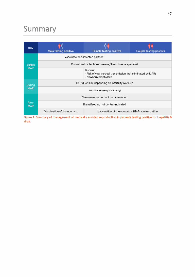
47
Summary
Figure 1: Summary of management of medically assisted reproduction in patients testing positive for Hepatitis B virus.

48
PART B: Hepatitis C Virus
B1. Prevalence and testing
NARRATIVE QUESTION: WHAT IS THE PREVALENCE OF HEPATITIS C VIRUS?
In 2015, 71 million persons were living with chronic Hepatitis C virus (HCV) infection with uneven global epidemic and differences across and within countries (WHO, 2017). The Eastern Mediterranean Region reporting the highest number of HCV RNA positive persons (±15 million), followed by the Western Pacific Region and European Region (both ± 14 million), the South-East Asia Region and African Region (both ± 10 million) and the Region of the Americas reporting the lowest number of HCV RNA positive persons (± 8 million).
The overall global prevalence of HCV infection in 2015 was 1%, with the highest estimated prevalence in the Eastern Mediterranean Region (2.3%), followed by the European Region (1.5%), the African Region (1.0%), the Region of the Americas and Western Pacific Region (both 0.7%) and the South-East Asian Region with the lowest estimated prevalence (0.5%).
In the European Union and Economic Area Countries the overall HCV prevalence in 2015 was estimated to be 1.1% (95% CI 0.9-1.4), equaling 5.6 million anti-HCV positive cases. The estimated prevalence of anti-HCV of the majority of countries was < 1%. Spain, Slovakia, Greece, Latvia, Romania and Italy had an estimated anti-HCV prevalence of 1.1%, 2.0%, 2.2%, 2.4%, 3.2% and 5.9% respectively (Hofstraat et al., 2017). In the Central European Region the estimated anti-HCV positive prevalence varies between 0.2% and 2.1% (Urbanek et al., 2016) and the estimated HCV RNA positive prevalence between 0.2% and 3.5% (Madalinski et al., 2015).
The global estimates of HCV prevalence appear to decrease in the last years (Cornberg et al., 2011, Lanini et al., 2016, WHO, 2017).
Unsafe health-care-practices and injection drug use remain the leading modes of transmission. In high income countries the prevalence of chronic HCV is generally below 2%. However, there is variability within countries with similar socio-economic conditions because of differences in size of high-risk population. Countries with the highest prevalence (>5%) are mainly low-middle income countries and include Egypt, Gabon, Uzbekistan, Cameroon, Mongolia, Pakistan, Nigeria, Georgia (Lanini, et al., 2016).
NARRATIVE QUESTION: HOW SHOULD TESTING OF HEPATITIS C STATUS PRIOR TO MEDICALLY ASSISTED REPRODUCTION BE PERFORMED?
Virological markers of Hepatitis C virus (HCV) infection are anti-HCV antibodies, HCV core antigen, HCV RNA and HCV genotype (Pawlotsky, 2002).
49
Screening for HCV is based on detection of total HCV antibodies (IgM and IgG). Enzyme immunoassay (EIA) and chemiluminescence immunoassay (CIA) are most commonly used for detection of anti HCV antibodies in serum. Whole blood or plasma also qualify. Serological tests have evolved over time yielding several generations of EIA’s. Still anti-HCV assays have disadvantages (e.g. prolonged window of detection after infection, low positive predictive value) and therefore confirmation tests are widely used (Cobb et al., 2014, Saludes et al., 2014, Uliana et al., 2014).
In recent years rapid diagnostic tests (RDTs) enabled screening for HCV antibodies at a lower cost compared to classical EIAs (Chevaliez and Pawlotsky, 2018, Smith et al., 2012).
Performance of the different HCV antibody tests varies (Khuroo et al., 2015, Tang et al., 2017).
Confirmatory antibody testing can be done with recombinant immunoblot assays (RIBA) for individuals who have tested positive by EIA. A main problem of RIBA is the occurrence of intermediate results (Ponde, 2013).
Confirmation of an HCV infection and circulating viral genome is based on detection of HCV RNA. Nucleic Acid Technology (NAT) is the gold standard and most commonly used as confirmation test (Benjamin, 2001, Sarrazin, 2002, Stramer, 2002).
Polymerase chain reaction (PCR), in particular real time PCR, is the most relevant of NAT (Carithers et al., 2000, Colucci, 2012, Gullett and Nolte, 2015, Irshad et al., 2016, Kessler and Stelzl, 2017, Krajden, 2000, Pawlotsky, 2003, Schutten, 2008, Scott and Gretch, 2007, Singh et al., 2017). New technologies in the field of PCR are digital droplet PCR, next-generation sequencing, lab-on-a-chip, digital antigen assays (Roth, 2019).
Alternatively, transcription mediated amplification (TMA) and branched-DNA assays can be used for detecting HCV RNA (Al Olaby and Azzazy, 2011, Albertoni et al., 2014).
HCV core Antigen can be used as an indirect marker of HCV replication and assays have the potential to replace NAT (Freiman et al., 2016). They have the advantage of reduced costs compared to PCR and can be performed on the same diagnostic platforms as some EIA assays.
HCV genotyping can be performed by antibody tests and by a PCR technique, but is only essential in the treatment of HCV (Al Olaby and Azzazy, 2011, Podzorski, 2002, Wilson et al., 2017, Yang and Wei, 2018).
Conclusion
HCV testing is mandatory according to the European Tissues and Cells Directive as a preventative measure to reduce the risks of transmission to partners and offspring.
REFERENCES Al Olaby RR, Azzazy HM. Hepatitis C virus RNA assays: current and emerging technologies and their clinical applications. Expert review of molecular diagnostics 2011;11: 53-64. Albertoni G, Castelo Girao MJ, Schor N. Mini review: current molecular methods for the detection and quantification of hepatitis B virus, hepatitis C virus, and human immunodeficiency virus type 1. International journal of infectious diseases : IJID : official publication of the International Society for Infectious Diseases 2014;25: 145-149.

50
Benjamin RJ. Nucleic acid testing: update and applications. Seminars in hematology 2001;38: 11-16. Carithers RL, Jr., Marquardt A, Gretch DR. Diagnostic testing for hepatitis C. Seminars in liver disease 2000;20: 159-171. Chevaliez S, Pawlotsky JM. New virological tools for screening, diagnosis and monitoring of hepatitis B and C in resource-limited settings. Journal of hepatology 2018;69: 916-926. Cobb B, Heilek G, Vilchez RA. Molecular diagnostics in the management of chronic hepatitis C: key considerations in the era of new antiviral therapies. BMC infectious diseases 2014;14 Suppl 5: S8. Colucci G. Molecular diagnostic and predictive tests in the evolution of chronic hepatitis C anti-viral therapies. BMC infectious diseases 2012;12 Suppl 2: S8. Cornberg M, Razavi HA, Alberti A, Bernasconi E, Buti M, Cooper C, Dalgard O, Dillion JF, Flisiak R, Forns X et al. A systematic review of hepatitis C virus epidemiology in Europe, Canada and Israel. Liver international : official journal of the International Association for the Study of the Liver 2011;31 Suppl 2: 30-60. Freiman JM, Tran TM, Schumacher SG, White LF, Ongarello S, Cohn J, Easterbrook PJ, Linas BP, Denkinger CM. Hepatitis C Core Antigen Testing for Diagnosis of Hepatitis C Virus Infection: A Systematic Review and Meta-analysis. Annals of internal medicine 2016;165: 345-355. Gullett JC, Nolte FS. Quantitative nucleic acid amplification methods for viral infections. Clinical chemistry 2015;61: 72-78. Hofstraat SHI, Falla AM, Duffell EF, Hahne SJM, Amato-Gauci AJ, Veldhuijzen IK, Tavoschi L. Current prevalence of chronic hepatitis B and C virus infection in the general population, blood donors and pregnant women in the EU/EEA: a systematic review. Epidemiology and infection 2017;145: 2873-2885. Irshad M, Gupta P, Mankotia DS, Ansari MA. Multiplex qPCR for serodetection and serotyping of hepatitis viruses: A brief review. World journal of gastroenterology 2016;22: 4824-4834. Kessler HH, Stelzl E. Profile of Roche's cobas(R) HCV tests. Expert review of molecular diagnostics 2017;17: 311-319. Khuroo MS, Khuroo NS, Khuroo MS. Diagnostic accuracy of point-of-care tests for hepatitis C virus infection: a systematic review and meta-analysis. PloS one 2015;10: e0121450. Krajden M. Hepatitis C virus diagnosis and testing. Canadian journal of public health = Revue canadienne de sante publique 2000;91 Suppl 1: S34-39, s36-42. Lanini S, Easterbrook PJ, Zumla A, Ippolito G. Hepatitis C: global epidemiology and strategies for control. Clinical microbiology and infection : the official publication of the European Society of Clinical Microbiology and Infectious Diseases 2016;22: 833-838. Madalinski K, Zakrzewska K, Kolakowska A, Godzik P. Epidemiology of HCV infection in Central and Eastern Europe. Przeglad epidemiologiczny 2015;69: 459-464, 581-454. Pawlotsky JM. Molecular diagnosis of viral hepatitis. Gastroenterology 2002;122: 1554-1568. Pawlotsky JM. Use and interpretation of hepatitis C virus diagnostic assays. Clinics in liver disease 2003;7: 127-137. Podzorski RP. Molecular testing in the diagnosis and management of hepatitis C virus infection. Archives of pathology & laboratory medicine 2002;126: 285-290. Ponde RA. Enzyme-linked immunosorbent/chemiluminescence assays, recombinant immunoblot assays and nucleic acid tests in the diagnosis of HCV infection. European journal of clinical microbiology & infectious diseases : official publication of the European Society of Clinical Microbiology 2013;32: 985-988. Roth WK. History and Future of Nucleic Acid Amplification Technology Blood Donor Testing. Transfusion medicine and hemotherapy : offizielles Organ der Deutschen Gesellschaft fur Transfusionsmedizin und Immunhamatologie 2019;46: 67-75. Saludes V, Gonzalez V, Planas R, Matas L, Ausina V, Martro E. Tools for the diagnosis of hepatitis C virus infection and hepatic fibrosis staging. World journal of gastroenterology 2014;20: 3431-3442.

51
Sarrazin C. Highly sensitive hepatitis C virus RNA detection methods: molecular backgrounds and clinical significance. Journal of clinical virology : the official publication of the Pan American Society for Clinical Virology 2002;25 Suppl 3: S23-29. Schutten M. Comparison of the Abbott Realtime HIV-1 and HCV viral load assays with commercial competitor assays. Expert review of molecular diagnostics 2008;8: 369-377. Scott JD, Gretch DR. Molecular diagnostics of hepatitis C virus infection: a systematic review. JAMA : the journal of the American Medical Association 2007;297: 724-732. Singh MP, Galhotra S, Saigal K, Kumar A, Ratho RK. Quantitative nucleic acid amplification methods and their implications in clinical virology. International journal of applied & basic medical research 2017;7: 3-9. Smith BD, Jewett A, Drobeniuc J, Kamili S. Rapid diagnostic HCV antibody assays. Antiviral therapy 2012;17: 1409-1413. Stramer SL. US NAT yield: where are we after 2 years? Transfusion medicine (Oxford, England) 2002;12: 243-253. Tang W, Chen W, Amini A, Boeras D, Falconer J, Kelly H, Peeling R, Varsaneux O, Tucker JD, Easterbrook P. Diagnostic accuracy of tests to detect Hepatitis C antibody: a meta-analysis and review of the literature. BMC infectious diseases 2017;17: 695. Uliana CV, Riccardi CS, Yamanaka H. Diagnostic tests for hepatitis C: recent trends in electrochemical immunosensor and genosensor analysis. World journal of gastroenterology 2014;20: 15476-15491. Urbanek P, Kristian P, Makara M, Hunyady B, Tomasiewicz K. Epidemiology of HCV infection in the Central European region. Clin Exp Hepatol 2016;2: 2-6. WHO. Global hepatitis report, 2017. 2017. Wilson EM, Rosenthal ES, Kattakuzhy S, Tang L, Kottilil S. Clinical Laboratory Testing in the Era of Directly Acting Antiviral Therapies for Hepatitis C. Clinical microbiology reviews 2017;30: 23-42. Yang R, Wei L. Profile of the VERSANT HCV genotype 2.0 assay. Expert review of molecular diagnostics 2018;18: 995-1004.
52
B2. Prevention of transmission before medically assisted reproduction
PICO QUESTION: WHAT ARE THE RISKS OF HEPATITIS C VIRUS TRANSMISSION THROUGH VAGINAL/ANAL INTERCOURSE?
Evidence A systematic review including 38 studies, of which 9 controlled studies among sexual contacts of non-haemodialysis, non-renal transplantation patients and 29 uncontrolled studies, resulting in a total of 4250 stable sexual heterosexual contacts reported 573/4250 testing positive for anti-HCV antibodies. Calculated pooled prevalence among the spouses was 13.48% (95% CI 12.48 to 14.55). In the 9 controlled studies, spouses of anti-HCV-positive patients in areas non-endemic for HCV have a significantly higher prevalence of anti-HCV than spouses of negative controls (15.2% vs. 0.9%, OR 20.57, 95% CI 6.05 to 84.08). Two studies reported prevalence of anti-HCV among female contacts of HCV-infected males and male contacts of female infected. Only in the group of male contacts of HCV-infected females was the prevalence of HCV infection significantly higher than in controls (OR 2.14, 95% CI 1.12 to 4.08) (Ackerman et al., 1998).
A cross-sectional study enrolled 500 heterosexual, monogamous patients chronically infected with HCV in a relationship ≥ 36 months and their partners and ≥ 3 sexual contacts in preceding 6 months. 20/500 (4.0%) partners tested anti-HCV-positive and of these 13/20 (65.0%) tested HCV-PCR-positive. (9/20 (45.0%) concordant, 8/20 (40.0%) discordant, 3/20 (15.0%) indeterminant). Prevalence potentially attributable to sex contact was 3/500 (0.6%; 95% CI 0.0% to 1.3%) resulting in an estimated incidence of minimum 3.6/10,000 person-years (95% CI 0.0 to 7.7) based on 3 confirmed couples and maximum 7.2/10,000 person-years (95% CI 1.3 to 13.0) based on 3 confirmed and 3 unknown couples. The estimated risk per sex act was minimum 1/380,000 (95% CI 1/600,000 to 1/280,000) and maximum 1/190,000 (95% CI 1/1.3 million to 1/100,000) (Terrault et al., 2013).
A case control study with 60 HCV-PCR-positive patients. Two spouses (3.39%) were anti-HCV-positive and one was HCV-PCR-positive. Relationship duration of 2 positive spouses with index patient was > 15 years (Hajiani et al., 2006).
A cross sectional study among 53 haemodialysis patients (of which 16 both anti-HCV and HCV-PCR-positive). None of spouses was anti-HCV-positive (0/16) (Fadil-Romao et al., 2006).
One study group conducted a retrospective cohort analysis among 600 chronically infected HCV patients (320 male + 280 female) and their partners as well as a prospective cohort analysis among 216 anti-HCV-negative partners from these chronically infected HCV patients. In the retrospective cohort 12/600 (2%) of partners (4/280 female and 8/320 male) were anti-HCV-positive and 11/600 (1.8%) were HCV-RNA-positive. All 11 couples were concordantly infected with the same genotype (type 1b). The rate of sexual intercourse was 1.73/week. In the prospective cohort analysis, the intercourse rate was 1.9/week. No partners were anti-HCV-positive (Tahan et al., 2005).

53
A prospective cohort analysis among 112 anti-HCV and HCV-RNA-positive patients (75 men and 37 women) and their anti-HCV negative spouses. One seroconversion (one female partner, genotype 1b) occurred after 20 months, resulting in a transmission risk of 2.33/1000 person-years (Kao et al., 2000).
In a cross-sectional study among 585 anti-HCV and HCV-RNA-positive patients and their 455 spouses HCV prevalence among spouses was 71/455 (15.6%). Of the infected patients, 19.8% had a relationship > 20 years vs. 8% had a relationship < 20 years (OR 2.8, 95% CI 1.5 to 5.3) (Caporaso et al., 1998).
A cross-sectional study analysed 121 patients with chronic liver disease (anti-HCV positive and 116 were also HCV-PCR-positive) and their spouses. 21/121 (17.4%) spouses were anti-HCV-positive of which 19 HCV-PCR-positive. 12/19 (63.2%) couples were concordant for genotype (Koda et al., 1996).
A cross-sectional study among 68 anti-HCV and HCV-PCR-positive patients with chronic hepatitis and their partners showed 4/68 (5.9%) of spouses were anti-HCV positive, of which 2 were HCV-PCR-positive (concordant type 1). Duration of relationship for HCV-positive vs. HCV-negative spouses was 25 years (15-30 years) vs. 10 years (2-43 years) (Tong et al., 1995).
A prospective cohort analysis among 895 heterosexual anti-HCV and HCV-PCR-positive patients and their anti-HCV-negative partners reported a mean frequency of intercourse 1.8/week (no anal intercourse, and no condom use). Three spouses HCV seroconverted (2 with concordant genotype for 1b and 2a, 1 discordant (had dental implant prior to test), resulting in a transmission rate of 0.37/1000 person-years. Transmission rate for concordant couples was 0.25/1000 person-years (Vandelli et al., 2004).
Recommendation
In a monogamous heterosexual relationship of more than 12 months, there is no indication for the use of barrier contraceptives to reduce the risk of Hepatitis C virus (HCV) transmission in a serodiscordant infected couple.
Conditional ⊕⊕
Justification The presented large prospective studies show a very low transmission between sexual partners. The major transmission route of HCV is parenteral transmission and not sexual intercourse in the absence of STDs or medical comorbidities such as HIV or liver pathology. A correlation was found between risk of sexual HCV transmission and the number of partners.
Only one study reports in detail the sexual behaviour and shows that there is no significant difference in HCV transmission between vaginal and anal intercourse.
54
PICO QUESTION: IS THERE A PRE-TREATMENT (BEFORE MAR) THRESHOLD BELOW WHICH TRANSMISSION OF HEPATITIS C VIRUS IS UNLIKELY?
Evidence Horizontal transmission
We identified no studies reporting a Hepatitis C virus (HCV) RNA serum threshold below which horizontal transmission does not occur.
Vertical transmission
No publications could be identified where maternal HCV viral load was measured before pregnancy.
Recommendation
All patients with an active or chronic HCV-infection must be reviewed by an infectious disease/ liver specialist before initiating any medically assisted reproduction treatment (MAR).
GPP
Commencing with MAR treatments in patients positive for HCV should be a joint decision between the patient, their partner, the fertility doctor and the infectious disease/ liver specialist.
Strong ⊕
In the case of the female testing positive for HCV, the possibility of viral vertical transmission should be discussed prior to MAR treatment.
GPP
Justification All identified studies focus on measuring the HCV-RNA viral load during pregnancy, and therefore there is not enough evidence to indicate a specific cut-off point for viremia before MAR to avoid vertical transmission and HCV infection in the offspring.
Vertical transmission has gained importance as the primary HCV transmission route among children once the blood products screening has been implemented. The estimates of HCV vertical transmission from HCV positive/HIV negative women ranged from 1.1 to 10.7% (Benova et al., 2014). The pooled risk of vertical HCV infection is 5-8%.
Clinical cohort studies evaluating the correlation between pre-pregnancy HCV-RNA viral load and vertical transmission are required to find a threshold value beyond which neonatal infection is more likely.

55
REFERENCES Ackerman Z, Paltiel O, Glikberg F, Ackerman E. Hepatitis C virus in various human body fluids: A systematic review. Hepatology Research 1998;11: 26-40. Benova L, Mohamoud YA, Calvert C, Abu-Raddad LJ. Vertical transmission of hepatitis C virus: systematic review and meta-analysis. Clinical infectious diseases : an official publication of the Infectious Diseases Society of America 2014;59: 765-773. Caporaso N, Ascione A, Stroffolini T. Spread of hepatitis C virus infection within families. Investigators of an Italian Multicenter Group. Journal of viral hepatitis 1998;5: 67-72. Fadil-Romao MA, Pomar LN, Romao Jr JE. Low risk of sexual transmission of hepatitis C virus to spouses of infected hemodialysis patients. Dialysis and Transplantation 2006;35: 252-259. Hajiani E, Masjedizadeh R, Hashemi J, Azmi M, Rajabi T. Hepatis C virus transmission and its risk factors within families of patients infected with hepatitis C virus in southern Iran: Khuzestan. World journal of gastroenterology 2006;12: 7025-7028. Kao JH, Liu CJ, Chen PJ, Chen W, Lai MY, Chen DS. Low incidence of hepatitis C virus transmission between spouses: a prospective study. Journal of gastroenterology and hepatology 2000;15: 391-395. Koda T, Yonaha M, Hayashi A, Ishikawa K. Hepatitis C transmission between spouses. Journal of gastroenterology and hepatology 1996;11: 1001-1005. Tahan V, Karaca C, Yildirim B, Bozbas A, Ozaras R, Demir K, Avsar E, Mert A, Besisik F, Kaymakoglu S et al. Sexual transmission of HCV between spouses. The American journal of gastroenterology 2005;100: 821-824. Terrault NA, Dodge JL, Murphy EL, Tavis JE, Kiss A, Levin TR, Gish RG, Busch MP, Reingold AL, Alter MJ. Sexual transmission of hepatitis C virus among monogamous heterosexual couples: the HCV partners study. Hepatology (Baltimore, Md) 2013;57: 881-889. Tong MJ, Lai PP, Hwang SJ, Lee SY, Co RL, Chien RN, Kuo G. Evaluation of sexual transmission in patients with chronic hepatitis C infection. Clinical and diagnostic virology 1995;3: 39-47. Vandelli C, Renzo F, Romanò L, Tisminetzky S, De Palma M, Stroffolini T, Ventura E, Zanetti A. Lack of evidence of sexual transmission of hepatitis C among monogamous couples: results of a 10-year prospective follow-up study. The American journal of gastroenterology 2004;99: 855-859.
56
B3. Assisted reproduction techniques and impact on outcomes
PICO QUESTION: SHOULD IUI, IVF OR ICSI BE PREFERENTIALLY USED FOR MAR IN HEPATITIS C INFECTED COUPLES?
Evidence In a prospective cohort study, 35 couples with viraemic and chronically Hepatitis C virus (HCV)-infected male partners underwent either ovarian stimulation and IUI (n=14) or ICSI (n=21) after semen processing (gradient centrifugation and swim-up). There was no case of horizontal or vertical transmission after IUI or ICSI (Savasi et al., 2013).
A cross-sectional observational study including 60 women testing positive for HCV (30 HCV+RNA- and 30 HCV+RNA+ women) investigated the risk of vertical transmission after ICSI. There was no vertical transmission in women who were RNA-, however, 1 infant born to an HCV+RNA+ women tested positive for HCV (Nesrine and Saleh, 2012).
A prospective cohort study, including 40 HCV-serodiscordant couples (male HCV positive) reported the results of ICSI treatment in these couples. None of the female partners seroconverted (Garrido et al., 2004).
Conclusion
From the perspective of horizontal and vertical transmission, there is currently not enough evidence to recommend one technique (IUI/IVF/ICSI) over another in patients infected with Hepatitis C.
Recommendation
The cause of infertility should dictate the specific technique (IUI/IVF/ICSI) used for MAR in couples where one or both partners test positive for HCV.
Strong ⊕
Women testing positive for HCV should be informed that MAR does not eliminate the risk of vertical transmission.
GPP

57
PICO QUESTION: CAN HEPATITIS C VIRAL RNA BE DETECTED IN OOCYTES/ SPERM/ PLACENTA?
Evidence RNA in sperm
No studies could be retrieved investigating the integration of HCV in sperm.
RNA in oocytes
The experimental study by Papaxanthos-Roche et al. investigated the susceptibility of human oocytes from Hepatitis C virus (HCV) RNA-positive women to HCV contamination during assisted reproductive technology (Papaxanthos-Roche et al., 2004). HCV RNA was detected in 17/24 unfertilized oocytes, 6/7 oocytes after an ICSI attempt and 11/17 after a conventional IVF attempt. HCV RNA was found in 19/20 (95%) follicular fluid samples. A weak correlation was found between plasma and follicular fluid HCV RNA loads (Papaxanthos-Roche, et al., 2004).
Placenta
An experimental study reported that primary human trophoblast cells and an extravillous trophoblast cell line (HTR8), from first and second trimester of pregnancy, express receptors relevant for HCV binding/entry and are permissive for HCV uptake (Giugliano et al., 2015).
Conclusion
The possibility of Hepatitis C viral RNA presence in oocytes cannot be excluded. However, the risk of Hepatitis C transmission through the use of reproductive material remains to be proven.
PICO QUESTION: DOES HEPATITIS C VIRUS AND/OR TREATMENT OF HEPATITIS C VIRUS BEFORE MAR IMPACT THE OUTCOME OF MAR?
MALE INFECTED
Evidence No articles in the literature search evaluating impact of antiviral treatment before MAR on reproductive outcomes.
A retrospective cohort study including 60 Hepatitis C virus (HCV)-serodiscordant and 69 non-infected couples investigated the reproductive outcomes of ICSI. Fertilization rate was significantly lower in HCV-serodiscordant couples as compared to controls (61% vs. 75%). However, there was no statistical difference in pregnancy rate per cycle (17.6% (18/102) vs. 20.2% (14/69)), miscarriages rate per cycle (11.1% (2/18) vs. 28.5% (4/14)), or live birth per cycle (15.7% (16/102) vs. 15.9% (11/69)) (Cito et al., 2019).
A retrospective cohort study including 78 HCV-serodiscordant and 1256 non-infected couples investigated reproductive outcomes of IVF. There was no statistical difference between HCV-serodiscordant couples and controls in fertilization rate (80.99±19.95 vs. 78.14±19.73), clinical

58
pregnancy rate (48.6% (36/74) vs. 55.0% (691/1256)) or miscarriage rate (18.9% (14/74) vs. 19.0% (239/1256)) (Yang et al., 2015).
A prospective case-control study including 28 HCV-serodiscordant couples and 46 non-infected controls compared reproductive outcomes of IVF-ICSI. Fertilization rate was significantly lower in HCV-serodiscordant couples as compared to controls (54.7% vs. 68.2%). However, there was no statistical difference between HCV-serodiscordant couples and controls in implantation rate (12.8% vs. 4.2%), clinical pregnancy rate per embryo transfer (17.5% vs. 7.0%) or the number of children born (8/28 vs. 2/46) (Prisant et al., 2010).
Another retrospective cohort study including 12 HCV-serodiscordant (9 male and 3 female testing positive for HCV) and 27 non-infected couples, investigated the reproductive outcomes of IVF-ICSI. There was no statistical difference in fertilization rate between HCV-serodiscordant and control couples (63.9% vs. 75.9%), however, there was a significant difference in clinical pregnancy rate per cycle (0% (0/12) vs. 41% (11/27)) (Pirwany et al., 2004).
Conclusion
There are contradictory results evaluating effects of male HCV infection on infertility treatments outcomes. Although the fertilization rate has been reported significantly lower in couples with HCV-RNA positive men, other studies report that HCV infection does not affect the IVF-ICSI cycle outcomes in these couples.
FEMALE INFECTED
Evidence No articles in the literature search evaluating impact of antiviral treatment before MAR on reproductive outcomes.
A retrospective cohort study compared the clinical outcomes of 25 first IVF-ICSI cycles in Hepatitis C virus (HCV)-serodiscordant with 25 matched control cycles. There was a statistically significant reduction in fertilization rates (67% vs. 86%) and implantation rates (6% (3/47) vs. 23% (11/47)) between HCV-serodiscordant couples and controls. However, there was no statistical difference between HCV-serodiscordant couples and controls in clinical pregnancy rate (12% (3/25) vs. 36% (9/25)), miscarriage rate (4% 1/25 vs. 12% (3/25)) or the number of children born (2/25 vs. 7/25). A subgroup of 17 patients with confirmed active HCV replication also showed no implantation (0% (0/22) vs. 26% (6/23)) and no live birth occurred in HCV positive patients with active replication (Shaw-Jackson et al., 2017).
A retrospective cohort study including 90 HCV-serodiscordant and 1256 non-infected couples investigated reproductive outcomes of IVF. There was no statistical difference between HCV-serodiscordant couples and controls in fertilization rate (76.93±19.18 vs. 78.14±19.73), clinical pregnancy rate (45.6% (41/90) vs. 55.0% (691/1256)) or miscarriage rate (23.3% (21/90) vs. 19.0% (239/1256)) (Yang, et al., 2015).
A retrospective cohort study including 40 HCV RNA+, 40 HCV RNA- seropositive and 40 HCV PCR- seronegative women compared clinical outcomes of ICSI. Comparing HCV PCR+ and PCR – women and
59
controls, significantly more cycles were cancelled (52% vs. 30% vs. 5%) and fertilization rate was reduced (28% vs. 32% vs. 67%), respectively. In addition, pregnancy rates were significantly lower in HCV PCR+ women compared to HCV PCR- and controls (5% (2/40) vs. 32.5% (13/40) vs. 47.5% (19/40)). Furthermore, the investigators reported a negative correlation between number of oocytes and HCV viral load (Hanafi et al., 2011).
A prospective case-control study including 22 HCV-serodiscordant couples and 42 non-infected controls compared reproductive outcomes of IVF-ICSI. There was no statistical difference between HCV-serodiscordant couples and controls in fertilization rate (71.1% vs. 70.2%), implantation rate (5.1% vs. 9.6%), clinical pregnancy rate per embryo transfer (10.8% vs. 12.8%) or the number of children born (2/22 vs. 4/42) (Prisant, et al., 2010).
A retrospective cohort study compared the outcomes of IVF-ICSI cycles in 42 HCV-serodiscordant and 84 matched non-infected couples. There was no statistical difference between HCV-serodiscordant couples and controls in fertilization rate (56.4% vs. 59%), implantation rate (19% vs. 19.2%) and pregnancy rate per embryo transfer (28.5% (8/28) vs. 29.3% (22/75)). However, significantly more cycles were cancelled in the HCV-serodiscordant couples (Englert et al., 2007).
Another retrospective cohort study including 12 HCV-serodiscordant (9 male and 3 female HCV-infected) and 27 non-infected couples, investigated the reproductive outcomes of IVF-ICSI. There was no statistical difference in fertilization rate between HCV-serodiscordant and control couples (63.9% vs. 75.9%), however, there was a significant difference in clinical pregnancy rate per cycle (0% (0/12) vs. 41% (11/27)) (Pirwany, et al., 2004).
Conclusion
There are contradictory results evaluating effects of female HCV infection on infertility treatments outcomes. Although some studies report significantly reduced implantation rates, higher cycle cancellations, and higher FSH use in HCV positive women, other report no significant differences.
REFERENCES Cito G, Coccia ME, Fucci R, Picone R, Cocci A, Russo GI, Rizzello F, Trotta M, Badolato L, Basile V et al. Influence of male human immunodeficiency virus (HIV) and hepatitis C virus (HCV) infection on the reproductive outcomes in serodiscordant couples: a case-control study. Andrology 2019;7: 852-858. Englert Y, Moens E, Vannin AS, Liesnard C, Emiliani S, Delbaere A, Devreker F. Impaired ovarian stimulation during in vitro fertilization in women who are seropositive for hepatitis C virus and seronegative for human immunodeficiency virus. Fertility and sterility 2007;88: 607-611. Garrido N, Meseguer M, Bellver J, Remohi J, Simon C, Pellicer A. Report of the results of a 2 year programme of sperm wash and ICSI treatment for human immunodeficiency virus and hepatitis C virus serodiscordant couples. Human reproduction (Oxford, England) 2004;19: 2581-2586. Giugliano S, Petroff MG, Warren BD, Jasti S, Linscheid C, Ward A, Kramer A, Dobrinskikh E, Sheiko MA, Gale M, Jr. et al. Hepatitis C Virus Sensing by Human Trophoblasts Induces Innate Immune Responses and Recruitment of Maternal NK Cells: Potential Implications for Limiting Vertical Transmission. Journal of immunology (Baltimore, Md : 1950) 2015;195: 3737-3747. Hanafi NF, Abo Ali AH, Abo el kheir HF. ICSI outcome in women who have positive PCR result for hepatitis C virus. Human reproduction (Oxford, England) 2011;26: 143-147.

60
Nesrine F, Saleh H. Hepatitis C virus (HCV) status in newborns born to HCV positive women performing intracytoplasmic sperm injection. African health sciences 2012;12: 58-62. Papaxanthos-Roche A, Trimoulet P, Commenges-Ducos M, Hocke C, Fleury HJ, Mayer G. PCR-detected hepatitis C virus RNA associated with human zona-intact oocytes collected from infected women for ART. Human reproduction (Oxford, England) 2004;19: 1170-1175. Pirwany IR, Phillips S, Kelly S, Buckett W, Tan SL. Reproductive performance of couples discordant for hepatitis B and C following IVF treatment. Journal of assisted reproduction and genetics 2004;21: 157-161. Prisant N, Tubiana R, Lefebvre G, Lebray P, Marcelin AG, Thibault V, Rosenblum O, Bonmarchand M, Vauthier-Brouzes D, Golmard JL et al. HIV-1 or hepatitis C chronic infection in serodiscordant infertile couples has no impact on infertility treatment outcome. Fertility and sterility 2010;93: 1020-1023. Savasi V, Oneta M, Parrilla B, Cetin I. Should HCV discordant couples with a seropositive male partner be treated with assisted reproduction techniques (ART)? European journal of obstetrics, gynecology, and reproductive biology 2013;167: 181-184. Shaw-Jackson C, Capraro M, Ameye L, Vandromme J, Manigart Y, Rozenberg S, Autin C. In vitro fertilization for women infected by hepatitis C virus: a matched case-control study and a systematic literature review. Journal of assisted reproduction and genetics 2017;34: 587-597. Yang L, Zhao R, Zheng Y, Song X. Effect of hepatitis C virus infection on the outcomes of in vitro fertilization. International journal of clinical and experimental medicine 2015;8: 6230-6235.

61
B4. Prevention/ reduction of transmission during assisted reproduction
PICO QUESTION: WHICH TECHNIQUES CAN BE USED TO PREVENT/ REDUCE HEPATITIS C VIRUS TRANSMISSION DURING MAR?
SEMEN PROCESSING The evidence on semen processing will be discussed in detail in the next section (B5).
ANTIVIRAL TREATMENT No studies on antiviral treatment and MAR were identified.
Conclusion
There are no data regarding antiviral therapy in men or women with HCV without co-infections requiring MAR in order to reduce the risk of HCV transmission. None of the currently available HCV antiviral drugs are licensed for use in pregnancy.

62
B5. Semen processing
PICO QUESTION: WHAT IS THE BEST TECHNIQUE FOR SEMEN PROCESSING TO REDUCE HEPATITIS C VIRAL LOAD?
Evidence In a sub group of 93 couples with HIV, HCV or HBV-positive men requiring ICSI, 23 couples were analysed with an HCV-positive male (incl. 1 HCV concordant couple). Semen processing by density gradient (80-40%) centrifugation and washing followed by real time PCR of HIV RNA, HCV RNA and HBV DNA before ICSI. No semen samples were positive for HCV RNA after washing. 48 ICSI cycles performed yielded 8 children and no HCV seroconversion detected in the women or their children (Molina et al., 2014).
A small cohort study, including 35 HCV-serodiscordant (male infected) couples reported no seroconversion after IUI and ICSI. Semen was processed by density gradient centrifugation and swim-up, however, was not PCR tested after processing (Savasi et al., 2013).
In a small cohort study of 16 males testing positive for HCV/HIV-1 requiring MAR, processing of semen was performed by density gradient centrifugation (80-40%), washing and swim-up. HCV RNA was found in seminal plasma and non-sperm cells (31.5%) after density gradient alone. All washed motile sperm samples before and after swim-up were HCV RNA negative (Savasi et al., 2010).
A prospective cohort study included 86 HCV-serodiscordant couples (76 men HCV positive and 10 men HCV/HIV positive). Semen was processed by density gradient (90-70-50%) centrifugation and swim-up. 20.4% of seminal plasma samples were positive for HCV RNA. All sperm fractions were HCV RNA negative. The infants were tested for HCV 3 months after birth, women were not tested. In 135 MAR cycles (10 IVF, 78 ICSI, 12 frozen embryo transfer, 35 IUI- cycles) 36 pregnancies occurred, 28 live births and none of the babies tested positive for HCV (Bourlet et al., 2009).
In a small cohort study of 7 males testing positive for HCV (6 HIV/HCV-positive and 1 HCV-positive), sperm was processed by 1:1 (vol/vol) dilution and centrifugation followed twice by 1:1 dilution of pellet and centrifugation, no swim-up was performed. 7 spermatozoa samples were HCV negative and 1 data of HCV PCR was not available (Garrido et al., 2006).
In another study, semen samples from a small cohort of 20 males testing positive for HIV/HCV not requiring MAR were divided into two aliquots. One total sperm and one for density gradient (90-45%) centrifugation. Following centrifugation three semen fractions originated (seminal plasma, non-motile cells and motile spermatozoa). 100uL of the three fractions for virological analysis and 900µL of the three fractions were washed 3 times and motile sperm fraction was submitted to swim-up. 5% (1/20) of total sperm tested HCV-positive. 0% of three pre-wash semen fractions HCV-positive. 0% of post-wash semen fractions HCV-positive (Canto et al., 2006).
In a cohort study of 32 males testing positive for HCV requiring MAR (HIV and HBV-negative), 11 MAR cycles were performed with proven HCV-negative sperm after triple layer density gradient centrifugation. 5 women became pregnant and 9 babies were born, none were HCV positive at birth or at 6 months of age. Women were not tested for HCV (Bourlet et al., 2002).

63
A cohort study of 35 infertile couples requiring MAR with a male partner testing positive for HCV without HBV or HIV co-infection where 50 frozen thawed semen samples were used after semen processing with density gradient (90-45%) centrifugation. Only 90% density gradient fraction used for IVF-cycles (n=26) and ICSI-cycles (n=24). 14% of total semen was HCV RNA positive. 2% of the 45% fraction was HCV-RNA-positive and 0% of the 90% fraction was HCV-RNA-positive. HCV viral load in all HCV-positive total semen samples measured below 600 IU/ml. No women or children tested positive for HCV after IVF or ICSI (Cassuto et al., 2002).
A prospective controlled trial included 34 men testing positive for HIV of which 21 also testing positive for HCV. Semen samples were processed by a triple density gradient centrifugation followed by swim up. 12.2% of samples were HIV-positive and 23.8% HCV positive with nested PCR-test versus 0% of samples HIV-positive and 0% HCV-positive with one round PCR-test (Meseguer et al., 2002).
Surgically retrieved sperm
One small cohort study comprising 4 males testing positive for HCV with obstructive azoospermia, anejaculation and cryptozoospermia. Two men underwent testicular sperm extraction (TESE) and microsurgical epididymal sperm aspiration (MESA), two underwent TESE. Epididymal spermatozoa preparation by density gradient (90-45%) centrifugation followed by resuspension of sperm pellet and centrifugation. Testicular spermatozoa preparation by centrifugation of spermatozoa/medium suspension over one layer (45%) gradient followed by resuspension of sperm pellet and centrifugation. No HCV was detected in the final sperm samples after MESA and TESE. No transmission of HCV to female partner after ICSI and 1 healthy child born (Leruez-Ville et al., 2013).
Recommendation
A discontinuous gradient centrifugation followed by swim-up and washing is recommended for semen processing in patients testing positive for HCV.
Strong ⊕
Justification No studies were identified comparing routine semen preparation with advanced semen processing (such as gradient centrifugation and swim-up) in male testing positive for HCV without co-infections.
Current evidence shows that semen can test positive for HCV after single continuous density centrifugation or after discontinuous density centrifugation without wash steps.
PICO QUESTION: IS THERE A NEED FOR PCR TESTING OF POST-WASHED SPERM?
Evidence In a sub group of 93 couples with HIV, HCV or HBV-positive men requiring ICSI, 23 couples were analysed with an HCV-positive male (incl. 1 HCV-concordant couple). Semen processing by density gradient (80-40%) centrifugation and washing followed by real time PCR of HIV RNA, HCV RNA and HBV DNA before ICSI. No semen samples were positive for HCV RNA after washing (Molina, et al., 2014).

64
In a small cohort study of 16 HCV/HIV1-positive men requiring MAR, processing of semen was performed by density gradient centrifugation (80-40%), washing and swim-up. HCV RNA was found in seminal plasma and non-sperm cells (31.5%) after density gradient alone. All washed motile sperm samples before and after swim-up were HCV RNA negative (Savasi, et al., 2010).
A prospective cohort study included 86 HCV-serodiscordant couples (76 men HCV-positive and 10 men HCV/HIV-positive). Semen was processed by density gradient (90-70-50%) centrifugation and swim-up. 20.4% of seminal plasma samples were positive for HCV RNA. All sperm fractions were HCV RNA negative (Bourlet, et al., 2009).
A small cohort study of 7 males testing positive for HCV (6 HIV/HCV-positive and 1 HCV-positive) processed the sperm by 1:1 (vol/vol) dilution and centrifugation followed twice by 1:1 dilution of pellet and centrifugation, no swim-up was performed. 7 spermatozoa samples were HCV negative and for 1 sample the PCR result was not available (Garrido, et al., 2006).
Semen samples from a small cohort of 20 HIV/HCV-positive men not requiring MAR were divided into two aliquots. One total sperm and one for density gradient (90-45%) centrifugation. Following centrifugation three semen fractions originated (seminal plasma, non-motile cells and motile spermatozoa). 100uL of the fractions were washed 3 times with RPMI and motile sperm fraction was submitted to swim-up. 5% (1/20) of total sperm tested HCV positive. 0% of three pre-wash semen fractions HCV positive. 0% of post-wash semen fractions HCV positive (Canto, et al., 2006).
A cohort study of 35 infertile couples requiring MAR with an HCV-positive male partner without HBV or HIV co-infection. 50 frozen semen samples after semen processing with density gradient (90-45%) centrifugation. Only 90% density gradient fraction used for IVF-cycles (n=26) and ICSI-cycles (n=24). 14% of total semen was HCV RNA positive. 2% of the 45% fraction was HCV RNA positive and 0% of the 90% fraction was HCV RNA positive. HCV viral load in all HCV positive total semen samples < 600 IU/ml (Cassuto, et al., 2002).
One small cohort study comprising 4 males testing positive for HCV with obstructive azoospermia, anejaculation and cryptozoospermia. Two men underwent TESE and MESA, two underwent TESE. Epididymal spermatozoa preparation by density gradient (90-45%) centrifugation followed by resuspension of sperm pellet and centrifugation. Testicular spermatozoa preparation by centrifugation of spermatozoa/medium suspension over one layer (45%) gradient followed by resuspension of sperm pellet and centrifugation. No HCV was detected in the final sperm samples after MESA and TESE (Leruez-Ville, et al., 2013).
Recommendation
After advanced semen processing, PCR testing for HCV is not necessary.
Strong ⊕
Justification All available evidence show that semen sample test PCR negative after advanced semen processing. In addition, the viral load in semen is low. Thus, no PCR testing for HCV is necessary after advanced processing.
65
PICO QUESTION: IS THERE A NEED FOR SEMEN PROCESSING WHEN BOTH THE MALE AND FEMALE ARE INFECTED?
Evidence No studies could be found investigating this PICO question.
Recommendation
Good laboratory practice regarding semen processing should be applied irrespective of whether only the male or both partners are testing positive for HCV.
GPP
Recommendation There is no evidence that semen processing might make a difference when both partners test positive for HCV. Practices on semen processing in these cases may differ between centers, however, especially for cryopreservation and storage purposes, it might be justified to perform semen processing.
The fact that both partners are infected does not mean it is justified to lower sperm processing standards. Good laboratory practice is required also when both partners are testing positive for HCV.
PICO QUESTION: DOES THE PLASMA VIRAL LOAD CORRELATE WITH HEPATITIS C VIRUS DETECTION IN SEMEN?
Evidence A small cohort study including 32 HCV+RNA+ (26 chronic and 4 acute HCV-infected) men investigated the association between serum and semen HCV viral load. The mean blood viral load was significantly higher in patients positive for HCV RNA in semen (n=4) than in those negative for HCV RNA in semen (n=28) (6.52 ± 0.55 vs. 5.88 ± 0.46 log copies/ml) (Bourlet, et al., 2002).
In a sub group analysis, including 30 HCV-positive HIV-negative men, Bradshaw et al. reported that the median blood HCV RNA levels in those with detectable HCV RNA was higher than that in those with undetectable HCV RNA in semen (6.2 log IU/mL; interquartile range (IQR) 5.7 to 6.7 log IU/mL vs. 6.0 log IU/mL; (IQR 5.3 to 6.2 log IU/mL), however the difference was not statistically significant (Bradshaw et al., 2015).
Conclusion
High plasma HCV viral load is likely to be predictive of the presence of HCV RNA in semen. Strong evidence for the correlation of HCV viral load between serum and semen is currently lacking.
66
REFERENCES Bourlet T, Levy R, Maertens A, Tardy J-C, Grattard F, Cordonier H, Laurent J-L, Guerin J-F, Pozzetto B. Detection and characterization of hepatitis C virus RNA in seminal plasma and spermatozoon fractions of semen from patients attempting medically assisted conception. Journal of clinical microbiology 2002;40: 3252-3255. Bourlet T, Lornage J, Maertens A, Garret AS, Saoudin H, Tardy JC, Jimenez C, Guerin JF, Pozzetto B, Levy R. Prospective evaluation of the threat related to the use of seminal fractions from hepatitis C virus-infected men in assisted reproductive techniques. Human reproduction (Oxford, England) 2009;24: 530-535. Bradshaw D, Lamoury F, Catlett B, Applegate TL, McAllister J, Dore GJ, Matthews GV, Danta M. A comparison of seminal hepatitis C virus (HCV) RNA levels during recent and chronic HCV infection in HIV-infected and HIV-uninfected individuals. The Journal of infectious diseases 2015;211: 736-743. Canto CL, Segurado AC, Pannuti C, Cedenho A, Srougi M, Spaine D, Fernandes S, Carretiero N, Bernal MC, Levi JE. Detection of HIV and HCV RNA in semen from Brazilian coinfected men using multiplex PCR before and after semen washing. Revista do Instituto de Medicina Tropical de Sao Paulo 2006;48: 201-206. Cassuto NG, Sifer C, Feldmann G, Bouret D, Moret F, Benifla JL, Porcher R, Naouri M, Neuraz A, Alvarez S et al. A modified RT-PCR technique to screen for viral RNA in the semen of hepatitis C virus-positive men. Human reproduction (Oxford, England) 2002;17: 3153-3156. Garrido N, Remohi J, Pellicer A, Meseguer M. The effectiveness of modified sperm washes in severely oligoasthenozoospermic men infected with human immunodeficiency and hepatitis C viruses. Fertility and sterility 2006;86: 1544-1546. Leruez-Ville M, Thiounn N, Poirot C, Launay O, Sogni P, Grabar S, Dulioust E. Intracytoplasmic sperm injection with microsurgically retrieved spermatozoa in azoospermic men infected with human immunodeficiency virus 1 or hepatitis C virus: the EP43 AZONECO ANRS study. Fertility and sterility 2013;99: 713-717. Meseguer M, Garrido N, Gimeno C, Remohi J, Simon C, Pellicer A. Comparison of polymerase chain reaction-dependent methods for determining the presence of human immunodeficiency virus and hepatitis C virus in washed sperm. Fertility and sterility 2002;78: 1199-1202. Molina I, Carmen Del Gonzalvo M, Clavero A, Angel Lopez-Ruz M, Mozas J, Pasquau J, Sampedro A, Martinez L, Castilla JA. Assisted reproductive technology and obstetric outcome in couples when the male partner has a chronic viral disease. International journal of fertility & sterility 2014;7: 291-300. Savasi V, Oneta M, Parrilla B, Cetin I. Should HCV discordant couples with a seropositive male partner be treated with assisted reproduction techniques (ART)? European journal of obstetrics, gynecology, and reproductive biology 2013;167: 181-184. Savasi V, Parrilla B, Ratti M, Oneta M, Clerici M, Ferrazzi E. Hepatitis C virus RNA detection in different semen fractions of HCV/HIV-1 co-infected men by nested PCR. European journal of obstetrics, gynecology, and reproductive biology 2010;151: 52-55.

67
B6. Reducing/ avoiding vertical transmission
PICO QUESTION: WHICH INTERVENTIONS CAN BE USED TO REDUCE/AVOID VERTICAL TRANSMISSION OF HEPATITIS C VIRUS TO THE NEWBORN?
ELECTIVE CAESAREAN SECTION
Evidence A systematic review and meta-analysis including 8 cohort studies, compared HCV seroconversion rates in children after vaginal delivery and caesarean section (CS) and reported 7% seroconversion after vaginal delivery (36/510) versus 6.1% (8/131) after CS (OR 1.1, 95% CI 0.45 to 2.67). CS does not decrease the risk of HCV transmission to the infant (Ghamar Chehreh et al., 2011).
Recommendation
Caesarean delivery is not recommended on the basis of maternal HCV-positivity alone.
Strong ⊕
Justification There is no evidence that the risk of HCV transmission from mother to child after caesarean section is lower compared to that after vaginal delivery.
BREASTFEEDING
Evidence A systematic review and meta-analysis including 14 cohort studies (2971 mother–infant pairs), investigated the risk of HCV transmission to the infant by breastfeeding and reported no association between breastfeeding and risk of transmission to the infant (Cottrell et al., 2013). Methodologic shortcomings in the poor quality studies included failure to perform statistical adjustment on potential confounders and insufficient information to determine comparability of groups at baseline stratified by breastfeeding status (Cottrell, et al., 2013).
Recommendation
Breastfeeding is not contra-indicated in women testing positive for HCV.
Strong ⊕⊕
68
Justification We identified no association between breastfeeding and the risk of HCV transmission from mother to child. Breastfeeding has significant health benefits. This recommendation is supported by the guidelines of the European Association for the Study of the Liver (EASL) (Pawlotsky et al., 2020).
There are 4 studies included in the systematic review by Cottrell et al. (2013) possibly including HCV mothers co-infected with HIV which may influence the reported results (Cottrell, et al., 2013).
REFERENCES Cottrell EB, Chou R, Wasson N, Rahman B, Guise JM. Reducing risk for mother-to-infant transmission of hepatitis C virus: a systematic review for the U.S. Preventive Services Task Force. Annals of internal medicine 2013;158: 109-113. Ghamar Chehreh ME, Tabatabaei SV, Khazanehdari S, Alavian SM. Effect of cesarean section on the risk of perinatal transmission of hepatitis C virus from HCV-RNA+/HIV- mothers: a meta-analysis. Archives of gynecology and obstetrics 2011;283: 255-260. Pawlotsky J-M, Negro F, Aghemo A, Berenguer M, Dalgard O, Dusheiko G, Marra F, Puoti M, Wedemeyer H. EASL recommendations on treatment of hepatitis C: Final update of the series. Journal of hepatology 2020;73: 1170-1218.

69
Summary
Figure 2: Summary of management of medically assisted reproduction in patients testing positive for Hepatitis C virus.
70
PART C: Human Immunodeficiency Virus
C1. Prevalence and testing
NARRATIVE QUESTION: WHAT IS THE PREVALENCE OF HUMAN IMMUNODEFICIENCY VIRUS?
Human Immunodeficiency virus (HIV) continues to be a major global public health issue, having claimed almost 33 million lives so far. There were an estimated 38 million people living with HIV at the end of 2019. However, with increasing access to effective HIV prevention, diagnosis, treatment and care, including for opportunistic infections, HIV infection has become a manageable chronic health condition, enabling people with HIV to lead long and healthy lives. In 2019, 68% of adults and 53% of children living with HIV globally were receiving lifelong antiretroviral therapy.
Over two thirds of all people living with HIV live in the WHO African Region (25.7 million). While HIV is prevalent among the general population in this region, an increasing number of new infections occur among key population groups (WHO, 2018).
Since the late 1990s, the progressive availability and success of combination antiretroviral therapy has reduced the risk of opportunistic infections and malignancies in people living with HIV, remarkably decreasing morbidity and mortality (UNAIDS, 2015).
Although HIV infection is preventable, significant HIV transmission continues across the WHO European Region. In 2019, 136 449 newly diagnosed HIV infections were reported in 47 of the 53 Member States in the Region, including 24 801 from countries of the European Union/European Economic Area (EU/EEA). This corresponds to a crude rate of 15.6 newly diagnosed infections per 100 000 population (ECDC and WHO, 2020).
In 2014, the Joint United Nations Program on HIV and AIDS (UNAIDS) and partners set the ‘90-90-90 targets’; aiming by 2020 to (a) diagnose 90% of all HIV positive people; (b) provide antiretroviral therapy for 90% of those diagnosed; and achieve viral suppression for 90% of those treated (UNAIDS, 2015).
The first lentivirus, SIVmac was identified in 1984 in rhesus macaques and HIV was transmitted from chimpanzees to humans in the beginning of the 20th century. The mature HIV virion consists of 2 copies of single-stranded RNA surrounded by structural proteins, a matrix shell, and lipid envelope.
HIV targets the immune system and weakens people's defence against many infections and some types of cancer, which people with a healthy immune system can fight off. As the virus destroys and impairs the function of immune cells, infected individuals gradually become immunodeficient. Immune function is typically measured by CD4 cell count. The most advanced stage of HIV infection is acquired immunodeficiency syndrome (AIDS), which can take many years to develop if not treated, depending

71
on the individual. AIDS is defined by the development of certain cancers, infections or other severe long-term clinical manifestations (WHO, 2018).
There are two major viral species of HIV known, HIV-1 and 2. The HIV-1 strain was identified first, is the more virulent, the most prevalent and most researched strain. There are a number of distinct HIV-1 lineages (or genetic subtypes), designated by letters. This genetic variability is caused by the reverse transcriptase enzyme, which is error-prone, resulting in mutations and recombinations. A recombination can occur when a person is infected with 2 or more virus strains. According to internationally defined nomenclature, 106 distinct circulating recombinant forms are known thus far. This diversification of the HIV virus has significant consequences for diagnosis, screening and management (for instance drug resistance), but also for vaccine development (Hemelaar et al., 2020).
The HIV-2 strain was first isolated in 1986, and has a serologic profile more closely related to the SIVmac than HIV-1. Like HIV-1, 9 distinct lineages (A-I) have been identified. Contrary to HIV-1, only two recombinant forms have been described so far. Epidemiological data for HIV-2 are very limited and rather outdated. HIV-2 is mainly restricted to West Africa where it infected up to 1–2 million people. In Europe, the two countries with the highest prevalence are Portugal and France. According to data from 2008, 4.5% of AIDS diagnosis in Portugal and 1-2% in France are due to HIV-2. HIV-2 transmission is slower than for HIV-1 due to lower viral loads with HIV-2 infections. The lower plasmatic viral load of HIV-2 is also reflected in semen, resulting in a 4 times lower likelihood of sexual transmission compared to HIV-1. Treatment of HIV-2 is challenging, since it displays a high level of intrinsic resistance against antiretroviral drugs developed for HIV-1 therapy. (Visseaux et al., 2016).
NARRATIVE QUESTION: HOW SHOULD TESTING OF HUMAN IMMUNODEFICIENCY VIRUS STATUS PRIOR TO MEDICALLY ASSISTED REPRODUCTION BE PERFORMED?
After human Immunodeficiency virus (HIV)-1 infection, HIV-1-specific markers appear in the blood in a chronologic order: HIV-1 RNA, p24 antigen, HIV-1 IgM antibody, and HIV-1 IgG antibody. Time from HIV acquisition to reactivity for an assay depends on which target is being detected, when that target can be detected after infection, concentration of the target in the specimen, the volume of specimen tested, and the test’s analytical sensitivity (Branson, 2019, Hurt et al., 2017).
The standard of care test for diagnosing HIV in a clinical setting is the serum immunoassay test (EIA), known as the HIV fourth-generation test, which is a combination antibody (Ab) and antigen (Ag) test. With this test, antibodies against both HIV-1 and HIV-2 are detected, as well as the p24 antigen, allowing for earlier HIV detection after exposure. If there is a strong suspicion of a very early HIV infection (less than 14 days), a nucleic acid test (NAT) can be performed to detect HIV RNA (as early as 5-10 days after the transmission, depending on the sensitivity of the assay). NAT should also be performed if the fourth-generation test is inconclusive. The false-positive rates of both the third and fourth generations are very low. Data show a false positive rate of third-generation testing (with confirmatory western blot) to be as low as 0.0004% to 0.0007%. (Branson, 2019, Huynh and Kahwaji, 2020).
HIV self-testing and rapid tests are some of the new strategies to encourage HIV diagnosis. HIV self-testing is a process whereby a person who wants to know his or her HIV status collects a specimen, performs a test, and interprets the test results in private or with someone they trust. HIV self-testing does not provide a definitive HIV-positive diagnosis, but it could be used as an initial test to be followed
72
by confirmatory testing (such as NAT or serum western blot) by a health worker. Many countries are now using innovative approaches to develop and support HIV self-testing using digital platforms and online support for help with the testing procedure and linkage to services (WHO, 2018). Rapid tests (mostly oral swab tests) are primarily ELISA tests, and provide the result in 20-30 minutes. The advantage of rapid tests is that they can be performed outside of a clinical setting. However, the results need to be confirmed by a confirmatory test (Huynh and Kahwaji, 2020, Parekh et al., 2019). In resource limited settings, HIV testing algorithms include a combination of two or three rapid tests, usually in a serial algorithm, depending on HIV prevalence; this has produced suitable sensitivities and specificities and improved the accuracy of testing results in point of care settings (Parekh, et al., 2019).
The sexual partners and drug-injecting partners of people diagnosed with HIV infection have an increased risk of also being HIV-positive. WHO recommends voluntary assisted HIV partner notification services as a simple and effective way to reach these partners – many of whom are undiagnosed and unaware of their HIV exposure and may welcome support and an opportunity to be tested for HIV (WHO, 2018).
Conclusion
HIV testing is mandatory according to the European Tissues and Cells Directive as a preventative measure to reduce the risks of transmission to partners and offspring.
REFERENCES Branson BM. HIV Diagnostics: Current Recommendations and Opportunities for Improvement. Infectious disease clinics of North America 2019;33: 611-628. ECDC, WHO. HIV/AIDS surveillance in Europe 2020 – 2019 data. 2020. World Health Organization, Copenhagen. Hemelaar J, Elangovan R, Yun J, Dickson-Tetteh L, Kirtley S, Gouws-Williams E, Ghys PD. Global and regional epidemiology of HIV-1 recombinants in 1990-2015: a systematic review and global survey. The lancet HIV 2020;7: e772-e781. Hurt CB, Nelson JAE, Hightow-Weidman LB, Miller WC. Selecting an HIV Test: A Narrative Review for Clinicians and Researchers. Sexually transmitted diseases 2017;44: 739-746. Huynh K, Kahwaji CI. HIV Testing StatPearls. 2020. StatPearls Publishing Parekh BS, Ou CY, Fonjungo PN, Kalou MB, Rottinghaus E, Puren A, Alexander H, Hurlston Cox M, Nkengasong JN. Diagnosis of Human Immunodeficiency Virus Infection. Clinical microbiology reviews 2019;32. UNAIDS. UNGASS country level reports (GARPR). Global aids response progress reporting. 2015. 2015. Joint United Nations Programme on HIV/AIDS (UNAIDS). Visseaux B, Damond F, Matheron S, Descamps D, Charpentier C. Hiv-2 molecular epidemiology. Infection, genetics and evolution : journal of molecular epidemiology and evolutionary genetics in infectious diseases 2016;46: 233-240. WHO. HIV/AIDS, fact sheet. 2018. World Health Organization, Geneva.

73
C2. Prevention of transmission before medically assisted reproduction
PICO QUESTION: WHAT ARE THE RISKS OF HUMAN IMMUNODEFICIENCY VIRUS TRANSMISSION THROUGH VAGINAL/ANAL INTERCOURSE?
Evidence A systematic review and meta-analysis reported that among heterosexual sex partners, with the index case on antiretroviral therapy (with varying levels of viral load) 23 linked transmissions were identified over 9922 person-years (pooled incidence 0.23 transmissions/100 person-years, 95% CI 0.15 to 0.35, 10 studies). Among couples where the index case had suppressed viral load, no transmissions over 1327 person-years were identified (pooled incidence 0.00 transmissions/100 person-years, 95% CI 0.00 to 0.28, 2 studies). Among serodiscordant couples who reported “always” using condoms, there were 1.14 HIV transmissions per 100 person-years (95% CI 0.56 to 2.04) (LeMessurier et al., 2018).
A systematic review and meta-analysis, including 15 studies, calculated that the summary estimate for HIV infectiousness of anal intercourse per act is 1.8% (95% CI 0.3 to 3.2) (Baggaley et al., 2010).
A further 8 prospective observational studies, not included in the systematic reviews, reported HIV seroconversion rates in HIV-serodiscordant couples. Ma et al., including 231 HIV-serodiscordant couples, reported 45 seroconversion before the start of the study and 2 during the follow-up (seroconversion rate of 0.39 per 100 person-years) (Ma et al., 2019). In the study by Colombe et al., 14 unaffected partners HIV-1 seroconverted in 105 serodiscordant couples (Colombe et al., 2019). In the study by Rodgers et al., including 888 HIV-1-serodiscordant couples (548 heterosexual and 340 male homosexual) contributing to 1238 eligible couple-years of follow-up, 11 HIV-1 unaffected partners seroconverted (Rodger et al., 2016). In the study by Quinn et al., 90 HIV-1 unaffected partners seroconverted in 415 HIV-1-serodiscordant couples during 30 months of follow-up (Quinn et al., 2000). In the study by Ragni et al., 39 partnered HIV-infected haemophilic men not on antiretroviral therapy were included. Five out of 39 transmitted HIV to their unaffected partner (Ragni et al., 1998). In the study by Operskalski et al., including 18 HIV-1-serodiscordant couples, 2 unaffected partners already seroconverted before the study and 4 seroconverted during 23 person-years of observation (Operskalski et al., 1997). In the study by Deschamps et al., 135 HIV-serodiscordant couples were included, of which 19 unaffected partners seroconverted during follow-up (Deschamps et al., 1996). De Vincenzi et al. reported that 12 unaffected partners out of 256 couples HIV seroconverted (2.3/100 person-years (95% CI 1.2 to 4.0)) (de Vincenzi, 1994).
In the study by Ma et al., the seroconversions occurred in couples where the index person did not immediately receive antiretroviral therapy after HIV diagnosis (Ma, et al., 2019). Similarly, Rodgers et al. reported that the estimated rate for transmission through any unprotected intercourse with the HIV-1-positive partner on antiretroviral therapy with HIV-1 load less than 200 copies/mL was zero, with an upper 95% confidence limit of 0.30 per 100 couple-years of follow-up (Rodger, et al., 2016). Also, in the study by Quinn et al. the rate of HIV-1 transmission was zero among the 51 couples in which the HIV-1–positive partner had undetectable serum levels of HIV-1 RNA or less than 1500 copies/ml (Quinn, et

74
al., 2000). In contrast, in the study by Zheng et al., compared with those who were on antiretroviral therapy, the OR for HIV acquisition in the antiretroviral therapy-naïve group was 1.14 (95% CI 0.91 to 1.43), there was no significant difference between the antiretroviral therapy and antiretroviral therapy naïve group (Zheng et al., 2018).
Deschamps et al. reported that the incidence of HIV infection was 1.0 per 100 person-years for persons who always used condoms and 6.8 per 100 person-years for persons who used condoms irregularly or not at all (Deschamps, et al., 1996). Similarly, De Vincenzi et al. reported no seroconversions in couples with consistent condom use (de Vincenzi, 1994).
Two retrospective cohort studies reported respectively 53 seroconversions within 5218 person-years of follow-up in 4481 HIV-serodiscordant couples (incidence rate of 1.02 (95%CI 0.76±1.33) per 100 person-years) and 72 seroconversions in 239 monogamous couples (index partner not on antiretroviral therapy) (rate of HIV-1 transmission per coital act was 0.0012 (95% CI 0.0009 to 0.0015)) (Tang et al., 2016, Wawer et al., 2005).
Recommendation
Human immunodeficiency virus (HIV)-1-serodiscordant couples should be informed that there is a risk of sexual transmission of the virus to the unaffected partner. To reduce this risk, couples must be advised to use barrier contraception and seek active therapy to reduce viral load.
Strong ⊕⊕
In individuals testing positive for HIV-1, antiretroviral therapy can suppress viral replication. These patients should remain on antiretroviral therapy and providing undetectable viral loads in serum can be achieved and sustained, the risk of horizontal transmission through unprotected intercourse is minimal in the absence of other sexually transmitted diseases.
Strong ⊕⊕
Justification The HIV-1 viral presence cannot be eliminated to date, however, anti-retroviral therapy can reduce the HIV-1 viral load to undetectable levels, thereby minimizing the risk of horizontal transmission.

75
PICO QUESTION: IS THERE A THRESHOLD BELOW WHICH TRANSMISSION OF HUMAN IMMUNODEFICIENCY VIRUS IS UNLIKELY?
Evidence Horizontal transmission
A systematic review and meta-analysis including 11 cohorts reporting on 5021 couples and 461 human Immunodeficiency virus (HIV) transmission events, reported that the HIV transmission rate from people not on antiretroviral therapy was 0.16 (95% CI 0.02 to 1.13 per 100 person-years, based on one episode of HIV, 6 studies). Furthermore, no HIV transmission events occurred in discordant heterosexual couples if the partner testing positive for HIV was treated with antiretroviral therapy and had a viral load below 400 copies/ml (Attia et al., 2009).
Two prospective observational studies, not included in the systematic review, reported on the risk of transmission in relation to the HIV-1 viral load. In the study by Rodger et al., the estimated rate of HIV-1 transmission through unprotected intercourse was zero when the partner testing positive for HIV-1 was on antiretroviral therapy with a viral load <200 copies/ml (upper 95% confidence limit of 0.30 per 100 couple-years of follow-up) (Rodger, et al., 2016). Pedraza et al. reported that HIV-1 transmitters had a higher viral load than non-transmitters (21.139 vs. 5.484 RNA copies/ml) (Pedraza et al., 1999)
Vertical transmission
We were unable to retrieve studies that investigated the maternal viral load before MAR and the risk of vertical transmission to the newborn.
Recommendation
Commencing with medically assisted reproduction (MAR) treatments in patients positive for HIV-1 or 2 should be a joint decision between the patient, their partner, the fertility doctor and the infectious disease specialist.
Strong ⊕
All patients testing positive for HIV, wishing to have a child should be counselled about the risk of horizontal and vertical transmission. In the case of the male testing positive for HIV, antiretroviral therapy can reduce the viral load in blood and semen to undetectable levels, allowing the possibility of natural conception. Reproductive counselling should include fertility and antiretroviral covariates.
GPP

76
In the case of the female testing positive for HIV-1 or 2, and even with undetectable viremia, the possibility of viral vertical transmission should be discussed prior to MAR treatment.
GPP
Justification Patients newly diagnosed with HIV infection, should get advice from an infectious disease specialist to discuss treatment options and start treatment to reduce viral load. Engaging with a HIV specialist when planning fertility treatment makes it possible to have a multi-disciplinary pre-conception counselling which could include the optimization of health status, including reviewing antiretroviral agents and reviewing blood viral load.
REFERENCES Attia S, Egger M, Muller M, Zwahlen M, Low N. Sexual transmission of HIV according to viral load and antiretroviral therapy: systematic review and meta-analysis. AIDS (London, England) 2009;23: 1397-1404. Baggaley RF, White RG, Boily MC. HIV transmission risk through anal intercourse: systematic review, meta-analysis and implications for HIV prevention. International journal of epidemiology 2010;39: 1048-1063. Colombe S, Beard J, Mtenga B, Lutonja P, Mngara J, de Dood CJ, van Dam GJ, Corstjens P, Kalluvya S, Urassa M et al. HIV-seroconversion among HIV-1 serodiscordant married couples in Tanzania: a cohort study. BMC infectious diseases 2019;19: 518. de Vincenzi I. A longitudinal study of human immunodeficiency virus transmission by heterosexual partners. European Study Group on Heterosexual Transmission of HIV. The New England journal of medicine 1994;331: 341-346. Deschamps MM, Pape JW, Hafner A, Johnson WD, Jr. Heterosexual transmission of HIV in Haiti. Annals of internal medicine 1996;125: 324-330. LeMessurier J, Traversy G, Varsaneux O, Weekes M, Avey MT, Niragira O, Gervais R, Guyatt G, Rodin R. Risk of sexual transmission of human immunodeficiency virus with antiretroviral therapy, suppressed viral load and condom use: a systematic review. CMAJ : Canadian Medical Association journal = journal de l'Association medicale canadienne 2018;190: E1350-e1360. Ma G, Chen H, Chen J, Jiang Z, Ma T. HIV transmission between spouses living in Lu'an city, Anhui province, China: a longitudinal study. Epidemiology and infection 2019;147: e205. Operskalski EA, Stram DO, Busch MP, Huang W, Harris M, Dietrich SL, Schiff ER, Donegan E, Mosley JW. Role of viral load in heterosexual transmission of human immunodeficiency virus type 1 by blood transfusion recipients. Transfusion Safety Study Group. American journal of epidemiology 1997;146: 655-661. Pedraza MA, del Romero J, Roldan F, Garcia S, Ayerbe MC, Noriega AR, Alcami J. Heterosexual transmission of HIV-1 is associated with high plasma viral load levels and a positive viral isolation in the infected partner. Journal of acquired immune deficiency syndromes (1999) 1999;21: 120-125. Quinn TC, Wawer MJ, Sewankambo N, Serwadda D, Li C, Wabwire-Mangen F, Meehan MO, Lutalo T, Gray RH. Viral load and heterosexual transmission of human immunodeficiency virus type 1. Rakai Project Study Group. The New England journal of medicine 2000;342: 921-929.

77
Ragni MV, Faruki H, Kingsley LA. Heterosexual HIV-1 transmission and viral load in hemophilic patients. Journal of acquired immune deficiency syndromes and human retrovirology : official publication of the International Retrovirology Association 1998;17: 42-45. Rodger AJ, Cambiano V, Bruun T, Vernazza P, Collins S, van Lunzen J, Corbelli GM, Estrada V, Geretti AM, Beloukas A et al. Sexual Activity Without Condoms and Risk of HIV Transmission in Serodifferent Couples When the HIV-Positive Partner Is Using Suppressive Antiretroviral Therapy. JAMA : the journal of the American Medical Association 2016;316: 171-181. Tang H, Wu Z, Mao Y, Cepeda J, Morano J. Risk Factor Associated with Negative Spouse HIV Seroconversion among Sero-Different Couples: A Nested Case-Control Retrospective Survey Study in 30 Counties in Rural China. PloS one 2016;11: e0164761. Wawer MJ, Gray RH, Sewankambo NK, Serwadda D, Li X, Laeyendecker O, Kiwanuka N, Kigozi G, Kiddugavu M, Lutalo T et al. Rates of HIV-1 transmission per coital act, by stage of HIV-1 infection, in Rakai, Uganda. The Journal of infectious diseases 2005;191: 1403-1409. Zheng Z, Li Y, Jiang Y, Liang X, Qin S, Nehl EJ. Population HIV transmission risk for serodiscordant couples in Guangxi, Southern China: A cohort study. Medicine 2018;97: e12077.
78
C3. Assisted reproduction techniques and impact on outcomes
PICO QUESTION: SHOULD IUI, IVF OR ICSI BE PREFERENTIALLY USED FOR MAR IN HUMAN IMMUNODEFICIENCY VIRUS INFECTED COUPLES?
Evidence A systematic review and meta-analysis included 14 studies reporting on the outcomes of IUI in couples testing positive for human Immunodeficiency virus (HIV)-1. In the 14 studies on IUI, 12 studies reported on serodiscordant couples with a male patient testing positive for HIV-1 and 4 with a female patient testing positive for HIV-1. Results on IUI in males testing positive for HIV-1 comprising of 2393 patients, showed a cumulative pregnancy rate of 17% (95 CI 15% to 20%) and 14% in female patients testing positive for HIV-1 (n=28) (95 CI 25% to 35%). The miscarriage rate for IUI in serodiscordant couples with male patients testing positive for HIV-1 (n=2393) was 19% (95 CI 14% to 25%) and for female patients testing positive for HIV-1 (n=25) was 13% (95 CI 1% to 34%). No HIV-1 transmission was observed in the seronegative partner of a total of 8212 IUI cycles (Barnes et al., 2014). The meta-analysis also included 15 studies on the outcomes of IVF/ICSI in HIV-1 infected couples. In these 15 studies, 12 studies reported on serodiscordant couples with a male partner testing positive for HIV-1 and 7 with a female partner testing positive for HIV-1. The clinical pregnancy rate was 30% (95 CI 25% to 35%) in couples with male partners testing positive for HIV-1 (n=780) and 16% (95 CI 13% to 20%) for couples where the female partner tested positive for HIV-1 (n= 253). No HIV-1 transmission was observed in 1254 IVF/ICSI cycles in serodiscordant couple with a male partner testing positive for HIV-1 (Barnes, et al., 2014).
A systematic review and meta-analysis included 11 studies reporting on the outcome of 3900 IUI cycles in 1184 couples and 738 IVF/ICSI cycles performed in 579 serodiscordant couples with a male partner testing positive for HIV-1 (Vitorino et al., 2011). This meta-analysis does include 4 studies that were also included in the structured review of Barnes et al. The median clinical pregnancy rate was 18% (range from 14.5% to 23%) for IUI and 38% (range 24.8% to 46.2%) for IVF/ICSI. There was no HIV-1 seroconversion in the female partners of couples where the male partner tested positive for HIV-1 and no vertical transmission was found in children at birth and 3-6 months after delivery (Vitorino, et al., 2011).
Recommendation
HIV infection status is not a reason to deny MAR treatment.
Strong ⊕

79
The cause of infertility should dictate the specific technique (IUI/IVF/ICSI) used for MAR in couples where one or both partners test positive for HIV.
Strong ⊕⊕
Justification There are no studies published comparing different MAR techniques in similar patient populations testing positive for HIV-1. Evidence from 2 structured reviews, although having overlapping studies in their results, showed that the pregnancy outcomes in IUI are lower compared to IVF/ICSI cycles as is the case in patients testing negative for HIV-1. The decision on which type of MAR treatment is chosen, is to be solely based on the fertility status of the couples.
Conflicting results in the outcome can be the origin of different types of subfertility in couples, certainly in studies describing smaller patient cohorts, moreover the patients are sometimes co-infected with other infectious diseases like HCV and/or HBV in several studies, and the infertility aetiology is also not always clearly described in the studies. The systematic review of Barnes (Barnes, et al., 2014), showed that in women on antiretroviral therapy and testing positive for HIV-1, 32%-75% suffered tubal factor infertility (5 studies), 8%-12.5% ovarian factor infertility (2 studies), 2% were diagnosed with endometriosis (1 study), 10%-37% had partners with male infertility (5 studies) and 12%-20% were ‘other’ or unknown factors (2 studies). It was clear that for HIV-1 infected women, tubal factor infertility was more prevalent than in the general infertility population where isolated tubal infertility is about 8% (CDC et al., 2011).
There is no preferred MAR method of choice based on infection status of the patient or the couple. The current evidence shows that safety is equal in all MAR techniques after specific semen processing. Reassuringly, evidence in the reported papers, showed no seroconversion in female partners of serodiscordant couples where the male tested positive for HIV-1 and no vertical transmission has been shown in the babies born from MAR in couples testing positive for HIV-1. It could be argued that MAR in couples where the male is HIV-1 positive is a therapeutic intervention that reduces the risk of horizontal transmission.
PICO QUESTION: CAN HUMAN IMMUNODEFICIENCY VIRUS DNA BE DETECTED IN OOCYTES/ SPERM/ PLACENTA?
Evidence DNA integration in sperm
An original research paper using semen samples from sperm donors testing negative for human Immunodeficiency virus (HIV)-1, co-incubated with HIV, reported a dose-dependent binding of HIV to spermatozoa. This binding occurred with low affinity and superficially on the sperm surface (Young et al., 2019).
An observational cohort study in 10 males tested positive for HIV, on antiviral therapy for 6 months undergoing bilateral orchiectomy for gender confirmation treatment, reported HIV persistence in the testis. The study analysing HIV diversity and genetic compartmentalization, reported dynamics of

80
proliferation of latently HIV-infected cells to be different between blood and testes. The authors noticed marked differences in the quantity and distribution of identical HIV sequences between patients and between anatomical sites within each patient, which may complicate the complete eradication on HIV in patients (Miller et al., 2019).
An original basic research study showed the presence of HIV-1 protein and viral RNA in seminal vesicles in testicular tissue section of 7/9 male patients testing positive for HIV-1. Viral integrated cells were observed either in the stroma close to the epithelium or in the lumen of the seminal vesicles (Deleage et al., 2011).
An observational study, including semen of 22 male patients testing positive for HIV-1 compared to 12 males testing negative for HIV-1, observed that in 75% of all semen samples tested; T cells were most commonly infected with HIV-1, followed by macrophages in 38% of all samples. Viral DNA was, in this study, never detected in motile spermatozoa (Quayle et al., 1997).
An original basic research study showed sperm-associated virus-like particles in the spermatozoa of 15 male patients testing positive for HIV-1. In contrast, 15 male patients testing negative for HIV-1 did not exhibit sperm-associated virus-like particles, however spermatozoa derived from patients testing negative for HIV-1 co-incubated with HIV-1 showed sperm-associated virus-like particles in the spermatozoa. This study described the presence of the putative HIV-1 receptor (GalAAG) on spermatozoa and found virus-like particles in an 8-cell stage embryo after IVF using sperm of a male patient testing positive for HIV-1 (Baccetti et al., 1994).
A basic research study where semen samples of 17 male persons testing negative with HIV-1 were co-incubated with HIV-1, observed the binding of HIV-1 to the plasma membrane of the spermatozoa and the presence of virus-like particles in vacuoles in the apical nuclear region of the spermatozoa (Dussaix et al., 1993).
DNA integration in oocytes
An original basic research study using 41 oocytes of a female donor testing negative for HIV-1, injected 28 of these oocytes with very high amounts of HIV-1 (4x10⁴ cop/ml) and observed in 3/28 oocytes (11%), HIV-1 viral integration (Steenvoorden et al., 2012).
A series of 4 cases described by Bertrand et al. investigating 24 follicular fluid samples, 15 follicular flushing samples and 1 cumulus cells samples obtained from 4 female patients testing positive for HIV-1 of which 3 had an undetectable viral load in plasma, did not detect HIV-1 RNA in any of the samples analysed. The patient with a detectable viral load showed HIV-1 RNA in 1 follicular fluid sample and 1 follicular flushing sample. The other 2 flushing samples of this patient were HIV-1 RNA negative (Bertrand et al., 2004).
An original basic research study where 100 oocytes from 15 female patients testing negative for HIV-1 were co-incubated with HIV-1, found no cell associated HIV-1 antigen neither virus-like particles in any of the experiments. The putative HIV-1 receptor (GalAAG) could not be detected on the oocytes (Baccetti et al., 1999).
Placenta
In a prospective cohort study, including 37 women testing positive for HIV-1 (39 pregnancies), HIV-1 was detected in 12/37 placentas with immunohistochemistry (IHC), staining syncytiotrophoblast and
81
villous mesenchymal cells. In addition, 3/18 placentas tested positive for HIV-1 by PCR. However, only for 1 placenta, the tests were concordant (Dictor et al., 2001).
Another cohort study, investigating the placentas 75 women testing positive for HIV-1 reported that no HIV-1 protein positive cells were found in the frozen sections of the placentas by IHC. Furthermore, in-situ hybridisation (ISH) also showed no HIV-1 proteins, regardless of the clinical status of the mother (Peuchmaur et al., 1991).
A third cohort study investigated 27 placentas (19 from women testing positive for HIV, 4 women testing negative for HIV and 4 from untested women considered low risk). P24/25 antigen was identified in 5 (26%) of the 19 placentas from seropositive pregnancies and in none of the 8 placentas of seronegative or untested, low-risk pregnancies. HIV was isolated from 3 (27%) of the 11 placentas from HIV-seropositive pregnancies and from none of the 3 placentas of HIV-seronegative pregnancies. Two of the 3 HIV culture positive placentas also had p24/55 antigen detected by immunoperoxidase staining and 1 was negative (Mattern et al., 1992).
Conclusion
There is no evidence of HIV nucleic acid integration in the genome in human spermatozoa. Semen can be prepared to collect spermatozoa free from HIV RNA and DNA and used for MAR. Similarly, HIV RNA/DNA has not been detected in human oocytes.
Advanced semen processing should be used for male patients testing positive for HIV-1 to reduce the likelihood of viral presence.
Strong ⊕
No special laboratory techniques are needed for processing of oocytes from female patients testing positive for HIV.
Strong ⊕
Justification DNA integration in sperm
Viral DNA and RNA can be detected in semen and on spermatozoa of males testing positive for HIV, also co-incubation experiments show the presence of HIV viral-like particles in spermatozoa, albeit it has never been proven that these are infectious HIV virions.
The virus particles were found between the plasma membrane and the outer acrosomal membrane in the sperm head, the neck or in the mitochondrial districts. The particles did have the diameter of a virus particle, but they never showed a nucleoid-like structure, hence the authors concluded that viral particles were found in the sperm cytoplasm and these represented infecting but not replicating virions (Baccetti, et al., 1994).
Seminal vesicles, macrophages and T-cells are most probably the carriers of HIV particles in a semen sample. Semen processing techniques are therefore necessary to eliminate these contaminating cells

82
(Quayle, et al., 1997). Although there is a theoretical risk for introducing HIV particles through ICSI, the probability is very small with processed semen as shown in the studies presented.
DNA integration in oocytes
There are very few studies reporting on the RNA/DNA detection in oocytes from female patients testing positive for HIV. Viral DNA and RNA cannot be detected in oocytes when co-incubation experiments with HIV are performed. No observation of virus-like particles in the oocytes were found, whereas similar experiments using sperm, did detect virus-like particles in the spermatozoa (Baccetti, et al., 1999). HIV receptors could not be detected on oocytes (not found on granulosa cells nor the zona pellucida) (Baccetti, et al., 1999). It is therefore unlikely that HIV-1 will bind to and infect oocytes.
Oocytes could get infected with HIV upon performing ICSI, however the study of Steenvoorden et al. showed that this risk, although theoretically possible, is highly unlikely to occur when using a processed semen sample of a patient testing positive for HIV (Steenvoorden, et al., 2012). The viral integration shown in this study was <2% when 40 copies of HIV were directly injected into an oocyte. The detection limit for HIV PCR testing at the moment is 10-40 cop/ml. Processed semen samples testing negative by PCR exhibit thus an even lower risk for injecting a viral particle in an oocyte. The theoretical chance of viral integration of human oocytes through ICSI using processed semen was calculated to be 0.00002% (Steenvoorden, et al., 2012).
There is 1 study describing the presence of HIV-1 RNA in follicular fluid and follicular flushes of a female patient testing positive for HIV with a detectable viral load, whereas the samples from the 3 HIV-infected women with an undetectable viral load, did not show viral RNA or DNA (Bertrand, et al., 2004).
PICO QUESTION: DOES HUMAN IMMUNODEFICIENCY VIRUS AND/OR TREATMENT OF HUMAN IMMUNODEFICIENCY VIRUS BEFORE MAR IMPACT THE OUTCOME OF MAR?
MALE INFECTED
Evidence Male infected
A case control study reported on the outcomes of ICSI cycles in 24 serodiscordant couples with a male partner testing positive for Human Immunodeficiency virus (HIV) compared to 69 couples testing negative for HIV. No statistical differences were observed in the following outcome measures all calculated per ET: clinical pregnancy rate (21.7% vs. 20.3%), rate of pregnancy loss (20% vs. 28.5%) and live birth rate (17.4% vs. 15.9%) between the serodiscordant and control couples, respectively (Cito et al., 2019).
A case control study reported on the outcomes of 44 IVF/ICSI day 2 ET cycles in 28 serodiscordant couples with a male partner testing positive for HIV-1 matched to a control group of 41 couples testing negative for HIV-1, having the same age, aetiology of infertility, rank of oocyte retrieval and type of MAR. The authors reported lower but not statistically significant differences in clinical pregnancy rate per ET (10.8% vs. 22.2%) or live birth rate per ET (6 vs. 4) between serodiscordant couples and control group (Prisant et al., 2010).
83
A case-control study including data from 84 HIV-1-serodiscordant couples compared IUI outcomes (294 cycles) with 90 heterosexual couples undergoing donor insemination (320 cycles) due to male sterility in the same timeframe. There was no difference in baby take home rate (52.4% vs. 41.1%), clinical pregnancy rate per IUI cycle (expressed as HCG+ >2000 mIU) (18.0% vs. 14.7%) or miscarriage rate per IUI cycle (17% vs. 21.3%) between HIV-1+ serodiscordant couples and couples testing negative for HIV-1. This study reported zero seroconversions in the seronegative female partners (Bujan et al., 2007).
An age-matched control study compared outcomes from 43 serodiscordant couples with a male partner testing positive for HIV-1 (55 ICSI cycles) with 50 age-matched couples testing negative for HIV-1 (55 ICSI cycles). There was no significant difference in clinical pregnancy rate per ET (45% vs. 40%) or miscarriage rate per ET between serodiscordant couples and the control group. No seroconversions were observed in the female partners and all 17 babies born from serodiscordant couples were HIV-1 negative, 3 months after their birth (Sauer and Chang, 2002).
Female infected
A systematic review and meta-analysis summarized publications on outcomes of IVF/ICSI from 10 studies. Results were compiled from in total 342 serodiscordant couples with an HIV+ female partner and 516 IVF/ICSI cycles. The clinical pregnancy rate per ET ranged from 9.1% to 63% for HIV+ females. In this review, 6/10 studies comprised a matched control study (2 studies), a lower pregnancy rate was observed for HIV+ women and in 4 case control studies, there was no significant difference in pregnancy outcome for HIV+ women compared to seronegative control couples, although lower values were observed in 3 studies and a higher value in 1 study (Marques et al., 2015).
Recommendation
Serodiscordant couples with a male partner testing positive for HIV-1 should be informed that the efficacy of MAR is not impacted compared to HIV-seronegative couples.
Strong ⊕
Serodiscordant couples with a female partner testing positive for HIV should be informed that the efficacy of IVF/ICSI could be reduced compared to HIV-seronegative couples.
Conditional ⊕
Justification MAR outcomes in this section are described in female or male patients testing positive for HIV. Studies where patients testing positive for HIV and other infectious diseases like HBV or HCV were excluded from this analysis except for the structured review of Marques et al. where individual studies did not always report the co-infectious status of the women involved (Marques, et al., 2015). MAR efficacy in HIV serodiscordant couples is conflicted by the HIV infection of the female partner. This outcome is probably not related to the co-infectious status in these studies, as the proportion of females testing positive for HIV and other co-infections is most likely lower than 10%.

84
MAR efficacy in HIV serodiscordant couples is not negatively impacted by the HIV-1 infection of the male partner.
When performing MAR in serodiscordant couples with a male partner testing positive for HIV-1, there is zero seroconversion in the seronegative female partner. The follow-up of the children born from serodiscordant couples show that there are no vertical transmissions reported, whether it be that the male partner is testing positive for HIV, or the female partner. There is no harm in performing MAR in serodiscordant couples and above all the outcome in MAR for couples with a male testing positive for HIV is similar to couples where both partners test negative for HIV.
REFERENCES Baccetti B, Benedetto A, Burrini AG, Collodel G, Ceccarini EC, Crisa N, Di Caro A, Estenoz M, Garbuglia AR, Massacesi A et al. HIV-particles in spermatozoa of patients with AIDS and their transfer into the oocyte. The Journal of cell biology 1994;127: 903-914. Baccetti B, Benedetto A, Collodel G, Crisa N, di Caro A, Garbuglia AR, Piomboni P. Failure of HIV-1 to infect human oocytes directly. Journal of acquired immune deficiency syndromes (1999) 1999;21: 355-361. Barnes A, Riche D, Mena L, Sison T, Barry L, Reddy R, Shwayder J, Parry JP. Efficacy and safety of intrauterine insemination and assisted reproductive technology in populations serodiscordant for human immunodeficiency virus: a systematic review and meta-analysis. Fertility and sterility 2014;102: 424-434. Bertrand E, Zissis G, Marissens D, Gerard M, Rozenberg S, Barlow P, Delvigne A. Presence of HIV-1 in follicular fluids, flushes and cumulus oophorus cells of HIV-1-seropositive women during assisted-reproduction technology. AIDS (London, England) 2004;18: 823-825. Bujan L, Sergerie M, Kiffer N, Moinard N, Seguela G, Mercadier B, Rhone P, Pasquier C, Daudin M. Good efficiency of intrauterine insemination programme for serodiscordant couples with HIV-1 infected male partner: a retrospective comparative study. European journal of obstetrics, gynecology, and reproductive biology 2007;135: 76-82. CDC, ASRM, SART. 2009 Assisted reproductive technology success rates: national summary and fertility clinic reports. . 2011, Atlanta: U.S. . Cito G, Coccia ME, Fucci R, Picone R, Cocci A, Russo GI, Rizzello F, Trotta M, Badolato L, Basile V et al. Influence of male human immunodeficiency virus (HIV) and hepatitis C virus (HCV) infection on the reproductive outcomes in serodiscordant couples: a case-control study. Andrology 2019;7: 852-858. Deleage C, Moreau M, Rioux-Leclercq N, Ruffault A, Jegou B, Dejucq-Rainsford N. Human immunodeficiency virus infects human seminal vesicles in vitro and in vivo. The American journal of pathology 2011;179: 2397-2408. Dictor M, Lindgren S, Bont J, Anzen B, Lidman K, Wallin KL, Naver L, Bohlin AB, Ehrnst A. HIV-1 in placentas of untreated HIV-1-infected women in relation to viral transmission, infectious HIV-1 and RNA load in plasma. Scandinavian journal of infectious diseases 2001;33: 27-32. Dussaix E, Guetard D, Dauguet C, D'Almeida M, Auer J, Ellrodt A, Montagnier L, Auroux M. Spermatozoa as potential carriers of HIV. Research in virology 1993;144: 487-495. Marques C, Guerreiro C, Soares SR. Lights and Shadows about the Effectiveness of IVF in HIV Infected Women: A Systematic Review. Infectious diseases in obstetrics and gynecology 2015;2015: 517208. Mattern CF, Murray K, Jensen A, Farzadegan H, Pang J, Modlin JF. Localization of human immunodeficiency virus core antigen in term human placentas. Pediatrics 1992;89: 207-209. Miller RL, Ponte R, Jones BR, Kinloch NN, Omondi FH, Jenabian MA, Dupuy FP, Fromentin R, Brassard P, Mehraj V et al. HIV Diversity and Genetic Compartmentalization in Blood and Testes during Suppressive Antiretroviral Therapy. Journal of virology 2019;93.

85
Peuchmaur M, Delfraissy JF, Pons JC, Emilie D, Vazeux R, Rouzioux C, Brossard Y, Papiernik E. HIV proteins absent from placentas of 75 HIV-1-positive women studied by immunohistochemistry. AIDS (London, England) 1991;5: 741-745. Prisant N, Tubiana R, Lefebvre G, Lebray P, Marcelin AG, Thibault V, Rosenblum O, Bonmarchand M, Vauthier-Brouzes D, Golmard JL et al. HIV-1 or hepatitis C chronic infection in serodiscordant infertile couples has no impact on infertility treatment outcome. Fertility and sterility 2010;93: 1020-1023. Quayle AJ, Xu C, Mayer KH, Anderson DJ. T lymphocytes and macrophages, but not motile spermatozoa, are a significant source of human immunodeficiency virus in semen. The Journal of infectious diseases 1997;176: 960-968. Sauer MV, Chang PL. Establishing a clinical program for human immunodeficiency virus 1-seropositive men to father seronegative children by means of in vitro fertilization with intracytoplasmic sperm injection. American journal of obstetrics and gynecology 2002;186: 627-633. Steenvoorden MM, Cornelissen M, van Leeuwen E, Schuurman NM, Egberink HF, Berkhout B, van der Veen F, Repping S. Integration of immunodeficiency virus in oocytes via intracytoplasmic injection: possible but extremely unlikely. Fertility and sterility 2012;98: 173-177. Vitorino RL, Grinsztejn BG, de Andrade CA, Hokerberg YH, de Souza CT, Friedman RK, Passos SR. Systematic review of the effectiveness and safety of assisted reproduction techniques in couples serodiscordant for human immunodeficiency virus where the man is positive. Fertility and sterility 2011;95: 1684-1690. Young CD, Tatieng S, Kongmanas K, Fongmoon D, Lomenick B, Yoon AJ, Kiattiburut W, Compostella F, Faull KF, Suree N et al. Sperm can act as vectors for HIV-1 transmission into vaginal and cervical epithelial cells. American journal of reproductive immunology (New York, NY : 1989) 2019;82: e13129.

86
C4. Prevention/ reduction of transmission during assisted reproduction
PICO QUESTION: WHICH TECHNIQUES CAN BE USED TO PREVENT/ REDUCE HUMAN IMMUNODEFICIENCY VIRUS TRANSMISSION DURING MAR?
SEMEN PROCESSING
Evidence The evidence on semen processing will be discussed in detail in the next section (C5).
PRE-EXPOSITION PROPHYLAXIS (PREP)
Evidence We could not identify any studies investigating the effect of PreP during MAR on the risk of vertical transmission of human Immunodeficiency virus.

87
C5. Semen processing
PICO QUESTION: WHAT IS THE BEST TECHNIQUE FOR SEMEN PROCESSING TO REDUCE HUMAN IMMUNODEFICIENCY VIRUS VIRAL LOAD?
Evidence A systematic review and meta-analysis included 40 studies on 4257 Human Immunodeficiency virus (HIV)-serodiscordant couples with a male partner testing positive for HIV and 11,915 MAR cycles. This review summarized the semen processing techniques of 38 studies, where 29 studies performed a density gradient combined with a swim-up procedure. In 8 studies, only a density gradient centrifugation was performed and only 1 study reported on the HIV status of the post-wash semen: 2.9% of the semen samples were HIV+ (DNA). There was 1 study that reported on a double swim-up technique. In all these studies, 93.8% of the female partners were tested for HIV and no (0/3994 women) seroconversions in 11,585 MAR cycles were reported. There was no vertical transmission in the 1026 babies born (Zafer et al., 2016).
In the cohort study of Zamora et al., 269 semen processing procedures were performed in 183 serodiscordant couples with a male partner testing positive for HIV-1. The semen was used in 234 completed ICSI cycles. Semen was processed via triple density gradient (90-70-45%) centrifugation followed by swim-up. Even after this preparation, 1.86% of the samples tested positive for HIV-1 (Zamora et al., 2016).
The study of Persico et al. including 55 male patients testing positive for HIV-1 where the semen was prepared through a density gradient (90-47%) followed by swim-up. HIV-1 RNA was found in 2% of the samples after density gradient and all samples tested negative for HIV-1 after the swim-up procedure (Persico et al., 2006).
In the cohort study of Leruez-Ville et al., semen processing through density gradient was performed in 125 men testing positive for HIV-1. Semen was processed using a density gradient (90-45%) followed by 2 semen washes. HIV-1 RNA and DNA were detected in the seminal fraction of 40.7% of the samples, HIV-1 RNA was found in 6.4% and HIV-1 DNA in 1.6% of the spermatozoa fraction (Leruez-Ville et al., 2002).
In the cohort study of Pasquier et al. including 32 men testing positive for HIV-1 (co-infected with HCV) reported HIV-1 RNA positivity in the seminal plasma of 30% of the 51 semen samples. In the spermatozoa fraction, 0% of the samples tested positive for HIV-1. The semen was prepared via triple gradient (90-70-50%) centrifugation followed by swim-up. The study reported that the 50% fraction of the gradient tested positive for HIV-1 whereas none of the pellet fractions tested positive for HIV-1. After swim-up, none of the semen samples tested positive for HIV-1 (Pasquier et al., 2000).
Two studies described the use of an insert device for semen preparation of HIV-1 positive samples. The descriptive cohort study of Fourie et al. including 95 men testing positive for HIV-1 and the preparation of 186 semen samples reported 1.9% of samples being HIV-1+ after density gradient (80-40%) centrifugation using an insert tube (Fourie et al., 2015). The descriptive cohort study of Inoue et al. including 129 serodiscordant couples with a male partner testing positive for HIV-1 and reporting on the outcomes of the semen processing procedure of 183 ejaculates showed 2.2% samples HIV-1+ after

88
semen processing. The semen procedure consisted of a continuous gradient (80-0%) centrifugation followed by a swim-up procedure where the sample was introduced via an insert tube. This study also described the outcomes of 334 MAR procedures where no seroconversion was observed in the female partner up to 3 months after embryo transfer. In pregnant women, HIV-1 tests were negative at 36 weeks of gestation, at delivery and 6 months after birth. There were no obvious malformations at birth and 100% of the 91 live births tested negative for HIV-1 (Inoue et al., 2017).
Surgically retrieved sperm
There are only a few case reports on the preparation of surgically retrieved sperm.
Nicopoullos et al. reported on a case of a 40-year old male patient testing positive for HIV and diagnosed with congenital bilateral absence of vas deference (CBAVD) experiencing obstructive azoospermia in need of an ICSI treatment. Microsurgical epididymal sperm aspiration (MESA) was performed, but the sperm quality was not suitable for cryopreservation. Hence, MESA was performed on the day of oocyte retrieval. The MESA sample was processed through semen preparation via discontinuous gradient (90-45%) centrifugation and 3 subsequent washes followed by a swim-up (Nicopoullos et al., 2004). The post-wash sample tested negative for HIV-1. Successful fertilization was obtained in this case report however, the embryo transfer did not result in a pregnancy.
Garrido et al. reported on 1 case of a couple with a man testing positive for HIV (without coinfections) undergoing testicular sperm extraction (TESE) owing to the absence of sperm in two consecutive sperm analyses. The TESE fragments were minced mechanically with sterile slides. The suspension was transferred to a tube and centrifuged at 600g for 5 minutes. The pellet was suspended in bicarbonate buffered medium and incubated at 37°C, 5% CO2 for 1h and samples were frozen. Upon thawing, the samples were washed with bicarbonate buffer at 600g for 5 min. The supernatant was carefully removed and the sample was suspended in bicarbonate medium and washed 2 times and processed to a final volume of 0.5-1 ml. Half of the sample was processed for HIV proviral DNA testing. All samples tested HIV-negative after processing. The TESE samples were used in MAR, but did not results in any pregnancies in the HIV serodiscordant couple. The female partner was tested and found HIV negative (Garrido et al., 2009).
Leruez-Ville et al. reported the outcome of MAR treatment in 2 non-obstructive azoospermic males testing positive for HIV-1 but with undetectable plasmatic HIV-1 viral load. The testicular tissue pieces were washed to eliminate blood contamination. Sterile needles were used for seminiferous tubules dilacerations. The suspension was processed through density centrifugation (1 ml gradient 45%) at 300g for 20 min. The sperm pellet was collected and resuspended in 5 ml medium and centrifuges for 10 min. at 600g. After 2 washes, the pellet was resuspended and used for ICSI in only 1 couple as the TESE samples of the other male patient did not contain any spermatozoa. The final processed samples of both patients were negative for HIV-1 RNA testing. Although no pregnancies were obtained, the female partner tested negative for HIV after the ICSI cycle (Leruez-Ville et al., 2013).

89
Recommendation
The technique recommended for processing ejaculated semen for males testing positive for HIV is to perform a discontinuous density gradient centrifugation followed by 2 semen washing steps, followed by swim-up.
Strong ⊕⊕
Justification Current evidence is in support of a thorough semen preparation. Most papers cite a method where sperm is prepared at minimum through a density gradient (80-40% or 90-(50)45%) centrifugation (400 g (10-20 min)) after which the pellet is washed 2 times by adding 5 ml of HEPES buffered medium the first time, 2.5 ml the second time. After these 2 washes, the pellet can be resuspended 1:1 in HEPES buffered medium and swim-up can be performed (1h, angle of 45° at 37°C). Before the density gradient is performed, it is possible to dilute the sperm sample 1:1 in HEPES buffered medium pelleting of the sperm by centrifugation. This pellet is then suspended and used for the density gradient centrifugation. This optional first step is used to remove the excess of seminal plasma immediately before sperm is processed, hence removing the largest amount of virus particles. Although the discontinuous density gradient, followed by 2 wash steps combined with a swim-up has been most described in literature, the technique is not 100% failure proof. Indeed, studies show that post-preparation semen samples can test positive for HIV RNA and/or DNA.
The detection limit for the PCR testing is less sensitive in earlier studies then the detection limit of the PCR testing in more recent studies. This may affect the results of post-preparation PCR testing.
The study of Politch et al. reported on a comparison of a double tube insert and gradient density centrifugation compared to sperm processing in a standard tube using semen samples of men testing negative for HIV-1 and artificially spiked with HIV. A gradient (90-47%) centrifugation with or without a tube insert was used followed by a swim-up procedure. The methods were compared to direct swim-up procedures of samples that were washed 2 times by centrifugation to a pellet and resuspending. All samples were spiked with the same viral load. HIV-1 RNA was detected in the motile fraction of all techniques. This study showed that a direct swim-up procedure with no prior preparation steps is less suitable for removal of HIV particles. At first, a discontinuous gradient centrifugation is needed to remove the majority of the HIV particles. The use of an insert device of a double tube which made it possible to obtain the pellet fraction without contamination of the upper layers, showed superiority in removing of HIV particles (Politch et al., 2004).
The reported procedures with or without a tube insert, are easy to implement in an IVF laboratory and there is a small extra cost compared to a single sperm preparation step. An extra PCR test also needs to be taken into account. This extra cost is small compared to the benefits of performing a safe MAR procedure. When multiple steps are used in semen processing, viable spermatozoa can be lost along the way. Hence, male patients testing positive with HIV and with poor semen quality might benefit from a shorter semen processing procedure and there are some case reports describing preparation techniques for surgical retrieved sperm in males tested positive for HIV obtaining HIV-negative samples which can be used for ICSI. The intervention of a multi-step semen processing procedure for obtaining negative HIV test on the post-preparation semen sample is justified and accepted.

90
PICO QUESTION: IS THERE A NEED FOR PCR TESTING OF POST-WASHED SPERM?
Evidence The systematic review and meta-analysis by Zafer et al. mentioned before reported that in 4/29 studies, there were post-wash semen samples that tested Human Immunodeficiency virus (HIV) positive by PCR. The range for positive HIV post-preparation semen samples in these studies ranged from 1.3% to 7.7% (Zafer, et al., 2016).
The descriptive cohort study of Inoue et al. including 129 serodiscordant couples with a male partner testing positive for HIV-1 reported a positive HIV PCR test in 2.2% of the 183 semen samples after continuous gradient (80-0%) centrifugation followed by a swim-up procedure where the sample was introduced via an insert tube (Inoue, et al., 2017).
In the cohort study of Zamora et al., 269 semen processing procedures were performed in 183 serodiscordant couples with a male partner testing positive for HIV-1. The semen was processed via triple density gradient (90-70-45%) centrifugation followed by swim-up and 1.86% of the resulting samples tested positive for HIV-1 (Zamora, et al., 2016).
The study of Persico et al. including 55 male patients testing positive for HIV-1 where the semen was prepared via density gradient (90-47%) centrifugation followed by swim-up. HIV-1 RNA was found in 2% of the samples after density gradient and all samples tested negative for HIV-1 after the swim-up procedure (Persico, et al., 2006).
The study of Fiore et al. investigated 8 semen samples obtained from 8 men testing negative for HIV-1, spiked with different concentration of plasma of a person testing positive with HIV-1. In semen samples containing 1x10³ to 5x 10⁴ HIV-1 RNA copies/ml, no virus was detected after density gradient. In semen samples containing 1x10⁵ to 5x 10⁵ HIV-1 RNA copies/ml, complete removal of the virus was only observed after swim-up (Fiore et al., 2005).
In the cohort study of Leruez-Ville et al., semen processing in 125 males testing positive for HIV-1 via density gradient (90-45%) centrifugation followed by 2 semen washes, showed the detection of HIV-1 RNA and DNA in the seminal fraction in 40.7% of the samples; HIV RNA was found in 6.4% and HIV-1 DNA in 1.6% of the spermatozoa fraction (Leruez-Ville, et al., 2002).
In the cohort study of Pasquier et al. including 32 men testing positive for HIV-1 (co-infected with HCV) reported HIV-1 RNA positivity in the seminal plasma of 30% of the 51 semen samples after processing via triple gradient (90-70-50%) centrifugation followed by swim-up. In the spermatozoa fraction, 0% of the samples tested positive for HIV-1 (Pasquier, et al., 2000).
The study of Kuji et al investigated the buoyant density of HIV-1 and showed that the density of HIV-1 was approximately 1.042 in isopyknic centrifugation. After centrifugation at 1600g for 40 minutes, almost all HIV particles remained in the superficial fractions, where the density was near or less than 1.042 which means that the sedimentation velocity of HIV is very low in the continuous gradients (80-0% or 90-0%) used in this paper. However, a very small amount of virus was found in be clustered at the bottom of the tube. It is thus very important that washing procedures are PCR tested for effectiveness in removing HIV particles from semen (Kuji et al., 2008).

91
Recommendation
Regardless of the semen processing technique used, the post-preparation sample that is going to be used in MAR from males tested positive for HIV should be HIV PCR tested.
Strong ⊕⊕
In serodiscordant couples with the male testing positive for HIV, only a HIV negative tested sperm sample should be used for MAR.
Strong ⊕⊕
Justification Evidence shows that post-prepared semen is not 100% virus-free. There are variations between the studies published. It is thus important to PCR test the semen after preparation and this sample must be negative to be used in MAR. Fluctuations in these data can be the origin of semen samples originating from males with different HIV disease status, using anti-viral drugs or not exerting variable viral loads. Moreover, the technical execution of a semen processing procedure for semen originating from males testing positive for HIV should be performed by a well-trained embryologist. Technical variability in the execution of the procedures can also lead to variability in the post-processing HIV PCR result.
There is very strong evidence in literature that if PCR-tested HIV-negative semen samples are used in MAR, there is minimal risk of transmission.
Under strict conditions, national policies may allow the use of processed semen samples of HIV-infected males without confirmatory PCR testing.
PICO QUESTION: IS THERE A NEED FOR SEMEN PROCESSING WHEN BOTH THE MALE AND FEMALE ARE INFECTED?
Evidence No studies could be found investigating this PICO question.
Recommendation
Good laboratory practice regarding semen processing should be applied irrespective of whether only the male or both partners are testing positive for HIV.
GPP
Justification Most studies excluded couples where both partners tested positive for HIV. Only two studies were found where these couples were included (Stora et al., 2016) and (Santulli et al., 2011). The latter study
92
included 17 couples were both partners tested HIV positive. In this study, conception in these couples had been sought by unprotected intercourse.
There is no evidence that semen processing might make a difference when both partners test positive for HIV. Practices on semen processing in these cases may differ between centers, however, especially for cryopreservation and storage purposes, consideration should be given to performing semen processing and post-processing HIV PCR testing.
The fact that both partners are infected does not mean it is justified to lower sperm processing standards. Good laboratory practice is required also when both partners are HIV-infected.
PICO QUESTION: DOES THE PLASMA VIRAL LOAD CORRELATE WITH HUMAN IMMUNODEFICIENCY VIRUS DETECTION IN SEMEN?
Evidence The structured review and meta-analysis of Kalichman et al. reported 19 empirical studies on Human Immunodeficiency virus (HIV) RNA load in blood and semen samples. The review included in total 1226 patients (both on highly active antiretroviral therapy and therapy-naïve patients). An overall moderate mean correlation coefficient was observed between HIV-1 RNA viral load in semen and blood of 0.45 that ranged from 0.07 (very weak in certain studies) to 0.64 (strong in others) (Kalichman et al., 2008).
The study of Kariuki et al. including 43 anti-retroviral therapy naive males testing positive for HIV-1 with a median detectable blood viral load of 4.10 log10 cop/ml found that viral loads in semen correlated moderately with viral loads in blood (Kariuki et al., 2020).
The study of Pasquier et al. on 1396 paired semen and blood samples from 362 males testing positive for HIV-1 (299/362 on antiretroviral therapy) reported HIV-1 shedding in semen in 13% (46/362) of the patients (Pasquier et al., 2017).
The study of Cheret et al. including 19 males tested positive for HIV-1 (on combined anti-retroviral therapy). At the start of the combined anti-retroviral therapy, all patients had a detectable viral load in plasma and 10/19 patients tested positive for HIV-1 DNA in semen. After 2 years of combined therapy, all patients had an undetectable plasmatic viral load and an undetectable HIV-1 RNA load in semen. Semen HIV-1 RNA load correlated well with plasmic viral load in patients with acute infection, but not in patients with a recent infection (Cheret et al., 2017).
The study of Du et al. including 19 men testing positive for HIV-1 (on antiretroviral therapy for 6 months) of which paired semen and blood samples were analysed for HIV-1 RNA reported that HIV-1 RNA was undetectable in 17/19 persons and that seminal HIV-1 RNA was detected in 16/19 persons (Du et al., 2016).
The cross-sectional cohort study of Politch et al. including 52 men testing positive for HIV (on highly active antiretroviral therapy for at least 3 months with undetectable viral load in the blood), observed HIV RNA in semen of 19% (10/52) of the patients ranging from 59 to 800 cop/ml (Politch et al., 2016).
The study of Ferraretto et al., including 88 male patients testing positive for HIV-1 (on antiretroviral therapy with undetectable viral load for more than 6 months), reported that HIV-1 RNA was detected in 7.5% (23/306) of 306 semen samples analysed (Ferraretto et al., 2014).

93
The study of Lambert-Niclot et al. including 304 males testing positive for HIV-1 (on highly active antiretroviral therapy with an undetectable blood viral load) providing in total 628 paired blood and semen samples reported that 6.6% of the patients exhibited at least one semen sample testing positive for HIV-1 RNA although the blood viral load was undetectable. The HIV-1 RNA viral load in semen ranged from 135 to 2365 cop/ml (Lambert-Niclot et al., 2012).
The study of Politch et al. including 101 HIV-1 positive tested men (80% were on highly active antiretroviral therapy for > 1 year), reported an HIV-1 RNA viral load in blood in 18% of the men and of 30% (RNA and/or DNA) in the compared semen sample (Politch et al., 2012).
The cross-sectional cohort study of Sheth et al. including 25 untreated HIV-1 positive tested men where in total 116 paired semen and blood samples were collected over a period of 24 weeks of the start of anti-retroviral therapy, reported that semen shedding was present during 16.4% of the study visits (19/116) with a compared blood samples presenting an undetectable viral load. No association was found between the isolated semen shedding and specific antiretroviral agents or classes (Sheth et al., 2009).
The study of Bujan et al. reported on the HIV-1 RNA viral load of 281 paired blood and semen samples 94 males testing positive for HIV-1 (78/94 were on antiretroviral therapy). HIV-1 RNA was detected in the blood of 72.2% of the patients corresponding to 53.7% of all the blood samples tested with a range of 3 to 130,000 cop/ml. HIV-1 RNA was detected in 38 semen samples ranging from 5 to 277,500 cop/ml. HIV-1 RNA in blood and seminal plasma were not correlated in this study. When blood HIV-1 RNA was detected, 19.4% of semen samples were positive for HIV-1 RNA. When blood HIV-1 RNA was undetectable, 7.9% of seminal plasma tested positive for HIV-1 RNA. HIV-1 DNA was detected in 8.7% of native semen samples (Bujan et al., 2004).
The cross-sectional cohort study of Leruez-Ville et al. reported on the HIV-1 viral loads in paired semen and blood samples of 125 males testing positive for HIV-1 (mixed treatment regimens). In the total population, HIV-1 RNA was detected in the blood of 40% (50/125) of the patients and in the semen of 6.4% (8/125) of the patients, 2 patients (1.6%) tested HIV-1-DNA-positive in their semen sample. This study showed a strong correlation between HIV-1 RNA loads in blood and in seminal plasma (Leruez-Ville, et al., 2002).
The cross-sectional study of Gupta et al. included 18 asymptomatic males testing positive for HIV-1, with a median CD4 cell count of 434 cells/mm3 (range: 117 to 935 cells/mm3) (no protease inhibitor therapy during the study period). Paired semen and blood samples were collected over a period of 10 weeks. This study showed 3 patterns of viral shedding in semen: (i) the intermittent shedder where the plasma viral load remained stable of the 10 weeks, where the semen viral load fluctuated, (ii) the persistent shedder where the plasma HIV-1 viral load and the viral load in semen showed some association and (iii) the non-shedder where the viral load in blood was low to median and there was no viral detection in semen. The data in this study shows that shedding of HIV-1 in semen is different and can be complex in males testing positive for HIV-1 (Gupta et al., 2000).
The cross-sectional cohort study of Ball et al. including 34 male patients testing positive for HIV-1 (no anti-viral therapy in the preceding 3 months of the study), reported on a moderate correlation between blood and semen viral RNA (r=0.5156, p<0.005). Blood proviral DNA was found in 100% of the samples tested, with a median of 496 cop/ml ranging from 9 to 5678 cop/ml. Proviral HIV-1 DNA was found in 47% of the semen samples tested with a median of <6 cop/ml ranging from <6 to 2171 cop/ml. HIV-1

94
viral RNA was detected in 76% of the blood samples analysed with a median of 18,600 cop/ml ranging from <2000 to 977,600 cop/ml. Viral RNA was detected in 63% of the semen samples with a median of 5600 cop/ml ranging from <2000 to 667,800 cop/ml. Proviral DNA in semen was associated with concomitant viral RNA in semen (p<0.05) and proviral DNA and viral RNA were significantly higher in blood compared to the corresponding semen (Ball et al., 1999).
The study of Xu et al. including 74 untreated men tested positive for HIV-1 reported on HIV-1 DNA detection in 100% of the blood samples ranging from 20 to 2500 cop/ml and in 65% of the semen samples ranging from <10 to 5000 cop/ml. There was a weak correlation (r=0.35, p<0.05) between HIV DNA detected in blood compared to semen (Xu et al., 1997).
The cross-sectional study of Liuzzi et al. including 23 male patients tested HIV-1 positive (no antiretroviral therapy) reported that there was no correlation between HIV-1 RNA levels in blood and semen (Liuzzi et al., 1996).
Recommendation
Advanced semen processing is recommended for male patients testing positive for HIV, regardless of the viral load in the serum and therapy status.
Strong ⊕
Justification Although there are studies reporting on various correlation coefficients between HIV viral load in semen and blood, it is clear that there is no definite strong correlation between the parameters. There is strong evidence showing that there is a variability in the correlation between the viral load in semen and blood and there is only very weak evidence showing a strong correlation between viral load in blood and semen. There is variability in patient parameters in the studies where patients were on highly active antiretroviral therapy, others had not been treated yet. Even in studies with patients on highly active antiretroviral therapy and having a stable undetectable viral load in blood, there was a small percentage of semen samples testing positive for HIV.
REFERENCES Ball JK, Curran R, Irving WL, Dearden AA. HIV-1 in semen: determination of proviral and viral titres compared to blood, and quantification of semen leukocyte populations. Journal of medical virology 1999;59: 356-363. Bujan L, Daudin M, Matsuda T, Righi L, Thauvin L, Berges L, Izopet J, Berrebi A, Massip P, Pasquier C. Factors of intermittent HIV-1 excretion in semen and efficiency of sperm processing in obtaining spermatozoa without HIV-1 genomes. AIDS (London, England) 2004;18: 757-766. Cheret A, Durier C, Melard A, Ploquin M, Heitzmann J, Lecuroux C, Avettand-Fenoel V, David L, Pialoux G, Chennebault JM et al. Impact of early cART on HIV blood and semen compartments at the time of primary infection. PloS one 2017;12. Du P, Liu A, Jiao Y, Liu C, Jiang T, Zhu W, Zhu Y, Wu H, Sun L. HIV RNA and proviral HIV DNA can be detected in semen after 6 months of antiretroviral therapy although HIV RNA is undetectable in blood. Microbiology and immunology 2016;60: 187-195.

95
Ferraretto X, Estellat C, Damond F, Longuet P, Epelboin S, Demailly P, Yazbeck C, Llabador MA, Pasquet B, Yazdanpanah Y et al. Timing of intermittent seminal HIV-1 RNA shedding in patients with undetectable plasma viral load under combination antiretroviral therapy. PloS one 2014;9: e88922. Fiore JR, Lorusso F, Vacca M, Ladisa N, Greco P, De Palo R. The efficiency of sperm washing in removing human immunodeficiency virus type 1 varies according to the seminal viral load. Fertility and sterility 2005;84: 232-234. Fourie JM, Loskutoff N, Huyser C. Semen decontamination for the elimination of seminal HIV-1. Reproductive biomedicine online 2015;30: 296-302. Garrido N, Gil-Salom M, Martinez-Jabaloyas JM, Meseguer M. First report of the absence of viral load in testicular sperm samples obtained from men with hepatitis C and HIV after washing and their subsequent use. Fertility and sterility 2009;92: 1012-1015. Gupta P, Leroux C, Patterson BK, Kingsley L, Rinaldo C, Ding M, Chen Y, Kulka K, Buchanan W, McKeon B et al. Human immunodeficiency virus type 1 shedding pattern in semen correlates with the compartmentalization of viral Quasi species between blood and semen. The Journal of infectious diseases 2000;182: 79-87. Inoue O, Kuji N, Ito H, Yamada M, Hamatani T, Oyadomari A, Kato S, Hanabusa H, Isaka K, Tanaka M. Clinical efficacy of a combination of Percoll continuous density gradient and swim-up techniques for semen processing in HIV-1 serodiscordant couples. Asian journal of andrology 2017;19: 208-213. Kalichman SC, Di Berto G, Eaton L. Human immunodeficiency virus viral load in blood plasma and semen: review and implications of empirical findings. Sexually transmitted diseases 2008;35: 55-60. Kariuki SM, Selhorst P, Norman J, Cohen K, Rebe K, Williamson C, Dorfman JR. Detectable HIV-1 in semen in individuals with very low blood viral loads. Virology journal 2020;17: 29. Kuji N, Yoshii T, Hamatani T, Hanabusa H, Yoshimura Y, Kato S. Buoyant density and sedimentation dynamics of HIV-1 in two density-gradient media for semen processing. Fertility and sterility 2008;90: 1983-1987. Lambert-Niclot S, Tubiana R, Beaudoux C, Lefebvre G, Caby F, Bonmarchand M, Naouri M, Schubert B, Dommergues M, Calvez V et al. Detection of HIV-1 RNA in seminal plasma samples from treated patients with undetectable HIV-1 RNA in blood plasma on a 2002-2011 survey. AIDS (London, England) 2012;26: 971-975. Leruez-Ville M, de Almeida M, Tachet A, Dulioust E, Guibert J, Mandelbrot L, Salmon D, Jouannet P, Rouzioux C. Assisted reproduction in HIV-1-serodifferent couples: the need for viral validation of processed semen. AIDS (London, England) 2002;16: 2267-2273. Leruez-Ville M, Thiounn N, Poirot C, Launay O, Sogni P, Grabar S, Dulioust E. Intracytoplasmic sperm injection with microsurgically retrieved spermatozoa in azoospermic men infected with human immunodeficiency virus 1 or hepatitis C virus: the EP43 AZONECO ANRS study. Fertility and sterility 2013;99: 713-717. Liuzzi G, Chirianni A, Clementi M, Bagnarelli P, Valenza A, Cataldo PT, Piazza M. Analysis of HIV-1 load in blood, semen and saliva: evidence for different viral compartments in a cross-sectional and longitudinal study. AIDS (London, England) 1996;10: F51-56. Nicopoullos JD, Frodsham LC, Ramsay JW, Almeida PA, Rozis G, Gilling-Smith C. Synchronous sperm retrieval and sperm washing in an intracytoplasmic sperm injection cycle in an azoospermic man who was positive for human immunodeficiency virus. Fertility and sterility 2004;81: 670-674. Pasquier C, Daudin M, Righi L, Berges L, Thauvin L, Berrebi A, Massip P, Puel J, Bujan L, Izopet J. Sperm washing and virus nucleic acid detection to reduce HIV and hepatitis C virus transmission in serodiscordant couples wishing to have children. AIDS (London, England) 2000;14: 2093-2099. Pasquier C, Walschaerts M, Raymond S, Moinard N, Saune K, Daudin M, Izopet J, Bujan L. Patterns of residual HIV-1 RNA shedding in the seminal plasma of patients on effective antiretroviral therapy. Basic and clinical andrology 2017;27: 17. Persico T, Savasi V, Ferrazzi E, Oneta M, Semprini AE, Simoni G. Detection of human immunodeficiency virus-1 RNA and DNA by extractive and in situ PCR in unprocessed semen and

96
seminal fractions isolated by semen-washing procedure. Human reproduction (Oxford, England) 2006;21: 1525-1530. Politch JA, Mayer KH, Anderson DJ. HIV-1 is undetectable in preejaculatory secretions from HIV-1-infected men on suppressive HAART. AIDS (London, England) 2016;30: 1899-1903. Politch JA, Mayer KH, Welles SL, O'Brien WX, Xu C, Bowman FP, Anderson DJ. Highly active antiretroviral therapy does not completely suppress HIV in semen of sexually active HIV-infected men who have sex with men. AIDS (London, England) 2012;26: 1535-1543. Politch JA, Xu C, Tucker L, Anderson DJ. Separation of human immunodeficiency virus type 1 from motile sperm by the double tube gradient method versus other methods. Fertility and sterility 2004;81: 440-447. Santulli P, Gayet V, Fauque P, Chopin N, Dulioust E, Wolf JP, Chapron C, de Ziegler D. HIV-positive patients undertaking ART have longer infertility histories than age-matched control subjects. Fertility and sterility 2011;95: 507-512. Sheth PM, Kovacs C, Kemal KS, Jones RB, Raboud JM, Pilon R, la Porte C, Ostrowski M, Loutfy M, Burger H et al. Persistent HIV RNA shedding in semen despite effective antiretroviral therapy. AIDS (London, England) 2009;23: 2050-2054. Stora C, Epelboin S, Devouche E, Matheron S, Epelboin L, Yazbeck C, Damond F, Longuet P, Dzineku F, Rajguru M et al. Women infected with human immunodeficiency virus type 1 have poorer assisted reproduction outcomes: a case-control study. Fertility and sterility 2016;105: 1193-1201. Xu C, Politch JA, Tucker L, Mayer KH, Seage GR, 3rd, Anderson DJ. Factors associated with increased levels of human immunodeficiency virus type 1 DNA in semen. The Journal of infectious diseases 1997;176: 941-947. Zafer M, Horvath H, Mmeje O, van der Poel S, Semprini AE, Rutherford G, Brown J. Effectiveness of semen washing to prevent human immunodeficiency virus (HIV) transmission and assist pregnancy in HIV-discordant couples: a systematic review and meta-analysis. Fertility and sterility 2016;105: 645-655.e642. Zamora MJ, Obradors A, Woodward B, Vernaeve V, Vassena R. Semen residual viral load and reproductive outcomes in HIV-infected men undergoing ICSI after extended semen preparation. Reproductive biomedicine online 2016;32: 584-590.

97
C6. Reducing/ avoiding vertical transmission
PICO QUESTION: WHICH INTERVENTIONS CAN BE USED TO REDUCE/AVOID VERTICAL TRANSMISSION OF HUMAN IMMUNODEFICIENCY VIRUS TO THE NEWBORN?
ELECTIVE CAESAREAN SECTION
Evidence A systematic review and meta-analysis investigated the association between mode of delivery and the risk of vertical Human Immunodeficiency virus (HIV) transmission. One RCT was included, which reported significantly fewer HIV infections among infants delivered by elective caesarean section (ECS) (1.7%) versus vaginal delivery (10.6%) (OR 0.2; 95% CI 0.0 to 0.5; 1 RCT; 385 infants). The OR was nonsignificant for women who received zidovudine in pregnancy (OR 0.4; 95% CI 0 to 1.4) compared with the OR for women who received no zidovudine in pregnancy (OR 0.2; 95% CI 0 to 0.8). Meta-analysis of all observational studies showed that ECS was also associated with a decreased risk of infant HIV infection with ECS compared to vaginal delivery (OR 0.43, 95% CI 0.30 to 0.63, 13 studies, 16204 infants). Stratifying to patients receiving combined antiretroviral therapy, the association between ECS and lower infant HIV infection was no longer statistically significant (OR 0.82, 95% CI 0.47 to 1.43, 4 studies, 8823 infants) (Kennedy et al., 2017).
A large cohort study, including 2297 women testing positive for HIV-1, compared vaginal delivery with elective and non-elective CS. Last maternal HIV-1 viral load before delivery of ≤ 400 copies/ml was 93% in the vaginal delivery group, 73% in the ECS group and 79% in the non-elective CS group. In mothers with viral load ≤ 400 copies/ml, HIV-1 transmission to the infant occurred in 4 (0.4%) in the vaginal delivery group, 1 (0.1%) in the ECS group and none in the non-elective CS group. In mothers with viral load > 400 copies/ml, HIV-1 transmission to the infant occurred in 2 (0.2%) %) in the vaginal delivery group, 3 (0.4%) in the ECS group and 2 (0.5%) in the non-elective CS group (Livingston et al., 2016).
Edathodu et al. compared 11 vaginal deliveries with 28 elective CS in women testing positive for HIV-1 (on antiretroviral therapy during pregnancy and delivery) and reported no perinatal transmission of HIV-1 at 18 months of age (Edathodu et al., 2010).
A prospective cohort study including 259 infants born to women testing positive for HIV-1, reported the risk of transmission by mode of delivery in 2 periods of time (with and without standard maternal zidovudine treatment). In period 1, without maternal zidovudine treatment, 105 women delivered vaginally and 27 by CS. The risk of transmission was 20.6% (28/136; 95% CI 14.1 to 28.4%). In period 2, with maternal zidovudine treatment, 101 women delivered vaginally and 24 by CS. The risk of transmission of HIV was 9.8% (12/123; 95% CI 5.1 to 16.4%). There was no difference between vaginal and CS delivery in HIV transmission over both periods (14.8% (30/203) vs. 14.9 (7/47)) (Simpson et al., 1997).
A prospective cohort study, including 848 women testing positive for HIV-1 and their infants, investigated the vertical transmission rate by mode of delivery. 723/848 infants were delivered

98
vaginally and 121/848 by CS. At 18 months of age, 171/848 infants were infected with HIV-1. However, the risk of transmission did not differ according to the type of delivery (RR 1.0; 95% CI 0.7 to 1.4) (Mayaux et al., 1995)
In a retrospective cohort study, including 580 pregnancies of women testing positive for HIV, and reported 142 (24.5%) vaginal deliveries, 323 (55.7%) CS and 115 (19.8%) non-elective CS. Vertical transmission occurred in none of the vaginal deliveries (0/139), 0.3% (1/316) of CS and 0.9% (1/113) of non-elective CS (Tibaldi et al., 2019).
In a retrospective case comparison study, including 389 pregnancies of women testing positive for HIV-1 (all on antiretroviral therapy at delivery), the cut-off for vaginal delivery was a viral load ≤1000 copies/ml. 130/389 (33.4%) deliveries were vaginal, 158/389 (40.6%) ECS and 101/389 emergency CS (26%). No perinatal HIV-1 infection occurred (Orbaek et al., 2017).
In a retrospective cohort study, including 8237 samples from infants born to mothers testing positive for HIV were tested for HIV. There was no significant difference in HIV transmission rates between vaginal and CS delivery if both mother and infant received PMTCT interventions (Torpey et al., 2012).
A registry study from Finland, including 212 women testing positive for HIV, reported 7.8% CS (21/264) because of poorly controlled HIV between 1999 and 2013. The overall rate of vaginal delivery in this period was 74.5%. No perinatal HIV transmission occurred (Aho et al., 2018).
Recommendation
Caesarean section is recommended in women with detectable HIV viral loads.
Strong ⊕⊕
Justification Published cohort data from the European countries have shown vertical transmission rates of <0.5% in women with plasma HIV RNA <50 HIV RNA copies/mL on antiretroviral therapy, irrespective of mode of delivery. These studies support the practice of recommending planned vaginal delivery for women on antiretroviral therapy with plasma viral load <50 HIV RNA cop/mL. The risk of transmission increases significantly with increased viral load.
BREASTFEEDING
Evidence A randomised controlled trial, including 425 women testing positive for HIV-1, assigned 213 women to the formula feeding arm and 212 women to the breastfeeding arm. Ninety-two infants acquired HIV-1 infection during the study, 21% (31/204) in the formula feeding arm and 37% (61/197) in the breastfeeding arm. There was no significant difference in mortality rates at 2 years of age between formula-fed and breastfed infants (Mbori-Ngacha et al., 2002).
A prospective cohort study, including 318 women testing positive for HIV-1 and their infants, investigated the risk of HIV-1 infection in their infants with breastfeeding (with vs. without antiretroviral

99
therapy) and not breastfeeding. The rate of perinatal HIV-1 infection significantly higher in breastfed infants compared with non-breastfed infants (OR 7.08; 95% CI 3.27 to 13.30). The use of antiretroviral therapy during breastfeeding was not associated with post-natal HIV-1 infection among infants (OR 0.02; 95% CI 0.004 to 0.091) compared to not on antiretroviral therapy during breastfeeding (OR 54.94; 95% CI 10.94 to 276.00) (Imade et al., 2010)
A prospective cohort study, including 562 women testing positive for HIV-1, investigated the risk of HIV-1 transmission by breastfeeding versus formula feeding. All included women received highly active antiretroviral therapy during pregnancy and all infants received immunoprophylaxis for one week irrespective of the feeding choice. 240 women (42.7%) preferred to breastfeed under highly active antiretroviral therapy and 322 women (57.3%) chose to bottle feed. At 6 weeks of age, the cumulative probability of HIV-1 transmission was 1.3 (95% CI 0.4 to 4.1) with breastfeeding versus 1 (95% CI, 0.3 to 3.0) with formula feeding. At 9 months of age, the cumulative probability of HIV-1 transmission was 1.8 (95% CI 0.7 to 4.8) with breastfeeding versus 1 (95% CI 0.3 to 3.0) with formula feeding (Peltier et al., 2009).
A prospective cohort study including 182 infants born to women testing positive for HIV, compared the mortality and HIV-free survival of infants by feeding practices. All women and infants received antiretroviral therapy or immunoprophylaxis if indicated. 59% of mothers chose to breastfeed and 41% chose to formula-feed. At one month of age, 13.0% (12/92) of the breastfed infants tested positive for HIV compared to 4.4% (3/69) of the formula-fed infants. There was no statistically significant difference in HIV-free survival with formula feeding compared to breastfeeding (86% vs. 96%; adjusted HR 2.8, 95% CI 0.67 to 11.7) (Kagaayi et al., 2008).
A prospective cohort study, including 306 infants born to women testing positive for HIV, compared HIV transmission rates between exclusive breastfeeding, exclusive formula feeding and mixed feeding. At 6 weeks of age, the HIV transmission rate was 11.2% (17/152) with exclusive breastfeeding, 3.4% (4/117) with exclusive formula feeding and 17.1% (6/35) with mixed feeding. At 6 months of age, HIV transmission rates were 16.0% (19/119) with exclusive breastfeeding, 3.7% (4/108) with exclusive formula feeding and 20.4% (10/49) with mixed feeding (Magoni et al., 2005).
A prospective cohort study including 203 infants born to women testing positive for HIV-1 investigated risk of vertical transmission by feeding practices. At 3 months of age, 59.1% (120/203) of infants were exclusively breastfed, while 39.9% (81/203) were mixed fed and 1.0% (2/203) were formula fed. HIV-1 incidence was 8.33 and 8.64 per 100 child months for breastfed only and mixed fed infants, respectively. There was no formula-only fed infant diagnosed as HIV-1 infected at 3 months of age (Olayinka et al., 2000).
A prospective cohort study, including 549 women testing positive for HIV-1 and their infants, investigated the HIV-1 transmission rate by feeding practices (exclusive breastfeeding, formula feeding or mixed feeding). At 3 months of age, the HIV-1 transmission rate in the formula feeding group was 18.8%, compared to 24.1% in the mixed feeding group and 14.6% in the breastfeeding group (Coutsoudis, 2000).
A cohort study including 961 infants born to women testing positive for HIV-1 (part of the subjects were enrolled prospectively and part retrospectively) assessed the effect of breastfeeding on HIV-1 transmission to the infants. 168 infants were breastfed and 793 infants were bottle-fed. The estimated

100
adjusted infection ratio for one day of breastfeeding versus bottle feeding was 1.19 (95% CI 1.10 to 1.28) (De Martino et al., 1992).
A large retrospective cohort study, including 1086 infants born to mothers testing positive for HIV-1, compared exclusive breastfeeding with exclusive formula feeding and mixed feeding. At 3 months of age, 61.1% (663/1086) were on exclusive formula feeding, 37.6% (408/1086) were on exclusive breastfeeding and 1.4% (15/1086) were mixed-fed. Vertical transmission was 3.8% with exclusive formula feeding, 2.7% with exclusive breastfeeding and 21.4% with mixed feeding. Stratified according to maternal antiretroviral therapy, HIV vertical transmission rates were 1.7% (10/566) with combined antiretroviral therapy, 1.9% (8/411) with zidovudine, and 19.2% (21/109) without antiretroviral therapy (Njom Nlend et al., 2018).
A retrospective cohort study, including 857 infants born to women testing positive for HIV, investigated the HIV-free survival of the infants by feeding practices. Immunoprophylaxis was provided to 77.2% of infants in the breastfeeding group and 75.0% of infants in the formula feeding group. The cumulative probability of HIV-free survival of infants in the breastfeeding group was 95% and 93% at 180 and 360 days respectively. However, in the formula-fed group, this cumulative probability was 97% (Assefa et al., 2017).
In a retrospective cohort study, data from 432 infants born to women testing positive for HIV-1 (naïve for zidovudine) were assessed for the risk of transmission by feeding practices. Infants who were breastfed had a significantly higher risk of being infected with HIV-1 than those who were never breastfed (21% vs. 13%). No clear pattern in risk of transmission by duration of breastfeeding could be observed (Tess et al., 1998).
Recommendation
A female testing positive for HIV should refrain from breastfeeding when and where she has safe nutritional alternatives.
Strong ⊕⊕
Justification Suppressive maternal antiretroviral therapy significantly reduces, but does not eliminate, the risk of vertical transmission of HIV through breastfeeding.
In Europe and other high-income settings, the safest way to feed infants born to women with HIV is with formula milk, as there is on-going risk of HIV exposure after birth. We recommend that women testing positive for HIV feed their babies with formula milk. Current WHO advice on breastfeeding for women with HIV is aimed at low- and middle-income countries where there is a high risk of infant morbidity and mortality from diarrhoea and other infections, and where formula feeding is not available for many families (WHO, 2016).

101
COMBINED NEONATAL PROPHYLAXIS (CNP)
Evidence A systematic review and meta-analysis, compared infant prophylaxis with a single drug with a multidrug regimen. Transmission rates of HIV for infants receiving single-drug prophylaxis ranged from 2% (95% CI 0.3% to 5.2%) to 4.8% (95% CI 3.2% to 7.1%). In the multidrug arm HIV transmission rates were 2.2% (95% CI 1.2% to 3.9%) in the 2-drug arm, and ranging from 0.4% (95% CI 0.1% to 1.4%) to 2.4% (95% CI 1.4% to 4.3%) in the 3 drug arm. In the EPPICC study, however, transmission rates were higher in the multidrug group (6.3% vs. 3.4%; OR 1.41; 95% CI 0.97 to 2.05). The higher transmission rate in this study, and in contrast with other studies, is likely the result of severe confounding by indication (Beste et al., 2018).
A systematic review and meta-analysis, including 4459 children from HIV-infected women, reported that mother-to-child transmission (MTCT) rates were 3.4% (95% CI 2.7 to 4.0), 6.3% (95% CI 4.8 to 7.6) and 17.7% (95% CI 13.9 to 21.5) for one-drug neonatal prophylaxis, combined neonatal prophylaxis (CNP) and no neonatal prophylaxis, respectively. Crude MTCT rates were 1.8% (39/2140) and 4.2% (29/681; adjusted OR 1.97; 95% CI 1.14 to 3.39) in one drug and CNP groups, respectively, among infants whose mothers received antenatal antiretroviral therapy; 7.0% (18/257) and 5.9% (8/134; adjusted OR 0.86; 95% CI 0.28 to 2.64) in those whose mothers received no antenatal or intrapartum antiretroviral prophylaxis; 8.0% (42/523) and 13.7% (27/198) (adjusted OR 1.57; 95% CI 0.81 to 3.08) among those whose mothers received only intrapartum prophylaxis (Chiappini et al., 2013).
A systematic review and meta-analysis, including 10 studies, reported that the combined transmission rate for arms that used antiretroviral therapy (both in mothers to reduce viral load as in neonates as prophylaxis) is 10.6% (95% CI 8.6 to 13.1), while the combined transmission rate for arms that used placebo is 21.0% (95% CI 15.5 to 27.7). Using the combined transmission rates above, the efficacy of using antiretroviral therapy to reduce MTCT is approximately 50% (1–10.6/21.0) (Chigwedere et al., 2008).
An RCT including 1522 infants born to women testing positive for HIV-1 (29% on antiretroviral therapy in each treatment arm), compared extending the infant Nevirapine (NVP) prophylaxis treatment beyond 6 weeks until 6 months of age with not extending prophylaxis. All infants had received NVP prophylaxis until 6 weeks of age. At 6 months of age, HIV-1 infection was significant lower in the NVP arm (1.1%; 95% CI 0.3 to 1.8%) compared to 2.4% (95% CI 1.3 to 3.6%) in the placebo arm. However, at 18 months of age, the HIV-1 infection rates were no longer significantly different (Fowler et al., 2014).
An RCT including 407 infants born to women testing positive for HIV-1, compared two prophylaxis regimes (6 months NVP vs. 6 weeks NVP) with placebo. In the placebo arm, 7.2% (7/97) infants tested positive for HIV-1 compared to 2.7% (4/146) with 6 months NVP treatment and 5.3% (3/57) with 6 weeks of NVP treatment (Aizire et al., 2012).
An RCT including 1829 women testing positive for HIV and their infants, compared a maternal antiretroviral therapy regime (according to the ruling regulations) with an infant NVP regime (during breastfeeding) and a control group (no intervention after the initial 7 days prophylaxis). At 48 weeks of age, the cumulative risk of HIV-1 transmission was significantly higher in the control group (7%; 95% CI 5 to 9) compared to the maternal regime (4%; 3 to 6) or the infant regime group (4%; 2 to 5) (Jamieson et al., 2012).

102
An RCT, including 1890 infants born to women testing positive for HIV-1, compared a single dose of NVP during labour for the mother and a single dose after birth for the infant with a single dose for the mother during labour and an extended 6-weeks regimen for the infant (during breastfeeding). HIV transmission was 8.9% in the extended-dose group compared to 10.4% in the single-dose group, but the difference was not significant (RR 0.87, 95% CI 0.65 to 1.15) (Omer, 2011).
An RCT including 3126 infants born to women testing positive for HIV-1, reporting the follow-up results at 9 months of age of those infants not infected at birth. The control group received a single dose of NVP at birth combined with one week of ZDV. Group A received the control regimen combined with extended NVP treatment until 14 weeks of age. Group B received the control regimen combined with NVP and ZDV until 14 weeks of age. At 9 months of age, significantly more HIV infections had occurred in the control arm (11.1%; 95% CI 9.3 to 13.3) compared to 5.0% (95% CI 3.8 to 6.6) in the extended NVP arm and 6.0% (95% CI 4.7 to 7.7) of the extended NVP/ZDV combination arm. At 2 years of age, significantly more HIV infections had occurred in the control arm (15.6%) compared to the extended NVP arm and the extended NVP/ZDV combination arm (10.8% and 11.2% respectively) (Taha et al., 2011).
An RCT including 2369 mothers testing positive for HIV-1 and their infants, compared providing prophylaxis during pregnancy with providing prophylaxis to the infant (during breastfeeding) with a control group (no extended postnatal antiretroviral therapy). At 2 weeks of age, there was no significant different in risk of HIV-1 transmission between the control group (5.4%; 95% CI 3.9 to 7.4), the maternal regimen group (5.5%; 95% CI 4.1 to 7.2) and the infant regimen group (4.4%; 95% CI 3.2 to 6.0). Among infants who were HIV-1–negative at 2 weeks of age, the estimated risk of HIV-1 infection by 28 weeks of age was 5.7% in the control group, 2.9% in the maternal regimen group, and 1.7% in the infant regimen group (Chasela et al., 2010).
An RCT including 1200 women testing positive for HIV-1, compared 1 month of ZDV prophylaxis in the infant with or without a birth-dose of NVP or placebo (formula feeding group) with 6 months of ZDV prophylaxis in the infant (breastfeeding group). Late HIV-1 transmission (after 1 month of age) occurred in 4.4% (24/547) of infants in the breastfeeding group, compared to 2 infants in the formula-fed group (Shapiro et al., 2009).
Recommendation
Combined neonatal prophylaxis (CNP) is recommended for neonates born to mothers testing positive for HIV.
Strong ⊕⊕⊕
Justification There is no doubt that the introduction of antiretroviral prophylaxis of neonates has significantly reduced the rates of vertical transmission of HIV. Due to the complexity of available regimens coupled with the challenges of neonatal pharmacokinetics, the GDG advises to consult national and International guidelines for more details.
103
REFERENCES Aho I, Kaijomaa M, Kivela P, Surcel HM, Sutinen J, Heikinheimo O. Most women living with HIV can deliver vaginally-National data from Finland 1993-2013. PloS one 2018;13: e0194370. Aizire J, Fowler MG, Wang J, Shetty AK, Stranix-Chibanda L, Kamateeka M, Brown ER, Bolton SG, Musoke PM, Coovadia H. Extended prophylaxis with nevirapine and cotrimoxazole among HIV-exposed uninfected infants is well tolerated. AIDS (London, England) 2012;26: 325-333. Assefa M, Worku A, Yesuf A. HIV-free survival and morbidity among breast-fed and formula-fed infants and young children in a prevention of MTCT of HIV program in Addis Ababa, Ethiopia, 2014. Asian Pacific Journal of Tropical Disease 2017;7: 225-232. Beste S, Essajee S, Siberry G, Hannaford A, Dara J, Sugandhi N, Penazzato M. Optimal Antiretroviral Prophylaxis in Infants at High Risk of Acquiring HIV: A Systematic Review. The Pediatric infectious disease journal 2018;37: 169-175. Chasela CS, Hudgens MG, Jamieson DJ, Kayira D, Hosseinipour MC, Kourtis AP, Martinson F, Tegha G, Knight RJ, Ahmed YI et al. Maternal or infant antiretroviral drugs to reduce HIV-1 transmission. The New England journal of medicine 2010;362: 2271-2281. Chiappini E, Galli L, Giaquinto C, Ene L, Goetghebuer T, Judd A, Lisi C, Malyuta R, Noguera-Julian A, Ramos JT et al. Use of combination neonatal prophylaxis for the prevention of mother-to-child transmission of HIV infection in European high-risk infants. AIDS (London, England) 2013;27: 991-1000. Chigwedere P, Seage GR, Lee TH, Essex M. Efficacy of antiretroviral drugs in reducing mother-to-child transmission of HIV in Africa: a meta-analysis of published clinical trials. AIDS research and human retroviruses 2008;24: 827-837. Coutsoudis A. Influence of infant feeding patterns on early mother-to-child transmission of HIV-1 in Durban, South Africa. Annals of the New York Academy of Sciences 2000;918: 136-144. De Martino M, Tovo PA, Tozzi AE, Pezzotti P, Galli L, Livadiotti S, Caselli D, Massironi E, Ruga E, Fioredda F et al. HIV-1 transmission through breast-milk: Appraisal of risk according to duration of feeding. AIDS (London, England) 1992;6: 991-997. Edathodu J, Halim MM, Dahham MB, Alrajhi AA. Mother-to-child transmission of HIV: experience at a referral hospital in Saudi Arabia. Annals of Saudi medicine 2010;30: 15-17. Fowler MG, Coovadia H, Herron CM, Maldonado Y, Chipato T, Moodley D, Musoke P, Aizire J, Manji K, Stranix-Chibanda L et al. Efficacy and safety of an extended nevirapine regimen in infants of breastfeeding mothers with HIV-1 infection for prevention of HIV-1 transmission (HPTN 046): 18-month results of a randomized, double-blind, placebo-controlled trial. Journal of acquired immune deficiency syndromes (1999) 2014;65: 366-374. Imade PE, Uwakwe NO, Omoregie R, Eghafona NO. Post-natal maternal antiretroviral therapy and HIV prevalence among breast-fed infants in Benin, Nigeria. North American journal of medical sciences 2010;2: 427-429. Jamieson DJ, Chasela CS, Hudgens MG, King CC, Kourtis AP, Kayira D, Hosseinipour MC, Kamwendo DD, Ellington SR, Wiener JB et al. Maternal and infant antiretroviral regimens to prevent postnatal HIV-1 transmission: 48-week follow-up of the BAN randomised controlled trial. Lancet 2012;379: 2449-2458. Kagaayi J, Gray RH, Brahmbhatt H, Kigozi G, Nalugoda F, Wabwire-Mangen F, Serwadda D, Sewankambo N, Ddungu V, Ssebagala D et al. Survival of infants born to HIV-positive mothers, by feeding modality, in Rakai, Uganda. PloS one 2008;3: e3877. Kennedy CE, Yeh PT, Pandey S, Betran AP, Narasimhan M. Elective cesarean section for women living with HIV: a systematic review of risks and benefits. AIDS (London, England) 2017;31: 1579-1591. Livingston EG, Huo Y, Patel K, Tuomala RE, Scott GB, Stek A. Complications and Route of Delivery in a Large Cohort Study of HIV-1-Infected Women-IMPAACT P1025. Journal of acquired immune deficiency syndromes (1999) 2016;73: 74-82.

104
Magoni M, Bassani L, Okong P, Kituuka P, Germinario EP, Giuliano M, Vella S. Mode of infant feeding and HIV infection in children in a program for prevention of mother-to-child transmission in Uganda. AIDS (London, England) 2005;19: 433-437. Mayaux MJ, Blanche S, Rouzioux C, Le Chenadec J, Chambrin V, Firtion G, Allemon MC, Vilmer E, Vigneron NC, Tricoire J et al. Maternal factors associated with perinatal HIV-1 transmission: the French Cohort Study: 7 years of follow-up observation. The French Pediatric HIV Infection Study Group. Journal of acquired immune deficiency syndromes and human retrovirology : official publication of the International Retrovirology Association 1995;8: 188-194. Mbori-Ngacha D, Nduati R, John G, Reilly M, Richardson B. Morbidity and mortality in breastfed and formula-fed infants of HIV-1-infected women: a randomized clinical trial. Obstetrical and Gynecological Survey 2002;57: 277-278. Njom Nlend AE, Motaze ACN, Sandie A, Fokam J. HIV-1 transmission and survival according to feeding options in infants born to HIV-infected women in Yaounde, Cameroon. BMC pediatrics 2018;18: 69. Olayinka B, Oni AO, Mbajiorgu FE. Impact of infant feeding practices on the risk of mother to child transmission of HIV-1 in Zimbabwe. Journal of paediatrics and child health 2000;36: 313-317. Omer SB. Twelve-month follow-up of Six Week Extended Dose Nevirapine randomized controlled trials: differential impact of extended-dose nevirapine on mother-to-child transmission and infant death by maternal CD4 cell count. AIDS (London, England) 2011;25: 767-776. Orbaek M, Thorsteinsson K, Helleberg M, Moseholm E, Katzenstein TL, Storgaard M, Johansen IS, Pedersen G, Weis N, Lebech AM. Assessment of mode of delivery and predictors of emergency caesarean section among women living with HIV in a matched-pair setting with women from the general population in Denmark, 2002-2014. HIV medicine 2017;18: 736-747. Peltier CA, Ndayisaba GF, Lepage P, van Griensven J, Leroy V, Pharm CO, Ndimubanzi PC, Courteille O, Arendt V. Breastfeeding with maternal antiretroviral therapy or formula feeding to prevent HIV postnatal mother-to-child transmission in Rwanda. AIDS (London, England) 2009;23: 2415-2423. Shapiro RL, Smeaton L, Lockman S, Thior I, Rossenkhan R, Wester C, Stevens L, Moffat C, Arimi P, Ndase P et al. Risk factors for early and late transmission of HIV via breast-feeding among infants born to HIV-infected women in a randomized clinical trial in Botswana. The Journal of infectious diseases 2009;199: 414-418. Simpson BJ, Shapiro ED, Andiman WA. Reduction in the risk of vertical transmission of HIV-1 associated with treatment of pregnant women with orally administered zidovudine alone. Journal of acquired immune deficiency syndromes and human retrovirology : official publication of the International Retrovirology Association 1997;14: 145-152. Taha TE, Li Q, Hoover DR, Mipando L, Nkanaunena K, Thigpen MC, Taylor A, Kumwenda J, Fowler MG, Mofenson LM et al. Postexposure prophylaxis of breastfeeding HIV-exposed infants with antiretroviral drugs to age 14 weeks: updated efficacy results of the PEPI-Malawi trial. Journal of acquired immune deficiency syndromes (1999) 2011;57: 319-325. Tess BH, Rodrigues LC, Newell ML, Dunn DT, Lago TD. Infant feeding and risk of mother-to-child transmission of HIV-1 in Sao Paulo State, Brazil. Sao Paulo Collaborative Study for Vertical Transmission of HIV-1. Journal of acquired immune deficiency syndromes and human retrovirology : official publication of the International Retrovirology Association 1998;19: 189-194. Tibaldi C, Masuelli G, Sansone M, Tassis B, Cetin I, Franceschetti L, Spinillo A, Simonazzi G, Vimercati A, Dalzero S et al. Vaginal delivery in women with HIV in Italy: results of 5 years of implementation of the national SIGO-HIV protocol. Infection 2019;47: 981-990. Torpey K, Kabaso M, Weaver MA, Kasonde P, Mukonka V, Bweupe M, Mukundu J, Mandala J. Infant feeding options, other nonchemoprophylactic factors, and mother-to-child transmission of HIV in Zambia. Journal of the International Association of Physicians in AIDS Care (Chicago, Ill : 2002) 2012;11: 26-33. WHO. Updates on HIV and infant feeding. 2016. World Health Organization, Geneva, pp. 68.

105
Summary
Figure 3: Summary of management of medically assisted reproduction in patients testing positive for human immunodeficiency virus.

106
PART D: Human Papilloma virus
D1. Prevalence and testing
NARRATIVE QUESTION: WHAT IS THE PREVALENCE OF HUMAN PAPILLOMA VIRUS?
Human papillomavirus (HPV) is the most common sexually transmitted infection (Ciavattini et al., 2020).
HPV infection is generally asymptomatic, with the worldwide rate in women with normal cytology of 11.4% (Bruni et al., 2018). Globally, prevalence of genital HPV infection in men is higher than in women (Serrano et al., 2018). HPV is more prevalent among young women, with a rate of 24% in <25 years old women (Serrano, et al., 2018). As reviewed by Serrano et al., among men, HPV prevalence is highest at the penis and lowest at the urethra; among women, HPV prevalence is highest at the cervix and vagina and lower at the vulvar epithelium (Serrano, et al., 2018). About 90% of HPV infections cause no symptoms and resolve spontaneously within two years (Baseman and Koutsky, 2005).
Considerable regional differences and substantial variations between studies have been documented on the prevalence of HPV (Serrano, et al., 2018). A high rate of HPV infection has been found in Africa and Oceania (Forman et al., 2012). Epidemiological studies also indicate that nearly 80% of HPV-positive women acquire genital HPV by age 50 (Coscia et al., 2015). No differences in the HPV type distribution between different geographical areas has been reported (Ciavattini, et al., 2020). The most common types of HPV worldwide are HPV16 (3.2%), HPV18 (1.4%), HPV52 (0.9%), HPV31 (0.8%), and HPV58 (0.7%).
In the natural history of HPV infections, the HPV virions can induce two different pathways, namely the clonal transforming pathway and the infectious virion producing pathway. When the detected viral HPV DNA originates from a dividing cell, this DNA is never infectious (dividing cells do not support virion production) and does not affect fertility since it cannot interact with HPV receptors (syndecan-1) present on spermatozoa or endometrial cells (Depuydt FVV 2016a). However, the viral DNA can transform the dividing cell which could in time lead to pre-cancer and cancer (Garolla et al., 2013). HPV infection has been identified to be involved in cervical, oropharyngeal, vulvar, vaginal and anal cancers. High-risk, oncogenic HPV types, such as HPV16/18 are cumulatively associated with over 99% of all cervical cancers (Smith et al., 2007). Only the infectious HPV virions can bind cells that influence fertility in both men (spermatozoa) and women (endometrial cells) via the Syndecan-1 receptor. The bulk of the detected HPV DNA whether in men or women is infectious from origin. In women with HPV-induced cancer, the detected viral HPV DNA is inside dividing cells and (Depuydt et al., 2016a).
The role of HPV in infertility remains to be elucidated. Determining the origin of HPV DNA is the key to predict the impact of the HPV infection (Depuydt, et al., 2016a). A growing number of studies demonstrated a correlation between HPV sperm infection and unexplained asthenozoospermia and unexplained infertility (Foresta et al., 2010, Foresta et al., 2011, Garolla et al., 2012, Garolla, et al., 2013, Gizzo et al., 2014, Lee et al., 2002).

107
Although in the majority of cases only one of the two HPV induced pathways is present, in almost one fifth of HPV infected women (18%) both the virion producing and HPV transforming pathway occurs at the same time. In most cases multiple HPV types are present but it is also possible that only one HPV type is detected (Depuydt et al., 2016b).
Independent of the number of HPV types detected, serial measurement of type specific viral load allows to identify the origin of the detected HPV DNA and makes it possible to assess the impact of the HPV infection for cancer screening or fertility.
Although HPV is not a lytic virus, harbouring its DNA will ultimately lead to cell death or immortality depending on the cell type.
NARRATIVE QUESTION: HOW SHOULD TESTING OF HUMAN PAPILLOMA VIRUS STATUS PRIOR TO MEDICALLY ASSISTED REPRODUCTION BE PERFORMED?
Human papillomaviruses (HPVs) are small DNA viruses that mainly infect mucosal epithelia of anogenital and upper respiratory tracts (Tommasino, 2019).
HPVs can be divided into high-risk and low risk groups according to their ability to induce cellular transformation and carcinogenesis. Despite the well-known role of HPV in cervical carcinogenesis, the current standard screening test for cervical cancer and CIN lesions is a cytological staining based technique, known as the pap-smear test. A more sensitive technique, which is the HPV-specific qualitative PCR, has been adopted only in certain circumstances (Berkowitz, 2013). So far, cytology-based screening programs have reduced cervical cancer incidence/mortality. However, the highest impact of this screening protocol has already been reached, in terms of cervical cancer incidence (Berkowitz, 2013).
Efforts to detect HPV, both DNA and antibodies, have increased significantly in recent years. The actual gold standard for HPV detection is Nucleic Acid Tests (NAT), that allow also for the genotyping of the virus. The NAT currently uses PCR techniques, such as qPCR and ddPCR (droplet digital PCR) as well as blotting tests, such as: (i) Line blot assay, (ii) Linear Array and (iii) Dot-blot hybridization.
Quantitative real-time PCR (qPCR) has been widely employed due to (i) its use in HPV DNA load quantification, its broad detection-range of target molecules and multiplexing potential (Malagutti et al., 2020). However, qPCR has several intrinsic limitations, including: (i) low sensitivity in quantifying the amount of viral DNA when present in a low-copy number (Caraguel et al., 2011); (ii) lack of precision in estimating small differences in copy number among samples (Hindson et al., 2011); (iii) the need for calibration curves, represented by plasmid vectors carrying viral DNAs, thereby increasing the risk of false-positive results. Previous studies also reported on HPV DNA detection/quantification employing ddPCR methods (Biron et al., 2016, Jeannot et al., 2016, Lillsunde Larsson and Helenius, 2017). These studies are addressed to the identification of a single specific HPV type. Consequently, a large number of experimental runs is required, as HPV type specific primer sets and TaqMan probes are employed in each experiment.
A recent study also reported the implementation of the HPV-specific ddPCR method ensuring the simultaneous detection of different human papillomavirus types and quantification of their viral DNA load in clinical specimens (Rotondo et al., 2020).

108
The assays to detect HPV antibody response in serum/plasma/blood can be divided in (i) Neutralization assays; (ii) Competitive immunoassay; (iii) Enzyme Immunoassays (EIA). The first include Pseudovirion-based neutralization assay and PsV encapsidating. The second are based on Competitive luminex immunoassay (cLIA) and 9-plex competitive Luminex Immuno Assay (9-plex cLIA). The third are mainly Direct/Indirect Enzyme linked immunosorbent assay (ELISA), enzyme multiplied immunoassay technique (EMIT), Bio-Plex and digital ELISA tests.
In conclusion, a test characterized by optimized clinical sensitivity and specificity that may be used in the clinic for routine testing is needed. To this aim, the work of Rotondo et al. (2020) is proposed (Rotondo, et al., 2020).
Conclusion
HPV testing of couples pursuing MAR treatment is not included in the European Tissue and Cells Directive, however, this is a rapidly developing area and focussed research is urgently needed. Testing for HPV is challenging and its importance still needs to be clarified in the field of medically assisted reproduction.
In couples attempting IUI, it may be beneficial to test for the presence of HPV. The results of the testing could be informative for the cause of unexplained infertility since male HPV positivity contributes to the risk of male infertility. The role of HPV infection in females undergoing assisted reproduction is still controversial.
REFERENCES Baseman JG, Koutsky LA. The epidemiology of human papillomavirus infections. Journal of clinical virology : the official publication of the Pan American Society for Clinical Virology 2005;32 Suppl 1: S16-24. Berkowitz RP. 2012 updated consensus guidelines for the management of abnormal cervical cancer screening tests and cancer precursors. Obstetrics and gynecology 2013;122: 393. Biron VL, Kostiuk M, Isaac A, Puttagunta L, O'Connell DA, Harris J, Côté DW, Seikaly H. Detection of human papillomavirus type 16 in oropharyngeal squamous cell carcinoma using droplet digital polymerase chain reaction. Cancer 2016;122: 1544-1551. Bruni L, Albero G, Serrano B, Mena M, Gómez D, Muñoz J, Bosch FX, de Sanjosé S. ICO/IARC Information Centre on HPV and Cancer (HPV Information Centre). Human Papillomavirus and related cancers, Fact Sheet 2018. 2018. Caraguel CG, Stryhn H, Gagné N, Dohoo IR, Hammell KL. Selection of a cutoff value for real-time polymerase chain reaction results to fit a diagnostic purpose: analytical and epidemiologic approaches. Journal of veterinary diagnostic investigation : official publication of the American Association of Veterinary Laboratory Diagnosticians, Inc 2011;23: 2-15. Ciavattini A, Giannella L, De Vincenzo R, Di Giuseppe J, Papiccio M, Lukic A, Delli Carpini G, Perino A, Frega A, Sopracordevole F et al. HPV Vaccination: The Position Paper of the Italian Society of Colposcopy and Cervico-Vaginal Pathology (SICPCV). Vaccines 2020;8. Coscia MF, Monno R, Ballini A, Mirgaldi R, Dipalma G, Pettini F, Cristallo V, Inchingolo F, Foti C, de Vito D. Human papilloma virus (HPV) genotypes prevalence in a region of South Italy (Apulia). Annali dell'Istituto superiore di sanita 2015;51: 248-251. Depuydt CE, Beert J, Bosmans E, Salembier G. Human Papillomavirus (HPV) virion induced cancer and subfertility, two sides of the same coin. Facts, views & vision in ObGyn 2016a;8: 211-222.

109
Depuydt CE, Thys S, Beert J, Jonckheere J, Salembier G, Bogers JJ. Linear viral load increase of a single HPV-type in women with multiple HPV infections predicts progression to cervical cancer. International journal of cancer 2016b;139: 2021-2032. Foresta C, Garolla A, Zuccarello D, Pizzol D, Moretti A, Barzon L, Palù G. Human papillomavirus found in sperm head of young adult males affects the progressive motility. Fertility and sterility 2010;93: 802-806. Foresta C, Patassini C, Bertoldo A, Menegazzo M, Francavilla F, Barzon L, Ferlin A. Mechanism of human papillomavirus binding to human spermatozoa and fertilizing ability of infected spermatozoa. PloS one 2011;6: e15036. Forman D, de Martel C, Lacey CJ, Soerjomataram I, Lortet-Tieulent J, Bruni L, Vignat J, Ferlay J, Bray F, Plummer M et al. Global burden of human papillomavirus and related diseases. Vaccine 2012;30 Suppl 5: F12-23. Garolla A, Lenzi A, Palù G, Pizzol D, Bertoldo A, De Toni L, Foresta C. Human papillomavirus sperm infection and assisted reproduction: a dangerous hazard with a possible safe solution. Human reproduction (Oxford, England) 2012;27: 967-973. Garolla A, Pizzol D, Bertoldo A, De Toni L, Barzon L, Foresta C. Association, prevalence, and clearance of human papillomavirus and antisperm antibodies in infected semen samples from infertile patients. Fertility and sterility 2013;99: 125-131.e122. Gizzo S, Ferrari B, Noventa M, Ferrari E, Patrelli TS, Gangemi M, Nardelli GB. Male and couple fertility impairment due to HPV-DNA sperm infection: update on molecular mechanism and clinical impact--systematic review. BioMed research international 2014;2014: 230263. Hindson BJ, Ness KD, Masquelier DA, Belgrader P, Heredia NJ, Makarewicz AJ, Bright IJ, Lucero MY, Hiddessen AL, Legler TC et al. High-throughput droplet digital PCR system for absolute quantitation of DNA copy number. Analytical chemistry 2011;83: 8604-8610. Jeannot E, Becette V, Campitelli M, Calméjane MA, Lappartient E, Ruff E, Saada S, Holmes A, Bellet D, Sastre-Garau X. Circulating human papillomavirus DNA detected using droplet digital PCR in the serum of patients diagnosed with early stage human papillomavirus-associated invasive carcinoma. The journal of pathology Clinical research 2016;2: 201-209. Lee CA, Huang CT, King A, Chan PJ. Differential effects of human papillomavirus DNA types on p53 tumor-suppressor gene apoptosis in sperm. Gynecologic oncology 2002;85: 511-516. Lillsunde Larsson G, Helenius G. Digital droplet PCR (ddPCR) for the detection and quantification of HPV 16, 18, 33 and 45 - a short report. Cellular oncology (Dordrecht) 2017;40: 521-527. Malagutti N, Rotondo JC, Cerritelli L, Melchiorri C, De Mattei M, Selvatici R, Oton-Gonzalez L, Stomeo F, Mazzoli M, Borin M et al. High Human Papillomavirus DNA loads in Inflammatory Middle Ear Diseases. Pathogens (Basel, Switzerland) 2020;9. Rotondo JC, Oton-Gonzalez L, Mazziotta C, Lanzillotti C, Iaquinta MR, Tognon M, Martini F. Simultaneous Detection and Viral DNA Load Quantification of Different Human Papillomavirus Types in Clinical Specimens by the High Analytical Droplet Digital PCR Method. Frontiers in microbiology 2020;11: 591452. Serrano B, Brotons M, Bosch FX, Bruni L. Epidemiology and burden of HPV-related disease. Best practice & research Clinical obstetrics & gynaecology 2018;47: 14-26. Smith JS, Lindsay L, Hoots B, Keys J, Franceschi S, Winer R, Clifford GM. Human papillomavirus type distribution in invasive cervical cancer and high-grade cervical lesions: a meta-analysis update. International journal of cancer 2007;121: 621-632. Tommasino M. HPV and skin carcinogenesis. Papillomavirus research (Amsterdam, Netherlands) 2019;7: 129-131.

110
D2. Prevention of transmission before medically assisted reproduction PICO QUESTION: WHAT ARE THE RISKS OF HUMAN PAPILLOMA VIRUS TRANSMISSION THROUGH VAGINAL/ANAL INTERCOURSE?
Evidence A prospective cohort study investigating transmission of human papilloma virus (HPV) between partners included 25 couples who were in a monogamous heterosexual relationship for at least 25 months. At each visit, the transmission rate from female to male was higher than from male to female. The overall transmission rate for female anogenital (genital and anal sites combined) to male anogenital areas between visit 1 and the other visits was 21.35 per 100 person-months, and the overall transmission rate for male-to-female transmission was 9.23 per 100 person-months (Widdice et al., 2013).
In the study by Burchell et al., 263 couples were screened for HPV (types 6, 11, 16, 18, 26, 31, 33, 34, 35, 39, 40, 42, 44, 45, 51, 52, 53, 54, 56, 58, 59, 61, 62, 66, 67, 68, 69, 70, 71, 72, 73, 81, 82, 83, 84, and 89) in genital samples: vaginal swabs for women, epithelial cells from the penis (the glans up to and including the external opening of the meatus, coronal sulcus, penile shaft and foreskin) for men. HPV was detected in 56% of women and men. Prevalence was higher among persons with infected partners (85%) than in those whose partners were negative (19%). Type-specific detection was substantially higher among women (OR 55.2; 95% CI 38.0 to 80.1) and men (OR 58.7; 95% CI 39.8 to 86.3) if their partner harboured the type under consideration. Furthermore, the prevalence among women and men with 10 or more lifetime partners was 15.4 (95% CI 5.9 to 40.2) and 9.5 (95% CI 4.4 to 19.8) times higher than among those with 1 partner (Burchell et al., 2010).
A prospective cohort study, including 25 heterosexual, non-pregnant, monogamous couples, investigated the HPV transmission between partners. A total of 53 heterosexual transmission events were observed among 16 couples (14 male-to-female and 39 female-to male). All infections transmitted from male to female partners originated from the penis with or without additional involvement of the scrotum. Transmission from female to male partners originated from the cervix and/or urine to the male genitalia (Hernandez et al., 2008).
In the study by Kjaer et al., a cohort of 11,088 women was screened for HPV 16 in cervical swabs, Pap smears and blood samples. All of the virgins who stayed virginal throughout the study continued to be HPV DNA negative at follow-up. Risk factors for acquisitions of HPV DNA were the number of sex partners. women with ≥3 partners having a 9.1 times increased risk (95% CI 1.8 to 48.5) for acquiring HPV DNA when compared to women with one partner during follow-up. Likewise, the number of sex partners was also a risk factor for being HPV DNA-positive at follow-up among women who had one partner at enrolment in the study. Women who at follow-up reported to have had three or more partners had a 9.4 times higher risk (95% CI 2.1 to 41.3) of being HPV positive than women with one partner (Kjaer et al., 2001).
Dillner et al. reported that the proportion of HPV-16-seropositive subjects increased linearly at approximately 4% per partner (P < 0.001), from 4% among those with 1 lifetime partner to 35% among

111
those with >5 lifetime partners. The seroprevalence of HPV-33 and HPV-18 were also linearly dependent on the number of partners, with respectively 4% and 3% increase per partner (Dillner et al., 1996).
Conclusion
HPV transmission occurs during vaginal intercourse and the risk of transmission increases with the number of partners.
The use of condoms does not completely prevent HPV infection as it can be transmitted via skin-to-skin contact, however, it can lower the risk of HPV transmission.
Recommendation
The use of barrier contraception during sexual intercourse is advised to lower the risk of Human Papilloma virus (HPV) transmission.
GPP
Justification The HPV virus can be found on scrotal skin, thus the use of condoms does not prevent HPV infection (Weaver et al., 2004). However, it can lower the chance of transmitting HPV.
PICO QUESTION: IS THERE A THRESHOLD BELOW WHICH TRANSMISSION OF HUMAN PAPILLOMA VIRUS IS UNLIKELY?
Evidence Vertical transmission
A small study including 15 pregnant women by Kaye et al. reported that transmitters had a significant higher Human Papilloma virus (HPV)-16 viral load compared to non-transmitters (mean 2 standard deviation; 4.35 ± 2.84 U/PCR sample vs. 1.83 ± 1.12 U). The viral copy number was also significantly higher in transmitters versus non-transmitters (35 to 5 x 10⁶ cop/PCR sample (629,886 ± 1,765,883) vs. between 17-195 copies (70.8 ±65.25 copies) P < 0.05) (Kaye et al., 1994).
A cohort study by Hahn et al. reported a statistical non-significant association of higher maternal HPV load with infection in neonates. There was no difference in maternal HPV copy number per cervical cell between HPV positive and HPV negative neonates (Hahn et al., 2013).
Conclusion
There is no evidence that there is a specific HPV DNA copy number threshold below which (horizontal or vertical) transmission is unlikely.

112
All women starting medically assisted reproduction (MAR) should undergo testing to detect HPV-related cervical lesions.
GPP
REFERENCES Burchell AN, Tellier PP, Hanley J, Coutlee F, Franco EL. Influence of partner's infection status on prevalent human papillomavirus among persons with a new sex partner. Sexually transmitted diseases 2010;37: 34-40. Dillner J, Kallings I, Brihmer C, Sikstrom B, Koskela P, Lehtinen M, Schiller JT, Sapp M, Mardh PA. Seropositivities to human papillomavirus types 16, 18, or 33 capsids and to Chlamydia trachomatis are markers of sexual behavior. The Journal of infectious diseases 1996;173: 1394-1398. Hahn HS, Kee MK, Kim HJ, Kim MY, Kang YS, Park JS, Kim TJ. Distribution of maternal and infant human papillomavirus: risk factors associated with vertical transmission. European journal of obstetrics, gynecology, and reproductive biology 2013;169: 202-206. Hernandez BY, Wilkens LR, Zhu X, Thompson P, McDuffie K, Shvetsov YB, Kamemoto LE, Killeen J, Ning L, Goodman MT. Transmission of human papillomavirus in heterosexual couples. Emerging infectious diseases 2008;14: 888-894. Kaye JN, Cason J, Pakarian FB, Jewers RJ, Kell B, Bible J, Raju KS, Best JM. Viral load as a determinant for transmission of human papillomavirus type 16 from mother to child. Journal of medical virology 1994;44: 415-421. Kjaer SK, Chackerian B, van den Brule AJ, Svare EI, Paull G, Walbomers JM, Schiller JT, Bock JE, Sherman ME, Lowy DR et al. High-risk human papillomavirus is sexually transmitted: evidence from a follow-up study of virgins starting sexual activity (intercourse). Cancer epidemiology, biomarkers & prevention : a publication of the American Association for Cancer Research, cosponsored by the American Society of Preventive Oncology 2001;10: 101-106. Weaver BA, Feng Q, Holmes KK, Kiviat N, Lee SK, Meyer C, Stern M, Koutsky LA. Evaluation of genital sites and sampling techniques for detection of human papillomavirus DNA in men. The Journal of infectious diseases 2004;189: 677-685. Widdice L, Ma Y, Jonte J, Farhat S, Breland D, Shiboski S, Moscicki AB. Concordance and transmission of human papillomavirus within heterosexual couples observed over short intervals. The Journal of infectious diseases 2013;207: 1286-1294.
113
D3. Assisted reproduction techniques and impact on outcomes PICO QUESTION: SHOULD IUI, IVF OR ICSI BE PREFERENTIALLY USED FOR MAR IN HUMAN PAPILLOMA VIRUS INFECTED COUPLES?
We identified no studies that have compared different techniques for MAR in couples where one partner is infected with human papilloma virus (HPV) in terms of risk of transmission.
Recommendation
The cause of infertility should dictate the specific technique (IUI/IVF/ICSI) used for MAR in couples where one or both partners test positive for HPV.
Strong ⊕
Women infected with HPV should be informed that MAR does not eliminate the risk of vertical transmission.
GPP
Justification From the perspective of horizontal and vertical transmission, there is currently not enough evidence to recommend one technique (IUI/IVF/ICSI) over another in patients infected with HPV.
PICO QUESTION: CAN HUMAN PAPILLOMA VIRUS DNA BE DETECTED IN OOCYTES/ SPERM/ PLACENTA?
Evidence DNA integration
Capra et al. investigated the presence of HPV in total semen and semen fractions after washing and found viral HPV DNA in total sperm, cell fraction and seminal plasma. However, no HPV DNA was found in sperm heads recovered after swim-up and lysis procedures (Capra et al., 2019).
Specimens of semen were collected from 308 male partners of couples undergoing IVF to detect the presence of human papilloma virus DNA. HPV DNA was found in 24/308 semen samples, and in-situ hybridisation (ISH) showed a clear localization of HPV at the equatorial region of the sperm head in infected samples (Schillaci et al., 2013).
The presence of 35 types of HPV was examined on DNA from semen samples of 188 Danish sperm donors using a sensitive HPV array. Characteristic protrusions at or near the equatorial segment of the sperm head were found (Kaspersen et al., 2011).

114
Similar findings were reported by Foresta et al. In sperm cells of a HPV-16 positive male, FISH analysis showed that HPV localizes at the equatorial region of the sperm head. Furthermore, they showed that sperm exposed to HPV can transfer the virus into oocytes (Foresta et al., 2011).
An older study by Lai et al. included 24 randomly selected males and investigated the possible presence and expression of HPV in sperm cells. HPV-16 DNA was found in 2 seminal plasma samples and in 6 sperm cell samples. HPV-16 RNA was only detected in 2 sperm cell specimens, not in seminal plasma samples. HPV-18 DNA was detected in 8 seminal plasma and 11 sperm cell samples. HPV-18 RNA was found in 2 seminal plasma and 5 sperm cell samples (Lai et al., 1996).
Placenta
A prospective cohort study, including 72 pregnant HPV positive women, reported a vertical transmission rate of 20.8% (15 neonates). However, all placenta and cord blood samples were negative for HPV by both PCR and Immunohistochemistry (IHC) (Hahn et al., 2013).
A prospective cohort study investigated the vertical transmission of HPV in 329 pregnant women and their newborns. HPV DNA was detected in 4.2% (13/306) of placental samples and in 3.5% (11/311) of cord blood samples. Oral HPV carriage in newborns was most significantly associated with the detection of HPV in the placenta (OR 14.0; 95% CI 3.7 to 52.2) (Koskimaa et al., 2012).
Another prospective cohort study investigated the transplacental transmission of HPV and included 49 HPV positive pregnant women at delivery. Twelve out of 49 placentas (24.5%) tested positive for HPV DNA, of which 5 on the fetal side of the placenta, 2 on the maternal side and 5 on both sides of the placenta. Eleven newborns tested positive for HPV; in 5 cases (10.2%) there was HPV type-specific agreement between genital/placenta/newborn samples (Rombaldi et al., 2008).
Conclusion
The association between HPV and male and female infertility is one of the research priorities at this point.
PICO QUESTION: DOES HUMAN PAPILLOMA VIRUS IMPACT THE OUTCOME OF MAR?
Evidence Male infected
In a prospective multi-centre study, Depuydt et al. investigated the clinical pregnancy rate of 732 couples undergoing 1713 IUI cycles. The clinical pregnancy rate was significantly lower in women inseminated with HPV positive semen (2.9 % per cycle) versus IUI with HPV negative semen (11.1 % per cycle). Above a ratio of 0.66 HPV virions/spermatozoon no pregnancies occurred (Depuydt et al., 2019).
In an observational prospective cohort study of 226 infertile couples the reproductive outcome after IUI or ICSI was studied in HPV positive and HPV negative men. In the IUI treated group the clinical pregnancy rate was 20 % (12/60) for HPV negative and 9.5 % (2/21) for HPV positive men. In the ICSI treated group, the clinical pregnancy rate was 40/98 (40.8%) for HPV negative and 6/33 (18.2%) for the HPV positive patients. The cumulative pregnancy rate (IUI and ICSI) for HPV positive men was 14.2 %

115
(5/54) compared to 38.4 % (66/172) for HPV-negative men while the miscarriage rate was significantly higher in HPV-positive versus HPV-negative men (62.5 % vs. 16.7 %) (Garolla et al., 2016).
In another prospective study 199 infertile couples were treated with IVF (n=33, 16.6 %) or ICSI (n=166, 83.4 %). HPV-positivity was documented in 9.5 % of men (19/199) and 17.5 % (35/199) of women. Both partners were HPV-positive in 4.5 % (9/199). The pregnancy rate was not different in couples with HPV+ and HPV- males (31.6% vs. 33.3%) while all pregnancies in HPV-positive couples resulted in miscarriage, whereas there was a 15.9% overall miscarriage rate in HPV-negative couples (P<.001) (Perino et al., 2011).
514 donor sperm samples from 3 different sperm banks were retrospectively examined for 18 different HPV types. Overall, 3.9% (20/514) of tested donor sperm was positive for HPV, with different prevalence among the 3 different sperm banks (3.6% bank A, 3.1% bank B and 16.7% bank C). Also, the HPV virion per spermatozoon ratio in donor samples was similar across the different sperm banks (95% CI 0.01 to 1.07 HPV virions/spermatozoon). When HPV-positive donor sperm was used, no clinical pregnancies resulted, whereas when HPV-negative donor sperm was used the clinical pregnancy rate was 14.6% (Depuydt et al., 2018).
Female infected
A systematic review and meta-analysis including 7 cohort studies (4 prospective and 3 retrospective) with a total of 1390 participants, investigated the impact of HPV infection on the risk of clinical pregnancy rate after MAR. The pooled results indicated no significant association between HPV infection and clinical pregnancy rate in MAR, with a pooled RR of 1.04 (95% CI 0.64 to 1.70). Three studies reported infection with a high-risk HPV alone with a pooled RR of 1.66 (95% CI 0.29 to 9.63). The other four studies reported mixed-type infection with high risk/low risk-HPV with a pooled RR of 0.85 (95% CI 0.66 to 1.09). The risk ratio of spontaneous abortion in MAR pregnancies was 1.47 (95% CI 0.86 to 2.50) (Xiong et al., 2018).
In a cohort study of 1044 Chinese women undergoing IVF for tubal infertility or male subfertility, no association was found between IVF-ET outcome and cervical infection, cytopathologic result, HPV detection, or result from the colposcopy or biopsy (Wang et al., 2008).
Recommendation
The possibility of HPV testing could be discussed with couples undergoing IUI.
Research only
Couples with a known positive HPV test should be advised that HPV is a transient infection, and postponing MAR treatment is an option depending on the individual circumstances.
GPP

116
Justification There are over 200 HPV subtypes and most people will test positive for a HPV type at some point during life. It is currently not clear which types of HPV are responsible for the observed reproductive effects, making it impossible to recommend routine testing of HPV outside research settings. Furthermore, HPV is a transient infection which most often clears spontaneously. It is unknown how fast infectious HPV is cleared in males and females.
Emerging evidence indicates that HPV infection in males affects sperm parameters and may cause reduced pregnancy and increase miscarriage rates. For this reason and in the case of appropriate research settings, couples may be advised to proceed with HPV testing prior to IUI.
REFERENCES Capra G, Schillaci R, Bosco L, Roccheri MC, Perino A, Ragusa MA. HPV infection in semen: results from a new molecular approach. Epidemiology and infection 2019;147: e177. Depuydt CE, Donders G, Verstraete L, Vanden Broeck D, Beert J, Salembier G, Bosmans E, Dhont TN, Van Der Auwera I, Vandenborne K et al. Time has come to include Human Papillomavirus (HPV) testing in sperm donor banks. Facts, views & vision in ObGyn 2018;10: 201-205. Depuydt CE, Donders GGG, Verstraete L, Vanden Broeck D, Beert JFA, Salembier G, Bosmans E, Ombelet W. Infectious human papillomavirus virions in semen reduce clinical pregnancy rates in women undergoing intrauterine insemination. Fertility and sterility 2019;111: 1135-1144. Foresta C, Patassini C, Bertoldo A, Menegazzo M, Francavilla F, Barzon L, Ferlin A. Mechanism of human papillomavirus binding to human spermatozoa and fertilizing ability of infected spermatozoa. PloS one 2011;6: e15036. Garolla A, Engl B, Pizzol D, Ghezzi M, Bertoldo A, Bottacin A, Noventa M, Foresta C. Spontaneous fertility and in vitro fertilization outcome: new evidence of human papillomavirus sperm infection. Fertility and sterility 2016;105: 65-72.e61. Hahn HS, Kee MK, Kim HJ, Kim MY, Kang YS, Park JS, Kim TJ. Distribution of maternal and infant human papillomavirus: risk factors associated with vertical transmission. European journal of obstetrics, gynecology, and reproductive biology 2013;169: 202-206. Kaspersen MD, Larsen PB, Ingerslev HJ, Fedder J, Petersen GB, Bonde J, Hollsberg P. Identification of multiple HPV types on spermatozoa from human sperm donors. PloS one 2011;6: e18095. Koskimaa HM, Waterboer T, Pawlita M, Grenman S, Syrjanen K, Syrjanen S. Human papillomavirus genotypes present in the oral mucosa of newborns and their concordance with maternal cervical human papillomavirus genotypes. The Journal of pediatrics 2012;160: 837-843. Lai YM, Yang FP, Pao CC. Human papillomavirus deoxyribonucleic acid and ribonucleic acid in seminal plasma and sperm cells. Fertility and sterility 1996;65: 1026-1030. Perino A, Giovannelli L, Schillaci R, Ruvolo G, Fiorentino FP, Alimondi P, Cefalu E, Ammatuna P. Human papillomavirus infection in couples undergoing in vitro fertilization procedures: impact on reproductive outcomes. Fertility and sterility 2011;95: 1845-1848. Rombaldi RL, Serafini EP, Mandelli J, Zimmermann E, Losquiavo KP. Transplacental transmission of Human Papillomavirus. Virology journal 2008;5: 106. Schillaci R, Capra G, Bellavia C, Ruvolo G, Scazzone C, Venezia R, Perino A. Detection of oncogenic human papillomavirus genotypes on spermatozoa from male partners of infertile couples. Fertility and sterility 2013;100: 1236-1240. Wang Y, Wang C, Qiao J, Wang L, Liang S. Relationship of cytopathology and cervical infection to outcome of in-vitro fertilization and embryo transfer. International journal of gynaecology and obstetrics: the official organ of the International Federation of Gynaecology and Obstetrics 2008;101: 21-26.

117
Xiong YQ, Mo Y, Luo QM, Huo ST, He WQ, Chen Q. The Risk of Human Papillomavirus Infection for Spontaneous Abortion, Spontaneous Preterm Birth, and Pregnancy Rate of Assisted Reproductive Technologies: A Systematic Review and Meta-Analysis. Gynecologic and obstetric investigation 2018.

118
D4. Prevention/ reduction of transmission during assisted reproduction
PICO QUESTION: WHICH TECHNIQUES CAN BE USED TO PREVENT/ REDUCE HUMAN PAPILLOMA VIRUS TRANSMISSION DURING MAR?
VACCINATION
Evidence No studies investigating the association between adjuvant human papilloma virus (HPV) vaccination in HPV+ males or females in MAR are published yet.
We found no studies that investigate the effect of vaccination during MAR on the risk of transmission to partner/infant.
We identified 2 studies investigating the effect of vaccination on HPV viral load in semen.
Foresta et al. studied the effect of prophylactic vaccination in males to improve the clearance of semen HPV infection (Foresta et al., 2015). 179 out of 619 infertile patients, showing HPV-DNA detection in semen by FISH analysis, were enrolled in the study. Subjects were split into 91 vaccine-sensitive (VSPs) and 88 non-vaccine-sensitive patients (NVSPs) by INNO-LiPA. 19 VSPs showed vaccine-type specific seroconversion at recruitment. Compared to seronegative patients, VSP seroconverted at recruitment showed a reduced prevalence of HPV semen infection at 12 (p=0.039), 18 (p=0.034) and 24 months (p=0.034) of follow-up. Vaccinated VSP showed improved healing (p=0.001 at 6 months and p=0.001 at 12 months vs. seroconverted VSP), achieving clearance in 12 months (Foresta, et al., 2015).
A retrospective analysis was performed on 151 infertile couples with detection of HPV in semen. Patients were counselled to receive adjuvant HPV vaccination. Seventy-nine accepted vaccination (vaccine group) whilst 72 did not (control group). Forty-one spontaneous pregnancies, 11 in the control group and 30 in the vaccine group, were recorded (respectively 15% and 38.9%, p < 0,05) and resulted into 4 deliveries and 7 miscarriages (control group) and 29 deliveries and one miscarriage (vaccine group, p < 0.05 vs. control) (Garolla et al., 2018).
Conclusion
There is weak evidence that therapeutic HPV vaccination in HPV-positive men may increase pregnancy rates in natural conception and reduce miscarriage rates. However, more studies are necessary.
Justification The effect of systematic HPV vaccination upon the reproductive potential of young adolescents (male and female) in some countries has to be evaluated in the future.
119
SEMEN PROCESSING
Evidence An observational study was performed on a small series of 15 clinically HPV-positive men. Gradient separation followed by swim-up from the washed pellet was used. HPV DNA was tested in all fractions including seminal plasma, cell pellet, round cells, non-motile spermatozoa and motile spermatozoa. Of the 15 subjects, 67% were positive in at least one of the seminal fractions. HPV was never found in the fraction of motile spermatozoa after sperm-wash. The sperm-washing technique, which was previously successfully used to remove HIV, can efficiently remove HPV from spermatozoa (Fenizia et al., 2020).
HPV testing was performed on semen samples from infected patients (n= 22), control subjects (n= 13) and on pooled control sperm samples incubated with HPV16-L1 (HPV capsid), before and after direct swim-up and modified swim-up (with added Heparinase-III). Direct swim-up reduces the number of HPV-infected sperm by approximately 24% (P< 0.01), while modified swim-up is able to remove completely HPV DNA both from naturally and artificially infected sperm (Garolla et al., 2012).
In a cross-sectional clinical study a group of 32 infertile patients positive for HPV in semen were studied. Semen analysis and in-situ hybridization for HPV detection were performed before and after semen processing, discontinuous gradient centrifugation, and swim-up protocols. Sperm washing centrifugation showed no changes in the number of infected samples and in the percentage of infected cells. Density gradient centrifugation and swim-up protocols induced a slight reduction in the number of infected samples (30 and 26, respectively) (Foresta et al., 2011).
Semen samples from 85 volunteers were studied, 45 with historical or clinical evidence of genital HPV infection (study group) and 40 were healthy, clinically HPV-negative semen donors. PCR detected HPV in 21 of 32 subjects (66%) with identifiable lesions and six of 53 (11%) without them (P <.001). Swim-up washings of all 27 prewash sperm cells with HPV reduced cellular HPV DNA below detectable levels in only two cases. Simple semen processing does not clear HPV in sperm (Olatunbosun et al., 2001).
In one study prewashed sperm were equally divided and sperm in one portion were exposed to L1 HPV DNA fragments for 30 min at 37 degrees C. Untreated washed sperm served as the control. After transfection, the sperm were washed by either centrifuge, two-layer Isolate colloid wash, or test-yolk buffer procedures. The objective was to compare three types of semen processing procedures for their capacity to remove exogenous HPV DNA from infected sperm. The obtained data showed that washing would not remove exogenous HPV DNA from sperm cells (Brossfield et al., 1999).
Recommendation
HPV-positive males should be informed that no current semen preparation technique can eliminate the virus from the infected semen sample.
GPP
Justification Current techniques for semen processing in medical assisted reproduction laboratories are not effective in removing HPV which remains adherent to the sperm cells.

120
More studies are needed to recommend any kind of sperm preparation technique to remove HPV DNA from washed spermatozoa.
Any further question on semen processing will not be discussed.
PICO QUESTION: DOES THE PLASMA VIRAL LOAD CORRELATE WITH HUMAN PAPILLOMA VIRUS DETECTION IN SEMEN?
Evidence We identified no publications investigating the correlation between plasma and semen human papilloma virus (HPV) load.
REFERENCES Brossfield JE, Chan PJ, Patton WC, King A. Tenacity of exogenous human papillomavirus DNA in sperm washing. Journal of assisted reproduction and genetics 1999;16: 325-328. Fenizia C, Vittori C, Oneta M, Parrilla B, Granata A, Ibba S, Biasin M, Clerici M, Trabattoni D, Savasi V. Human papillomavirus in spermatozoa is efficiently removed by washing: A suitable approach for assisted reproduction. Reproductive biomedicine online 2020. Foresta C, Garolla A, Parisi S, Ghezzi M, Bertoldo A, Di Nisio A, De Toni L. HPV prophylactic vaccination in males improves the clearance of semen infection. EBioMedicine 2015;2: 1487-1493. Foresta C, Pizzol D, Bertoldo A, Menegazzo M, Barzon L, Garolla A. Semen washing procedures do not eliminate human papilloma virus sperm infection in infertile patients. Fertility and sterility 2011;96: 1077-1082. Garolla A, De Toni L, Bottacin A, Valente U, De Rocco Ponce M, Di Nisio A, Foresta C. Human Papillomavirus Prophylactic Vaccination improves reproductive outcome in infertile patients with HPV semen infection: a retrospective study. Scientific reports 2018;8: 912. Garolla A, Lenzi A, Palù G, Pizzol D, Bertoldo A, De Toni L, Foresta C. Human papillomavirus sperm infection and assisted reproduction: a dangerous hazard with a possible safe solution. Human reproduction (Oxford, England) 2012;27: 967-973. Olatunbosun O, Deneer H, Pierson R. Human papillomavirus DNA detection in sperm using polymerase chain reaction. Obstetrics and gynecology 2001;97: 357-360.

121
D5. Reducing/ avoiding vertical transmission
PICO QUESTION: WHICH INTERVENTIONS CAN BE USED TO REDUCE/AVOID VERTICAL TRANSMISSION OF HUMAN PAPILLOMA VIRUS TO THE NEWBORN?
ELECTIVE CAESAREAN SECTION
Evidence A systematic review and meta-analysis including 9 studies investigated the risk of transmission between caesarean and vaginal delivery. There was no statistically significant difference in risk of vertical transmission between caesarean and vaginal delivery (RR 0.912; 95% CI 0.226 to 3.674; n= 421 mother-infant pairs) (Zouridis et al., 2018).
An older systematic review and meta-analysis including 8 cohort studies, also investigated the prevalence of HPV in the offspring of HPV-infected women in association to their mode of delivery. They concluded that caesarean section is associated with significantly lower rates of HPV transmission than vaginal birth (14.9% vs. 28.2%; RR 0.515; 95% CI 0.34 to 0.78; n=446 mother-infant pairs). The number of caesarean sections needed to prevent one case of perinatal infection (number needed to treat) would be 7.5 (Chatzistamatiou et al., 2016).
A pilot study including 268 healthy infants and children investigated the association between the detection of HPV in the oral cavity and the method of delivery and reported that they found no statistically significant association (Summersgill et al., 2001).
A prospective cohort study, including 36 HPV infected women and 37 controls, also investigated the risk of vertical transmission and reported a fetal transmission rate of 50% (7/14) with vaginal delivery as compared to 33.3% (4/12) with caesarean section (Wang et al., 1998).
Recommendation
Caesarean delivery is not recommended on the basis of maternal HPV-positivity alone.
Strong ⊕⊕
Justification Current evidence does not support the use of caesarean section to lower the risk or prevent mother-to-infant transmission of HPV.
The CDC guidelines on HPV state that caesarean section is indicated for women with anogenital warts if the pelvic outlet is obstructed or if vaginal delivery would result in excessive bleeding (Workowski and Bolan, 2015).
122
BREASTFEEDING
Evidence Louvante et al. performed a study including 308 breastfeeding mothers. HPV DNA was found in breast milk in 10.1% (31/308), 20.1% (39/194) and 28.8% (17/59) of samples at day 3, months 2 and 6, respectively. Breast milk HPV persisted among 5.5% (9/164) of the lactating mothers. No significant associations were detected between the persistent breast milk HPV and the offspring's oral incidence of HPV infection. The authors concluded that HPV in breast milk is prevalent among lactating mothers and HPV can also persist in breast milk. According to their results breast milk is a potential vehicle for HPV transmission to oral mucosa of the spouse but not of the offspring (Louvanto et al., 2017).
High-risk HPV was identified in milk samples of 6 of 40 (15%) from normal Australian lactating women. The presence of high-risk HPV in human milk suggests the possibility of milk transmission of these viruses. Given the rarity of viral associated malignancies in young people, it is possible but unlikely, that such transmission is associated with breast or other cancers (Glenn et al., 2012).
Yoshida et al. analysed maternal milk samples (n=80) for high-risk HPV DNA. High-risk HPV-16 was detected in two of 80 samples (2.5%), and in these two cases, high-risk HPV was not detected in the uterine cervix or oral cavity of the child. They concluded that the infection of HPV in maternal milk is rare (2/80); vertical transmission through maternal milk was not detected in this study (0/80) (Yoshida et al., 2011).
Conclusion
Breastfeeding is probably not contra-indicated in HPV-positive women.
Conditional ⊕
Justification Transmission of HPV to the offspring by breastfeeding is very rare. To date there is no evidence of harm to the newborn by vertical transmission of HPV.
REFERENCES Chatzistamatiou K, Sotiriadis A, Agorastos T. Effect of mode of delivery on vertical human papillomavirus transmission - A meta-analysis. Journal of obstetrics and gynaecology : the journal of the Institute of Obstetrics and Gynaecology 2016;36: 10-14. Glenn WK, Whitaker NJ, Lawson JS. High risk human papillomavirus and Epstein Barr virus in human breast milk. BMC research notes 2012;5: 477. Louvanto K, Sarkola M, Rintala M, Syrjanen K, Grenman S, Syrjanen S. Breast Milk Is a Potential Vehicle for Human Papillomavirus Transmission to Oral Mucosa of the Spouse. The Pediatric infectious disease journal 2017;36: 627-630. Summersgill KF, Smith EM, Levy BT, Allen JM, Haugen TH, Turek LP. Human papillomavirus in the oral cavities of children and adolescents. Oral surgery, oral medicine, oral pathology, oral radiology, and endodontics 2001;91: 62-69. Wang X, Zhu Q, Rao H. Maternal-fetal transmission of human papillomavirus. Chinese medical journal 1998;111: 726-727.

123
Workowski KA, Bolan GA. Sexually transmitted diseases treatment guidelines, 2015. MMWR Recommendations and reports : Morbidity and mortality weekly report Recommendations and reports 2015;64: 1-137. Yoshida K, Furumoto H, Abe A, Kato T, Nishimura M, Kuwahara A, Maeda K, Matsuzaki T, Irahara M. The possibility of vertical transmission of human papillomavirus through maternal milk. Journal of obstetrics and gynaecology : the journal of the Institute of Obstetrics and Gynaecology 2011;31: 503-506. Zouridis A, Kalampokas T, Panoulis K, Salakos N, Deligeoroglou E. Intrauterine HPV transmission: a systematic review of the literature. Archives of gynecology and obstetrics 2018;298: 35-44.

124
Summary
Figure 4: Summary of management of medically assisted reproduction in patients testing positive for human papilloma virus.
125
PART E: Human T-lymphotropic virus I/II
E1. Prevalence and testing
NARRATIVE QUESTION: WHAT IS THE PREVALENCE OF HUMAN T-LYMPHOTROPIC VIRUS I/II?
The human T-lymphotropic virus, human T-cell lymphotropic virus, or human T-cell leukemia-lymphoma virus (HTLV) family of viruses are a group of human retroviruses. These are known to cause a type of cancer called adult T-cell leukemia/lymphoma and a demyelinating disease called HTLV-1 associated myelopathy/tropical spastic paraparesis (HAM/TSP).
The HTLVs belong to a larger group of primate T-lymphotropic viruses (PTLVs). Members of this family that infect humans are called HTLVs, and the ones that infect Old World monkeys are called Simian T-lymphotropic viruses (STLVs). To date, four types of HTLVs (HTLV-I, HTLV-II, HTLV-III, and HTLV-IV) and four types of STLVs (STLV-1, STLV-2, STLV-3, and STLV-5) have been identified. HTLV types HTLV-I and HTLV-II viruses are the first retroviruses which were discovered. Human T-lymphotropic virus types I and II are two closely related retroviruses belonging to the Retrovirus family and the Deltaretrovirus genus. While HTLV-I and II are retroviruses, they differ from the Lentivirus genus to which the more common human immunodeficiency virus types 1 and 2 (HIV-1 and 2) belong.
HTLV-I and HTLV-II are both involved in actively spreading epidemics, affecting 15-20 million people worldwide. HTLV-I is the most clinically significant of the two: at least 500,000 of the individuals infected with HTLV-I eventually develop an often rapidly fatal leukemia, while others will develop a debilitative myelopathy, and yet others will experience uveitis, infectious dermatitis, or another inflammatory disorder. HTLV-II is associated with milder neurologic disorders and chronic pulmonary infections. In the United States, HTLV-I/II seroprevalence rates among volunteer blood donors average 0.016 percent.
Recently, two new genetically distinct, but closely related viruses have been described from Africa (named HTLV-III and IV), but their epidemiology and disease-causing properties are as yet unknown. Therefore, and since they seem very rare, they are not included in this risk assessment. HTLV-I and II are RNA viruses which can reverse transcribe their genome into DNA and integrate into their host T-lymphocytes. Infection with both viruses is chronic and lifelong and only a fraction of those infected eventually develop disease (see below). No vaccine exists against infection by either of the viruses (European Centre for Disease Prevention and Control, 2012).
HTLV-1 has six reported subtypes (subtypes A to F). The great majority of infections are caused by the cosmopolitan subtype A. HTLV was discovered by Robert Gallo and colleagues in 1980. Between 1 in 20 and 1 in 25 infected people are thought to develop cancer as a result of the virus. HTLV-I infection is thought to spread only through dividing cells since reverse transcriptase generates proviral DNA from

126
genomic viral RNA, and the provirus is integrated into the host genome by viral integrase after transmission. Quantification of provirus reflects the number of HTLV-I-infected cells. An increase in numbers of HTLV-I-infected cell division, promoted by the activity of accessory viral genes, especially Tax, may provide an enhancement of infectivity. Tax expression induces proliferation, inhibits the apoptosis of HTLV-I-infected cells and, conversely, evokes the host immune response, including cytotoxic T cells, to kill virus-infected cells (Li et al., 2014).
HTLV-II is prevalent among injecting drug users in the United States and in Europe; more than 80% of HTLV-I/II seropositivity in drug users in the United States is due to HTLV-II infection. HTLV-II also appears to be endemic in American Indian populations, including the Guaymi Indians in Panama and North American Indians in Florida and New Mexico. Approximately half of U.S. volunteer blood donors seropositive for HTLV-I/II are infected with HTLV-II. HTLV-II-infected blood donors most often report either a history of injecting drug use or a history of sexual contact with an injecting drug user. A smaller percentage report a history of blood transfusion (CDC, 1993).
HTLV-I/II is endemic in Southern Japan, the Caribbean Islands and parts of central Africa. Transmission is by sexual contact, intravenous drug abuse, from an infected mother to her child, mainly via breast milk and by non-leukoreduced blood transfusion. HTLV-I causes Adult T-Cell Leukaemia (ATL) in 2-4% of infected individuals and typically after long latency periods. Once diagnosed with ATL life expectancy is typically less than a year. HTLV-I Associated Myelopathy (HAM) / Tropical Spastic Paraparesis (TSP) occurs in approximately 0.25-4% of HTLV-I infected individuals usually after a latency period of up to 20 years, although HAM/TSP may occur after a few months when HTLV-I infection is acquired through a blood transfusion. Patients with HAM/TSP may live with significant disability for 20-30 years post HAM/TSP diagnosis. HTLV-II infrequently causes HAM/TSP, increased incidence of pneumonia and bronchitis and perhaps higher all-cause and cancer mortality (Vermeulen et al., 2019).
NARRATIVE QUESTION: HOW SHOULD TESTING OF HUMAN T-LYMPHOTROPIC VIRUS I/II STATUS PRIOR TO MEDICALLY ASSISTED REPRODUCTION BE PERFORMED?
At present, the initial diagnosis of human T-lymphotropic virus (HTLV) I/II-infection is based mainly on screening for antibodies in bloodserum by Enzyme Linked Immuno Sorbent Assay (ELISA/EIA), Line immunoassays [LIA] or Particle Agglutination (PA). ELISA includes both HTLV I and II antigens and therefore detects antibodies for both viral types but cannot discriminate. Since the introduction of HTLV I/II assays in the mid 1980’s, considerable progress has been made over the last decades in the field of HTLV I/II-antibody testing enabling discrimination between HTLV I and II, development of next generation EIA’s (Berini et al., 2008, Caterino-de-Araujo, 2009, da Silva Brito et al., 2018, Jacob et al., 2009, Jacob et al., 2007, Verdonck et al., 2009), Chemiluminescent Microparticle Immunoassay [CLIA] (Malm et al., 2010, Tosswill and Taylor, 2018), Electrochemiluminescence Immunoassay [ECLIA] (Kapprell et al., 2010, Laperche et al., 2017, Qiu et al., 2008, Yun et al., 2019), Chemiluminescent Enzyme Immunoassay (Ishihara et al., 2014), Luciferase Immunoprecipitation System [LIPS] (Furuta et al., 2015).
A positive HTLV I/II antibody test needs to be confirmed. The techniques most commonly applied are Immuno Blot (IB) and Western Blot (WB) (Miller, 2016). Polymerase chain reaction (PCR) is commonly

127
used for confirm the diagnosis and can be used to of the presence as well as quantify HTLV proviral DNA.
A WHO international standard of Nucleic Acid Amplification Techniques (NAT’s) for HTLV has not been established and no gold standard PCR test method for HTLV I or HTLV II has been identified. Moreover, a commercial kit is not available which is why numerous in-house quantitative PCR assays have been developed to quantify HTLV I or HTLV II.
These include:
• quantitative real time PCR (qPCR, rtPCR) for HTLV I (Altamirano et al., 2010, Castro et al., 2013, Davidson et al., 2006, Dehee et al., 2002, Ishihara, et al., 2014, Kamihira et al., 2003, Kamihira et al., 2010, Kuramitsu et al., 2019, Miley et al., 2000, Naderi et al., 2012, Rosadas et al., 2013, Vitone et al., 2006)
• quantitative nested PCR (nested-PCR) for HTLV I (Dezzutti et al., 2003, Ishihara, et al., 2014) • non-radioisotope PCR for HTLV I (Costa et al., 2006) • quantitative real time PCR (qPCR, rtPCR) for HTLV I and HTLV II (Andrade et al., 2010, Estes
and Sevall, 2003, Kuramitsu, et al., 2019, Lee et al., 2004, Waters et al., 2011) • quantitative nested PCR (nested-PCR) for HTLV I and HTLV II (Gallego et al., 2004) • digital droplet PCR (ddPCR) for HTLV I and HTLV II (Thulin Hedberg et al., 2018) • quantitative real time PCR (qPCR, rtPCR) for HTLV I and HTLV II and HTLV III (Besson and
Kazanji, 2009, Moens et al., 2009)
A diagnostic test strategy can be of use in the screening and detection of HTLV I/II (Costa et al., 2011, Stramer et al., 2018, Thorstensson et al., 2002).
Conclusion
HTLV-I antibody testing must be performed for people living in, or originating from, high-incidence areas or with sexual partners or parents originate from those areas.
REFERENCES Altamirano NA, Rocco C, Aulicino P, Sen L, Mangano A. Quantitation of HTLV-I proviral load by a real-time PCR assay using SYBR Green: comparison of two methods for DNA isolation. Journal of virological methods 2010;170: 160-164. Andrade RG, Ribeiro MA, Namen-Lopes MS, Silva SM, Basques FV, Ribas JG, Carneiro-Proietti AB, Martins ML. Evaluation of the use of real-time PCR for human T cell lymphotropic virus 1 and 2 as a confirmatory test in screening for blood donors. Revista da Sociedade Brasileira de Medicina Tropical 2010;43: 111-115. Berini CA, Susana Pascuccio M, Bautista CT, Gendler SA, Eirin ME, Rodriguez C, Pando MA, Biglione MM. Comparison of four commercial screening assays for the diagnosis of human T-cell lymphotropic virus types 1 and 2. Journal of virological methods 2008;147: 322-327. Besson G, Kazanji M. One-step, multiplex, real-time PCR assay with molecular beacon probes for simultaneous detection, differentiation, and quantification of human T-cell leukemia virus types 1, 2, and 3. Journal of clinical microbiology 2009;47: 1129-1135. Castro GM, Balangero MC, Maturano E, Mangeaud A, Gallego SV. Development and validation of a real-time PCR assay for a novel HTLV-1 tax sequence detection and proviral load quantitation. Journal of virological methods 2013;189: 383-387.
128
Caterino-de-Araujo A. Best screening assays for the diagnosis of human T-cell lymphotropic virus types 1 and 2 in South America. Journal of virological methods 2009;156: 150-151. CDC. Recommendations for counseling persons infected with human T-lymphotrophic virus, types I and II. Centers for Disease Control and Prevention and U.S. Public Health Service Working Group. MMWR Recommendations and reports : Morbidity and mortality weekly report Recommendations and reports 1993;42: 1-13. Costa EA, Magri MC, Caterino-de-Araujo A. The best algorithm to confirm the diagnosis of HTLV-1 and HTLV-2 in at-risk individuals from Sao Paulo, Brazil. Journal of virological methods 2011;173: 280-286. Costa JM, Sumita LM, Segurado AC. Non-radioisotope detection of pol sequences of HTLV-1 proviral DNA: Standardisation and sensitivity analysis. Journal of virological methods 2006;137: 29-33. da Silva Brito V, Santos FLN, Goncalves NLS, Araujo THA, Nascimento DSV, Pereira FM, Boa-Sorte NCA, Grassi MFR, Caterino-de-Araujo A, Galvao-Castro B. Performance of Commercially Available Serological Screening Tests for Human T-Cell Lymphotropic Virus Infection in Brazil. Journal of clinical microbiology 2018;56. Davidson F, Lycett C, Jarvis LM, Kerr D, Lumley S, Petrik J, Dow BC. Detection of HTLV-I and -II in Scottish blood donor samples and archive donations. Vox sanguinis 2006;91: 231-236. Dehee A, Cesaire R, Desire N, Lezin A, Bourdonne O, Bera O, Plumelle Y, Smadja D, Nicolas JC. Quantitation of HTLV-I proviral load by a TaqMan real-time PCR assay. Journal of virological methods 2002;102: 37-51. Dezzutti CS, Guenthner PC, Daniel S, Utz U, Cabrera T, Marshall JH, Bianco C, Lal RB, Cowan EP. Detection of human T-lymphotropic virus (HTLV) tax sequences in New York City blood donors seronegative for HTLV types 1 and 2. Clinical and diagnostic laboratory immunology 2003;10: 715-717. Estes MC, Sevall JS. Multiplex PCR using real time DNA amplification for the rapid detection and quantitation of HTLV I or II. Molecular and cellular probes 2003;17: 59-68. European Centre for Disease Prevention and Control. Risk assessment of HTLV-I/II transmission by tissue/cell transplantation. Part 2: Risks by tissue type, impact of processing and effectiveness of prevention measures. 2012. ECDC, Stockholm, pp. 45. Furuta RA, Ma G, Matsuoka M, Otani S, Matsukura H, Hirayama F. Reevaluation of confirmatory tests for human T-cell leukemia virus Type 1 using a luciferase immunoprecipitation system in blood donors. Transfusion 2015;55: 880-889. Gallego S, Mangano A, Gastaldello R, Sen L, Medeot S. Usefulness of a Nested-polymerase chain reaction for molecular diagnosis of human T-cell lymphotropic virus type I/II. Memorias do Instituto Oswaldo Cruz 2004;99: 377-380. Ishihara K, Inokuchi N, Tsushima Y, Tsuruda K, Morinaga Y, Hasegawa H, Yanagihara K, Kamihira S. Relevance of molecular tests for HTLV-1 infection as confirmatory tests after the first sero-screening. Journal of immunoassay & immunochemistry 2014;35: 74-82. Jacob F, Magri MC, Costa EA, Santos-Fortuna E, Caterino-de-Araujo A. Comparison of signal-to-cutoff values in first, second, and third generation enzyme immunoassays for the diagnosis of HTLV-1/2 infection in "at-risk" individuals from Sao Paulo, Brazil. Journal of virological methods 2009;159: 288-290. Jacob F, Santos-Fortuna Ede L, Azevedo RS, Caterino-de-Araujo A. Performances of HTLV serological tests in diagnosing HTLV infection in high-risk population of Sao Paulo, Brazil. Revista do Instituto de Medicina Tropical de Sao Paulo 2007;49: 361-364. Kamihira S, Dateki N, Sugahara K, Hayashi T, Harasawa H, Minami S, Hirakata Y, Yamada Y. Significance of HTLV-1 proviral load quantification by real-time PCR as a surrogate marker for HTLV-1-infected cell count. Clinical and laboratory haematology 2003;25: 111-117. Kamihira S, Yamano Y, Iwanaga M, Sasaki D, Satake M, Okayama A, Umeki K, Kubota R, Izumo S, Yamaguchi K et al. Intra- and inter-laboratory variability in human T-cell leukemia virus type-1 proviral load quantification using real-time polymerase chain reaction assays: a multi-center study. Cancer science 2010;101: 2361-2367.

129
Kapprell HP, Stieler M, Oer M, Goller A, Hausmann M, Schochetman G, Devare SG, Qiu X. Evaluation of a new third-generation ARCHITECT rHTLV-I/II assay for blood screening and diagnosis. Diagnostic microbiology and infectious disease 2010;67: 61-69. Kuramitsu M, Okuma K, Tezuka K, Nakamura H, Sagara Y, Kurane I, Hamaguchi I. Development and evaluation of human T-cell leukemia virus-1 and -2 multiplex quantitative PCR. Microbiology and immunology 2019;63: 458-464. Laperche S, Sauleda S, Piron M, Muhlbacher A, Schennach H, Schottstedt V, Queiros L, Uno N, Yanagihara K, Imdahl R et al. Evaluation of Sensitivity and Specificity Performance of Elecsys HTLV-I/II Assay in a Multicenter Study in Europe and Japan. Journal of clinical microbiology 2017;55: 2180-2187. Lee TH, Chafets DM, Busch MP, Murphy EL. Quantitation of HTLV-I and II proviral load using real-time quantitative PCR with SYBR Green chemistry. Journal of clinical virology : the official publication of the Pan American Society for Clinical Virology 2004;31: 275-282. Li X, Chen Y, Wu Z, Zhang N. Prevalence of human T-lymphotropic virus type 1 infection among blood donors in mainland China: a meta-analysis. International Journal of Infectious Diseases 2014;25: 94-99. Malm K, Kjerstadius T, Andersson S. Evaluation of a new screening assay for HTLV-1 and -2 antibodies for large-scale use. Journal of medical virology 2010;82: 1606-1611. Miley WJ, Suryanarayana K, Manns A, Kubota R, Jacobson S, Lifson JD, Waters D. Real-time polymerase chain reaction assay for cell-associated HTLV type I DNA viral load. AIDS research and human retroviruses 2000;16: 665-675. Miller L. Profile of the MP Diagnostics HTLV Blot 2.4 test: a supplemental assay for the confirmation and differentiation of antibodies to HTLV-1 and HTLV-2. Expert review of molecular diagnostics 2016;16: 135-145. Moens B, Lopez G, Adaui V, Gonzalez E, Kerremans L, Clark D, Verdonck K, Gotuzzo E, Vanham G, Cassar O et al. Development and validation of a multiplex real-time PCR assay for simultaneous genotyping and human T-lymphotropic virus type 1, 2, and 3 proviral load determination. Journal of clinical microbiology 2009;47: 3682-3691. Naderi M, Paryan M, Azadmanesh K, Rafatpanah H, Rezvan H, Mirab Samiee S. Design and development of a quantitative real time PCR assay for monitoring of HTLV-1 provirus in whole blood. Journal of clinical virology : the official publication of the Pan American Society for Clinical Virology 2012;53: 302-307. Qiu X, Hodges S, Lukaszewska T, Hino S, Arai H, Yamaguchi J, Swanson P, Schochetman G, Devare SG. Evaluation of a new, fully automated immunoassay for detection of HTLV-I and HTLV-II antibodies. Journal of medical virology 2008;80: 484-493. Rosadas C, Cabral-Castro MJ, Vicente AC, Peralta JM, Puccioni-Sohler M. Validation of a quantitative real-time PCR assay for HTLV-1 proviral load in peripheral blood mononuclear cells. Journal of virological methods 2013;193: 536-541. Stramer SL, Townsend RL, Foster GA, Johnson R, Weixlmann B, Dodd RY. Discordant human T-lymphotropic virus screening with Western blot confirmation: evaluation of the dual-test algorithm for US blood donations. Transfusion 2018;58: 638-640. Thorstensson R, Albert J, Andersson S. Strategies for diagnosis of HTLV-I and -II. Transfusion 2002;42: 780-791. Thulin Hedberg S, Eriksson L, Demontis MA, Molling P, Sundqvist M, Taylor G, Malm K, Andersson S. Droplet digital PCR for absolute quantification of proviral load of human T-cell lymphotropic virus (HTLV) types 1 and 2. Journal of virological methods 2018;260: 70-74. Tosswill JHC, Taylor GP. Interpretation of low reactivity in the Abbott Architect rHTLV I/II assay. Transfusion medicine (Oxford, England) 2018;28: 326-330. Verdonck K, Gonzalez E, Maldonado F, Agapito D, Van Dooren S, Vandamme AM, Silva-Santisteban A, Vanham G, Clark D, Gotuzzo E. Comparison of three ELISAs for the routine diagnosis of human T-

130
lymphotropic virus infection in a high-prevalence setting in Peru. Transactions of the Royal Society of Tropical Medicine and Hygiene 2009;103: 420-422. Vermeulen M, Sykes W, Coleman C, Custer B, Jacobs G, Jaza J, Kaidarova Z, Hlela C, Gessain A, Cassar O et al. The prevalence of human T-lymphotropic virus type 1 & 2 (HTLV-1/2) in South African blood donors. Vox sanguinis 2019;114: 451-458. Vitone F, Gibellini D, Schiavone P, D'Antuono A, Gianni L, Bon I, Re MC. Human T-lymphotropic virus type 1 (HTLV-1) prevalence and quantitative detection of DNA proviral load in individuals with indeterminate/positive serological results. BMC infectious diseases 2006;6: 41. Waters A, Oliveira AL, Coughlan S, de Venecia C, Schor D, Leite AC, Araujo AQ, Hall WW. Multiplex real-time PCR for the detection and quantitation of HTLV-1 and HTLV-2 proviral load: addressing the issue of indeterminate HTLV results. Journal of clinical virology : the official publication of the Pan American Society for Clinical Virology 2011;52: 38-44. Yun SG, Kim SW, Sohn JY, Cho Y. Evaluation of Elecsys HTLV-I/II assay in comparison with ARCHITECT rHTLV-I/II assay with Korean samples. Journal of clinical laboratory analysis 2019;33: e22909.

131
E2. Prevention of transmission before medically assisted reproduction
PICO QUESTION: WHAT ARE THE RISKS OF HUMAN T-LYMPHOTROPIC VIRUS I/II TRANSMISSION THROUGH VAGINAL/ANAL INTERCOURSE?
Evidence A prospective cohort study, including 85 couples, investigated sexual transmission of human T-lymphotropic virus (HTLV)-I and II. During the 13 years of follow-up, only 2 seroconversions per virus occurred, for both viruses one from male to female and one from female to male. The incidence rate for sexual transmission of HTLV-I was calculated to be 0.9/100 person-years (95% CI 0.1 to 3.3). The incidence rate for sexual transmission of HTLV-II was calculated to be 0.5 (95% CI 0.06 to 1.8) (Roucoux et al., 2005).
A prospective cohort study, including 342 married couples (97 HTLV-I-discordant, 95 HTLV-I-concordant, 342 negative concordant) investigated sexual transmission of HTLV-1. A man was 6.8 times more likely to be seropositive if the wife was positive than if she was negative (59.7% vs. 8.8%). Similarly, a woman was 4.7 times more likely to be seropositive if the husband was positive than if he were negative (74.2% vs. 15.8) (Stuver et al., 1993).
Recommendation
It is suggested to inform Human T-cell lymphotropic virus (HTLV I/II)-serodiscordant couples that there is a risk of sexual transmission of the virus to the unaffected partner. To reduce this risk, couples could be advised to use barrier contraception and receive reproductive counselling if they want to conceive.
Conditional ⊕
Justification There is a risk of sexual transmission of HTLV I/II. The risk appears to be higher from male to female.
PICO QUESTION: IS THERE A THRESHOLD BELOW WHICH TRANSMISSION OF HUMAN T-LYMPHOTROPIC VIRUS I/II IS UNLIKELY?
Evidence Horizontal transmission
A prospective cohort study including 19 seroconcordant and 37 serodiscordant human T-lymphotropic virus (HTLV)-I-infected couples investigated the risk of sexual transmission and reported that HTLV-I

132
pro-viral load was higher among seroconcordant couples than among serodiscordant couples (mean 363±433 vs. 145±145 copies/104 PBMC; by real-time PCR) (Paiva et al., 2017).
A prospective cohort study including 125 seropositive donors (40 men and 85 women) who brought their partners to enrol in the study, investigated the sexual transmission of HTLV-I and II. HTLV transmitter men had been in their relationships longer (mean 225 months vs. 122 months) and had higher viral loads (mean 257,549 vs. 2,945 copies/300,000 cells for HTLV-I; 5,541 vs.118 copies/300,000 cells for HTLV-II; by qPCR) than non-transmitters (Kaplan et al., 1996).
A prospective cohort study, including 342 married couples (97 HTLV-I-discordant, 95 HTLV-I-concordant, 342 negative concordant) investigated sexual transmission of HTLV-1. Four carrier husbands whose wives seroconverted had HTLV-I titers ≥1:1024 (by passive particle agglutination assay) and were anti-Tax positive (Stuver, et al., 1993).
Vertical transmission
We were unable to retrieve studies that investigated the maternal viral load before MAR and the risk of vertical transmission to the newborn.
Conclusion
Based on current evidence, we cannot define a threshold of HTLV I/II viral load below which horizontal or vertical transmission of HTLV I/II is not occurring.
REFERENCES Kaplan JE, Khabbaz RF, Murphy EL, Hermansen S, Roberts C, Lal R, Heneine W, Wright D, Matijas L, Thomson R et al. Male-to-female transmission of human T-cell lymphotropic virus types I and II: association with viral load. The Retrovirus Epidemiology Donor Study Group. Journal of acquired immune deficiency syndromes and human retrovirology : official publication of the International Retrovirology Association 1996;12: 193-201. Paiva A, Smid J, Haziot MEJ, Assone T, Pinheiro S, Fonseca LAM, de Oliveira ACP, Casseb J. High risk of heterosexual transmission of human T-cell lymphotropic virus type 1 infection in Brazil. Journal of medical virology 2017;89: 1287-1294. Roucoux DF, Wang B, Smith D, Nass CC, Smith J, Hutching ST, Newman B, Lee TH, Chafets DM, Murphy EL. A prospective study of sexual transmission of human T lymphotropic virus (HTLV)-I and HTLV-II. The Journal of infectious diseases 2005;191: 1490-1497. Stuver SO, Tachibana N, Okayama A, Shioiri S, Tsunetoshi Y, Tsuda K, Mueller NE. Heterosexual transmission of human T cell leukemia/lymphoma virus type I among married couples in southwestern Japan: an initial report from the Miyazaki Cohort Study. The Journal of infectious diseases 1993;167: 57-65.
133
E3. Assisted reproduction techniques and impact on outcomes
PICO QUESTION: SHOULD IUI, IVF OR ICSI BE PREFERENTIALLY USED FOR MAR IN HUMAN T-LYMPHOTROPIC VIRUS I/II INFECTED COUPLES?
Evidence No studies have compared different techniques for MAR in couples where one partner is infected with human T-lymphotropic virus (HTLV) I/II.
Recommendation
The cause of infertility should dictate the specific technique (IUI/IVF/ICSI) used for medically assisted reproduction (MAR) in couples where one or both partners test positive for HTLV I/II.
Strong ⊕
Women testing positive for HTLV I/II should be informed that MAR does not eliminate the risk of vertical transmission.
GPP
Justification From the perspective of horizontal and vertical transmission, there is currently not enough evidence to recommend one technique (IUI/IVF/ICSI) over another in patients infected with HTLV I/II.
PICO QUESTION: CAN HUMAN T-LYMPHOTROPIC VIRUS I/II VIRUS DNA BE DETECTED IN OOCYTES/ SPERM/ PLACENTA?
Evidence Sperm
One very old study, including 3 adult T-cell leukemia virus positive males, found that about 1% of mononuclear cells in semen express ATLA (Nakano et al., 1984).
Placenta
In a case report the placental villi from 9 HTLV-I-positive and 3 HTLV-I seronegative pregnant women at term were obtained. Histochemical double stain revealed that the placental epithelial cells were positive for HTLV-I. Six out of nine placentas from HTLV-I seropositive mothers were infected by HTLV-I (Fujino et al., 1992).
134
Conclusion
Studies on HTLV I/II viruses are dated and the technology to detect these viruses has changed a lot since. Therefore, the possibility of HTLV I/II presence in gametes or placenta cannot be confirmed or excluded. To date, the risk of HTLV I/II transmission through the use of infected semen or oocytes remains to be proven.
PICO QUESTION: DOES HUMAN T-LYMPHOTROPIC VIRUS I/II AND/OR TREATMENT OF HTLV I/II BEFORE MAR IMPACT THE OUTCOME OF MAR?
Evidence One cohort study was identified comparing ICSI in 32 human T-lymphotropic virus (HTLV)-I-infected women and 62 non-infected (historic) controls (Mansouri Torshizi et al., 2014). There was no difference in pregnancy rates between HTLV-I-infected and control groups (46% (15/32) vs. 45% (28/62)). No data were reported regarding vertical transmission or safety (Mansouri Torshizi, et al., 2014).
Conclusion
The impact of female HTLV I-infection on MAR outcomes remains unknown.
REFERENCES Fujino T, Fujiyoshi T, Yashiki S, Sonoda S, Otsuka H, Nagata Y. HTLV-I transmission from mother to fetus via placenta [1]. Lancet 1992;340: 1157. Mansouri Torshizi M, Khalighi AR, Fadavi Islam M, Aram R, Sabouri E, Khalilifar H, Roustaee H. Effect of human T-cell lymphotrophic virus type 1 (HTLV-1) in seropositive infertile women on intracytoplasmic sperm injection (ICSI) outcome. Iranian journal of reproductive medicine 2014;12: 15-18. Nakano S, Ando Y, Ichijo M, Moriyama I, Saito S, Sugamura K, Hinuma Y. Search for possible routes of vertical and horizontal transmission of adult T-cell leukemia virus. Gan 1984;75: 1044-1045.

135
E4. Prevention/ reduction of transmission during assisted reproduction
PICO QUESTION: WHICH TECHNIQUES CAN BE USED TO PREVENT/ REDUCE HUMAN T-LYMPHOTROPIC VIRUS I/II TRANSMISSION DURING MAR?
SEMEN PROCESSING
Evidence No studies were identified comparing routine semen preparation with advanced semen processing in males testing positive for HTLV I/II.
Conclusion
There are no techniques known for prevention/reduction of transmission of HTLV I/II during MAR
Any further questions on semen processing will not be discussed.
PICO QUESTION: DOES THE PLASMA VIRAL LOAD CORRELATE WITH HUMAN T-LYMPHOTROPIC VIRUS I/II DETECTION IN SEMEN?
Evidence We found no studies investigating the correlation between viral load in semen and serum in human T-lymphotropic virus (HTLV) I/II-infected patients.

136
E5. Reducing/ avoiding vertical transmission
PICO QUESTION: WHICH INTERVENTIONS CAN BE USED TO REDUCE/AVOID VERTICAL TRANSMISSION OF HUMAN T-LYMPHOTROPIC VIRUS I/II TO THE NEWBORN?
ELECTIVE CAESAREAN SECTION
Evidence A cross-sectional study including 134 mothers and their 288 children who were tested for human T-lymphotropic virus (HTLV)-I infection investigated the effect of mode of delivery. Out of 121 vaginal deliveries, 22 children tested positive for HTLV, versus 7 out of 74 caesarean deliveries. Protection from vertical transmission of HTLV-I by caesarean section did not reach statistical significance (OR 0.47; 95% CI 0.19 to 1.16) (Paiva et al., 2018).
Recommendation
Caesarean delivery is not recommended on the basis of maternal HTLV I/II-positivity alone.
Strong ⊕
Justification There is only very limited and low quality evidence comparing the risk of vertical transmission between vaginal and caesarean delivery. Caesarean section is a major surgery with higher risks of complications and a longer recovery process as compared to vaginal delivery. Therefore, we cannot recommend caesarean section for the sole purpose of reducing the risk of vertical transmission of HTLV I/II.
BREASTFEEDING
Evidence A systematic review and meta-analysis including 7 cohort studies investigated the effect of infant feeding practices on vertical transmission of HTLV-I. Pooled OR for breastfeeding versus bottle feeding was 3.48 (95% CI 1.58 to 7.64). Subgroup analysis according to the duration of breastfeeding showed a pooled OR of 0.912 (95% CI 0.45 to 1.80) for breastfeeding up to 6 months versus bottle feeding. In contrast, a pooled OR of 3.83 (95% CI 1.80 to 8.10) was calculated for breastfeeding beyond 6 months (Boostani et al., 2018).
A prospective cohort study including 150 HTLV-I positive mothers and their 154 children presented data on risk of HLTV-I transmission via breastfeeding with at least 18 months of follow-up. Compared to non-HTLV-I-infected children, OR for breastfeeding ≤6 months was 10.8 (95% CI 2.0 to 57.8). The risk of transmission was 4.4 fold higher among children breastfed for 6-12 months and 10.2 fold higher among

137
children breastfed for >12 months as compared to children breastfed for ≤6 months (Hisada et al., 2002).
A prospective cohort study, including 34 index children from mothers testing positive for HTLV-I investigated the risk of HLTV-I transmission through breastfeeding. In the group of mothers who breastfed for more than 6 months, 4/19 children tested positive for HTLV-I. In contrast, in the group of mothers who breastfed for 6 months or less, only 1 child was infected with HTLV-I (RR 3.2; 95% CI 0.4 to 22.1) (Wiktor et al., 1993).
In the study by Ando et al., 35 women testing positive for HTLV-I were included. Twenty four infants were breastfed and 11 infants were bottle fed. Twelve months after birth, 11/24 breastfed infants tested positive for HTLV 1 as compared to 1/11 in the bottle fed group (Ando et al., 1987).
A cross-sectional study including 134 mothers with their 288 children who were tested for HTLV-I infection. Out of the 288 included children, 253 were breastfed, and 41 tested positive for HTLV-1. Breastfeeding for 12 months or more was associated with an increased risk of transmission (OR 6.15; 95%CI 2.62 to 14.41) (Paiva, et al., 2018).
Recommendation
A female testing positive for HTLV I/II should refrain from breastfeeding when and where she has safe nutritional alternatives.
Strong ⊕
Justification Current evidence indicates that breastfeeding is associated with an increased risk of vertical transmission of HTLV I. Therefore, avoiding breastfeeding should be considered in women testing positive for HTLV I/II when and where safe nutritional alternatives exist.
REFERENCES Ando Y, Nakano S, Saito K, Shimamoto I, Ichijo M, Toyama T, Hinuma Y. Transmission of adult T-cell leukemia retrovirus (HTLV-I) from mother to child: comparison of bottle- with breast-fed babies. Japanese journal of cancer research : Gann 1987;78: 322-324. Boostani R, Sadeghi R, Sabouri A, Ghabeli-Juibary A. Human T-lymphotropic virus type I and breastfeeding; systematic review and meta-analysis of the literature. Iranian journal of neurology 2018;17: 174-179. Hisada M, Maloney EM, Sawada T, Miley WJ, Palmer P, Hanchard B, Goedert JJ, Manns A. Virus markers associated with vertical transmission of human T lymphotropic virus type 1 in Jamaica. Clinical infectious diseases : an official publication of the Infectious Diseases Society of America 2002;34: 1551-1557. Paiva AM, Assone T, Haziot MEJ, Smid J, Fonseca LAM, Luiz ODC, de Oliveira ACP, Casseb J. Risk factors associated with HTLV-1 vertical transmission in Brazil: longer breastfeeding, higher maternal proviral load and previous HTLV-1-infected offspring. Scientific reports 2018;8: 7742. Wiktor SZ, Pate EJ, Murphy EL, Palker TJ, Champegnie E, Ramlal A, Cranston B, Hanchard B, Blattner WA. Mother-to-child transmission of human T-cell lymphotropic virus type I (HTLV-I) in Jamaica:

138
association with antibodies to envelope glycoprotein (gp46) epitopes. Journal of acquired immune deficiency syndromes (1999) 1993;6: 1162-1167.

139
Summary
Figure 5: Summary of management of medically assisted reproduction in patients testing positive for human T-lymphotropic virus.

140
PART F: Zika virus F1. Prevalence and testing
NARRATIVE QUESTION: WHAT IS THE PREVALENCE OF ZIKA VIRUS?
Zika virus (ZIKV) is a positive sense, single stranded RNA arthropod-borne flavivirus first isolated in 1947 in a febrile sentinel Rhesus macaque in Uganda (Dick et al., 1952). Other similar arboviral infections include dengue, chikungunya and yellow fever. Recent outbreaks of ZIKV have predominantly, but not exclusively, been in South America, particularly Brazil in 2015, and have been linked to adverse pregnancy outcome. Earlier outbreaks were reported in the Yap Islands in 2007 and French Polynesia in 2013 (Nugent et al., 2017). By early 2018, over 85 countries have reported evidence of ZIKV globally, with 500,000 suspected cases reported in Latin America and the Caribbean (WHO, 2018). With an increase in overseas travel, a fair proportion of European patients may be at risk of contracting ZIKV just prior to having medically assisted reproduction (MAR). Within the European Union, as of March 2017, 20 of 1737 cases with a known route of transmission were acquired through sexual transmission (WHO, 2018), hence awareness of ZIKV is important for fertility specialists (Ioos et al., 2014).
Two major lineages of ZIKV, known as the Asian and African lineages, have been identified. The Asian lineage was first identified in Asia and subsequently spread to the Pacific Islands and then to the Americas. The 2015-16 epidemic in the Americas was caused by a strain of the Asian lineage commonly referred to as the American strain. The 2018 ZIKV outbreak in India was due to the Asian lineage-Asian strain, demonstrating the epidemic potential of this older Asian strain. Accurate and up-to-date epidemiologic data on ZIKV are limited in many areas of the world. The majority of ZIKV infections are asymptomatic, and when disease occurs, symptoms are generally mild and non-specific, and therefore may not be detected or reported. Many countries lack or have limited systems for routine surveillance, case detection and reporting. Although numerous cases of travel associated ZIKV infections have been reported in European travellers, no countries in the region have reported autochthonous, mosquito borne transmission of ZIKV (WHO, 2018).
A recent systematic review looking at the prevalence of ZIKV in blood donations, estimated that the population prevalence of ZIKV might be 0.53% (Eick et al., 2019), with up to 75% of infections being asymptomatic, making a lack of viraemic symptoms unreliable (Flamand et al., 2019). This figure is higher at 0.88% for pregnant women in endemic regions (Borges et al., 2017). This will likely differ between countries and be higher in endemic areas at times of outbreak. This heterogeneity has led others to postulate that pooled prevalence estimates are probably not robust (Haby et al., 2018).
If ZIKV is contracted in the first trimester of pregnancy, 10% of infections are likely to result in neurological damage such as microcephaly, resulting in lifelong developmental issues (Reynolds et al., 2017). Given that ZIKV infection is a worldwide problem, new global alliances are needed to enable the combination of efforts and resources more effectively, to combine interventions known to be effective against multiple arboviral diseases (Wilder-Smith et al., 2017). The US ‘Centers for Disease Control and Prevention’ (CDC), has established a registry for ZIKV pregnancy and infancy, to further learning and
141
make recommendations for affected families and healthcare providers (https://www.cdc.gov/pregnancy/zika/research/registry.html).
NARRATIVE QUESTION: HOW SHOULD TESTING OF ZIKA VIRUS STATUS PRIOR TO MEDICALLY ASSISTED REPRODUCTION BE PERFORMED?
As the clearance of ZIKV is usually fairly quick and testing not conclusive, waiting the advised time period before MAR is the better and more reliable option.
Should testing be considered necessary, it can include antigen-antibody (Ag/Ab), PCR and viral culture (Paz-Bailey et al., 2018). More recent studies have suggested that being PCR positive may not necessarily indicate infectiousness by viral culture but publications reporting this are limited to small case series (Counotte et al., 2018). Arsuaga at al. conducted a review of viral culture studies on 22 men in 11 reports and concluded that median duration in semen was 9.5 days (95% CI 1.2-20.3), with a maximum of 69 days. Intermittent shedding is possible, so a single negative RT-PCR may not indicate definitive clearance (Arsuaga et al., 2016, Barzon et al., 2016).
WHO recommends nucleic acid testing (NAT) in patients presenting with onset of symptoms < 7 days. RT-PCR, primer and probe sets for Zika virus-specific assays have been published (Charrel et al., 2016). When using NAT, negative results should be interpreted with caution. This does not rule out infection, as viraemia drops rapidly 7 days after onset of symptoms and may not be detected by the test at the lower end of sensitivity. For patients presenting with onset of symptoms ≥ 7 days, serology is the preferred testing method. Recommended serological assays include enzyme immunoassays (EIAs) and immunofluorescence assays (IFA) detecting IgM antibodies using viral lysate, cell culture supernatant or recombinant proteins as well as neutralization assays such as plaque-reduction neutralization tests (PRNT). In general, a reactive result for Zika virus IgM in the absence of IgM to Dengue or other flaviviruses suggests recent exposure to Zika virus (WHO, 2016).
Zika IgM antibodies can persist for months to years following infection. Therefore, detecting Zika IgM antibodies might not indicate a recent infection. There is notable cross-reactivity between Dengue IgM and Zika IgM antibodies in serologic tests. Antibodies generated by a recent Dengue virus infection can cause the Zika IgM to be falsely positive. Asymptomatic non-pregnant patients should not routinely be tested for Dengue or Zika viruses or testing performed as part of preconception screening in non-endemic areas. For symptomatic non pregnant patients, Dengue and Zika virus NAT should be performed on serum collected ≤7 days after symptom onset. Dengue and Zika virus IgM antibody testing should be performed on NAT-negative serum specimens or serum collected >7 days after onset of symptoms. For symptomatic pregnant women, serum and urine specimens should be collected as soon as possible within 12 weeks of symptom onset for concurrent Dengue and Zika virus NAT and IgM antibody testing. Positive IgM antibody test results with negative NAT results should be confirmed by neutralizing antibody tests when clinically or epidemiologically indicated, including for all pregnant women. (CDC; https://www.cdc.gov/zika/hc-providers/testing-guidance.html; (2020).
Viraemia levels below which a ‘safe’ level is considered have not been fully established, however Paz-Bailey et al., (2018), when validating the Trioplex RT-PCR assay in semen considered a test to be positive if target amplification was detected within 38 threshold cycles (Paz-Bailey, et al., 2018).
142
Conclusion
Zika virus testing of couples pursuing MAR treatment is not included in the European Tissue and Cells Directive. Patients that have visited an endemic Zika region should be managed according to regional policies depending on risk and prevalence.
REFERENCES Arsuaga M, Bujalance SG, Díaz-Menéndez M, Vázquez A, Arribas JR. Probable sexual transmission of Zika virus from a vasectomised man. The Lancet Infectious diseases 2016;16: 1107. Barzon L, Pacenti M, Franchin E, Lavezzo E, Trevisan M, Sgarabotto D, Palù G. Infection dynamics in a traveller with persistent shedding of Zika virus RNA in semen for six months after returning from Haiti to Italy, January 2016. Euro surveillance : bulletin Europeen sur les maladies transmissibles = European communicable disease bulletin 2016;21. Borges E, Jr., Braga D, Zanetti BF, Setti AS, Provenza RR, Iaconelli A, Jr. Zika Virus Outbreak - Should assisted reproduction patients avoid pregnancy? JBRA assisted reproduction 2017;21: 208-211. Charrel RN, Leparc-Goffart I, Pas S, de Lamballerie X, Koopmans M, Reusken C. Background review for diagnostic test development for Zika virus infection. Bulletin of the World Health Organization 2016;94: 574-584d. Counotte MJ, Kim CR, Wang J, Bernstein K, Deal CD, Broutet NJN, Low N. Sexual transmission of Zika virus and other flaviviruses: A living systematic review. PLoS medicine 2018;15: e1002611. Dick GW, Kitchen SF, Haddow AJ. Zika virus. I. Isolations and serological specificity. Transactions of the Royal Society of Tropical Medicine and Hygiene 1952;46: 509-520. Eick SM, Dale AP, McKay B, Lawrence C, Ebell MH, Cordero JF, Welton M. Seroprevalence of Dengue and Zika Virus in Blood Donations: A Systematic Review. Transfusion medicine reviews 2019;33: 35-42. Flamand C, Bailly S, Fritzell C, Berthelot L, Vanhomwegen J, Salje H, Paireau J, Matheus S, Enfissi A, Fernandes-Pellerin S et al. Impact of Zika Virus Emergence in French Guiana: A Large General Population Seroprevalence Survey. The Journal of infectious diseases 2019;220: 1915-1925. Haby MM, Pinart M, Elias V, Reveiz L. Prevalence of asymptomatic Zika virus infection: a systematic review. Bulletin of the World Health Organization 2018;96: 402-413d. Ioos S, Mallet HP, Leparc Goffart I, Gauthier V, Cardoso T, Herida M. Current Zika virus epidemiology and recent epidemics. Medecine et maladies infectieuses 2014;44: 302-307. Nugent EK, Nugent AK, Nugent R, Nugent K. Zika Virus: Epidemiology, Pathogenesis and Human Disease. The American journal of the medical sciences 2017;353: 466-473. Paz-Bailey G, Rosenberg ES, Doyle K, Munoz-Jordan J, Santiago GA, Klein L, Perez-Padilla J, Medina FA, Waterman SH, Gubern CG et al. Persistence of Zika Virus in Body Fluids - Final Report. The New England journal of medicine 2018;379: 1234-1243. Reynolds MR, Jones AM, Petersen EE, Lee EH, Rice ME, Bingham A, Ellington SR, Evert N, Reagan-Steiner S, Oduyebo T et al. Vital Signs: Update on Zika Virus-Associated Birth Defects and Evaluation of All U.S. Infants with Congenital Zika Virus Exposure - U.S. Zika Pregnancy Registry, 2016. MMWR Morbidity and mortality weekly report 2017;66: 366-373. WHO. Laboratory testing for Zika virus infection. 2016. World Health Organization, Geneva, pp. 5. WHO. Zika virus, fact sheet. 2018. World Health Organization, Geneva. Wilder-Smith A, Preet R, Renhorn KE, Ximenes RA, Rodrigues LC, Solomon T, Neyts J, Lambrechts L, Willison H, Peeling R et al. ZikaPLAN: Zika Preparedness Latin American Network. Global health action 2017;10: 1398485.

143
F2. Prevention of transmission before medically assisted reproduction
PICO QUESTION: WHAT ARE THE RISKS OF ZIKA VIRUS TRANSMISSION THROUGH VAGINAL/ANAL INTERCOURSE?
Evidence A systematic review was identified including 36 cases where transmission is presumed to have occurred sexually. In most cases transmission was from male to female (34/36), where it was not clear vaginal or anal, one case was female to male and one case male to male (Counotte et al., 2018).
In a retrospective cohort study, all cases in Rio de Janeiro were collected between 2013 and 2015. The regression results indicated that the incidence of Zika infection was significantly higher for sexually active women (1.8; 95% CI 0.500 to 3.053). However, being sexually active alone was not a significant predictor of Zika incidence (0.2; 95% CI 1.207 to 0.783) (Coelho et al., 2016).
A small prospective cohort study including 11 index cases (5 women, 6 men) and 6 sexual contacts, reported no sexual transmission of ZIKV (Sánchez-Montalvá et al., 2018).
Another small cohort study including 16 symptomatic index cases (9 women, 7 men) with confirmed Zika virus infection. One sexual partner of a male index case got infected (Sokal et al., 2016).
A case series including 5 male travellers reported that the sexual partners of 3/5 index cases were symptomatic. One of the sexual partners tested positive for ZIKV RNA, one tested positive on serology (not tested for ZIKV RNA) and one tested negative for ZIKV RNA (García-Bujalance et al., 2017).
MAR related, a case report described a pregnant woman (after frozen embryo transfer) and her husband. The husband travelled back and forth to an endemic Zika region. The couples had sexual relations in early pregnancy. Placental tissue tested positive for ZIKV RNA after birth. Serology on the couple was not possible due to it being too far removed from the date of suspected infection (Yarrington et al., 2019).
Recommendation
A male diagnosed with ZIKV-infection or returning from a ZIKV endemic region should use barrier contraception with any partner, for 3 months.
GPP
A female diagnosed with ZIKV-infection or returning from a ZIKV endemic region should use barrier contraception and avoid pregnancy for 2 months.
GPP
144
Justification Quality of the evidence is very low, as there are only case reports and case series. Currently, the risk of transmission cannot be quantified based on the available evidence.
The recommendation to wait 3 months for men and 2 months for females before attempting conception, and use barrier contraception, is in line with the WHO recommendations.
PICO QUESTION: IS THERE A THRESHOLD BELOW WHICH TRANSMISSION OF ZIKA VIRUS IS UNLIKELY?
Evidence We identified no studies investigating maternal ZIKV viral load and the risk of vertical transmission or ZIKV viral load in partner and risk of horizontal transmission.
Conclusion
There is no agreed threshold described in the literature below which transmission of ZIKV is unlikely. We advocate the use of barrier contraception to prevent horizontal transmission and avoiding pregnancy for 3 months after diagnosis or return from a ZIKV endemic area to reduce vertical transmission.
REFERENCES Coelho FC, Durovni B, Saraceni V, Lemos C, Codeco CT, Camargo S, de Carvalho LM, Bastos L, Arduini D, Villela DA et al. Higher incidence of Zika in adult women than adult men in Rio de Janeiro suggests a significant contribution of sexual transmission from men to women. International journal of infectious diseases : IJID : official publication of the International Society for Infectious Diseases 2016;51: 128-132. Counotte MJ, Kim CR, Wang J, Bernstein K, Deal CD, Broutet NJN, Low N. Sexual transmission of Zika virus and other flaviviruses: A living systematic review. PLoS medicine 2018;15: e1002611. García-Bujalance S, Gutiérrez-Arroyo A, De la Calle F, Díaz-Menéndez M, Arribas JR, García-Rodríguez J, Arsuaga M. Persistence and infectivity of Zika virus in semen after returning from endemic areas: Report of 5 cases. Journal of clinical virology : the official publication of the Pan American Society for Clinical Virology 2017;96: 110-115. Sánchez-Montalvá A, Pou D, Sulleiro E, Salvador F, Bocanegra C, Treviño B, Rando A, Serre N, Pumarola T, Almirante B et al. Zika virus dynamics in body fluids and risk of sexual transmission in a non-endemic area. Tropical medicine & international health : TM & IH 2018;23: 92-100. Sokal A, D'Ortenzio E, Houhou-Fidouh N, Brichler S, Dorchies J, Cabras O, Leparc-Goffart I, Yazdanpanah Y, Matheron S. Zika virus infection: report of the first imported cases in a Paris travel centre. Journal of travel medicine 2016;24. Yarrington CD, Hamer DH, Kuohung W, Lee-Parritz A. Congenital Zika syndrome arising from sexual transmission of Zika virus, a case report. Fertility research and practice 2019;5: 1.

145
F3. Assisted reproduction techniques and impact on outcomes
PICO QUESTION: SHOULD IUI, IVF OR ICSI BE PREFERENTIALLY USED FOR MAR IN ZIKA VIRUS INFECTED COUPLES?
Evidence No studies have proven that MAR is safe in couples where one partner is infected with Zika virus. All current guidance advises against active therapy.
Recommendation
If a patient or partner has been diagnosed with ZIKV-infection or returning from a ZIKV endemic region in the last 3 months, medically assisted reproduction (MAR) treatment should be postponed.
GPP
In case of fertility preservation, the approach should be tailored to the individual situation.
GPP
PICO QUESTION: CAN ZIKA VIRUS RNA BE DETECTED IN OOCYTES/ SPERM/ PLACENTA?
This question was formulated to identify Zika-related specific risks of vertical transmission through MAR.
Evidence Sperm
One study reported ZIKV-RNA-positivity in different semen factions after serial swim-up tests. After submitting the samples to a swim-up method to isolate motile spermatozoa, 7/11 samples tested ZIKV-RNA-positive (Joguet et al., 2017).
Oocytes
One case report from a ZIKV-positive woman (male tested negative) undergoing MAR showed that 2/7 retrieved oocytes tested positive for ZIKV RNA. However, follicular fluid and cumulus cells were negative for ZIKV RNA (Filho et al., 2019).
Placenta
A retrospective cohort study including 68 women with available amniotic fluid samples reported ZIKV-RNA-positivity in 39/68 samples. In 15/68 patients, ZIKV RNA was identified in amniotic fluid only. For 12 patients with concurrent serum and amniotic fluid samples 9/12 tested ZIKV-RNA-positive in amniotic fluid only and 3/12 in both (Reyes et al., 2020).

146
Similarly, a retrospective case control study analysed 291 fetal samples/placentas from ZIKV-infected women and reported that among infected placentas from infants with congenital ZIKV-infection, positive RT-PCR at birth were found in 51/58 (87.9; 95% CI 76.7 to 95.0) of placentas tested. They also reported that among these infected placentas, 27/43 (62.8%; 95% CI 48.3 to 77.2) demonstrated pathological anomalies (Pomar et al., 2019).
In a prospective case series including 8 cases, 3/8 cases placenta tested positive for ZIKV RNA, in 5/8 placenta tested negative despite detection of ZIKV in amniotic fluid (Schaub et al., 2017).
A case series of 49 pregnant women with ZIKV-infection, of which 17 placentas were available for sampling reported ZIKV-positivity in 14/17 placentas. No correlation was observed between the dissemination of ZIKV in the placenta and the presence of fetal findings (Venceslau et al., 2020).
A retrospective case series including 4 twins from infected women, 2 placentas discordant for ZIKV PCR testing, 1 both placentas infected, 1 both placentas negative (Sobhani et al., 2019).
A case series including 52 women with suspected ZIKV-infection during pregnancy reported placental tissue positivity from 32/52 (62%) case patients. In 12 of the 17 case-patients with adverse pregnancy outcomes, ZIKV RNA was detected by RT-PCR in placentas/umbilical cord/fetal tissues; all had symptom onset during the first trimester. The relative levels of ZIKV RNA in the first trimester placentas (13.10; 95% CI 1.718 to 99.87 cop/cell) were 25-fold higher than those in the second or third trimester or full-term placentas. The time frame from maternal symptom onset to detection of ZIKV RNA by RT-PCR in placentas was 15 to 210 (mean 81) days (Bhatnagar et al., 2017).
A case series including 24 women who contracted ZIKV-infection in different stages of pregnancy reported that placental Hofbauer cells showed immunostaining of flavivirus and anti-ZIKV antibodies, regardless of the gestational age when ZIKV occurred (de Noronha et al., 2018).
Case series including 3 mothers with ZIKV-infection at different stages during pregnancy reported that immunofluorescence staining for ZIKV protein co-localized with Hofbauer cells, indicating infection of the placenta, regardless of the pregnancy trimester in which ZIKV-infection occurred. The ZIKV proteins were present in the placenta up to delivery, without causing any physical harm to the newborn infant (Lum et al., 2019).
A case series including 5 cases of maternal ZIKV-infection during various stages of pregnancy. In 3/5 cases there was placental tissue available for testing. In all cases the placental tissue tested positive for ZIKV RNA by RT-PCR (de Noronha et al., 2016).
In a case report immunohistochemistry showed ZIKV viral particles in the decidua, fibroblasts and chorion and in the circulating cells in the intravascular compartment (Santos et al., 2020).
Recommendation
In the case of fertility preservation, there is insufficient data on the risk of viral transmission using gametes potentially infected with ZIKV. An individual risk assessment is advised before using these gametes.
GPP
147
Conclusion
Zika virus has been found on the surface of sperm and placental tissue, however, it does not contain the retrotranscription activity or enzymes to allow DNA-integration into gametes. The possible contamination of the oocytes with maternal blood, as a cause of positivity, has not been ruled out.
There is insufficient evidence on the association between ZIKV-infection and gametes or potential of transmission to offspring in the absence of maternal infection
PICO QUESTION: DOES ZIKA VIRUS IMPACT THE OUTCOME OF MAR?
Evidence There were no studies investigating the effect of Zika virus on the outcome of medically assisted reproduction.
If ZIKV-infection is diagnosed in male or female during MAR treatment, cycle should be stopped, and the couple should be advised to use barrier contraception for 3 months.
GPP
REFERENCES Bhatnagar J, Rabeneck DB, Martines RB, Reagan-Steiner S, Ermias Y, Estetter LB, Suzuki T, Ritter J, Keating MK, Hale G et al. Zika Virus RNA Replication and Persistence in Brain and Placental Tissue. Emerging infectious diseases 2017;23: 405-414. de Noronha L, Zanluca C, Azevedo ML, Luz KG, Santos CN. Zika virus damages the human placental barrier and presents marked fetal neurotropism. Memorias do Instituto Oswaldo Cruz 2016;111: 287-293. de Noronha L, Zanluca C, Burger M, Suzukawa AA, Azevedo M, Rebutini PZ, Novadzki IM, Tanabe LS, Presibella MM, Duarte Dos Santos CN. Zika Virus Infection at Different Pregnancy Stages: Anatomopathological Findings, Target Cells and Viral Persistence in Placental Tissues. Frontiers in microbiology 2018;9: 2266. Filho EA, Facio CL, Machado-Paula LA, Oliveira MA, Martinhago CD, Araujo LP, Araujo LFP. Case report of Zika virus during controlled ovarian hyperstimulation: results from follicular fluid, cumulus cells and oocytes. JBRA assisted reproduction 2019;23: 172-174. Joguet G, Mansuy JM, Matusali G, Hamdi S, Walschaerts M, Pavili L, Guyomard S, Prisant N, Lamarre P, Dejucq-Rainsford N et al. Effect of acute Zika virus infection on sperm and virus clearance in body fluids: a prospective observational study. The Lancet Infectious diseases 2017;17: 1200-1208. Lum FM, Narang V, Hue S, Chen J, McGovern N, Rajarethinam R, Tan JJ, Amrun SN, Chan YH, Lee CY et al. Immunological observations and transcriptomic analysis of trimester-specific full-term placentas from three Zika virus-infected women. Clinical & translational immunology 2019;8: e01082. Pomar L, Lambert V, Madec Y, Vouga M, Pomar C, Matheus S, Fontanet A, Panchaud A, Carles G, Baud D. Placental infection by Zika virus in French Guiana. Ultrasound in obstetrics & gynecology : the official journal of the International Society of Ultrasound in Obstetrics and Gynecology 2019. Reyes MM, Ailes EC, Daza M, Tong VT, Osorio J, Valencia D, Turca AR, Galang RR, Gonzalez Duarte M, Ricaldi JN et al. Zika Virus Detection in Amniotic Fluid and Zika-Associated Birth Defects. American journal of obstetrics and gynecology 2020.

148
Santos GR, Pinto CAL, Prudente RCS, Bevilacqua E, Witkin SS, Passos SD. Histopathologic Changes in Placental Tissue Associated With Vertical Transmission of Zika Virus. International journal of gynecological pathology : official journal of the International Society of Gynecological Pathologists 2020;39: 157-162. Schaub B, Vouga M, Najioullah F, Gueneret M, Monthieux A, Harte C, Muller F, Jolivet E, Adenet C, Dreux S et al. Analysis of blood from Zika virus-infected fetuses: a prospective case series. The Lancet Infectious diseases 2017;17: 520-527. Sobhani NC, Avvad-Portari E, Nascimento ACM, Machado HN, Lobato DSS, Pereira JP, Esquivel MS, Vasconcelos ZC, Zin AA, Tsui I et al. Discordant Zika virus findings in twin pregnancies complicated by antenatal Zika virus exposure: a prospective cohort. The Journal of infectious diseases 2019. Venceslau EM, Guida JPS, Nobrega GM, Samogim AP, Parise PL, Japecanga RR, de Toledo-Teixeira DA, Forato J, Antolini-Tavares A, Souza A et al. Adequate Placental Sampling for the Diagnosis and Characterization of Placental Infection by Zika Virus. Frontiers in microbiology 2020;11: 112.

149
F4. Prevention/ reduction of transmission during assisted reproduction
PICO QUESTION: WHICH TECHNIQUES CAN BE USED TO PREVENT/ REDUCE ZIKA VIRUS TRANSMISSION DURING MAR?
SEMEN PROCESSING
Evidence No studies were found comparing different techniques of semen processing in males with Zika virus infection.
One study reported ZIKV-RNA-positivity in different semen fractions after serial swim-up tests. After submitting the samples to a swim-up method to isolate motile spermatozoa, 7/11 samples tested ZIKV-RNA-positive (Joguet et al., 2017).
A more recent case report showed that ZIKV RNA was detected in the ejaculate by RT-PCR, but not in the prepared sperm after a bilayer gradient centrifugation (42 days after onset of symptoms). However, ZIKV RNA was detected in the prepared sperm after repeat testing 8 and 13 days after the initial test (Cassuto et al., 2018).
Conclusion
There are currently no semen processing techniques available that can completely remove ZIKV from semen.
Justification Currently there is limited evidence that sperm preparation techniques can clear ZIKV from sperm. Semen processing will only reduce ZIKV RNA in 75-80% of cases of known active disease (based on a very small study of 14 patients) and so cannot be considered curative. Semen processing could be considered as risk reducing but not an effective intervention to eliminate risk.
Furthermore, ZIKV-infection is transient (Biava et al., 2018, Kurscheidt et al., 2019, Oliveira Souto et al., 2018). Therefore, it is recommended to postpone medically assisted reproduction for 3 months after diagnosis or on return from a Zika virus endemic area.
Any further questions on semen processing will not be discussed.
PICO QUESTION: DOES THE PLASMA VIRAL LOAD CORRELATE WITH ZIKA VIRUS DETECTION IN SEMEN?
Evidence One prospective cohort study, including 15 men with acute ZIKV-infection, identified 3 different patterns of viral shedding in seminal plasma: a) non-shedding patients, with consistently negative ZIKV

150
RNA detection in seminal plasma during follow-up (4/15); b) seminal shedders with concomitant sera and/or urine shedding (6/15); c) persistent seminal shedders after virus clearance in sera and urine, i.e. discordant shedding patients (5/15) (Joguet, et al., 2017).
A prospective cohort study investigated ZIKV shedding in serum and semen in 55 men, and viral shedding in vaginal secretions in 50 women. Only 1/50 women had ZIKV RNA in vaginal secretions. ZIKV RNA was present in at least one semen sample of 31/55 male participants. Chance-corrected agreement of RNA detection was low in paired samples of semen and serum (Paz-Bailey et al., 2018).
In another prospective cohort study investigating viral shedding in semen samples, ZIKV RNA was demonstrated in 5/10 male patients. There was no significant association between viremia and detection of ZIKV in semen (Barzon et al., 2018).
A case series reported ZIKV-RNA-positivity in 5/17 semen samples. Plasma, collected at the same time as positive semen, tested negative for ZIKV RNA in 6/8 collections (Musso et al., 2017).
One prospective study compared the clearance of ZIKV in urine and semen. Out of 184 men participating, 60 had at least one PCR-positive semen sample. From the samples submitted within 30 days of disease onset, 61% tested positive for ZIKV. In contrast, only 1% of urine samples was positive for ZIKV RNA and the viral load was lower than in semen (Mead et al., 2018).
Recommendation
MAR is not advised even if serum is free of ZIKV because of poor correlation between serum and semen viral load.
Strong ⊕
Justification All infected patients, regardless of serum viral load, may be infectious through semen. The clearance of Zika virus is slower from semen compared to blood. Therefore, a negative test in plasma/serum does not offer 100% reassurance.
It was not specified in the methods of the papers whether seminal plasma or sperm cells were tested.
REFERENCES Barzon L, Percivalle E, Pacenti M, Rovida F, Zavattoni M, Del Bravo P, Cattelan AM, Palu G, Baldanti F. Virus and Antibody Dynamics in Travelers With Acute Zika Virus Infection. Clinical infectious diseases : an official publication of the Infectious Diseases Society of America 2018;66: 1173-1180. Biava M, Caglioti C, Castilletti C, Bordi L, Carletti F, Colavita F, Quartu S, Nicastri E, Iannetta M, Vairo F et al. Persistence of ZIKV-RNA in the cellular fraction of semen is accompanied by a surrogate-marker of viral replication. Diagnostic implications for sexual transmission. The new microbiologica 2018;41: 30-33. Cassuto NG, Marras G, Jacomo V, Bouret D. Persistence of Zika virus in gradient sperm preparation. Journal of gynecology obstetrics and human reproduction 2018;47: 211-212. Joguet G, Mansuy JM, Matusali G, Hamdi S, Walschaerts M, Pavili L, Guyomard S, Prisant N, Lamarre P, Dejucq-Rainsford N et al. Effect of acute Zika virus infection on sperm and virus clearance in body fluids: a prospective observational study. The Lancet Infectious diseases 2017;17: 1200-1208.
151
Kurscheidt FA, Mesquita CSS, Damke G, Damke E, Carvalho A, Suehiro TT, Teixeira JJV, da Silva VRS, Souza RP, Consolaro MEL. Persistence and clinical relevance of Zika virus in the male genital tract. Nature reviews Urology 2019;16: 211-230. Mead PS, Duggal NK, Hook SA, Delorey M, Fischer M, Olzenak McGuire D, Becksted H, Max RJ, Anishchenko M, Schwartz AM et al. Zika Virus Shedding in Semen of Symptomatic Infected Men. The New England journal of medicine 2018;378: 1377-1385. Musso D, Richard V, Teissier A, Stone M, Lanteri MC, Latoni G, Alsina J, Reik R, Busch MP. Detection of Zika virus RNA in semen of asymptomatic blood donors. Clinical microbiology and infection : the official publication of the European Society of Clinical Microbiology and Infectious Diseases 2017;23: 1001.e1001-1001.e1003. Oliveira Souto I, Alejo-Cancho I, Gascón Brustenga J, Peiró Mestres A, Muñoz Gutiérrez J, Martínez Yoldi MJ. Persistence of Zika virus in semen 93 days after the onset of symptoms. Enfermedades infecciosas y microbiologia clinica 2018;36: 21-23. Paz-Bailey G, Rosenberg ES, Doyle K, Munoz-Jordan J, Santiago GA, Klein L, Perez-Padilla J, Medina FA, Waterman SH, Gubern CG et al. Persistence of Zika Virus in Body Fluids - Final Report. The New England journal of medicine 2018;379: 1234-1243.

152
F5. Reducing/ avoiding vertical transmission
PICO QUESTION: WHICH INTERVENTIONS CAN BE USED TO REDUCE/AVOID VERTICAL TRANSMISSION OF ZIKA VIRUS TO THE NEWBORN?
ELECTIVE CAESAREAN SECTION No relevant studies could be found in the literature.
BREASTFEEDING
Evidence A meta-analysis was identified including 9 case reports (10 mother-infant pairs). In 5 cases, the onset of maternal symptoms occurred before birth. In 2 out of 5 cases, the newborn tested positive for ZIKV by RT-PCR or nested PCR and one newborn’s test result was inconclusive. In 4 out of 5 cases, the presence of ZIKV in breast milk was confirmed. In one case, the maternal symptoms started 2 days after delivery. The newborn tested negative for ZIKV at birth. The breast milk sample tested positive for ZIKV on the first day of breastfeeding; the infant tested positive for ZIKV the day after initiating breastfeeding. However, there was no replicative ZIKV detected in cell culture. In 2 cases, ZIKV-infection occurred after birth. In the first case the mother’s breast milk tested negative for ZIKV, the infant was not tested. In the second case, the mother’s breast milk tested positive and the infant was asymptomatic. In one case, infective ZIKV particles were found in the breast milk and the mother refrained from breastfeeding for the duration of her symptoms. The infant tested negative for ZIKV-infection (Sampieri and Montero, 2019).
Several case reports have been published and not included in the meta-analysis. A case report including four breastfeeding mother-infant pairs reported no ZIKV in the infants (Cavalcanti et al., 2017).
One case report showed secondary microcephaly, in an exclusively breastfed infant, in the presence of ZIKV-positive breast milk, but ZIKV-negative blood samples in both mother and infant (Siqueira Mello et al., 2019).
Conclusion
ZIKV has been found in breast milk of women with confirmed ZIKV-infection.
The possibility of transmission of ZIKV through breastfeeding has only been assessed in 12 mother-child pairs. This provides insufficient evidence to establish a recommendation.
REFERENCES Cavalcanti MG, Cabral-Castro MJ, Goncalves JL, Santana LS, Pimenta ES, Peralta JM. Zika virus shedding in human milk during lactation: an unlikely source of infection? International journal of

153
infectious diseases : IJID : official publication of the International Society for Infectious Diseases 2017;57: 70-72. Sampieri CL, Montero H. Breastfeeding in the time of Zika: a systematic literature review. PeerJ 2019;7: e6452. Siqueira Mello A, Pascalicchio Bertozzi APA, Rodrigues MMD, Gazeta RE, Moron AF, Soriano-Arandes A, Sarmento SGP, Vedovello D, Silva ACB, Grillo Fajardo TC et al. Development of Secondary Microcephaly After Delivery: Possible Consequence of Mother-Baby Transmission of Zika Virus in Breast Milk. The American journal of case reports 2019;20: 723-725.
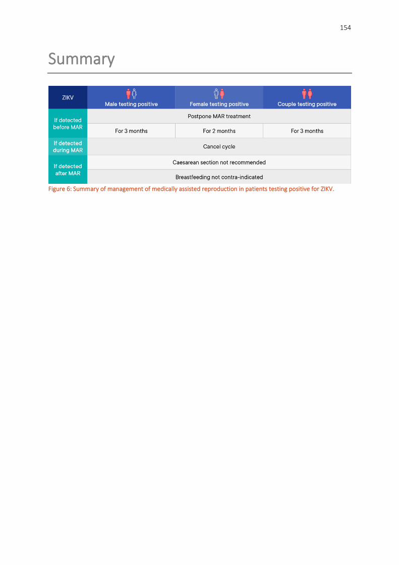
154
Summary
Figure 6: Summary of management of medically assisted reproduction in patients testing positive for ZIKV.

155
PART G: SARS-CoV-2 In the circumstances of the current situation on the SARS-CoV-2 global pandemic, the guideline group considered introducing a chapter on this virus. However, the evidence is emerging and changing constantly. The best source of information with the latest evidence about the impact of this virus on MAR practice can been found here:
https://www.eshre.eu/Home/COVID19WG

156
PART H: Laboratory safety
With respect to the risk of cross-contamination by infectious agents in laboratory, the primary means of prevention is strict adherence to good laboratory practice, including careful management of cryobanks using appropriate methods of cryostorage.
PICO QUESTION: CAN SEPARATE CRYO TANK STORAGE PREVENT CROSS CONTAMINATION OF STORED MATERIAL?
Evidence Bacteria, fungi and viruses have been shown to survive in liquid nitrogen (LN2) cryobanks (Mirabet et al., 2012). Viruses have been detected after storage at -70°C for HIV (9.1 years), HBV (4 years) and HCV (9 years) (Baleriola et al., 2011). HCV has also been shown to be stable for 6 months at -80°C (Halfon et al., 1996).
One study (Hawkins et al., 1996) showed viral transmission through LN2, related to physical damage of stored material. In this study, transmission of HBV via damage to infected stored bone marrow was shown, which led to six patients becoming infected with the virus (Hawkins et al, 1996). Whilst this study did not involve reproductive cells or tissue, and also involved ethyl vinyl bags which are not used in MAR cryopreservation and have a 10% failure rate in liquid nitrogen (Khuu et al., 2002), viral transmission through LN2 was demonstrated.
A more recent study showed that no viruses could be detected in the LN2 used to vitrify oocytes (n=14) and embryos (n=10) from patients infected with HIV, HCV, and HBV using open devices (Cobo et al., 2012), nor detection of HCV in the LN2 from the long-term storage tank for a patient infected with HCV (Cobo, et al., 2012). However, the sample size was small.
In one animal study, screened bovine embryos were vitrified using open devices or in plastic cryovials, and then stored in LN2 experimentally contaminated with 3 bovine viruses (BVDV, BHV, BIV) for 3-5 weeks. Warmed embryos showed infection with BVDV & BHV-1 (83 batches, 13/61, 21.3%; but no infection with BIV (22 batches, 0/22, 0%) (Bielanski et al., 2000).
Recommendation
Since viruses can survive and be transmitted via liquid nitrogen (LN2), separate storage of reproductive cells according to viral positive and viral negative status is recommended.
GPP

157
Emptied and dried cryo tanks and transport shippers should be disinfected according to local standard operating procedures to reduce the potential of cross-contamination.
GPP
Individual clinics must risk assess to decide the number of cryo tanks needed.
GPP
Separate cryopreservation dewars should be used to quarantine gametes and embryos from patients with unknown infectious status.
GPP
Justification There is a lot of variation in practice. Some clinics store all viral positive samples together, and some clinics have separate storage per type of virus.
Before attempting disinfection of cryo-tanks, contact the manufacturers regarding the warranty conditions. We are unaware of any disinfection procedure for the absorbent material in dry transport shippers.
PICO QUESTION: CAN THE TYPE OF CRYOSTORAGE ENVIRONMENT (LIQUID VERSUS VAPOUR/OPEN VERSUS CLOSED SYSTEMS) PREVENT CROSS CONTAMINATION OF STORED MATERIAL?
Evidence Liquid versus vapour
Unlike LN2 vapour phase (LNVP) storage vessels, LN2 storage vessels will accumulate particulate matter from the atmosphere. This includes pathogenic organisms which may remain viable by immersion in LN2. Pathogens can accumulate on the surface of cryodevices placed into LN2 storage, creating a contamination risk, particularly when removed from storage and warmed (Grout and Morris, 2009). Contamination of samples in LNVP also carries potential risk.
Mirabet et al. aimed to identify microbiological agent in the liquid nitrogen containers, while comparing different types of tanks (liquid nitrogen vs. gas phase vs. half gas half liquid). The vapour phase tank yielded less contamination than the liquid phase (Mirabet, et al., 2012).
Contamination found in the lower part of the liquid nitrogen vessels after thawing are generally environmental germs and exclusively contamination of water without cross-contamination of the samples themselves (Molina et al., 2016).
In an animal study investigating bovine embryos and viruses (BVDV and BHV-1), contaminated and non-contaminated samples of embryos and semen were stored in proximity in LNVP in open containers (dry

158
shippers) for 7 days. No cross-contamination took place, indicating that LNVP is a safe means for short-term storage of embryos and semen in dry shipper dewars (Bielanski, 2005).
Open versus closed systems
No original research studies could be found.
Recommendation
Vapour phase cryopreservation could be considered over liquid nitrogen in terms of safety to reduce the risk of cross-contamination.
Conditional ⊕
Provided the cryomaterial is not compromised, cryodevices, such as sealed semen straws/vials, should be cleaned with a disinfectant wipe after removal from LN2 storage to mitigate risk of transmission of pathogens from the cryodevice surface.
GPP
Justification No storage environment can guarantee 100% prevention of cross-contamination. Current evidence shows that the risk of cross-contamination is smaller with the use of vapour phase as compared to liquid nitrogen.
Many embryologists are understandably cautious about storing vitrified oocytes/embryos in vapour phase. The risks of undesired warming in vapour phase need to be weighed against the risk of cross-contamination in the liquid phase. The risk of warming is less for cryopreserved sperm samples compared to vitrified oocytes/embryos, as the rate of warming of frozen sperm is slower, hence the vapour may be considered more acceptable for sperm samples.
Open versus closed systems
Joaquim et al 2017 states:
• devices must be chosen carefully to minimize the risk of disease transmission • there is no scientific consensus on the safety of claimed high-security closed methods in
comparison with current open vitrification systems. More investment should be made to make closed systems more efficient (Joaquim et al., 2017)
Dubaut et al. state that for ZIKV, “the specimen should be labelled and stored separately reflecting potential increased risk, and if liquid nitrogen is required, open carriers should not be used”.
It is recommended that dry shippers should be used in accordance with manufacturer’s instructions, as “dry” shippers rather than filled with liquid nitrogen.

159
PICO QUESTION: CAN THE TYPE OF VIALS PREVENT CROSS-CONTAMINATION OF STORED MATERIAL?
Evidence LN2 infiltrates inner thread vials and may cause a high rate of microbial contamination (and risk of explosion on thawing). Vials with a heat-sealable/ hermetically-sealable membrane are resistant to LN2 penetration and contamination (Chen et al., 2006).
Recommendation
Hermetical sealing of cryovials with additional covers could reduce the risk of cross-contamination of stored material.
Conditional ⊕
Justification Liquid nitrogen infiltrates inner thread vials, therefore increasing the risk of cross-contamination or explosion of the vial upon warming. Sealing the vials, using a membrane resistant to liquid nitrogen can overcome this issue. Application of such a secondary enclosure has been termed "double bagging” (Bielanski, 2014).
PICO QUESTION: CAN HIGH SECURITY STRAWS PREVENT CROSS CONTAMINATION OF STORED MATERIAL?
Evidence When three types of straws (24 each of PVC, PETG and IR) were filled with HIV-1, then sealed either ultrasonically or thermally, prior to storage in LN2, only thermal sealed ionomeric resin (IR) straws showed no contamination on the outside of the straw upon thawing (Letur-Konirsch et al., 2003). Similarly, IR straws filled with HCV+ seminal plasma, and thermally sealed prior to LN2 storage, showed no contamination on the straw exterior upon thawing (Maertens et al., 2004).
Recommendation
The use of high security straws in combination with thermal sealing is the preferred approach as it minimises the risk of cross-contamination.
Strong ⊕
At the time of thawing, decontamination of the exterior of the straw and the single use of sterile scissors will reduce the risk of contaminating the stored contents with potential pathogens.
GPP
160
Justification Cryostorage devices should provide closure integrity and sample stability, without risk of infection from the cryo-tank.
Caution should be used to specifically consider avoidance of cross-contamination upon sample removal from the cryo-tank.
PICO QUESTION: CAN THE USE OF SEPARATE LABS PREVENT CROSS CONTAMINATION?
Evidence One study showed that for baseline swabs (n=79) taken in a total laboratory automation (TLA) system during routine clinical use after running a small number of high-titre HCV samples, low level HBV (n=10) and HCV (n=8) contamination was detected on equipment and exposed surfaces, even when good lab practice was adhered to (Bryan et al., 2016).
When specimen containers were exteriorly coated with a fluorescent powder to enable the visualization of gross contamination using UV light, experienced lab technologists using standard personal protective equipment (PPE) showed contamination of PPE (gloves and laboratory coat cuffs), bare hands, biosafety cabinets (8/36; 22% tests) and testing accessory items (29/32; 91% tests) (Yarbrough et al., 2018).
For dentistry, after sterilization of invasive medical instruments with 2% glutaraldehyde, HBV was still detectable on the sterilized instruments (Zhou et al., 2006).
Contamination of samples by LN2-borne bacteria (S. minor Sclerotia) during cooling in controlled rate freezers, in vitrification procedures or in vapour phase vessels has been demonstrated (Grout and Morris, 2009).
A study by Cobo et al. included 24 patients testing positive for HBV having oocyte vitrification or IVF. No virus particles were detected in follicular fluid, oocyte or embryo culture media after IVF (Cobo, et al., 2012).
Recommendation
Given that personal protective equipment (PPE), laboratory equipment and exposed surfaces can be contaminated even after good laboratory practice, disinfection and changing PPE between cases can reduce the risk of cross-contamination.
GPP
The recommended procurement, processing, release and storage procedures should be used for all samples, not only virally positive samples.
GPP

161
Justification We found no studies investigating if the use of separate labs will enhance the prevention of cross-contamination of samples.
Good practice according to the ESHRE guideline should be applied in all MAR laboratories, acknowledging that each center and country may have different legislation.
A large multi-center study is needed to investigate the need of separate labs for viral positive samples.
Use of single use devices (e.g. ICSI pipettes) mitigates the risk of cross-contamination.
REFERENCES Baleriola C, Johal H, Jacka B, Chaverot S, Bowden S, Lacey S, Rawlinson W. Stability of hepatitis C virus, HIV, and hepatitis B virus nucleic acids in plasma samples after long-term storage at -20degreeC and -70degreeC. Journal of clinical microbiology 2011;49: 3163-3167. Bielanski A. Non-transmission of bacterial and viral microbes to embryos and semen stored in the vapour phase of liquid nitrogen in dry shippers. Cryobiology 2005;50: 206-210. Bielanski A. Biosafety in embryos and semen cryopreservation, storage, management and transport. Advances in experimental medicine and biology 2014;753: 429-465. Bielanski A, Nadin-Davis S, Sapp T, Lutze-Wallace C. Viral contamination of embryos cryopreserved in liquid nitrogen. Cryobiology 2000;40: 110-116. Bryan A, Cook L, Atienza EE, Kuypers J, Cent A, Baird GS, Coombs RW, Jerome KR, Wener MH, Butler-Wu SM. Bloodborne Viral Pathogen Contamination in the Era of Laboratory Automation. Clinical chemistry 2016;62: 973-981. Chen HI, Tsai CD, Wang HT, Hwang SM. Cryovial with partial membrane sealing can prevent liquid nitrogen penetration in submerged storage. Cryobiology 2006;53: 283-287. Cobo A, Bellver J, de los Santos MJ, Remohi J. Viral screening of spent culture media and liquid nitrogen samples of oocytes and embryos from hepatitis B, hepatitis C, and human immunodeficiency virus chronically infected women undergoing in vitro fertilization cycles. Fertility and sterility 2012;97: 74-78. Grout BW, Morris GJ. Contaminated liquid nitrogen vapour as a risk factor in pathogen transfer. Theriogenology 2009;71: 1079-1082. Halfon P, Khiri H, Gerolami V, Bourliere M, Feryn JM, Reynier P, Gauthier A, Cartouzou G. Impact of various handling and storage conditions on quantitative detection of hepatitis C virus RNA. Journal of hepatology 1996;25: 307-311. Hawkins AE, Zuckerman MA, Briggs M, Gilson RJ, Goldstone AH, Brink NS, Tedder RS. Hepatitis B nucleotide sequence analysis: linking an outbreak of acute hepatitis B to contamination of a cryopreservation tank. Journal of virological methods 1996;60: 81-88. Joaquim DC, Borges ED, Viana IGR, Navarro PA, Vireque AA. Risk of Contamination of Gametes and Embryos during Cryopreservation and Measures to Prevent Cross-Contamination. BioMed research international 2017;2017: 1840417. Khuu HM, Cowley H, David-Ocampo V, Carter CS, Kasten-Sportes C, Wayne AS, Solomon SR, Bishop MR, Childs RM, Read EJ. Catastrophic failures of freezing bags for cellular therapy products: description, cause, and consequences. Cytotherapy 2002;4: 539-549. Letur-Konirsch H, Collin G, Sifer C, Devaux A, Kuttenn F, Madelenat P, Brun-Vezinet F, Feldmann G, Benifla JL. Safety of cryopreservation straws for human gametes or embryos: a study with human immunodeficiency virus-1 under cryopreservation conditions. Human reproduction (Oxford, England) 2003;18: 140-144.

162
Maertens A, Bourlet T, Plotton N, Pozzetto B, Levy R. Validation of safety procedures for the cryopreservation of semen contaminated with hepatitis C virus in assisted reproductive technology. Human reproduction (Oxford, England) 2004;19: 1554-1557. Mirabet V, Alvarez M, Solves P, Ocete D, Gimeno C. Use of liquid nitrogen during storage in a cell and tissue bank: contamination risk and effect on the detectability of potential viral contaminants. Cryobiology 2012;64: 121-123. Molina I, Mari M, Martínez JV, Novella-Maestre E, Pellicer N, Pemán J. Bacterial and fungal contamination risks in human oocyte and embryo cryopreservation: open versus closed vitrification systems. Fertility and sterility 2016;106: 127-132. Yarbrough ML, Kwon JH, Wallace MA, Hink T, Shupe A, Fraser VJ, Dubberke ER, Burnham CA. Frequency of Instrument, Environment, and Laboratory Technologist Contamination during Routine Diagnostic Testing of Infectious Specimens. Journal of clinical microbiology 2018;56. Zhou LF, Zhu HH, Lin J, Hu MJ, Chen F, Chen Z. Surveillance of viral contamination of invasive medical instruments in dentistry. J Zhejiang Univ Sci B 2006;7: 745-748.
163
Annexes
Annex 1: Guideline development group
Annex 2: Abbreviations
Annex 3: Recommendations for research
Annex 4: Methodology
Annex 5: Stakeholder consultation
Annex 6: Literature study: flowcharts, list of excluded studies
Annex 7: Evidence tables

Annex 1: Guideline development group This guideline was composed of (previous) members of the SIG Safety and Quality in ART, Ethics and Law and the former task force on Viral Infections, with addition of experts in the field that replied on a call for experts to the ESHRE audience.
Chair of the GDG Edgar Mocanu Rotunda Hospital, Department of Reproductive Medicine, Royal
College of Surgeons in Ireland, Dublin (Ireland)
GDG members Andrew Drakeley Liverpool Women’s Hospital, Department of Reproductive Medicine,
Liverpool (UK) Markus S. Kupka Gynaekologicum Hamburg, Department Gynaecology and Obstetrics,
Hamburg (Germany) Evelin E. Lara-Molina IVI RMA Global Barcelona, Department of Egg Donation, Barcelona
(Spain) Willem Ombelet Genk Institute for Fertility technology, ZOL Hospitals, Genk
Faculty of Medicine and Life Sciences, Hasselt University, Hasselt (Belgium)
Catherine Patrat APHP.centre – University of Paris, Cochin, Service de Biologie de la Reproduction - CECOS, Paris (France)
Guido Pennings Bioethics Institute Ghent (BIG) Ghent University, Department of Philosophy and Moral Science, Gent (Belgium)
Augusto Enrico Semprini University of Milan, Department of Biomedical and Clinical Sciences, Milan (Italy)
Kelly Tilleman Ghent University Hospital, Department for Reproductive Medicine, Gent (Belgium)
Mauro Tognon University of Ferrara School of Medicine, Department of Medical Sciences, Ferrara (Italy)
Nino Tonch Amsterdam University Medical Centre, location AMC, Department of Reproductive Medicine, Amsterdam (Netherlands)
Bryan Woodward X&Y Fertility, Leicester (UK)
Invited expert Mark Atkins (clinical microbiologist/virologist)
Micropathology Ltd, Coventry (UK)
Methodological support Nathalie Le Clef European Society of Human Reproduction and Embryology (Belgium)

165
DECLARATIONS OF INTEREST
All members of the guideline development group were asked to declare possible conflicts of interest by means of the disclosure forms (see ESHRE Manual for Guideline Development).
Conflicts of interest
Edgar Mocanu None declared.
Andrew Drakeley
Research fees from Ferring and Merck Consulting fees from ferring Sponsering for scientific meetings from Ferring, Gedeon-Richter and Pharmasure
Markus S. Kupka None declared. Evelin E. Lara-Molina None declared. Willem Ombelet None declared. Catherine Patrat Speakers fees from Merck and MSD Guido Pennings None declared. Augusto Enrico Semprini None declared.
Tilleman Kelly Speakers fees from Cooper Surgical and Ferring. Consultancy for educational purposes as member of the advisory board BioTeam of Ferring.
Mauro Tognon None declared. Nino Tonch None declared. Bryan Woodward None declared. Nathalie Le Clef None declared.

166
Annex 2: Abbreviations Ab Antibody Ag Antigen CI Confidence interval CNP Combined neonatal prophylaxis CS Caesarean section DNA Desoxyribonucleic acid EIA Enzyme immune assay ELISA Enzyme-Linked Immuno Sorbent Assay ET Embryo transfer FISH Fluorescence in situ hybridization GDG Guideline development group GPP Good practice point HBcAg Hepatitis B core antigen HBeAb Hepatitis B e-antibody HBeAg Hepatitis B e-antigen HBIG Hepatitis B Immune globulin HBsAg Hepatitis B surface antigen HBV Hepatitis B virus HBxAg Hepatitis B x-antigen HCV Hepatitis C virus HIV Human immunodeficiency virus HR Hazard ratio HTLV Human T-cell lymphotrophic virus ICSI Intracytoplasmic sperm injection IgG Immunoglobulin G IgM Immunoglobulin M IHC Immunohistochemistry IQR Interquartile range ISH In situ hybridization IU International unit/infectious units IUI Intra-uterine insemination IVF In vitro fertilization LN2 Liquid nitrogen LNVP Liquid nitrogen vapour phase MAR Medically assisted reproduction MD Mean difference MESA Microsurgical epididymal sperm aspiration MTCT Mother-to-child-transmission NAT Nucleic acid testing NVP Nevirapine OR Odds ratio RD Risk difference PCR Polymerase Chain Reaction FTCT Father-to-child-transmission RCT Randomized controlled trial

167
RDT Rapid diagnostic test RNA Ribonucleic acid ROC-AUC Receiver operating characteristic – area under the curve RR Relative risk/risk ratio SET Single embryo transfer SMD Standardized mean difference TESE Testicular sperm extraction WMD Weighted mean difference ZIKV Zika virus ZDV Zidovudine

168
Annex 3: Recommendations for research on MAR in couples with a viral infection/disease
From the literature and discussion of the available evidence, several topics were identified for which evidence is inconsistent, insufficient or non-existing. For the benefit of patients with a viral infection/disease, the GDG recommends that future research, where possible in well-designed RCTs, should focus on these research gaps.
Considered are:
- HPV and the association with infertility o HPV as a cause of male infertility o HPV subtypes involved in infertility
- Semen processing in HBV and HCV: comparison between standard semen processing and advanced semen processing
- Semen processing for HTLV I/II - Association between SARS-CoV-2 and infertility - Laboratory safety procedures for SARS-CoV-2

169
Annex 4: Methodology
GUIDELINE DEVELOPMENT
The European Society of Human Reproduction and Embryology (ESHRE) guidelines are developed based on the Manual for ESHRE guideline development (N. Vermeulen, N. Le Clef, S. Mcheik, A. D'Angelo , K. Tilleman, Z. Veleva, W.L.D.M. Nelen, Manual for ESHRE guideline development, version 2019), which can be consulted on the ESHRE website (www.eshre.eu/guidelines). The principal aim of this manual is to provide stepwise advice on ESHRE guideline development for members of ESHRE guideline development groups. The manual describes a 12-step procedure for writing clinical management guidelines by the guideline development group, supported by the ESHRE methodological expert:
The current guideline was developed and funded by ESHRE, which covered expenses associated with the guideline meetings (travel, hotel and catering expenses) associated with the literature searches (library costs, costs associated with the retrieval of papers) and with the implementation of the guideline (printing, publication costs). Except for reimbursement of their travel expenses, GDG members did not receive any payment for their participation in the guideline development process.
The scope of the guideline and first version of the key questions were drafted by members of the ESHRE Special Interest Group Safety and Quality in ART, Ethics and Law and members of the former task force on Viral Diseases. A call was launched for experts in the field interested in joining the guideline development group. All applications were reviewed, and experts were selected based on expertise and geographical location. We strived towards a balance in gender and location within Europe. A meeting of the guideline development group was organized to discuss the key questions and redefine them through the PICO process (patients – interventions – comparison – outcome). This resulted in a final list of 9 key questions for 6 viruses each. Based on the defined key words, literature searches were performed by the methodological expert (Dr. N. Le Clef). Key words were sorted to importance and used for searches in PUBMED/MEDLINE, the Cochrane library, EMBASE and GIM. We searched the databases from inception up to 3 November 2020.
Literature searches were performed as an iterative process. In a first step, systematic reviews and meta-analyses were collected. If no results were found, the search was extended to randomized controlled trials, and further to cohort studies and case reports, following the hierarchy of the levels of evidence. References were selected or excluded by the methodological expert and expert GDG member based on title and abstract and knowledge of the existing literature. If necessary, additional searches were performed in order to get the final list of papers. The quality of the selected papers was assessed by means of the quality assessment checklist, defined in the ESHRE guideline manual. Furthermore, the

170
evidence was collected and summarized in an evidence table according to GIN format (http://www.g-i-n.net/activities/etwg). The quality assessment and evidence tables were constructed by the expert GDG members.
Normally, summary of findings (SoF) tables would be prepared following the GRADE approach for intervention studies with at least 2 studies per outcome. However, since the body of evidence mainly consisted of cohort and cross-sectional studies, case series and case reports, which were often difficult to compare in a direct manner, the added value of summarizing the evidence in SoF tables was limited and thus not performed for this guideline. The critical outcomes for this guideline were: - Safety:
o risk of horizontal transmission to partner/family/healthcare providers o risk of vertical transmission to the infant
- Efficacy: implantation rates, pregnancy rates, live birth rates, miscarriage rates
GDG meetings were organized to discuss the draft recommendations and the supporting evidence and to reach consensus on the final formulation of the recommendations. In a final step, all evidence and recommendations were combined in the ESHRE guideline: “Medically assisted reproduction in patients with a viral infection/disease”.
FORMULATION OF RECOMMENDATIONS We labelled the recommendations as either ‘‘strong’’ or ‘‘conditional’’ according to the GRADE approach. We used the words ‘‘we recommend’’ for strong recommendations and ‘‘we probably recommend’’ for conditional recommendations. Suggested interpretation of strong and conditional recommendations by patients, clinicians and health care policy makers is as follows:
Implications for Strong recommendation Conditional recommendation Patients Most individuals in this situation would
want the recommended course of action, and only a small proportion would not
The majority of individuals in this situation would want the suggested course of
action, but many would not Clinicians Most individuals should receive the
intervention Adherence to this recommendation
according to the guideline could be used as a quality criterion or performance indicator
Formal decision aids are not likely to be needed to help individuals make decisions
consistent with their values and preferences
Recognise that different choices will be appropriate for individual patients and that
you must help each patient arrive at a management decision consistent with his
or her values and preferences Decision aids may be useful in helping
individuals to make decisions consistent with their values and preferences
Policy makers The recommendation can be adopted as policy in most situations
Policy making will require substantial debate and involvement of various
stakeholders
For each recommendation it is mentioned whether it is strong or conditional and what the quality of the supporting evidence was. In the justification section, more data are provided on the considerations taken into account when formulating the recommendations: balance between desirable and undesirable effects, certainty of the evidence of effects, certainty in how people value the outcome, acceptability and feasibility of the intervention. Impact on health equity and resource impact were only discussed where relevant.
171
STRATEGY FOR REVIEW OF THE GUIDELINE DRAFT After finalization of the guideline draft, the review process was initiated. The draft guideline was published on the ESHRE website, accompanied by the reviewers’ comments form and a short explanation of the review process. The guideline was open for review between 18 February and 1 April 2021.
To notify interested clinicians, we sent out an invitation to review the guideline by email to all members of the ESHRE SIG Safety and Quality in ART, Ethics and Law, SIG embryology and ESHRE members with an interest in viral disease.
Selected reviewers were invited personally by email. These reviewers included: • Coordinators and deputies of the ESHRE SIG Ethics and law and the ESHRE SIG Quality and Safety
in ART. • Contact persons of patient organizations across Europe.
• Contact persons of international and national societies focused on IVF/ICSI across Europe.
All reviewers are listed in annex 6. The Reviewer comments processing report, including further information on the review and a list of all comments per reviewer with the response formulated by the GDG will be published on the ESHRE website.
GUIDELINE IMPLEMENTATION STRATEGY The standard dissemination procedure for all ESHRE guidelines comprises publishing and announcement.
Each guideline is published on the ESHRE Website and in Human Reproduction Open. The announcement procedure includes a news item in “Focus on Reproduction”, a newsflash on the ESHRE website homepage and a short presentation at the ESHRE Annual meeting. All participants in the annual ESHRE meeting will be informed about the development and release of new guidelines; all related national societies and patient organizations are informed about the guideline release. They are asked to encourage local implementation by, for instance, translations or condensed versions, but they are also offered a website link to the original document.
Patient versions of the guideline will be developed by a subgroup of the GDG together with patient representatives. The patient version is a translation of the recommendations in everyday language, with emphasis on questions important to patients. It aims to help patients understand the guideline’s recommendations and facilitates clinical decision-making.
To further enhance implementation of the guideline, the members of the GDG, as experts in the field, will be asked to select recommendations for which they believe implementation will be difficult and make suggestions for tailor-made implementation interventions (e.g. option grids, flow-charts, additional recommendations, addition of graphic/visual material to the guideline).

172
SCHEDULE FOR UPDATING THE GUIDELINE The current guideline will be considered for revision in 2025 (four years after publication). An intermediate search for new evidence will be performed two years after publication, which will inform the GDG of the necessity of an update.
Every care is taken to ensure that this publication is correct in every detail at the time of publication. However, in the event of errors or omissions, corrections will be published in the web version of this document, which is the definitive version at all times. This version can be found at www.eshre.eu/guidelines.
For more details on the methodology of ESHRE guidelines, visit www.eshre.eu/guidelines

173
Annex 5: Stakeholder consultation
As per routine development procedures, the guideline draft was open for review for 6 weeks, between 18 February and 1 April 2021. All reviewers, their comments and the reply of the guideline development group are summarized in the review report, which is published on the ESHRE website as supporting documentation to the guideline. The list of representatives of professional organization, and of individual experts that provided comments to the guideline are summarized below.
Representative Organization Harish M Bhandari British Fertility Society, UK
Reviewer Country Thomas Mitchell, James Duffy, Anastasia Mania, Niki Konsta, Ippokratis Sarris UK
Pierre Boyer France Carlos Calhaz-Jorge Portugal Stefan Matik North Macedonia Qianhong Ma, Fang Ma China Charalampos Siristatidis Greece Liana Bosco Italy Kimball O. Pomeroy USA Janek von Byern Austria

174
Copyright © European Society of Human Reproduction and Embryology - All rights reserved
The content of these ESHRE guidelines has been published for personal and educational use only. No commercial use is authorised. No part of the ESHRE guidelines may be translated or reproduced in any form without prior written permission of the ESHRE communications manager.
Copyright © 2022 FDOKUMEN