Mechanistic insights into viral clearance during the ...
9
RESEARCH ARTICLE Mechanistic insights into viral clearance during the chromatography steps in antibody processes by using virus surrogates Rachel Dyer | Yuanli Song | Jie Chen | Elizabeth Bigelow | Jennifer McGinnis | Lauren Jenkins | Sanchayita Ghose | Zheng Jian Li Biologics Development, Bristol-Myers Squibb, Devens, Massachusetts Correspondence Jie Chen, Biologics Development, Bristol- Myers Squibb, 38 Jackson Road, Devens, MA 01434. Email: [email protected] Abstract Viral safety is required for biological products to treat human diseases, and the burden of inactivation and or virus removal lies on the downstream purification process. Minute virus of mice (MVM) is a nonenveloped parvovirus commonly used as the worst-case model virus in validation studies because of its small size and high chemical stability. In this study, we investigated the use of MVM-mock virus particle (MVP) and bacteriophage ΦX174 as surrogates for MVM to mimic viral clearance studies, with a focus on chromatography operations. Based on structural models and comparison of log reduction value among MVM, MVP, and ΦX174, it was demonstrated that MVP can be used as a noninfectious surrogate to assess viral clearance during process devel- opment in multiple chromatography systems in a biosafety level one (BSL-1) labora- tory. Protein A (ProA) chromatography was investigated to strategically assess the impact of the resin, impurities, and the monoclonal antibody product on virus removal. KEYWORDS ΦX174, chromatography, minute virus of mice (MVM), MVM-mock virus particle (MVP), viral clearance 1 | INTRODUCTION Biologic drugs produced by mammalian cell culture processes could potentially be contaminated by endogenous and/or pathogenic adven- titious viruses, mainly from animal-based raw materials, or from the manufacturing process. 1 Demonstration of viral clearance, including virus removal and virus inactivation, is critical for any biopharmaceutical manufacturing process to ensure patient safety. Currently, viral clear- ance is assessed by spiking infectious model viruses during good lab practice (GLP) compliant viral validation studies using a scaled-down model of a downstream purification process. Since the handling of infectious viruses requires special safety facilities (minimally, biosafety level 2), these validation studies are usually performed at a contract research organization (CRO), which are costly, time-consuming, and logistically challenging. Noninfectious and robust virus surrogates are, therefore, highly desired for process development to gain an early in- house assessment of the viral clearance capability of the manufacturing process before the GLP viral clearance validation. Adequate virus safety for enveloped viruses can be achieved by viral filtration and viral inactivation methods, such as low pH, deter- gent, or heat. 2,3 However, since inactivation procedures are ineffec- tive for nonenveloped virus, virus removal by filtration must be supplemented by other orthogonal methods, such as chromatography, in order to provide adequate virus clearance across the overall down- stream process. Minute virus of mice (MVM), a nonenveloped parvo- virus, has been used often as a worst-case virus in validation studies because it is more challenging to remove in the downstream process due to its small size and chemical stability. 4,5 Rachel Dyer and Yuanli Song contributed equally to this study. [Correction added on September 3 2020, after first online publication: URL for peer review history has been corrected.] Received: 10 July 2020 Revised: 24 July 2020 Accepted: 27 July 2020 DOI: 10.1002/btpr.3057 Biotechnol Progress. 2020;36:e3057. wileyonlinelibrary.com/journal/btpr © 2020 American Institute of Chemical Engineers 1 of 9 https://doi.org/10.1002/btpr.3057
-
Upload
khangminh22 -
Category
Documents
-
view
2 -
download
0
Transcript of Mechanistic insights into viral clearance during the ...

R E S E A R CH A R T I C L E
Mechanistic insights into viral clearance during thechromatography steps in antibody processes by using virussurrogates
Rachel Dyer | Yuanli Song | Jie Chen | Elizabeth Bigelow |
Jennifer McGinnis | Lauren Jenkins | Sanchayita Ghose | Zheng Jian Li
Biologics Development, Bristol-Myers Squibb,
Devens, Massachusetts
Correspondence
Jie Chen, Biologics Development, Bristol-
Myers Squibb, 38 Jackson Road, Devens, MA
01434.
Email: [email protected]
Abstract
Viral safety is required for biological products to treat human diseases, and the burden
of inactivation and or virus removal lies on the downstream purification process.
Minute virus of mice (MVM) is a nonenveloped parvovirus commonly used as the
worst-case model virus in validation studies because of its small size and high chemical
stability. In this study, we investigated the use of MVM-mock virus particle (MVP) and
bacteriophage ΦX174 as surrogates for MVM to mimic viral clearance studies, with a
focus on chromatography operations. Based on structural models and comparison of
log reduction value among MVM, MVP, and ΦX174, it was demonstrated that MVP
can be used as a noninfectious surrogate to assess viral clearance during process devel-
opment in multiple chromatography systems in a biosafety level one (BSL-1) labora-
tory. Protein A (ProA) chromatography was investigated to strategically assess the
impact of the resin, impurities, and the monoclonal antibody product on virus removal.
K E YWORD S
ΦX174, chromatography, minute virus of mice (MVM), MVM-mock virus particle (MVP), viral
clearance
1 | INTRODUCTION
Biologic drugs produced by mammalian cell culture processes could
potentially be contaminated by endogenous and/or pathogenic adven-
titious viruses, mainly from animal-based raw materials, or from the
manufacturing process.1 Demonstration of viral clearance, including
virus removal and virus inactivation, is critical for any biopharmaceutical
manufacturing process to ensure patient safety. Currently, viral clear-
ance is assessed by spiking infectious model viruses during good lab
practice (GLP) compliant viral validation studies using a scaled-down
model of a downstream purification process. Since the handling of
infectious viruses requires special safety facilities (minimally, biosafety
level 2), these validation studies are usually performed at a contract
research organization (CRO), which are costly, time-consuming, and
logistically challenging. Noninfectious and robust virus surrogates are,
therefore, highly desired for process development to gain an early in-
house assessment of the viral clearance capability of the manufacturing
process before the GLP viral clearance validation.
Adequate virus safety for enveloped viruses can be achieved by
viral filtration and viral inactivation methods, such as low pH, deter-
gent, or heat.2,3 However, since inactivation procedures are ineffec-
tive for nonenveloped virus, virus removal by filtration must be
supplemented by other orthogonal methods, such as chromatography,
in order to provide adequate virus clearance across the overall down-
stream process. Minute virus of mice (MVM), a nonenveloped parvo-
virus, has been used often as a worst-case virus in validation studies
because it is more challenging to remove in the downstream process
due to its small size and chemical stability.4,5
Rachel Dyer and Yuanli Song contributed equally to this study.
[Correction added on September 3 2020, after first online publication: URL for peer review
history has been corrected.]
Received: 10 July 2020 Revised: 24 July 2020 Accepted: 27 July 2020
DOI: 10.1002/btpr.3057
Biotechnol Progress. 2020;36:e3057. wileyonlinelibrary.com/journal/btpr © 2020 American Institute of Chemical Engineers 1 of 9
https://doi.org/10.1002/btpr.3057

Over the years, there have been numerous efforts to use virus
surrogates to assess viral clearance during bioprocessing. The use of
virus surrogates for assessing viral filtration, which is a size-based
virus removal unit operation, has been very well established using
model virus surrogates, such as bacteriophages PP7, PR772, and
ΦX174.6-10 Virus surrogates have also been utilized to define the virus
particle binding mechanism to various chromatography resins, such as
anion exchange (AEX), hydrophobic interaction (HIC), and multimodal
(MMC) resins.11 Hydrophobicity and isoelectric point (pI) of viruses
have been reported to guide the development of chromatography
steps for optimal virus removal, but the impact of the product protein
and resin properties, as well as impurity levels, all have an effect on
the retention of viruses, are not as easily predicted.12-15 However, in
some studies, bacteriophages have been shown to be different from
MVM in charge and/or hydrophobicity.12 Due to these differences,
the interactions to chromatography resins, protein of interest, and
impurities may not be representative, and, therefore, bacteriophages
may be limited in chromatographic applications to consistently trend
with MVM removal. Recently, a new noninfectious MVM-mock virus
particle (MVP), with similar size, hydrophobicity, and pI as MVM, has
been developed and showed promise as a virus surrogate to evaluate
viral clearance by filtration and AEX.16-18
In this work, we compared the performance of ΦX174 (a well-
studied bacteriophage) and MVP (the new noninfectious recombinant
virus particle) as virus surrogates to MVM to perform preliminary, in-
house viral clearance risk assessments for chromatography steps, with
the ultimate goal of being able to assess the impact of process
changes during development activities. In addition to experimental
results, a molecular structural model was implemented to illustrate the
surface properties of MVM, MVP, and ΦX174 in order to better
understand the results we observed. Furthermore, we elucidate the
mechanistic behaviors of viral clearance during Protein A (ProA) chro-
matography. We investigate the interactions between virus particles,
monoclonal antibody (mAb), impurities, and chromatography resin as
well as the pH impact on viral removal during ProA chromatography.
The understanding of the interactions of virus-resin, virus-protein,
and virus-impurity is helpful to guide process development and
improve process robustness.
2 | MATERIALS AND METHODS
2.1 | Materials
All chemicals used in chromatography buffers were purchased from
the following vendors: J.T. Baker, Amresco, Avantor, Macron, Croda,
EMD Millipore, Ajinomoto, and Thermo Fisher Scientific. Resins
including MabSelect™ Sure LX, Capto™ Phenyl (high sub), Capto™
MMC, and cyanogen bromide (CNBr)-activated Sepharose® were
purchased from GE Healthcare. Other resins including POROS®
50 HQ and POROS® 50 XS were purchased from Thermo Fisher Sci-
entific. Resins were packed into Omnifit columns (ID 0.66cm). The
virus surrogates ΦX174 and MVP were purchased from Carolina
Biological Supply Company (Burlington, NC) and MockV Solutions
(Rockville, MD), respectively. All mAbs were produced at Bristol-
Myers Squibb using a Chinese Hamster Ovary cell expression system.
ΦX174 quantitative polymerase chain reaction (qPCR) reagents
including Invitrogen™ PureLink™ Pro 96 Viral RNA/DNA Purification
Kit and Real-Time Quantitative PCR (RT-qPCR) reagents were pur-
chased from Thermo Fisher Scientific.
2.2 | Methods
2.2.1 | Virus and virus surrogate quantification
To quantify ΦX174, DNA was extracted using the Invitrogen™ Pur-
eLink™ Pro 96 Viral RNA/DNA Purification Kit following the vendor's
protocol. An internal quantitative PCR (qPCR) method was developed
to quantitate ΦX174 genomic DNA. The method was performed using
a fluorogenic probe and flanking forward and reverse primers
designed to bind to a repetitive sequence within the ΦX174
genome.19 The forward primer sequence was 50-CGCCATTAA
TAATGTTTTCCGTAA-30, the reverse primer sequence was 50-CAT-
CCCGTCAACATTCAAACG-30, and the probe sequence was 6FAM-
50-CGCCTTCCATGATGAGA-30-MGBNFQ. QPCR experiments for
ΦX174 were performed using a QuantStudio™ 7 Flex Real Time PCR
system (Thermo Fisher Scientific). Results were calculated using the
QuantStudio Analysis Software.
To quantify MVP, Immuno-qPCR using the MVM-MVP kit from
MockV Solutions was conducted. First, the samples were applied to
the wells of a 96-well plate that was pre-coated with a capture anti-
body. After incubation, the wells were washed and a DNA conjugated
detector antibody was added. After another incubation step, the wells
were washed and a buffer was added to recover the DNA. The sam-
ples were then transferred to a nonbinding deep well plate. qPCR was
performed according to vendor's instructions using a ViiA 7 Real-Time
qPCR System (Thermo Fisher Scientific).
MVM data were generated at CROs using quantitative PCR or
plaque assays following viral clearance validation study protocols.
2.2.2 | Virus surrogate comparability assessment
Several monoclonal antibodies with validated MVM clearance data
were used in this study, and these mAbs cover a broad range of pI
and hydrophobicity. For experiments using ΦX174, the load mate-
rial was spiked to 1 × 10⁷ phage/mL using a ΦX174 stock with a
titer of 2 × 10¹⁰ phage/mL. For experiments using MVP, the load
material was spiked to 1 × 10⁹ particles/mL using an MVP stock
with a titer of 1 × 10¹² particles/mL. All chromatography experi-
ments were performed in duplicate on an ÄKTA Avant 150 system
(GE Healthcare) at room temperature. The operating conditions
were the same as the validated MVM viral clearance studies con-
ducted at CROs, including, but not limited to, column loading, pH,
conductivity, flow rate, and column bed height (details not listed).
2 of 9 DYER ET AL.

The virus removal capability of multiple modalities, including ProA,
AEX, CEX, HIC, and MMC, were evaluated using different mAbs
spiked with the virus surrogates (Table 1). ProA and CEX chroma-
tography were performed in bind-elute mode, AEX and HIC chroma-
tography were performed in flow-through mode, and MMC
chromatography was operated in both flow-through and bind-elute
modes. Load and eluate or flow-through samples were collected for
ΦX174 or MVP quantification. For select processes, fractions from
other steps, including washes and strips, were also collected for
virus particle quantification.
2.2.3 | Virus particle structural modeling
Structural models of MVM, ΦX174, and MVP were generated using
deposited structures with PDB codes 1MVM, 2BPA, and 4ZPY,
respectively. Size and surface properties, such as charge and hydro-
phobicity, were analyzed using PyMol.20-22 The virus particles were
color coded according to the distance from the center of the particle
ranging from 110 Å (blue) to 160 Å (red). The hydrophobicity of the
virus particles was analyzed with normalized consensus hydrophobic-
ity scale and displayed in PyMol with the most hydrophilic residue
(Arg) in white and the most hydrophobic residue (Ile) in red.23 The
electrostatic potential of virus particles was calculated using Adaptive
Poisson-Boltzmann Solver (APBS) with the Poisson-Boltzmann
model.24 The charged surface of each individual capsid protein in the
virus particle was displayed in PyMol with positive charges in blue and
negative charges in red.
2.2.4 | Investigation of viral clearance in protein Achromatography
In order to investigate the mechanism of virus removal in ProA chro-
matography, we evaluated the interaction of MVP with mAbs, the
Protein A resin, and impurities.
First, we conducted a study to assess the interaction between
MVP and different mAbs in a simple system. Several mAbs were
conjugated to CNBr-activated Sepharose® resin following the ven-
dor's instructions, which is optimized for the conjugation of mAbs to
the resin base matrix. Briefly, lyophilized resin was resuspended and
conjugated with 10 g mAb/L of resin. Conjugated resin was washed
with Tris to remove any unconjugated mAb and block any unreacted
CNBr sites, and the washes were analyzed for protein content to
ensure that the resin was conjugated with an equivalent amount of
mAb. The conjugated and washed resin was packed in a 0.66 cm ID
Omifit column. A volume of 100 μL of the MVP stock (containing
1 × 10¹² particles/mL) was injected into the column using an ÄKTA
Avant 150 chromatography system (GE Healthcare), and the retention
time of the MVP was measured using the UV absorbance profile at
280 nm. A column packed with unconjugated resin (no mAb) was also
run as control condition.
Furthermore, we studied the impact of impurity content in
the feed stream and impact of the impurity wash pH to deconvolute
the interactions among MVP, mAb, Protein A resin, and impurities.
The ProA chromatography experiments were performed on an ÄKTA
Avant 150 chromatography system (GE Healthcare) at room tempera-
ture. The ProA experiments involve equilibration, load, wash 1 (chase,
PBS buffer), wash 2 (impurity reduction, acetate, Tris, or carbonate
buffer ranging from pH 5 to pH 10), wash 3 (bridging, Tris buffer at
pH 7.5), elution (sodium acetate buffer at pH 3.2–4.0), strip, clean in
place, and storage phase. The load, flow-through, wash, elution, and
strip pools were collected and analyzed for MVP content. Each feed
stream, containing different impurity contents, was spiked with
1 × 10⁹ particles/mL using an MVP stock with a titer of
1 × 10¹² particles/mL. To evaluate the interaction of MVP and the
Protein A resin (base matrix and ligand), we used phosphate-buffered
saline (PBS) as ProA load. To investigate the interaction of MVP and
impurities, we used null cell culture material. To investigate the inter-
action between MVP and the product protein, we used purified mAb
buffer exchanged into PBS at a concentration of 4.9 g/L. As a process
control, we concentrated the purified mAb load to 68 g/L and spiked
into the null cell culture material to a concentration of 4.9 g/L, in
order to investigate the clearance in a more representative feed
stream. ProA chromatography were performed using either a pH 9 or
pH 10 impurity clearing wash.
TABLE 1 Monoclonal antibodyproperties and experiment in this work
Molecule pI Hydrophobicitya Subtype Unit operations Surrogate
mAb1 8.8 Mid IgG1/IgG2 Protein A, HIC MVP
mAb2 7.9 Mid IgG4 Protein A, AEX MVP, ΦX174
mAb3 6.9 High IgG4 Protein A, CEX, AEX MVP
mAb4 7.0 High IgG4 Protein A, CEX MVP, ΦX174
mAb5 6.8 Mid IgG4 MMC MVP
mAb6 7.7 Low IgG4 HIC MVP, ΦX174
mAb7 5.9 Mid IgG4 N/A MVP
mAb8 8.2 High IgG1 N/A MVP
Abbreviation: MVP, MVM-mock virus particle.amAb relative hydrophobicity was determined by measuring elution salt concentration during a HIC linear
salt gradient experiment, data not shown.
DYER ET AL. 3 of 9

3 | RESULTS AND DISCUSSION
3.1 | Evaluation of ΦX174 and MVP as virussurrogates for MVM
In order to evaluate noninfectious virus surrogates to be used for pre-
liminary assessment of viral clearance risk during process develop-
ment, two virus surrogates, ΦX174 and MVP, were selected based on
their similar physicochemical properties to MVM including size, pI,
and hydrophobicity (Table 2). Following the vendor's suggestion, pre-
liminary spike recovery studies were performed to screen buffers to
ensure accurate quantification of the virus surrogates. We also dem-
onstrated the virus surrogates can undergo up to two freeze thaw
cycles (data not shown). The mAbs used in this study cover a wide
range of pI and hydrophobicity, and different isotypes (Table 1). To
compare ΦX174 and MVP as virus surrogates for MVM, we con-
ducted the spiking study using the same operating conditions as the
validated viral clearance studies performed at the CRO. Load, elution,
or flow-through pools were analyzed by qPCR method to quantify
ΦX174 titer, or by immuno-qPCR method to quantify MVP titer. Log
reduction value (LRV) was calculated based on the following equation
to align with the CRO on MVM LRV calculation.
LRV= log10
Load virus concentration× Load volumeElution or flow through pool virus concentraiton×Pool volume
� �
Figure 1 shows a comparison of LRV results between the sur-
rogate data and data generated at the CRO using MVM. Several
chromatography steps were evaluated including ProA, AEX, CEX,
HIC, and MMC. In Figure 1a, we observed a correlation in LRVs
between the ΦX174 surrogate results and the results using MVM
for ProA chromatography. However, for HIC and AEX chromatog-
raphy, ΦX174 was not capable of reflecting the MVM perfor-
mance. In contrast, we observed better correlation in LRVs
between the MVP surrogate results and the results using MVM
over a broader range of chromatography operations, including
ProA, AEX, CEX, and MMC chromatography (Figure 1b). Interest-
ingly, a trend of slightly higher LRV for the MVP compared to the
MVM was observed in ProA chromatography. One reason could be
that the MVP is not real virus and lacks of the same post-
translational modification, such as glycosylation and phosphoryla-
tion, as MVM. Thus, such difference could cause MVP relatively
easier to be washed off in ProA chromatography.
TABLE 2 Comparison of MVM and viral surrogate properties
Attribute MVM ΦX174 MVP
Type Parvovirus Bacteriophage Virus-like particle
Enveloped No No No
Genome ssDNA ssDNA No DNA
Capsid shape Icosahedral Icosahedral Icosahedral
Size (nm) 10,17 21–28 26–32 22–28
pI 17 5.99 6.6 5.81
Hydrophobicity 17 Mild Low Mild
Expression system Mammalian E. Coli Insect
Abbreviations: MVM, minute virus of mice; MVP, MVM-mock virus particle; ssDNA, single stranded DNA.
F IGURE 1 Viral clearance correlationbetween surrogates and MVM.(a) Correlation between ΦX174 andMVM clearance for Protein Achromatography (black circle) and forpolishing chromatography steps HIC(gray square) and AEX (gray triangle).(b) Correlation between MVP and MVMfor capture step Protein A (black circle)and polishing chromatography steps CEX(gray square), MMC (gray invertedtriangle), and AEX (gray triangle). MVM,minute virus of mice; MVP, MVM-mockvirus particle
4 of 9 DYER ET AL.

In order to evaluate predictive capability of the two virus surro-
gates for MVM clearance in a polishing step, their clearance in anion
exchange chromatography was performed. AEX is one of the most
commonly used polishing steps during bioprocessing, serving an
important function to remove impurities such as host cell proteins
(HCPs), DNA, as well as viral contaminants.5,25 In this study, we tested
the impact of load conductivity on viral clearance at 2, 5, and 8 mS/cm
at a constant pH of 7.2, and compared these values to that from a val-
idated MVM clearance study. The MVM, MVP, and ΦX174 LRV
results are compared in Figure 2. The results show that the MVP
clearance was improved with increasing conductivity from 2 to
8 mS/cm, and that this trend was also consistent with the result from
the validated MVM clearance study. ΦX174 clearance data, however,
do not follow the same trend as MVM. In an AEX system, virus binds
to the resin, primarily, due to electrostatic interactions and it has also
been reported that virus particles behave similarly to pro-
teins.13,15,25,26 Consequently, it is expected that viruses will bind to
AEX resin more strongly than mAbs, and this interaction should be
disrupted by increasing the salt concentration in the mobile phase.
Previous literature has shown that under a low conductivity operating
range (<9 mS/cm), the LRV could increase slightly with increasing con-
ductivity.25,27 In this study, MVP trended qualitatively with MVM
clearance data, further demonstrating that MVP is a good virus surro-
gate to MVM, especially in chromatographic systems. To further
explain this phenomenon, we used modeling to illustrate the differ-
ences in the structure of these two virus surrogates.
3.2 | Structural analysis of MVM, MVP,and ΦX174
It is known that MVM particle is comprised of 60 subunits including
three viral proteins, designated VP1, VP2, and VP3 with size of
83.3 kDa, 64.3 kDa, and 61.4 kDa, respectively.20 These three viral
proteins start from different residue number at the N-terminus and
share an identical sequence at their C-terminus. VP2 is the dominant
variant in both full and empty viral particles. We selected 1MVM as
the structural model for full MVM particle. MVP is assembled using
recombinant VP2 without nucleic acids. We selected empty MVM
structure 4ZPY as model for MVP. We selected ΦX174 atomic
structure 2BPA as the model structure. The ΦX174 particle contains
60 copies of the J, F, and G proteins, as well as 12 copies of the H
protein, per virion. Particle size, accessible exposed hydrophobic
surface area, and accessible exposed net negative surface area were
analyzed based on the selected model protein structure using
PyMol, as shown in Table 3. The full MVM model (Figure 3a) is
slightly larger than the MVP model (Figure 3c). The ΦX174 model
has a larger radius up to 160 Å, including the surface protrusions, as
shown in Figure 3b. The MVM model (Figure 3d) demonstrates simi-
lar accessible surface hydrophobicity to the MVP model (Figure 3f),
but with higher accessible surface hydrophobicity than the ΦX174
model (Figure 3e). The accessible exposed net negative charge sur-
face area of three models is not significantly different (Figure 3g–i).
Our results are consistent with previous research that ΦX174 is less
hydrophobic than MVM, and also slightly different in pI.11 ΦX174
has large protrusions, which may reduce the virus particle contact to
the resin (either AEX or HIC), and less surface contact will result in
weaker interactions. Accessible exposed hydrophobic surface areas
of MVM and MVP are similar, while that of ΦX174 is less than that
of both MVM and MVP. Very subtle difference among accessible
exposed net negative surface areas of MVM, ΦX174, and MVP is
observed. The similarity in physicochemical properties between
MVP and MVM support why we observed that MVP is a better sur-
rogate for MVM in chromatographic systems than the bacterio-
phage ΦX174, as shown in Figure 1. In addition, the structural
model can be utilized to explain the different performance between
MVP and MVM. There are slightly less accessible hydrophobic
exposed area and accessible exposed net negative surface area of
MVP compared to MVM (Table 3), which could cause MVP bind to
POROS HQ resin slightly weaker than MVM, and a lower LRV as a
result (Figure 2).
Above all, it is clear that MVP is more similar to MVM from
molecular structure level, which makes MVP a better virus surrogate
F IGURE 2 Virus clearance comparison of MVM, MVP, andΦX174 in mAb3 process at the AEX step under AEX load conductivityof 2 mS/cm (black bar), 5 mS/cm (light gray bar), and 8 mS/cm (darkgray bar). Error bars indicate variation from duplicate experiments.MVM, minute virus of mice; MVP, MVM-mock virus particle
TABLE 3 Summary of MVM, ΦX174,and MVP structural analysis
Attribute MVM ΦX174 MVP
Distance of center to surface (Å) 110–128 115–160 108–125
Accessible exposed hydrophobic surface area (Å2) 35,974 25,694 34,656
Accessible exposed net negative surface area (Å2) 14,967 12,973 13,789
Abbreviations: MVM, minute virus of mice; MVP, MVM-mock virus particle.
DYER ET AL. 5 of 9
to MVM. Hence, all subsequent studies have used MVP as the
virus surrogate.
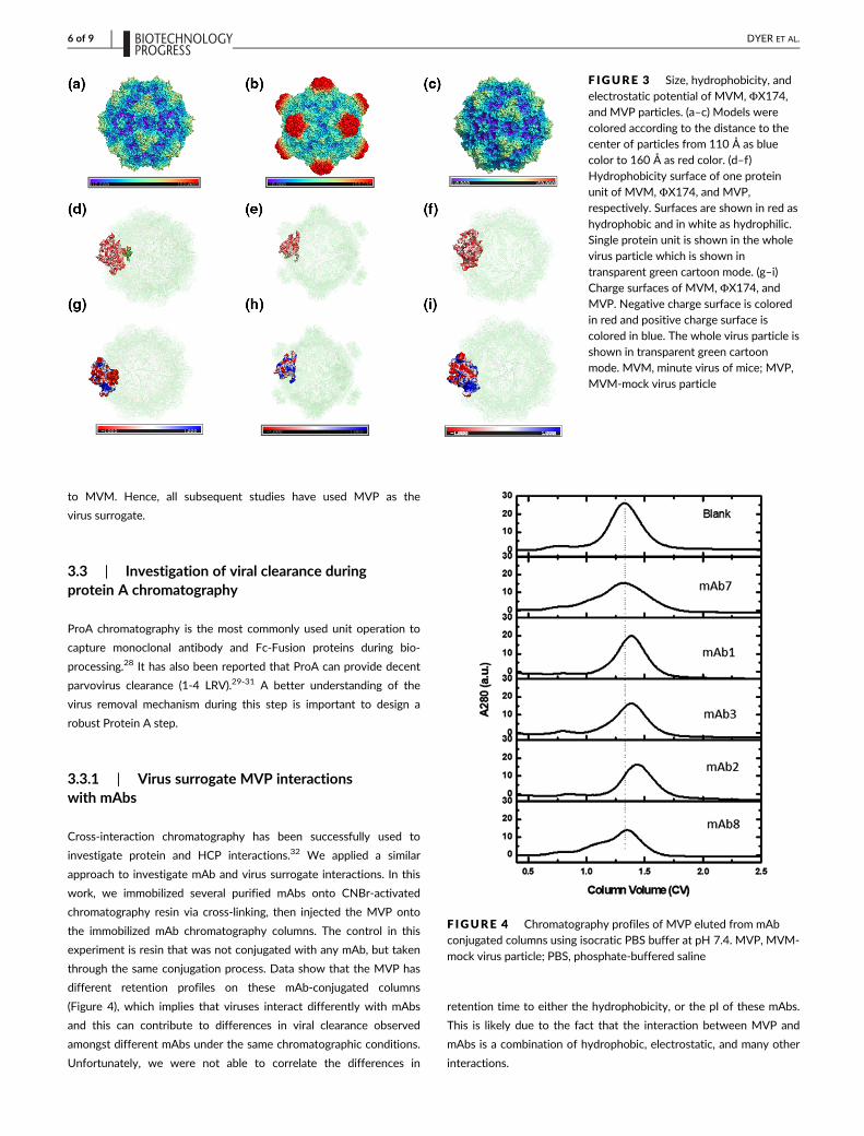
3.3 | Investigation of viral clearance duringprotein A chromatography
ProA chromatography is the most commonly used unit operation to
capture monoclonal antibody and Fc-Fusion proteins during bio-
processing.28 It has also been reported that ProA can provide decent
parvovirus clearance (1-4 LRV).29-31 A better understanding of the
virus removal mechanism during this step is important to design a
robust Protein A step.
3.3.1 | Virus surrogate MVP interactionswith mAbs
Cross-interaction chromatography has been successfully used to
investigate protein and HCP interactions.32 We applied a similar
approach to investigate mAb and virus surrogate interactions. In this
work, we immobilized several purified mAbs onto CNBr-activated
chromatography resin via cross-linking, then injected the MVP onto
the immobilized mAb chromatography columns. The control in this
experiment is resin that was not conjugated with any mAb, but taken
through the same conjugation process. Data show that the MVP has
different retention profiles on these mAb-conjugated columns
(Figure 4), which implies that viruses interact differently with mAbs
and this can contribute to differences in viral clearance observed
amongst different mAbs under the same chromatographic conditions.
Unfortunately, we were not able to correlate the differences in
retention time to either the hydrophobicity, or the pI of these mAbs.
This is likely due to the fact that the interaction between MVP and
mAbs is a combination of hydrophobic, electrostatic, and many other
interactions.
F IGURE 3 Size, hydrophobicity, andelectrostatic potential of MVM, ΦX174,and MVP particles. (a–c) Models werecolored according to the distance to thecenter of particles from 110 Å as bluecolor to 160 Å as red color. (d–f)Hydrophobicity surface of one proteinunit of MVM, ΦX174, and MVP,respectively. Surfaces are shown in red as
hydrophobic and in white as hydrophilic.Single protein unit is shown in the wholevirus particle which is shown intransparent green cartoon mode. (g–i)Charge surfaces of MVM, ΦX174, andMVP. Negative charge surface is coloredin red and positive charge surface iscolored in blue. The whole virus particle isshown in transparent green cartoonmode. MVM, minute virus of mice; MVP,MVM-mock virus particle
F IGURE 4 Chromatography profiles of MVP eluted from mAbconjugated columns using isocratic PBS buffer at pH 7.4. MVP, MVM-
mock virus particle; PBS, phosphate-buffered saline
6 of 9 DYER ET AL.

3.3.2 | Impact of protein A wash pH on virusclearance
Literature has also shown an impact of impurity levels on virus
removal in chromatographic separations.5,15,26 Given this information,
we hypothesize that virus clearance can also be impacted by the inter-
actions with both mAb and impurities in the feed stock. In an attempt
to better understand this more complex interaction with the “dirty”
feed stocks of ProA, we investigated the impact of wash conditions
and different feed stocks on virus clearance in the ProA step.
We first performed a comprehensive study using four different
mAbs and varying pH (5, 7, 9, 10, and) of the intermediate wash buffer
(referred to as wash 2 buffer). Both MVP and MVM were used in this
study and the summary of the LRV data is shown in Figure 5. As can be
seen from the figure, for all four mAbs, virus clearance improved with
increasing pH of the wash 2 buffer. More importantly, both MVM
(Figure 5a) and MVP (Figure 5b) showed similar qualitative trends (if not
exact LRVs) further building confidence in our virus surrogate model. A
high pH wash in ProA chromatography has been reported to enhance
the removal of impurities such as HCP and DNA.33,34 It is expected
that, similar to impurities, interactions with the virus may also be
impacted by pH (pH > 8), hence the improvement in virus clearance
observed at high pH conditions in these studies.
To further evaluate the impact of the wash 2 pH on ProA virus
clearance, we conducted a set of experiments using two different pHs
(pH 9 and pH 10). During ProA chromatography, the load sample as
well as flow through, wash 1, wash 2, wash 3, elution, and strip frac-
tions were collected. From these fractions, MVP levels were quanti-
fied and LRV was calculated for each individual fraction as shown in
Figure 6. The strip sample MVP content was below the limit of quanti-
fication, so the data are not shown. From the data in Figure 6, it is
clear that the lowest LRV, which is high MVP content, was found in
the flow-through fraction, indicating that MVP flowed through the
column while the antibody was retained on the resin. This is consis-
tent with a previously discovered virus removal mechanism in ProA.31
Interestingly, the wash 2 and wash 3 fractions show lower LRV when
using the pH 10 wash and subsequent higher LRV in the elution pools
indicating more MVP is removed with the higher pH wash. This result
suggests that we can modify wash 2 conditions in order to optimize
virus clearance during ProA. Furthermore, it shows that we can use
the MVP as a virus surrogate to optimize the ProA step for viral clear-
ance during process development.
3.3.3 | Impact of protein A load material on virusclearance
In order to further explore the mechanism of removal and the com-
plex relationship between MVP, resin, mAbs, and impurities in the
ProA load, four sets of experiments were performed to de-convolute
these interactions. We conducted each experiment with different
feed streams with wash 2 buffer at pH 9 or pH 10. MVP levels of the
elution pools were quantified and LRV was calculated (Table 4). We
observed different interactions with each feed stream. MVP clear-
ance from the PBS and null cell harvest material were very high (>4
LRV), out of the quantitation range, indicating the MVP is not being
retained strongly on the column without the presence of the mAb.
The MVP removal from feed streams containing the purified mAb,
however, shows lower LRV, indicating a strong interaction with the
F IGURE 5 Protein A wash 2 bufferpH impact showing pH 5 (black bar),pH 7 (medium gray bar), pH 9 (dark graybar), and pH 10 (light gray bar). (a) LRVdata for MVM. (b) LRV data for MVP.Error bars indicate variation fromduplicate experiments. LRV, logreduction value; MVM, minute virus ofmice; MVP, MVM-mock virus particle
F IGURE 6 LRV of MVP in Protein A chromatography fractionswith mAb1. During Protein A chromatography, the load sample aswell as flow through, wash 1, wash 2, wash 3, elution, and strip poolswere collected and LRV values were calculated. Strip data are notshown (all < LOQ). The molecule underwent experiments withimpurity clearing wash (wash 2) at pH 9 (black) and pH 10 (gray). Errorbars indicate variation from duplicate experiments. LOQ, limit ofquantitation; LRV, log reduction value; MVP, MVM-mock virusparticle
DYER ET AL. 7 of 9

mAb. In addition, it showed MVP clearance was not improved with
pH 10 wash as expected from the previous studies. However, in
the last set of the experiments, with the more representative feed
stream containing the mAb with the impurities, it shows an increase
in LRV from 3.21 to ≥4.65 with the pH 10 wash. Given this result,
as well as previous studies demonstrating improved impurity
removal with increased wash pH,33,34 the data suggest that MVP
clearance improvement is due to the increased clearance of impuri-
ties. This set of studies also suggests that the pH 9 wash is not
strong enough to overcome the mAb interaction that we observed,
hence the lower LRV value in the pH 9 experiments with feed
streams containing the mAb with or without impurities. From this
study, we observed that the MVP displays a low-level interaction
with the resin and stronger interactions with the impurities as well
as the mAb product. With the increase in impurity clearance with
this molecule (data not shown) using the pH 10 wash, MVP appears
to be removed along with the impurities, leading to improved clear-
ance. In this study, the product aggregate level in the load material
was <0.5%, which poses minimal risk of interacting with the virus
surrogate. In future studies, the impact of aggregate levels could be
explored further.
4 | CONCLUSIONS
In this work, we identified MVP, a noninfectious virus-like particle, as
a predictive surrogate to MVM in a wide variety of chromatographic
applications, including ProA, CEX, AEX, HIC, and MMC. This virus sur-
rogate can be utilized for preliminary viral clearance assessments, as
well as for research studies to enhance process understanding and
process robustness. It could be widely utilized as it does not carry the
risk that bacteriophages pose to bacterial cell lines. The quantifiable
range of the MVP assay is currently the primary limitation for broader
applications, as it does not allow for an accurate determination of LRV
larger than 4. Due to this assay limitation, most viral clearance studies
using AEX and VF are not quantifiable. However, with MVP as a sur-
rogate, and a focus on unit operations and process conditions that
result in virus reduction within the quantifiable assay range, we were
able to demonstrate the use of the MVP in process development
applications. Our structure analysis demonstrated that MVP had
higher similarity to MVM with respect to size, topology, surface
hydrophobicity, and surface charge. This provides the confidence that
the data generated using this surrogate can predict the similar trend
to that of the true virus removal. With this confidence in the surro-
gate, we investigated the mechanism of MVP removal at the ProA
chromatography step. Our data suggest that the product (mAb)-MVP
interaction plays a role on MVP removal in ProA chromatography,
however, impurities (such as HCPs)-MVP interaction dictates the
MVP removal, and the measures that improve impurity removal could
also enhance MVP removal as a result. The impact of the product
(mAb)-MVP interaction will likely play a significant role in polishing
chromatography where impurity levels are much lower, this will be
investigated further.
ACKNOWLEDGMENTS
The authors would like to acknowledge Satish Sharma in process ana-
lytical group at Bristol Myers Squibb for his input regarding assays, as
well as Juan Wang for initial ΦX174 assay development. They would
also like to acknowledge the downstream development group for pro-
viding MVM data, and their suggestions and support throughout this
project. They would like to acknowledge David Cetlin, CEO of MockV
Solutions (MockV Solutions was recently acquired by Cygnus Tech-
nologies), for his technical suggestions. They would also like to
acknowledge Magfur Alam for reviewing and providing feedback on
this manuscript.
PEER REVIEW
The peer review history for this article is available at https://publons.
com/publon/10.1002/btpr.3057.
ORCID
Jie Chen https://orcid.org/0000-0001-7975-5817
REFERENCES
1. International Conference on Harmonization (ICH) Quality Guideline
Q5A: Viral Safety Evaluation of Biotechnology Products Derived from
Cell Lines of Human or Animal Origin. 1998
2. Aranha H, Forbes S. Viral clearance strategies for biopharmaceutical
safety. Pharm Technol. 2001;25(4):22-22.
3. Chinniah S, Hinckley P, Connell-Crowley L. Characterization of oper-
ating parameters for XMuLV inactivation by low pH treatment. Bio-
technol Prog. 2016;32(1):89-97.
4. Gefroh E, Dehghani H, McClure M, Connell-Crowley L, Vedantham G.
Use of MMV as a single worst-case model virus in viral filter valida-
tion studies. PDA J Pharm Sci Technol. 2014;68(3):297-311.
5. Miesegaes G, Lute S, Brorson K. Analysis of viral clearance unit opera-
tions for monoclonal antibodies. Biotechnol Bioeng. 2010;106(2):
238-246.
6. Smith K, Comparing Virus Ultrafiltration of Bacteriophages with Filtra-
tion of Minute Virus of Mice. Thesis, University of Arkansas, Fayette-
ville, 2014.
7. Bolton G, Cabatingan M, Rubino M, Lute S, Brorson K, Bailey M. Nor-
mal-flow virus filtration: detection and assessment of the endpoint in
bioprocessing. Biotechnol Appl Biochem. 2005;42(2):133-142.
8. Lute S, Aranha H, Tremblay D, et al. Characterization of coliphage
PR772 and evaluation of its use for virus filter performance testing.
Appl Environ Microbiol. 2004;70(8):4864-4871.
TABLE 4 Protein A chromatography MVP clearance withdifferent feed streams and wash pH
Feed stream
Elution pool LRV
pH 9 wash pH 10 wash
PBS ≥4.21 ≥4.46
Null cell harvest ≥4.01 ≥4.39
Purified mAb 3.11 2.64
Null cell harvest + purified mAb 3.21 ≥4.65
Abbreviations: LRV, log reduction value; MVP, MVM-mock virus particle;
PBS, phosphate-buffered saline.
8 of 9 DYER ET AL.

9. Aranah-Creado H, Brandwein H. Application of bacteriophages as sur-
rogates for mammalian viruses: a case for use in filter validation based
on precedents and current practices in medical and environmental
virology. PDA J Pharm Sci Technol. 1999;53:75-82.
10. Lute S, Riordan W, Pease LF, et al. A consensus rating method for
small virus-retentive filters. I. Method development. PDA J Pharm Sci
Technol. 2008;62(5):318-333.
11. Brown MR, Johnson SA, Brorson KA, Lute SC, Roush DJ. A step-wise
approach to define binding mechanisms of surrogate viral particles to
multi-modal anion exchange resin in a single solute system. Biotechnol
Bioeng. 2017;114(7):1487-1494.
12. Johnson SA, Walsh A, Brown MR, et al. The step-wise framework to
design a chromatography-based hydrophobicity assay for viral parti-
cles. J Chromatogr B. 2017;1061:430-437.
13. Strauss DM, Lute S, Tebaykina Z, et al. Understanding the mechanism
of virus removal by Q sepharose fast flow chromatography during the
purification of CHO-cell derived biotherapeutics. Biotechnol Bioeng.
2009;104(2):371-380.
14. Heldt CL, Zahid A, Vijayaragavan KS, Mi X. Experimental and compu-
tational surface hydrophobicity analysis of a non-enveloped virus and
proteins. Colloids Surf B Biointerfaces. 2017;153:77-84.
15. Connell-Crowley L, Larimore EA, Gillespie R. Using high throughput
screening to define virus clearance by chromatography resins. Bio-
technol Bioeng. 2013;110(7):1984-1994.
16. Cetlin D, Pallansch M, Fulton C, et al. Use of a noninfectious surro-
gate to predict minute virus of mice removal during nanofiltration.
Biotechnol Prog. 2018;34(5):1213-1220.
17. Johnson S, Brorson KA, Frey DD, Dhar AK, Cetlin DA. Characteriza-
tion of non-infectious virus-like particle surrogates for viral clearance
applications. Appl Biochem Biotechnol. 2017;183(1):318-331.
18. HerbigK, CetlinD, Johnson J, BrownS,DembrowD.ModelingVirusClear-
ance: Use of a noninfectious surrogate of mouse minute virus as a tool for
evaluating an anion-exchange chromatography method. BioProcess Inter-
nantional, 2019.
19. Myers MB, Mittelstaedt RA, Heflich RH. Using ΦX174 DNA as an
exogenous reference for measuring mitochondrial DNA copy number.
Biotechniques. 2009;47(4):867-869.
20. Llamas-Saiz A, Agbandje-McKenna M, Wikoff W, Bratton J,
Tattersall P, Rossmann M. Structure determination of minute virus of
mice. Acta Crystallogr D Biol Crystallogr. 1997;53(1):93-102.
21. McKenna R, Xia D, Willingmann P, et al. Atomic structure of single-
stranded DNA bacteriophage φX174 and its functional implications.
Nature. 1992;355(6356):137.
22. Guerra P, Valbuena A, Querol-Audí J, et al. Structural basis for biolog-
ically relevant mechanical stiffening of a virus capsid by cavity-
creating or spacefilling mutations. Sci Rep. 2017;7(1):1-13.
23. Eisenberg D, Schwarz E, Komaromy M, Wall R. Analysis of membrane
and surface protein sequences with the hydrophobic moment plot.
J Mol Biol. 1984;179(1):125-142.
24. Baker NA, Sept D, Joseph S, Holst MJ, McCammon JA. Electrostatics
of nanosystems: application to microtubules and the ribosome. Proc
Natl Acad Sci U S A. 2001;98(18):10037-10041.
25. Curtis S, Lee K, Blank GS, Brorson K, Xu Y. Generic/matrix evaluation
of SV40 clearance by anion exchange chromatography in flow-
through mode. Biotechnol Bioeng. 2003;84(2):179-186.
26. Iskra T, Sacramo A, Gallo C, et al. Development of a modular virus
clearance package for anion exchange chromatography operated in
weak partitioning mode. Biotechnol Prog. 2015;31(3):750-757.
27. Strauss DM, Gorrell J, Plancarte M, Blank GS, Chen Q, Yang B. Anion
exchange chromatography provides a robust, predictable process to
ensure viral safety of biotechnology products. Biotechnol Bioeng.
2009;102(1):168-175.
28. Ghose S, Allen M, Hubbard B, Brooks C, Cramer SM. Antibody vari-
able region interactions with Protein A: implications for the develop-
ment of generic purification processes. Biotechnol Bioeng. 2005;92(6):
665-673.
29. Brorson K, Brown J, Hamilton E, Stein KE. Identification of protein A
media performance attributes that can be monitored as surrogates
for retrovirus clearance during extended re-use. J Chromatogr A.
2003;989(1):155-163.
30. Zhang M, Miesegaes GR, Lee M, et al. Quality by design approach for
viral clearance by protein A chromatography. Biotechnol Bioeng. 2014;
111(1):95-103.
31. Brorson K, Swann PG, Lizzio E, Maudru T, Peden K, Stein KE. Use of
a quantitative product-enhanced reverse transcriptase assay to moni-
tor retrovirus levels in mAb cell-culture and downstream processing.
Biotechnol Prog. 2001;17(1):188-196.
32. Aboulaich N, Chung WK, Thompson JH, Larkin C, Robbins D, Zhu M.
A novel approach to monitor clearance of host cell proteins associ-
ated with monoclonal antibodies. Biotechnol Prog. 2014;30(5):1114-
1124.
33. Chollangi S, Parker R, Singh N, Li Y, Borys M, Li Z. Development of
robust antibody purification by optimizing protein-A chromatography
in combination with precipitation methodologies. Biotechnol Bioeng.
2015;112(11):2292-2304.
34. Shukla AA, Hinckley P. Host cell protein clearance during protein A
chromatography: development of an improved column wash step.
Biotechnol Prog. 2008;24(5):1115-1121.
How to cite this article: Dyer R, Song Y, Chen J, et al.
Mechanistic insights into viral clearance during the
chromatography steps in antibody processes by using virus
surrogates. Biotechnol Progress. 2020;36:e3057. https://doi.
org/10.1002/btpr.3057
DYER ET AL. 9 of 9