MDR1-P-glycoprotein behaves as an oncofetal protein that promotes cell survival in gastric cancer...
12
MDR1-P-glycoprotein behaves as an oncofetal protein that promotes cell survival in gastric cancer cells Alba Rocco 1, * , Debora Compare 1, * , Eleonora Liguori 1 , Alessandra Cianflone 1 , Giuseppe Pirozzi 2 , Virginia Tirino 2 , Alessandra Bertoni 3 , Margherita Santoriello 3 , Corrado Garbi 3 , Maria D’Armiento 4 , Stefania Staibano 4 and Gerardo Nardone 1 P-glycoprotein (P-gp), traditionally linked to cancer poor prognosis and multidrug resistance, is undetectable in normal gastric mucosa and overexpressed in gastric cancer (GC). We propose that P-gp may be involved in Helicobacter pylori (Hp)-related gastric carcinogenesis by inhibiting apoptosis. Aim of the study was to evaluate the expression of P-gp in fetal stomach and in Hp-related gastric carcinogenesis, the epigenetic control of the multi-drug resistance-1 (MDR1) gene, the localization and interaction between P-gp and Bcl-x L and the effect of the selective silencing of P-gp on cell survival. P-gp and Bcl-xl expression was evaluated by immunohistochemistry on 28 spontaneously abortive human fetuses, 66 Hp-negative subjects, 138 Hp-positive chronic gastritis (CG) of whom 28 with intestinal metaplasia (IM) and 45 intestinal type GCs. P-gp/Bcl-x L colocalization was investigated by confocal immunofluorescence microscopy and protein–protein interaction by co-immunoprecipitation, in basal conditions and after stress-induced apoptosis, in GC cell lines AGS and MKN-28 and hepatocellular carcinoma cell line Hep-G2. The role of P-gp in controlling apoptosis was evaluated by knocking down its expression with a specific small interfering RNAs in stressed AGS and MKN-28 cell lines. P-gp is expressed in the gastric mucosa of all human fetuses while, it is undetectable in adult normal mucosa and re-expressed in 30/110 Hp-positive non-IM-CG, 28/28 IM-CG and 40/45 GCs. P-gp expression directly correlates with that of Bcl-x L and with the promoter hypomethylation of the MDR1 gene. In GC cell lines, P-gp is localized on the plasma membrane and mitochondria where it colocalizes with Bcl-x L . Co-immunoprecipitation confirms the physical interaction between P-gp and Bcl-x L in AGS, MKN-28 and Hep-G2, at both basal level and after stress-induced apoptosis. The selective silencing of P-gp sensitizes GC cells to stress-induced apoptosis. P-gp behaves as an oncofetal protein that, by cross-talking with Bcl-x L , acts as an anti-apoptotic agent in Hp-related gastric carcinogenesis. Laboratory Investigation advance online publication, 2 July 2012; doi:10.1038/labinvest.2012.100 KEYWORDS: apoptosis; gastric carcinogenesis; H. pylori; P-glycoprotein; stomach morphogenesis P-glycoprotein (P-gp), the main product of the multi-drug resistance-1 (MDR1) gene, is a membrane-associated glyco- protein, normally expressed on the apical membrane of various cell types, 1 where it extrudes drugs and other toxic agents by acting as an energy-dependent efflux pump. 2 P-gp has been related to the intrinsic and acquired drug resistance in several neoplasms and represents one of the leading cause of the failure of chemotherapeutic treatments and poor prognosis of cancer. 3,4 In the gastrointestinal tract, under physiological conditions, P-gp expression progressively decreases from ileum, where it shows the maximum level of expression, to the stomach where it is absent. 5 In contrast, P-gp is generally overexpressed in gastric cancer (GC), where it has been traditionally linked to poor prognosis and multidrug resistance. 6,7 In a previous study, we found P-gp expressed in a subset of chronic gastritis (CG) patients directly related to the severity of the inflammatory state of gastric mucosa and Helicobacter pylori (Hp) infection. 8 More interestingly, P-gp was expressed in both metaplastic and non-metaplastic areas of gastric Received 6 December 2011; revised 14 May 2012; accepted 14 May 2012; published online 2 July 2012 1 Department of Clinical and Experimental Medicine, Gastroenterology Unit, ‘Federico II’ University of Naples, Naples, Italy; 2 Department of Experimental Oncology, National Cancer Institute, Naples, Italy; 3 Department of Cellular and Molecular Biology and Pathology, ‘Federico II’ University of Naples, Naples, Italy and 4 Department of Biomorphological and Functional Science, Pathology Unit, ‘Federico II’ University of Naples, Naples, Italy Correspondence: Professor G Nardone, MD, Department of Clinical and Experimental Medicine, Gastroenterology Unit, Federico II University of Naples, via Pansini 5, 80131 Naples, Italy. E-mail: [email protected] *These authors equally contributed to the work. Laboratory Investigation (2012), 1–12 & 2012 USCAP, Inc All rights reserved 0023-6837/12 $32.00 www.laboratoryinvestigation.org | Laboratory Investigation | Volume 00 00 2012 1
Transcript of MDR1-P-glycoprotein behaves as an oncofetal protein that promotes cell survival in gastric cancer...

MDR1-P-glycoprotein behaves as an oncofetal proteinthat promotes cell survival in gastric cancer cellsAlba Rocco1,*, Debora Compare1,*, Eleonora Liguori1, Alessandra Cianflone1, Giuseppe Pirozzi2, Virginia Tirino2,Alessandra Bertoni3, Margherita Santoriello3, Corrado Garbi3, Maria D’Armiento4, Stefania Staibano4 andGerardo Nardone1
P-glycoprotein (P-gp), traditionally linked to cancer poor prognosis and multidrug resistance, is undetectable in normalgastric mucosa and overexpressed in gastric cancer (GC). We propose that P-gp may be involved in Helicobacter pylori(Hp)-related gastric carcinogenesis by inhibiting apoptosis. Aim of the study was to evaluate the expression of P-gp infetal stomach and in Hp-related gastric carcinogenesis, the epigenetic control of the multi-drug resistance-1 (MDR1) gene,the localization and interaction between P-gp and Bcl-xL and the effect of the selective silencing of P-gp on cell survival.P-gp and Bcl-xl expression was evaluated by immunohistochemistry on 28 spontaneously abortive human fetuses, 66Hp-negative subjects, 138 Hp-positive chronic gastritis (CG) of whom 28 with intestinal metaplasia (IM) and 45 intestinaltype GCs. P-gp/Bcl-xL colocalization was investigated by confocal immunofluorescence microscopy and protein–proteininteraction by co-immunoprecipitation, in basal conditions and after stress-induced apoptosis, in GC cell lines AGS andMKN-28 and hepatocellular carcinoma cell line Hep-G2. The role of P-gp in controlling apoptosis was evaluated byknocking down its expression with a specific small interfering RNAs in stressed AGS and MKN-28 cell lines. P-gp isexpressed in the gastric mucosa of all human fetuses while, it is undetectable in adult normal mucosa and re-expressed in30/110 Hp-positive non-IM-CG, 28/28 IM-CG and 40/45 GCs. P-gp expression directly correlates with that of Bcl-xL andwith the promoter hypomethylation of the MDR1 gene. In GC cell lines, P-gp is localized on the plasma membrane andmitochondria where it colocalizes with Bcl-xL. Co-immunoprecipitation confirms the physical interaction between P-gpand Bcl-xL in AGS, MKN-28 and Hep-G2, at both basal level and after stress-induced apoptosis. The selective silencing ofP-gp sensitizes GC cells to stress-induced apoptosis. P-gp behaves as an oncofetal protein that, by cross-talking withBcl-xL, acts as an anti-apoptotic agent in Hp-related gastric carcinogenesis.Laboratory Investigation advance online publication, 2 July 2012; doi:10.1038/labinvest.2012.100
KEYWORDS: apoptosis; gastric carcinogenesis; H. pylori; P-glycoprotein; stomach morphogenesis
P-glycoprotein (P-gp), the main product of the multi-drugresistance-1 (MDR1) gene, is a membrane-associated glyco-protein, normally expressed on the apical membrane ofvarious cell types,1 where it extrudes drugs and other toxicagents by acting as an energy-dependent efflux pump.2 P-gphas been related to the intrinsic and acquired drug resistancein several neoplasms and represents one of the leading causeof the failure of chemotherapeutic treatments and poorprognosis of cancer.3,4 In the gastrointestinal tract, underphysiological conditions, P-gp expression progressively
decreases from ileum, where it shows the maximum level ofexpression, to the stomach where it is absent.5 In contrast,P-gp is generally overexpressed in gastric cancer (GC), whereit has been traditionally linked to poor prognosis andmultidrug resistance.6,7
In a previous study, we found P-gp expressed in a subset ofchronic gastritis (CG) patients directly related to the severityof the inflammatory state of gastric mucosa and Helicobacterpylori (Hp) infection.8 More interestingly, P-gp was expressedin both metaplastic and non-metaplastic areas of gastric
Received 6 December 2011; revised 14 May 2012; accepted 14 May 2012; published online 2 July 2012
1Department of Clinical and Experimental Medicine, Gastroenterology Unit, ‘Federico II’ University of Naples, Naples, Italy; 2Department of Experimental Oncology,National Cancer Institute, Naples, Italy; 3Department of Cellular and Molecular Biology and Pathology, ‘Federico II’ University of Naples, Naples, Italy and 4Department ofBiomorphological and Functional Science, Pathology Unit, ‘Federico II’ University of Naples, Naples, ItalyCorrespondence: Professor G Nardone, MD, Department of Clinical and Experimental Medicine, Gastroenterology Unit, Federico II University of Naples, via Pansini 5,80131 Naples, Italy.E-mail: [email protected]
*These authors equally contributed to the work.
Laboratory Investigation (2012), 1–12
& 2012 USCAP, Inc All rights reserved 0023-6837/12 $32.00
www.laboratoryinvestigation.org | Laboratory Investigation | Volume 00 00 2012 1
mucosa and directly related to Bcl-xL, a well-known anti-apoptotic agent.9 Based on these observations, it seemsconceivable to postulate that P-gp could mean other than‘multidrug resistance’ and have a more general function ingastric mucosa. Some reports suggest that P-gp may act as aprimary anti-apoptotic agent by reducing the pool of plasmamembrane phospholipids or by inhibiting caspase-mediatedapoptosis.10,11 In leukemia and many types of cancers,including hepatocellular, colorectal and prostate cancer, P-gphas been demonstrated to promote cell survival.12–15 Untilnow, no data are available on the possible anti-apoptotic roleof P-gp in gastric carcinogenesis.
It is universally accepted that the molecular pathways thatunderlie normal organogenesis are similar to those perturbedduring carcinogenesis. Apoptosis or programmed cell deathis crucial for both organogenesis and carcinogenesis. Acoordinated regulation of apoptosis, indeed, is critical forthe appropriate architectural development of organs andtissues,16 whereas the inhibition of cell death represents oneof the fundamental hallmarks of carcinogenesis.17
In this study, we evaluate the expression of P-gp in humanfetal stomach and in Hp-related gastric carcinogenesis, itsimplication in controlling apoptosis by interacting withBcl-xL and the effect of the selective silencing of P-gp on cellsurvival in an in vitro model.
MATERIALS AND METHODS‘Ex Vivo’ StudyPopulation and tissue samplingGastric tissue specimens were obtained from 28 abortivefetuses (18 male and 10 female), 204 consecutive subjects(132 men and 72 women; age range: 20–73 years) recruitedbetween those referred to our Endoscopy Unit on account ofdyspepsia and 45 patients (27 men and 18 women; age range48–75 years) undergoing gastric surgery for primaryintestinal type GC. Gastric mucosa samples from humanfetuses were obtained from spontaneously abortive humanfetuses.
In dyspeptic patients and in patients with GC, mucosaspecimens were obtained at the time of upper gastrointestinalendoscopy and surgery, respectively. Hp infection was diag-nosed by concordance between histology and rapid ureasetest. Dyspeptic patients with Hp-related gastric diseases werealso examined by upper endoscopy at least 12 months aftereradication therapy (7 days of treatment with omeprazole20 mg twice a day, clarithromycin 500 mg twice a day andamoxocyllin 1000 mg twice a day).
In all cases, tissue samples were stored at �80 1C or fixedin buffered formalin for molecular, morphological andimmunohistochemical examinations.
The study was performed in accordance with the guide-lines of the Institutional Ethics Committee of Federico IIUniversity of Naples. Informed consent for the use of per-sonal data and tissues was obtained from all patients andfrom parents of the human fetuses.
Histopathological diagnosisThe histopathological diagnosis was made on hematoxylinand eosin-stained sections; the modified Giemsa stain wasused for Hp identification. In each biopsy sample, the pre-sence of Hp, degree of inflammation, glandular atrophy andIM was evaluated and classified according to the updatedSydney system.18 IM and GC were classified according to Jassand Filipe19 and Lauren,20 respectively.
ImmunohistochemistryFor each biopsy, 4-lm thick serial sections were cut fromparaffin blocks, mounted on acid-cleaned glass slides andheated at 55 1C for 60 min. Slides were de-waxed and re-hydrated, then endogenous peroxidase activity was inhibitedby incubation with 3% H2O2 in methanol (20 min at roomtemperature). To reduce nonspecific background staining,slides were incubated with 5% goat serum (15 min at roomtemperature). Finally, slides were incubated with the appro-priate primary antisera in a moist chamber overnight at 4 1C.Anti-MDR1-glycoprotein p (Biogenex, San Ramon, CA,USA, dilution 1:150) and anti-Bcl-xL (Santa Cruz Bio-technology, Santa Cruz, CA, USA, dilution 1:100) were usedfor P-gp and Bcl-xL detection, respectively. The avidin–biotinperoxidase complex procedure (ABC standard, Vector La-boratories, Burlingame, CA, USA) was then performed.Peroxidase activity was detected with diaminobenzidine asthe substrate. Finally, sections were weakly counterstainedwith Harris’s hematoxylin and mounted. Two independentapproaches were used to confirm the specificity of the im-munohistochemical signal: serial dilution of the primaryantibody until the signal disappeared; and the use of non-immune rabbit IgG or non-immune mouse IgG instead ofprimary antibody, which failed to reveal relevant staining.
The intensity of the staining of both P-gp and Bcl-xL wasestimated on a scale from 0 (absent) to 4 (strong) and thearea of positivity was assessed by providing values of 1 (focalor o10%), 2 (10–30%), 3 (30–50%) or 4 (450%). Thehistological and immunohistochemical evaluations wereperformed on all biopsy samples, independently by twopathologists (SS and MDA). Following the independentreviews, each case was evaluated jointly and eventual dis-agreements were resolved by consensus.
Analysis of MDR1 methylation statusDNA was extracted from frozen samples using standardmethods as described elsewhere.21 DNA modification formethylation specific polymerase chain reaction (PCR) wasperformed according to the manufacturer’s instructions,using the CpGenome DNA Modification Kit (Millipore,Billerica, MA, USA). Primer sequences and amplificationconditions were as described elsewhere.22,23 Briefly, DNA wasextracted from gastric mucosa samples using standard phe-nolchloroform methods. After extraction, 2mg of DNA wereused for bisulfite treatment. DNA was denatured in 0.2 NNaOH at 37 1C for 10 min and incubated with 3 M sodium
P-gp regulates apoptosis in gastric cancer
A Rocco et al
2 Laboratory Investigation | Volume 00 00 2012 | www.laboratoryinvestigation.org

bisulfite at 50 1C for 16 h. DNA was then purified using theWizard cleanup system (Promega) and desulfonated with0.3 N NaOH at 25 1C for 5 min. DNA was then precipitatedwith ammonium acetate and ethanol, washed with 70%ethanol, dried and resuspended in H2O. Primer pairs werespecifically designed to amplify both methylated and un-methylated DNA, leading to PCR products of, 181 and205 bp, respectively. A sample of colon cancer tissue was usedas positive control.
Western blottingGastric mucosa samples were homogenized in 2 ml of ice-cold lysis buffer containing 2% SDS, 5% (v/w) mercap-toethanol, 10% (v/w) glycerol and 62.5 mM TRIS (pH 6.8),using a tissue grinder. Protein concentration was determinedby a modified Bradford method (Biorad assay kit, Bio-RadLaboratories, Segrate, Italy) using bovine serum albuminas standard. Equal amounts of total proteins (10 mg) wereloaded in 12% SDS-PAGE and electro-blotted onto anitrocellulose membrane. Nonspecific binding was blockedwith 5% non-fat milk in PBS for 1 h at room temperature.The membrane was then incubated with anti-MDR1-P-gpantibody (Biogenex, dilution 1:500) overnight at 4 1C, rinsedfor three times with TBST, followed by incubation with HRP-labeled goat anti-mouse IgG for 1 h. After washing threetimes with TBST, the bands were developed with theenhanced chemiluminescence (ECL) reagent for 5 min.Chemiluminescent signals were quantified by scanning den-sitometry (Amersham Biosciences, Piscataway, NJ, USA).
‘In Vitro’ StudyCell linesHuman gastric adenocarcinoma cell lines AGS and MKN-28,and human hepatocarcinoma cell line Hep-G2, purchasedfrom ATCC (American Type Culture Collection, Manassas,VA, USA), were used to carry out in vitro experiments. AGSand MKN-28 were cultured in RPMI 1640, whereas Hep-G2cells were cultured in DMEM medium. Both medium culturewere supplemented with 10% FBS (Lonza, Basel, Switzer-land), 100 units/ml penicillin and 100 mg/ml streptomycin(Mediatech, Herndon, VA, USA) at 37 1C in a humidifiedincubator containing 5% CO2.
Colocalization of P-gp and Bcl-xL in AGS and MKN-28 cell linesP-gp and Bcl-xL were colocalized in AGS and MKN-28 cellsby using double immunofluorescence method. Briefly, cellswere plated onto glass coverslips and fixed for 15 min infreshly prepared paraformaldehyde (4%). After being washedin 0.1 M PBS, cells were processed for permeabilization andblocking of nonspecific binding sites by a treatment with0.2% Triton X-100 in PBS (5 min) and 10% goat serum(blocking buffer). Subsequently, cells were exposed overnightat 4 1C to mouse monoclonal anti-P-gp (Biogenex, dilution1:250), rabbit polyclonal anti-Bcl-xL (Santa Cruz Bio-technology, dilution 1:250) primary antibodies, which were
also diluted in the blocking buffer. After incubation, cellswere rinsed in PBS (3� 10 min) and subsequently incubatedat room temperature for 60 min with goat anti-mouse AlexaFluor 488 (1:200; Molecular Probes) and goat anti-rabbitAlexa Fluor 546 (1:200; Molecular Probes) secondary anti-bodies, diluted in the blocking buffer. Appropriate negativecontrols were carried out omitting the primary antibodies.Finally, cells were rinsed in PBS (3� 10 min) and mountedin PBS-glycerol (1:1, vol/vol). Fluorescence analysis wasperformed with the confocal laser scanner microscope ZeissLSM 510.
To confirm the mitochondrial localization of P-gp, AGSand MKN-28 cells were stained with mouse monoclonal anti-Pgp antibody and MitoTracker Red (Molecular Probes,Invitrogen, Milan, Italy) a specific fluorescent probe formitochondria. Briefly, cells were incubated in 300 nMprewarmed medium solution of MitoTracker Red at 37 1Cfor 45 min, washed with fresh prewarmed medium, fixedand permeabilized. After immunostaining, coverslips weremounted on microscope slides using PBS/glycerol (1:1)mixture.
Preparation of mitochondrial fractionCells were washed twice with ice-cold PBS, and the pellet wassuspended in 500 ml of ice-cold buffer A (20 mM HEPES, pH7.5, 1.5 mM MgCl2, 10 mM KCl, 1 mM EDTA, 1 mM EGTA,1 mM dithiothreitol, 0.1 mM phenylmethylsulfonyl fluoride,and 10 mg/ml each leupeptin, aprotinin and pepstatin A)containing 250 mM sucrose. To lyse the cells, the cell sus-pension was passed five times through a 26-gauge needlefitted to a syringe. Unbroken cells, large plasma membranepieces, and nuclei were removed by centrifuging the homo-genates at 1000 g at 4 1C for 10 min. The resulting super-natant was subjected to 10 000 g centrifugation at 4 1C for20 min. The pellet fraction (ie, mitochondria) was firstwashed with the above buffer A containing sucrose and thensolubilized in 50 ml of TNC buffer (10 mM Tris-acetate, pH8.0, 0.5% Igepal CA-630, 5 mM CaCl2). The supernatant wasrecentrifuged at 100 000 g (4 1C, 1 h) to generate cytosol.
Western blot analysis of the cell fractionsCells were washed twice with PBS and then lysed by usinglysis buffer (30 mM Tris-HCl pH 7.4, 1% SDS). Cell lysateswere centrifuged at 13 000 g, at 4 1C, for 15 min. The totalcellular protein content was determined with BCAt proteinassay kit (Pierce, Rockford, IL, USA). Subsequently, cellularproteins were dissolved in sample loading buffer and run on10% SDS-PAGE gels. Proteins were electrotransferred onPVDF membranes (100 V, 1 h). The membranes were rinsedwith PBS and blocked with 10% non-fat milk in PBS for 1 hat room temperature. Membranes were then incubated withthe primary antibody anti-Pgp (Biogenex, dilution 1:200),and anti-Bcl-xL (Santa Cruz Biotechnology, dilution 1:1000),in 10% non-fat milk. After primary antibody incubation,membranes were rinsed in TBST wash buffer for 5 min� 6
P-gp regulates apoptosis in gastric cancer
A Rocco et al
www.laboratoryinvestigation.org | Laboratory Investigation | Volume 00 00 2012 3

times. Membranes were then incubated with the secondaryantibody (HRP-conjugated goat anti-mouse and anti-rabbitIgG, 1:2500, Pierce) for 1 h at room temperature and rinsedin TBST wash buffer for 5 min� 6 times. The protein–anti-body complexes were visualized by chemiluminescence (ECLsystem, Amersham Biosciences).
Immunoprecipitation and immunoblot analysisImmunoprecipitation and immunoblot analysis were per-formed in AGS, MKN-28 and Hep-G2 cell lines in basalcondition and in AGS cells also after overexpression of P-gpinduced by oxidative stress (6-h incubation with 3% H2O2).Briefly, cells were homogenized and sonicated in lysis buffer(20 mM Tris-HCl, pH 7.4, 0.15 M NaCl, 10 mM EDTA, 0.5%Nonidet P-40) containing aprotinin (5mg/ml), leupeptin(10 mg/ml), pepstatin (2mg/ml), 0.5 mM phenylmethylsulfo-nyl fluoride, 2 mM orthovanadate and 10 mM NaF. The ly-sates were cleared by centrifugation at 14 000 g for 15 min.Cell lysates (1 mg) were subjected to immunoprecipitationusing anti-P-gp (Biogenex, 4mg/ml) and anti-Bcl-xL (Santa
Cruz Biotechnology, 4 mg/ml) primary antibody, overnight at4 1C, then with protein A/G-Sepharose beads for 3 h. Nega-tive controls with nonspecific IgG were performed by addingmouse anti-IgG. Immune complexes were collected by cen-trifugation, washed three times with cold PBS and theneluted in lysis buffer. Eluted protein precipitates and samplesof cells lysates (100 mg) were separated on 10% SDS-PAGE.After being transferred to PROTRAN membrane, sampleswere immunoblotted with specific antibodies, then withhorseradish peroxidase-conjugated secondary antibodies.Chemiluminescence (ECL) signal were quantified by scan-ning densitometry (ECL system, Amersham Biosciences).
Transfection of small interfering RNA (siRNA) against MDR1AGS and MKN-28 cells were seeded in six-well plates or in60-mm culture plates. After 48 h, cells were transientlytransfected, in serum-free medium, with Metafectenes ProTransfection Reagent (Biontex Laboratories GmbH) and5 mM siRNA solution in 1X siRNA buffer (Thermo Fisher
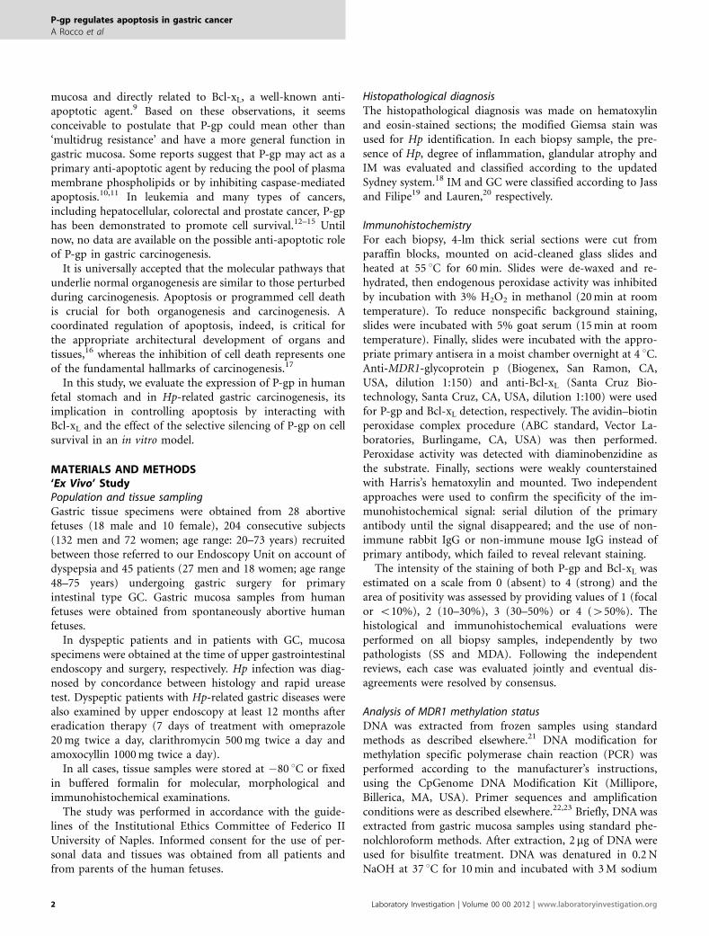
Figure 1 Immunohistochemical expression of P-gp and Bcl-xL in gastric mucosa of human fetuses. Top panel shows the representative immunopattern
of P-gp in gastric mucosa at 20th week (a), 24th week (b) and 38th week of gestational age (c). Note the intense immunostaining in both superficial
and glandular epithelium. Bottom panel shows Bcl-xL immunolocalization in gastric mucosa at 20th week (e), 24th week (f) and 38th week of gestational
age (g). Note that the distribution of Bcl-xL in gastric epithelium is similar even if less intense of that of P-gp (ABC technique, original magnification b,
f � 100; a, c, e, g � 200; details � 400). Negative controls (d, h) obtained by using nonimmune rabbit IgG instead of primary antibodies (original magnification
d � 200, h � 100).
Figure 2 Immunohistochemical expression of P-gp and Bcl-xL in adult gastric mucosa. Top panel shows the immunohistochemical localization of P-gp in a
case of Hp-negative normal mucosa (a), Hp-positive non-metaplastic atrophic gastritis (b), Hp-positive metaplastic atrophic gastritis (c), intestinal type GC
(d). Note the absence of the immunohistochemical signal in normal mucosa and the progressive increase in the intensity of immunostaining from Hp-
positive non-metaplastic atrophic gastritis to GC. Bottom panel shows Bcl-xL immunolocalization in a case of Hp-negative normal mucosa (f), Hp-positive
non-metaplastic atrophic gastritis (g), Hp-positive metaplastic atrophic gastritis (h) and intestinal type GC (i). Note that the cellular distribution of Bcl-xL
parallels that of P-gp (ABC technique, original magnification b, c, g � 100; d � 150; a, f, h, i � 200). Negative controls (e, j) obtained by using nonimmune
rabbit IgG instead of primary antibodies (original magnification d � 150, h � 200).
P-gp regulates apoptosis in gastric cancer
A Rocco et al
4 Laboratory Investigation | Volume 00 00 2012 | www.laboratoryinvestigation.org
P-gp regulates apoptosis in gastric cancer
A Rocco et al
www.laboratoryinvestigation.org | Laboratory Investigation | Volume 00 00 2012 5

Scientific) in serum-free medium and incubated at 37 1C in5% CO2 for 48 h.
Flow cytometry analysisAfter transfection, AGS and MKN 28 cells (control, scrambleand siRNA transfected) were analyzed baseline and after 6-hincubation with 3% H2O2. Cells were washed with bindingbuffer and suspended in 100 ml Annexin V-binding buffer.Cells were then incubated with 5 ml Annexin V-FITC and 5 mlpropidium iodide for 15 min at room temperature away fromlight before addition of 400 ml Annexin V-binding buffer.The cell pellets were analyzed using a FACS Vantage flowcytometer (BD Biosciences, San Jose, CA, USA).
Statistical analysisData were analyzed using the SPSS package for Windows(version 17.0). The categorical variables were analyzed usingthe w2 test or Fisher’s exact test. The correlation betweenthese variables was assessed using Spearman’s correlationcoefficient. Stepwise binary logistic regression was used toidentify the association between the variables. P-values of0.05 and 0.1 were selected as cutoff points to, respectively,enter and exit the stepwise procedure. Differences wereconsidered to be significant at the 5% level.
RESULTS‘Ex Vivo’ StudyP-gp and Bcl-xL expression in gastric mucosa of human fetusesAccording to gestational age, 28 spontaneously abortivehuman fetuses were divided in three group: (i) 9 cases from18th to 20th week; (ii) 15 cases from 21th to 36th week; and(iii) 4 cases from 37th to 38th week. In the gastric samples,P-gp was strongly expressed in the cytoplasm of all cellsof both superficial and glandular epithelium with an
immunopositivity that paralleled the epithelial differentiationduring stomach development (Figures 1a–c). The patternof immunohistochemical expression of Bcl-xL was similar tothat of P-gp being localized mainly in the cytoplasm ofgastric epithelial cells (Figures 1e–g).
P-gp and Bcl-xL expression in Hp-related gastric carcinogenesisPatients with dyspeptic symptoms were classified accordingto endoscopic and histologic findings in 66 Hp-negative and138 Hp-positive cases. In the Hp-negative group, all subjectspresented a normal gastric mucosa with minimal infiltrationof lymphocytes and monocytes in the lamina propria. In theHp-positive group, gastric atrophy was detected in 42/138cases; it was mild in 28, moderate in 10 and severe in 6 cases.Complete, type I IM, always associated with atrophy, wasfound in 28/138 cases.
In Hp-negative gastric mucosa P-gp protein wasundetectable in all cases, whereas it was expressed in 27%(30/110) cases of Hp-positive CG without IM and in 100%(28/28) of Hp-positive chronic atrophic gastritis with IM.In non-metaplastic CG, P-gp immunostaining rangedfrom mild-to-moderate and was distributed in the peri-nuclear area of the cytoplasm; conversely, in IM areas, P-gpshowed an intense and diffuse cytoplasmic immunopositivity(Figures 2a–c).
Figure 3 Expression of P-gp in gastric tissues by western blot analysis. A
representative autoradiograph performed on tissue lysates of Hp-negative
subjects, Hp-positive patients, GC patients and human fetuses. GAPDH
expression was used as internal control. The histogram at bottom reports
the mean densitometric values of P-gp/GAPDH ratio obtained from three
independent experiments. *One-way ANOVA: Po0.0001.
Figure 4 Methylation specific polymerase chain reaction results for MDR1
gene. DNA from: (a) fetal gastric mucosa, (b) gastritis biopsies and (c) GCs.
Arrow in the panel b indicates the positive control from colon cancer tissue
showing the presence of methylated promoter of P-gp-MDR1. Arrow in the
panel c indicates the GC sample scored as methylated. M, methylated; U,
unmethylated.
P-gp regulates apoptosis in gastric cancer
A Rocco et al
6 Laboratory Investigation | Volume 00 00 2012 | www.laboratoryinvestigation.org

By western blot analysis, the levels of P-gp were sig-nificantly higher in Hp-positive than in Hp-negative gastricmucosa (Po0.05) (Figure 3).
Bcl-xL was undetectable in gastric mucosa samples ofHp-negative subjects (Figure 2f), and expressed in 20/110(18%) patients with Hp-positive CG without IM and in24/28 (86%) of those with Hp-positive CG associated withIM (Figures 2g, h).
By using Sperman’s correlation, the levels of immuno-staining of P-gp were positively correlated with Hpinfection (Po0.05), atrophy (P¼ 0.02), IM (P¼ 0.001) andBcl-xL (P¼ 0.001) expression. However, when the data wereanalyzed by logistic regression, only Hp infection (Po0.05)and Bcl-xL expression (Po0.01) were significantly associatedwith P-gp expression.
Of the 138 patients who underwent Hp eradicationtherapy, 28 dropped-out. In the remaining 110 patients, Hpinfection was successfully eradicated in 88 cases. In this groupof patients, Pg-p and Bcl-xL were overexpressed only in thepresence of IM (16 subjects), whereas they were undetectablein the remaining 72 cases.
P-gp and Bcl-xL expression in GCIntestinal type GCs were 7 well, 26 moderately and 12 poorlydifferentiated. Twelve were confined to the mucosa orsubmucosa and 33 locally advanced. A strong P-gp immuno-staining was found in 88% (40/45) of the cases, stronger thanin CG and uniformly distributed in the cytoplasm ofepithelial cells (Figure 2d). Bcl-xL expression was found in75% (34/45) of the cases (Figure 2i).
By western blot analysis, the molecular expression ofP-gp was significantly higher in GC than Hp-positive CG(Po0.01).
In GC, the expression of P-gp significantly correlated withthe high grade of differentiation (r¼ 0.50, 95% CI: 0.66–0.28;Po0.0001) and Bcl-xL expression (r¼ 0.38, 95% CI: 0.57–0.15; Po0.001).
Analysis of MDR1 methylation statusThe methylation status of P-gp-MDR1 gene was analyzed in10 fetuses (gestational age 18th–20th week: 4 cases; 24th–36thweek: 4 cases; 38th week: 2 cases), 8 Hp-negative, 10Hp-positive and 8 intestinal type GCs. The P-gp-MDR1
Figure 5 Immunofluorescence localization of P-gp and Bcl-xL in AGS cells. (a–c) Cells were double stained with anti-P-gp and anti-Bcl-xL antibodies. Both
proteins stained a cytoplasmic membranous compartment. The merge evidenced a high degree of colocalization. (d–f) Cells were double stained with anti-
P-gp antibodies and the mitochondrion-selective probe MitoTracker Red. The merge in f shows that P-gp staining superimposes with the MitoTracker
staining. Bar¼ 15 mm. All the experiments were performed in triplicate.
P-gp regulates apoptosis in gastric cancer
A Rocco et al
www.laboratoryinvestigation.org | Laboratory Investigation | Volume 00 00 2012 7

promoter region was unmethylated in the fetal mucosa, inCG and all GCs but one (Figure 4).
‘In Vitro’ StudyLocalization of P-gp and Bcl-xL by confocal microscopyIn both AGS and MKN-28 cell lines, P-gp staining waslocalized mainly in the perinuclear region, at the level of
membranous organelles, where it colocalized with Bcl-xL
(Figure 5). By staining the cells with both anti-Pgp antibodyand MitoTracker Red, a specific mitochondrial dye, asignificant fraction of P-gp was found localized at level of themitochondria (Figure 5).
Expression of P-gp in the mitochondrial fractionTo confirm the presence of P-gp on the mitochondria, wefractionated total cell lysates of AGS and MKN-28 cells andisolated the mitochondrial fraction. As shown in Figure 6,the mitochondrial fraction contains a substantial amount ofP-gp.
Immunoprecipitation and immunoblot analysisImmunoprecipitation of total proteins extracted from AGSand MKN-28 cells with anti-P-gp or anti-Bcl-xL antibodiesshowed the presence of Pg-p in Bcl-xL immunocomplexes, aswell as the presence of Bcl-xL in P-gp immunocomplexes(Figure 7, panel a). Western blot on whole cell lysates con-
Figure 7 Immunoprecipitation of P-gp and Bcl xL. The figure shows the interaction between P-gp and Bcl-xL proteins in human GC cell lines, AGS and MKN-
28, (panels a, b) and hepatocellular carcinoma (Hep-G2) cell line (panels c, d). Extracts derived from AGS, MKN-28 and Hep-G2 cells were subjected to
immunoprecipitation with Bcl-xL or P-gp antibody. The immunoprecipitates (panels a, c) and whole cell lysates (panels b, d) were resolved on an 10% SDS-
PAGE and immunoblotted with anti-Pgp or anti- Bcl-xL antibody. Negative controls were obtained by using nonspecific mouse IgG. The experiments were
performed in triplicate.
Figure 6 Western blot analyses of P-gp in whole cell lysate, mitochondrial
and cytosolic fractions isolated from AGS and MKN-28 cells. Figure
representative of three separate experiments. Each experiment was performed
in triplicate. Note the substantial amount of P-gp in the mitochondrial fraction.
P-gp regulates apoptosis in gastric cancer
A Rocco et al
8 Laboratory Investigation | Volume 00 00 2012 | www.laboratoryinvestigation.org

firmed the expression of P-gp and Bcl-xL in GC celllines (Figure 7, panel b). These data indicate the physicalinteraction of the two proteins in both GC cell lines.The interaction between P-gp and Bcl-xL was confirmed byimmunoprecipitation and immunoblot also in Hep-G2 celllysates (Figure 7, panels c and d). Stress-induced P-gp over-expression in AGS cell line resulted in an increased inter-action between the two proteins (Figure 8).
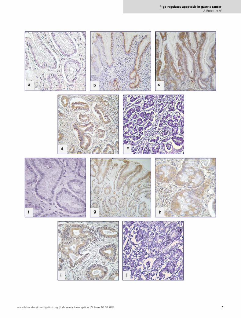
Inhibition of MDR1 expression by siRNA increases cellsusceptibility to pro-apoptotic stimuliTo test the role of P-gp in the control of apoptosis, weperformed experiments of stress-induced apoptosis in AGSand MKN-28 cell lines in which the expression of P-gp wasdownregulated by transfection with specific siRNA targetingP-gp. The expression of mitochondrial P-gp was abolishedby MDR1-targeted siRNA in both cell lines (Figure 9). Thiseffect was specific, because nonspecific control or scrambledsiRNAs did not affect P-gp levels.
Cells, in which P-gp expression was knocked down, weresubjected to oxidative stress by hydrogen peroxide and the
fraction of apoptotic, necrotic or viable cells was measuredby FACS analysis. The selective silencing of P-gp by siRNAresults in a significant rise of the apoptotic index in both GCcell lines exposed to oxidative stress (AGS: from 22.6±0.6 to90.3±1.2%; Po0.0001 and MKN-28: from 7.3±0.9 to56.2±1.1%; Po0.0001) (Table 1 and Figure 9).
DISCUSSIONIn this study, we found that P-gp is expressed in the fetalgastric mucosa, undetectable in adult normal gastric mucosaand re-expressed in a subset of Hp-related CG and in GC,thus behaving as a gastric oncofetal protein. Tissue differen-tiation during development is largely under epigeneticcontrol. Considerable experimental evidence indicates thatepigenetic alteration, including DNA methylation, is impor-tant in tumor initiation and progression, as well as thatreactivation of developmentally regulated genes may resultfrom the aberrant hypomethylation observed in many tumortypes.24–26 In our study, the expression of P-gp in gastricmucosa seems to be dependent on the methylation status ofits promoter, as typically reported for oncofetal proteins.
Table 1 Mean percentage of necrotic, apoptotic and viable AGS and MKN-28 cells baseline and after H2O2-induced stress beforeand after knock-down of P-gp by siRNA
Basal Basal+H2O2 Scramble Scramble+H2O2 Silenced Silenced+H2O2
Mean % ±s.d. Mean % ±s.d. Mean % ±s.d. Mean % ±s.d. Mean % ±s.d. Mean % ±s.d.
AGS
Necrosis 2.3±0.2 14.5±0.7 2.6±0.4 15.2±0.6 13.6±0.8 7.9±0.3
Apotosis 6.4±0.5 22.6±0.6 10.1±0.4 23.1±0.5 9.4±0.5 90.3±1.2
Viable 88.3±0.8 62.9±1.1 87.3±0.8 61.7±0.5 80±0.9 1.8±0.4
MKN-28
Necrosis 9.2±0.9 21.7±1.3 8.5±0.3 23±0.9 14±1.5 11±0.3
Apotosis 3.3±0.6 7.3±0.9 3.6±0.2 7.6±0.7 9.8±0.4 56.2±1.1
Viable 87.5±0.8 71±0.8 87.9±1.9 69.4±0.8 76.2±1.1 32.9±1.0
Figure 8 Immunoprecipitation of P-gp and Bcl xL in AGS cell lines after stress-induced P-gp overexpression. The figure shows the basal expression of P-gp
and Bcl-xL protein in AGS cells (panel a). Panel b shows the interaction between P-gp, overexpressed after H2O2-induced stress, and Bcl-xL. Note the
increased interaction between the proteins.
P-gp regulates apoptosis in gastric cancer
A Rocco et al
www.laboratoryinvestigation.org | Laboratory Investigation | Volume 00 00 2012 9
A positive relation has been reported between the ex-pression of P-gp and survivin, a member of the inhibitorsof apoptosis protein family, capable of blocking apoptosisthrough caspase-dependent or caspase-independent path-ways.27 In our series of cases, the co-expression of P-gp andBcl-xL suggests that, during stomach development, P-gp mayhave a physiological role, that is, to warrant survival ofspecific cellular populations, which characterizes the histo-logical architecture and function of the mature organ.28,29 In
contrast, in the adult, the co-expression of P-gp and Bcl-xL,during Hp-related gastric carcinogenesis, could result inprolonged survival of abnormal cells in which the sequentialaccumulation of molecular alterations can ultimately leadto tumor promotion and progression. Hp is the mostimportant single factor responsible for inflammatoryand neoplastic gastric diseases.30 The exposure of gastricepithelial cells to Hp results in a complex inflammatory reac-tion with the generation of reactive oxygen species and an
Figure 9 Transfection of siRNA against MDR1 and flow cytometry analysis. Selective knock down of P-gp amplifies stress-induced apoptosis in GC cell
lines. MKN-28 and AGS cell lines were transfected with scrambled or P-gp-targeted siRNA. After 48 h, the cell were collected and divided in two
aliquots: the first was lysed and total extracts blotted with anti-P-gp antibodies (a). Another aliquot was exposed to 3% H2O2 for 6 h and analyzed by FACS
(b). The early apoptotic cells (Annexin-FITC positive and propidium iodide (PI) negative) were located in the lower right quadrant. The late apoptotic
or necrotic cells (Annexin-FITC positive and PI positive) were located in the upper right quadrant. Healthy cells (negative for both probes) were
located in the lower left quadrant. The results are expressed as percentage of positively stained cells in total cells. The panel c shows a quantitative
representation of the targeting experiments. All experiments were performed in triplicate. *Po0.0001; #Po0.0001.
P-gp regulates apoptosis in gastric cancer
A Rocco et al
10 Laboratory Investigation | Volume 00 00 2012 | www.laboratoryinvestigation.org

increased level of nitric oxide synthase that exposes the cellsto a significant oxidative stress.31 The cells with high levels ofP-gp and Bcl-xL could escape stress-imposed death andaccumulate DNA damage ultimately leading to cell trans-formation and cancer development. Opposite, the eradica-tion of the Hp infection with the concomitant resolution ofgastric inflammation may lead to the downregulation oreven suppression of P-gp expression. In our study, indeed,after the successful eradication of Hp infection, P-gp wasundetectable in all cases but persisted only in IM areas,where, it likely represents only an epiphenomenon of the‘intestinal phenotype’.
In support to the procarcinogenetic role of P-gp, it hasbeen reported that a downregulation of the protein expres-sion may be associated with reduced development of cancer.Kankesan et al32 reported that PSC833 (PSC), a potentinhibitor of P-gp, inhibited the development of N-methyl-N-nitrosourea-induced mammary cancer in female Sprague–Dawley rats and the growth of 1,2-dimethylhydrazine-induced liver cancer in rats. Mochida et al33 introducingdisrupted alleles of the murine P-gp gene, MDR1a, into Apc(Min/þ ) mice found that the number of polyps and cancerswas markedly decreased in the MDR1a (�/�), Apc (Min/þ )mutant mice, suggesting that P-gp has an active role duringintestinal tumorigenesis.34
To elucidate the mechanisms through which P-gp elicits itsanti-apoptotic function, we set an in vitro system, by usingtwo GC cell lines, AGS and MKN-28, well and poorly dif-ferentiated, respectively. By means of immunofluorescenceconfocal microscopy, we found that P-gp and Bcl-xL colo-calized at the mitochondria level in both cell lines. The mito-chondrial localization of P-gp has been previously shownin human hepatocarcinoma, hepatoma, breast cancer andmyeloid leukemia cell lines.35–38 Mitochondria are the sitewhere converge many of the factors, including members ofBcl-2 family, which elicit apoptosis.39 This subcellular dis-tribution suggests that P-gp may participate in the regulationof apoptosis presumably via the mitochondrial pathway. Inthis study, by co-immunoprecipitation experiments, for thefirst time we demonstrated that in GC cell lines, P-gp phy-sically interacts with Bcl-xL. Interestingly, the interactionbetween the two proteins was even more marked after stress-induced P-gp overexpression. These new findings furthersupport the hypothesis that P-gp may have a fundamentalrole in controlling apoptosis. The occurrence of P-gp–Bcl-xL
interaction also in hepatocarcinoma cell line Hep-G2 in-dicates that this new relationship in not distinctive of GC.Finally, the selective silencing of P-gp by siRNA resulted in asignificant rise of the cell apoptotic index in both GC celllines exposed to oxidative stress, thus confirming the im-plication of P-gp in controlling apoptosis.
In conclusion, P-gp behaves as a gastric oncofetal proteinthat by acting as anti-apoptotic agent, confers a strongsurvival advantage to GC cells by cross-talking with Bcl-xL.The expression of P-gp in pre-neoplastic gastric mucosa
could indicate a functional implication of this protein sincethe early phases of Hp-related gastric carcinogenesis andmight represent a useful biomarker to select patients at majorrisk of a progressive gastric disease.
ACKNOWLEDGEMENTS
This research was supported by grants from the Italian Ministry of University
and Research (MURST) to the Department of Clinical and Experimental
Medicine, Gastroenterology Unit, ‘Federico II’ University of Naples. We are
grateful to Enrico Vittorio Avvedimento for helpful comments on the
manuscript.
DISCLOSURE/CONFLICT OF INTEREST
The authors declare no conflict of interest.
1. Li Y, Yuan H, Yang K, et al. The structure and functions ofP-glycoprotein. Curr Med Chem 2010;17:786–800.
2. Mizutani T, Masuda M, Nakai E, et al. Genuine functions ofP-glycoprotein (ABCB1). Curr Drug Metab 2008;9:167–174.
3. Goda K, Bacso Z, Szabo G. Multidrug resistance through the spectacleof P-glycoprotein. Curr Cancer Drug Targets 2009;9:281–297.
4. Wang B, Li XQ, Ma X, et al. Immunohistochemical expression andclinical significance of P-glycoprotein in previously untreatedextranodal NK/T-cell lymphoma, nasal type. Am J Hematol 2008;83:795–799.
5. Mouly S, Paine MF. P-glycoprotein increases from proximal to distalregions of human small intestine. Pharm Res 2003;20:1595–1599.
6. Zhang D, Fan D. Multidrug resistance in gastric cancer: recent researchadvances and ongoing therapeutic challenges. Expert Rev AnticancerTher 2007;7:1369–1378.
7. Xu HW, Xu L, Hao JH, et al. Expression of P-glycoprotein and multidrugresistance-associated protein is associated with multidrug resistance ingastric cancer. J Int Med Res 2010;38:34–42.
8. Nardone G, Rocco A, Vaira D, et al. Expression of COX-2, mPGE-synthase1, MDR-1 (P-gp), and Bcl-xL: a molecular pathway of H pylori-related gastric carcinogenesis. J Pathol 2004;202:305–312.
9. Chipuk JE, Fisher JC, Dillon CP, et al. Mechanism of apoptosis inductionby inhibition of the anti-apoptotic BCL-2 proteins. Proc Natl Acad SciUSA 2008;105:20327–20332.
10. Tainton KM, Smyth MJ, Jackson JT, et al. Mutational analysisof P-glycoprotein: suppression of caspase activation in the absenceof ATP-dependent drug efflux. Cell Death Differ 2004;11:1028–1037.
11. Mantovani I, Cappellini A, Tazzari PL, et al. Caspase-dependentcleavage of 170-kDa Pglycoprotein during apoptosis of humanT-lymphoblastoid CEM cells. J Cell Physiol 2006;207:836–844.
12. Guenova ML, Balatzenko GN, Nikolova VR, et al. An anti-apoptoticpattern correlates with multidrug resistance in acute myeloidleukemiapatients: a comparative study of active caspase-3, cleaved PARPs, Bcl-2,Survivin and MDR1 gene. Hematology 2010;15:135–143.
13. Takanishi K, Miyazaki M, Ohtsuka M, et al. Inverse relationship betweenP-glycoprotein expression and its proliferative activity in hepato-cellular carcinoma. Oncology 1997;54:231–237.
14. Potocnik U, Glavac D, Dean M. Common germline MDR1/ABCB1functional polymorphisms and haplotypes modify susceptibility tocolorectal cancers with high microsatellite instability. Cancer GenetCytogenet 2008;183:28–34.
15. Van Brussel JP, Jan Van Steenbrugge G, Van Krimpen C, et al.Expression of multidrug resistance-related proteins and proliferativeactivity is increased in advanced clinical prostate cancer. J Urol 2001;165:130–135.
16. Hanahan D, Weinberg RA. The hallmarks of cancer. Cell 2000;100:57–70.
17. Danial NN, Korsmeyer SJ. Cell death: critical control points. Cell2004;116:205–219.
18. Dixon MF, Genta RM, Yardley JH, et al. Classification and grading ofgastritis. Am J Surg Pathol 1996;20:1161–1181.
19. Jass JR, Filipe MI. The mucin profiles of normal gastric mucosa,intestinal metaplasia and its variants and gastric carcinoma. HistochemJ 1981;13:931–939.
P-gp regulates apoptosis in gastric cancer
A Rocco et al
www.laboratoryinvestigation.org | Laboratory Investigation | Volume 00 00 2012 11
20. Lauren P. The two histological main types of gastric carcinoma: diffuseand so-called intestinal type carcinoma. Acta Path Microbiol Scand1965;64:31–49.
21. Ottini L, Falchetti M, Saieva C, et al. MRE11 expression is impairedin gastric cancer with microsatellite instability. Carcinogenesis2004;25:2337–2343.
22. Garcia-Manero G, Daniel J, Smith TL, et al. DNA methylation of multiplepromoter associated CpG islands in adult acute lymphocytic leukemia.Clin Cancer Res 2002;8:2217–2224.
23. van Rijnsoever M, Grieu F, Elsaleh H, et al. Characterization of colorectalcancers showing hypermethylation at multiple CpG islands. Gut2002;51:797–802.
24. Hu M, Shivdasani RA. Overlapping gene expression in fetal mouseintestine development and human colorectal cancer. Cancer Res2005;65:8715–8722.
25. Lum L, Beachy PA. The Hedgehog response network: sensors, switches,and routers. Science 2004;304:1755–1759.
26. Vogelstein B, Kinzler KW. Cancer genes and the pathways they control.Nat Med 2004;10:789–799.
27. Liu F, Liu S, He S, et al. Survivin transcription is associated with P-glycoprotein/MDR1 overexpression in the multidrug resistance ofMCF-7 breast cancer cells. Oncol Rep 2010;23:1469–1475.
28. van Kalken CK, Giaccone G, van der Valk P, et al. Multidrug resistancegene (P-glycoprotein) expression in the human fetus. Am J Pathol1992;141:1063–1072.
29. Menard D, Arsenault P. Cell proliferation in developing humanstomach. Anat Embryol (Berl) 1990;182:509–516.
30. The EUROGAST Study Group. An international association betweenHelicobacter pylori infection and gastric cancer. Lancet 1993;341:1359–1362.
31. Nardone G, Rocco A, Malfertheiner P. Review article: helicobacter pyloriand molecular events in precancerous gastric lesions. AlimentPharmacol Ther 2004;20:261–270.
32. Kankesan J, Vanama R, Yusuf A, et al. Effect of PSC 833, aninhibitor of P-glycoprotein on Nmethyl-N-nitrosourea inducedmammary carcinogenesis in rats. Carcinogenesis 2004;25:425–430.
33. Kankesan J, Yusuf A, Laconi E, et al. Effect of PSC 833, an inhibitor of P-glycoprotein, on 1,2- dimethylhydrazine-induced liver carcinogenesisin rats. Carcinogenesis 2003;24:1977–1984.
34. Mochida Y, Taguchi K, Taniguchi S, et al. The role of P-glycoproteinin intestinal tumorigenesis: disruption of mdr1a suppressespolyp formation in Apc(Min/+) mice. Carcinogenesis 2003;24:1219–1224.
35. Solazzo M, Fantappie O, Lasagna N, et al. P-g localization inmitochondria and its functional characterization in multiple drug-resistant cell lines. Exp Cell Res 2006;312:4070–4078.
36. Shen Y, Chu Y, Yang Y, et al. Mitochondrial localization ofP-glycoprotein in the human breast cancer cell line MCF-7/ADM and its functional characterization. Oncol Rep 2012;27:1535–1540.
37. Ling X, He Y, Zhang G, et al. Increased P-glycoprotein expression inmitochondria is related to acquired multidrug resistance in humanhepatoma cells depleted of mitochondrial DNA. Int J Oncol2012;40:109–118.
38. Munteanu E, Verdier M, Grandjean-Forestier F, et al. Mitochondriallocalization and activity of P-glycoprotein in doxorubicin-re sistantK562 cells. Biochem Pharmacol 2006;71:1162–1174.
39. Green DR, Kroemer G. The pathophysiology of mitochondrial celldeath. Science 2004;305:626–629.
P-gp regulates apoptosis in gastric cancer
A Rocco et al
12 Laboratory Investigation | Volume 00 00 2012 | www.laboratoryinvestigation.org