Mcm2, but Not RPA, Is a Component of the Mammalian Early ...
14
The Rockefeller University Press, 0021-9525/99/08/709/14 $5.00 The Journal of Cell Biology, Volume 146, Number 4, August 23, 1999 709–722 http://www.jcb.org 709 Mcm2, but Not RPA, Is a Component of the Mammalian Early G1-Phase Prereplication Complex Daniela S. Dimitrova,* Ivan T. Todorov, ‡ Thomas Melendy, § and David M. Gilbert* *Department of Biochemistry and Molecular Biology, S.U.N.Y. Health Science Center, Syracuse, New York 13210; ‡ Desmos, Inc., San Diego, California 92121; and § Department of Microbiology, S.U.N.Y. at Buffalo, School of Medicine and Biomedical Sciences, Buffalo, New York 14214 Abstract. Previous experiments in Xenopus egg ex- tracts identified what appeared to be two indepen- dently assembled prereplication complexes (pre-RCs) for DNA replication: the stepwise assembly of ORC, Cdc6, and Mcm onto chromatin, and the FFA-1–medi- ated recruitment of RPA into foci on chromatin. We have investigated whether both of these pre-RCs can be detected in Chinese hamster ovary (CHO) cells. Early- and late-replicating chromosomal domains were pulse-labeled with halogenated nucleotides and prela- beled cells were synchronized at various times during the following G1-phase. The recruitment of Mcm2 and RPA to these domains was examined in relation to the formation of a nuclear envelope, specification of the di- hydrofolate reductase (DHFR) replication origin and entry into S-phase. Mcm2 was loaded gradually and cu- mulatively onto both early- and late-replicating chro- matin from late telophase throughout G1-phase. Dur- ing S-phase, detectable Mcm2 was rapidly excluded from PCNA-containing active replication forks. By contrast, detergent-resistant RPA foci were undetect- able until the onset of S-phase, when RPA joined only the earliest-firing replicons. During S-phase, RPA was present with PCNA specifically at active replication forks. Together, our data are consistent with a role for Mcm proteins, but not RPA, in the formation of mam- malian pre-RCs during early G1-phase. Key words: pre-RC • Mcm • RPA • cell cycle • DNA replication domains I N the past several years, much progress has been made in the identification of proteins that assemble onto chromatin to mediate the once-per-cell-cycle replica- tion of eukaryotic chromosomes (Gilbert, 1998). Most of these gene products were originally identified in S. cerevi- siae and homologues were subsequently found in higher eukaryotes (Dutta and Bell, 1997; Kearsey and Labib, 1998; Tye, 1999). Studies with Xenopus egg extracts have allowed a preliminary biochemical evaluation of the roles of these gene products in replication (Rowles and Blow, 1997). When Xenopus sperm chromatin is incubated in a Xenopus egg extract, two independent complexes of es- sential replication proteins are assembled before the initi- ation of replication. Both of these complexes have been termed prereplication complexes or prereplication centers (pre-RC) 1 . The first type of pre-RCs to be identified (Ada- chi and Laemmli, 1992, 1994) contained replication pro- tein A (RPA), a complex of three polypeptides that consti- tutes the single-stranded DNA-binding protein (SSB) required for the replication of eukaryotic DNA (Wold, 1997). Previous studies in viral replication systems have shown that stabilization of unwound origin DNA by RPA is one of the earliest steps of the initiation reaction (Tsurimoto et al., 1989). The Xenopus laevis RPA homo- logue (xlRPA) was purified as a stable complex of three protein subunits and shown to be essential for replication of sperm chromatin in Xenopus egg extract (Fang and Newport, 1993; Adachi and Laemmli, 1994). xlRPA was found to be rapidly organized into discrete foci on sperm chromatin (Adachi and Laemmli, 1992, 1994; Yan and Newport, 1995a) in a process that requires a partially purified activity termed focus forming activity (FFA-1) and one additional cytosolic fraction (Yan and Newport, Address correspondence to David M. Gilbert, Department of Biochemis- try and Molecular Biology, S.U.N.Y. Health Science Center, 750 East Ad- ams Street, Syracuse, NY 13210. Tel.: (315) 464-8723. Fax: (315) 464-8750. E-mail: [email protected] 1. Abbreviations used in this paper: BrdU, bromodeoxyuridine; CldU, 5-chloro-29-deoxyuridine; DHFR, dihydrofolate reductase; FFA, focus forming activity; IdU, 5-iodo-29-deoxyuridine; ODP, origin decision point; ORC, origin recognition complex; PCNA, proliferating cell nuclear anti- gen; pre-RCs, prereplication complexes or centers; RPA, replication pro- tein A; SSB, single-stranded DNA-binding protein; TxRed, Texas Red; xlRPA, Xenopus laevis RPA homologue.
-
Upload
khangminh22 -
Category
Documents
-
view
3 -
download
0
Transcript of Mcm2, but Not RPA, Is a Component of the Mammalian Early ...
The Rockefeller University Press, 0021-9525/99/08/709/14 $5.00The Journal of Cell Biology, Volume 146, Number 4, August 23, 1999 709–722http://www.jcb.org 709
Mcm2, but Not RPA, Is a Component of the Mammalian Early G1-Phase Prereplication Complex
Daniela S. Dimitrova,* Ivan T. Todorov,
‡
Thomas Melendy,
§
and David M. Gilbert*
*Department of Biochemistry and Molecular Biology, S.U.N.Y. Health Science Center, Syracuse, New York 13210;
‡
Desmos, Inc., San Diego, California 92121; and
§
Department of Microbiology, S.U.N.Y. at Buffalo, School of Medicine and Biomedical Sciences, Buffalo, New York 14214
Abstract.
Previous experiments in
Xenopus
egg ex-tracts identified what appeared to be two indepen-dently assembled prereplication complexes (pre-RCs) for DNA replication: the stepwise assembly of ORC, Cdc6, and Mcm onto chromatin, and the FFA-1–medi-ated recruitment of RPA into foci on chromatin. We have investigated whether both of these pre-RCs can be detected in Chinese hamster ovary (CHO) cells. Early- and late-replicating chromosomal domains were pulse-labeled with halogenated nucleotides and prela-beled cells were synchronized at various times during the following G1-phase. The recruitment of Mcm2 and RPA to these domains was examined in relation to the formation of a nuclear envelope, specification of the di-hydrofolate reductase (DHFR) replication origin and
entry into S-phase. Mcm2 was loaded gradually and cu-mulatively onto both early- and late-replicating chro-matin from late telophase throughout G1-phase. Dur-ing S-phase, detectable Mcm2 was rapidly excluded from PCNA-containing active replication forks. By contrast, detergent-resistant RPA foci were undetect-able until the onset of S-phase, when RPA joined only the earliest-firing replicons. During S-phase, RPA was present with PCNA specifically at active replication forks. Together, our data are consistent with a role for Mcm proteins, but not RPA, in the formation of mam-malian pre-RCs during early G1-phase.
Key words: pre-RC • Mcm • RPA • cell cycle • DNA replication domains
I
N
the past several years, much progress has been madein the identification of proteins that assemble ontochromatin to mediate the once-per-cell-cycle replica-
tion of eukaryotic chromosomes (Gilbert, 1998). Most ofthese gene products were originally identified in
S
.
cerevi-siae
and homologues were subsequently found in highereukaryotes (Dutta and Bell, 1997; Kearsey and Labib,1998; Tye, 1999). Studies with
Xenopus
egg extracts haveallowed a preliminary biochemical evaluation of the rolesof these gene products in replication (Rowles and Blow,1997). When
Xenopus
sperm chromatin is incubated in a
Xenopus
egg extract, two independent complexes of es-sential replication proteins are assembled before the initi-ation of replication. Both of these complexes have beentermed prereplication complexes or prereplication centers(pre-RC)
1
. The first type of pre-RCs to be identified (Ada-
chi and Laemmli, 1992, 1994) contained replication pro-tein A (RPA), a complex of three polypeptides that consti-tutes the single-stranded DNA-binding protein (SSB)required for the replication of eukaryotic DNA (Wold,1997). Previous studies in viral replication systems haveshown that stabilization of unwound origin DNA by RPAis one of the earliest steps of the initiation reaction(Tsurimoto et al., 1989). The
Xenopus laevis
RPA homo-logue (xlRPA) was purified as a stable complex of threeprotein subunits and shown to be essential for replicationof sperm chromatin in
Xenopus
egg extract (Fang andNewport, 1993; Adachi and Laemmli, 1994). xlRPA wasfound to be rapidly organized into discrete foci on spermchromatin (Adachi and Laemmli, 1992, 1994; Yan andNewport, 1995a) in a process that requires a partiallypurified activity termed focus forming activity (FFA-1)and one additional cytosolic fraction (Yan and Newport,
Address correspondence to David M. Gilbert, Department of Biochemis-try and Molecular Biology, S.U.N.Y. Health Science Center, 750 East Ad-ams Street, Syracuse, NY 13210. Tel.: (315) 464-8723. Fax: (315) 464-8750.E-mail: [email protected]
1.
Abbreviations used in this paper:
BrdU, bromodeoxyuridine; CldU,
5-chloro-2
9
-deoxyuridine; DHFR, dihydrofolate reductase; FFA, focus
forming activity; IdU, 5-iodo-2
9
-deoxyuridine; ODP, origin decision point;ORC, origin recognition complex; PCNA, proliferating cell nuclear anti-gen; pre-RCs, prereplication complexes or centers; RPA, replication pro-
tein A; SSB, single-stranded DNA-binding protein; TxRed, Texas Red;xlRPA,
Xenopus laevis
RPA homologue.
The Journal of Cell Biology, Volume 146, 1999 710
1995b). These RPA foci were shown to form on chromatinbefore DNA unwinding, and under conditions in whichthe initiation of replication was prevented. Hence, theyhave been referred to as prereplication centers (pre-RCs).
Subsequent to the identification of RPA pre-RCs, the
Xenopus
homologues of the
S
.
cerevisiae
origin recogni-tion complex (ORC), Cdc6, and Mcm2-7 proteins wereidentified, shown to be essential for replication in
Xenopus
egg extracts, and found to rapidly associate with spermchromatin introduced into
Xenopus
egg extract (Kubotaet al., 1995; Madine et al., 1995; Carpenter et al., 1996;Chong et al., 1995; Coleman et al., 1996). The sequentialorder of association of these proteins was shown to beORC, Cdc6, and Mcm, with the binding of each dependentupon the prior association of the upstream proteins (Cole-man et al., 1996; Romanowski et al., 1996b). The
S
.
cerevi-siae
counterparts of these proteins were subsequentlyshown to assemble in this same order onto the specific se-quences that function as origins of replication (Aparicioet al., 1997; Tanaka et al., 1997). Since the ORC-Cdc6-Mcm pathway appears to be conserved in eukaryotes, isrequired for replication, and is completed before the initi-ation step, this multiprotein complex is also referred to asa prereplication complex (pre-RC). However, the rela-tionship between these pre-RCs and the RPA-containingpre-RCs is unclear.
In mammalian cells, Mcm’s have been shown to betightly bound to chromatin during the second half of G1-phase and to subsequently be displaced from replicatingchromatin during S-phase (Todorov et al., 1995; Krude et al.,1996), consistent with a role for Mcm proteins in DNAreplication, most likely at a step before initiation. How-ever, these studies did not examine the association of Mcmproteins with chromatin immediately after exit from mito-sis and during the early stages of G1-phase to determineprecisely when Mcm’s are recruited to chromatin. In addi-tion, it has been suggested that differential loading of Mcmproteins onto early- vs. late-replication origins might beresponsible for the precise temporal order of chromo-somal DNA replication (Kearsey and Labib, 1998). Thisissue had not previously been addressed, due to the lack ofappropriate experimental approaches to examine the G1-phase association of proteins with early- vs. late-replicat-ing chromosomal domains.
Studies of RPA in mammalian cells have not produced aclear picture for its role in the preparation of the genomefor replication (Wold, 1997). Evidence for a diffuse nu-clear distribution of RPA during G1-phase suggested thatits role as a pre-RC component may be evolutionarily con-served (Brenot-Bosc et al., 1995; Murti et al., 1996). Onthe other hand, controversial reports on the cell cycle–reg-ulated dissociation of the extremely stable RPA complexor the lack of dynamic localization of some of the RPAsubunits to replication foci during S-phase (Cardoso et al.,1993; Murti et al., 1996) have produced considerable con-fusion. Clearly, elucidating the role of RPA is critical toour understanding of replication origin assembly in mam-malian cells.
One approach to study the roles of initiation proteins isto determine their order of assembly during the cell cyclerelative to other defined replication hallmarks. Recently,we identified a distinct early G1-phase step at which CHO
cell nuclei become committed to initiate replication atspecific sites within the dihydrofolate reductase (DHFR)locus, termed the origin decision point (ODP) (Wu andGilbert, 1996). We reasoned that a detailed examinationof the association of Mcm and RPA proteins with chro-matin during early G1-phase, particularly in relation tothe ODP, would help to clarify whether both of the previ-ously defined pre-RCs exist in mammalian cells and howtheir assembly relates to the selection of specific origins ofreplication at the ODP. We applied a newly developedapproach (Dimitrova, D.S., and D.M. Gilbert, manuscriptsubmitted for publication) in which the DNA segmentsthat replicate either early or late in S-phase are differen-tially tagged and monitored throughout the CHO cell cy-cle. Here we demonstrate that this approach can beapplied to examine the association of proteins withearly- and late-replicating chromatin during G1-phase.We found that Mcm2 proteins bind to both early- andlate-replicating chromatin very early in G1-phase, beforethe ODP. Unexpectedly, we observed that the amount ofchromatin-associated Mcm2 increased gradually through-out G1-phase. However, no qualitative changes or bipha-sic transitions in this association were observed at theODP, suggesting that the simple act of Mcm2 loadingonto chromatin is not directly related to the selection ofspecific origin sites at the DHFR locus. By contrast, RPAassociated with earliest-replicating genomic sequencesonly at the onset of S-phase and dynamically redistributedto sites of ongoing DNA synthesis throughout S-phase,forming punctate foci that closely resembled and colocal-ized with those formed by the proliferating cell nuclearantigen (PCNA), a well-characterized replication forkprotein. We conclude that mammalian Mcm2 associationwith chromatin is consistent with the formation of a pre-RC complex on chromatin before the specification of rep-lication origins. However, we find no evidence for a roleof RPA in the formation of pre-RCs in mammalian nu-clei. Instead, our data provide a global and unifying viewfor the role of RPA in genome replication in simple andcomplex organisms.
Materials and Methods
Cell Culture and Synchronization
CHOC 400 is a CHO cell derivative in which a 243-kb segment of DNAcontaining the DHFR gene has been amplified
z
500-fold by stepwise se-lection in methotrexate (Hamlin et al., 1994). CHOC 400 cells were grownas monolayer cultures in DME (GIBCO) supplemented with nonessentialamino acids and 5% FBS (GIBCO) at 37
8
C in a 5% CO
2
atmosphere. Ho-mogeneous populations of cells blocked in metaphase (
$
95%) were ob-tained by mechanical shake off after 4–5 h incubation with nocodazole(Calbiochem-Novabiochem) at 50 ng/ml as described (Gilbert et al.,1995). G1-phase populations were prepared by washing mitotic cells withwarm medium and plating in free medium (cells were collected at 2 and 6 hto obtain pre-ODP or post-ODP cells, respectively). G1/S-phase cellswere prepared by releasing mitotic cells in free medium for 2–6 h, afterwhich aphidicolin (Calbiochem-Novabiochem) was added at 10
m
g/ml andthe cells were incubated for another 8–12 h. Cells were released from theaphidicolin block by washing with warm PBS and subsequently incubatingfor 5 min at 37
8
C in free medium; under these conditions,
z
70–95% of thecells entered S-phase. CHOC 400 S-phase cell populations were preparedby further incubation in free medium for 3 h (early S-phase) or 6–8 h (mid/late S-phase).

Dimitrova et al.
Mammalain Prereplication Complexes
711
Labeling of Nascent DNA withHalogenated Nucleotides
CHOC 400 cells, synchronized at the G1/S border as described above,were pulse-labeled with 10
m
M 5-chloro-2
9
-deoxyuridine (CldU; SigmaChemical Co.) for 5–20 min (designated earliest-replicating sequences).The CldU-medium was removed, cells were washed with warm PBS,chased with 200
m
M thymidine (Sigma Chemical Co.), and then regrownin fresh medium. CHOC 400 cells in late S-phase (typically 6–8 h after re-lease from aphidicolin block) were pulse-labeled with 10
m
M 5-iodo-2
9
-deoxyuridine (IdU; Sigma Chemical Co.) for 10 min (designated late-replicating sequences), the IdU-medium was removed, the cells werewashed with warm PBS and regrown in fresh medium.
Immunofluorescent Microscopy
The differential staining of DNA sites, substituted with CldU or IdU, wasperformed according to the protocol described in (Aten et al., 1992), withsome modifications. In experiments where only CldU- or IdU-substitutedDNA was detected, cells were washed with PBS, fixed with cold 70% eth-anol and stored at 4
8
C for an indefinite period of time. Immediately be-fore immunostaining, cells were incubated for 30 min at room tempera-ture in 1.5 N HCl, then washed and incubated with the primary antibody.Sites of CldU incorporation were detected using rat anti-bromodeoxyuri-dine (BrdU) antibody (no. MAS250b; Harlan-Sera Lab) and FITC-conju-gated donkey anti–rat IgG (no. 712-095-153; Jackson ImmunoResearchLaboratories). Sites of IdU incorporation were detected using mouse anti-BrdU antibody (no. 347580; Becton Dickinson) and FITC-conjugateddonkey anti–mouse IgG (no. 715-095-151; Jackson ImmunoResearch Lab-oratories). Antibody concentrations were adjusted specifically for everyexperiment. Incubations with antibodies were carried out in a humidifiedchamber for 0.5–1 h at room temperature, or for longer time periods at4
8
C. Multiple controls, using nonlabeled DNA or omitting the respectiveprimary antibodies, were performed to ensure that there was absolutelyno cross-reactivity in double-staining experiments.
For immunostaining of protein antigens, CHOC 400 cells grown oncoverslips were washed with cold PBS and cold cytoskeleton buffer (CSK:10 mM Hepes-KOH, pH 7.4, 300 mM sucrose, 100 mM NaCl, 3 mMMgCl
2
) and then extracted for 2 min on ice with 0.5% Triton X-100 (Tri-ton; Sigma Chemical Co.) in CSK buffer supplemented with 1 mM PMSF(Boehringer Mannheim), 1
m
g/ml each pepstatin, chymostatin, leupeptin,and aprotinin (Sigma Chemical Co.), 50 mM sodium fluoride and 0.1 mMsodium vanadate. The cells were fixed for 20 min at room temperaturewith 4% formaldehyde in PBS, washed with PBS, treated for 5 min with0.5% NP-40 (Boehringer Mannheim) in PBS, and stored in PBS at 4
8
C.The following primary antibodies were used for detection of protein anti-gens: (a) affinity-purified rabbit polyclonal anti-human Mcm2 antibody(Todorov et al., 1995); (b) rabbit polyclonal anti-human RPA antibody(Din et al., 1990); and (c) mouse monoclonal anti-human PCNA anti-body (PC10 mAb, no. sc-56; Santa Cruz Biotechnology). The secondaryantibodies used were Texas Red (TxRed)-conjugated donkey anti–rabbitIgG (no. 711-075-152; Jackson ImmunoResearch Laboratories), FITC-conjugated donkey anti–rabbit IgG (no. 711-095-152; Jackson Immuno-Research Laboratories) and TxRed-conjugated donkey anti–mouse IgG(no. 715-075-151; Jackson ImmunoResearch Laboratories). The order ofaddition of the primary antibodies was found to be essential in theMcm2
1
PCNA staining experiment in Fig. 4 A. Incubating the cells simul-taneously with the monoclonal anti-PCNA and the polyclonal anti-Mcm2antibodies resulted in normal staining for Mcm2, but complete lack of orvery faint staining for PCNA. Hence, the cells were first incubated withthe anti-PCNA, followed by the anti-Mcm2 antibodies. This effect was notobserved in the RPA
1
PCNA staining experiment in Fig. 4 B, in which thecells were incubated simultaneously with the monoclonal anti-PCNA andthe polyclonal anti-RPA antibodies. All washes after antibody incuba-tions were done with 0.5% Tween-20 (Sigma Chemical Co.) in PBS (PBS/Tween) at room temperature.
In double-staining experiments where proteins were colocalized withsites of CldU- or IdU-substituted DNA, the cells were fixed according tothe protocol described for the respective protein and immunolabeling ofthe protein antigen preceded detection of the halogenated nucleotides.The primary and secondary antibodies specific for RPA, Mcm2, or PCNAwere fixed in place with 4% formaldehyde for 20 min at room tempera-ture, the cells were treated for 5 min with 0.5% NP-40 in PBS, DNA wasdepurinated with HCl and the coverslips were subsequently washed withPBS/Tween and incubated with the anti-CldU or anti-IdU antibodies as
described above. Coverslips were mounted in Vectashield (Vector Labo-ratories).
Conventional epifluorescence microscopy was performed with a NikonLabophot-2 microscope equipped with a Nikon PlanApo 100
3
1.4 NAoil-immersion objective, a dual FITC/Rhodamine (Merge images) and sin-gle FITC and TxRed fluorescence filters. Photographs were taken onKodak Ektachrome P1600 films, scanned with a Nikon Coolscan deviceand assembled in a Power MacIntosh and Apple G3 computers usingAdobe Photoshop 5.0.2 and Claris Draw 1.0v4 software. In Figs. 2 C and 4,dual-color confocal laser scanning microscopy was performed with a MRC1024 ES system (Bio-Rad Laboratories) equipped with a Nikon EclipseE600 microscope. A Nikon PlanApo 60
3
1.4 NA oil-immersion objectivewas used, and a Krypton/Argon laser to excite FITC and TxRed (at 488and 568 nm, respectively). Optical sections of 512 pixels
3
512 pixels
3
8bits/pixel were collected through the nuclei at 0.5-
m
m intervals (with Kal-man averaging of 6 images). The fluorescence signals from the two fluoro-chromes were recorded sequentially. Images were processed using Laser-Sharp software and assembled in an Apple G3 computer using AdobePhotoshop 5.0.2 and Claris Draw 1.0v4 software.
Cell Fractionation
CHOC 400 cells were trypsinized, washed with cold PBS and CSK buffer,and then resuspended at 2.5
3
10
7
cells/ml in CSK buffer containing 0.5%Triton, 1 mM PMSF, 1
m
g/ml each pepstatin, chymostatin, leupeptin, andaprotinin, 50 mM sodium fluoride and 0.1 mM sodium vanadate. Cell ex-traction was carried out for 5 min on ice. Identical results were obtainedwith different types and concentrations of nonionic detergent (0.05–0.5%Triton or NP-40) and different extraction times (1–10 min). Cell lysateswere separated into a soluble fraction and a nuclear pellet by centrifuga-tion for 3 min at 1,500
g
at 4
8
C. The pelleted nuclei were washed once withCSK buffer and resuspended in CSK at
2.5
3
10
7
nuclei/ml. To analyze theamount of total nuclear protein, intact nuclei were prepared by cell per-meabilization with digitonin as described (Gilbert et al., 1995; Dimitrovaand Gilbert, 1998), with some modifications. CHOC 400 cells (5
3
10
6
cells/ml) were incubated for 5 min on ice in transport buffer (TB) contain-ing 70–80
m
g/ml digitonin, 1 mM PMSF, 1
m
g/ml each pepstatin, chymo-statin, leupeptin, and aprotinin, 50 mM sodium fluoride, and 0.1 mMsodium vanadate. Cytosolic proteins were removed by immediate centrif-ugation (without addition of BSA stop-solution) at 1,500
g
for 2 min at4
8
C. The intactness of nuclei was verified before they were used further inthe experiment. Digitonin-permeabilized nuclei were prepared by raisingthe concentration of digitonin to 250
m
g/ml (Dimitrova and Gilbert, 1998).Nuclear pellets were washed once with cold TB and resuspended in TB at2.5
3
10
7
nuclei/ml.
Immunoblotting
Proteins were separated by electrophoresis in SDS–polyacrylamide gels asdescribed (Laemmli, 1970) and electroblotted to nylon membranes (Im-mobilon, Millipore) using a semi-dry system (Bio-Rad Transblot SD). Themembranes were blocked for 1 h in 4% nonfat dry milk in TBS-T buffer(20 mM Tris-HCl, pH 7.5, 137 mM NaCl, and 0.05% Tween-20) andprobed with the respective primary antibodies, followed by horseradishperoxidase-conjugated goat anti–rabbit (for Mcm2 and RPA; no. A-6154;Sigma Chemical Co.) or anti–mouse (for PCNA; no. A-4416; SigmaChemical Co.) IgG. Antibody binding was detected by enhanced chemilu-minescence system (ECL; Amersham).
Mapping Replication Origins by the Early Labeled Fragment Hybridization Assay
Specificity of initiation in the DHFR locus in pre- and post-ODP CHOC400 nuclei was determined by the early labeled fragment hybridization(ELFH) assay as described (Dimitrova and Gilbert, 1998; Gilbert et al.,1995). In brief,
z
5
3
10
5
intact nuclei were incubated for 90 min in a
Xe-nopus
egg extract supplemented with 100
m
g/ml aphidicolin. Nuclei werethen washed free of aphidicolin and the earliest-replicating nascent DNAchains were labeled briefly with
a
-[
32
P]dATP (New England Nuclear). 17unique probes (Gilbert et al., 1995; Lawlis et al., 1996) distributed over a120-kb region that includes the DHFR ori-
b
were immobilized on nylonfilters (Hybond N
1
; Amersham) and hybridized to the
32
P-labeled earlyreplication intermediates. Relative cpm were obtained by PhosphorImag-ing analysis (Molecular Dynamics) and adjusted for differences in probesize, deoxyadenine content, and hybridization efficiency by normalizing to
The Journal of Cell Biology, Volume 146, 1999 712
the corresponding values for parallel hybridizations with labeled replica-tion intermediates from exponentially growing cells.
Results
Differential Tagging of Earliest-Replicating andLate-Replicating DNA Domains withHalogenated Nucleotides
In principle, a pre-RC protein could either associate withall replicons during G1-phase or it might associate onlywith the very first replicons preparing to initiate S-phase.We wanted to design a protocol that could distinguishthese two possibilities. In mammalian cells, several repli-cation proteins (DNA polymerase
a
, cyclinA/cdk2, DNAmethyltransferase, DNA ligase I, and PCNA) have beenshown to be present at sites of DNA synthesis by simulta-neous indirect immunofluorescent labeling of protein anti-gens and BrdU-substituted nascent DNA (Madsen andCelis, 1985; Kill et al., 1991; Leonhardt et al., 1992; Car-doso et al., 1993; Hozak et al., 1993; Montecucco et al.,1995). However, by design this approach cannot detect theassembly of pre-RCs because it requires that DNA synthe-sis be already ongoing in order to label sites on DNA. Wereasoned that if we could tag early- and/or late-replicatingDNA sequences in one cell cycle, then we could follow theassociation of various proteins with the prelabeled DNAsites during G1-phase of the subsequent cell cycle.
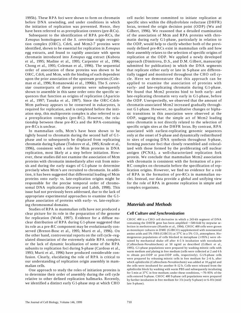
To accomplish this, we took advantage of the ability ofspecific antibodies to distinguish segments of DNA substi-tuted with iodinated or chlorinated nucleotide analogues(Bakker et al., 1991; Aten et al., 1992) to develop amethod of differentially tagging the earliest- and the later-replicating DNA (Fig. 1). CHOC 400 cells were syn-chronized at the G1/S border by mitotic selection andsubsequent incubation in the presence of aphidicolin.Aphidicolin inhibits the processive elongation of nascentDNA strands but does not prevent the initiation of repli-cation and the formation of short (100–500 bp) primers(Nethanel and Kaufmann, 1990). Thus, cells accumulatewith replication forks arrested close to their sites of initia-tion. These cells were then released into S-phase by re-moval of aphidicolin and the very earliest-replicatingchromosomal domains were briefly labeled with CldU,followed by a chase period of several hours. Late-replicat-ing DNA was then labeled with IdU, the cells were subse-quently synchronized in metaphase by mitotic shake offand released into G1-phase.
It has been shown that the distribution of replicationsites in the nucleus follows a defined spatio-temporal pro-gram during S-phase, which is typical for each cell type(Nakayasu and Berezney, 1989; Manders et al., 1992;O’Keefe et al., 1992). Examples of these patterns inCHOC 400 cells (a derivative of CHO cells in which theDHFR locus has been amplified 500-fold) are shown inFig. 1. Each of the fluorescent foci consists of a cluster ofreplicons that are synchronously replicated within thespan of
z
60 min (Manders et al., 1996; Ma et al., 1998;Dimitrova, D.S., and D.M. Gilbert, manuscript submittedfor publication). Early replication patterns (visualized as afew tens to a few hundred small fluorescent foci scatteredthroughout the nuclear interior) persist for the first 5–6 h
of a 10–12-h S-phase and consist of multiple sets of repli-con clusters. With the protocol shown in Fig. 1, only thevery earliest subset of these replicon clusters is labeledwith CldU. Since cells do not traverse S-phase in perfectsynchrony, the late S-phase IdU–label highlights all threelate spatio-temporal replication patterns, providing a con-venient means to visualize the entire spectrum of late-rep-licating domains within the same cell preparations. Wehave demonstrated that each of these spatio-temporal pat-terns of labeled replicon clusters persists throughout inter-phase and is reproduced in the subsequent cell cyclewithin 2 h after metaphase. Furthermore, we have shownthat the earliest subset of clusters tagged with CldU are re-producibly activated at the onset of subsequent S-phases(Dimitrova, D.S., and D.M. Gilbert, manuscript submittedfor publication). Based on this evidence, we conclude thatit is feasible to compare the assembly of pre-RCs onto theearliest- vs. later-replicating chromatin by following thebehavior of the CldU-tagged and IdU-tagged DNA sitesduring G1-phase of the subsequent cell cycle.
Mcm2 Binds Simultaneously to Both Early- andLate-replicating Chromatin upon Exit from Mitosis
We examined the localization of chromatin-bound Mcm2relative to earliest- and late-replicating chromatin inCHOC 400 cell populations prelabeled as described aboveand released in the subsequent cell cycle for different peri-ods of time. Cells were first extracted with Triton X-100 toremove soluble nuclear proteins, then fixed and stainedwith a polyclonal antibody specific for Mcm2 and a mono-clonal antibody specific for either CldU or IdU. We ob-
Figure 1. Experimental protocol (see text for details). Represen-tative photographs of the types of replication patterns prevailingin the early (first half, euchromatic) or late (second half, three se-quential heterochromatic patterns) stages of the 10–12 h S-phasein CHOC 400 cells are displayed.
Dimitrova et al.
Mammalain Prereplication Complexes
713
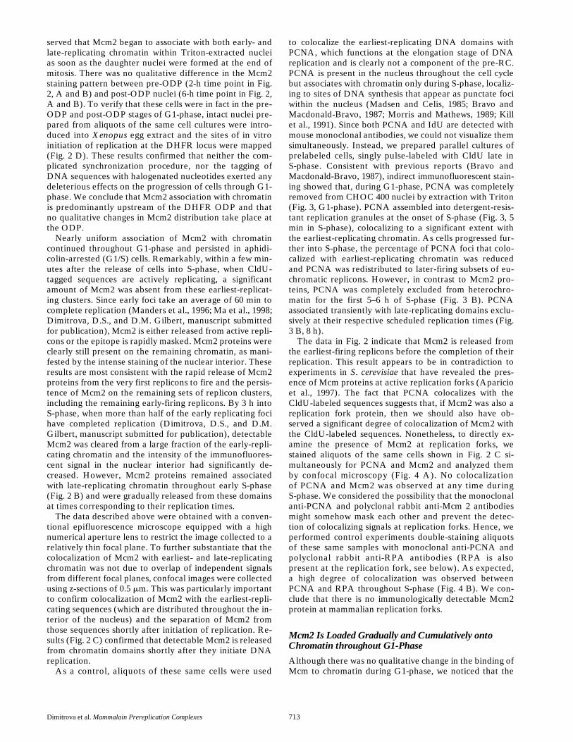
served that Mcm2 began to associate with both early- andlate-replicating chromatin within Triton-extracted nucleias soon as the daughter nuclei were formed at the end ofmitosis. There was no qualitative difference in the Mcm2staining pattern between pre-ODP (2-h time point in Fig.2, A and B) and post-ODP nuclei (6-h time point in Fig. 2,A and B). To verify that these cells were in fact in the pre-ODP and post-ODP stages of G1-phase, intact nuclei pre-pared from aliquots of the same cell cultures were intro-duced into
Xenopus
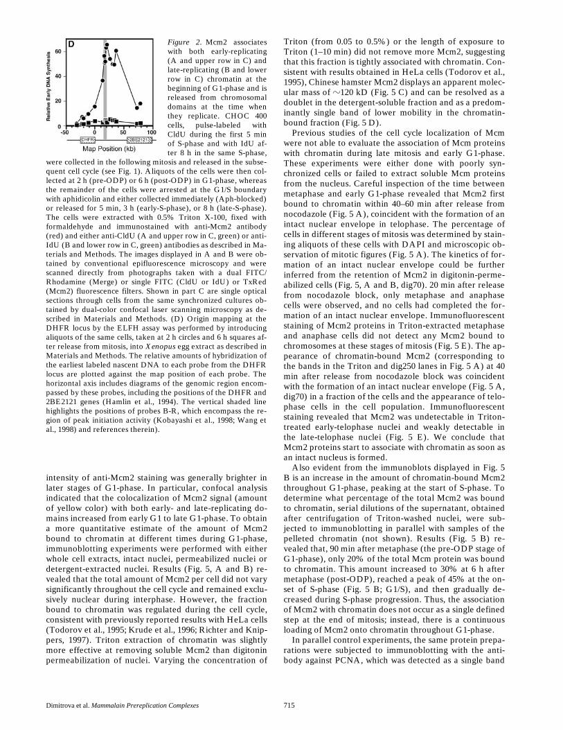
egg extract and the sites of in vitroinitiation of replication at the DHFR locus were mapped(Fig. 2 D). These results confirmed that neither the com-plicated synchronization procedure, nor the tagging ofDNA sequences with halogenated nucleotides exerted anydeleterious effects on the progression of cells through G1-phase. We conclude that Mcm2 association with chromatinis predominantly upstream of the DHFR ODP and thatno qualitative changes in Mcm2 distribution take place atthe ODP.
Nearly uniform association of Mcm2 with chromatincontinued throughout G1-phase and persisted in aphidi-colin-arrested (G1/S) cells. Remarkably, within a few min-utes after the release of cells into S-phase, when CldU-tagged sequences are actively replicating, a significantamount of Mcm2 was absent from these earliest-replicat-ing clusters. Since early foci take an average of 60 min tocomplete replication (Manders et al., 1996; Ma et al., 1998;Dimitrova, D.S., and D.M. Gilbert, manuscript submittedfor publication), Mcm2 is either released from active repli-cons or the epitope is rapidly masked. Mcm2 proteins wereclearly still present on the remaining chromatin, as mani-fested by the intense staining of the nuclear interior. Theseresults are most consistent with the rapid release of Mcm2proteins from the very first replicons to fire and the persis-tence of Mcm2 on the remaining sets of replicon clusters,including the remaining early-firing replicons. By 3 h intoS-phase, when more than half of the early replicating focihave completed replication (Dimitrova, D.S., and D.M.Gilbert, manuscript submitted for publication), detectableMcm2 was cleared from a large fraction of the early-repli-cating chromatin and the intensity of the immunofluores-cent signal in the nuclear interior had significantly de-creased. However, Mcm2 proteins remained associatedwith late-replicating chromatin throughout early S-phase(Fig. 2 B) and were gradually released from these domainsat times corresponding to their replication times.
The data described above were obtained with a conven-tional epifluorescence microscope equipped with a highnumerical aperture lens to restrict the image collected to arelatively thin focal plane. To further substantiate that thecolocalization of Mcm2 with earliest- and late-replicatingchromatin was not due to overlap of independent signalsfrom different focal planes, confocal images were collectedusing z-sections of 0.5
m
m. This was particularly importantto confirm colocalization of Mcm2 with the earliest-repli-cating sequences (which are distributed throughout the in-terior of the nucleus) and the separation of Mcm2 fromthose sequences shortly after initiation of replication. Re-sults (Fig. 2 C) confirmed that detectable Mcm2 is releasedfrom chromatin domains shortly after they initiate DNAreplication.
As a control, aliquots of these same cells were used
to colocalize the earliest-replicating DNA domains withPCNA, which functions at the elongation stage of DNAreplication and is clearly not a component of the pre-RC.PCNA is present in the nucleus throughout the cell cyclebut associates with chromatin only during S-phase, localiz-ing to sites of DNA synthesis that appear as punctate fociwithin the nucleus (Madsen and Celis, 1985; Bravo andMacdonald-Bravo, 1987; Morris and Mathews, 1989; Killet al., 1991). Since both PCNA and IdU are detected withmouse monoclonal antibodies, we could not visualize themsimultaneously. Instead, we prepared parallel cultures ofprelabeled cells, singly pulse-labeled with CldU late inS-phase. Consistent with previous reports (Bravo andMacdonald-Bravo, 1987), indirect immunofluorescent stain-ing showed that, during G1-phase, PCNA was completelyremoved from CHOC 400 nuclei by extraction with Triton(Fig. 3, G1-phase). PCNA assembled into detergent-resis-tant replication granules at the onset of S-phase (Fig. 3, 5min in S-phase), colocalizing to a significant extent withthe earliest-replicating chromatin. As cells progressed fur-ther into S-phase, the percentage of PCNA foci that colo-calized with earliest-replicating chromatin was reducedand PCNA was redistributed to later-firing subsets of eu-chromatic replicons. However, in contrast to Mcm2 pro-teins, PCNA was completely excluded from heterochro-matin for the first 5–6 h of S-phase (Fig. 3 B). PCNAassociated transiently with late-replicating domains exclu-sively at their respective scheduled replication times (Fig.3 B, 8 h).
The data in Fig. 2 indicate that Mcm2 is released fromthe earliest-firing replicons before the completion of theirreplication. This result appears to be in contradiction toexperiments in
S
.
cerevisiae
that have revealed the pres-ence of Mcm proteins at active replication forks (Aparicioet al., 1997). The fact that PCNA colocalizes with theCldU-labeled sequences suggests that, if Mcm2 was also areplication fork protein, then we should also have ob-served a significant degree of colocalization of Mcm2 withthe CldU-labeled sequences. Nonetheless, to directly ex-amine the presence of Mcm2 at replication forks, westained aliquots of the same cells shown in Fig. 2 C si-multaneously for PCNA and Mcm2 and analyzed themby confocal microscopy (Fig. 4 A). No colocalizationof PCNA and Mcm2 was observed at any time duringS-phase. We considered the possibility that the monoclonalanti-PCNA and polyclonal rabbit anti-Mcm 2 antibodiesmight somehow mask each other and prevent the detec-tion of colocalizing signals at replication forks. Hence, weperformed control experiments double-staining aliquotsof these same samples with monoclonal anti-PCNA andpolyclonal rabbit anti-RPA antibodies (RPA is alsopresent at the replication fork, see below). As expected,a high degree of colocalization was observed betweenPCNA and RPA throughout S-phase (Fig. 4 B). We con-clude that there is no immunologically detectable Mcm2protein at mammalian replication forks.
Mcm2 Is Loaded Gradually and Cumulatively onto Chromatin throughout G1-Phase
Although there was no qualitative change in the binding ofMcm to chromatin during G1-phase, we noticed that the
The Journal of Cell Biology, Volume 146, 1999 714
Figure 2.
Dimitrova et al.
Mammalain Prereplication Complexes
715
intensity of anti-Mcm2 staining was generally brighter inlater stages of G1-phase. In particular, confocal analysisindicated that the colocalization of Mcm2 signal (amountof yellow color) with both early- and late-replicating do-mains increased from early G1 to late G1-phase. To obtaina more quantitative estimate of the amount of Mcm2bound to chromatin at different times during G1-phase,immunoblotting experiments were performed with eitherwhole cell extracts, intact nuclei, permeabilized nuclei ordetergent-extracted nuclei. Results (Fig. 5, A and B) re-vealed that the total amount of Mcm2 per cell did not varysignificantly throughout the cell cycle and remained exclu-sively nuclear during interphase. However, the fractionbound to chromatin was regulated during the cell cycle,consistent with previously reported results with HeLa cells(Todorov et al., 1995; Krude et al., 1996; Richter and Knip-pers, 1997). Triton extraction of chromatin was slightlymore effective at removing soluble Mcm2 than digitoninpermeabilization of nuclei. Varying the concentration of
Triton (from 0.05 to 0.5%) or the length of exposure toTriton (1–10 min) did not remove more Mcm2, suggestingthat this fraction is tightly associated with chromatin. Con-sistent with results obtained in HeLa cells (Todorov et al.,1995), Chinese hamster Mcm2 displays an apparent molec-ular mass of
z
120 kD (Fig. 5 C) and can be resolved as adoublet in the detergent-soluble fraction and as a predom-inantly single band of lower mobility in the chromatin-bound fraction (Fig. 5 D).
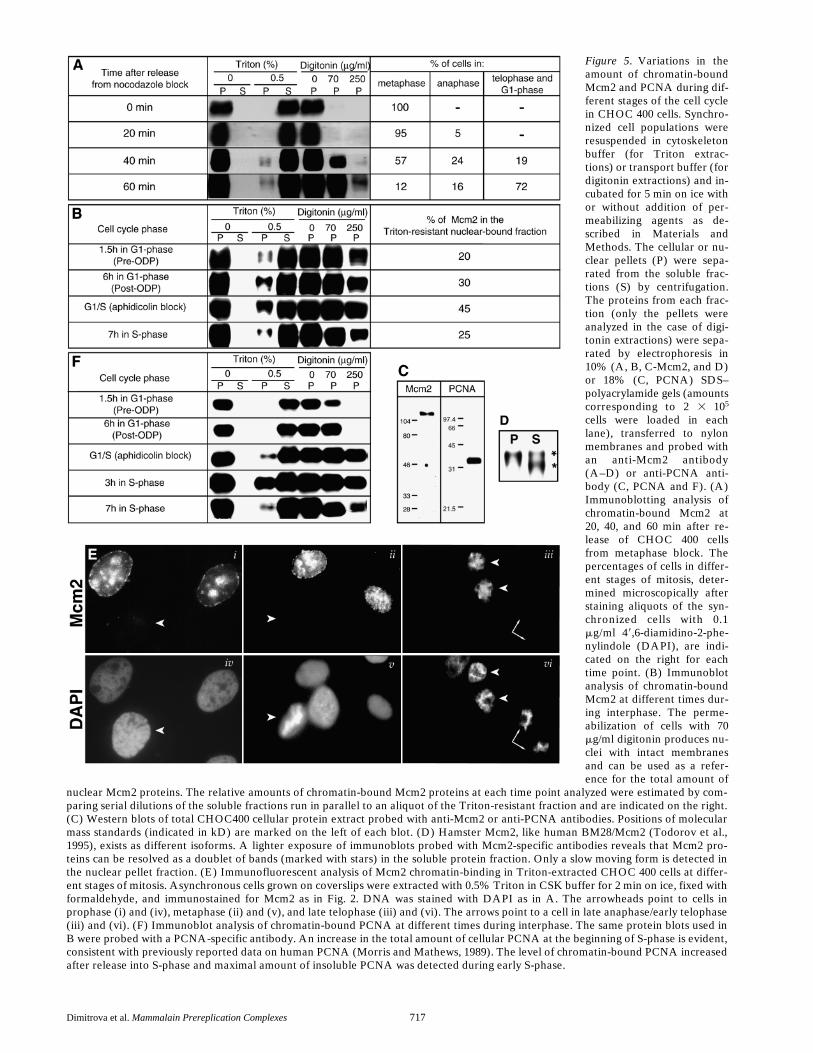
Previous studies of the cell cycle localization of Mcmwere not able to evaluate the association of Mcm proteinswith chromatin during late mitosis and early G1-phase.These experiments were either done with poorly syn-chronized cells or failed to extract soluble Mcm proteinsfrom the nucleus. Careful inspection of the time betweenmetaphase and early G1-phase revealed that Mcm2 firstbound to chromatin within 40–60 min after release fromnocodazole (Fig. 5 A), coincident with the formation of anintact nuclear envelope in telophase. The percentage ofcells in different stages of mitosis was determined by stain-ing aliquots of these cells with DAPI and microscopic ob-servation of mitotic figures (Fig. 5 A). The kinetics of for-mation of an intact nuclear envelope could be furtherinferred from the retention of Mcm2 in digitonin-perme-abilized cells (Fig. 5,
A and B, dig70). 20 min after releasefrom nocodazole block, only metaphase and anaphasecells were observed, and no cells had completed the for-mation of an intact nuclear envelope. Immunofluorescentstaining of Mcm2 proteins in Triton-extracted metaphaseand anaphase cells did not detect any Mcm2 bound tochromosomes at these stages of mitosis (Fig. 5 E). The ap-pearance of chromatin-bound Mcm2 (corresponding tothe bands in the Triton and dig250 lanes in Fig. 5 A) at 40min after release from nocodazole block was coincidentwith the formation of an intact nuclear envelope (Fig. 5 A,dig70) in a fraction of the cells and the appearance of telo-phase cells in the cell population. Immunofluorescentstaining revealed that Mcm2 was undetectable in Triton-treated early-telophase nuclei and weakly detectable inthe late-telophase nuclei (Fig. 5 E). We conclude thatMcm2 proteins start to associate with chromatin as soon asan intact nucleus is formed.
Also evident from the immunoblots displayed in Fig. 5B is an increase in the amount of chromatin-bound Mcm2throughout G1-phase, peaking at the start of S-phase. Todetermine what percentage of the total Mcm2 was boundto chromatin, serial dilutions of the supernatant, obtainedafter centrifugation of Triton-washed nuclei, were sub-jected to immunoblotting in parallel with samples of thepelleted chromatin (not shown). Results (Fig. 5 B) re-vealed that, 90 min after metaphase (the pre-ODP stage ofG1-phase), only 20% of the total Mcm protein was boundto chromatin. This amount increased to 30% at 6 h aftermetaphase (post-ODP), reached a peak of 45% at the on-set of S-phase (Fig. 5 B; G1/S), and then gradually de-creased during S-phase progression. Thus, the associationof Mcm2 with chromatin does not occur as a single definedstep at the end of mitosis; instead, there is a continuousloading of Mcm2 onto chromatin throughout G1-phase.
In parallel control experiments, the same protein prepa-rations were subjected to immunoblotting with the anti-body against PCNA, which was detected as a single band
Figure 2. Mcm2 associateswith both early-replicating(A and upper row in C) andlate-replicating (B and lowerrow in C) chromatin at thebeginning of G1-phase and isreleased from chromosomaldomains at the time whenthey replicate. CHOC 400cells, pulse-labeled withCldU during the first 5 minof S-phase and with IdU af-ter 8 h in the same S-phase,
were collected in the following mitosis and released in the subse-quent cell cycle (see Fig. 1). Aliquots of the cells were then col-lected at 2 h (pre-ODP) or 6 h (post-ODP) in G1-phase, whereasthe remainder of the cells were arrested at the G1/S boundarywith aphidicolin and either collected immediately (Aph-blocked)or released for 5 min, 3 h (early-S-phase), or 8 h (late-S-phase).The cells were extracted with 0.5% Triton X-100, fixed withformaldehyde and immunostained with anti-Mcm2 antibody(red) and either anti-CldU (A and upper row in C, green) or anti-IdU (B and lower row in C, green) antibodies as described in Ma-terials and Methods. The images displayed in A and B were ob-tained by conventional epifluorescence microscopy and werescanned directly from photographs taken with a dual FITC/Rhodamine (Merge) or single FITC (CldU or IdU) or TxRed(Mcm2) fluorescence filters. Shown in part C are single opticalsections through cells from the same synchronized cultures ob-tained by dual-color confocal laser scanning microscopy as de-scribed in Materials and Methods. (D) Origin mapping at theDHFR locus by the ELFH assay was performed by introducingaliquots of the same cells, taken at 2 h circles and 6 h squares af-ter release from mitosis, into Xenopus egg extract as described inMaterials and Methods. The relative amounts of hybridization ofthe earliest labeled nascent DNA to each probe from the DHFRlocus are plotted against the map position of each probe. Thehorizontal axis includes diagrams of the genomic region encom-passed by these probes, including the positions of the DHFR and2BE2121 genes (Hamlin et al., 1994). The vertical shaded linehighlights the positions of probes B-R, which encompass the re-gion of peak initiation activity (Kobayashi et al., 1998; Wang etal., 1998) and references therein).

The Journal of Cell Biology, Volume 146, 1999 716
with an apparent molecular mass of 35 kD (Fig. 5 C). Asexpected, PCNA was detected in the nucleus throughoutthe cell cycle. It was absent from insoluble nuclear struc-tures during G1-phase, first associated with nuclear com-ponents at the G1/S-phase transition and a fraction re-mained nuclear bound throughout S-phase (Fig. 5 F).Whereas the maximal Mcm2 bound was observed at theG1/S boundary within aphidicolin-blocked cells, the maxi-mal amount of PCNA bound was observed after releasefrom the aphidicolin block, in early S-phase.
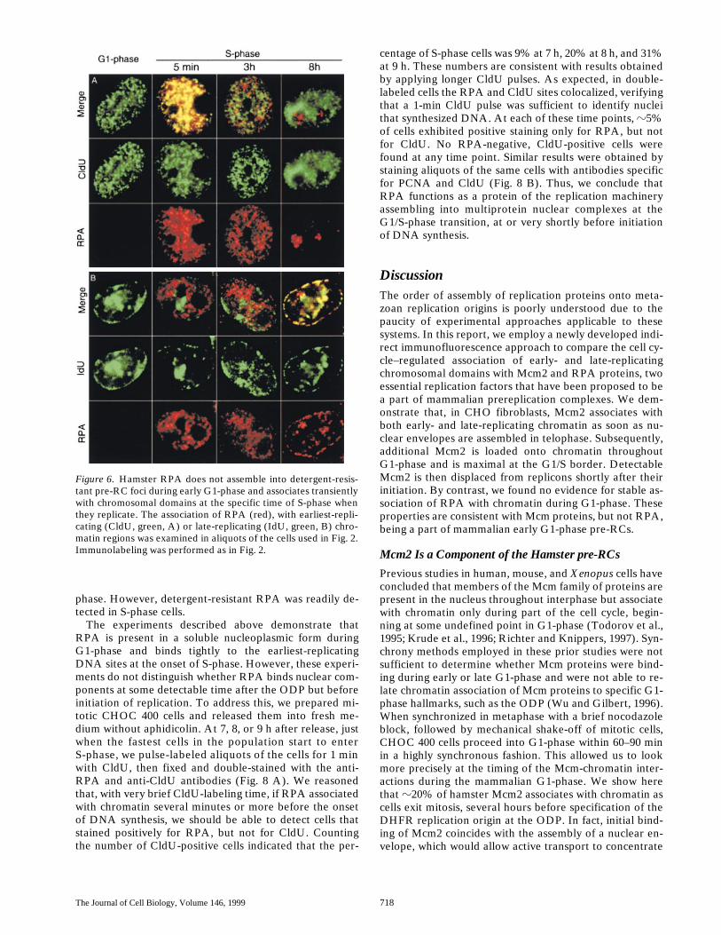
RPA Associates Specifically with Earliest-Replicating Genomic Sequences at the G1/S Border and Redistributes to Sites of Ongoing DNA Synthesisduring S-Phase
Previous studies of RPA in mammalian cells have de-
tected discrete nuclear RPA sites in non-S-phase HeLacells (Krude, 1995) or presented direct evidence for a dif-fuse nuclear distribution of RPA during G1-phase in hu-man cells (Brenot-Bosc et al., 1995; Murti et al., 1996).These results suggested that mammalian RPA may formpre-RCs similar to those observed in
Xenopus
egg extractand encouraged us to determine exactly when they form inrelation to Mcm binding and the ODP. Since the optimaltechnical protocol for immunostaining of RPA (Dim-itrova, D.S., and D.M. Gilbert, manuscript in preparation)was the same as for Mcm2, we were able to use the samepopulations of synchronized cells shown in Fig. 2, A andB, allowing a direct comparison of the cell cycle behaviorof these two proteins. Simultaneous staining of 2 h or 6 hG1-phase nuclei with the anti-RPA antibody and eitheranti-CldU, or anti-IdU antibody, revealed that RPA wascompletely absent from G1-phase chromatin (Fig. 6, G1-phase). RPA bound to chromatin at the onset of S-phase(Fig. 6, 5 min in S-phase), when it formed discrete foci thatcolocalized with sites of early-, but not late-replicatingchromatin. Late in S-phase (Fig. 6, 8 h), RPA continued toexhibit punctate distribution but at that time it associatedexclusively with late-replicating chromatin domains. Con-trary to previous reports (Yan and Newport, 1995a; Murtiet al., 1996), RPA foci were not detected in G2-phase nu-clei (not shown).
To confirm that there was not a chromatin-bound frac-tion of RPA in G1-phase nuclei that is not detectable byimmunofluorescence, we performed immunoblotting ex-periments with aliquots of the same protein extracts fromsynchronized cells shown in Fig. 5. Using the same poly-clonal antibody used in Fig. 6, Chinese hamster proteinsof 14- (not visible), 30-, and 70-kD apparent molecularmasses were detected (Fig. 7 A). Results with synchro-nized cells (Fig. 7 B) revealed that, as with PCNA, noRPA was detected associated with chromatin during G1-
Figure 3. PCNA does not bind nuclear components during G1-phase and associates transiently with chromosomal domains atthe time when they replicate. (A) The association of PCNA (red)with the earliest-replicating chromatin regions (CldU-tagged,green) was examined in aliquots of the cells used in the experi-ment displayed in Fig. 2. (B) Late-replicating genomic regionswere pulse-labeled with CldU (green) at 8 h in S-phase in a paral-lel culture of CHOC 400 cells, mitotic cells were collected fewhours later, released in the subsequent cell cycle for the same pe-riods of time and processed in the same way as in Fig. 2.
Figure 4. RPA, but not Mcm2, is present at PCNA-containingactive mammalian replication forks. Aliquots of the cell cultures(synchronized in early or mid/late S-phase) used in Fig. 2 wereimmunostained with PCNA-specific (red) and, either Mcm2-spe-cific (A, green), or RPA-specific (B, green) antibodies. Shownare single sections obtained by dual-color confocal laser scanningmicroscopy.
Dimitrova et al.
Mammalain Prereplication Complexes
717
Figure 5. Variations in theamount of chromatin-boundMcm2 and PCNA during dif-ferent stages of the cell cyclein CHOC 400 cells. Synchro-nized cell populations wereresuspended in cytoskeletonbuffer (for Triton extrac-tions) or transport buffer (fordigitonin extractions) and in-cubated for 5 min on ice withor without addition of per-meabilizing agents as de-scribed in Materials andMethods. The cellular or nu-clear pellets (P) were sepa-rated from the soluble frac-tions (S) by centrifugation.The proteins from each frac-tion (only the pellets wereanalyzed in the case of digi-tonin extractions) were sepa-rated by electrophoresis in10% (A, B, C-Mcm2, and D)or 18% (C, PCNA) SDS–polyacrylamide gels (amountscorresponding to 2 3 105
cells were loaded in eachlane), transferred to nylonmembranes and probed withan anti-Mcm2 antibody(A–D) or anti-PCNA anti-body (C, PCNA and F). (A)Immunoblotting analysis ofchromatin-bound Mcm2 at20, 40, and 60 min after re-lease of CHOC 400 cellsfrom metaphase block. Thepercentages of cells in differ-ent stages of mitosis, deter-mined microscopically afterstaining aliquots of the syn-chronized cells with 0.1mg/ml 49,6-diamidino-2-phe-nylindole (DAPI), are indi-cated on the right for eachtime point. (B) Immunoblotanalysis of chromatin-boundMcm2 at different times dur-ing interphase. The perme-abilization of cells with 70mg/ml digitonin produces nu-clei with intact membranesand can be used as a refer-ence for the total amount of
nuclear Mcm2 proteins. The relative amounts of chromatin-bound Mcm2 proteins at each time point analyzed were estimated by com-paring serial dilutions of the soluble fractions run in parallel to an aliquot of the Triton-resistant fraction and are indicated on the right.(C) Western blots of total CHOC400 cellular protein extract probed with anti-Mcm2 or anti-PCNA antibodies. Positions of molecularmass standards (indicated in kD) are marked on the left of each blot. (D) Hamster Mcm2, like human BM28/Mcm2 (Todorov et al.,1995), exists as different isoforms. A lighter exposure of immunoblots probed with Mcm2-specific antibodies reveals that Mcm2 pro-teins can be resolved as a doublet of bands (marked with stars) in the soluble protein fraction. Only a slow moving form is detected inthe nuclear pellet fraction. (E) Immunofluorescent analysis of Mcm2 chromatin-binding in Triton-extracted CHOC 400 cells at differ-ent stages of mitosis. Asynchronous cells grown on coverslips were extracted with 0.5% Triton in CSK buffer for 2 min on ice, fixed withformaldehyde, and immunostained for Mcm2 as in Fig. 2. DNA was stained with DAPI as in A. The arrowheads point to cells inprophase (i) and (iv), metaphase (ii) and (v), and late telophase (iii) and (vi). The arrows point to a cell in late anaphase/early telophase(iii) and (vi). (F) Immunoblot analysis of chromatin-bound PCNA at different times during interphase. The same protein blots used inB were probed with a PCNA-specific antibody. An increase in the total amount of cellular PCNA at the beginning of S-phase is evident,consistent with previously reported data on human PCNA (Morris and Mathews, 1989). The level of chromatin-bound PCNA increasedafter release into S-phase and maximal amount of insoluble PCNA was detected during early S-phase.
The Journal of Cell Biology, Volume 146, 1999 718
phase. However, detergent-resistant RPA was readily de-tected in S-phase cells.
The experiments described above demonstrate thatRPA is present in a soluble nucleoplasmic form duringG1-phase and binds tightly to the earliest-replicatingDNA sites at the onset of S-phase. However, these experi-ments do not distinguish whether RPA binds nuclear com-ponents at some detectable time after the ODP but beforeinitiation of replication. To address this, we prepared mi-totic CHOC 400 cells and released them into fresh me-dium without aphidicolin. At 7, 8, or 9 h after release, justwhen the fastest cells in the population start to enterS-phase, we pulse-labeled aliquots of the cells for 1 minwith CldU, then fixed and double-stained with the anti-RPA and anti-CldU antibodies (Fig. 8 A). We reasonedthat, with very brief CldU-labeling time, if RPA associatedwith chromatin several minutes or more before the onsetof DNA synthesis, we should be able to detect cells thatstained positively for RPA, but not for CldU. Countingthe number of CldU-positive cells indicated that the per-
centage of S-phase cells was 9% at 7 h, 20% at 8 h, and 31%at 9 h. These numbers are consistent with results obtainedby applying longer CldU pulses. As expected, in double-labeled cells the RPA and CldU sites colocalized, verifyingthat a 1-min CldU pulse was sufficient to identify nucleithat synthesized DNA. At each of these time points,
z
5%of cells exhibited positive staining only for RPA, but notfor CldU. No RPA-negative, CldU-positive cells werefound at any time point. Similar results were obtained bystaining aliquots of the same cells with antibodies specificfor PCNA and CldU (Fig. 8 B). Thus, we conclude thatRPA functions as a protein of the replication machineryassembling into multiprotein nuclear complexes at theG1/S-phase transition, at or very shortly before initiationof DNA synthesis.
Discussion
The order of assembly of replication proteins onto meta-zoan replication origins is poorly understood due to thepaucity of experimental approaches applicable to thesesystems. In this report, we employ a newly developed indi-rect immunofluorescence approach to compare the cell cy-cle–regulated association of early- and late-replicatingchromosomal domains with Mcm2 and RPA proteins, twoessential replication factors that have been proposed to bea part of mammalian prereplication complexes. We dem-onstrate that, in CHO fibroblasts, Mcm2 associates withboth early- and late-replicating chromatin as soon as nu-clear envelopes are assembled in telophase. Subsequently,additional Mcm2 is loaded onto chromatin throughoutG1-phase and is maximal at the G1/S border. DetectableMcm2 is then displaced from replicons shortly after theirinitiation. By contrast, we found no evidence for stable as-sociation of RPA with chromatin during G1-phase. Theseproperties are consistent with Mcm proteins, but not RPA,being a part of mammalian early G1-phase pre-RCs.
Mcm2 Is a Component of the Hamster pre-RCs
Previous studies in human, mouse, and
Xenopus
cells haveconcluded that members of the Mcm family of proteins arepresent in the nucleus throughout interphase but associatewith chromatin only during part of the cell cycle, begin-ning at some undefined point in G1-phase (Todorov et al.,1995; Krude et al., 1996; Richter and Knippers, 1997). Syn-chrony methods employed in these prior studies were notsufficient to determine whether Mcm proteins were bind-ing during early or late G1-phase and were not able to re-late chromatin association of Mcm proteins to specific G1-phase hallmarks, such as the ODP (Wu and Gilbert, 1996).When synchronized in metaphase with a brief nocodazoleblock, followed by mechanical shake-off of mitotic cells,CHOC 400 cells proceed into G1-phase within 60–90 minin a highly synchronous fashion. This allowed us to lookmore precisely at the timing of the Mcm-chromatin inter-actions during the mammalian G1-phase. We show herethat
z
20% of hamster Mcm2 associates with chromatin ascells exit mitosis, several hours before specification of theDHFR replication origin at the ODP. In fact, initial bind-ing of Mcm2 coincides with the assembly of a nuclear en-velope, which would allow active transport to concentrate
Figure 6. Hamster RPA does not assemble into detergent-resis-tant pre-RC foci during early G1-phase and associates transientlywith chromosomal domains at the specific time of S-phase whenthey replicate. The association of RPA (red), with earliest-repli-cating (CldU, green, A) or late-replicating (IdU, green, B) chro-matin regions was examined in aliquots of the cells used in Fig. 2.Immunolabeling was performed as in Fig. 2.

Dimitrova et al.
Mammalain Prereplication Complexes
719
Mcm proteins in the nuclear compartment. Significantly,our novel indirect immunofluorescence approach for visu-alization of proteins and tagged DNA sites allowed us todemonstrate for the first time that Mcm2 proteins bind si-multaneously to both early- and late-replicating chromatinregions at the very beginning of G1-phase.
Quantitative immunoblotting analysis showed that anadditional 25% of Mcm2 binds chromatin gradually andcumulatively throughout G1-phase until, at the G1/S bor-der,
z
45% of Mcm2 is bound, with the remainder presentin the soluble nucleosolic fraction. A similar increase inthe amount of chromatin-bound Mcm proteins during G1-phase has been documented in
S
. cerevisiae (Aparicio et al.,1997; Tanaka et al., 1997; Zou and Stillman, 1998). The ob-servation that the initial binding of hamster Mcm2 to chro-matin occurs in late telophase is similar to the situation inyeast and Xenopus egg extracts and implicates this proteinas a pre-RC component. The fact that this event is up-stream of the ODP suggests that the formation of Mcm-containing pre-RCs is not sufficient for the specification of
mammalian replication origins. This view is also supportedby studies in Xenopus egg extracts, where ORC-Cdc6-Mcm–dependent replication occurs without the use of spe-cific origins (Gilbert, 1998). However, since we found thatadditional Mcm is continuously loaded onto hamster chro-matin throughout G1-phase, it is still formally possiblethat a critical threshold of loaded Mcm in some way fo-cuses initiation to specific sites. Alternatively, the specifi-cation of origins could be a gradual process, with differentorigins specified at different times in G1-phase corre-sponding to the binding of Mcm proteins. To date, theODP has been determined only for the DHFR origin.
In eukaryotic nuclei, the activation of replication originsoccurs according to a strictly regulated temporal program(Fangman and Brewer, 1992; Diller and Raghuraman,1994). Although the mechanism that establishes this pro-gram has not been elucidated, it has been suggested thatreplication timing might be influenced by the amount or
Figure 7. Immunoblot analysis of chromatin-bound RPA duringG1 and S-phase. (A) Proteins in a total extract from CHOC 400cells (2.5 3 105 cells) were separated by electrophoresis in 12.5%SDS–polyacrylamide gels, transferred to a nylon membrane andprobed with anti-RPA antibodies. The rabbit polyclonal anti-RPA serum [raised against human RPA trimeric complex (Dinet al., 1990)] recognized protein bands corresponding to the large(apparent molecular mass of z70 kD), middle (z30 kD), andsmall (z14 kD, a very faint band invisible on this blot) RPA sub-units. Positions of molecular mass standards (indicated in kD)are marked on the left. (B) Western blots of chromatin-boundRPA in early G1 and S-phase. Aliquots of the same protein ex-tracts from synchronized CHOC 400 cells used in Fig. 5, B and F,were subjected to SDS-PAGE (12.5% polyacrylamide gels,equivalents of 2 3 105 cell per lane), transferred to nylon mem-branes and probed with anti-RPA polyclonal antibodies. Only re-sults for the middle (p30) and large (p70) RPA subunits are dis-played, since the signal for the small RPA subunit was very weak(we have found no evidence for disassembly of the RPA complexduring the cell cycle in control immunolocalization experiments,using monoclonal antibodies specific for the individual RPA sub-units together with the polyclonal serum against the heterotri-meric RPA complex; Dimitrova, D.S., and D.M. Gilbert, manu-script in preparation). An increase of total cellular RPA proteinand a maximal level of RPA in the nuclear pellet fraction was de-tected during early S-phase (see Fig. 5 F).
Figure 8. Hamster RPA and PCNA are assembled into deter-gent-resistant replicative complexes within less than an hourfrom the start of S-phase. CHOC 400 cells, synchronized in mito-sis, were released for 8 h in the following cell cycle (20% of thecells had entered S-phase at this time). The cells were pulse-labeled with 100 mM CldU for 1 min, extracted with Triton andfixed with formaldehyde as described in Materials and Methods.Aliquots of the cells were stained with anti-CldU antibodies(green) and either anti-RPA antibodies (red, A) or anti-PCNAantibodies (red, B). S-phase cells display yellow coloration result-ing from the colocalization of RPA or PCNA (red) with nascentDNA (CldU, green). Arrowheads point to nuclei that exhibitpositive staining for RPA or PCNA, but no CldU label. Arrowsindicate the positions of G1-phase cells (barely visible as faintshadows).
The Journal of Cell Biology, Volume 146, 1999 720
kinetics of Mcm loading onto early- vs. late-replicatingchromatin (Kearsey and Labib, 1998). The techniquedescribed in this report allowed us to distinguish the as-sociation of proteins with early- and late-replicatingchromosomal domains during G1-phase. We consistentlyobserved equivalent association of Mcm2 with early- andlate-replicating chromatin. Hence, the establishment of achromosomal domain as early- or late-replicating does notappear to involve quantitative differences in the associa-tion of Mcm2 proteins.
Are Mcm Proteins Present at DNA Replication Forks: The Mcm Paradox
In S. cerevisiae chromatin immunoprecipitation experi-ments have produced direct evidence for the binding ofmembers of the Mcm protein family (Mcm4 and Mcm7) toyeast chromosomal replication origins (Aparicio et al.,1997; Tanaka et al., 1997). One of these studies furthershowed that shortly after origin firing Mcm4 and Mcm7proteins dissociate from origins and move with replicationforks, along with DNA polymerase e (Aparicio et al.,1997). This observation raised an apparent paradox re-garding the behavior of Mcm proteins (Newlon, 1997),since previous reports (and our own unpublished data)have shown that, in Xenopus egg extracts and in culturedmammalian cells, members of the Mcm family do not colo-calize with sites of newly synthesized DNA (Krude, 1995;Todorov et al., 1995; Romanowski et al., 1996a). However,these results are difficult to interpret because proteins arelocalized to their positions at the moment of fixation,whereas sites of newly synthesized DNA were labeled be-fore fixation. In the approach described in this report, pro-teins can be localized to sites that are actively engaged inreplication. Mcm2 was still associated with the earliest-fir-ing replicon clusters after initiation (at the aphidicolin-arrested step) but was cleared from those earliest replicat-ing clusters within 5 min after DNA synthesis was allowed toproceed. At this time, these CldU-tagged sequences wereactively being replicated and colocalized with the replica-tion fork proteins PCNA and RPA. Since these earliestreplicons take z60 min to complete replication (Manderset al., 1996; Ma et al., 1998; Dimitrova, D.S., and D.M. Gil-bert, manuscript submitted for publication), we are forcedto conclude that all detectable (by immunofluorescence)mammalian Mcm2 is cleared from replicons shortly afterinitiation (see Fig. 2, A and C, 5 min in S-phase) and doesnot associate with replication forks thereafter. Further-more, direct inspection of Mcm2 and PCNA revealed acomplete lack of colocalization (Fig. 4 A). The possibilityremains that other members of the Mcm complex, such asMcm4 (Aparicio et al., 1997), behave differently thanMcm2. However, Tanaka et al. (1997) did not observe as-sociation of Mcm4 with S. cerevisiae replication forks usingthe same technique as (Aparicio et al., 1997). It is also pos-sible that there is a distinct minor population of Mcm pro-teins that escape immunodetection but are present at rep-lication forks. This would imply the existence of threepopulations of Mcm proteins: a soluble form (at leasthalf), a chromatin-bound immunodetectable form that iscleared upon initiation, and a replication fork-associatedform.
RPA Does Not Form pre-RCs during Early-G1 Phase in Mammalian Nuclei
While Mcm2-binding to chromatin before the ODP impli-cates it as a component of the CHO pre-RCs, the behaviorof RPA is most consistent with its involvement in the initi-ation and elongation of nascent DNA strands. RPA is awell-characterized complex of three polypeptides that is akey participant in the initiation step of DNA replication.Several groups (Adachi and Laemmli, 1992, 1994; Kubotaet al., 1995; Yan and Newport, 1995a; Coleman et al., 1996;Coue et al., 1996) have shown that, in Xenopus egg ex-tracts, RPA associates with sperm chromatin before thestart of DNA synthesis. We have applied the same immu-nofluorescent technique employed to examine the associa-tion of Mcm2 with chromatin and failed to detect anystable interaction between RPA and chromatin duringG1-phase in CHOC 400 cells. Hamster RPA assembledinto distinct nuclear granules within a few minutes beforeDNA synthesis and could be detected in association withthe earliest-firing replicon clusters at the onset of S-phase,similar to PCNA. This coincides with the initial appear-ance of replicative megacomplexes, termed replicationfactories, which have been observed to assemble at theG1/S transition in human fibroblasts (Kill et al., 1991;Hozak et al., 1994) and contain both PCNA and DNApolymerase a. Our results are also consistent with recentchromatin immunoprecipitation studies in S. cerevisiaethat did not detect any association of RPA with yeast chro-matin until the onset of S-phase (Tanaka and Nasmyth,1998). We suggest that previous data on G1- or G2-phasenuclear association of RPA proteins (Brenot-Bosc et al.,1995; Murti et al., 1996) derive from the presence of a solu-ble, detergent-extractable form of RPA (Dimitrova, D.S.,and D.M. Gilbert, manuscript in preparation) whose func-tional significance for DNA replication remains unclear.
The approach described in this report also allowed us todemonstrate that RPA does not associate with late-rep-licating chromosomal regions until the late stages ofS-phase, at the time when these sequences engage in repli-cation. This behavior parallels that of PCNA and supportsa role for RPA in the initiation and elongation steps ofreplication, not the formation of pre-RCs.
What Are the Xenopus RPA-containing pre-RCs?
Most of our knowledge of the role of RPA in replicationhas resulted from in vitro studies of SV-40 replication orXenopus sperm chromatin replicating in Xenopus egg ex-tracts. In the SV-40 system, RPA has been shown to be re-quired for the stabilization of unwound origin DNA dur-ing the initiation step (Tsurimoto et al., 1989). In Xenopusegg extracts, RPA is essential for replication and formspunctate foci on sperm chromatin that resemble sites ofDNA synthesis (Adachi and Laemmli, 1994; Yan andNewport, 1995b). The assembly of RPA foci onto chroma-tin precedes DNA unwinding and the initiation of replica-tion and is independent of cdk2 activity in the extracts, im-plying that RPA may play a role at an earlier, preinitiationstage of chromosome replication (Adachi and Laemmli,1994; Yan and Newport, 1995a). Subsequently, it wasshown that the formation of prereplicative RPA foci onsperm chromatin is dependent on the presence of another
Dimitrova et al. Mammalain Prereplication Complexes 721
protein, FFA-1, later shown to be the Xenopus homologueof a human DNA helicase defective in individuals withWerner syndrome (Yan and Newport, 1995b; Yan et al.,1998). RPA-containing pre-RCs do not colocalize with Xe-nopus Mcm proteins (Coue et al., 1996) and are assembledin extracts that have been immunodepleted of XenopusORC and Cdc6 proteins and cannot form Mcm-containingpre-RCs (Coleman et al., 1996). Based on these findings, ithas been concluded that two separate and independent as-sembly pathways are essential for the initiation of replica-tion in eukaryotic cells.
After these studies, it has been assumed that similar pre-replicative RPA foci exist in mammalian cells. However,we could find no evidence for their existence and recentstudies in S. cerevisiae found no evidence for RPA associa-tion with yeast chromatin in G1-phase (Tanaka andNasmyth, 1998). In fact, it still remains to be demonstratedthat FFA-1/RPA foci formed in Xenopus egg extracts areinvolved in DNA replication. Oddly, RPA foci were foundto persist on sperm chromatin even after completion ofDNA replication (in G2-phase; Yan and Newport, 1995b).Most importantly, while it has been shown that RPA focido not form in FFA-1–depleted extracts, the critical exper-iment to determine whether FFA-1–depleted extractscould support sperm DNA replication was not performed(Yan et al., 1998). FFA-1 is a homologue of Werner’s heli-case which, like RPA, and possibly together with RPA, isinvolved in DNA repair and recombination (Fry andLoeb, 1998; Brosh et al., 1999; Suzuki et al., 1999). Hence,it remains possible that FFA-1–mediated formation ofRPA foci is unrelated to DNA replication and that therole of RPA in DNA replication is restricted to its role asdefined in the SV-40 in vitro studies.
This work was supported by grants from the National Institutes of Health(NIH, GM57233-01) and the March of Dimes (FY98-0499) to D.M. Gil-bert. Research in the laboratory of T. Melendy is supported by grantsfrom the NIH (GM56406) and the American Cancer Society (RPG-98-076-01-GMC).
Submitted: 19 January 1999Revised: 22 July 1999Accepted: 23 July 1999
References
Adachi, Y., and U.K. Laemmli. 1992. Identification of nuclear pre-replicationcenters poised for DNA synthesis in Xenopus egg extracts: immunolocaliza-tion study of replication protein A. J. Cell Biol. 119:1–15.
Adachi, Y., and U.K. Laemmli. 1994. Study of the cell cycle-dependent assem-bly of the DNA pre-replication centres in Xenopus egg extracts. EMBO(Eur. Mol. Biol. Organ.) J. 13:4153–4164.
Aparicio, O.M., D.M. Weinstein, and S.P. Bell. 1997. Components and dynam-ics of DNA replication complexes in S. cerevisiae: redistribution of MCMproteins and Cdc45p during S phase. Cell. 91:59–69.
Aten, J.A., P.J.M. Bakker, J. Stap, G.A. Boschman, and C.H.N. Veenhof. 1992.DNA double labelling with IdUrd and CldUrd for spatial and temporalanalysis of cell proliferation and DNA replication. Histochem. J. 24:251–259.
Bakker, P.J., J. Stap, C.J. Tukker, C.H. van Oven, C.H. Veenhof, and J. Aten.1991. An indirect immunofluorescence double staining procedure for the si-multaneous flow cytometric measurement of iodo- and chlorodeoxyuridineincorporated into DNA. Cytometry. 12:366–372.
Bravo, R., and H. Macdonald-Bravo. 1987. Existence of two populations of cy-clin/proliferating cell nuclear antigen during the cell cycle: association withDNA replication sites. J. Cell Biol. 105:1549–1554.
Brenot-Bosc, F., S. Gupta, R.L. Margolis, and R. Fotedar. 1995. Changes in thesubcellular localization of replication initiation proteins and cell cycle pro-teins during G1- to S-phase transition in mammalian cells. Chromosoma.103:517–527.
Brosh, R.M., Jr., D.K. Orren, J.O. Nehlin, P.H. Ravn, M.K. Kenny, A. Machwe,and V.A. Bohr. 1999. Functional and physical interaction between WRN he-
licase and human replication protein A. J. Biol Chem. 274:18341–18350.Cardoso, M.C., H. Leonhardt, and B. Nadal-Ginard. 1993. Reversal of terminal
differentiation and control of DNA replication: cyclin A and Cdk2 specifi-cally localize at subnuclear sites of DNA replication. Cell. 74:979–992.
Carpenter, P.B., P.R. Mueller, and W.G. Dunphy. 1996. Role for a XenopusORC2-related protein in controlling DNA replication. Nature. 379:357–360.
Chong, J.P.J., H.M. Mahbubani, C.-Y. Khoo, and J.J. Blow. 1995. Purificationof an MCM-containing complex as a component of the DNA replication li-censing system. Nature. 375:418–424.
Coleman, T.R., P.B. Carpenter, and W.G. Dunphy. 1996. The Xenopus Cdc6protein is essential for the initiation of a single round of DNA replication incell-free extracts. Cell. 87:53–63.
Coue, M., S.E. Kearsey, and M. Mechali. 1996. Chromatin binding, nuclear lo-calization and phosphorylation of Xenopus cdc21 are cell-cycle dependentand associated with the control of initiation of DNA replication. EMBO(Eur. Mol. Biol. Organ.) J. 15:1085–1097.
Diller, J.D., and M.K. Raghuraman. 1994. Eukaryotic replication origins: con-trol in space and time. Trends Biochem. Sci. 19:320–325.
Dimitrova, D., and D. Gilbert. 1998. Regulation of mammalian replication ori-gin usage in Xenopus egg extracts. J. Cell Sci. 111:2989–2998.
Din, S., S.J. Brill, M.P. Fairman, and B. Stillman. 1990. Cell-cycle-regulatedphosphorylation of DNA replication factor A from human and yeast cells.Genes Dev. 4:968–977.
Dutta, A., and S.P. Bell. 1997. Initiation of DNA replication in eukaryotic cells.Annu. Rev. Cell Dev. Biol. 13:293–332.
Fang, F., and J.W. Newport. 1993. Distinct roles of cdk2 and cdc2 in RP-Aphosphorylation during the cell cycle. J. Cell Sci. 106:983–994.
Fangman, W.L., and B.J. Brewer. 1992. A question of time: replication originsof eukaryotic chromosomes. Cell. 71:363–366.
Fry, M., and L.A. Loeb. 1998. The three faces of the WS helicase. Nat. Genet.19:308–309.
Gilbert, D.M. 1998. Replication origins in yeast vs. metazoa: separation of thehaves and the have nots. Curr. Opin. Genet. Dev. 8:194–199.
Gilbert, D.M., H. Miyazawa, and M.L. DePamphilis. 1995. Site-specific initia-tion of DNA replication in Xenopus egg extract requires nuclear structure.Mol. Cell Biol. 15:2942–2954.
Hamlin, J.L., P.J. Mosca, and V.V. Levenson. 1994. Defining origins of replica-tion in mammalian cells. Biochim. Biophys. Acta. 1198:85–111.
Hozak, P., A.B. Hassan, D.A. Jackson, and P.R. Cook. 1993. Visualization ofreplication factories attached to nucleoskeleton. Cell. 73:361–373.
Hozak, P., D.A. Jackson, and P.R. Cook. 1994. Replication factories and nu-clear bodies: the ultrastructural characterization of replication sites duringthe cell cycle. J. Cell Sci. 107:2191–2202.
Kearsey, S.E., and K. Labib. 1998. MCM proteins: evolution, properties, androle in DNA replication. Biochim. Biophys. Acta. 1398:113–136.
Kill, I.R., J.M. Bridger, K.H. Campbell, C.G. Maldonado, and C.J. Hutchison.1991. The timing of the formation and usage of replicase clusters in S-phasenuclei of human diploid fibroblasts. J. Cell Sci. 100:869–876.
Kobayashi, T., T. Rein, and M. DePamphilis. 1998. Identification of primaryinitiation sites for DNA replication in the hamster DHFR gene initiationzone. Mol. Cell Biol. 18:3266–3277.
Krude, T. 1995. Chromatin assembly factor 1 (CAF-1) colocalizes with replica-tion foci in HeLa cell nuclei. Exp. Cell Res. 220:304–311.
Krude, T., C. Musahl, R.A. Laskey, and R. Knippers. 1996. Human replicationproteins hCdc21, hCdc46 and P1Mcm3 bind chromatin uniformly beforeS-phase and are displaced locally during DNA replication. J. Cell Sci. 109:309–318.
Kubota, Y., S. Mimura, S. Nishimoto, H. Takisawa, and H. Nojima. 1995. Iden-tification of the yeast MCM3-related protein as a component of XenopusDNA replication licensing factor. Cell. 81:601–609.
Laemmli, U.K. 1970. Cleavage of structural proteins during the assembly of thehead of bacteriophage T4. Nature. 227:680–685.
Lawlis, S.J., S.M. Keezer, J.-R. Wu, and D.M. Gilbert. 1996. Chromosome ar-chitecture can dictate site-specific initiation of DNA replication in Xenopusegg extracts. J. Cell Biol. 135:1–12.
Leonhardt, H., A.W. Page, H.U. Weier, and T.H. Bestor. 1992. A targeting se-quence directs DNA methyltransferase to sites of DNA replication in mam-malian nuclei. Cell. 71:865–873.
Ma, H., J. Samarabandu, R.S. Devdhar, R. Acharya, P. Cheng, C. Meng, and R.Berezney. 1998. Spatial and temporal dynamics of DNA replication sites inmammalian cells. J. Cell Biol. 143:1415–1425.
Madine, M.A., C.-Y. Khoo, A.D. Mills, and R.A. Laskey. 1995. MCM3 com-plex required for cell cycle regulation of DNA replication in vertebrate cells.Nature. 375:421–424.
Madsen, P., and J.E. Celis. 1985. S-phase patterns of cyclin (PCNA) antigenstaining resemble topographical patterns of DNA synthesis. a role for cyclinin DNA replication? FEBS Lett. 193:5–11.
Manders, E.M., J. Stap, G.J. Brakenhoff, R. van Driel, and J.A. Aten. 1992. Dy-namics of three-dimensional replication patterns during the S-phase, analy-sed by double labelling of DNA and confocal microscopy. J. Cell Sci. 103:857–862.
Manders, E.M., J. Stap, J. Strackee, R. van Driel, and J.A. Aten. 1996. Dynamicbehavior of DNA replication domains. Exp. Cell Res. 226:328–335.
Montecucco, A., E. Savini, F. Weighardt, R. Rossi, G. Ciarrocchi, A. Villa, andG. Biamonti. 1995. The N-terminal domain of human DNA ligase I containsthe nuclear localization signal and directs the enzyme to sites of DNA repli-
Dimitrova et al. Mammalain Prereplication Complexes 722
cation. EMBO (Eur. Mol. Biol. Organ.) J. 14:5379–5386.Morris, G.F., and M.B. Mathews. 1989. Regulation of proliferating cell nuclear
antigen during the cell cycle. J. Biol Chem. 264:13856–13864.Murti, K., D. He, B. Brinkley, R. Scott, and S.-H. Lee. 1996. Dynamics of hu-
man replication protein A subunit distribution and partitioning in the cellcycle. Exp. Cell Res. 223:279–289.
Nakayasu, H., and R. Berezney. 1989. Mapping replicational sites in the eucary-otic cell nucleus. J. Cell Biol. 108:1–11.
Nethanel, T., and G. Kaufmann. 1990. Two DNA polymerases may be requiredfor synthesis of the lagging DNA strand of simian virus 40. J. Virol. 64:5912–5918.
Newlon, C. 1997. Putting it all together. Cell. 91:717–720.O’Keefe, R.T., S.C. Henderson, and D.L. Spector. 1992. Dynamic organization
of DNA replication in mammalian cell nuclei—spatially and temporally de-fined replication of chromosome-specific alpha-satellite DNA sequences. J.Cell Biol. 116:1095–1110.
Richter, A., and R. Knippers. 1997. High-molecular-mass complexes of humanminichromosome-maintenance proteins in mitotic cells. Eur. J. Biochem.247:136–141.
Romanowski, P., M.A. Madine, and R.A. Laskey. 1996a. XMCM7, a novelmember of the Xenopus MCM family, interacts with XMCM3 and colocal-izes with it throughout replication. Proc. Natl. Acad. Sci. USA. 93:10189–10194.
Romanowski, P., M.A. Madine, A. Rowles, J.J. Blow, and R.A. Laskey. 1996b.The Xenopus origin recognition complex is essential for DNA replicationand MCM binding to chromatin. Curr. Biol. 6:1416–1425.
Rowles, A., and J.J. Blow. 1997. Chromatin proteins involved in the initiationof DNA replication. Curr. Opin. Genet. Dev. 7:152–157.
Suzuki, N., M. Shiratori, M. Goto, and Y. Furuichi. 1999. Werner syndrome he-licase contains a 59→ 39 exonuclease activity that digests DNA and RNAstrands in DNA/DNA and RNA/DNA duplexes dependent on unwinding.Nucleic Acids Res. 27:2361–2368.
Tanaka, T., D. Knapp, and K. Nasmyth. 1997. Loading of an Mcm protein ontoDNA replication origins is regulated by Cdc6p and CDKs. Cell. 90:649–660.
Tanaka, T., and K. Nasmyth. 1998. Association of RPA with chromosomal rep-lication origins requires an Mcm protein, and is regulated by Rad53, and cy-clin- and Dbf4-dependent kinases. EMBO (Eur. Mol. Biol. Organ.) J. 17:5182–5191.
Todorov, I.T., A. Attaran, and S.E. Kearsey. 1995. BM28, a human member ofthe MCM2-3-5 family, is displaced from chromatin during DNA replication.J. Cell Biol. 129:1433–1445.
Tsurimoto, T., M.P. Fairman, and B. Stillman. 1989. Simian virus 40 DNA repli-cation in vitro: identification of multiple stages of initiation. Mol. Cell Biol.9:3839–3849.
Tye, B.K. 1999. Mcm proteins in DNA replication. Annu. Rev. Biochem. 68:649–686.
Wang, S., P. Dijkwel, and J. Hamlin. 1998. Lagging-strand, early-labelling, andtwo-dimensional gel assays suggest multiple potential initiation sites in theChinese hamster dihydrofolate reductase origin. Mol. Cell Biol. 18:39–50.
Wold, M.S. 1997. Replication protein A: a heterotrimeric, single-strandedDNA-binding protein required for eukaryotic DNA metabolism. Annu.Rev. Biochem. 66:61–92.
Wu, J.-R., and D.M. Gilbert. 1996. A distinct G1 step required to specify thechinese hamster DHFR replication origin. Science. 271:1270–1272.
Yan, H., C.Y. Chen, R. Kobayashi, and J. Newport. 1998. Replication focus-forming activity 1 and the Werner syndrome gene product. Nat. Genet. 19:375–378.
Yan, H., and J. Newport. 1995a. An analysis of the regulation of DNA synthesisby cdk2, Cip1, and licensing factor. J. Cell Biol. 129:1–15.
Yan, H., and J. Newport. 1995b. FFA-1, a protein that promotes the formationof replication centers within nuclei. Science. 269:1883–1885.
Zou, L., and B. Stillman. 1998. Formation of a preinitiation complex by S-phasecyclin CDK-dependent loading of Cdc45p onto chromatin. Science. 280:593–596.