Mathematical biomarkers for the autonomic regulation of cardiovascular system
9
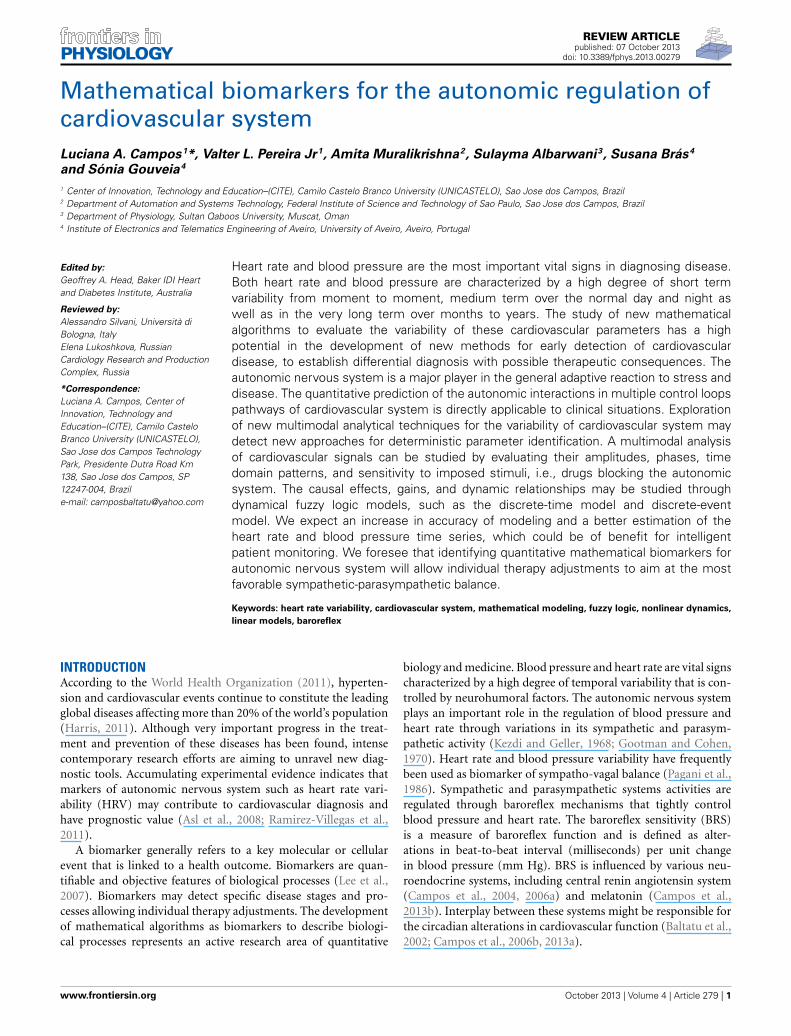
REVIEW ARTICLE published: 07 October 2013 doi: 10.3389/fphys.2013.00279 Mathematical biomarkers for the autonomic regulation of cardiovascular system Luciana A. Campos 1 *, Valter L. Pereira Jr 1 , Amita Muralikrishna 2 , Sulayma Albarwani 3 , Susana Brás 4 and Sónia Gouveia 4 1 Center of Innovation, Technology and Education–(CITE), Camilo Castelo Branco University (UNICASTELO), Sao Jose dos Campos, Brazil 2 Department of Automation and Systems Technology, Federal Institute of Science and Technology of Sao Paulo, Sao Jose dos Campos, Brazil 3 Department of Physiology, Sultan Qaboos University, Muscat, Oman 4 Institute of Electronics and Telematics Engineering of Aveiro, University of Aveiro, Aveiro, Portugal Edited by: Geoffrey A. Head, Baker IDI Heart and Diabetes Institute, Australia Reviewed by: Alessandro Silvani, Università di Bologna, Italy Elena Lukoshkova, Russian Cardiology Research and Production Complex, Russia *Correspondence: Luciana A. Campos, Center of Innovation, Technology and Education–(CITE), Camilo Castelo Branco University (UNICASTELO), Sao Jose dos Campos Technology Park, Presidente Dutra Road Km 138, Sao Jose dos Campos, SP 12247-004, Brazil e-mail: [email protected] Heart rate and blood pressure are the most important vital signs in diagnosing disease. Both heart rate and blood pressure are characterized by a high degree of short term variability from moment to moment, medium term over the normal day and night as well as in the very long term over months to years. The study of new mathematical algorithms to evaluate the variability of these cardiovascular parameters has a high potential in the development of new methods for early detection of cardiovascular disease, to establish differential diagnosis with possible therapeutic consequences. The autonomic nervous system is a major player in the general adaptive reaction to stress and disease. The quantitative prediction of the autonomic interactions in multiple control loops pathways of cardiovascular system is directly applicable to clinical situations. Exploration of new multimodal analytical techniques for the variability of cardiovascular system may detect new approaches for deterministic parameter identification. A multimodal analysis of cardiovascular signals can be studied by evaluating their amplitudes, phases, time domain patterns, and sensitivity to imposed stimuli, i.e., drugs blocking the autonomic system. The causal effects, gains, and dynamic relationships may be studied through dynamical fuzzy logic models, such as the discrete-time model and discrete-event model. We expect an increase in accuracy of modeling and a better estimation of the heart rate and blood pressure time series, which could be of benefit for intelligent patient monitoring. We foresee that identifying quantitative mathematical biomarkers for autonomic nervous system will allow individual therapy adjustments to aim at the most favorable sympathetic-parasympathetic balance. Keywords: heart rate variability, cardiovascular system, mathematical modeling, fuzzy logic, nonlinear dynamics, linear models, baroreflex INTRODUCTION According to the World Health Organization (2011), hyperten- sion and cardiovascular events continue to constitute the leading global diseases affecting more than 20% of the world’s population (Harris, 2011). Although very important progress in the treat- ment and prevention of these diseases has been found, intense contemporary research efforts are aiming to unravel new diag- nostic tools. Accumulating experimental evidence indicates that markers of autonomic nervous system such as heart rate vari- ability (HRV) may contribute to cardiovascular diagnosis and have prognostic value (Asl et al., 2008; Ramirez-Villegas et al., 2011). A biomarker generally refers to a key molecular or cellular event that is linked to a health outcome. Biomarkers are quan- tifiable and objective features of biological processes (Lee et al., 2007). Biomarkers may detect specific disease stages and pro- cesses allowing individual therapy adjustments. The development of mathematical algorithms as biomarkers to describe biologi- cal processes represents an active research area of quantitative biology and medicine. Blood pressure and heart rate are vital signs characterized by a high degree of temporal variability that is con- trolled by neurohumoral factors. The autonomic nervous system plays an important role in the regulation of blood pressure and heart rate through variations in its sympathetic and parasym- pathetic activity (Kezdi and Geller, 1968; Gootman and Cohen, 1970). Heart rate and blood pressure variability have frequently been used as biomarker of sympatho-vagal balance (Pagani et al., 1986). Sympathetic and parasympathetic systems activities are regulated through baroreflex mechanisms that tightly control blood pressure and heart rate. The baroreflex sensitivity (BRS) is a measure of baroreflex function and is defined as alter- ations in beat-to-beat interval (milliseconds) per unit change in blood pressure (mm Hg). BRS is influenced by various neu- roendocrine systems, including central renin angiotensin system (Campos et al., 2004, 2006a) and melatonin (Campos et al., 2013b). Interplay between these systems might be responsible for the circadian alterations in cardiovascular function (Baltatu et al., 2002; Campos et al., 2006b, 2013a). www.frontiersin.org October 2013 | Volume 4 | Article 279 | 1
-
Upload
independent -
Category
Documents
-
view
3 -
download
0
Transcript of Mathematical biomarkers for the autonomic regulation of cardiovascular system
REVIEW ARTICLEpublished: 07 October 2013
doi: 10.3389/fphys.2013.00279
Mathematical biomarkers for the autonomic regulation ofcardiovascular systemLuciana A. Campos1*, Valter L. Pereira Jr1, Amita Muralikrishna2, Sulayma Albarwani3, Susana Brás4
and Sónia Gouveia4
1 Center of Innovation, Technology and Education–(CITE), Camilo Castelo Branco University (UNICASTELO), Sao Jose dos Campos, Brazil2 Department of Automation and Systems Technology, Federal Institute of Science and Technology of Sao Paulo, Sao Jose dos Campos, Brazil3 Department of Physiology, Sultan Qaboos University, Muscat, Oman4 Institute of Electronics and Telematics Engineering of Aveiro, University of Aveiro, Aveiro, Portugal
Edited by:
Geoffrey A. Head, Baker IDI Heartand Diabetes Institute, Australia
Reviewed by:
Alessandro Silvani, Università diBologna, ItalyElena Lukoshkova, RussianCardiology Research and ProductionComplex, Russia
*Correspondence:
Luciana A. Campos, Center ofInnovation, Technology andEducation–(CITE), Camilo CasteloBranco University (UNICASTELO),Sao Jose dos Campos TechnologyPark, Presidente Dutra Road Km138, Sao Jose dos Campos, SP12247-004, Brazile-mail: [email protected]
Heart rate and blood pressure are the most important vital signs in diagnosing disease.Both heart rate and blood pressure are characterized by a high degree of short termvariability from moment to moment, medium term over the normal day and night aswell as in the very long term over months to years. The study of new mathematicalalgorithms to evaluate the variability of these cardiovascular parameters has a highpotential in the development of new methods for early detection of cardiovasculardisease, to establish differential diagnosis with possible therapeutic consequences. Theautonomic nervous system is a major player in the general adaptive reaction to stress anddisease. The quantitative prediction of the autonomic interactions in multiple control loopspathways of cardiovascular system is directly applicable to clinical situations. Explorationof new multimodal analytical techniques for the variability of cardiovascular system maydetect new approaches for deterministic parameter identification. A multimodal analysisof cardiovascular signals can be studied by evaluating their amplitudes, phases, timedomain patterns, and sensitivity to imposed stimuli, i.e., drugs blocking the autonomicsystem. The causal effects, gains, and dynamic relationships may be studied throughdynamical fuzzy logic models, such as the discrete-time model and discrete-eventmodel. We expect an increase in accuracy of modeling and a better estimation of theheart rate and blood pressure time series, which could be of benefit for intelligentpatient monitoring. We foresee that identifying quantitative mathematical biomarkers forautonomic nervous system will allow individual therapy adjustments to aim at the mostfavorable sympathetic-parasympathetic balance.
Keywords: heart rate variability, cardiovascular system, mathematical modeling, fuzzy logic, nonlinear dynamics,
linear models, baroreflex
INTRODUCTIONAccording to the World Health Organization (2011), hyperten-sion and cardiovascular events continue to constitute the leadingglobal diseases affecting more than 20% of the world’s population(Harris, 2011). Although very important progress in the treat-ment and prevention of these diseases has been found, intensecontemporary research efforts are aiming to unravel new diag-nostic tools. Accumulating experimental evidence indicates thatmarkers of autonomic nervous system such as heart rate vari-ability (HRV) may contribute to cardiovascular diagnosis andhave prognostic value (Asl et al., 2008; Ramirez-Villegas et al.,2011).
A biomarker generally refers to a key molecular or cellularevent that is linked to a health outcome. Biomarkers are quan-tifiable and objective features of biological processes (Lee et al.,2007). Biomarkers may detect specific disease stages and pro-cesses allowing individual therapy adjustments. The developmentof mathematical algorithms as biomarkers to describe biologi-cal processes represents an active research area of quantitative
biology and medicine. Blood pressure and heart rate are vital signscharacterized by a high degree of temporal variability that is con-trolled by neurohumoral factors. The autonomic nervous systemplays an important role in the regulation of blood pressure andheart rate through variations in its sympathetic and parasym-pathetic activity (Kezdi and Geller, 1968; Gootman and Cohen,1970). Heart rate and blood pressure variability have frequentlybeen used as biomarker of sympatho-vagal balance (Pagani et al.,1986). Sympathetic and parasympathetic systems activities areregulated through baroreflex mechanisms that tightly controlblood pressure and heart rate. The baroreflex sensitivity (BRS)is a measure of baroreflex function and is defined as alter-ations in beat-to-beat interval (milliseconds) per unit changein blood pressure (mm Hg). BRS is influenced by various neu-roendocrine systems, including central renin angiotensin system(Campos et al., 2004, 2006a) and melatonin (Campos et al.,2013b). Interplay between these systems might be responsible forthe circadian alterations in cardiovascular function (Baltatu et al.,2002; Campos et al., 2006b, 2013a).
www.frontiersin.org October 2013 | Volume 4 | Article 279 | 1

Campos et al. Cardiovascular mathematical biomarkers
BRS may be determined pharmacologically through injec-tion of vasoactive substances (such as phenylephrine and sodiumnitroprusside) and invasive determination of arterial pressure(Campos et al., 2013b). Non-invasive methods for BRS evalua-tion may be through measurement of heart rate/blood pressurechanges in response to deep breath, Valsalva maneuver (forcedexpiration against resistance), or tilt testing (passive body move-ment from a supine position to an upright tilt). These testsprovide an index of cardiovagal function of baroreflex (Levin,1966; Wheeler and Watkins, 1973; Borst et al., 1982; Cookeet al., 1998; Shields, 2009). However, they may be altered bysome factors, including rating and depth of breathing, positionof the subject, and the presence of some diseases, such as dia-betic neuropathy (Sundkvist et al., 1982; Low, 1993). Continuousmonitoring of heart rate offers the possibility of evaluating thespontaneous adaptations of BRS as result of dynamic variationsof blood pressure (Baltatu et al., 2001; Gouveia et al., 2009).Spectral analysis of blood pressure and HRV is applied to eval-uate spontaneous BRS (Head et al., 2001). Low and high fre-quency spectral components of the blood pressure oscillationsare related with the frequency oscillations in R-R interval dueto baroreflex activity (La Rovere et al., 2008). These methodsfor BRS evaluation have different clinical implications such asin diagnostic and clinical management of cardiovascular diseases(La Rovere et al., 2008).
HEART RATE VARIABILITYHeart rate can be defined as the number of cardiac cycles per unitof time. Cardiac cycle includes a period of relaxation (diastole)and a period of contraction (systole) of the heart, representingthe time period necessary for the given event to repeat itself.The time interval between two successive R waves in the electro-cardiogram (ECG) equals one cardiac cycle. Thus, the ECG canmeasure heart rate through the recording of electrical potentialsgenerated by the heart’s electrical activity. The frequency (f ) isthe inverse of the period (T), thus, f = 1/T. HRV is the variationof beat-to-beat intervals, also known as R-R intervals. The HRVindexes are obtained by analyzing the intervals between R waves,which can be captured by instruments including electrocardio-graph, digital-to-analog converter and cardio frequency meterfrom surface electrodes that are placed at specific points on thebody (Rajendra Acharya et al., 2006).
Time domain and frequency domain are two types of methodscommonly used to analyze cardiovascular variability. Both meth-ods apply to linear data structure. Time domain uses continuousmonitoring of cardiovascular parameters while frequency domainuses spectral analysis to express heart rate oscillation (Task force,1996).
TIME DOMAIN INDEXES OF CARDIOVASCULAR VARIABILITYLinear HRV analysis in time domain employs statistical methods.Data needs to be normalized before analysis. In order to make acomparison between different data sets, these have to be acquiredover similar periods of time. The mostly used periods of time are24 h (long term) and 5–30 min (short term). The time domainindexes are based on normal sinus beat-to-beat intervals (normal-to-normal, or NN), and the most commonly used are:
(a) standard deviation of all NN intervals (SDNN, milliseconds;Table 1). Its values depend on the length of the recordingdata: longer the length higher SDNN values. Therefore, acomparison of SDNN values of different length may lead toinappropriate interpretation (Task force, 1996). Low SDNNvalues are a predictor of high mortality in cardiovasculardiseases (Kleiger et al., 1987; Task force, 1996; Nolan et al.,1998);
(b) root mean square of the successive differences (RMSSD,milliseconds; Table 1). It is an indication of short-termHRV components (Task force, 1996), reflects parasympa-thetic activity and is correlated with sudden death in epilepsy(DeGiorgio et al., 2010) and fibrillation (Dash et al., 2009).
(c) adjacent successive NN intervals differing more than 50 ms(NN50; Table 1) and its percentage (pNN50). It indicatesshort-term HRV components and reflects parasympatheticactivity (Task force, 1996). The values of NN50 have beencorrelated with autonomic neuropathy in diabetic patients(Ewing et al., 1991).
(d) triangular index, calculated from the number of all NN inter-vals divided by the maximum of the density distribution(Table 1). Estimate the overall HRV over 24 h (Task force,1996) and it is influenced mainly by low frequencies (Maliket al., 1989).
(e) triangular interpolation of NN interval histogram (TINN;Table 1). It represents the baseline width of the distributionmeasured as a base of a histogram triangle approximating theRR interval distribution (Malik and Camm, 1993; Vanderleiet al., 2009). Estimate overall HRV over 24 h (Task force,1996) and it is influenced mainly by low frequencies (Maliket al., 1989).
FREQUENCY DOMAIN INDEXES OF CARDIOVASCULAR VARIABILITYLinear HRV analysis in frequency domain employs mathematicalalgorithms for frequency assignment. Physiological data collectedas a time series can be considered as a sum of sinusoidal oscilla-tions with different frequencies. The conversion of time domainanalysis to frequency domain can be done through a mathemat-ical transformation developed nearly two centuries ago (1807)by a French mathematician named Jean Baptiste Joseph Fourier(1768–1830). This process, called spectral analysis, allows signaldecomposition originated from time series (tachogram) in its dif-ferent frequency components, or in the so-called frequency bands.Noteworthy is that frequency refers to the number of times that aparticular phenomenon occurs related to time. Typically, the unitfor frequency is Hertz (Hz), which is equivalent to one cycle persecond.
From continuous recordings of heart rate, the total power (TP)is decomposed into three distinct bands in humans:
1. high frequency band (HF; Table 1) with frequency rangingfrom 0.15 to 0.40 Hz, related to the heart rate variations asso-ciated with the respiratory cycle, commonly called respiratorysinus arrhythmia. It is usually modulated by the parasympa-thetic nervous system (Kuusela et al., 2003; Rajendra Acharyaet al., 2006). HF is also known as “respiratory” band because
Frontiers in Physiology | Integrative Physiology October 2013 | Volume 4 | Article 279 | 2

Campos et al. Cardiovascular mathematical biomarkers
Table 1 | Time and frequency domain measures of heart rate variability (Kamen and Tonkin, 1995; Task force, 1996; Brennan et al., 2001).
Time and frequency domain heart rate variability indexes
Variable Units Description Formula
Tim
edo
mai
nin
dexe
s
SDNN ms Standard deviation of NN intervals SDNN =√
1N − 1
N∑j = 1
(RRj − RR)2
RMSSD ms Root mean square of successive NNdifferences
RMSD =√
1N − 1
N − 1∑j = 1
(RRj + 1 − RRj )2
NN50 count # Number of successive NN differenceslarger than 50 ms
Number of (RRj + 1 − RRj ) > 50
pNN50 % NN50 count divided by the totalnumber of all NN intervals
pNN50 = NN50N − 1
× 100
Triangular index Number of all NN intervals divided bythe maximum of the densitydistribution
NY
, where Y is the maximum of the NN
sample density distribution
TINN ms Robust range of NN duration M-N, where M and N representrespectively maximum and minimum NNwidth, obtained by triangular interpolationof NN histogram
Poincare plot SD1 ms Standard deviation perpendicular tothe line-of-identity axis
SD12 = 12
SDSD2, where SDSD is the
standard deviation of successive NNinterval differences
Poincare plot SD2 ms Standard deviation along theline-of-identity axis
SD22 = 2SDNN2 − 12
SDSD2
Freq
uenc
ydo
mai
nin
dexe
sFB
={V
LF,L
F,H
F,TF
}
fFB Hz Peak frequency Frequency with maximum amplitude in FB
pFB ms2 Absolute power power in FB
FB norm % Normalized powerpFB
pTF − pVLF× 100
FB, frequency bands; VLF, very low frequency; LF, low frequency; HF, high frequency; TF, total frequency.
it corresponds to the R-R fluctuations caused by breathing(Bernardi et al., 1989; Ori et al., 1992; Bernardi et al., 1995);
2. low frequency band (LF; Table 1) with frequency rangingfrom 0.04 to 0.15 Hz is modulated by both sympatheticand parasympathetic nervous systems (Kuusela et al., 2003;Rajendra Acharya et al., 2006);
3. very low frequency band (VLF; Table 1) with frequency rang-ing from 0.003 to 0.04 Hz, it is a variable which depends onthe renin-angiotensin system, whose regulation is also affectedby sympathetic and parasympathetic nervous systems (Tayloret al., 1998; Kuusela et al., 2003; Acharya et al., 2004). Thelower frequency of the VLF band depends on window length.It was demonstrated an association of VLF with mortality aftercardiac infarction (Bigger et al., 1992).
In the analysis of fetal heart rate (FHR) tracings, these frequencybands are usually adjusted to VLF (0–0.03 Hz), LF (0.03–0.15 Hz),and HF (0.5–1 Hz), and hold the same physiological associations
as in human adults. FHR recordings additionally exhibit a MFfrequency band related to the fetal movements and maternalbreathing (Signorini et al., 2003).
These frequency bands are not applicable for smaller animalssuch as rats and mice. In rats, the components of VLF to HF ofHRV power spectra vary between 0.0 and 3.0 Hz (Task force, 1996;Baltatu et al., 2001; Bezerra et al., 2001; Ushizima et al., 2001; Silvaet al., 2009), while in mice, the components of LF to HF varybetween 0.1 and 5.0 Hz (Ishii et al., 1996; Joaquim et al., 2004;Baudrie et al., 2007).
Measurement of spectral components is typically made inabsolute power (ms2). However, the values of LF and HF can alsobe expressed in normalized units (NU) representing the value ofeach component relative to the TP minus the VLF component(Task force, 1996).
Both time and frequency domain methods for variability eval-uation analyze linear properties of the data, as described above.These methods have limitations, as they require windowing of the
www.frontiersin.org October 2013 | Volume 4 | Article 279 | 3
Campos et al. Cardiovascular mathematical biomarkers
data and it uses deterministic algorithms that are valid only toperiodic phenomena. Therefore, there is a need to identify a hier-archy of models, each suited for a different type of investigationor to different parts of the system and devise strategies to couplethem, using a multiscale framework. Systems of differential-algebraic equations can be employed to study systemic behaviorand auto-regulation mechanisms.
Heart period and blood pressure are coupled in a closedloop. The coupling between heart period and blood pressurevia baroreflex regulation can be evaluated using cross-correlationanalysis (Silvani et al., 2008, 2011), regression analysis by theevents technique (Gouveia et al., 2009) and causal estimates(Acharya et al., 2004; Porta et al., 2011). Cross-correlationanalysis gives a correlation coefficient between variations ofheart period and blood pressure (Silvani et al., 2011). GeoffreyHead et al. demonstrated that cross-spectral coherence trans-fer function between heart period and blood pressure couldbe used effectively to reproducibly estimate BRS (Head et al.,2001). The transfer function gives information about coher-ence, gain and phase relation of both signals (Baltatu et al.,2001). Coupling between heart period and systolic blood pres-sure in healthy subjects varies among wake-sleep states (Silvaniet al., 2008). It was suggested that cross-correlation function isapplicable to study the balance between central autonomic andbaroreflex control of heart rate. For instance, there are posi-tive and negative correlations between heart period and bloodpressure values that result from baroreflex and central auto-nomic controls, respectively (Silvani et al., 2011). However, incases of BRS impairment, cross-correlation is likely to be sta-tistically not significant, due to autonomic balance alterations.To address this shortcoming, the so called events techniquebased on baroreflex events, was proposed to maximize simul-taneously the number of beats considered for BRS estimationand the correlation between the corresponding systolic bloodpressure and RR values, in order to provide a more accuratebaroreflex slope estimate (Gouveia et al., 2009). This techniquehas shown to accurately detect baroreflex alterations, associ-ated with postural changes (Gouveia et al., 2009) and drug-induced BRS stimulation (Beloka et al., 2009). This techniquehas also been demonstrated to be capable of providing segmentsof short and of long beat length, which were associated withparasympathetic and the sympathetic ANS activities, respectively(Gouveia et al., 2010).
One of the disadvantages is that cross-correlation and linearregression methods are able to estimate only the linear com-ponent of heart period and systolic blood pressure coupling(Silvani et al., 2008; Gouveia et al., 2009). The causal relation-ship theory between heart period and systolic blood pressurewas introduced by Oppenheim (Oppenheim and Schafer, 1975).This led to studies to find the dominant causal direction in theinteractions between heart period and systolic blood pressure.Cross-conditional entropy was used to study heart pulse-systolicblood pressure causality during head-up tilt test in heart trans-planted patients, and compared with classical approach of linearmethods. The head-up tilt induced progressive shift from theprevalent causal direction to the reverse causality in healthy sub-jects (Porta et al., 2011). Although cross-conditional entropy is a
tool to determine the causality, it is unable to account for the com-bined exogenous influences of respiration on R-R interval andsystolic blood pressure variability (Baselli et al., 1994; Cooke et al.,1999; Westerhof et al., 2006; Porta et al., 2011).
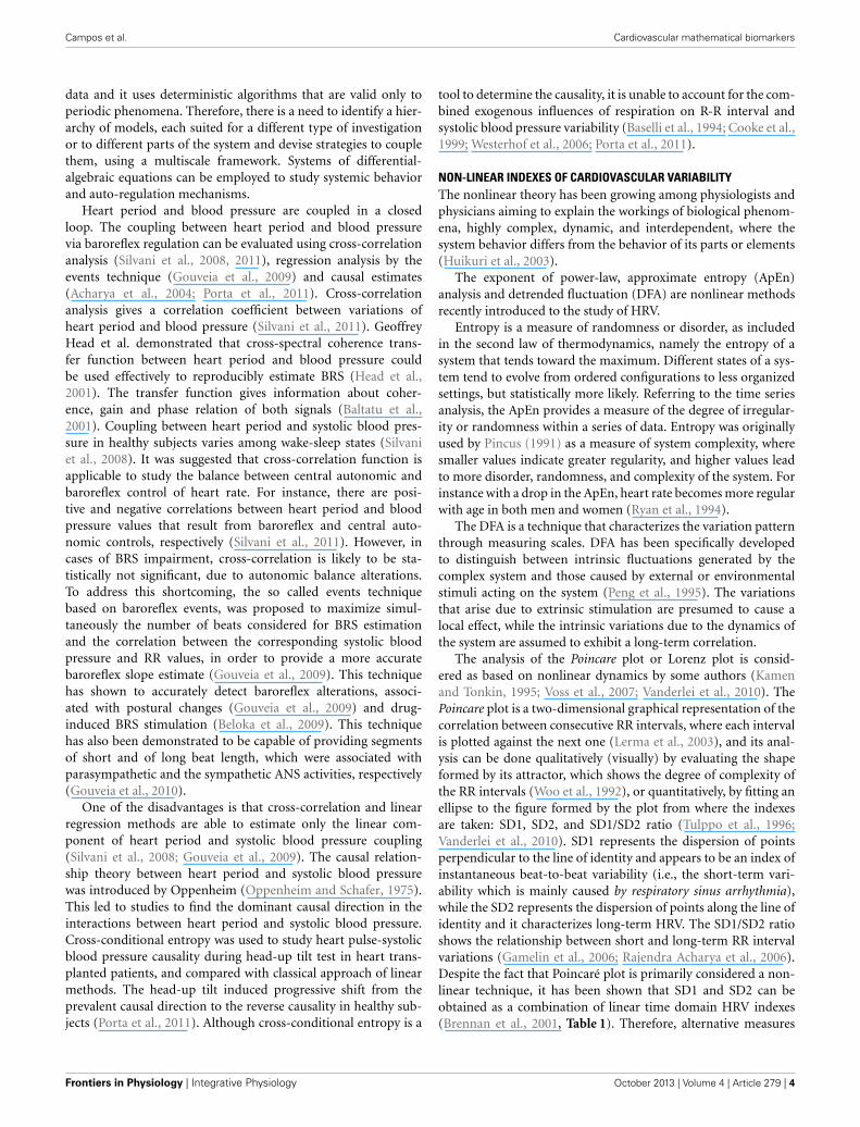
NON-LINEAR INDEXES OF CARDIOVASCULAR VARIABILITYThe nonlinear theory has been growing among physiologists andphysicians aiming to explain the workings of biological phenom-ena, highly complex, dynamic, and interdependent, where thesystem behavior differs from the behavior of its parts or elements(Huikuri et al., 2003).
The exponent of power-law, approximate entropy (ApEn)analysis and detrended fluctuation (DFA) are nonlinear methodsrecently introduced to the study of HRV.
Entropy is a measure of randomness or disorder, as includedin the second law of thermodynamics, namely the entropy of asystem that tends toward the maximum. Different states of a sys-tem tend to evolve from ordered configurations to less organizedsettings, but statistically more likely. Referring to the time seriesanalysis, the ApEn provides a measure of the degree of irregular-ity or randomness within a series of data. Entropy was originallyused by Pincus (1991) as a measure of system complexity, wheresmaller values indicate greater regularity, and higher values leadto more disorder, randomness, and complexity of the system. Forinstance with a drop in the ApEn, heart rate becomes more regularwith age in both men and women (Ryan et al., 1994).
The DFA is a technique that characterizes the variation patternthrough measuring scales. DFA has been specifically developedto distinguish between intrinsic fluctuations generated by thecomplex system and those caused by external or environmentalstimuli acting on the system (Peng et al., 1995). The variationsthat arise due to extrinsic stimulation are presumed to cause alocal effect, while the intrinsic variations due to the dynamics ofthe system are assumed to exhibit a long-term correlation.
The analysis of the Poincare plot or Lorenz plot is consid-ered as based on nonlinear dynamics by some authors (Kamenand Tonkin, 1995; Voss et al., 2007; Vanderlei et al., 2010). ThePoincare plot is a two-dimensional graphical representation of thecorrelation between consecutive RR intervals, where each intervalis plotted against the next one (Lerma et al., 2003), and its anal-ysis can be done qualitatively (visually) by evaluating the shapeformed by its attractor, which shows the degree of complexity ofthe RR intervals (Woo et al., 1992), or quantitatively, by fitting anellipse to the figure formed by the plot from where the indexesare taken: SD1, SD2, and SD1/SD2 ratio (Tulppo et al., 1996;Vanderlei et al., 2010). SD1 represents the dispersion of pointsperpendicular to the line of identity and appears to be an index ofinstantaneous beat-to-beat variability (i.e., the short-term vari-ability which is mainly caused by respiratory sinus arrhythmia),while the SD2 represents the dispersion of points along the line ofidentity and it characterizes long-term HRV. The SD1/SD2 ratioshows the relationship between short and long-term RR intervalvariations (Gamelin et al., 2006; Rajendra Acharya et al., 2006).Despite the fact that Poincaré plot is primarily considered a non-linear technique, it has been shown that SD1 and SD2 can beobtained as a combination of linear time domain HRV indexes(Brennan et al., 2001, Table 1). Therefore, alternative measures
Frontiers in Physiology | Integrative Physiology October 2013 | Volume 4 | Article 279 | 4

Campos et al. Cardiovascular mathematical biomarkers
are still needed to characterize nonlinear features in Poincaré plotgeometry.
FUZZY LOGIC CONCEPTSThe possibility of using mathematical methods and theories fordata analysis has opened up a range of possibilities for the studyof pathophysiological behaviors of cardiovascular variability (Huet al., 2009; Sassi et al., 2009; Gieraltowski et al., 2013). Largevolume of data can be more easily assessed and analyzed withfuzzy logic. In order to better understand the onset and devel-opment of important pathologies, the autonomic nervous systemactivity can be explored through dynamical fuzzy logic models,such as the discrete-time model and the discrete-event model.Fuzzy logic approaches are able to perform non-linear mappingor predictions involving more than one cardiovascular parameterand to explore possible relations among these parameters, whichnormally would not be considered as a possibility. Fuzzy logicrepresents a flexible system that adequately describes nonlinearand complex systems since the resulting function can be writ-ten as a weighted linear combination of the system inputs and,therefore, it can resemble a nonlinear function as needed. Forthis reason, fuzzy logic methods are a feasible solution to con-sider in the absence of prior mathematical description betweeninput-output variables (Kovacic and Bogdan, 2006).
Considering the Sugeno Fuzzy Logic formulation, the systemoutput z can be modeled from
z =
N∑i = 1
wizi
N∑i = 1
wi
,
where N corresponds to the number of fuzzy rules and zi =n∑
j = 1aixj + ci is a linear combination of the system inputs xj, j =
1, . . . n. The rule weights are obtained as wi =n∏
j = 1�Fi
j(xj) where
�Fij
is the membership function of rule i and input xj. Although
membership functions may assume different shapes, the Gaussianfunction is rather a popular choice in the literature due to itssymmetry and dependence on mean and variance, which corre-spond respectively to the center and the width of the membershipfunction.
Fuzzy logic has the singular characteristic to combine empir-ical knowledge (described as linguistic rules) and knowledgedirectly extracted from the data (Sadegh-Zadeh, 1999), enablingan easier way to interpret the outcomes in a physiological per-spective. This mathematical model may be a reliable method toevaluate the influence of the autonomic nervous system over car-diovascular control in healthy and diseased subjects (Carvalhoet al., 2002).
The main advantage of the use of fuzzy logic systems comesfrom their power to deal adequately with the uncertainty (Zadeh,1975; Kovacic and Bogdan, 2006). In particular, this approach tol-erates imprecise data, and it is focused on the “plausibility” ofoccurrence rather than the traditional binary response “0” or “1.”
For example, while a given measurement of a certain biologicalvariable such as stress may convey a person as being “content,”the same measurement may reveal a status of “dissatisfaction” foranother one. Thus, biological variables that vary from person toperson and are closely influenced by external and internal changesdirect themselves toward fuzzy logic model of analysis, wherethe application of methods of investigation based on zero andone, true and false does not apply (Zadeh, 1975). Cardiovascularsignals are characterized by a great intra- and inter-individualvariability, besides imprecise measurements due to limited res-olution of acquisition systems. Additionally, it is believed thattraditional statistical methods may not capture all the informa-tion needed to describe disease in its complexity and dynamics(Grossi, 2005). In this context, fuzzy logic may be a more reliablealternative to traditional methods.
APPLICATIONS OF FUZZY LOGICS TO THE ANALYSIS OFCARDIOVASCULAR VARIABILITYFuzzy logic approaches have been recently used in the cardiovas-cular field in different contexts including applications in signalprocessing and monitoring, classification, prediction or control.One approach consists of extracting the relevant features fromone or more cardiovascular signals, which are then integrated intoa fuzzy logic scheme aiming at the identification of the presenceor the quantification of a pathological state.
Fuzzy logic methods have been successfully integrated in con-trol systems. For instance during anesthesia, mean arterial pres-sure was controlled based on the error between desired andmeasured values, allowing it to control the balance between theunconsciousness and the side effects caused by the hypnoticdrug (Meier et al., 1992). Also during anesthesia, hemodynamicchanges were successfully modeled considering drug dose levelalterations as inputs of the fuzzy system (Nunes and Amorim,2008). In hemodialysis condition, fuzzy logic has also shownto be capable of effectively control blood pressure trends, usingultra-filtration rate as input (Mancini et al., 2007). Such a systemallowed an overall reduction of 40% of the most severe episodesin hypotension-prone subjects.
Abnormal cardiac rhythms have been identified using artificialneural network and fuzzy interactions based on nonlinear heartperiod R-R features, such as spectral entropy, Poincare SD1/SD2,and Lyapunov exponent (Acharya et al., 2004). Also based onR-R features, fuzzy logic was used for ECG beat classificationto detect arrhythmic and ischemic heartbeats (Tsipouras et al.,2007). Fuzzy logic approaches showed efficiency in improvingoscillometric cuff pressure measurements by properly detectingoutliers and noise artifacts (Lin et al., 2003).
With the goal of evaluating autonomic nervous system func-tion, fuzzy logic has been used to choose the optimum subsetof time, frequency and nonlinear variables related to sympa-thetic and parasympathetic activities on HRV (Petkovic et al.,2013). Fuzzy logic approach has been used in a classifica-tion scheme to jointly evaluate results of several autonomictests, e.g., head-up tilt test and active postural change, usingboth time and spectral analysis of heart rate and of dias-tolic blood pressure series (Carvalho et al., 2002). Similarfuzzy logic schemes were used for the information fusion of
www.frontiersin.org October 2013 | Volume 4 | Article 279 | 5

Campos et al. Cardiovascular mathematical biomarkers
relevant features extracted from multimodal cardiovascular sig-nals, such as heart period R-R and systolic blood pressure, forthe detection of life threatening states in cardiac care units(Kannathal et al., 2006).
Recently, fuzzy logic methods have been employed to effec-tively describe blood pressure and heart period R-R couplingand, therefore, have the potential to improve time domainBRS estimation (Liu et al., 2008; Gouveia and Bras, 2012).The autoregressive linear analysis approach for BRS estimationhas limitations when cardiovascular regulation is depressed. Liuet al. proposed a hybrid model consisting of a parallel mod-ular structure with an autoregressive and a fuzzy logic sys-tem, to study simultaneously linear and non-linear heart rateand blood pressure coupling mechanisms (Liu et al., 2008).This approach illustrates the utility of combining more tradi-tional methods with fuzzy logic, which could be of advantagein diseased conditions when cardiovascular system regulation isafflicted.
Time domain BRS methods based on spontaneous data typi-cally assume blood pressure and heart period R-R linearity andprovide single slope estimation, regardless of the blood pres-sure value (Beloka et al., 2009; Gouveia et al., 2009). In thiscontext, fuzzy logic methods can contribute to establish a BRSdependent of blood pressure level, similarly to time domainblood pressure pharmacological methods. Recently, fuzzy logichas been used to analyze spontaneous R-R series as a function ofblood pressure values, comparing performances in real and sur-rogate data (Gouveia and Bras, 2012). As an illustrative example,Figure 1 shows fuzzy logic curves and the membership functionsfor a healthy subject. The fuzzy logic curve obtained from realdata is much less flatter than that obtained from isodistributionsurrogate data (gained by shuffling the RR data) and exhibitssignificantly lower average modeling errors (Figure 1A). Also,the non-uniform location of the membership functions suggeststhat different systolic blood pressure values contribute differentlyto RR modeling (Figure 1B). These results indicate that fuzzylogic is able to model R-R changes connected to blood pressurealterations, besides the mean value and, thus, has the potentialto improve time domain BRS estimation in spontaneous con-ditions. It remains to be assessed the clinical impact of thesefindings and inherent repercussion on BRS estimation. Finally,Figure 2 illustrates the flexibility of fuzzy logic-based modelingin the identification of heterogeneous shapes and patterns insystolic blood pressure and RR association for a set of severalsubjects.
Given the complexity of the mechanisms regulating heartrate and its non-linear characteristics, it is reasonable to assumethat HRV analysis based on non-linear methods would gener-ate valuable knowledge on the systems involved in the HRVregulation. The mainly used non-linear method of HRV anal-ysis is Poincaré plot, which although simple and easy to use,it does not reflect the number of samples at each point of thegraph, leading to errors of judgment of its plots (Hnatkovaet al., 1995). The use of fuzzy logic and the consequent useof a “plausibility” rather than a binary logic may help toovercome the drawbacks found in the traditional methods ofanalysis.
FIGURE 1 | Fuzzy functions obtained for one healthy subject: (A) Fuzzy
curve in solid line was obtained from real data (represented in gray)
and Fuzzy curve in dashed line was estimated from an isodistribution
surrogate realization; (B) membership functions obtained from real
data.
FIGURE 2 | Fuzzy surfaces obtained for a set of 23 subjects in Lying
condition [EuroBaVar dataset (Gouveia and Bras, 2012)], with a curve
in each subplot according to one subject. Subplot (A) shows the fuzzycurves obtained from real data and subplot (B) shows the fuzzy curvesestimated from an isodistribution surrogate data.
CONCLUDING REMARKSFuzzy logic is a suitable choice when the system deals withuncertainty data, when linguistic interpretation is needed andwhen data “plausibility” should be taken into account. Fuzzylogic has been successfully used in different scenarios forthe analysis of cardiovascular variability, with special empha-sis on control and classification systems. Recent studies pointout fuzzy logic also as a modeling alternative for cardiovas-cular time series, with potential impact on the estimation ofjoint parameters, e.g., arterial BRS. Therefore, fuzzy logic isa promising approach for the analysis of cardiovascular sys-tem and its regulatory mechanisms in normal and diseasedconditions.
We expect an increase in accuracy of modeling and a bet-ter estimation of the heart rate and blood pressure time series,which could be of benefit for intelligent patient monitoring. Weforesee that identifying quantitative mathematical biomarkers forautonomic nervous system will allow individual therapy adjust-ments to aim at the most favorable sympathetic-parasympatheticbalance.
ACKNOWLEDGMENTSThis review was partially supported by São Paulo ResearchFoundation (Grants 2013/06698-0, 2013/50137-2), EuropeanRegional Development Fund (FEDER) through the programmeCOMPETE and by the Portuguese Government through the
Frontiers in Physiology | Integrative Physiology October 2013 | Volume 4 | Article 279 | 6

Campos et al. Cardiovascular mathematical biomarkers
FCT, Fundação para a Ciência e a Tecnologia, in thescope of the projects FCOMP-01-0124-FEDER-022682 (FCTref. PEst-C/EEI/UI0127/2011), Incentivo/EEI/UI0127/2013(Instituto de Engenharia Electrónica e Telemática de Aveiro,IEETA/UA, Aveiro, www.ieeta.pt), and Cloud Thinking
(CENTRO-07-ST24-FEDER-002031, co-funded by QREN, “MaisCentro” program). Susana Brás acknowledges the postdoctoralgrant by Cloud Thinking Project (ref. BPD/DETI/5259/2013).Sónia Gouveia acknowledges the postdoctoral grant by FCT (ref.SFRH/BPD/87037/2012).
REFERENCESAcharya, R., Kumar, A., Bhat, P. S., Lim,
C. M., Iyengar, S. S., Kannathal, N.,et al. (2004). Classification of car-diac abnormalities using heart ratesignals. Med. Biol. Eng. Comp. 42,288–293. doi: 10.1007/BF02344702
Asl, B. M., Setarehdan, S. K., andMohebbi, M. (2008). Supportvector machine-based arrhythmiaclassification using reduced featuresof heart rate variability signal.Artif. Intell. Med. 44, 51–64. doi:10.1016/j.artmed.2008.04.007
Baltatu, O., Afeche, S. C., Jose dosSantos, S. H., Campos, L. A.,Barbosa, R., Michelini, L. C.,et al. (2002). Locally synthe-sized angiotensin modulatespineal melatonin generation.J. Neurochem. 80, 328–334. doi:10.1046/j.0022-3042.2001.00701.x
Baltatu, O., Janssen, B. J., Bricca,G., Plehm, R., Monti, J., Ganten,D., et al. (2001). Alterations inblood pressure and heart ratevariability in transgenic ratswith low brain angiotensinogen.Hypertension 37, 408–413. doi:10.1161/01.HYP.37.2.408
Baselli, G., Cerutti, S., Badilini, F.,Biancardi, L., Porta, A., Pagani, M.,et al. (1994). Model for the assess-ment of heart period and arte-rial pressure variability interactionsand of respiration influences. Med.Biol. Eng. Comput. 32, 143–152. doi:10.1007/BF02518911
Baudrie, V., Laude, D., and Elghozi,J. L. (2007). Optimal frequencyranges for extracting informationon cardiovascular autonomic con-trol from the blood pressure andpulse interval spectrograms inmice. Am. J. Physiol. Regul. Integr.Comp. Physiol. 292, R904–R912.doi: 10.1152/ajpregu.00488.2006
Beloka, S. P., Gouveia, S., Gujic, M.,Naeije, R., Rocha, A. P., and vande Borne, P. (2009). Differentialeffects of oral beta blockadeon cardiovascular and sympa-thetic regulation. J. Cardiovasc.Pharmacol. Ther. 14, 323–331. doi:10.1177/1074248409350137
Bernardi, L., Bianchini, B., Spadacini,G., Leuzzi, S., Valle, F., Marchesi, E.,et al. (1995). Demonstrable cardiacreinnervation after human hearttransplantation by carotid barore-flex modulation of RR interval.
Circulation 92, 2895–2903. doi:10.1161/01.CIR.92.10.2895
Bernardi, L., Keller, F., Sanders, M.,Reddy, P. S., Griffith, B., Meno,F., et al. (1989). Respiratory sinusarrhythmia in the denervatedhuman heart. J. Appl. Physiol. 67,1447–1455.
Bezerra, S. M., dos Santos, C. M.,Moreira, E. D., Krieger, E. M., andMichelini, L. C. (2001). ChronicAT(1) receptor blockade altersautonomic balance and sympa-thetic responses in hypertension.Hypertension 38, 569–575. doi:10.1161/hy09t1.095393
Bigger, J. T. Jr., Fleiss, J. L., Steinman,R. C., Rolnitzky, L. M., Kleiger,R. E., and Rottman, J. N. (1992).Frequency domain measures ofheart period variability and mor-tality after myocardial infarction.Circulation 85, 164–171. doi:10.1161/01.CIR.85.1.164
Borst, C., Wieling, W., van Brederode,J. F., Hond, A., de Rijk, L. G., andDunning, A. J. (1982). Mechanismsof initial heart rate response to pos-tural change. Am. J. Physiol. 243,H676–H681.
Brennan, M., Palaniswami, M., andKamen, P. (2001). Do existing mea-sures of Poincaré plot geometryreflect nonlinear features of heartrate variability? IEEE Trans. BiomedEng. 48, 1342–1347. doi: 10.1109/10.959330
Campos, L. A., Cipolla-Neto, J.,Amaral, F. G., Michelini, L.C., Bader, M., and Baltatu, O.C. (2013a). The Angiotensin-melatonin axis. Int. J. Hypertens.2013, 521783. doi: 10.1155/2013/521783
Campos, L. A., Cipolla-Neto, J., andMichelini, L. C. (2013b). Melatoninmodulates baroreflex control viaarea postrema. Brain Behav. 3,171–177. doi: 10.1002/brb3.123
Campos, L. A., Couto, A. S., Iliescu, R.,Santos, J. A., Santos, R. A., Ganten,D., et al. (2004). Differential reg-ulation of central vasopressinreceptors in transgenic ratswith low brain angiotensino-gen. Regul. Pept. 119, 177–182. doi:10.1016/j.regpep.2004.02.001
Campos, L. A., Iliescu, R., Fontes,M. A., Schlegel, W. P., Bader,M., and Baltatu, O. C. (2006a).Enhanced isoproterenol-induced
cardiac hypertrophy in transgenicrats with low brain angiotensino-gen. Am. J. Physiol. Heart Circ.Physiol. 291, H2371–H2376. doi:10.1152/ajpheart.01145.2005
Campos, L. A., Plehm, R., Cipolla-Neto, J., Bader, M., and Baltatu,O. C. (2006b). Altered circadianrhythm reentrainment to lightphase shifts in rats with low levelsof brain angiotensinogen. Am.J. Physiol. Regul. Integr. Comp.Physiol. 290, R1122–R1127. doi:10.1152/ajpregu.00703.2005
Carvalho, H. S., Junqueira, L. F.,and Souza-Neto, J. (2002). Acomputerized fuzzy logic systemfor evaluation of the cardiovas-cular autonomic function basedon multiple functional tests.Comput. Cardiol. 29, 173–176. doi:10.1109/CIC.2002.1166735
Cooke, W. H., Cox, J. F., Diedrich, A.M., Taylor, J. A., Beightol, L. A.,Ames J. E., et al. (1998). Controlledbreathing protocols probe humanautonomic cardiovascular rhythms.Am. J. Physiol. 274, H709–H718.
Cooke, W. H., Hoag, J. B., Crossman,A. A., Kuusela, T. A., Tahvanainen,K. U., and Eckberg, D. L.(1999). Human responses toupright tilt: a window on cen-tral autonomic integration.J. Physiol. 517(Pt 2), 617–628. doi:10.1111/j.1469-7793.1999.0617t.x
Dash, S., Chon, K. H., Lu, S., andRaeder, E. A. (2009). Automatic realtime detection of atrial fibrillation.Ann. Biomed. Eng. 37, 1701–1709.doi: 10.1007/s10439-009-9740-z
DeGiorgio, C. M., Miller, P.,Meymandi, S., Chin, A., Epps,J., Gordon, S., et al. (2010).RMSSD, a measure of vagus-mediated heart rate variability,is associated with risk factors forSUDEP: the SUDEP-7 Inventory.Epilepsy Behav. 19, 78–81. doi:10.1016/j.yebeh.2010.06.011
Ewing, D. J., Neilson, J. M., Shapiro,C. M., Stewart, J. A., and Reid, W.(1991). Twenty four hour heart ratevariability: effects of posture, sleep,and time of day in healthy controlsand comparison with bedside testsof autonomic function in diabeticpatients. Br. Heart J. 65, 239–244.doi: 10.1136/hrt.65.5.239
Gamelin, F. X., Berthoin, S., andBosquet, L. (2006). Validity of
the polar S810 heart rate monitorto measure R-R intervals at rest.Med. Sci. Sports Exerc. 38, 887–893.doi: 10.1249/01.mss.0000218135.79476.9c
Gieraltowski, J., Hoyer, D., Tetschke,F., Nowack, S., Schneider, U., andZebrowski, J. (2013). Developmentof multiscale complexity andmultifractality of fetal heart ratevariability. Auton. Neurosci. doi:10.1016/j.autneu.2013.01.009
Gootman, P. M., and Cohen, M. I.(1970). Efferent splanchnic activityand systemic arterial pressure. Am.J. Physiol. 219, 897–903.
Gouveia, S., and Bras, S. (2012).“Exploring the use of fuzzy logicmodels to describe the relationbetween, SBP, and RR values,” inConference proceedings: AnnualInternational Conference of theIEEE Engineering in Medicine, andBiology Society IEEE Engineeringin Medicine, and Biology SocietyConference 2012. 2827–2830.
Gouveia, S., Rocha, A. P., Laguna,P., and Lago, P. (2009). Timedomain baroreflex sensitivity assess-ment by joint analysis of sponta-neous SBP and RR series. Biomed.Signal. Proces. 4, 254–261. doi:10.1016/j.bspc.2009.03.003
Gouveia, S., Rocha, A. P., Laguna,P., and van de Borne, P. (2010).Correlation between time domainbaroreflex sensitivity and sym-pathetic nerve activity. Comput.Cardiol. 37, 5–8.
Grossi, E. (2005). Medical conceptsrelated to individual risk are betterexplained with “plausibility” ratherthan “probability”. BMC Cardiovasc.Disord. 5:31. doi: 10.1186/1471-2261-5-31
Harris, A. (2011). WHO: WorldHealth Organization. CharlestonAdvisor 12, 54–56. doi:10.5260/chara.12.4.54
Head, G. A., Lukoshkova, E. V., Burke,S. L., Malpas, S. C., Lambert,E. A., and Janssen, B. J. (2001).Comparing spectral and invasiveestimates of baroreflex gain. IEEEEng. Med. Biol. Mag. 20, 43–52. doi:10.1109/51.917723
Hnatkova, K., Copie, X., Staunton, A.,and Malik, M. (1995). Numericprocessing of Lorenz plots of R-Rintervals from long-term ECGs.Comparison with time-domain
www.frontiersin.org October 2013 | Volume 4 | Article 279 | 7

Campos et al. Cardiovascular mathematical biomarkers
measures of heart rate variabilityfor risk stratification after myocar-dial infarction. J. Electrocardiol.28(Suppl.), 74–80. doi: 10.1016/S0022-0736(95)80020-4
Hu, J., Gao, J., and Tung, W. W. (2009).Characterizing heart rate variabil-ity by scale-dependent Lyapunovexponent. Chaos 19, 028506. doi:10.1063/1.3152007
Huikuri, H. V., Makikallio, T. H., andPerkiomaki, J. (2003). Measurementof heart rate variability by methodsbased on nonlinear dynamics.J. Electrocardiol. 36(Suppl.), 95–99.doi: 10.1016/j.jelectrocard.2003.09.021
Ishii, K., Kuwahara, M., Tsubone, H.,and Sugano, S. (1996). Autonomicnervous function in mice and voles(Microtus arvalis): investigation bypower spectral analysis of heart ratevariability. Lab. Anim. 30, 359–364.doi: 10.1258/002367796780739880
Joaquim, L. F., Farah, V. M., Bernatova,I., Fazan, R. Jr., Grubbs, R., andMorris, M. (2004). Enhanced heartrate variability and baroreflex indexafter stress and cholinesterase inhi-bition in mice. Am.J. Physiol. HeartCirc. Physiol. 287, H251–H257. doi:10.1152/ajpheart.01136.2003
Kamen, P. W., and Tonkin, A. M.(1995). Application of the Poincaréplot to heart rate variability: anew measure of functional sta-tus in heart failure. Aust. NZ. J.Med. 25, 18–26. doi: 10.1111/j.1445-5994.1995.tb00573.x
Kannathal, N., Acharya, U. R., Ng,E. Y., Krishnan, S. M., Min, L.C., and Laxminarayan, S. (2006).Cardiac health diagnosis usingdata fusion of cardiovascular andhaemodynamic signals. Comput.Methods Prog. Biomed. 82, 87–96.doi: 10.1016/j.cmpb.2006.01.009
Kezdi, P., and Geller, E. (1968).Baroreceptor control of postgan-glionic sympathetic nerve discharge.Am. J. Physiol. 214, 427–435.
Kleiger, R. E., Miller, J. P., Bigger,J. T. Jr., and Moss, A. J. (1987).Decreased heart rate variability andits association with increased mor-tality after acute myocardial infarc-tion. Am. J. Cardiol. 59, 256–262.doi: 10.1016/0002-9149(87)90795-8
Kovacic, Z., and Bogdan, S. (2006).Fuzzy Controller Design, Theory andApplications. Boca Raton, FL: Taylorand Francis Group.
Kuusela, T. A., Kaila, T. J., and Kahonen,M. (2003). Fine structure of the low-frequency spectra of heart rate andblood pressure. BMC Physiol. 3:11.doi: 10.1186/1472-6793-3-11
La Rovere, M. T., Pinna, G. D.,and Raczak, G. (2008). Baroreflex
sensitivity: measurement and clin-ical implications. Ann. NoninvasiveElectrocardiol. 13, 191–207. doi:10.1111/j.1542-474X.2008.00219.x
Lee, J. W., Figeys, D., and Vasilescu,J. (2007). Biomarker assay transla-tion from discovery to clinical stud-ies in cancer drug development:quantification of emerging pro-tein biomarkers. Adv. Cancer Res.96, 269–298. doi: 10.1016/S0065-230X(06)96010-2
Lerma, C., Infante, O., Perez-Grovas,H., and Jose, M. V. (2003).Poincare plot indexes of heartrate variability capture dynamicadaptations after haemodialysis inchronic renal failure patients. Clin.Physiol. Func. Imag. 23, 72–80. doi:10.1046/j.1475-097X.2003.00466.x
Levin, A. B. (1966). A simple test of car-diac function based upon the heartrate changes induced by the Valsalvamaneuver. Am. J. Cardiol. 18, 90–99.doi: 10.1016/0002-9149(66)90200-1
Lin, C. T., Liu, S. H., Wang, J. J., andWen, Z. C. (2003). Reduction ofinterference in oscillometric arte-rial blood pressure measurementusing fuzzy logic. IEEE Trans.Biomed. Eng. 50, 432–441. doi:10.1109/TBME.2003.809502
Liu, J., McKenna, T. M., Gribok, A.,Beidleman, B. A., Tharion, W. J.,and Reifman, J. (2008). A fuzzy logicalgorithm to assign confidence lev-els to heart and respiratory rate timeseries. Physiol. Meas. 29, 81–94. doi:10.1088/0967-3334/29/1/006
Low, P. A. (1993). Autonomicnervous system function.J. Clin. Neurophysiol. 10, 14–27.doi: 10.1097/00004691-199301000-00003
Malik, M., and Camm, A. J. (1993).Components of heart ratevariability–what they really meanand what we really measure.Am. J. Cardiol. 72, 821–822. doi:10.1016/0002-9149(93)91070-X
Malik, M., Farrell, T., Cripps, T., andCamm, A. J. (1989). Heart rate vari-ability in relation to prognosis aftermyocardial infarction: selection ofoptimal processing techniques. Eur.Heart J. 10, 1060–1074.
Mancini, E., Mambelli, E., Irpinia, M.,Gabrielli, D., Cascone, C., Conte,F., et al. (2007). Prevention ofdialysis hypotension episodes usingfuzzy logic control system. Nephrol.Dial. Transpl. 22, 1420–1427. doi:10.1093/ndt/gfl799
Meier, R., Nieuwland, J., Zbinden, A.M., and Hacisalihzade, S. S. (1992).Fuzzy logic control of humanblood pressure during anesthesia.IEEE Control Syst. 12, 12–17. doi:10.1109/37.168811
Nolan, J., Batin, P. D., Andrews, R.,Lindsay, S. J., Brooksby, P., Mullen,M., et al. (1998). Prospectivestudy of heart rate variability andmortality in chronic heart failure:results of the United Kingdomheart failure evaluation and assess-ment of risk trial (UK-heart).Circulation 98, 1510–1516. doi:10.1161/01.CIR.98.15.1510
Nunes, C. S., and Amorim, P. (2008).“A neuro-fuzzy approach for pre-dicting hemodynamic responsesduring anesthesia,” in Conferenceproceedings: Annual InternationalConference of the IEEE Engineeringin Medicine and Biology Society IEEEEngineering in Medicine and BiologySociety Conference 2008, 5814–5817.
Oppenheim, A. V., and Schafer, R.W. (1975). Digital Signal Processing.Englewood Cliffs, NJ: Prentice Hall.
Ori, Z., Monir, G., Weiss, J., Sayhouni,X., and Singer, D. H. (1992). Heartrate variability. Frequency domainanalysis. Cardiol. Clin. 10, 499–537.
Pagani, M., Lombardi, F., Guzzetti, S.,Rimoldi, O., Furlan, R., Pizzinelli,P., et al, (1986). Power spectralanalysis of heart rate and arte-rial pressure variabilities as amarker of sympatho-vagal inter-action in man and consciousdog. Circulation 59, 178–193. doi:10.1161/01.RES.59.2.178
Peng, C. K., Havlin, S., Stanley,H. E., and Goldberger, A. L.(1995). Quantification of scalingexponents and crossover phenom-ena in nonstationary heartbeattime series. Chaos 5, 82–87. doi:10.1063/1.166141
Petkovic, D., Cojbasic, Z., and Lukic,S. (2013). Adaptive neuro fuzzyselection of heart rate variabilityparameters affected by auto-nomic nervous system. ExpertSyst. Appl. 40, 4490–4495. doi:10.1016/j.eswa.2013.01.055
Pincus, S. M. (1991). Approximateentropy as a measure of systemcomplexity. Proc. Natl. Acad.Sci. U.S.A. 88, 2297–2301. doi:10.1073/pnas.88.6.2297
Porta, A., Catai, A. M., Takahashi,A. C., Magagnin, V., Bassani,T., Tobaldini, E., et al. (2011).Causal relationships between heartperiod and systolic arterial pressureduring graded head-up tilt. Am.J. Physiol. Regul. Integr. Comp.Physiol. 300, R378–R386. doi:10.1152/ajpregu.00553.2010
Rajendra Acharya, U., Paul Joseph,K., Kannathal, N., Lim, C. M.,and Suri, J. S. (2006). Heart ratevariability: a review. Med. Biol.Eng. Comput. 44, 1031–1051. doi:10.1007/s11517-006-0119-0
Ramirez-Villegas, J. F., Lam-Espinosa,E., Ramirez-Moreno, D. F.,Calvo-Echeverry, P. C., andAgredo-Rodriguez, W. (2011).Heart rate variability dynamics forthe prognosis of cardiovascularrisk. PLoS ONE 6, e17060. doi:10.1371/journal.pone.0017060
Ryan, S. M., Goldberger, A. L., Pincus,S. M., Mietus, J., and Lipsitz, L. A.(1994). Gender- and age-related dif-ferences in heart rate dynamics: arewomen more complex than men?J. Am. Coll. Cardiol. 24, 1700–1707.doi: 10.1016/0735-1097(94)90177-5
Sadegh-Zadeh, K. (1999). Advancesin fuzzy theory. Artif. Intell. Med.15, 309–323. doi: 10.1016/S0933-3657(98)00060-8
Sassi, R., Signorini, M. G., and Cerutti,S. (2009). Multifractality and heartrate variability. Chaos 19, 028507.doi: 10.1063/1.3152223
Shields, R. W. Jr. (2009). Heart ratevariability with deep breathing as aclinical test of cardiovagal function.Cleve. Clin. J. Med. 76(Suppl. 2),S37–S40. doi: 10.3949/ccjm.76.s2.08
Signorini, M. G., Margenes, G.,Cerutti, S., and Arduini, D. (2003).Linear and nonlinear parame-ters for the analysis of fetal heartrate signal from cardiotoco-graphic recordings. IEEE Trans.Biomed. Eng. 50, 365–374. doi:10.1109/TBME.2003.808824
Silva, G. J., Ushizima, M. R., Lessa,P. S., Cardoso, L., Drager, L. F.,Atala, M. M., et al. (2009). Criticalanalysis of autoregressive and fastFourier transform markers ofcardiovascular variability in ratsand humans. Braz. J. Med. Biol.Res.42, 386–396. doi: 10.1590/S0100-879X2009000400012
Silvani, A., Grimaldi, D., Vandi, S.,Barletta, G., Vetrugno, R., Provini,F., et al. (2008). Sleep-dependentchanges in the coupling betweenheart period and blood pres-sure in human subjects. Am.J. Physiol. Regul. Integr. Comp.Physiol. 294, R1686–R1692. doi:10.1152/ajpregu.00756.2007
Silvani, A., Magosso, E., Bastianini,S., Lenzi, P., and Ursino, M.(2011). Mathematical model-ing of cardiovascular coupling:central autonomic commandsand baroreflex control. Auton.Neurosci. Basic Clin. 162, 66–71.doi: 10.1016/j.autneu.2011.04.003
Sundkvist, G., Lilja, B., and Almér, L.O. (1982). Deep breathing, Valsalva,and tilt table tests in diabetics withand without symptoms of auto-nomic neuropathy. Acta Med. Scand.211, 369–373. doi: 10.1111/j.0954-6820.1982.tb01964.x
Frontiers in Physiology | Integrative Physiology October 2013 | Volume 4 | Article 279 | 8

Campos et al. Cardiovascular mathematical biomarkers
Task force. (1996). Heart rate vari-ability: standards of measurement,physiological interpretation andclinical use. Task force of theeuropean society of cardiologyand the north american societyof pacing and electrophysiology.Circulation 93, 1043–1065. doi:10.1161/01.CIR.93.5.1043
Taylor, J. A., Carr, D. L., Myers, C.W., and Eckberg, D. L. (1998).Mechanisms underlying very-low-frequency RR-interval oscillationsin humans. Circulation 98,547–555. doi: 10.1161/01.CIR.98.6.547
Tsipouras, M. G., Exarchos, T. P., andFotiadis, D. I. (2007). “Integrationof global and local knowledge forfuzzy expert system creation: appli-cation to arrhythmic beat classifi-cation,” in Conference proceedings:Annual International Conference ofthe IEEE Engineering in Medicineand Biology Society IEEE Engineeringin Medicine and Biology SocietyConference 2007, 3840–3843. doi:10.1109/IEMBS.2007.4353170
Tulppo, M. P., Makikallio, T. H., Takala,T. E., Seppanen, T., and Huikuri,H. V. (1996). Quantitative beat-to-beat analysis of heart rate dynamics
during exercise. Am. J. Physiol. 271,H244–H252.
Ushizima, M. R., Moreira, E. D., Costa,E. T., Castiglioni, P., Krieger, E. M.,Di Rienzo, M., et al. (2001). Effectsof sinoaortic denervation on bloodpressure, pulse interval, sympatheticnerve activity and diaphragmaticelectromyogram variability in con-scious rats. Comput. Cardiol. 28,485–488.
Vanderlei, L. C., Pastre, C. M., Freitas,I. F. Jr., and Godoy, M. F. (2010).Geometric indexes of heart ratevariability in obese and eutrophicchildren. Arq. Bras. Cardiol.95, 35–40. doi: 10.1590/S0066-782X2010005000082
Vanderlei, L. C., Pastre, C. M., Hoshi,R. A., Carvalho, T. D., and Godoy,M. F. (2009). Basic notions of heartrate variability and its clinical appli-cability. Rev. Bras. Cir. Cardiovasc.24, 205–217. doi: 10.1590/S0102-76382009000200018
Voss, A., Schroeder, R., Truebner, S.,Goernig, M., Figulla, H. R., andSchirdewan, A. (2007). Comparisonof nonlinear methods symbolicdynamics, detrended fluctuation,and Poincare plot analysis in riskstratification in patients with dilated
cardiomyopathy. Chaos 17, 015120.doi: 10.1063/1.2404633
Westerhof, B. E., Gisolf, J., Karemaker,J. M., Wesseling, K. H., Secher,N. H., and van Lieshout, J. J.(2006). Time course analysis ofbaroreflex sensitivity during pos-tural stress. Am. J. Physiol. HeartCirc. Physiol. 291, H2864–H2874.doi: 10.1152/ajpheart.01024.2005
Wheeler, T., and Watkins, P. J. (1973).Cardiac denervation in diabetes.Br. Med. J. 4, 584–586. doi:10.1136/bmj.4.5892.584
Woo, M. A., Stevenson, W. G., Moser,D. K., Trelease, R. B., and Harper,R. M. (1992). Patterns of beat-to-beat heart rate variability inadvanced heart failure. Am. HeartJ. 123, 704–710. doi: 10.1016/0002-8703(92)90510-3
World Health Organization.(2011). Global Status Reporton Noncommunicable Diseases,2010. Geneva: World HealthOrganization.
Zadeh, L. A. (1975). Fuzzy logic andapproximate reasoning. Synthese 30,407–428. doi: 10.1007/BF00485052
Conflict of Interest Statement: Theauthors declare that the research
was conducted in the absence of anycommercial or financial relationshipsthat could be construed as a potentialconflict of interest.
Received: 30 July 2013; paper pendingpublished: 12 August 2013; accepted: 17September 2013; published online: 07October 2013.Citation: Campos LA, Pereira VL Jr,Muralikrishna A, Albarwani S, Brás Sand Gouveia S (2013) Mathematicalbiomarkers for the autonomic regulationof cardiovascular system. Front. Physiol.4:279. doi: 10.3389/fphys.2013.00279This article was submitted to IntegrativePhysiology, a section of the journalFrontiers in Physiology.Copyright © 2013 Campos, Pereira,Muralikrishna, Albarwani, Brás andGouveia. This is an open-access arti-cle distributed under the terms of theCreative Commons Attribution License(CC BY). The use, distribution or repro-duction in other forums is permitted,provided the original author(s) or licen-sor are credited and that the originalpublication in this journal is cited, inaccordance with accepted academic prac-tice. No use, distribution or reproductionis permitted which does not comply withthese terms.
www.frontiersin.org October 2013 | Volume 4 | Article 279 | 9