MAP17 and SGLT1 Protein Expression Levels as Prognostic Markers for Cervical Tumor Patient Survival
10
MAP17 and SGLT1 Protein Expression Levels as Prognostic Markers for Cervical Tumor Patient Survival Marco Perez 1 , Juan M. Praena-Fernandez 2 , Blanca Felipe-Abrio 1 , Maria A. Lopez-Garcia 1 , Antonio Lucena-Cacace 1 , Angel Garcia 3 , Matilde Lleonart 3 , Guiovanna Roncador 4 , Juan J. Marin 1,5 , Amancio Carnero 1 * 1 Instituto de Biomedicina de Sevilla, Hospital Universitario Virgen del Rocio, Consejo Superior de Investigaciones Cientificas, Universidad de Sevilla, Sevilla, Spain, 2 Fundacion FISEVI, Instituto de Biomedicina de Sevilla/ Hospital Universitario Virgen del Rocio, Sevilla, Spain, 3 Instituto de Recherca Val d’Hebron, Barcelona, Spain, 4 Centro Nacional de Investigaciones Oncologicas, Madrid, Spain, 5 Departamento de Medicina preventiva y salud pu ´ blica, Universidad de Sevilla, Sevilla, Spain Abstract MAP17 is a membrane-associated protein that is overexpressed in human tumors. Because the expression of MAP17 increases reactive oxygen species (ROS) generation through SGLT1 in cancer cells, in the present work, we investigated whether MAP17 and/or SGLT1 might be markers for the activity of treatments involving oxidative stress, such as cisplatin or radiotherapy. First, we confirmed transcriptional alterations in genes involved in the oxidative stress induced by MAP17 expression in HeLa cervical tumor cells and found that Hela cells expressing MAP17 were more sensitive to therapies that induce ROS than were parental cells. Furthermore, MAP17 increased glucose uptake through SGLT receptors. We then analyzed MAP17 and SGLT1 expression levels in cervical tumors treated with cisplatin plus radiotherapy and correlated the expression levels with patient survival. MAP17 and SGLT1 were expressed in approximately 70% and 50% of cervical tumors of different types, respectively, but they were not expressed in adenoma tumors. Furthermore, there was a significant correlation between MAP17 and SGLT1 expression levels. High levels of either MAP17 or SGLT1 correlated with improved patient survival after treatment. However, the patients with high levels of both MAP17 and SGLT1 survived through the end of this study. Therefore, the combination of high MAP17 and SGLT1 levels is a marker for good prognosis in patients with cervical tumors after cisplatin plus radiotherapy treatment. These results also suggest that the use of MAP17 and SGLT1 markers may identify patients who are likely to exhibit a better response to treatments that boost oxidative stress in other cancer types. Citation: Perez M, Praena-Fernandez JM, Felipe-Abrio B, Lopez-Garcia MA, Lucena-Cacace A, et al. (2013) MAP17 and SGLT1 Protein Expression Levels as Prognostic Markers for Cervical Tumor Patient Survival. PLoS ONE 8(2): e56169. doi:10.1371/journal.pone.0056169 Editor: Matthew L. Anderson, Baylor College of Medicine, United States of America Received August 27, 2012; Accepted January 7, 2013; Published February 13, 2013 Copyright: ß 2013 Perez et al. This is an open-access article distributed under the terms of the Creative Commons Attribution License, which permits unrestricted use, distribution, and reproduction in any medium, provided the original author and source are credited. Funding: This work was supported by grants from the Spanish Ministry of Science and Innovation and FEDER funds (SAF2009-08605), Consejeria de Ciencia e Innovacion and Consejeria de Salud of the Junta de Andalucia (CTS-6844 and PI-0142) and FIS (PI12/00137). AC’s laboratory is also funded by a fellowship from the Fundacion Oncologica FERO, supported by Fundacio Josep Botet. The funders had no role in study design, data collection and analysis, decision to publish, or preparation of the manuscript. Competing Interests: The authors have declared that no competing interests exist. * E-mail: [email protected] Introduction MAP17 is a small (17 kDa), non-glycosylated, membrane- associated protein located in the plasma membrane and Golgi apparatus [1]. The protein sequence shows a hydrophobic amino- terminus of 13 amino acids encoding a PDZ-binding domain and two transmembrane regions [2]. MAP17 acts as an atypical anchoring site for PDZK1 and interacts with the NaPi-IIa/ PDZK1 protein complex in renal proximal tubular cells [3]. The physiological role of MAP17 in proximal tubules is not well known; however, MAP17 stimulates SGLT transporters, increas- ing specific Na-dependent transport of mannose and glucose in Xenopus oocytes [1] and human tumor cells [4]. MAP17 is overexpressed, primarily through mRNA amplifica- tion, in a variety of human carcinomas [5,6,7]. Immunohisto- chemical analysis of MAP17 during cancer progression shows that overexpression strongly correlates with tumoral progression in prostate, breast and ovarian carcinomas [6,7]. Generalized MAP17 overexpression in human carcinomas indicates that MAP17 can be a good marker for tumorigenesis and especially for malignant progression. Tumor cells that overexpress MAP17 show increased tumoral phenotypes with enhanced proliferative capabilities in either the presence or absence of contact inhibition, decreased apoptotic sensitivity and increased migration. MAP17-expressing clones also grow more robustly in nude mice [8]. The increased malignant cell behaviors induced by MAP17 are associated with an increase in ROS production, and the treatment of MAP17-expressing cells with antioxidants results in a reduction in tumorigenic properties. The ROS increases induced by MAP17 lead to PTEN and AKT(T308)-phosphatase oxidation, maintaining AKT activation even in the absence of serum. Thus, MAP17 significantly decreases c-myc-induced caspase-3-like activity in Rat1 fibroblasts under low serum conditions, which is inhibited by ROS scavengers [9]. In contrast, Na+-dependent glucose transporter 1 (SGLT1) is the primary mediator of apical glucose uptake in tumors [10,11]. Previous studies have demonstrated that SGLT1 activation rescues PLOS ONE | www.plosone.org 1 February 2013 | Volume 8 | Issue 2 | e56169
-
Upload
independent -
Category
Documents
-
view
0 -
download
0
Transcript of MAP17 and SGLT1 Protein Expression Levels as Prognostic Markers for Cervical Tumor Patient Survival

MAP17 and SGLT1 Protein Expression Levels asPrognostic Markers for Cervical Tumor Patient SurvivalMarco Perez1, Juan M. Praena-Fernandez2, Blanca Felipe-Abrio1, Maria A. Lopez-Garcia1,
Antonio Lucena-Cacace1, Angel Garcia3, Matilde Lleonart3, Guiovanna Roncador4, Juan J. Marin1,5,
Amancio Carnero1*
1 Instituto de Biomedicina de Sevilla, Hospital Universitario Virgen del Rocio, Consejo Superior de Investigaciones Cientificas, Universidad de Sevilla, Sevilla, Spain,
2 Fundacion FISEVI, Instituto de Biomedicina de Sevilla/ Hospital Universitario Virgen del Rocio, Sevilla, Spain, 3 Instituto de Recherca Val d’Hebron, Barcelona, Spain,
4 Centro Nacional de Investigaciones Oncologicas, Madrid, Spain, 5 Departamento de Medicina preventiva y salud publica, Universidad de Sevilla, Sevilla, Spain
Abstract
MAP17 is a membrane-associated protein that is overexpressed in human tumors. Because the expression of MAP17increases reactive oxygen species (ROS) generation through SGLT1 in cancer cells, in the present work, we investigatedwhether MAP17 and/or SGLT1 might be markers for the activity of treatments involving oxidative stress, such as cisplatin orradiotherapy. First, we confirmed transcriptional alterations in genes involved in the oxidative stress induced by MAP17expression in HeLa cervical tumor cells and found that Hela cells expressing MAP17 were more sensitive to therapies thatinduce ROS than were parental cells. Furthermore, MAP17 increased glucose uptake through SGLT receptors. We thenanalyzed MAP17 and SGLT1 expression levels in cervical tumors treated with cisplatin plus radiotherapy and correlated theexpression levels with patient survival. MAP17 and SGLT1 were expressed in approximately 70% and 50% of cervical tumorsof different types, respectively, but they were not expressed in adenoma tumors. Furthermore, there was a significantcorrelation between MAP17 and SGLT1 expression levels. High levels of either MAP17 or SGLT1 correlated with improvedpatient survival after treatment. However, the patients with high levels of both MAP17 and SGLT1 survived through the endof this study. Therefore, the combination of high MAP17 and SGLT1 levels is a marker for good prognosis in patients withcervical tumors after cisplatin plus radiotherapy treatment. These results also suggest that the use of MAP17 and SGLT1markers may identify patients who are likely to exhibit a better response to treatments that boost oxidative stress in othercancer types.
Citation: Perez M, Praena-Fernandez JM, Felipe-Abrio B, Lopez-Garcia MA, Lucena-Cacace A, et al. (2013) MAP17 and SGLT1 Protein Expression Levels asPrognostic Markers for Cervical Tumor Patient Survival. PLoS ONE 8(2): e56169. doi:10.1371/journal.pone.0056169
Editor: Matthew L. Anderson, Baylor College of Medicine, United States of America
Received August 27, 2012; Accepted January 7, 2013; Published February 13, 2013
Copyright: � 2013 Perez et al. This is an open-access article distributed under the terms of the Creative Commons Attribution License, which permitsunrestricted use, distribution, and reproduction in any medium, provided the original author and source are credited.
Funding: This work was supported by grants from the Spanish Ministry of Science and Innovation and FEDER funds (SAF2009-08605), Consejeria de Ciencia eInnovacion and Consejeria de Salud of the Junta de Andalucia (CTS-6844 and PI-0142) and FIS (PI12/00137). AC’s laboratory is also funded by a fellowship from theFundacion Oncologica FERO, supported by Fundacio Josep Botet. The funders had no role in study design, data collection and analysis, decision to publish, orpreparation of the manuscript.
Competing Interests: The authors have declared that no competing interests exist.
* E-mail: [email protected]
Introduction
MAP17 is a small (17 kDa), non-glycosylated, membrane-
associated protein located in the plasma membrane and Golgi
apparatus [1]. The protein sequence shows a hydrophobic amino-
terminus of 13 amino acids encoding a PDZ-binding domain and
two transmembrane regions [2]. MAP17 acts as an atypical
anchoring site for PDZK1 and interacts with the NaPi-IIa/
PDZK1 protein complex in renal proximal tubular cells [3]. The
physiological role of MAP17 in proximal tubules is not well
known; however, MAP17 stimulates SGLT transporters, increas-
ing specific Na-dependent transport of mannose and glucose in
Xenopus oocytes [1] and human tumor cells [4].
MAP17 is overexpressed, primarily through mRNA amplifica-
tion, in a variety of human carcinomas [5,6,7]. Immunohisto-
chemical analysis of MAP17 during cancer progression shows that
overexpression strongly correlates with tumoral progression in
prostate, breast and ovarian carcinomas [6,7]. Generalized
MAP17 overexpression in human carcinomas indicates that
MAP17 can be a good marker for tumorigenesis and especially
for malignant progression.
Tumor cells that overexpress MAP17 show increased tumoral
phenotypes with enhanced proliferative capabilities in either the
presence or absence of contact inhibition, decreased apoptotic
sensitivity and increased migration. MAP17-expressing clones also
grow more robustly in nude mice [8]. The increased malignant cell
behaviors induced by MAP17 are associated with an increase in
ROS production, and the treatment of MAP17-expressing cells
with antioxidants results in a reduction in tumorigenic properties.
The ROS increases induced by MAP17 lead to PTEN and
AKT(T308)-phosphatase oxidation, maintaining AKT activation
even in the absence of serum. Thus, MAP17 significantly
decreases c-myc-induced caspase-3-like activity in Rat1 fibroblasts
under low serum conditions, which is inhibited by ROS scavengers
[9].
In contrast, Na+-dependent glucose transporter 1 (SGLT1) is
the primary mediator of apical glucose uptake in tumors [10,11].
Previous studies have demonstrated that SGLT1 activation rescues
PLOS ONE | www.plosone.org 1 February 2013 | Volume 8 | Issue 2 | e56169
enterocytes from cell apoptosis by activating PI3K [12], and the
inhibition of this membrane transport also inhibits MAP17-
dependent ROS increase and proliferation [8]. Together, these
results suggest that MAP17-dependent tumorigenic properties
depend on the activation of ROS by SGLT1 membrane transport.
SGLT1, conversely, has been previously related to cancer [13],
showing a correlation with prognosis.
ROS may promote either proliferation or cell death depending
on the intensity and location of the oxidative burst and the activity
of the antioxidant system [14,15]. Considering the proliferative
signals delivered by ROS to cancer cells and the consequent
resistance of cancer cells to pro-apoptotic signals, ROS-induced
tumor cell death is more likely to be induced by ROS-generating
antineoplastic therapies that increase the constitutive status above
the critical threshold required for cell death.
In experimental models, ROS generation in tumors and
subsequent oxidative stress are at sub-lethal levels; further ROS
increases might lead tumor cells to death [7,8,15,16,17]. We
hypothesized that MAP17, through SGLT1 activation, enhances
the oxidative stress in tumor cells close to the threshold separating
growth from death and, therefore, might be markers for tumors
with high oxidative stress. Therapies increasing ROS might help
cells cross this threshold and be beneficial to patients whose tumors
exhibit increased levels of MAP17 and/or SGLT1.
Cervical cancer is a malignant neoplasm of the cervix uteri or
the cervical area. Treatment consists of surgery (including local
excision) in early stages and chemotherapy plus radiotherapy in
advanced stages of the disease. Current standard treatment
includes radiotherapy and brachytherapy plus cisplatin, which
are ROS-inducing therapies, depending on the stage of the tumor
[18,19].
Patient prognosis depends on the cancer stage. The overall 5-
year survival rate is approximately 72% [19,20]. With treatment,
80 to 90% of women with stage I cancer and 50 to 65% of those
with stage II cancer survive to 5 years after diagnosis. However,
only 25 to 35% of women with stage III cancer and 15% or fewer
of those with stage IV cancer are alive after 5 years [19,20].
In the present work, we explored whether increases in MAP17
and its effector SGLT1 serve as prognostic markers for improved
survival in patients with cervical tumors currently treated with
cisplatin and radiation therapy.
Materials and Methods
Patient SelectionThis research followed the tenets of the Declaration of Helsinki
and was approved by the Hospital Val d’Hebron institutional
ethical review board. All samples were obtained after informed
consent was provided by the patients. The participants provided
their written informed consent to participate in the study. The
patients included in this retrospective study were selected from a
clinical database at Hospital Val d’Hebron.
Tissue Acquirement and PreparationHuman cervical carcinoma tissues were obtained from surgical
procedures and sent to the pathology laboratory for diagnosis. The
tissues were diagnosed using routine hematoxylin and eosin stain.
Tumor and normal counterparts from the remaining specimen
were saved in a tumor bank for subsequent experiments.
ImmunohistochemistryParaffin-embedded tumoral samples were kindly provided by
the Pathology Department at Hospital Val d’Hebron. Three-
micrometer slices were sectioned from the TMA block and applied
to coated, immunochemistry slides (DAKO, Glostrup, Denmark).
The slides were baked overnight in a 56uC oven, deparaffinized in
xylene for 20 min, rehydrated through a graded ethanol series and
washed with PBS. A heat-induced epitope retrieval step was
performed by heating a slide in a solution of sodium citrate buffer
pH 6.5 for 2 min in a conventional pressure cooker. After heating,
the slides were incubated with proteinase K for 10 min and rinsed
in cool running water for 5 min. Endogenous peroxide activity was
quenched with 1.5% hydrogen peroxide (DAKO) in methanol for
10 minutes, and incubation with the primary antibodies anti-
MAP17 (1:4) and anti-SGLT1 (Abcam #14685) was performed
for 40 min. After incubation, immunodetection was performed
with the EnVision (DAKO, Glostrup, Denmark) visualization
system using diaminobenzidine chromogen as the substrate,
according to the manufacturer’s instructions. Immunostaining
was performed in a TechMate 500 automatic immunostaining
device (DAKO) and measured through a double-blind visual
assessment using microscopic observation according to the
anatomopathological experience of pathologists.
A monoclonal MAP17 antibody was generated from bacterial-
purified GST-MAP17 protein. Several clonal antibodies were
tested for specificity and validated through antigen competition
(See [21] for full characterization).
Immunodetection of MAP17The cells were trypsinized and cytospin onto glass coverslips.
The following day, the cells were fixed with acetone for 10 min
and then incubated with MAP17 monoclonal antibody for 30 min.
The cells were washed three times with PBS and incubated for
additional 30 min with a secondary goat anti-mouse antibody
(DAKO Cytomation) diluted 1:50 in fetal bovine serum. After
incubation, immunodetection was performed with the EnVision
(DAKO, Glostrup, Denmark) visualization system using diamino-
benzidine chromogen as the substrate. After washing, the slides
were mounted with Aquatex (Merck). The images were acquired
using an Olympus Provis Microscope AX70.
Cell CultureHela malignant cervical tumor cells were obtained from the
ECACC human cell line repository and maintained in Dulbeccos
modified Eagles medium (Sigma) containing 10% fetal bovine
serum (Sigma), penicillin, streptomycin and fungizone. MAP17
full-length cDNA was cloned into pBabe puro and mass culture
generated by stable gene transfer in Hela cells. After selection with
2 mg/ML puromycin, mass cultures were used for the study. As a
control, Hela cells were transfected with pBabe puro alone and
selected.
Cytotoxicity StudiesCytotoxicity studies were performed as described previously
[22–23]. Briefly, 4,000 cells/well were seeded in 96-well plates and
left to attach and grow for 24 hours before treatment. The drugs
were weighed and then diluted with DMSO to generate a 10-mM
solution. From here, a ‘‘mother plate’’ with serial dilutions was
prepared at 200X the final concentration in culture. Eleven serial
dilutions of the drug from an initial 30-mM dose were assayed per
compound in a minimum of three independent experiments. The
medium was removed from the cells and replaced with 0.2 ml of
medium dosed with drug. Each concentration was assayed in
triplicate. Two sets of control wells were included on each plate
consisting of either medium only or medium with the same
concentration of DMSO. A third control set was established
consisting of untreated cells just before addition of the drugs (as the
seeding control; the number of cells starting the culture). The cells
MAP17 and SGLT1 Proteins as Prognostic Markers
PLOS ONE | www.plosone.org 2 February 2013 | Volume 8 | Issue 2 | e56169
were exposed to drug for 96 hours and then incubated with 3-(4,5-
dimethylthiazol-2-yl)-2,5-diphenyltetrazoliumbromide (MTT) sub-
strate. The resulting absorbance was measured by means of a
microplate reader at 410 nM (Bio-Rad, Hercules, CA, USA), and
the cytotoxic effect of each treatment was assessed by calculating
the concentration necessary to induce 50% cell death (the IC50
value) using PRISM software.
Analysis of the Transcription of Genes Regulating ROSTotal RNA was purified using the TRI-REAGENT (Molecular
Research Center, Cincinnati, OH, USA). Reverse transcription was
performed with 5 mg of mRNA using MMLV reverse transcriptase
(Promega) and oligo dT primer, according to the manufacturer’s
recommendations. RNA was collected from all samples, and reverse
transcription was performed using the RT2 First Strand kit
(SuperArray Biosciences, Frederick, MD, USA). Polymerase chain
reactions (PCR) were performed to evaluate expression of 84 genes
using the RT2 profiler PCR array (Human Oxidative Stress and
Antioxidant Defense SuperArray) on the ABI Fast 7900 with RT2
Real-Time SYBR green PCR master mix PA-011. The thermo-
cycler parameters were 95uC for 10 min followed by 40 cycles of
95uC for 15 s and 60uC for 1 min. Relative changes in gene
expression levels were calculated using the comparative threshold
cycle (DDCt) method. This method first subtracts the Ct (threshold
cycle number) of the gene-average Ct of the three housekeeping
genes on the array (HPRT1, GAPDH, and ACTB) to normalize the
RNA amount. Finally the DDCt was calculated as the normalized
average Ct of the test group, vs the normalized average Ct of the
houakeeping genes group. This DDCt value was raised to the power
of 2 to calculate the degree of change. (http://www.sabiosciences.
com/rt_pcr_product/HTML/PAHS-065Z.html).
Quantitative RT-PCRTo measure human MAP17 expression, real-time PCR was
performed using the ABI 7900HT cycler (Applied Biosystems).
The reaction was performed in 96-well plates, and QPCR
reactions were run using Taqman Gene Expression assays
(Applied Biosystems). The detection of b-actin was used as an
internal control. Relative quantification values were expressed as
log10 of relative quantity. Relative quantity and statistical analyses
for QPCR data were calculated using Applied Biosystem RQ
Manager 1.2.1 software.
Western blot AnalysesWestern blot analyses were performed as described previously
[24,25]. Briefly, the cells were washed twice with PBS and lysed by
sonication in lysis buffer (50 mM Tris-HCl, pH 7.5; 1% NP-40;
2 mM Na3 VO4; 150 mM NaCl; 20 mM Na4P2O7; 100 mM
NaF; and complete protease inhibitor cocktail; Roche Molecular
Biochemicals). The samples were separated on 7.5–15% SDS-
PAGE gels, transferred to PVDF membranes (Immobilon-P,
Millipore) and immunostained. The following primary antibodies
were used: MAb anti-SGLT1 aSGLT1 (Abcam #14685); and
MAb anti-a-tubulin (Sigma 9026). Horseradish peroxidase-labeled
rabbit anti-mouse (Promega diluted 1:5000) and goat anti-rabbit
(Calbiochem 401315, diluted 1:4000) secondary antibodies were
used, respectively. The proteins were visualized using the ECL
detection system (Amersham Biosciences).
Glucose Transport AssaysA total of 105 logarithmically growing cells were seeded in 24-
well plates and preincubated with 1 mM glucose (Sigma Chemical
Co, St. Louis, MO, USA) for 1 h at 37uC. Then, 50 mCi/ml of D-
[2–3H] glucose (Amersham Biosciences, Buckinghamshire, UK)
was added and incubated for 4 h at 37uC. After washing to
remove the unincorporated radioactivity, the cells were lysed
(50 mM Tris-HCl pH 7.5, 1% NP-40, 150 mM NaCl) and
prepared for counting with an aqueous scintillation cocktail in
Wallac 1414.
Results
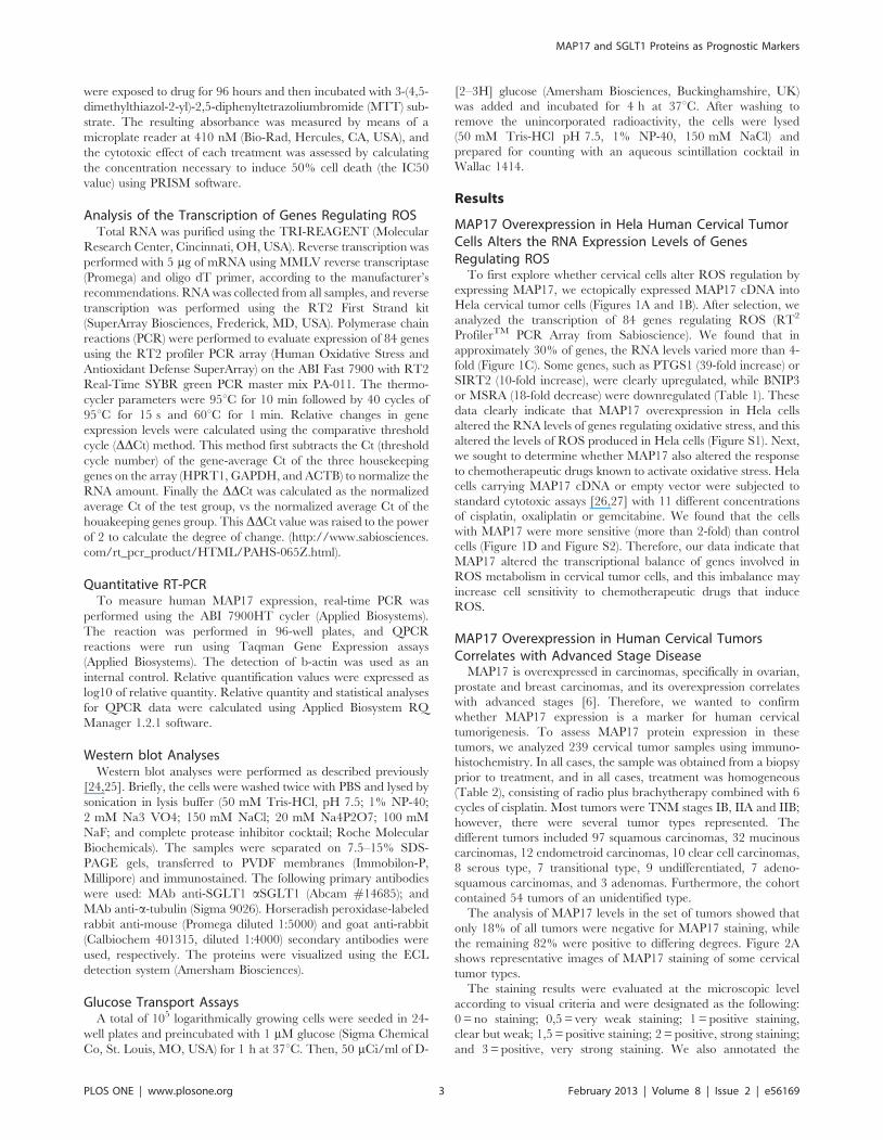
MAP17 Overexpression in Hela Human Cervical TumorCells Alters the RNA Expression Levels of GenesRegulating ROS
To first explore whether cervical cells alter ROS regulation by
expressing MAP17, we ectopically expressed MAP17 cDNA into
Hela cervical tumor cells (Figures 1A and 1B). After selection, we
analyzed the transcription of 84 genes regulating ROS (RT2
ProfilerTM PCR Array from Sabioscience). We found that in
approximately 30% of genes, the RNA levels varied more than 4-
fold (Figure 1C). Some genes, such as PTGS1 (39-fold increase) or
SIRT2 (10-fold increase), were clearly upregulated, while BNIP3
or MSRA (18-fold decrease) were downregulated (Table 1). These
data clearly indicate that MAP17 overexpression in Hela cells
altered the RNA levels of genes regulating oxidative stress, and this
altered the levels of ROS produced in Hela cells (Figure S1). Next,
we sought to determine whether MAP17 also altered the response
to chemotherapeutic drugs known to activate oxidative stress. Hela
cells carrying MAP17 cDNA or empty vector were subjected to
standard cytotoxic assays [26,27] with 11 different concentrations
of cisplatin, oxaliplatin or gemcitabine. We found that the cells
with MAP17 were more sensitive (more than 2-fold) than control
cells (Figure 1D and Figure S2). Therefore, our data indicate that
MAP17 altered the transcriptional balance of genes involved in
ROS metabolism in cervical tumor cells, and this imbalance may
increase cell sensitivity to chemotherapeutic drugs that induce
ROS.
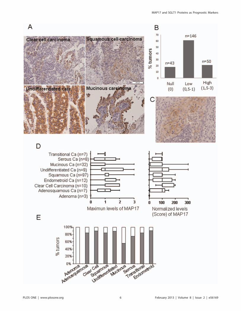
MAP17 Overexpression in Human Cervical TumorsCorrelates with Advanced Stage Disease
MAP17 is overexpressed in carcinomas, specifically in ovarian,
prostate and breast carcinomas, and its overexpression correlates
with advanced stages [6]. Therefore, we wanted to confirm
whether MAP17 expression is a marker for human cervical
tumorigenesis. To assess MAP17 protein expression in these
tumors, we analyzed 239 cervical tumor samples using immuno-
histochemistry. In all cases, the sample was obtained from a biopsy
prior to treatment, and in all cases, treatment was homogeneous
(Table 2), consisting of radio plus brachytherapy combined with 6
cycles of cisplatin. Most tumors were TNM stages IB, IIA and IIB;
however, there were several tumor types represented. The
different tumors included 97 squamous carcinomas, 32 mucinous
carcinomas, 12 endometroid carcinomas, 10 clear cell carcinomas,
8 serous type, 7 transitional type, 9 undifferentiated, 7 adeno-
squamous carcinomas, and 3 adenomas. Furthermore, the cohort
contained 54 tumors of an unidentified type.
The analysis of MAP17 levels in the set of tumors showed that
only 18% of all tumors were negative for MAP17 staining, while
the remaining 82% were positive to differing degrees. Figure 2A
shows representative images of MAP17 staining of some cervical
tumor types.
The staining results were evaluated at the microscopic level
according to visual criteria and were designated as the following:
0 = no staining; 0,5 = very weak staining; 1 = positive staining,
clear but weak; 1,5 = positive staining; 2 = positive, strong staining;
and 3 = positive, very strong staining. We also annotated the
MAP17 and SGLT1 Proteins as Prognostic Markers
PLOS ONE | www.plosone.org 3 February 2013 | Volume 8 | Issue 2 | e56169
percentage of tumoral cells positive for MAP17 to score the overall
levels of MAP17 observed. Out of the 239 tumors, only 43 (18%)
did not express MAP17. A total of 146 (61%) showed intermediate
levels, and 50 tumors (21%) showed very high MAP17 expression
levels (Figure 2B).
We observed two different subcellular distributions of MAP17.
The first distribution pattern involved cytoplasmic and membra-
nous staining, with broad cytoplasmic distribution, especially in
the perinuclear region (Figure 2A). The second distribution
pattern showed MAP17 staining only in the membrane
(Figure 2C). Most MAP17-positive tumors showed a cytoplasmic
plus membranal distribution; however, approximately 5% of
samples showed a clear membrane-only distribution. Remarkably,
a few tumors contained different populations showing both
subcellular localizations for MAP17; we do not currently know
the reason for such differential distributions. In cultured cells,
MAP17 shows primarily perinuclear cytoplasmic staining with
some plasma membrane localization. It is possible that the strong
anchoring of MAP17 to intracellular membranous compartments,
such as the Golgi, accounts for this intracellular localization.
Therefore, it is also possible that some tumors accumulate
intracellular membranous compartments and accumulate in-
creased levels of MAP17 in the cytosol, while in other tumors,
the level of internal membranes is minimal, and the cells
accumulate MAP17 in the cellular membrane. It may also depend
on the molecular context of the specific tumor or cellular clone.
However, more research is required to understand this distribution
variability.
Figure 1. MAP17 overexpression in Hela cells. Hela cancer cells expressing ectopic MAP17 cDNA were selected and analyzed for MAP17 proteinexpression by A) quantitative measurement of MAP17 mRNA expressed ectopically in Hela cells and B) immunodetection after cytospin centrifugation ontoslides. C) Map17 alters the transcription of genes involved in oxidative stress. A graph is shown depicting the distribution of gene transcriptional alterationsinduced by MAP17 in Hela cells. D) The IC50 values are shown for different chemotherapeutic drugs in Hela cells expressing MAP17 or with vector only.doi:10.1371/journal.pone.0056169.g001
Table 1. Genes involved in ROS regulation in whichtranscription is significantly altered by MAP17 expression inHela cells.
Genes upregulated .4-fold Genes downregulated .4-fold
Gene Symbol Fold Change Gene Symbol Fold Change
CCL5 7,3958 ALB 24,3486
GPX3 6,9993 ATOX1 27,0806
KRT1 5,5198 BNIP3 218,7871
MGST3 5,1573 CAT 26,293
PRDX2 8,0437 DHCR24 25,3102
PRDX5 4,054 DUOX1 25,2595
PRG3 7,0221 GPX1 24,3298
PTGS1 39,1571 MSRA 218,4663
PXDNL 5,2399 NCF2 27,5352
SCARA3 4,9336 OXR1 24,2456
SIRT2 9,8365 OXSR1 25,6898
SOD2 4,1722 PRDX3 28,1426
SOD3 5,5476 PRDX4 213,334
SRXN1 4,9745 SELS 24,7526
TXNRD1 4,4648 STK25 25,165
TXNRD2 5,0278 HPRT1 214,7514
doi:10.1371/journal.pone.0056169.t001
MAP17 and SGLT1 Proteins as Prognostic Markers
PLOS ONE | www.plosone.org 4 February 2013 | Volume 8 | Issue 2 | e56169
Next, we analyzed the MAP17 distribution levels for different
tumor types. For this task, we evaluated MAP17 levels in two
different ways: we determined the maximum level of MAP17
found in the tumor; or we normalized the levels by multiplying the
MAP17 signal intensity by the percentage of positive cells to
calculate the score (Figure 2D). We found that most tumor types
showed a broad range of levels with a median of 1: low but clear
levels of MAP17. However, mucinous type tumors appeared to
express very low MAP17 levels. Remarkably, adenoma benign
tumors did not express MAP17. Figure 2E shows the percentage of
each tumor type that showed positivity (at any level) for MAP17.
MAP17 Overexpression in Hela Tumor Cells IncreasesGlucose uptake and SGLT1 Content
Previous results have demonstrated that SGLT1 activation
rescues enterocytes from cell apoptosis by activating PI3K [12],
and the inhibition of this membrane transport inhibits MAP17-
dependent ROS increases and proliferation [8]. To explore the
relationship of this gene with MAP17 in the cervix, we first
measured glucose uptake in Hela cells expressing MAP17
(Figure 3A). It was found that glucose uptake was increased an
average of 4-fold and was inhibited upon treatment with the
SGLT inhibitor ploridzine. Furthermore, we found that SGLT1
protein levels increased an average of 2-fold in Hela cells
expressing MAP17 (Figure 3B).
SGLT1 Overexpression in Human Cervical TumorsCorrelates with MAP17 Levels
These and other, previous results indicate that MAP17-
dependent tumorigenic properties depend on the indirect activa-
tion of ROS by SGLT1 transport. Therefore, we measured
SGLT1 expression levels in the same cohort of cervical tumor
samples.
We found that the tumors showed positive SLGT1 staining
(Figure 3C), with approximately 40% tumors being positive for
SLGT1 (Figure 3D). However, only a few samples showed very
high staining levels. The distribution of the SLGT1-positive
tumors among the different cervical tumor types showed a pattern
similar to MAP17; adenoma benign tumors were clearly SLGT1
negative (Figure 3E).
Given the functional relationship between MAP17 and SLGT1,
we analyzed whether a correlation between the expression levels of
both proteins existed. We grouped the SGLT1 expression levels as
low (0–0,5), medium (0,5,x,1,5) or high (.1,5), and compared
the groups according to MAP17 expression levels (Anova,
Krustell-Wallis test). We found a direct, significant correlation
between MAP17 and SGLT1 levels within the groups (p,0,001;
Figure 4A). Next, we compared the levels of both proteins in the
same tumor samples and found a significant correlation (Spear-
man test, p,0,001) between the levels of both proteins in the same
tumor sample.
Again, we observed 2 different subcellular distributions for
SGLT1: cytoplasmic and membranous and membrane only.
Clearly, SGLT1 distribution patterns matched MAP17 distribu-
tion patterns in the same tumor (Figure 4B).
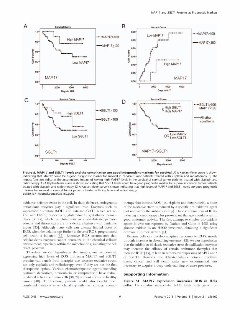
MAP17 and SGLT1 Expression Levels Correlate withSurvival in Patients with Cervical Tumors
Because the expression of MAP17 increases ROS [8], which
might be acting as a second messenger to increase the tumorigenic
properties of cancer cells, we hypothesized that further increases in
ROS might increase ROS levels beyond threshold and turn the
physiology of the cells towards apoptosis. We observed (Figure 1D)
that Hela cells overexpressing MAP17 increased by 2-fold their
sensitivity to several chemotherapeutic drugs known to induce
ROS. Therefore, MAP17 might be a marker for the activity of
treatments where oxidative stress plays a key role in the response,
such as cisplatin or radiotherapy.
After the samples were collected, the patients of our cohort were
treated with standard treatment for this type of tumor, radiother-
apy (50 Gy) + brachytherapy (15 Gy) + cisplatin (6 cycles: 40 mg/
m2). We analyzed whether MAP17 levels influenced response to
therapy. To that end, we explored whether the different MAP17
levels correlated with patient survival. Kaplan-Meier curves
showed that patients with tumors with medium or high levels of
MAP17 expression levels (.100 normalized score) showed
improved survival than patients with tumors with low or null
levels of MAP17 (Figure 5A). Overexpression of MAP17 had a
clear impact on patient survival (Figure 5B).
Therefore, a high level of MAP17 correlated with improved
survival and is a good prognostic factor in cervical cancer treated
with radiotherapy and cisplatin.
On the other hand, previous studies have demonstrated that
inhibition of SGLT1 membrane transport also inhibit MAP17-
dependent ROS increases and proliferation [5]. Because the
expression of SGLT1 correlated with MAP17, we studied whether
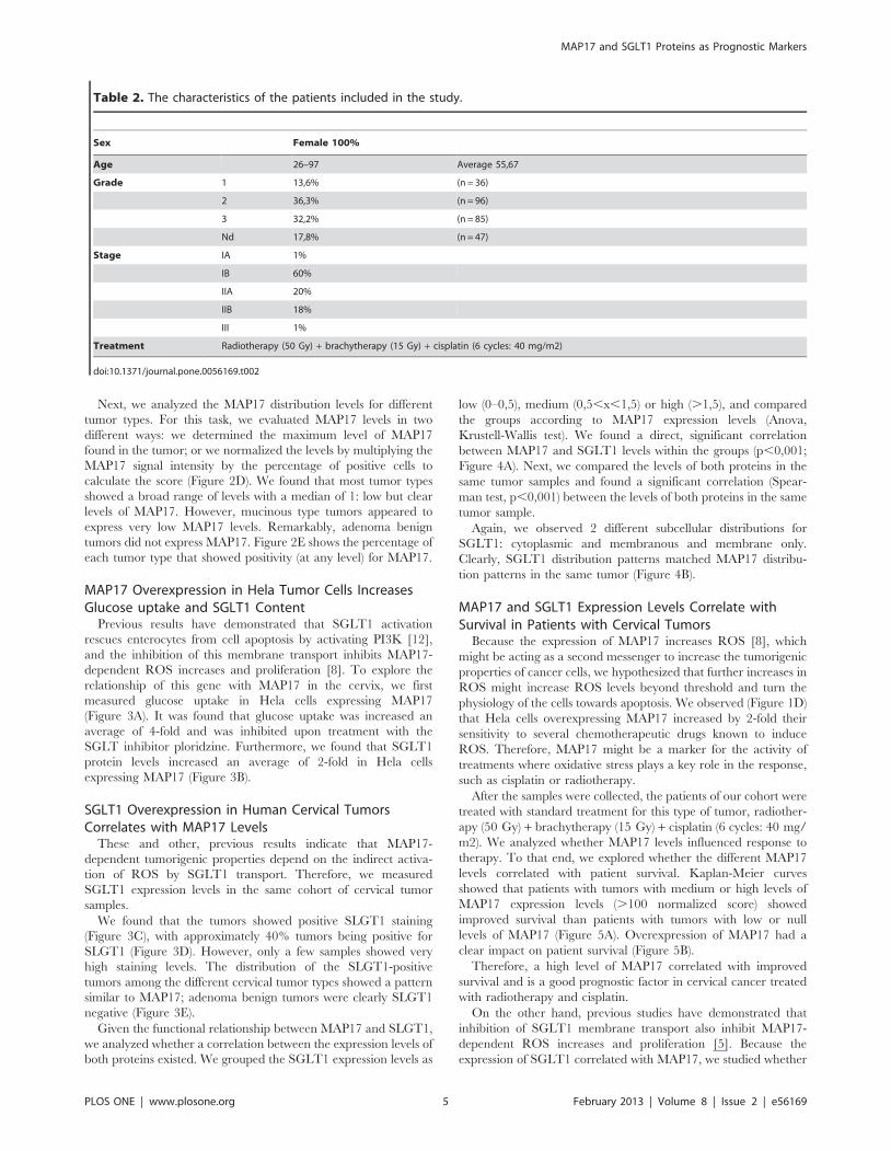
Table 2. The characteristics of the patients included in the study.
Sex Female 100%
Age 26–97 Average 55,67
Grade 1 13,6% (n = 36)
2 36,3% (n = 96)
3 32,2% (n = 85)
Nd 17,8% (n = 47)
Stage IA 1%
IB 60%
IIA 20%
IIB 18%
III 1%
Treatment Radiotherapy (50 Gy) + brachytherapy (15 Gy) + cisplatin (6 cycles: 40 mg/m2)
doi:10.1371/journal.pone.0056169.t002
MAP17 and SGLT1 Proteins as Prognostic Markers
PLOS ONE | www.plosone.org 5 February 2013 | Volume 8 | Issue 2 | e56169
MAP17 and SGLT1 Proteins as Prognostic Markers
PLOS ONE | www.plosone.org 6 February 2013 | Volume 8 | Issue 2 | e56169

the presence of SGLT1 in the same cohort of tumors was related
to prognosis independently and in connection with MAP17.
Similar to MAP17, SGLT1 levels showed a clear correlation
with patient survival. The patients whose tumors expressed
medium or high levels of SGLT1 (normalized levels $100)
showed improved survival than patients with low SGLT1 levels
(Figure 5C). However, the combination of both markers showed a
clear impact on patient survival. When exhibiting a combination
of high MAP17 levels (.100) and high SGLT1 levels ($100),
patients showed complete survival (p,0.001; Figure 5D), while the
patients with low MAP17 or low SGLT1 levels, or both, had the
worst prognosis (Figure 5D).
Discussion
ROS are beneficially involved in many signaling pathways that
control development and maintain cellular homeostasis [15].
Under physiological conditions, a tightly regulated redox balance
protects cells from injurious ROS activity. However, if altered,
ROS promote various pathological conditions including cancer
[16,28,29,30,31,32,33]. Understanding the duality of ROS as
cytotoxic molecules and key mediators in signaling cascades may
provide novel opportunities to improve cancer therapeutic
interventions.
MAP17 is overexpressed, primarily through increased mRNA
levels, in a variety of tumors. Because MAP17 expression increases
ROS through SGLT1 in cancer cells, we hypothesized that
MAP17 and SGLT1 might be markers for tumors with high
oxidative stress, and therefore, a further increase in ROS might
elevate the levels beyond the apoptotic threshold. In addition,
MAP17 and/or SGLT1 might be markers for the activity of
treatments where oxidative stress plays a key role in the response,
such as cisplatin or radiotherapy.
Figure 2. MAP17 overexpression in cervical tumors. A) Representative images of MAP17 immunostaining are shown for different tumor types.B) A graph is shown representing the percentage of cervical tumors with different MAP17 levels. C) Immunostaining was performed showingmembrane-only distribution of MAP17. D) The distribution of the MAP17 expression levels among different cervical tumor types is shown. Themaximum-levels graph refers to the maximum staining intensity observed in any part of the tumor (scale from 0 to 3). The normalized-levels (score)graph refers to maximum levels (0–3) scored by the percentage of cells (0–100). The normalized levels were obtained by multiplying the percentageof cells by the level of intensity observed. E) A graph representing the percentage of MAP17-positive tumors by tumor subtype is shown.doi:10.1371/journal.pone.0056169.g002
Figure 3. SGLT1 overexpression in cervical tumors. A) MAP17 expression in Hela cells increases glucose uptake that is blocked by the SGLTinhibitor phloridzine. B) Western blot analysis was performed for SGLT1 in Hela cells expressing MAP17 or vector alone (V). C) Representative imagesare shown of SGLT1 immunostaining of different cervical tumor subtypes. D) A graph is shown representing the percentage of cervical tumors withdifferent SGLT1 levels. E) A graph is shown representing the percentage of SGLT1-positive tumors by tumor subtype.doi:10.1371/journal.pone.0056169.g003
MAP17 and SGLT1 Proteins as Prognostic Markers
PLOS ONE | www.plosone.org 7 February 2013 | Volume 8 | Issue 2 | e56169
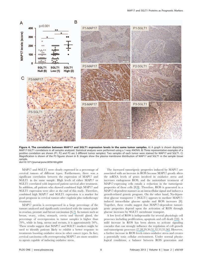
MAP17 and SGLT1 were clearly expressed in a percentage of
cervical tumors of different types. Furthermore, there was a
significant correlation between the expression of MAP17 and
SGLT1 in the same sample. High levels of either MAP17 or
SGLT1 correlated with improved patient survival after treatment.
In addition, all patients who showed combined high MAP17 and
SGLT1 expression were alive at the end of this study. Therefore,
combined high MAP17 and SGLT1 expression is a marker for
good prognosis in cervical tumors after cisplatin plus radiotherapy
treatment.
MAP17 protein is overexpressed in a large percentage of the
tumors analyzed and significantly correlated with the tumor grade
in ovarian, prostate and breast carcinomas [6,7]. In tumors such as
breast, ovary, colon, stomach, cervix and thyroid gland, the
percentage of overexpression in tumor samples is higher than
70%, while in lung, uterus and rectum, it is approximately 50%.
These results suggest that MAP17 and SGLT1 markers might be
used to identify patients likely to exhibit a better response to
treatments boosting oxidative stress in other cancer types. In fact,
cervical carcinoma cells overexpressing MAP17 are more sensitive
to agents capable of inducing oxidative stress.
The increased tumorigenic properties induced by MAP17 are
associated with an increase in ROS because MAP17 greatly alters
the mRNA levels of genes involved in oxidative stress and
increases endogenous ROS, and the antioxidant treatment of
MAP17-expressing cells entails a reduction in the tumorigenic
properties of these cells [8,9]. Therefore, ROS is generated in a
MAP17-dependent manner as an intracellular signal and induces a
growth-related genetic program. On the other hand, Na-depen-
dent glucose transporter 1 (SGLT1) appears to mediate MAP17-
induced intracellular glucose uptake and ROS increases [8].
Together, these results suggest that MAP17-dependent tumori-
genic properties depend upon the activation of ROS through
glucose increases by SGLT1 membrane transport.
A low level of ROS is indispensable for several physiologic cell
processes including proliferation, apoptosis and cell death [34]. A
mild increase in ROS has been shown to activate signaling
cascades that can strongly influence the regulation of cell growth
and tumorigenic processes ([7,28,29,30,31,32,33,35,36]. However,
a further increase in ROS levels raises oxidative stress and creates
a potentially toxic cellular environment. Under normal physio-
logical conditions, a balance between ROS generation and
Figure 4. The correlation between MAP17 and SGLT1 expression levels in the same tumor samples. A) A graph is shown depictingMAP17-SGLT1 correlations in all samples analyzed. Statistical analyses were performed using a 1-way ANOVA. B) Three representative examples of apositive correlation are shown (P1, P2 and P3 are 3 different tumor samples). Two samples of each tumor were stained for MAP17 and SGLT1. C)Magnification is shown of the P3 figures shown in B. Images show the plasma membrane distribution of MAP17 and SGLT1 in the sample tissuesample.doi:10.1371/journal.pone.0056169.g004
MAP17 and SGLT1 Proteins as Prognostic Markers
PLOS ONE | www.plosone.org 8 February 2013 | Volume 8 | Issue 2 | e56169
oxidative defenses exists in the cell. In these defenses, endogenous
antioxidant enzymes play a significant role. Enzymes such as
superoxide dismutase (SOD) and catalase (CAT), which act on
O2- and H2O2, respectively, glutaredoxins, glutathione peroxi-
dases (GPXs), which use glutathione as a co-substrate, peroxir-
edoxins and thioredoxins are in a delicate balance with oxidative
inputs [35]. Although many cells can tolerate limited doses of
ROS, when the balance tips further in favor of ROS, programmed
cell death is initiated [37]. Excessive ROS accumulates that
cellular detox enzymes cannot neutralize in the chemical cellular
environment, especially within the mitochondria, initiating the cell
death program.
Therefore, we can hypothesize that tumors, not just cervical,
expressing high levels of ROS producing MAP17 and SGLT1
proteins can benefit from therapies that increase oxidative stress,
not only cisplatin and radiotherapy, even if they are not the first
therapeutic option. Various chemotherapeutic agents including
platinum derivatives, doxorubicin or camptothecin have redox-
mediated activity on tumor cells [38,39] without effects on healthy
tissues [40]. Furthermore, patients could also benefit from
combined therapies in which, along with the cytotoxic chemo-
therapy that induces ROS (i.e., cisplatin and doxorubicin), a boost
of the oxidative stress is induced by a specific pro-oxidative agent
(not necessarily the antitumor drug). These combinations of ROS-
inducing chemotherapy plus pro-oxidant therapies could result in
good antitumor activity. The first attempt to employ pro-oxidant
agents in vivo was reported by Nathan and Cohn in 1981 using
glucose oxidase as an H2O2 precursor, obtaining a significant
decrease in tumor growth [41].
Because cells can develop adaptive responses to ROS, mostly
through increases in detoxifying enzymes [42], we can hypothesize
that the inhibition of classic oxidative stress detoxification enzymes
may increase the efficacy of certain antitumor therapies that
increase ROS [43], at least in tumors overexpressing MAP17 and/
or SGLT1. However, the delicate balance between oxidative
stress, cancer and cell death make new experimental tests
necessary to acquire a deep understanding of these processes.
Supporting Information
Figure S1 MAP17 expression increases ROS in Helacells. To visualize intracellular ROS levels, cells grown on
Figure 5. MAP17 and SGLT1 levels and the combination are good independent markers for survival. A) A Kaplan-Meier curve is shownindicating that MAP17 could be a good prognostic marker for survival in cervical tumor patients treated with cisplatin and radiotherapy. B) Theimpact function indicates the accumulated impact of having high MAP17 levels in the survival of cervical tumor patients treated with cisplatin andradiotherapy. C) A Kaplan-Meier curve is shown indicating that SGLT1 levels could be a good prognostic marker for survival in cervical tumor patientstreated with cisplatin and radiotherapy. D) A Kaplan-Meier curve is shown indicating that high levels of MAP17 and SGLT1 levels are good prognosticmarkers for survival in cervical tumor patients treated with cisplatin and radiotherapy.doi:10.1371/journal.pone.0056169.g005
MAP17 and SGLT1 Proteins as Prognostic Markers
PLOS ONE | www.plosone.org 9 February 2013 | Volume 8 | Issue 2 | e56169
coverslips were washed twice with warm PBS and then incubated
at 37uC with 8 mM of CM-H2DCFDA (Molecular Probes) in
warm PBS supplemented with 2.5 mM glucose for 15 min. Then,
PBS was replaced with 10% FBS supplemented DMEM, and cells
were incubated 10 min in the same conditions. Cells were washed
with warm PBS and fixed with 4% paraformaldehyde (Sigma) at
room temperature for 5 min. The fixed cells were washed three
times with PBS and the coverslips mounted in mowiol.
Intracellular ROS were visualized using a Confocal Ultra-spectral
microscope Leica TCS-SP2-AOBS-UV. Percentage of cells
showing immunofluorescence was calculated and refered to
parental cells (100%). The experiment was repeated 3 times
independently.
(DOC)
Figure S2 Cytotoxicity curves for different drugs.(DOC)
Author Contributions
Conceived and designed the experiments: AC. Performed the experiments:
MP BFA MALG ALC AC. Analyzed the data: JMPF JJM AC.
Contributed reagents/materials/analysis tools: GR AG ML. Wrote the
paper: AC.
References
1. Blasco T, Aramayona J, Alcalde A, Catalan J, Sarasa M, et al. (2003) Rat kidney
MAP17 induces cotransport of Na-mannose and Na-glucose in Xenopus laevisoocytes. Am J Physiol Renal Physiol 285: F799–810.
2. Jaeger C, Schaefer B, Wallich R, Kramer M (2000) The membrane-associatedprotein pKe#192/MAP17 in human keratinocytes. J Invest Dermatol 2000 115:
375–380.3. Pribanic S, Gisler S, Bacic D, Madjdpour C, Hernando N, et al. (2003)
Interactions of MAP17 with the NaPi-IIa/PDZK1 protein complex in renal
proximal tubular cells. Am J Physiol Renal Physiol 285: F784–791.4. Guijarro MV, Castro ME, Romero L, Moneo V, Carnero A (2007) Large scale
genetic screen identifies MAP17 as protein bypassing TNF-induced growtharrest. J Cell Biochem 101: 112–121.
5. Kocher O, Cheresh P, Lee SW (1996) Identification and partial characterization
of a novel membrane-associated protein (MAP17) up-regulated in humancarcinomas and modulating cell replication and tumor growth. Am J Pathol 149:
493–500.6. Guijarro MV, Leal JF, Fominaya J, Blanco-Aparicio C, Alonso S, et al. (2007)
MAP17 overexpression is a common characteristic of carcinomas. Carcinogen-esis 28: 1646–1652.
7. Guijarro MV, Vergel M, Marin JJ, Munoz-Galvan S, Ferrer I, et al. (2012)
p38alpha limits the contribution of MAP17 to cancer progression in breasttumors. Oncogene.
8. Guijarro MV, Leal JF, Blanco-Aparicio C, Alonso S, Fominaya J, et al. (2007)MAP17 enhances the malignant behavior of tumor cells through ROS increase.
Carcinogenesis 28: 2096–2104.
9. Guijarro MV, Link W, Rosado A, Leal JF, Carnero A (2007) MAP17 inhibitsMyc-induced apoptosis through PI3K/AKT pathway activation. Carcinogenesis
28: 2443–2450.10. Ganapathy V, Thangaraju M, Prasad PD (2009) Nutrient transporters in cancer:
relevance to Warburg hypothesis and beyond. Pharmacol Ther 121: 29–40.11. Wright EM, Turk E (2004) The sodium/glucose cotransport family SLC5.
Pflugers Arch 447: 510–518.
12. Huang CY, Hsiao JK, Lu YZ, Lee TC, Yu LC (2011) Anti-apoptotic PI3K/Aktsignaling by sodium/glucose transporter 1 reduces epithelial barrier damage and
bacterial translocation in intestinal ischemia. Lab Invest 91: 294–309.13. Hanabata Y, Nakajima Y, Morita KI, Kayamori K, Omura K (2011)
Coexpression of SGLT1 and EGFR is associated with tumor differentiation in
oral squamous cell carcinoma. Odontology.14. Haulica I, Boisteanu D, Neagu B (2001) [The role of oxidative stress in normal
and pathological adaptive reactions]. Rev Med Chir Soc Med Nat Iasi 105: 11–18.
15. Manda G, Nechifor M.T., Neagu T.M. (2009) Reactive Oxygen species, cancerand anticancer therapies. Curr Chem Biol 3: 342–366.
16. Behrend L, Henderson G, Zwacka RM (2003) Reactive oxygen species in
oncogenic transformation. Biochem Soc Trans 31: 1441–1444.17. Burdon RH, Gill V, Rice-Evans C (1990) Oxidative stress and tumour cell
proliferation. Free Radic Res Commun 11: 65–76.18. Kesic V (2006) Management of cervical cancer. Eur J Surg Oncol 32: 832–837.
19. Rodriguez Villalba S, Diaz-Caneja Planell C, Cervera Grau JM (2011) Current
opinion in cervix carcinoma. Clin Transl Oncol 13: 378–384.20. Lilic V, Lilic G, Filipovic S, Milosevic J, Tasic M, et al. (2009) Modern treatment
of invasive carcinoma of the uterine cervix. J BUON 14: 587–592.21. Guijarro MV, Leal JF, Fominaya J, Blanco-Aparicio C, Alonso S, et al. (2007)
MAP17 overexpression is a common characteristic of carcinomas. Carcinogen-
esis.22. Moneo V, Serelde BG, Leal JF, Blanco-Aparicio C, Diaz-Uriarte R, et al. (2007)
Levels of p27(kip1) determine Aplidin sensitivity. Mol Cancer Ther 6: 1310–1316.
23. Moneo V, Serelde BG, Fominaya J, Leal JF, Blanco-Aparicio C, et al. (2007)
Extreme sensitivity to Yondelis (Trabectedin, ET-743) in low passaged sarcoma
cell lines correlates with mutated p53. J Cell Biochem 100: 339–348.
24. Carnero A, Beach DH (2004) Absence of p21WAF1 cooperates with c-myc in
bypassing Ras-induced senescence and enhances oncogenic cooperation.
Oncogene 23: 6006–6011.
25. Castro ME, Ferrer I, Cascon A, Guijarro MV, Lleonart M, et al. (2008)
PPP1CA contributes to the senescence program induced by oncogenic Ras.
Carcinogenesis 29: 491–499.
26. Blanco-Aparicio C, Pequeno B, Moneo V, Romero L, Leal JF, et al. (2005)
Inhibition of phosphatidylinositol-3-kinase synergizes with gemcitabine in low-
passage tumor cell lines correlating with Bax translocation to the mitochondria.
Anticancer Drugs 16: 977–987.
27. Link W, Oyarzabal J, Serelde BG, Albarran MI, Rabal O, et al. (2009) Chemical
interrogation of FOXO3a nuclear translocation identifies potent and selective
inhibitors of phosphoinositide 3-kinases. J Biol Chem 284: 28392–28400.
28. Irani K, Xia Y, Zweier JL, Sollott SJ, Der CJ, et al. (1997) Mitogenic signaling
mediated by oxidants in Ras-transformed fibroblasts. Science 275: 1649–1652.
29. Sundaresan M, Yu ZX, Ferrans VJ, Irani K, Finkel T (1995) Requirement for
generation of H2O2 for platelet-derived growth factor signal transduction.
Science 270: 296–299.
30. Burdon RH (1996) Control of cell proliferation by reactive oxygen species.
Biochem Soc Trans 24: 1028–1032.
31. Bae GU, Seo DW, Kwon HK, Lee HY, Hong S, et al. (1999) Hydrogen
peroxide activates p70(S6k) signaling pathway. J Biol Chem 274: 32596–32602.
32. Klaunig JE, Xu Y, Isenberg JS, Bachowski S, Kolaja KL, et al. (1998) The role
of oxidative stress in chemical carcinogenesis. Environ Health Perspect 106
Suppl 1: 289–295.
33. Droge W (2002) Free radicals in the physiological control of cell function. Physiol
Rev 82: 47–95.
34. Storz P (2005) Reactive oxygen species in tumor progression. Front Biosci 10:
1881–1896.
35. Marra M, Sordelli IM, Lombardi A, Lamberti M, Tarantino L, et al. (2011)
Molecular targets and oxidative stress biomarkers in hepatocellular carcinoma:
an overview. J Transl Med 9: 171.
36. Finkel T, Holbrook NJ (2000) Oxidants, oxidative stress and the biology of
ageing. Nature 408: 239–247.
37. Fruehauf JP, Meyskens FL, Jr. (2007) Reactive oxygen species: a breath of life or
death? Clin Cancer Res 13: 789–794.
38. Simizu S, Takada M, Umezawa K, Imoto M (1998) Requirement of caspase-3(-
like) protease-mediated hydrogen peroxide production for apoptosis induced by
various anticancer drugs. J Biol Chem 273: 26900–26907.
39. Simizu S, Umezawa K, Takada M, Arber N, Imoto M (1998) Induction of
hydrogen peroxide production and Bax expression by caspase-3(-like) proteases
in tyrosine kinase inhibitor-induced apoptosis in human small cell lung
carcinoma cells. Exp Cell Res 238: 197–203.
40. Yoshikawa T, Kokura S, Tainaka K, Naito Y, Kondo M (1995) A novel cancer
therapy based on oxygen radicals. Cancer Res 55: 1617–1620.
41. Nathan CF, Cohn ZA (1981) Antitumor effects of hydrogen peroxide in vivo.
J Exp Med 154: 1539–1553.
42. Benhar M, Engelberg D, Levitzki A (2002) ROS, stress-activated kinases and
stress signaling in cancer. EMBO Rep 3: 420–425.
43. Nathan CF, Arrick BA, Murray HW, DeSantis NM, Cohn ZA (1981) Tumor
cell anti-oxidant defenses. Inhibition of the glutathione redox cycle enhances
macrophage-mediated cytolysis. J Exp Med 153: 766–782.
MAP17 and SGLT1 Proteins as Prognostic Markers
PLOS ONE | www.plosone.org 10 February 2013 | Volume 8 | Issue 2 | e56169