Jen Tooher Pathophysiology of Cervical Radiculopathy and ...
18
Jen Tooher Pathophysiology of Cervical Radiculopathy and Current Research for Evaluation and Treatment of Symptoms Introduction Cervical radiculopathy (CR) describes pain and/or sensorimotor deficits from the neck down the upper back, shoulder, and arm, and it occurs as a result of cervical nerve root compression. 1,2 90% of cervical radiculopathies are attributable to herniated nucleus pulposus (HNP) of the intervertebral disc or degenerative disc disease (DDD) and its’ associated pathologies, 3 including spinal stenosis, spondylosis of facet joints, osteophyte formation, ligamentous thickening, and spondylolisthesis. 1‐3 In the remaining 10% of cases, compression occurs from tumors, trauma, or instability. 4,5 For the purposes of this paper, discussion will focus on treatment and management of cervical radiculopathy as a result of HNP or DDD. Approximately 30-50% of adults experience neck pain each year, with highest incidence in people working in hospitals or offices. 1 Neck pain has been categorized into 4 grades according to the 2010 Joint and Bone Task Force, and cervical radiculopathy fits into grades II or III. 6 Prevalence of cervical radiculopathy is 3.5 per 1,000, and it is more common in males. 3 Peak incidence of herniated discs occurs in the 30s and 40s, whereas spondylosis tends to be seen in adults over 55. 3 Significant pain and disability can result. 7 In order to understand the pathology of cervical radiculopathy, a basic understanding of cervical spine anatomy is warranted. The cervical spine is composed of bone, cartilage, and nerves. Bone acts to transmit forces, 8 whereas cartilage attenuates compressive loads, 9 and nerves send signals between the brain and the body. 10 Seven cervical vertebrae with corresponding intervertebral discs and eight spinal nerves comprise the cervical spine. 2 The ligamentum flavum and laminae border the posterior
-
Upload
khangminh22 -
Category
Documents
-
view
0 -
download
0
Transcript of Jen Tooher Pathophysiology of Cervical Radiculopathy and ...

Jen Tooher
Pathophysiology of Cervical Radiculopathy and Current Research for Evaluation and Treatment of Symptoms
Introduction
Cervical radiculopathy (CR) describes pain and/or sensorimotor deficits
from the neck down the upper back, shoulder, and arm, and it occurs as a result of
cervical nerve root compression.1,2 90% of cervical radiculopathies are attributable
to herniated nucleus pulposus (HNP) of the intervertebral disc or degenerative disc
disease (DDD) and its’ associated pathologies,3 including spinal stenosis,
spondylosis of facet joints, osteophyte formation, ligamentous thickening, and
spondylolisthesis.1‐3 In the remaining 10% of cases, compression occurs from tumors,
trauma, or instability.4,5 For the purposes of this paper, discussion will focus on treatment
and management of cervical radiculopathy as a result of HNP or DDD.
Approximately 30-50% of adults experience neck pain each year, with highest
incidence in people working in hospitals or offices.1 Neck pain has been categorized into
4 grades according to the 2010 Joint and Bone Task Force, and cervical radiculopathy fits
into grades II or III.6 Prevalence of cervical radiculopathy is 3.5 per 1,000, and it is more
common in males.3 Peak incidence of herniated discs occurs in the 30s and 40s, whereas
spondylosis tends to be seen in adults over 55.3 Significant pain and disability can result.7
In order to understand the pathology of cervical radiculopathy, a basic
understanding of cervical spine anatomy is warranted. The cervical spine is composed of
bone, cartilage, and nerves. Bone acts to transmit forces,8 whereas cartilage attenuates
compressive loads,9 and nerves send signals between the brain and the body.10 Seven
cervical vertebrae with corresponding intervertebral discs and eight spinal nerves
comprise the cervical spine.2 The ligamentum flavum and laminae border the posterior

Jen Tooher
spinal canal; vertebral bodies and intervertebral discs border the spinal canal anteriorly.1
Spinal foramina form openings between the vertebrae where spinal nerves and
blood vessels pass obliquely at each level (Figure 1- Appendix A.)1,2 Cervical spinal
nerves divide into dorsal and ventral primary rami to supply the posterior neck, or
prevertebral, paraspinal muscles, and the brachial plexus, respectively.1 Intervertebral
foramina are funnel-shaped, with a narrow entrance closest to the spinal cord,3 whereas
nerve roots are largest in the central dural sac.3 Both intervertebral foramina and the
spinal canal are largest in the upper cervical spine and decrease in size until C7-T1,
causing disc pathology or narrowing to be more pronounced in the middle-lower cervical
spine; nerve root compression is most common between C5 and C7 vertebral levels.3
Current research suggests the diameter of the cervical spinal canal changes biomechanics
of cervical spine mobility and may predispose individuals to pathology. In one study of
295 subjects, congenital narrowing increased degeneration and cord compression in all
cervical segments except C2-3. The group with congenitally smaller canals also had
significantly higher segmental mobility at C4-5 and C6-7 and lower mobility at C3-4.11
Based on the orientation of facet joints in the cervical spine, lateral bending and axial
rotation are coupled motions in the cervical spine from C2-C7.12 Coupled extension and
rotation occur from C2-C5 while coupled flexion and rotation occur in C5-C7.13
Intervertebral discs attenuate compressive loads throughout the spine.9 To
effectively do this, discs are composed of an inner nucleus pulposus, a surrounding
annulus fibrosus, and superior and inferior cartilaginous vertebral end plates.9 The fibers
in the annulus fibrosus are made up of type 1 collagen and arranged in alternating oblique
layers to improve mobility and prevent excess tensile stress.1,9 The nucleus pulposus is

Jen Tooher
made of primarily type 2 collagen1 and functions using fluid dynamics: where the fluid is
distributed evenly throughout the disc to attenuate focal areas of stress.9 Type 2 collagen,
proteoglycans, and hyaluronan components of the nucleus pulposus are hydrophilic and
promote disc hydration. These minerals decrease with age, causing the discs to dry out
and start to bulge.1,14 Bone mass also decreases with age,15 which can prompt
ligamentum flavum thickening to compensate for instability that results from bone
degeneration and subsequent facet joint hypermobility.2,16 All these age-related factors
contribute to HNP, spondylosis, and the narrowing of the cervical canal.1
The pathophysiology of cervical radiculopathy depends on the cause of the
radicular symptoms. In the case of a herniated disc, the disc bulge, protrustion, or
extrusion causes pain as degeneration begins in the second decade from repetitive strain
on the posterolateral annulus.9 Circumferential tears cause fissures and allow the nucleus
pulposus to extrude.1 Pain is thought to occur as proteoglycans and phospholipases from
the nucleus pulposus initiate the inflammatory cascade, directly compress nerves, and
create symptoms down the arm.1,2 Inflammation and ischemia associated with root
compression appear to be contributing causes of symptoms in cervical radiculopathy.2,3
Spontaneous recovery occurs in weeks to months in many herniated discs, and the
presence of interleukins, prostaglandins, and inflammatory cytokines like MCP and the
fibronectin-aggrecan complex support the theory of inflammation.3,17,18 Adults have
modest lordosis of the cervical spine, but postural and age-related changes can decrease
or reverse lordosis, increasing pressure on the anterior disc to produce posterior-lateral
protrusion.1 Non-traumatic disc herniation rarely occurs at more than one level, but
spondylosis can involve multiple vertebral levels.3

Jen Tooher
DDD causes mechanical nerve root compression in the intervertebral foramina
because disc height decreases with age, the foramina and ligamentum flava hypertrophy,
and annular fibrous tissue decreases.1,9 Decreased disc height increases motion at facet
joints posteriorly, alters normal bony alignment, and increases strain on supporting
ligaments.1 Poor posture provides a feedforward mechanism that further aggravates
DDD.19 Degenerative changes are most common at C5-6, followed by C6-7.1,2
Spondylotic radiculopathy occurs after disc degeneration begins, and can cause facet joint
osteophyte formation because the mechanical behavior of the nucleus pulposus doesn’t
adequately distribute forces and focal areas of stress cause damage.20 Osteophytes can
compress nerves and cause patients with spondylotic radiculopathy to complain of
multilevel or bilateral radiculopathy.1
Clinical Evaluation
Patient history is essential to understanding the cause of cervical radiculopathy
and forming an appropriate treatment plan.1 Signs and symptoms of cervical
radiculopathy include insidious onset of pain, numbness, and/or tingling in the neck and
upper extremity, and muscle weakness.2 Pain is the presenting symptom in 70% of
patients with radiculopathy, and can range from a dull ache to burning, electrical pain.1
Pain often radiates to the medial border of the scapula, progressing down the arm and to
parts of the hand.1 Neck movements may increase pain, especially cervical extension,2
and coughing and sneezing can increase tingling in the arm.1 Neck pain is usually less
severe than arm symptoms.3 Atypical pain presentations include subscapular or chest
pain.3 Cervical extension, lateral bending, or rotation with axial loading narrow the
intervertebral foramina and can aggravate acute herniations.1 Acute disc problems

Jen Tooher
typically have quick onset of very severe pain, whereas chronic disc pathology and
spondylosis usually have less severe, gradual onset of pain or numbness.2 Risk factors
associated with CR include smoking, axial load bearing, high-risk occupations, and prior
lumbar radiculopathy.1 Smoking is thought to increase risk of radiculopathy due to
decreased vertebral blood supply, which accelerates ischemia.3
Weakness is more often associated with herniated discs than with spondylosis.1
Myotomes relate to muscles innervated by one spinal nerve, whereas dermatomes relate
to sensory distribution of a dorsal root.21 Paresthesias or anesthesias occur as often as
91% of the time along the involved dermatomes, reflexes will be diminished 70% of the
time, and muscle weakness can be present in a myotomal pattern up to 65% of the time.1,3
Using information from myotomes, dermatomes, and decreased reflex tests help identify
if and where cervical radiculopathy occurs (Table 1- Appendix B.)1,2
After the patient history, the objective portion of the clinical exam includes range
of motion, strength and myotomes, palpation, dermatomes, reflexes, and special tests.21
Patients should rate their pain using the Verbal Rating Scale (VRS) from 0-10 in all
locations to quantify pain severity.21 Cervical range of motion using a goniometer, along
with descriptions for which motions are limited due to pain, provides objective
information to understand impairments from cervical pathology.6 A stiff neck and head
tilt away from the side of injury are common in cervical radiculopathy.19 Decreased
active range of motion (AROM) in lateral bending and rotation can occur towards or
away from the side of the injury. If the motion is limited on the same side of the injury, it
is likely because the nerve root is compressed at the formina. Limited motion on the
opposite side is usually because of increased pressure on the nerve from a herniated disc.1

Jen Tooher
Myotome testing of the upper extremities denotes if muscle weakness exists from
radiculopathy. If weakness is found, comparison between sides is essential.1 Palpation of
spinous processes and articular pillars is reliable and helps to rule out excess movement
of vertebrae.6 With muscle palpation, a clinician should find tenderness, tightness, and
potential hypertonicity on ipsilateral cervical paraspinals and possibly along the muscle
where radicular symptoms present.1 Sensory exams should include both light touch and
pinprick, and should be done along a dermatomal distribution to identify if anesthesia or
hyperesthesia exists.3 Pinprick sensation is more reliable than light touch in people with
mild radiculopathy.1 Asymmetry of C5, C6, and C7 deep tendon reflexes should be
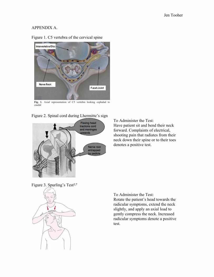
assessed, as they are suggestive of radiculopathy.7 Lhermitte’s sign, tested by flexing the
neck and asking about patient symptoms, may cause electrical pain down the spine and
possibly into the extremities for people with HNP, cervical cord involvement, severe
spondylosis with myelopathy, cervical tumor, and multiple sclerosis (Figure 2- Appendix
A.)19 However, Babinski and Hoffman’s should both be negative in CR; if these tests are
positive, cervical myelopathy or neurological processes must be ruled out.1
Five provocative tests can help identify cervical radiculopathy. These tests
include Spurling’s test, the valsalva maneuver, the shoulder abductor sign, the upper limb
tension test, and neck distraction.2 Spurling’s, shoulder abductor sign, and neck
distraction have high specificity but lower sensitivity. Spurling’s test is statistically best
at identifying cervical radiculopathy (Figure 3- Appendix A.)1 Based on psychometric
properties, these tests should be used to confirm a diagnosis of cervical radiculopathy, not
to screen for it. The upper limb tension test has 97% sensitivity, so it is the best screening
tool for cervical radiculopathy (Figure 4- Appendix A.)7 A clinical prediction rule (CPR)

Jen Tooher
for cervical radiculopathy has been developed, with 4 components: positive Spurling’s
test, positive neck distraction test, positive upper limb tension test, and less than 60
degrees of cervical range of motion to the involved side.22 3 out of 4 findings has 94%
specificity for cervical radiculopathy, with a positive likelihood ratio of 6.1.7 If all of the
4 components are found in a patient, there is 99% specificity and a positive likelihood
ratio of 30.3.22 The valsalva maneuver has insufficient psychometric testing to validate
its use, but may be practical in clinical settings to screen for cervical radiculopathy.2,3
Differential Diagnosis
According to the APTA Guide to PT Practice, cervical radiculopathy fits into
Pattern 4F: Impaired Joint Mobility, Motor Function, Muscle Performance, Range of
Motion, and Reflex Integrity Associated with Spinal Disorders.23 Because of the range of
symptoms associated with cervical radiculopathy, differential diagnoses need to be
considered and ruled out before continuing with treatment for cervical radiculopathy.
People with severe spondylosis or spondylolisthesis14 can develop cervical myelopathy,
which presents with radicular symptoms, gait changes, bowel or bladder dysfunction,
sensory dysfunction or weakness in the lower extremities. These patients need immediate
referral.1 Spondylotic myelopathy is degeneration of spine with osteophyte formation that
compresses the central canal. Spinal cord compression presenting with vascular
insufficiency and direct mechanical pressure can cause radicular symptoms (ie- central
cord syndrome) and serious, long-term effects if not treated promptly.1
Multiple sclerosis (MS) can present with a variety of symptoms, including
unilateral or bilateral arm paresthesias or anesthesias.24 Length of sensory changes,
Jen Tooher
positive upper motor neuron tests (clonus, Babinski, Hoffman’s), and a comprehensive
history help determine if referral to neurologist is necessary to rule out MS.25
Carpal tunnel syndrome (CTS) presents similar to cervical radiculopathy from C6
nerve root compression, but patients with CTS often have increased pain in the morning
and abductor pollicis brevis and thenar eminenance weakness. Spurling’s maneuver can
rule in cervical radiculopathy and rule out CTS, whereas Tinel’s and wrist flexion will
rule in CTS.2 For a more comprehensive look at other diagnoses that present with neck,
shoulder, and/or arm pain, weakness, or sensory changes, see Table 2 in Appendix B.
Imaging
Imaging is not indicated with grade I or II neck pain, it may be necessary in grade
III neck pain.6 Since most of the patients physical therapists see with CR will likely fit
into grades II or III, understanding imaging options helps guide referral decisions. Disc
herniations and stenosis are best identified using MRI (93% predictive),2,26 and can be
difficult to capture on x-rays.27 However, MRI is not supported as a necessary routine
screening tool for radiculopathy.6 X-rays show obvious spondolytic changes or
instability.2,27 CT scans are good if an individual is trying to identify bony ossification,
such as ossification of cervical ligaments or osteophytes of foramina.2 CT with
myelography combines bone and nerve root, but it is invasive and doesn’t show soft
tissue, making it more useful for foraminal narrowing and stenosis rather than disc
pathology.28 Electromyography (EMG) or nerve conduction studies (NCS) are useful if
double crush injury is suspected. It will show decreased nerve conduction if pathology
exists.2,3,29 Unfortunately, predictive capacity might be as low as 42% for compression.2
These tests help localize the level of the injury if other imaging has been inconclusive.29

Jen Tooher
Treatment
75-90% patients will have improved symptoms and pain with non-operative
management of cervical radiculopathy, so understanding current evidence-based
treatment options will help physical therapists effectively manage patients with this
condition.3 Urgent surgery is only indicated in small portion of patients, mostly in
patients with progressive weakness or extreme, unremitting pain.2
Evidence regarding physical therapy interventions for cervical radiculopathy is
limited.3,7 However, most studies have shown at least moderate effects of multi-modal
treatment for cervical radiculopathy.2,30 The general focus of exercise interventions
should be to restore function and decrease pain.1 Stretching tight scalenes, trapezius, and
pectoralis muscles improves postural alignment and decreases abnormal stresses on
vertebral bodies.31 Strengthening scapular stabilizers (rhomboids, middle trapezius) and
deep neck muscles (longus colli, longus capitis, rectus capitis anterior) sustains improved
posture and provides extrinsic support to the cervical spine.7 Press ups and push ups with
a plus at both low and high intensities are good exercises to target serratus anterior and
lower trapezius muscles while keeping the upper trapezius quiet.31 Mobilization has been
shown to decreased pain and increase range of motion.
Other interventions include range of motion exercises, neck relaxation techniques,
superficial heat, manual traction, TENS, cervical pillow, massage, and ultrasound.1,3
Literature suggests a combination of range of motion, heat, postural education, and neck
relaxation can successfully improve patient symptoms after six weeks.2 Modalities (ice,
heat, electrical stimulation) alone don’t significantly improve symptoms but may control
pain and inflammation, so can be used with other interventions to maximize treatment

Jen Tooher
effects.1 PT has shown improved pain and sensorimotor symptoms as much as surgery or
cervical collars 16 months after intervention, and it is a more cost effective intervention
than surgery.3 Strength exercises are more effective than aerobic exercises.1
Effective intervention conclusions are limited for individuals with cervical
radiculopathy because most studies identifying conservative exercise interventions for
neck pain do not separate CR from other neck diagnoses.7 Cervical traction may provide
temporary relief, reduce pain, and improve radicular symptoms for people with CR.32
High velocity-low amplitude (HVLA) manipulation has also been studied for cervical
radiculopathy with mixed results; some studies suggest improved symptoms for as long
as 9 months with no complications while others suggest complications of worsening
radiculopathy.2 The proposed mechanism for improvements involves relaxing resting
paraspinal muscle activity to provide relief.33 Many studies addressing effectiveness of
manipulation for neck pain exclude people with nerve root compression or radiating
symptoms, suggesting the diagnosis is a contraindication to this type of treatment.34‐36
Manipulation with cervical radiculopathy has increased risk of vertebral dissection and
spinal cord compression. Since superior results for manipulation haven’t been found
when compared to mobilization, mobilization is safer and is better technique to use for
people with CR.3,37 Neural dynamic mobilizations, cervical mobilizations, and Maitland
mobilizations may provide more benefits than other types of mobilizations.38
A treatment-based classification system has been developed for neck pain to
categorize and individualize treatments for subgroups of patients. Child’s classification
system categorizes cervical radiculopathy into the centralization classification.
Centralization focuses on repeated active or passive neck movements, traction, and

Jen Tooher
exercises to decrease peripheralization of symptoms.32 In order to correctly classify
patients and identify exercises that will benefit people with cervical radiculopathy,
identifying the pathology is essential.3 If the cervical radiculopathy is due to a herniated
disc, targeted exercises include chin retractions and repeated extension or oblique lateral
extension.32 Studies of the McKenzie method have shown moderate to strong
improvements in radicular symptoms when used appropriately.7
If cervical radiculopathy is the result of spondylosis, the focus of treatment should
be activity modification, neck immobilization, intermittent cervical traction, and
isometric exercises to improve symptoms.19 Semi-hard cervical collars control pain by
minimizing motion in acute stages but shouldn’t be used for long periods. Immobilization
causes paraspinal atrophy and decreases intrinsic cervical stability.3 Pain control should
be the initial goal of treatment for these patients, and then strengthening and postural
reeducation can begin.19 Gentle, active interventions have found better results.2
Additional Treatments
CR symptoms don’t improve significantly with NSAIDs or oral corticosteroids,
but NSAIDs are commonly used in acute stages to control pain.3 An artificial soluble
TNF receptor significantly decreases TNF-alpha, a cytokine that causes inflammation,
and may be a future solution to prevent inflammation and decrease spine pathology.17
Epidural steroid injections are another conservative treatment option for people with
cervical radiculopathy.1,3,39,40 Epidural steroid injections might be most effective for
patients who have MRI findings of central canal stenosis (approximately 60%
effective.)1,40 Studies suggest steroids provide short-term relief, but evidence about long-
term impact of steroids is unclear.2,6 Complications are also more common in epidural

Jen Tooher
steroid injections, especially transforaminal injections.2 Interlaminar injections might be
safer.1 One cohort study of transforaminal steroid injections found satisfactory recovery
in patients with CR, but another randomized controlled trial of transforaminal steroid
injections did not show positive effects compared to a control.3 Low-dose anticonvulsants
or antidepressants might provide some short-term relief.1
Surgery has been shown to be useful in some cases with cervical radiculopathy.6
Surgical options include anterior or posterior discectomy, arthroplasty, disc fusion,
foramenotomy, and decompression.3 Fast and long-term results have been seen with
anterior cervical discectomy with and without fusion. Anterior cervical discectomy with
fusion provides relief in approximately 90% of patients with CR.41,42 Arthroplasty has
shown similar results as anterior discectomy and improved results compared to cervical
fusion when done on patients with cervical radiculopathy.6,43 Accelerated spinal
degenerative at adjacent levels has been seen in patients with a history of cervical fusion,
so radicular symptoms may recur in patients after surgical fusion.3,44
Conclusion
Cervical radiculopathy is a clinical syndrome marked by neck, upper extremity
pain, and possible sensorimotor deficits.22 People with CR often have difficulty with
ADLs and can lose time from work and social obligations.7 The most difficult aspect of
clinical diagnosis is identifying the vertebral level where compression is occurring.3
Using anatomy, pathology, clinical, and treatment knowledge discussed in this paper, PTs
can more effectively identify, localize, and treat cervical radiculopathy efficiently and
effectively to decrease disability that often occurs as a result of cervical radiculopathy.
Jen Tooher
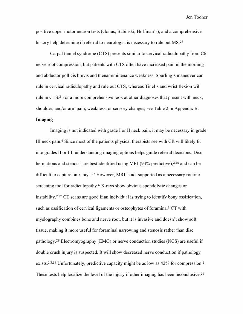
APPENDIX A. Figure 1. C5 vertebra of the cervical spine
Figure 2. Spinal cord during Lhermitte’s sign
To Administer the Test: Have patient sit and bend their neck forward. Complaints of electrical, shooting pain that radiates from their neck down their spine or to their toes denotes a positive test.
Figure 3. Spurling’s Test2,7
To Administer the Test: Rotate the patient’s head towards the radicular symptoms, extend the neck slightly, and apply an axial load to gently compress the neck. Increased radicular symptoms denote a positive test.
should be the rapid diagnosis and treatment of thiscondition in order to facilitate the return of the patient totheir normal state of health.
Methods
To accomplish the goal of this review, the PubMed databasewas searched for publications from January 2000 throughJanuary 2011. The search was limited to English languagearticles. Initially, the database was searched using the term“cervical radiculpathy.” This search returned a total of 1,978articles; when narrowed to full text, there were a total of374 articles. This subgroup was then searched for thekeywords “surgery,” “pathophysiology,” “diagnosis,” and“treatment”. The resulting articles were screened by titleand abstract for relevance to this review. A total of 84relevant resources were identi!ed. All relevant full-textmanuscripts found on the PubMed database using thementioned keywords were reviewed by independent authorsand the results are concisely presented here.
Results
The cervical spinal nerves exit the spinal cord and areoriented obliquely toward their respective neural foramen(Fig. 1). The cervical spinal nerves are named correspond-ing to the vertebral body below the nerve. The C8 nerveexits between C7 and T1 [5]. The neural foramen is madeup of the facet joint posteriorly and the intervertebral discanteriorly. The superior and inferior borders are comprisedof the pedicles of the vertebral bodies above and below,respectively. The foramina are largest in the upper cervicalspine and gradually narrow distally, with the C7/T1foramina being the most narrow. The most common causesof nerve root compression are spondylosis of the facet jointand herniation of the intervertebral disc [6]. Hypermobilityof the facet joint leads to ligamentous hypertrophy as wellas bony hypertrophy. An increase in the size of the superior
articulating process from the distal vertebra causes com-pression of the nerve. Intervertebral disc herniations canalso cause nerve root compression from the anterior aspectof the foramen [7]. Disc herniations can either be acute orchronic. Chronic herniations occur when the intervertebraldisc becomes degenerated and desiccated. This causescollapse of the disc space and bulging of the annulus intothe neural foramen. Chronic herniations and facet spondy-losis generally cause symptoms with an insidious onset thattend to be less severe. An acute herniation occurs when afragment of the nucleus pulposus extrudes through a defectin the annulus !brosis. This generally is associated with thesudden onset of severe symptoms, in contrast to thoseassociated with a chronic disc herniation [4, 8]. Researchershypothesize that pain syndromes and de!cits arise as aresult of both ischemia and in"ammation notions that wouldexplain why acute insults tend to result in more profoundsymptoms than slow, adaptable processes [9].
The patient history alone can diagnose cervical radicul-opathy in over 75% of cases [10]. The most commonsymptom associated with radiculopathy is arm pain orparesthesias in the dermatomal distribution of the affectednerve. Various different nerve compression syndromes arewell described (Table 1). Cervical radiculopathy may ormay not be associated with neck pain. In rheumatoidarthritis, atlanto-axial settling can lead to C2 radicular painwhich can manifest itself as eye and/or ear pain andheadache. C3 and C4 symptoms tend to be vague neckpain and trapezious pain. C5 pain occurs in the shoulder andradiates down the ventral arm to below the elbow. C6radiculopathy is associated with pain down the superiorlateral aspect of the arm into the !rst two digits. Commonly,there is overlap in the pain presentation of C6 radiculopathyand carpal tunnel syndrome. In carpal tunnel syndrome,patients often report worse symptoms in the morning. Ifpresent, the motor component of the two clinical entities isquite different. Classically, carpal tunnel syndrome motorde!cit is gauged by strength assessment of the abductorpollicis brevis which is innervated by the recurrent branchof the median nerve (which has a take-off point distal to thetransverse carpal ligament). Clinically, patients withadvanced carpal tunnel syndrome will have thenar atrophy.In contrast, the C6 nerve root provides motor innervationsin a shared fashion to elbow "exors and wrist extensors. Onclinical examination, Spurling’s maneuver will exacerbate
Fig. 1. Axial representation of C5 vertebra looking cephalad tocaudal
Table 1 Patterns of nerve root compression syndromes
Nerve root Pain pattern Weakness Re"exes
C2 Occipital, eyesC3 Neck, trapeziusC4 Neck, trapeziusC5 Shoulder, lateral UE DeltoidC6 Lateral forearm,
!rst two digitsBiceps Biceps absent
C7 Posterior forearm,third digit
Triceps Triceps absent
C8 Medical forearm,fourth and !fth digit
Finger abduction,grip
266 HSSJ (2011) 7:265–272

Jen Tooher
Figure 4. Upper Limb Tension Test-Median Nerve22
To Administer the Test: Depress and stabilize the scapula, move affected arm into 110 degrees of shoulder abduction. Then supinate the forearm, add ulnar deviation and wrist and finger extension. Externally rotate the shoulder, and then extend the elbow. Laterally bend the head away from the arm being tested. Monitor symptoms after each change before moving on to the next position. A test is positive if radicular symptoms worsen.

Jen Tooher
APPENDIX B. Table 1. Pain Patterns, Myotomes, Dermatomes, & Reflexes associated with Cervical Radiculopathy2,21 Pain Pattern Myotomes Dermatomes Reflexes C2 Occiput, eyes N/A Lateral occipital
protuberance
C3 Neck, Trapezius N/A Supraclavicular fossa, midclavicular line
C4 Neck, Trapezius Shoulder Elevation Acromioclavicular joint
C5
Shoulder, lateral UE Shoulder abduction or external rotation
Lateral antecubital fossa
Brachioradialis
C6 Lateral forearm, first 2 fingers
Elbow flexion or wrist extension
1st digit (thumb) Biceps
C7 Posterior forearm, third finger
Elbow extension or wrist flexion
3rd digit (middle finger)
Triceps
C8 Medial forearm, fourth and fifth finger
Thumb extension or ulnar deviation
Medial antecubital fossa
Table 2. Differential Diagnosis List for Cervical Radiculopathy2,3Acromioclavicular pathology Acute posterior cervical strain Adhesive capsulitis Aortic disease Arachnoiditis Arteriovenous malformation Back pain Bicipital tendonitis- rotator cuff tears, lateral epicondylitis Brainstem syndromes Calcareous tendonitis Carpal tunnel syndrome Cervical disk syndromes Cervical lymphadenitis Cervical rib Congenital spinal lesion Diskitis Double crush syndrome Elbow Epicondylitis Epidural absess Extrinsic neoplaia (usually metatastic) Facet joint pain Frozen shoulder syndromes Glenohumeral arthritis Gout (infrequently) Heart Disease Hyperabduction syndrome Intervertebral osteoarthritis
Idiopathic brachial plexopathy Intrinsic neoplasia Myocardial ischemic pain Nerve injuries Occipital neuralgia Osteomyelities Osteoarthritis of apophyseal joints Paget’s disease Pancoast’s tumor Parsonage-Turner syndrome Pharyngeal infections Postural disorders Psychogenic disorders Rheumatoid arthritis Rib-clavicle compression Rotator cuff injury Scale muscle tightness Septic arthritis Spinal cord tumors Sternocleidomastoid tendinitis Subacromial bursitis Synovial cysts Tabes dorsalis Thoracic disk Thoracic outlet syndrome Tropical spastic paraparesis Ulnar nerve entrapment

Jen Tooher
References 1. Roth D, Mukai A, Thomas P, Hudgins TH, Alleva JT. Cervical radiculopathy. Dis Mon. 2009;55(12):737-756. doi: 10.1016/j.disamonth.2009.06.004. 2. Caridi JM, Pumberger M, Hughes AP. Cervical radiculopathy: A review. HSS J. 2011;7(3):265-272. doi: 10.1007/s11420-011-9218-z. 3. Kuijper B, Tans JT, Schimsheimer RJ, et al. Degenerative cervical radiculopathy: Diagnosis and conservative treatment. A review. Eur J Neurol. 2009;16(1):15-20. doi: 10.1111/j.1468-1331.2008.02365.x; 10.1111/j.1468-1331.2008.02365.x. 4. Ashraf S. Intramedullary cervical spine germinoma: A case report. Med J Malaysia. 2012;67(2):207-209. 5. Kruse RA, Cambron JA. Large C4/5 spondylotic disc bulge resulting in spinal stenosis and myelomalacia in a klippel-feil patient. J Altern Complement Med. 2012;18(1):96-99. doi: 10.1089/acm.2010.0844. 6. Guzman J, Haldeman S, Carroll LJ, et al. Clinical practice implications of the bone and joint decade 2000-2010 task force on neck pain and its associated disorders: From concepts and findings to recommendations. Spine (Phila Pa 1976). 2008;33(4 Suppl):S199-213. doi: 10.1097/BRS.0b013e3181644641. 7. Langevin P, Roy JS, Desmeules F. Cervical radiculopathy: Study protocol of a randomised clinical trial evaluating the effect of mobilisations and exercises targeting the opening of intervertebral foramen [NCT01500044. BMC Musculoskelet Disord. 2012;13:10-2474-13-10. doi: 10.1186/1471-2474-13-10; 10.1186/1471-2474-13-10. 8. Gross M. Voicethread lecture: Bone. University of North Carolina at Chapel Hill: ; 2012. 9. Gross M. Voicethread lecture: Articular cartilage. [Electronic]. University of North Carolina at Chapel Hill: ; 2012. 10. Ellenberg MR, Honet JC, Treanor WJ. Cervical radiculopathy. Arch Phys Med Rehabil. 1994;75(3):342-352. 11. Morishita Y, Naito M, Hymanson H, Miyazaki M, Wu G, Wang JC. The relationship between the cervical spinal canal diameter and the pathological changes in the cervical spine. Eur Spine J. 2009;18(6):877-883. doi: 10.1007/s00586-009-0968-y. 12. Ishii T, Mukai Y, Hosono N, et al. Kinematics of the cervical spine in lateral bending in vivo three-dimensional analysis. Spine. 2006;31(2):155-160. 13. Ishii T, Mukai Y, Hosono N, et al. Kinematics of the subaxial cervical spine in rotation in vivo three-dimensional analysis. Spine. 2004;29(24):2826-2831. 14. Ogiela D. PubMed health. A.D.A.M. Medical Encyclopedia. Herniated Disk Web site. http://www.ncbi.nlm.nih.gov/pubmedhealth/PMH0001478/. Published June 7, 2012. Updated 2012. Accessed November 14, 2012. 15. Bottomley J, Lewis C. Comparing and contrasting age related changes in biology, physiology, anatomy, and function. In: Geriatric rehabilitation: A clinical approach. 2nd ed. Upper Saddle River, New Jersey: Pearson Education Inc; 2003:50-75.
Jen Tooher
16. Boss GR, Seegmiller JE. Age-related physiological changes and their clinical significance. West J Med. 1981;135(6):434-440. 17. Sinclair SM, Shamji MF, Chen J, et al. Attenuation of inflammatory events in human intervertebral disc cells with a tumor necrosis factor antagonist. Spine (Phila Pa 1976). 2011;36(15):1190-1196. doi: 10.1097/BRS.0b013e3181ebdb43. 18. Gajendran VK, Reuter MW, Golish SR, Hanna LS, Scuderi GJ. Is the fibronectin-aggrecan complex present in cervical disk disease? PM R. 2011;3(11):1030-1034. doi: 10.1016/j.pmrj.2011.07.003; 10.1016/j.pmrj.2011.07.003. 19. McCormack BM, Weinstein PR. Cervical spondylosis. an update. West J Med. 1996;165(1-2):43-51. 20. Hehir MK, Figueroa JJ, Zynda-Weiss AM, Stanton M, Logigian EL. Unexpected neuroimaging abnormalities in patients with apparent C8 radiculopathy: Broadening the clinical spectrum. Muscle Nerve. 2012;45(6):859-865. doi: 10.1002/mus.23319; 10.1002/mus.23319. 21. McMorris M. Musculoskeletal issues- cervical spine. [Powerpoint Presentation]. University of North Carolina at Chapel Hill: Division of Physical Therapy; 2011. 22. Wainner RS, Fritz JM, Irrgang JJ, Boninger ML, Delitto A, Allison S. Reliability and diagnostic accuracy of the clinical examination and patient self-report measures for cervical radiculopathy. Spine (Phila Pa 1976). 2003;28(1):52-62. doi: 10.1097/01.BRS.0000038873.01855.50. 23. American Physical Therapy Association. Interactive guide to physical therapist practice. . 2003. 24. National Multiple Sclerosis Society. Promoting function, independence, and mobility. http://www.nationalmssociety.org/living-with-multiple-sclerosis/mobility-and-accessibility/index.aspx. Updated 2011. Accessed August 23, 2012. 25. Khan F, Turner-Stokes L, Ng L, Kilpatrick T, Amatya B. Multidisciplinary rehabilitation for adults with multiple sclerosis. . 2007(2). doi: 10.1002/14651858.CD006036.pub2. 26. Kaale BR, Krakenes J, Albrektsen G, Wester K. Clinical assessment techniques for detecting ligament and membrane injuries in the upper cervical spine region- A comparison with MRI results. Manual Therapy. 2008;13:398-404. 27. Ardran G. The application and limitation of the use of X-rays in medical diagnosis. Mathematical and Physical Sciences. ;292(1390):147-156. 28. Song KJ, Choi BW, Kim GH, Kim JR. Clinical usefulness of CT-myelogram comparing with the MRI in degenerative cervical spinal disorders: Is CTM still useful for primary diagnostic tool? J Spinal Disord Tech. 2009;22(5):353-357. doi: 10.1097/BSD.0b013e31817df78e. 29. Nicholson KJ, Quindlen JC, Winkelstein BA. Development of a duration threshold for modulating evoked neuronal responses after nerve root compression injury. Stapp Car Crash J. 2011;55:1-24. 30. Hurwitz E, Carragee E, van der Velde G. <br />Treatment of neck pain: Noninvasive interventions: Results of the bone and joint decade 2000-2010 task force on neck pain and its associated disorders. Spine. 2008;33(4):123-52. 31. Andersen CH, Zebis MK, Saervoll C, et al. Scapular muscle activity from selected strengthening exercises performed at low and high intensities. J Strength Cond Res. 2012;26(9):2408-2416. doi: 10.1519/JSC.0b013e31823f8d24.

Jen Tooher
32. Childs J, Fritz J, Piva S, Whitman J. Proposal of a classification system for patients with neck pain. JOSPT. 2004;34:686-700. 33. Fryer G, Morris T, Gibbons P. Paraspinal muscles and intervertebral dysfunction: Part two. J Manipulative Physiol Ther. 2004;27(5):348-357. doi: 10.1016/j.jmpt.2004.04.008. 34. Dunning JR, Cleland JA, Waldrop MA, et al. Upper cervical and upper thoracic thrust manipulation versus nonthrust mobilization in patients with mechanical neck pain: A multicenter randomized clinical trial. J Orthop Sports Phys Ther. 2012;42(1):5-18. doi: 10.2519/jospt.2012.3894. 35. Puentedura E, Cleland J, Landers M, Mintken P, Louw A, Fernandez-de-las-Penasm C. Development of a clinical prediction rule to identify patients with neck pain likely to benefit from thrust joint manipulation to the cervical spine. JOSPT. 2012;42(7):577-592. 36. Cleland J, Mintken P, Carpenter K, et al. Examination of a clinical prediction rule to identify patients with neck pain likely to benefit from thoracic spine thrust manipulation and a general cervical range of motion exercise: Multi-center randomized clinical trial. Physical Therapy. 2010;90(9):1239-1250. 37. Kay TM, Gross A, Goldsmith CH, et al. Exercises for mechanical neck disorders. Cochrane Database Syst Rev. 2012;8:CD004250. doi: 10.1002/14651858.CD004250.pub4. 38. Gross A, Miller J, D'Sylva J. Manipulation or mobilisation for neck pain. Cochrane Database Syst Rev. 2010;1. 39. Choi GS, Ahn SH, Cho YW, Lee DG. Long-term effect of pulsed radiofrequency on chronic cervical radicular pain refractory to repeated transforaminal epidural steroid injections. Pain Med. 2012;13(3):368-375. doi: 10.1111/j.1526-4637.2011.01313.x; 10.1111/j.1526-4637.2011.01313.x. 40. Fish DE, Kobayashi HW, Chang TL, Pham Q. MRI prediction of therapeutic response to epidural steroid injection in patients with cervical radiculopathy. Am J Phys Med Rehabil. 2009;88(3):239-246. doi: 10.1097/PHM.0b013e3181951890. 41. Peolsson A, Peolsson M. Predictive factors for long-term outcome of anterior cervical decompression and fusion: A multivariate data analysis. Eur Spine J. 2008;17(3):406-414. doi: 10.1007/s00586-007-0560-2. 42. Fransen P. Prevention of scar tissue formation in spinal surgery: State of the art and review of the literature. J Neurosurg Sci. 2011;55(3):277-281. 43. Boselie TF, Willems PC, van Mameren H, de Bie R, Benzel EC, van Santbrink H. Arthroplasty versus fusion in single-level cervical degenerative disc disease. Cochrane Database Syst Rev. 2012;9:CD009173. doi: 10.1002/14651858.CD009173.pub2; 10.1002/14651858.CD009173.pub2. 44. Kepler CK, Hilibrand AS. Management of adjacent segment disease after cervical spinal fusion. Orthop Clin North Am. 2012;43(1):53-62, viii. doi: 10.1016/j.ocl.2011.08.003.