Hematologic Issues in Cervical Spine Surgery
15
13 L. Denaro et al. (eds.), Pitfalls in Cervical Spine Surgery, DOI: 10.1007/978-3-540-85019-9_2, © Springer-Verlag Berlin Heidelberg 2010 2.1 Changes in Blood Cell Count Parameters 2.1.1 Anemia Anemia is defined as a reduction of hemoglobin (Hb) levels of <13 g/dL for a man and <12 g/dL for a woman. It is very frequent among hospitalized patients and can be a serious problem in patients who undergo cervical spine surgery. Anemia can be classified as: Mild (Hb: >10 g/dL) • Moderate (Hb : 8–10 g/dL) • Severe (Hb : 6–8 g/dL) • Very severe (Hb: <6 g/dL) • To identify the causes of anemia, we can use functional and morphologic criteria. However, from a practical point of view, the morphologic criteria are preferred and require the reticulocytes absolute number and the mean corpuscular volume (MCV), respectively. The flowchart for identifying the causes of anemia using morphologic criteria is reported in Fig . 2.1. 2.1.1.1 Approach to the Anemic Patient Soon after a diagnosis of anemia has been made, the next step is to take an accurate clinical history and perform a physical examination to evaluate the signs and symptoms of anemia. These signs and symptoms may be: (a) directly related to anemia, and therefore, may present in all patients, independently from the cause of anemia, such as pallor, anorexia, fatigue, roaring in the ears, tachycardia, heart murmur, arrhythmia; until when Hematologic Issues in Cervical Spine Surgery Giuseppe Avvisati, Ombretta Annibali, Elisabetta Cerchiara, Marianna De Muro, Rosa Greco, Francesco Marchesi, Carolina Nobile, Odoardo Olimpieri, Azzurra Romeo, and Maria Cristina Tirindelli G. Avvisati () Hematology, University Campus Bio-Medico, Via Àlvaro del Portillo, 21, 00128 Rome, Italy e-mail: [email protected] 2 Contents 2.1 Changes in Blood Cell Count Parameters.............................................................. 13 2.1.1 Anemia..................................................................... 13 2.1.2 Polycythemia............................................................ 15 2.1.3 Reduction in the White Blood Cell Count ............... 16 2.1.4 Increase in White Blood Cell Count ........................ 17 2.1.5 Changes in Platelet Count ........................................ 17 2.1.6 How a Cervical Spine Surgeon should Manage Patients with Changes in Blood Cell Count ............ 18 2.2 Patients with Monoclonal Gammopathy .......................................................... 19 2.2.1 Monoclonal Gammopathy of Undetermined Significance (MGUS) .............................................. 19 2.2.2 Multiple Myeloma ................................................... 20 2.3 Preoperative Evaluation of the Hemorrhagic Risk.................................................. 20 2.3.1 Abnormal Hemostatic and Coagulation Parameters ........................................... 20 2.4 Preoperative Evaluation of the Thromboembolic Risk ........................................... 23 2.5 Thromboprophylaxis in Cervical Spine Surgery ......................................................... 24 References .......................................................................... 26
-
Upload
independent -
Category
Documents
-
view
0 -
download
0
Transcript of Hematologic Issues in Cervical Spine Surgery
13L. Denaro et al. (eds.), Pitfalls in Cervical Spine Surgery, DOI: 10.1007/978-3-540-85019-9_2, © Springer-Verlag Berlin Heidelberg 2010
2.1 Changes in Blood Cell Count
Parameters
2.1.1 Anemia
Anemia is defi ned as a reduction of hemoglobin (Hb) levels of <13 g/dL for a man and <12 g/dL for a woman. It is very frequent among hospitalized patients and can be a serious problem in patients who undergo cervical spine surgery. Anemia can be classifi ed as:
Mild (Hb: >10 g/dL) • Moderate (Hb : 8–10 g/dL) • Severe (Hb : 6–8 g/dL) • Very severe (Hb: <6 g/dL) •
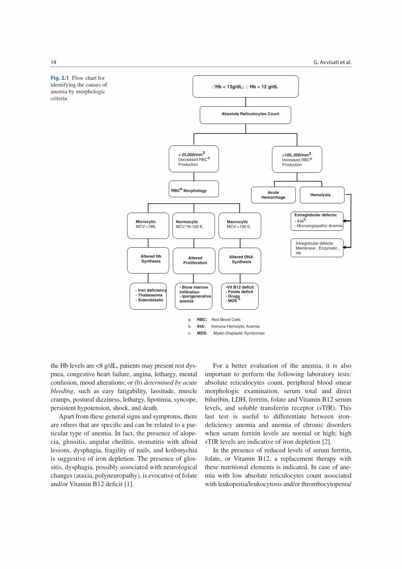
To identify the causes of anemia, we can use functional and morphologic criteria. However, from a practical point of view, the morphologic criteria are preferred and require the reticulocytes absolute number and the mean corpuscular volume (MCV), respectively. The fl owchart for identifying the causes of anemia using morphologic criteria is reported in Fig . 2.1 .
2.1.1.1 Approach to the Anemic Patient
Soon after a diagnosis of anemia has been made, the next step is to take an accurate clinical history and perform a physical examination to evaluate the signs and symptoms of anemia. These signs and symptoms may be: (a) directly related to anemia , and therefore, may present in all patients, independently from the cause of anemia, such as pallor, anorexia, fatigue, roaring in the ears, tachycardia, heart murmur, arrhythmia; until when
Hematologic Issues in Cervical Spine Surgery
Giuseppe Avvisati , Ombretta Annibali , Elisabetta Cerchiara ,
Marianna De Muro , Rosa Greco , Francesco Marchesi , Carolina
Nobile , Odoardo Olimpieri , Azzurra Romeo, and Maria Cristina
Tirindelli
G. Avvisati (*) Hematology , University Campus Bio-Medico , Via Àlvaro del Portillo, 21 , 00128 Rome , Italy e-mail: [email protected]
2
Contents
2.1 Changes in Blood Cell Count
Parameters .............................................................. 13
2.1.1 Anemia ..................................................................... 132.1.2 Polycythemia ............................................................ 152.1.3 Reduction in the White Blood Cell Count ............... 162.1.4 Increase in White Blood Cell Count ........................ 172.1.5 Changes in Platelet Count ........................................ 172.1.6 How a Cervical Spine Surgeon should Manage
Patients with Changes in Blood Cell Count ............ 18
2.2 Patients with Monoclonal
Gammopathy .......................................................... 19
2.2.1 Monoclonal Gammopathy of Undetermined Signifi cance (MGUS) .............................................. 19
2.2.2 Multiple Myeloma ................................................... 20
2.3 Preoperative Evaluation of the
Hemorrhagic Risk .................................................. 20
2.3.1 Abnormal Hemostatic and Coagulation Parameters ........................................... 20
2.4 Preoperative Evaluation of the
Thromboembolic Risk ........................................... 23
2.5 Thromboprophylaxis in Cervical
Spine Surgery ......................................................... 24
References .......................................................................... 26
14 G. Avvisati et al.
the Hb levels are <8 g/dL, patients may present rest dys-pnea, congestive heart failure, angina, lethargy, mental confusion, mood alterations; or (b) determined by acute
bleeding , such as easy fatigability, lassitude, muscle cramps, postural dizziness, lethargy, lipotimia, syncope, persistent hypotension, shock, and death.
Apart from these general signs and symptoms, there are others that are specifi c and can be related to a par-ticular type of anemia. In fact, the presence of alope-cia, glossitis, angular cheilitis, stomatitis with aftoid lesions, dysphagia, fragility of nails, and koilonychia is suggestive of iron depletion. The presence of glos-sitis, dysphagia, possibly associated with neurological changes (ataxia, polyneuropathy), is evocative of folate and/or Vitamin B12 defi cit [1] .
For a better evaluation of the anemia, it is also important to perform the following laboratory tests: absolute reticulocytes count, peripheral blood smear morphologic examination, serum total and direct biluribin, LDH, ferritin, folate and Vitamin B12 serum levels, and soluble transferrin receptor (sTfR). This last test is useful to differentiate between iron- defi ciency anemia and anemia of chronic disorders when serum ferritin levels are normal or high; high sTfR levels are indicative of iron depletion [2] .
In the presence of reduced levels of serum ferritin, folate, or Vitamin B12, a replacement therapy with these nutritional elements is indicated. In case of ane-mia with low absolute reticulocytes count associated with leukopenia/leukocytosis and/or thrombocytopenia/
< 25,000/mm3
Decreased RBCa
Production
- Iron deficiency
- Thalassemia
- Sideroblastic
-Vit B12 deficit - Folate deficit - Drugs - MDS
c
- Bone marrow
infiltration - Iporigenerative
anemia
♂Hb < 13g/dL; ♀ Hb < 12 g/dL
a. RBC: Red Blood Cells
b. IHA: Immune Hemolytic Anemia
c. MDS: Myelo-Displastic Syndromes
Absolute Reticulocytes Count
>100,.000/mm3
Increased RBCa
Production
RBCa Morphology
Hemolysis
Extraglobular defects:
- IHAb
- Microangiopathic Anemia
Intraglobular defects:
Membrane , Enzymatic ,
Hb
Microcytic
MCV:<78fLNormocytic
MCV:78-100 fL
Macrocytic
MCV:>100 fL
Altered
Proliferation
Altered Hb
Synthesis Altered DNA
Synthesis
Acute
Hemorrhage
Fig. 2.1 Flow chart for identifying the causes of anemia by morphologic criteria

2 Hematologic Issues in Cervical Spine Surgery 15
thrombocytosis, it is very important to seek the hema-tologist’s advice before any surgical intervention, because the cause of anemia may infl uence the possibil-ity to perform a surgical procedure such as in the case of a diagnosis of acute leukemia.
Moreover, anemia may also be a postoperative complication, related to blood loss during surgery. Therefore, a low hemoglobin level observed before cervical spine surgery requires investigation, and must be corrected to prevent more severe postoperative ane-mia, which can expose the patient to cardiovascular complications, delay healing of the tissue, and blood transfusions, with an increased risk of transfusion-related adverse events. Once the anemia has been cor-rected, we can reduce the exposure to homologous blood using autologous blood transfusions or acute normovolemic hemodilution.
2.1.1.2 Anemia’s Decalogue for the Surgeon
(a). The management of anemia should be tailored to the cause and severity of anemia.
(b). Blood transfusion therapy is indicated generally only in case of severe (Hb:6–8 g/dL) or very severe (Hb <6 g/dL) anemia not related to iron, folate, or Vitamin B12 defi ciency.
(c). In the presence of reduced levels of serum ferritin, folate, or Vitamin B12, replacement therapy with these nutritional elements is indicated.
(d). In anemia caused by iron, folate, or Vitamin B12 defi ciency, blood transfusions therapy may be required to avoid more severe complications, when symptoms or hemodynamic instability and/or co-morbidities are present.
(e). To correct iron-defi ciency (low serum ferritin lev-els) anemia, slow intravenous infusion (1 vial diluted in 250 mL of saline solution 0.9%, infused in 1 h) or oral iron can be used.
(f). The choice between intravenous and oral iron depends on the surgical urgency.
(g). Erythropoietin (EPO) therapy may be useful in case of moderate anemia (Hb: 8–10 g/dL) in the absence of co-morbidities.
(h). In the presence of severe co-morbidities, such as cardiac or pulmonary disease, blood transfusion therapy is mostly indicated to correct anemia.
(i). If surgery can be postponed, intravenous iron ther-apy combined with EPO therapy may increase the
Hb to levels that allow preoperative autologous blood collection (even in those patients with ane-mia of chronic diseases), thus reducing the use of homologous blood transfusions.
(j). In case of anemia with low absolute reticulocytes count associated with leukopenia/leukocytosis and/or thrombocytopenia/thrombocytosis, one should seek the hematologist’s advice before any surgical intervention.
2.1.2 Polycythemia
The diagnosis of polycythemia may be suspected in patients with abnormally high hematocrit (Hct), Hb concentration, and/or red blood cells (RBC) count even though RBC value is least often used to suggest this diagnosis, as patients with thalassemia minor may have an elevated RBC count, but a normal or reduced Hct or Hb, due to the presence of microcytic, poorly hemoglobinized, hypochromic red cells [3] . These three measurements (Hct, Hb, and RBC) are concen-trations, and therefore, dependent on the plasma vol-ume as well as the RBC mass. Hence, it is necessary to differentiate the relative (or apparent) and unapparent polycythemia from absolute polycythemia.
• Relative polycythemia is a condition in which there is a reduction in the plasmatic volume with a rela-tive increase in RBC mass. As a consequence, Hct, Hb concentration, and RBC count are apparently increased. It can be caused by dehydration, diuretic drugs, alcohol, obesity, and hypertension. • Unapparent polycythemia is a condition in which the RBC mass and plasma volume are equally increased; hence, Hb and Hct remain normal. In this case, erythrocytosis can only be detected via blood volume and genetic studies. Absolute polycythemia is a condition in which there • is a true (absolute) increase in RBC mass. It can be primary or secondary. Primary polycythemia: depending on an effective • hyper-production of RBC mass caused by an intrin-sic bone-marrow alteration, it is determined by an acquired or inherited DNA mutation leading to an abnormal RBC production; it includes the poly-
cythemia vera (PV) and the rare familial variants. Secondary polycythemia: caused by elevated EPO • synthesis, responsible for the increased RBC

16 G. Avvisati et al.
production by bone marrow. It is most often due to an EPO response to hypoxia, but can also result from increased EPO secretion by congenital or acquired causes such as: presence of a congenital oxygen high-affi nity Hb or a tumor (kidney, liver, little brain (cerebellum), adrenal glands, lung, uterus).
2.1.2.1 Polycythemia and Cervical
Spinal Surgery
If a patient has an increased Hct value (>52% in men and >48% in women) and an increased Hb concentra-tion (>18 g/dL and >16 g/dL, respectively), it is abso-lutely important to diagnose whether or not these altered values are due to a PV or are the consequence of a relative or secondary polycythemia. The prognosis and management are different depending on the diag-nosis. Therefore, orthopedic surgeons before surgery should ask for a hematologist’s consultation. The hematologist, through an accurate history, physical examination, and some laboratory tests, will determine the etiology of polycythemia and evaluate the presence of an increased risk of arterial and/or venous throm-
botic events or bleeding, and the type of the prophylac-tic therapy needed to avoid these complications.
2.1.2.2 Tips for the Orthopedic Surgeon
in the Presence of a PV
If a patient with PV needs cervical spinal surgery, the orthopedic surgeon must ask the hematologist about how to prevent the possible thromboembolic (TE) or hemorrhagic complications.
Although these patients always receive low-dose aspirin (75–100 mg/day) as prophylaxis for arterial thrombosis, its use cannot be recommended to reduce the TE risk following elective spinal surgery [4] .
However, as its use can predispose the patient to an increased risk of bleeding during surgery, aspirin should be discontinued at least 1 week before surgery to avoid bleeding complications. As for TE complica-tions, their incidence during elective spinal surgery is unknown; however, recent guidelines recommend that in case of additional risk factors (advanced age, can-cers, neurological defi cit, previous TE events, or ante-rior surgery access), TE prophylaxis should be performed. Therefore, because the patients with PV
have a neoplastic disease and very often might have suffered from a previous TE episode, they should receive antithrombotic prophylaxis for spinal surgery.
2.1.3 Reduction in the White
Blood Cell Count
In adults, a reduction in the white blood cells (WBC) count of <4,000/µL is defi ned as leukopenia. Its etiol-ogy may be related to disorders of production or distri-bution and turnover of the leukocytes, and to the use of some drugs or infectious diseases. Therefore, before a cervical spinal surgery intervention, the causes of leu-copenia should be carefully investigated. However, from a practical view point, only absolute neutropenia (neutrophiles <1,500/µl) and absolute lymphopenia (lymphocytes <1,000/µl) will be described.
2.1.3.1 Absolute Neutropenia
Absolute neutropenia (neutrophiles <1,500/µL) can predispose the patient to severe infective diseases. Neutrophiles play a pivotal role in the defense of the human body against infections. Therefore, the risk for acute or chronic infections must be evaluated before listing neutropenic patients for surgery. Also, bacterial, viral, parasitic, and rickettsial infections may be respon-sible for this condition. The main causes of absolute neutropenia are: acquired (infectious diseases, drugs, immune and autoimmune disorders, severe folate and Vitamin B12 defi ciency, hypersplenism, hematologic malignancies) or congenital (very rare and occurring in childhood) [5] . Furthermore, the absolute neutrophil count (ANC) is used to defi ne the severity of neutrope-nia as follows:
• Mild : ANC <1,500/µL and >1,000/µL • Moderate : ANC <1,000/µL and >500/µL • Severe : ANC <500/µL • Very severe : ANC <100/µL
Only those patients with severe or very severe neutro-penia are considered at high risk for infections by opportunistic or nonopportunistic microbial agents. Moreover, in a patient who is a candidate for surgery, unexplained neutropenia, associated with anemia and/or thrombocytopenia, must be carefully investigated
2 Hematologic Issues in Cervical Spine Surgery 17
by a trained hematologist before surgery is performed to exclude the presence of hematologic malignancies.
2.1.3.2 Absolute Lymphocytopenia
A condition of absolute lymphocytopenia (lympho-cytes <1,000/µL) may be caused by: drugs (i.e., gluco-corticoids, some anti-metabolites chemotherapy, and immune-suppressor drugs), some infectious diseases (i.e., HIV infection in AIDS phase, HBV, HCV, TBC, typhus fever, sepsis, pneumonia), radiotherapy, sarcoi-dosis, autoimmune disorders, chronic renal failure, some hematologic malignancies (i.e., Hodgkin’s lym-phoma in advanced phase of disease), and some rare congenital immune disorders [6] . To avoid complica-tions, the cause of lymphocytopenia should be deter-mined before surgery is performed.
2.1.4 Increase in White Blood Cell Count
An increase in WBC of >10,000/µL is defi ned as • leukocytosis, and may have many causes, which should be carefully investigated before any surgical intervention. Moreover, some pathologic conditions are responsible for an absolute increase in a single type (neutrophiles, lymphocytes, monocytes eosino-philes, basophiles) of WBC associated or not asso-ciated with leukocytosis. Therefore, the fi rst step in the diagnostic process of this condition is to defi ne which type of WBC is increased; as a consequence, in adults, depending on the type of WBC increased, we can observe the following conditions: • Absolute neutrophilia : neutrophiles >7,500/µL • Absolute lymphocytosis : lymphocytes >4,000/µL • Absolute monocytosis : monocytes >1,000/µL • Absolute eosinophilia : eosinophiles >500/µL • Absolute basophilia : basophiles >200/µL (very
rare)
Frequently, however, there is only a relative increase in a subtype of WBC, determining an alteration in the leukocytes formula, without an increase in the absolute number of some types of WBC. These conditions are defi ned as “relative” increase (i.e., in case of a WBC count of 4,270/µL with 20% of monocytes, despite the percentage of monocytes being high, its absolute
number is <1,000/µL; thus, we can conclude the condi-tion as “relative monocytosis”).
2.1.4.1 Absolute Neutrophilia
The presence of an absolute neutrophilia ( neutrophiles
>7,500/ µ L ) arises from various clinical conditions such as infections, chronic infl ammation, neoplasia or a hematological malignancy, or a fracture. Moreover, transient neutrophilia is normal in the postoperative period, caused by the response of the human body to the surgical stress. Persistent neutrophilia may be asso-ciated with a surgery-related infective complication. An increase in the absolute number of neutrophiles before surgery can be a good reason for a hematologic consultation.
2.1.4.2 Absolute Lymphocytosis
An increase in the absolute number of lymphocytes ( lymphocytes >4,000/µL ) may be caused by hemato-logic malignancies or by viral, certain bacterial, or parasitic infections. Moreover, vasculitis, infl amma-tory bowel diseases, emergency medical conditions, acute trauma, and hypersensitivity reactions (drug-induced or related to acute serum sickness) may cause absolute lymphocytosis. A trained hematologist can easily verify its pathogenesis.
2.1.4.3 Absolute Eosinophilia
and Absolute Monocytosis
These conditions are less frequent than absolute neu-trophilia and lymphocytosis; therefore, we will not describe these conditions.
2.1.5 Changes in Platelet Count
2.1.5.1 Abnormal Increase in Platelet Count
The normal platelet count ranges from 150,000 to 450,000/µL, and a platelet count >500,000/µL is defi ned as thrombocytosis [7] .
However, <10% of isolated thrombocytosis refl ects a hematological disorder. Therefore, most cases of

18 G. Avvisati et al.
thrombocytosis occur within a systemic disorder, sec-
ondary or reactive thrombocytosis [8] , such as those occurring because of:
Recent trauma or surgery • Prior surgical removal of the spleen • Local or systemic complaints suggesting infection • or infl ammation Iron defi ciency • Pregnancy • Malignancy •
A primary thrombocytosis may be due to a myelopro-liferative disorder, and when isolated, is generally pathognomonic of essential thrombocythemia (ET).
Therefore, in the presence of a pathologic increase in platelet count, it is very important to exclude that thrombocytosis is a reactive process. Once a reactive process has been excluded, the next step is to accu-rately classify whether or not the thrombocytosis is a result of a myeloproliferative disorder such as ET, chronic myelogenous leukemia (CML), idiopathic myelofi brosis (IMF), PV, myelodysplastic syndrome (MDS), and acute myeloid leukemia (AML) [9] . Thus, patients in whom a reactive process cannot be identi-fi ed, require a hematological consult and a bone mar-row examination with reticulin staining and cytogenetic studies to better defi ne the myeloproliferative disorder responsible for thrombocytosis.
Patients with primary thrombocytosis as well as those with thrombocytosis secondary to malignancy require a correct TE prophylaxis because the risk of TE episodes range from 10 to 25% and is not related to the number of platelets [10] .
2.1.5.2 Abnormal Decrease in Platelet Count
A platelet number <150,000/µL defi nes thrombocytope-nia, and may predispose to excessive bleeding [7] . However, bleeding during surgery because of a reduc-tion in the number of platelets does not generally occur until the platelet count is <50,000/µL, and clinical or spontaneous bleeding does not occur until the platelet count is <10,000–20,000/µL [11] . Once thrombocy-topenia has been reported, it should be confi rmed by repeated testing and examination of the peripheral blood smear to exclude cases of pseudothrombocytopenia. In the presence of a confi rmed result of thrombocytopenia, the initial step is to perform a comprehensive history and physical examination to understand the cause of it.
Drugs are a common cause of thrombocytopenia. The spectrum of drugs reported to cause thrombocy-topenia is broadening and changing progressively. A complete list and analysis of all published reports of drug-induced thrombocytopenia is available on the internet ( http://w3.ouhsc.edu/platelets ). Patients with thrombocytopenia may be asymptomatic, and thrombo-cytopenia may be fi rst detected on a routine complete blood count (CBC). The most common symptomatic presentation of thrombocytopenia is bleeding, charac-teristically mucosal and cutaneous. Following confi r-mation of thrombocytopenia, to diagnose its cause, it is better to consult a hematologist.
In thrombocytopenic patients at risk of, or com-plaining of, severe hemorrhagic complications, the guidelines of The Italian Society of Haemostasis and Thrombosis, may be applied [12] , which is summa-rized as follows:
• Prophylaxis of hemorrhagic events during surgery : High hemorrhagic risk and surgery procedures at • high risk of hemorrhage impossible to control by local hemostatic measures: 1 unit/10 kg of body weight of random donor platelets or 1 U of platelets pheresis Low hemorrhagic risk and surgery procedures at • high risk of hemorrhages: desmopressin if not con-traindicated (i.e., previous thrombosis or throm-boembolism, severe arteriopathy) Low hemorrhagic risk and minor surgery: local • hemostasis • Treatment of hemorrhagic episodes: • Major hemorrhagic event: 1 unit/10 kg of body weight of random donor platelets or 1 U of platelets pheresis • Minor hemorrhagic event: local hemostasis
2.1.6 How a Cervical Spine Surgeon
should Manage Patients with
Changes in Blood Cell Count
In the presence of changes in the blood cell count, sur-geons should recognize these alterations and seek a consultation with a trained hematologist, who, by means of laboratory and instrumental tests, will recognize the cause responsible for the changes observed in the blood cell count. In the meantime, the hematologist will pro-vide the surgeon with correct suggestions about the

2 Hematologic Issues in Cervical Spine Surgery 19
possibility of performing the surgical intervention immediately or after the solution of the problem respon-sible for the changes in the blood cell count.
2.2 Patients with Monoclonal
Gammopathy [13]
A monoclonal gammopathy (paraproteinemia or dys-proteinemia) is defi ned as the presence of immuno-logically homogeneous protein commonly referred to as a paraprotein or monoclonal protein (M-protein, where the “M” stands for monoclonal) in serum or urine, produced by a single clone of plasma cells. The routinely recommended method for the detection and quantifi cation of an M-protein in serum or concen-trated urine is the agarose gel electrophoresis, an inex-pensive and easy to perform screening procedure.
Before cervical spine surgery, serum protein elec-trophoresis should be considered in any patient with:
Elevated erythrocyte sedimentation rate • Elevated serum viscosity • Unexplained anemia • Back pain, weakness, or fatigue • Osteopenia • Osteolytic lesions • Spontaneous fractures • Renal insuffi ciency with a bland urine sediment • Heavy proteinuria in a patient over the age of 40 • years Hypercalcemia • Hypergammaglobulinemia • Immunoglobulin defi ciency • Bence Jones proteinuria • Unexplained peripheral neuropathy •
However, an M-protein can be present in a number of different disorders, including B-cell and plasma-cell proliferations. The most common of these are listed in Table 2.1 .
2.2.1 Monoclonal Gammopathy of
Undetermined Signifi cance (MGUS)
MGUS is found in approximately 1–2% of the patients. The incidence is generally higher in patients over 70 years. In well-performed epidemiologic studies, the
prevalence of MGUS in subjects ³ 50, ³ 70, and ³ 85 years of age was 3.2, 5.3, and 7.5%, respectively. Differentiation of a patient with MGUS from the one with multiple myeloma (MM) may sometimes be
Table 2.1 Disorders associated with the presence of a monoclonal gammopathy
Plasma cell disorders
Monoclonal gammopathy of undetermined signifi cance (MGUS)
Biclonal gammopathy of undetermined signifi cance
Idiopathic Bence Jones proteinuria
POEMS syndrome, Osteosclerotic myeloma
Castleman’s disease
AL (light chain) amyloidosis
Solitary plasmacytoma
Multiple myeloma (MM)
Smoldering MM
B-cell lymphoproliferative disorders
Non-Hodgkin’s lymphoma
Chronic lymphocytic leukemia
Lymphoplasmacytic lymphoma (Waldenstrom macroglobulinemia)
Posttransplant monoclonal gammopathies
Connective-tissue disorders
Systemic lupus erythematosus
Rheumatoid arthritis
Sjogren syndrome
Scleroderma
Psoriatic arthritis
Associated with infections
Hepatitis C virus infection
HIV/AIDS
Dermatologic disorders
Miscellaneous disorders
Cryoglobulinemia
Cryofi brinogenemia
Chronic myelogemous leukemia
Acquired von Willebrand disease
Myelodysplastic syndrome
The most common causes of monoclonal gammopathy are: MGUS and MM

20 G. Avvisati et al.
diffi cult at the time of initial presentation. By defi nition, the diagnosis of MGUS requires the absence of anemia, hypercalcemia, renal failure, and lytic bone lesions related to the plasma cell proliferative disorder. However, the mere presence of clinical fi ndings such as anemia, hypercalcemia, renal failure, or lytic bone lesions in conjunction with an M-protein does not automatically indicate MM or related malignancy, as these abnormali-ties may be due to other unrelated coexisting diseases (see Table 2. 2.1 ). To differentiate MGUS from a related plasma-cell malignancy, patients should have a CBC, serum creatinine, serum calcium, and a complete radio-graphic bone survey of the skeleton, including all long bones. Patients who exhibit abnormalities with respect to the above-mentioned tests should undergo additional tests to determine the etiology of the abnormalities, and whether they are indeed related to a plasma-cell prolif-erative disorder.
2.2.2 Multiple Myeloma
MM is caused by the malignant proliferation of plasma cells in the bone marrow producing an M-protein. Plasma cells frequently invade the adjacent bone, pro-ducing skeletal destruction that results in pathological fractures and bone pain. Other symptoms of this dis-ease are anemia, hypercalcemia, and impairment of renal function. An M-protein in the serum or urine is present in 97% of these patients.
In particular, a serum M-protein is observed in 80% of the patients at diagnosis, while immune-fi xation reveals an M-protein in over 90%. An IgG M-protein is found in about 50% of the patients, while 20% have an IgA M-protein, and monoclonal light chain alone (light-chain myeloma) is found in almost 20% of the patients. M-protein (Bence Jones protein) in urine is observed in approximately 75% of the patients.
The most critical criterion for symptomatic or treat-able disease is the evidence of organ or tissue impair-ment (end-organ damage) manifested by anemia, hypercalcemia, lytic bone lesions, renal insuffi ciency, hyperviscosity, amyloidosis, or recurrent infections. Most patients with MM are symptomatic. However, it is possible that during screening tests for unrelated dis-orders, patients without any symptoms attributable to myeloma may be diagnosed as having MM (asymp-tomatic myeloma). Other patients may demonstrate local symptoms owing to plasmacytoma. Solitary plas-macytoma is rare. The diagnosis requires histologic
evidence of a monoclonal plasma cell infi ltrate in the lesion, absence of other lesions, and lack of marrow plasmacytosis elsewhere.
2.3 Preoperative Evaluation
of the Hemorrhagic Risk
Besides thrombocytopenia, a quantitative defect of platelet number (see above), thrombocytopathy, a qualitative (abnormal function) defect of platelets, as well as the alterations in the coagulation parameters (prothrombin time = PT, activated partial thromboplas-tin time = aPTT, and fi brinogen) may increase the hem-orrhagic risk in cervical spine surgery.
2.3.1 Abnormal Hemostatic
and Coagulation Parameters
A good knowledge of the basic principles of the hemo-static system is mandatory in cervical spine surgery, as unexpected bleedings or thrombosis episodes can rap-idly become life-threatening emergencies in the intra-operative and postoperative period.
The reason of this extreme susceptibility to coagu-lation-related complications can be found in the vascu-lar anatomy of the cervical district [14] . The vertebral and basilar arteries provide up to 20% of the total cere-bral blood fl ow, and are the principal affl uent to the arterial circulation system of the cerebellum and brain-stem. Any alteration that involves the vertebrobasilar circulation can cause irreversible ischemic damages to vital structures.
The incidence of severe hemostatic alterations after cervical surgery is diffi cult to assess, overly depending on the patient’s conditions and comorbidities, but it has been estimated in 1:20,000 patients subjected to cervical manipulation, even though more stringent studies have documented the incidence rates as high as 1:4,500.
To prevent ischemic injuries from arterial occlusions, the vertebrobasilar system is rich in collateral vessels that can rapidly redirect the blood fl ow to the hypoper-fused zone. Nonetheless, any collateral system has intrinsic limits, and cannot overcome large fl ow inter-ruptions. Other possible elements involved in thrombotic alterations are blood fl ow interruptions during surgery, and possible endothelial damages following manipula-tion. Several studies showed that arterial blood fl ows can
2 Hematologic Issues in Cervical Spine Surgery 21
be stopped during surgery for up to 3 h without throm-botic complications, provided that an adequate anti-thrombotic prophylaxis had been performed. Nonetheless, the characteristic fl ow intermittency in vertebrobasilar artery during spinal surgery can be an additional stimu-lus for thrombus formation, and can lead to another hemostatic issue, namely, the endothelial damage.
Given these characteristics, the evaluation of thrombo-hemorrhagic risk factors for every single patient should be accurate and exhaustive, from his-tory taken at the bedside to preoperative laboratory screening tests. Only an adequate familiarity with the coagulation system can provide a safe approach to such challenging surgery.
2.3.1.1 Qualitative Platelets Defects
(Thrombocytopathies)
Platelets are important for primary hemostasis. Physiologically, when platelets are exposed to damaged endothelium, they adhere to the exposed basement membrane collagen and change their shape from smooth disks to spheres with pseudopodia. Subsequently, they secrete the contents of their granules, a process referred to as the release reaction. Finally, additional platelets aggregate on those platelets that have adhered to the vessel wall. As a result, the primary hemostatic plug is formed and bleeding is arrested.
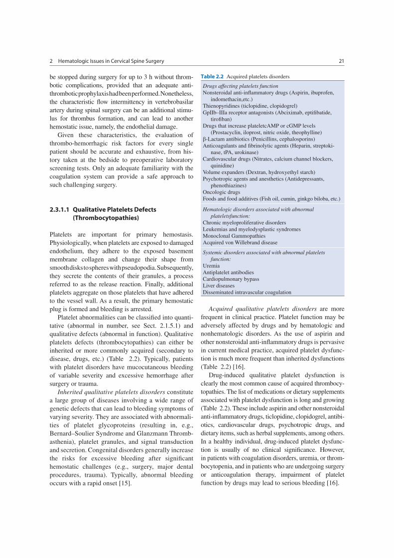
Platelet abnormalities can be classifi ed into quanti-tative (abnormal in number, see Sect. 2.1.5.1 ) and qualitative defects (abnormal in function). Qualitative platelets defects (thrombocytopathies) can either be inherited or more commonly acquired (secondary to disease, drugs, etc.) (Table 2.2 ). Typically, patients with platelet disorders have mucocutaneous bleeding of variable severity and excessive hemorrhage after surgery or trauma.
Inherited qualitative platelets disorders constitute a large group of diseases involving a wide range of genetic defects that can lead to bleeding symptoms of varying severity. They are associated with abnormali-ties of platelet glycoproteins (resulting in, e.g., Bernard–Soulier Syndrome and Glanzmann Thromb-asthenia), platelet granules, and signal transduction and secretion. Congenital disorders generally increase the risks for excessive bleeding after signifi cant hemostatic challenges (e.g., surgery, major dental procedures, trauma). Typically, abnormal bleeding occurs with a rapid onset [15] .
Acquired qualitative platelets disorders are more frequent in clinical practice. Platelet function may be adversely affected by drugs and by hematologic and nonhematologic disorders. As the use of aspirin and other nonsteroidal anti-infl ammatory drugs is pervasive in current medical practice, acquired platelet dysfunc-tion is much more frequent than inherited dysfunctions (Table 2.2 ) [16] .
Drug-induced qualitative platelet dysfunction is clearly the most common cause of acquired thrombocy-topathies. The list of medications or dietary supplements associated with platelet dysfunction is long and growing (Table 2.2 ). These include aspirin and other nonsteroidal anti-infl ammatory drugs, ticlopidine, clopidogrel, antibi-otics, cardiovascular drugs, psychotropic drugs, and dietary items, such as herbal supplements, among others. In a healthy individual, drug-induced platelet dysfunc-tion is usually of no clinical signifi cance. However, in patients with coagulation disorders, uremia, or throm-bocytopenia, and in patients who are undergoing surgery or anticoagulation therapy, impairment of platelet function by drugs may lead to serious bleeding [16] .
Table 2.2 Acquired platelets disorders
Drugs affecting platelets function Nonsteroidal anti- infl ammatory drugs (Aspirin, ibuprofen,
indomethacin,etc.) Thienopyridines (ticlopidine, clopidogrel) GpIIb–IIIa receptor antagonists (Abciximab, eptifi batide,
tirofi ban) Drugs that increase plateletcAMP or cGMP levels
(Prostacyclin, iloprost, nitric oxide, theophylline) b -Lactam antibiotics (Penicillins, cephalosporins) Anticoagulants and fi brinolytic agents (Heparin, streptoki-
nase, tPA, urokinase) Cardiovascular drugs (Nitrates, calcium channel blockers,
quinidine) Volume expanders (Dextran, hydroxyethyl starch) Psychotropic agents and anesthetics (Antidepressants,
phenothiazines) Oncologic drugs Foods and food additives (Fish oil, cumin, ginkgo biloba, etc.)
Hematologic disorders associated with abnormal
plateletsfunction: Chronic myeloproliferative disorders Leukemias and myelodysplastic syndromes Monoclonal Gammopathies Acquired von Willebrand disease
Systemic disorders associated with abnormal platelets
function: Uremia Antiplatelet antibodies Cardiopulmonary bypass Liver diseases Disseminated intravascular coagulation

22 G. Avvisati et al.
Aspirin is the most notable drug in this regard because of its frequent use, its irreversible effect on platelet prostaglandin synthesis, and its documented effect on hemostatic competency [17] . The inhibition of platelet release reaction occurs within 15–30 min after ingestion with aspirin doses as low as 40–80 mg, and persists as long as the affected platelet survives (8–10 days). Thus, a single small dose of aspirin impairs the release reaction for up to 96 h [18] . Other nonsteroidal
anti-infl ammatory drugs (such as ibuprofen, indometha-cin) reversibly inhibit platelet prostaglandin synthesis and usually have little effect on hemostasis [19] . The antiplatelet effect of a number of drugs has proved use-ful in preventing arterial thrombosis, but excessive bleeding can complicate their use. In addition to aspirin, these drugs include ticlopidine and clopidogrel [20] . Their effect on platelet aggregation may be seen within 24–48 h of the fi rst dose, but does not reach a maximum for 4–6 days. The effect on platelet function may last for 4–10 days after the drugs have been discontinued. A number of other drugs and a number of foods and food additives may affect platelet function, but these effects do not appear to be clinically signifi cant.
Hematologic diseases associated with abnormal platelet function include marrow processes in which platelets may be intrinsically abnormal, such as the clonal myeloid diseases, dysproteinemias in which abnormal plasma proteins can impair platelet function, and acquired forms of von Willebrand disease. Of the systemic diseases, renal failure is most prominently associated with abnormal platelet function because of retention of platelet inhibitory compounds. Platelet function may be abnormal in the presence of antiplate-let antibodies, following cardiopulmonary bypass, in association with liver disease, or in disseminated intra-vascular coagulation [16] .
2.3.1.2 Coagulopathies
These alterations require accurate preoperative evaluation [21] . The most important part of an accurate preopera-tive evaluation of possible coagulation alterations is his-tory taking. In this process, one should always consider that frequently normal people consider their bleeding to be excessive. In dubious cases, further help can be obtained by physical examination and laboratory tests.
Physical examination is important in assessing the possible coagulation problems, and the physician should have a particular focus on skin and mucosal hemorrhagic
manifestations, such as small red focal areas (<2 mm) that do not disappear with fi nger pressure (petechiae). Petechiae are more commonly found on sites subjected to shear and pressure stresses (under belt skin or perior-bitary regions, especially after crying or intense cough-ing); when they merge into larger areas, they are called purpura (<1 cm) or ecchymoses (>1 cm), and are fre-quently associated with platelets disorders. Primary coagulation disorders are, however, more frequently associated with large and spontaneous bruises, painful hemarthroses, and intracavitary blood effusions.
Every patient should, therefore, be asked about:
Past bleeding episodes associated with surgical pro-• cedures (e.g., appendectomy, circumcision, tonsil-lectomy, or dental extractions). Spontaneous bruises formation and/or petechial • lesions. History of frequent nosebleeds (if confi ned to a • single nostril, a hematologic systemic disease is improbable; if bilateral and recurrent in absence of major trauma, also consider hereditary hemorrhagic teleangiectasias or von Willebrand disease). Presence of recurrent hemorrhagic and/or throm-• botic episodes in family members. Excessive bleeding during childbearing or men-• strual cycles. Recurrent episodes of gingival hemorrhage in the • absence of local alterations. Excessive bleeding from minor cuts, with particular • regard to the time to stop and the need for direct pressure or tissue paper. Even a single episode of hemarthrose is worth for • further investigations for hemophilia, especially in infants and children. Comorbidities such as epatic diseases, alterations in • nutrient absorption, alcohol assumption history, or vitamin defi ciencies. Very important is the presence of renal failure or • liver diseases. Drug assumption (with particular attention to non-• steroidal anti-infl ammatories, oral anticoagulants such as warfarin and acenocoumarol, herbal reme-dies, and other medications available without prescription); NB: oral anticoagulants should be suspended at least 4 days before surgery and an adequate prophylaxis with low molecular weight heparin (LMWH) should be started at the same time; clopidogrel should be discontinued 14 days before any major surgical procedure, aspirin and

2 Hematologic Issues in Cervical Spine Surgery 23
lysine acetylsalicylate administration should be interrupted at least 7 days before surgery, whereas Clopidogrel and Ticlopidine should be interrupted 4–6 days prior to surgery.
2.3.1.3 The laboratory Interface
As pointed out earlier, laboratory tests can be extremely helpful in assessing possible coagulation disorders that can put at risk the patient’s life during cervical surgery [22] ; notwithstanding, the need for routine preoperative testing is controversial. While coagula-tion tests can in fact detect many asymptomatic disor-ders that may cause surgical bleedings, many studies bring into question their positive predictive value for perioperative hemorrhagic complications.
Coagulation test can be divided into routine tests and secondary tests.
Routine Tests
Prothrombin Time (PT) PT analyzes the “extrinsic and common pathway
factors,” and it is particularly sensible in detecting alter-ations of factors VII, X, V, thrombin, and fi brinogen.
The main causes of PT alterations are:
Sampling errors • Anticoagulant therapy • Factor VII, X, V, thrombin, or fi brinogen defi cien-• cies ( it is also mandatory to test the hepatic
function) Coagulation-acquired inhibitors (antibody against • factor VII, X, V, thrombin, or fi brinogen), antibod-ies, anti-phospholipids MM paraprotein •
Activated partial thromboplastin time (aPTT) The aPTT test evaluates the intrinsic pathway (com-
posed by factor XII, XI, VIII, and IX) together with the common pathway. Abnormalities in aPTT are caused by:
Sampling errors • Heparin or anticoagulant therapy • Factor XII, XI, IX, VIII, X, V, thrombin, or fi brino-• gen defi ciencies ( NB: consider hemophilia; testing
the hepatic function is also mandatory)
Coagulation-acquired inhibitors (antibody against • factor XII, XI, IX, VIII, X, V, thrombin, or fi brino-gen), antibodies, anti-phospholipids Von Willebrand disease • MM paraprotein •
Second-Line Tests
Second-line tests are special assays to further discrimi-nate the fi rst-line tests’ abnormalities, and should always be requested under the advice of an expert hematolo-gist. The most commonly performed second-line tests are single-factor concentration assays, used to charac-terize and evaluate the defi ciency of single coagulation factors, such as factor VIII and factor IX in hemophila A and B, respectively. dRVVT and KCT-SCT, specifi c aPTT-derived tests performed with low concentrations of phospholipids, are useful in detecting Lupus Anticoagulant. Moreover, two second-line tests could be useful to evaluate the presence of heparin in blood samples: thrombin time (TT) and reptilase time (RT). TT measures the clotting time after the addition of thrombin to plasma. It is prolonged only in the presence of heparin, MM paraprotein, and hypofi brinogenemia. In the presence of a prolonged TT, an RT test should be performed. RT is not sensitive to heparin, and hence, in the presence of an abnormal TT and a normal RT, the alteration is due to heparin in the tested sample.
As pointed out earlier, correct evaluation of the hemorrhagic risk is mandatory in every patient who is a candidate for cervical spine surgery, because it will reduce the risk of hemorrhagic complications that can rapidly become life-threatening emergencies in intra- and postoperative periods.
2.4 Preoperative Evaluation
of the Thromboembolic Risk
The vertebral and basilar arteries provide up to 20% of the total cerebral blood fl ow, and are the principal affl uent to the arterial circulation system of cerebellum and brain-stem. Any alteration that involves the vertebrobasilar cir-culation can cause irreversible ischemic damages to vital structures. Therefore, a correct evaluation, before sur-gery, of the TE risk in these patients may result in a sharp decrease in the ischemic risk [3] .
The term Thrombophilia is now used to describe a predisposition to an increased risk of thromboembolism.

24 G. Avvisati et al.
Thromboembolism is a multifactorial disorder produced by congenital abnormalities of anticoagulant or proco-agulant factors combined with acquired pathological conditions [23] . The patients-related risk factors for venous thromboembolism (VTE) are summarized in Table 2.3 .
A correct approach to the evaluation of TE risk should start with a full history dealing with previous TE episodes suffered by the patients or their relatives. Only in the presence of a positive personal or familiar
history for thromboembolism, we should perform labo-
ratory tests to identify congenital or acquired causes
of thrombophilia [24] . Moreover, in the evaluation of the TE risk, we also have to consider those pathologies frequently associated with an elevated risk of deep vein thrombosis (DVT) or pulmonary embolism (PE) [25] , such as cancer, autoimmune disorders, infl amma-tory bowel disease, hematologic malignancies, older age, previous history of thromboembolism, and recur-rent fetal loss (see Table 2.3 ).
In presence of one or more of the abovementioned risk factors, a correct program of thromboprophylaxis should be applied (see paragraph X5 below), even though the thrombotic risk is dependent on the type and duration of the surgery. Moreover, the need for a thrombophilic screening should always be suggested by an expert in thromboembolism. Depending on the laboratory results obtained and on the associated pathologic conditions, it is possible to identify which is the risk for the develop-ment of thromboembolism in patients (Table 2.4 ) who are candidates for cervical spine surgery.
2.5 Thromboprophylaxis in Cervical
Spine Surgery
During the last 15–20 years, spine surgery has changed radically, developing into a well-defi ned area of spe-cialist surgery. As a consequence, some attention is now being given to DVT and PE events in spinal sur-gery. The incidence of DVT during spine surgery is not
Table 2.4 Risk of thromboembolism in cervical spine surgery according to patient-, disease-, and surgery-related variables
Risk Hereditary factors Acquired factors
Low Heterozygous factor V
Leiden
Heterozygous prothrombin 20210
Oral contraceptives Pregnancy Elevated Factor VIII
Moderate Heterozygous ATIII, Protein C or Protein S defi ciency
Surgery for malignancy Sepsis Prolonged immobilization Antiphospholipid
antibodies Myeloproliferative
disorders PNH a
High Homozygous factor V
Leiden
Homozygous prothrombin 20210
Associated total hip and knee replacement
Hip fracture Anterior or anterior-
posterior combined surgical approach
Very high Homozygous or double heterozy-gous ATIII, protein C or protein S defi ciency
Mucin-secreting adenocarcinoma
a Paroxismal nocturnal hemoglobinuria
Table 2.3 Patient-related risk factors for VTE
Inherited Factor V Leiden Prothrombin 20210 mutation Protein C defi ciency Protein S defi ciency Antithrombin III defi ciency
Acquired Active heart or respiratory failure Acute medical illness Age over 60 years Antiphospholipid antibodies Autoimmune disorders Cancer and Chemotherapy Central venous catheter in situ Continuous travelformore than 3 h approximately 4 weeks
before or after surgery Hyperhomocystenemia Immobility (Paralysis or limb in plaster) Infl ammatory bowel disease Monoclonal gammopathy Myeloproliferative diseases Nephrotic syndrome Obesity (body mass index ³ 30 kg/m 2 ) Paroxismal nocturnal hemoglobinuria Personal or family history of venous thromboembolism Pregnancy and puerperium Recent myocardial infarction or stroke Severe infection Tobacco usage Trauma: Hip Fracture, Acute spinal injury Use of oral contraceptive or hormonal replacement therapy Varicose veins associated with phlebitis

2 Hematologic Issues in Cervical Spine Surgery 25
well documented, because only case reports or retro-spective studies are reported. Patients at greatest risk for VTE are those undergoing major lower extremity orthopedic surgery, and those who have experienced major trauma or spinal cord injury (SCI). However, while thromboprophylaxis guidelines are well known for general orthopedic surgery, especially in elective hip, knee, and trauma procedures, the optimal prophy-laxis against the risk of DVT and PE for patients under-going cranial or spinal procedures remains controversial [26] . Given the paucity of data, it is not possible to give fi rm recommendations about thromboprophylaxis in spine surgery patients. Moreover, some patients may not require any specifi c thromboprophylaxis. The risk of VTE appears to be low when any of the following methods of thromboprophylaxis is used: postoperative LDUH (Low Dose Unfractionated Heparin) or LMWH, or intraoperative and then postoperative GCS (Gradu-ated Compression Stockings), and/or IPC (Intermittent Pneumatic Compression). For spine surgery patients with additional VTE risk factors, such as a neurologic defi cit or prolonged immobility, advanced age, malig-nancy, previous VTE, or an anterior surgical approach, thromboprophylaxis with one of the above-mentioned options is recommended. Spine surgery includes many surgical procedures for a variety of pathologies, and involves a highly heterogeneous class of patients. Therefore, a careful analysis in terms of TE risks is required in each individual case (Tables 2.3 and 2.4 ). Three main variables need consideration:
1. Patient-related variables , such as age, gender (oral contraceptive use or hormonal substitutive therapy), bed rest, obesity and concomitant pathologies (hypertension, diabetes, varicose veins), genetic thrombophilic factors (see Table 2.3 )
2. Disease-related variables , such as trauma, tumor, deformity, degenerative pathology, and fi nally
3. Surgery-related variables , such as anterior, poste-rior, or combined approach (the higher risk of PE after combined anterior–posterior spinal fusions indicates that retraction and manipulation of the great vessels may lead to stasis or intimal damage that can predispose to clot formation), positioning, instrumentation, operating time, and location (cer-vical, thoracic, lumbar spine) [27] .
As there is no unique risk factor, and spinal surgery is so
varied, it is therefore not possible to suggest a standard-
ized thromboprophylaxis for spinal surgery , as can be
done for hip and knee surgery. In Tables 2.5 and 2.6 , we report the regimens to prevent VTE in elective spine surgery and acute SCI, respectively, suggested by The Eighth ACCP Conference on Antithrombotic and Thrombolytic Therapy, held in 2008 [28] .
Table 2.5 Thromboprophylaxis in elective spine surgery (Summarized from ref [28] )
No risk factors for VTE(see Tables 2.3 and 2.4 ) Early and persistent mobilization, not routinely use of specifi c
thromboprophylaxis Presence of some risk factors a (see Tables 2.3 and 2.4 ) One of the following thromboprophylaxis is recommended Additional risk factors for VTE (see Tables 2.3 and 2.4 ) Intra and postoperative mechanical prophylaxis with
intermittent pneumatic compression (IPC) ± graduated compression stockings (GCS) or
Postoperative LDUH or LMWH Multiple risk factors for VTE (see Tables 2.3 and 2.4 ) Intra and postoperative mechanical prophylaxis with IPC ±
GCS associatedwithpostoperative LMWH or LDUH
Patients with ruptured cranial or spinal vascular malforma-tions (e.g., brain aneurysms) should not be offered pharmacological prophylaxis until the lesion has been secured.
a Advanced age, malignancy, presence of a neurologic defi cit, previous VTE, or an anterior surgical approach
Table 2.6 Thromboprophylaxis in patients with acute spinal cord injury (SCI) (Summarized from ref [28] )
In all patients with acute SCI, routine thromboprophylaxis is recommended
Once primary hemostasis is evident,start thromboprophylaxis with LMWH, alternatives include the combined use of IPC and either LDUH or LWMH
In case anticoagulant thromboprophylaxis is contraindicated because of high bleeding risk early after injury,optimal use of IPC and/or GCS is recommended.When the high bleeding risk decreases, add pharmacologic thrombopro-phylaxis to the mechanical thromboprophylaxis
In patients with an incomplete SCI associated with the evidence of a spinal hematoma on CT or MRI, use mechanical thromboprophylaxis instead of anticoagulant thromboprophylaxis, at least for the fi rst few days after injury
Following acute SCI, do not use LDUH alone
Do not use an IVC (intravenous caval) fi lter for thromboprophylaxis
Following acute SCI, for patients undergoing rehabilitation, continue LMWH thromboprophylaxis or use an oral vitamin K antagonist (INR target, 2.5; range, 2.0–3.0)
26 G. Avvisati et al.
References
1. Marks PW, Glader B (2005) Approach to anemia in the adult and child. In: Hoffman R, Benz AI jr, Shattil SJ, Furie B, Cohen HJ, Silberstein LE, McGlave P (eds) Hematology, basic principles and practice, 4th edn. Elsevier, Churchill Livingstone, Amsterdam, New York
2. Weiss G, Goodnough LT (2005) Anemia of chronic disease. N Engl J Med 352:1011–1023
3. Bergqvist D (2007) Risk of venous thromboembolism in patients undergoing cancer surgery and options for thrombo-prophylaxis. J Surg Oncol 95:167–174
4. Ruggeri M, Rodeghiero F, Tosetto A et al; Gruppo Italiano Malattie Ematologiche dell’Adulto (GIMEMA) Chronic Myeloproliferative Diseases Working Party (2008) Postsurgery outcomes in patients with polycythemia vera and essential thrombocythemia: a retrospective survey. Blood 111:666–671
5. Palmblad JE, Von dem Borne AE (2002) Idiopathic, immune, infectious and idiosyncratic neutropenias. Semin Hematol 39:113–120
6. Kipps TJ (1995) Lymphocytosis and Lymphocytopenia. In. Beutler E, Lichtman MA, Coller BS, Kipps DJ (eds) Williams hematology. Mc Graw Hill, New York
7. Buckley MF, James JW, Brown DE et al (2000) A novel approach to the assessment of variations in the human plate-let count. Thromb Haemost 83:480–484
8. Schafer AI (2004) Thrombocytosis. N Engl J Med 350:1211–1219
9. Messinezy M, Westwood N, Sawyer B et al (1994) Primary thrombocythaemia: a composite approach to diagnosis. Clin Lab Haematol 16:139–148
10. Vannucchi AM, Barbui T (2007) Thrombocytosis and throm-bosis. Hematology Am Soc Hematol Educ Program 363–370
11. George JN, Woolf SH, Raskob GE et al (1996) Idiopathic thrombocytopenic purpura: a practice guideline developed by explicit methods for the American Society of Hematology. Blood 88:3–40
12. http://www.siset.org/lineeguida/LG4.pdf (Last revision 4 June 2007)
13. International Myeloma Working Group (2003) The International Myeloma Working Group criteria for the clas-sifi cation of monoclonal gammopathies, multiple myeloma and related disorders: a report of the International Myeloma Working Group. Br J Haematol 121:749–757
14. Lord RS (1973) Vertebrobasilar ischaemia and the extracra-nial arteries. Med J Aust 7:32–37
15. Hayward CPM, Rao AK, Cattaneo M (2006) Congenital platelet disorders: overview of their mechanisms, diagnostic evaluation and treatment. Haemophilia 12:128–136
16. Shen Y-M, Frenkel E (2007) Acquired platelet dysfunction. Hematol Oncol Clin N Am 21:647–661
17. Patrono C (1994) Aspirin as an antiplatelet drug. N Engl J Med 330:1287–1293
18. Hirsh J, Dalen JE, Fuster V et al (1995) Aspirin and other platelet-active drugs. The relationship among dose, effec-tiveness, and side effects. Chest 108:247–257
19. Antman EM, Benett JS, Daugherty A et al (2007) Use of nonsteroidal antiinfl ammatory drugs: an update for clini-cians: a scientifi c statement from the American Heart Association. Circulation 115:1634–1642
20. Jacobson AK (2004) Platelet ADP receptor antagonists: ticlopidine and clopidogrel. Best Pract Res Clin Haematol 17:55–64
21. Reding MT (2005) Hematologic problems in the surgical patient: bleeding and thrombosis. In: Hoffman R, Benz AI jr, Shattil SJ, Furie B, Cohen HJ, Silberstein LE, McGlave P (eds) Hematology, basic principles and practice, 4th edn. Elsevier, Churchill Livingstone, Amsterdam, New York
22. Kitchen S, Makris M (2005) Laboratory tests of hemostasis. In: O’Shaughnessy D, Makris M, Lillicrap D (eds) Practical hemostasis and thrombosis. Blackwell, USA
23. Ageno W, Squizzato A, Garcia D et al (2006) Epidemiology and risk factors of venous thromboembolism. Semin Thromb Hemost 32:651–658
24. Mazzolai L, Duchosal MA (2007) Hereditary thrombophilia and venous thromboembolism: critical evaluation of the clinical implications of screening. Eur J Vasc Endovasc Surg 34:483–488
25. Simioni P, Tormene D, Spiezia L (2006) Inherited thrombo-philia and venous thromboembolism. Semin Thromb Hemost 32:700–708
26. Epstein NE (2005) A review of the risk and benefi ts of dif-fering prophylaxis regimens for the treatment of deep venous
Core Messages
Before performing a cervical spine surgery pro- ›cedure, changes in blood cell count parameters as well as changes in the serum protein electro-phoresis profi le should be critically evaluated by an expert hematologist. Any risk factors for bleeding or thromboembo- ›lism should also be carefully evaluated to reduce hemorrhagic or thromboemblic complications that may complicate this type of surgery and become rapidly life-threatening emergencies during intra- and postoperative periods. It is very important to verify the presence of: ›Disorders associated with the presence of a monoclonal gammopathy (Table 2.1 ), Acquired platelets disorders (Table 2.2 ), Patient-related risk factors for VTE (Table 2.3 ), and the Risk of Thromboembolism according to patient-, dis-ease-, and surgery-related variables (Table 2.4 ) to better defi ne the type of thromboprophylaxis in elective spine surgery (Table 2.5 ) or in patients with acute SCI (Table 2.6 ). The management of anemia should be tailored ›to the cause (Fig . 2.1 ) and severity of anemia.

2 Hematologic Issues in Cervical Spine Surgery 27
thrombosis and pulmonary embolism in neurosurgery. Surg Neurol 64:295–302
27. Brambilla S, Ruosi C, La Maida GA et al (2004) Prevention of venous thromboembolism in spinal surgery. Eur Spine J Feb; 13(1):1–8
28. Geerts WH, Bergqvist D, Pineo GF, Heit JA et al (2008) Prevention of venous thromboembolism: American college of chest physicians evidence-based clinical practice guide-lines (8th edn). Chest 133:381S–453S