Long-Term In Vivo Imaging of Fibrillar Tau in the Retina of P301S Transgenic Mice
9
Long-Term In Vivo Imaging of Fibrillar Tau in the Retina of P301S Transgenic Mice Christian Scho ¨n 1,2 , Nadine A. Hoffmann 1,2 , Simon M. Ochs 1 , Steffen Burgold 1,2 , Severin Filser 1 , Sonja Steinbach 1 , Mathias W. Seeliger 3 , Thomas Arzberger 2 , Michel Goedert 4 , Hans A. Kretzschmar 2 , Boris Schmidt 5 , Jochen Herms 1 * 1 Department of Translational Brain Research, DZNE – German Centre for Neurodegenerative Diseases, Munich, Germany, 2 Center for Neuropathology, Ludwig- Maximilian-University, Munich, Germany, 3 Division of Ocular Neurodegeneration, Centre of Ophthalmology, Institute for Ophthalmic Research, University of Tuebingen, Tuebingen, Germany, 4 Medical Research Council Laboratory of Molecular Biology, Cambridge, United Kingdom, 5 Clemens Scho ¨ pf-Institute of Chemistry and Biochemistry, Technische Universita ¨t Darmstadt, Darmstadt, Germany Abstract Tauopathies are widespread neurodegenerative disorders characterised by the intracellular accumulation of hyperpho- sphorylated tau. Especially in Alzheimer’s disease, pathological alterations in the retina are discussed as potential biomarkers to improve early diagnosis of the disease. Using mice expressing human mutant P301S tau, we demonstrate for the first time a straightforward optical approach for the in vivo detection of fibrillar tau in the retina. Longitudinal examinations of individual animals revealed the fate of single cells containing fibrillar tau and the progression of tau pathology over several months. This technique is most suitable to monitor therapeutic interventions aimed at reducing the accumulation of fibrillar tau. In order to evaluate if this approach can be translated to human diagnosis, we tried to detect fibrillar protein aggregates in the post-mortem retinas of patients that had suffered from Alzheimer’s disease or Progressive Supranuclear Palsy. Even though we could detect hyperphosphorylated tau, we did not observe any fibrillar tau or Aß aggregates. In contradiction to previous studies, our observations do not support the notion that Ab or tau in the retina are of diagnostic value in Alzheimer’s disease. Citation: Scho ¨ n C, Hoffmann NA, Ochs SM, Burgold S, Filser S, et al. (2012) Long-Term In Vivo Imaging of Fibrillar Tau in the Retina of P301S Transgenic Mice. PLoS ONE 7(12): e53547. doi:10.1371/journal.pone.0053547 Editor: Tsuneya Ikezu, Boston University School of Medicine, United States of America Received July 11, 2012; Accepted November 28, 2012; Published December 31, 2012 Copyright: ß 2012 Scho ¨ n et al. This is an open-access article distributed under the terms of the Creative Commons Attribution License, which permits unrestricted use, distribution, and reproduction in any medium, provided the original author and source are credited. Funding: This work was supported by the German Federal Ministry for Education and Research (BMBF – 13N10636)(http://www.bmbf.de) and the UK Medical Research Council (U105184291) (http://www.mrc.ac.uk/index.htm). The funders had no role in study design, data collection and analysis, decision to publish, or preparation of the manuscript. Competing Interests: The authors have declared that no competing interests exist. * E-mail: [email protected] Introduction Intracellular inclusions of hyperphosphorylated tau protein are the defining pathological hallmark of neurodegenerative disorders called tauopathies [1]. In normal brain, tau is localized in axons and plays an important role in the assembly and stabilization of microtubules [2,3]. Under pathological conditions, tau is also found in the somatodendritic compartment in a hyperphosphory- lated state, promoting the aggregation of tau to form neurofibril- lary tangles (NFTs). The identification of disease-causing muta- tions in the microtubule associated protein tau (MAPT) has established that tau dysfunction is sufficient to cause neurodegen- eration and dementia [4]. Among tauopathies, Alzheimer’s disease (AD) is the most prominent form: In 2010, AD affected more than 50% of the estimated 35.6 million people worldwide suffering from dementia. Currently used diagnostic tools include cognitive tests, neuroima- ging and measurement of Amyloid-beta (Ab) or tau levels in the cerebrospinal fluid [5]. While modern imaging techniques like MRI or PET evolve to be the new hope for the diagnosis of AD, biomedical science focuses on the design of new molecular probes for the specific labelling of biomarkers like Ab and tau in vivo [6,7,8]. However, imaging techniques like PET are restricted to specialised clinics with cost-intensive equipment and the definitive diagnosis of AD still depends on the post mortem analysis of the brain. This fact imposes severe restrictions on an early in- tervention, which is widely acknowledged to be necessary for a positive outcome of therapeutic treatments. Therefore, novel techniques and reliable biomarkers are needed for early diagnosis prior to the onset of cognitive decline [5]. The retina as part of the central nervous system is easily accessible for widely-used imaging techniques like scanning laser ophthalmoscopy (SLO) and optical coherence tomography (OCT). Therefore, pathological alterations in the retina are discussed as potential biomarkers for the diagnosis of AD. Prominent examples are the reduced retinal nerve fibre layer thickness or the decrease in retinal blood flow rate and venous diameter of AD patients [9,10,11,12]. Another important biomarker could be the accumu- lation of Ab-plaques within the retina like proposed by recent publications [13,14,15,16]. In this study, we focused on the tau pathology in the retina. We chose the human P301S tau transgenic mouse line, a well established model of frontotemporal dementia with parkinsonism linked to chromosome 17 (FTDP-17). This model develops severe tau-pathology throughout the nervous system including the retina [17,18,19,20]. Using SLO, we were able to monitor the PLOS ONE | www.plosone.org 1 December 2012 | Volume 7 | Issue 12 | e53547
-
Upload
independent -
Category
Documents
-
view
4 -
download
0
Transcript of Long-Term In Vivo Imaging of Fibrillar Tau in the Retina of P301S Transgenic Mice

Long-Term In Vivo Imaging of Fibrillar Tau in the Retinaof P301S Transgenic MiceChristian Schon1,2, Nadine A. Hoffmann1,2, Simon M. Ochs1, Steffen Burgold1,2, Severin Filser1,
Sonja Steinbach1, Mathias W. Seeliger3, Thomas Arzberger2, Michel Goedert4, Hans A. Kretzschmar2,
Boris Schmidt5, Jochen Herms1*
1Department of Translational Brain Research, DZNE – German Centre for Neurodegenerative Diseases, Munich, Germany, 2Center for Neuropathology, Ludwig-
Maximilian-University, Munich, Germany, 3Division of Ocular Neurodegeneration, Centre of Ophthalmology, Institute for Ophthalmic Research, University of Tuebingen,
Tuebingen, Germany, 4Medical Research Council Laboratory of Molecular Biology, Cambridge, United Kingdom, 5Clemens Schopf-Institute of Chemistry and
Biochemistry, Technische Universitat Darmstadt, Darmstadt, Germany
Abstract
Tauopathies are widespread neurodegenerative disorders characterised by the intracellular accumulation of hyperpho-sphorylated tau. Especially in Alzheimer’s disease, pathological alterations in the retina are discussed as potentialbiomarkers to improve early diagnosis of the disease. Using mice expressing human mutant P301S tau, we demonstrate forthe first time a straightforward optical approach for the in vivo detection of fibrillar tau in the retina. Longitudinalexaminations of individual animals revealed the fate of single cells containing fibrillar tau and the progression of taupathology over several months. This technique is most suitable to monitor therapeutic interventions aimed at reducing theaccumulation of fibrillar tau. In order to evaluate if this approach can be translated to human diagnosis, we tried to detectfibrillar protein aggregates in the post-mortem retinas of patients that had suffered from Alzheimer’s disease or ProgressiveSupranuclear Palsy. Even though we could detect hyperphosphorylated tau, we did not observe any fibrillar tau or Aßaggregates. In contradiction to previous studies, our observations do not support the notion that Ab or tau in the retina areof diagnostic value in Alzheimer’s disease.
Citation: Schon C, Hoffmann NA, Ochs SM, Burgold S, Filser S, et al. (2012) Long-Term In Vivo Imaging of Fibrillar Tau in the Retina of P301S Transgenic Mice. PLoSONE 7(12): e53547. doi:10.1371/journal.pone.0053547
Editor: Tsuneya Ikezu, Boston University School of Medicine, United States of America
Received July 11, 2012; Accepted November 28, 2012; Published December 31, 2012
Copyright: � 2012 Schon et al. This is an open-access article distributed under the terms of the Creative Commons Attribution License, which permitsunrestricted use, distribution, and reproduction in any medium, provided the original author and source are credited.
Funding: This work was supported by the German Federal Ministry for Education and Research (BMBF – 13N10636)(http://www.bmbf.de) and the UK MedicalResearch Council (U105184291) (http://www.mrc.ac.uk/index.htm). The funders had no role in study design, data collection and analysis, decision to publish, orpreparation of the manuscript.
Competing Interests: The authors have declared that no competing interests exist.
* E-mail: [email protected]
Introduction
Intracellular inclusions of hyperphosphorylated tau protein are
the defining pathological hallmark of neurodegenerative disorders
called tauopathies [1]. In normal brain, tau is localized in axons
and plays an important role in the assembly and stabilization of
microtubules [2,3]. Under pathological conditions, tau is also
found in the somatodendritic compartment in a hyperphosphory-
lated state, promoting the aggregation of tau to form neurofibril-
lary tangles (NFTs). The identification of disease-causing muta-
tions in the microtubule associated protein tau (MAPT) has
established that tau dysfunction is sufficient to cause neurodegen-
eration and dementia [4].
Among tauopathies, Alzheimer’s disease (AD) is the most
prominent form: In 2010, AD affected more than 50% of the
estimated 35.6 million people worldwide suffering from dementia.
Currently used diagnostic tools include cognitive tests, neuroima-
ging and measurement of Amyloid-beta (Ab) or tau levels in the
cerebrospinal fluid [5]. While modern imaging techniques like
MRI or PET evolve to be the new hope for the diagnosis of AD,
biomedical science focuses on the design of new molecular probes
for the specific labelling of biomarkers like Ab and tau in vivo
[6,7,8]. However, imaging techniques like PET are restricted to
specialised clinics with cost-intensive equipment and the definitive
diagnosis of AD still depends on the post mortem analysis of the
brain. This fact imposes severe restrictions on an early in-
tervention, which is widely acknowledged to be necessary for
a positive outcome of therapeutic treatments. Therefore, novel
techniques and reliable biomarkers are needed for early diagnosis
prior to the onset of cognitive decline [5].
The retina as part of the central nervous system is easily
accessible for widely-used imaging techniques like scanning laser
ophthalmoscopy (SLO) and optical coherence tomography (OCT).
Therefore, pathological alterations in the retina are discussed as
potential biomarkers for the diagnosis of AD. Prominent examples
are the reduced retinal nerve fibre layer thickness or the decrease
in retinal blood flow rate and venous diameter of AD patients
[9,10,11,12]. Another important biomarker could be the accumu-
lation of Ab-plaques within the retina like proposed by recent
publications [13,14,15,16].
In this study, we focused on the tau pathology in the retina. We
chose the human P301S tau transgenic mouse line, a well
established model of frontotemporal dementia with parkinsonism
linked to chromosome 17 (FTDP-17). This model develops severe
tau-pathology throughout the nervous system including the retina
[17,18,19,20]. Using SLO, we were able to monitor the
PLOS ONE | www.plosone.org 1 December 2012 | Volume 7 | Issue 12 | e53547

progressing accumulation of fibrillar tau aggregates in the P301S
retina over several months. We further show that hyperpho-
sphorylated tau accumulates in the retina of patients with AD and
Progressive Supranuclear Palsy (PSP). Since the examined retinas
showed no fibrillar tau aggregates or Ab-plaques, these biomarkers
are of limited value for an ophthalmic diagnosis of human
tauopathies.
Materials and Methods
Ethics StatementThe mouse studies were carried out in accordance with an
animal protocol approved by the Ludwig-Maximilians-University
Munich and the government of Upper Bavaria (Az. 55.2-1-54-
2531-188-09). In vivo imaging was performed under anesthesia,
and all efforts were made to minimize suffering of the animals.
The use of human tissue samples was approved by the institutional
review board of the Ludwig-Maximilians-University Munich
(Brain-Net: Brain Banking Centre Munich – Project 068/00).
Patients provided written informed consent before the tissue
samples were collected and used for investigational purposes.
AnimalsWe used homozygous mice expressing human mutant P301S
tau [17] that were backcrossed for at least 7 generations to obtain
animals on a pure C57Bl/6 background. Age-matched C57Bl/6
wild-type mice served as controls. P301S mice were further crossed
with animals expressing yellow fluorescent protein (YFP) in a subset
of retinal ganglion cells (RGCs) (strain B6.Cg-Tg(Thy1-
YFPH)2Jrs/J, The Jackson Laboratory, Bar Harbor, USA). These
mice were backcrossed to obtain animals homozygous for P301S
tau x Thy1-YFPH. All groups used in this study were of mixed
gender.
Human subjectsRetina specimens from subjects with a neuropathologically
confirmed diagnosis of AD (n= 6) or PSP (n= 2) and of healthy
controls (n = 4) were collected (Table 1). The staging of AD
specimens was performed in routine analysis of post mortem tissue by
a experienced team of neuropathologists according to the Braak &
Braak and CERAD staging [21,22].
In vivo scanning of the mouse retinaThe ophthalmic examinations of the mouse retinas were
performed using a modified Spectralis HRA + OCT system
(Heidelberg Engineering, Dossenheim, Germany) with two differ-
ent laser wavelengths for the excitation of fluorophores (450 and
488 nm) and an integrated set of emission filters (LP 458 nm, BP
550/49 nm and BP 617/73 nm). Mice were anesthetized with an
intraperitoneal injection of ketamin (0.14 mg/g) and xylazin
(0.01 mg/g), followed by the dilation of their pupils with
Tropicamid eye drops (Mydriadicum Stulln, Pharma Stulln
GmbH, Stulln, Germany). During the scanning procedure,
a custom-made contact lens in combination with hydroxylpropyl
methylcellulose (Methocel 2%; OmniVision, Puchheim, Germany)
kept the eye moist and negated the refractive power of the
interface between air and cornea [23]. A custom-made mouse
holder allowed the realignment of the animal and retina for long-
term examinations and the suppression of moving artefacts.
Images were recorded in high resolution mode with the scanner set
to 30u field of view.
In vivo examination of FSB- and YFP-positive cellsCells containing fibrillar tau were labelled in vivo by the
fluorophore FSB ((E, E)-1-fluoro-2,5-bis(3-hydroxycarbonyl-4-hy-
droxy)styrylbenzene; Merck; product # 344101) [24]. 24–
48 hours before each scanning session, mice received an i.p.
injection of 10 mg/kg FSB dissolved in 10% DMSO and 90%
PBS containing 2% mouse albumin (Merck; product # 126674).
FSB-positive cells were excited at 450 nm (detection LP 458 nm),
YFP-positive cells at 488 nm (detection BP 550/49) and spots of
increased autofluorescence at 488 nm (detection BP 617/73).
Immunohistochemistry of murine tissueMice were transcardially perfused with PBS and 4% PFA before
the preparation of the retinal whole mounts and the post-fixation
in 4% PFA for 30 min. The retinas were permeabilized in PBS
containing 2% Triton-X over night and non-specific epitopes were
blocked by incubating the sections with Casein I-Block for 1 h
(Applied Biosystems, product # T2015). For immunohistochem-
ical stainings, the retinal whole mounts were treated over night
with the primary antibodies AT8 (1:200; Thermo Scientific,
product # MN1020), AT100 (1:200; Thermo Scientific, product
# MN1060) or anti-NeuN (1:300; Millipore, product #MAB377). After washing 3610 min with PBS, a secondary anti-
mouse antibody conjugated with Alexa647 (1:200; Invitrogen,
product # A-21236) was applied for 4 h, followed by washing
3610 min in PBS, the co-staining with FSB (0.001% in 50%
ethanol) for 30 min and a last washing step 3610 min in PBS.
Immunohistochemistry of human tissueHuman eyes were obtained at autopsy and fixed in 4% formalin
in PBS for 3–5 days. Afterwards, the tissue was embedded in
paraffin and 2–4 mm thick sections were cut. Sections were
deparaffinised and boiled for 30 min in 10 mM citrate buffer
(pH 6.0) (AT8 staining) or incubated 2 min in formic acid (4G8
staining) for antigen retrieval. After blocking 30 min with Casein-I,
probes were stained with antibodies against hyperphosphorylated
tau (AT8, 1:200; AT100, 1:200; AT180, 1:50, Thermo Scientific,
product #MN1040; AT270, 1:200, Thermo Scientific, product #MN1050 or PHF-1, 1:1000 [25]) and Ab (4G8, 1:1000;
Table 1. Examined human cases.
Case DiseaseStaging (Braak&Braak, CERAD)
Age,yr/Sex PI
AT8Retina
1 AD VI, C 39/M 7 h +
2 AD VI, C 73/F 20 h +
3 AD VI, C 85/M 27 h +
4 AD VI, C 56/F 22 h +
5 AD VI, C 37/M 24 h +
6 AD V, C 79/M 72 h 2
7 PSP - 67/M 40 h ++
8 PSP - 84/F 8 h ++
9 Control 0 53/M 24 h 2
10 Control 0 56/M 72 h 2
11 Control I 60/W 18 h 2
12 Control I 57/M 16 h 2
AD, Alzheimer’s disease; F, female; M, male; PI, post mortem interval; PSP,Progressive supranuclear palsy; 2, no AT8-positive cells; +, occasional AT8-positive cells; ++, many AT8-positive cells.doi:10.1371/journal.pone.0053547.t001
Imaging of Fibrillar Tau Aggregates in the Retina
PLOS ONE | www.plosone.org 2 December 2012 | Volume 7 | Issue 12 | e53547

Calbiochem, product # NE1002). Further, sections were treated
with biotin-conjugated anti-mouse immunoglobulins and the
staining was visualized by a horseradish peroxidase-diaminoben-
zidine reaction using the Multilink-detection kit HRP/DAB
(BioGenex, product # QD-200 OX).
For the immunofluorescence staining, sections were deparaffi-
nised and boiled like described. Sections were blocked with the
endogenous biotin blocking kit (Invitrogen, product #E21390)
and 30 min in Casein-I. Afterwards, the probes were treated with
biotin-labelled AT8 (1:200, Thermo Scientific, product
#MN1020B) over night. After washing 3610 min in PBS the
staining was completed using the TSA-KIT with HRP-Streptavi-
din and Tyramid Alexa 647 (Invitrogen, product #T20936)
according to the manufacturer’s recommendation. After a further
washing step, sections were co-staining with FSB (0.001% in 50%
ethanol) for 30 min and washed 3610 min in PBS. Alternatively,
sections were co-stained with Thioflavin-S (1% in PBS) for 10 min
and washed in 100% EtOH, 70% EtOH, 50% EtOH (5 min each
wash) and PBS (3610 min). Adjacent Sections were silver-
impregnated using the method of Gallyas to visualize fibrillar
tau pathology.
MicroscopyFluorescence images were acquired with a confocal laserscan-
ning microscope (LSM 510, Carl Zeiss MicroImaging GmbH,
Jena, Germany). The fluorophores were separated according to
their excitation and emission properties: FSB was excited at
458 nm (detection LP 475 nm), YFP at 514 nm (detection LP
530 nm) and Alexa647 at 633 nm (detection LP 650 nm). FSB-
positive cells within the retinal whole mounts were imaged with
a completely opened pinhole, to ensure the detection of all cells on
different levels of the retinal ganglion cell layer (GCL).
Quantification and statistical analysisDuring the long-term in vivo examinations, images were
realigned and all observed FSB-positive cells within the scanning
field of 30u were counted. Areas not present at all time points were
excluded from the analysis. Curves display the mean values of all
analyzed retinas at specific time points. For the ex vivo analysis,
RGCs co-stained with AT8 and FSB were counted and
normalized to the area of the retinal tissue. Each data point in
the quantifications represents one retina. The statistical analysis
was performed with GraphPad Prism 5.0b (GraphPad Software,
San Diego, CA). To confirm statistical significance, we conducted
repeated measures ANOVA test on in vivo data and ANOVA on ex
vivo data. Results are shown as mean values +/2 SD.
Results
In vivo detection of fibrillar tau aggregates in the retinaof P301S miceMice transgenic for the human P301S tau mutation develop
fibrillar inclusions of hyperphosphorylated tau protein within cells
of the GCL [18]. Using a modified SLO in combination with
a custom-made mouse holder (Fig. 1A), we were able to detect
these fibrillar tau aggregates in the retinas of living P301S mice.
Fluorescent signals could be imaged 24–48 hours after the
systemic administration of the fluorophore FSB in aged P301S
animals (Fig. 1D). In comparison, no FSB-positive signal could be
observed in the retina of age-matched wild-type mice (C57Bl/6,
n = 3) (Fig. 1E). A major drawback for the examination of
fluorescent dyes within the retina are dots of increased autofluor-
escence which are very prominent in the mouse retina when
excited at 488 nm [26,27]. To exclude false-positive signals
emanating from autofluorescence, we examined the eyes prior to
the injection of FSB (Fig. 1B, C). Furthermore, the FSB signal can
be differentiated from autofluorescence by its spectral properties;
FSB is limited to efficient excitation at 450 nm, whereas spots of
autofluorescence became apparent upon excitation at 450 or
488 nm (Fig. 1 H, I).
To further confirm the in vivo observed FSB-positive spots as
cells containing fibrillar tau aggregates, we crossed P301S with
Thy1-YFPH mice that express YFP in RGCs. In retinal whole
mounts, FSB-positive cells were counterstained with antibodies
against hyperphosphorylated tau and localized in the cell layer
containing the YFP-positive RGCs (Fig. S1 A–A’’’’). 1.9% of the
RGCs labelled with FSB were also YFP-positive. No pathological
alterations could be observed in their dendritic arbors. In vivo,
Thy1-YFPH mice show a sparse labelling of YFP-expressing
ganglion cells with a unique pattern for the identification of
discrete RGCs [28,29]. After the administration of FSB, the SLO
imaging revealed FSB-positive cells adjacent to YFP-positive cells
(Fig. 2A, B). The two fluorophores can be separated based on their
spectral properties: YFP can be excited at 450 and 488 nm, while
FSB can only be detected at 450 nm. In addition, retinal whole
mounts of the same mice were prepared for the relocation of the in
vivo observed FSB-positive cells based on the unique pattern of the
ganglion cells containing YFP. A consecutive counterstaining with
the antibody AT8 definitely identified the in vivo observed FSB-
positive spots (Fig. 2B) as cells within the GCL containing
hyperphosphorylated tau (Fig. 2C, D). An intensive AT8-positive
staining was also detected in the axons of RGCs.
Binding kinetics of FSB in vivoTo monitor the binding kinetics of FSB to fibrillar tau within the
P301S retina in more detail in vivo images were obtained after
a single FSB injection at 450 nm excitation. No signal could be
observed in the retina 10 min after the injection (Fig. 3A), whereas
bright fluorescence was prominent within the retinal blood vessels
1 hour and 12 hours post injection (h.p.i.) (Fig. 3B, C). 24 h.p.i.
the fluorophore was almost completely cleared from the blood-
stream and distinct FSB-positive cells could be distinguished from
the background fluorescence (Fig. 3D). The observed FSB-positive
cells were still fluorescent 48 h.p.i., 72 h.p.i. and even 1 month
after the first injection without any sign of bleaching (Fig. 3E–G).
New FSB-positive cells did not appear until a second injection of
the fluorophore (Fig. 3H).
In vivo long-term imaging of the formation of fibrillar tauaggregatesThe capability to detect fibrillar tau in vivo allowed us to follow
up the progression of the tau pathology in the P301S retina over
a long period of time. Homozygous P301S mice were scanned
between 2 and 5 months of age in an interval of 1 month (n= 5)
(Fig. 4 A–D). In a second experiment, P301S mice were imaged
between 5 and 6.5 months of age in an interval of 2 weeks (n = 4)
(Fig. 4 F–I). 24–48 hours before each image acquisition at 450 nm
excitation, FSB was administered systemically. The precise
repositioning of the animal in front of the optical lens was enabled
by the custom-made mouse head holder (Fig. 1A). Thus, the same
retinal region could be imaged repeatedly and new appearing
FSB-positive cells were detected over the whole period of time
(Fig. 4 A’–I’). For quantification, FSB-positive cells were discrim-
inated from points of increased autofluorescence according to their
spectral properties (Fig. 1 H, I). The quantification revealed
a constant increase in the number of FSB-positive cells from
2 months (3.561.3, mean 6 SD) to 5 months (12.362.1) of age
(Fig. 4E). This steady growth in number of FSB-positive cells was
Imaging of Fibrillar Tau Aggregates in the Retina
PLOS ONE | www.plosone.org 3 December 2012 | Volume 7 | Issue 12 | e53547

still present in a second imaged cohort of aged mice from 5
(13.564.2) to 6.5 months (15.564.4) of age (Fig. 4J). Interestingly,
no loss of cells after the formation of fibrillar tau was noticed. At
the most advanced state of the tangle-formation in this mouse
model (6.5 months) all FSB–positive cells could be counterstained
in retinal whole mounts with the neuronal marker NeuN,
confirming the viability of neurons containing fibrillar tau (Fig. S1
B–B’’).
Ex vivo analysis of the formation of fibrillar tauaggregatesFSB-positive cells were counted in retinal whole mounts of
P301S-mice (Fig. 5A) in order to validate the in vivo detected
constant increase of fibrillar tau aggregates. The ex vivo observed
FSB-positive cells could all be counterstained with the antibodies
AT8 (Fig. 5B) or AT100 (data not shown). Consistent with the in
vivo data, a constant growth in the number of FSB-positive cells
was found in homozygous animals (n = 5–6) between 2 months
(1.160.1, mean 6 SD), 4 months (2.760.6) and 5 months
(3.160.6) of age (Fig. 5C). This increase in the number of FSB-
positive cells was also detected in further aged animals
(6.5 months; 3.560.6). In comparison, the retinas of heterozygous
P301S mice at 14 months of age (n = 5) showed a far less
developed tau pathology (1.060.2). Remarkably, the retinal
pathology preceded the cortical pathology. Only weak immuno-
reactivity against hyperphosphorylated tau (antibody AT8) was
detected in the cerebral cortex of 2-month-old animals (Fig. 5D),
while numerous AT8-positive neurons and strong neuropil
labelling could be found by the age of 5 months (Fig. 5E).
Pathological alterations of tau and Ab in the retinas ofAD and PSP patientsTo clarify whether the in vivo detection of fibrillar tau in the
mouse retina can be translated to the diagnostic question
concerning human tauopathies, we performed immunohistochem-
ical stainings on paraffin-embedded retinas of patients, which had
suffered from different tauopathies (Table 1). In this study, 5 out of
6 AD- and 2 examined PSP-cases showed AT8-positive inclusions
of hyperphosphorylated tau in the retinas. Besides the plexiform
layers and the GCL, cells in the inner nuclear layer (INL) were
intensively stained by the antibody (Fig. 6 A–C). Remarkably,
already very young patients with forms of familial Alzheimer’s
disease (Cases #1 and#5) revealed AT8-positive inclusions (Fig. 6
B, B’). In contrast, the retinas of control patients did not show any
comparable staining (Fig. 6 D).
The observed inclusions of hyperphosphorylated tau could not
be co-stained with the fluorophore FSB (Fig. 6 E–E’’). Further
attempts to confirm the presence of fibrillar tau aggregates using
Thioflavin-S, Gallyas-Silver or the antibodies AT100, AT180,
AT270 or PHF-1 did not result in any positive staining of the
human retinas (data not shown). In addition, we performed
histological stainings against Ab, the second neuropathological
hallmark of AD. No evidence was found for fibrillar accumulations
of Ab in the retinas from AD patients with the antibody 4G8 (Fig. 6
F) nor by using the fluorophores FSB and Thioflavin-S (data not
shown). Brain slices of human AD patients were used to confirm
the staining results.
Figure 1. In vivo imaging of fibrillar tau aggregates in the retinaof P301Smice. A SLO examination of the mouse retina using a custommade mouse holder. B, C Pre-examination revealing spots of increasedautofluorescence in the retinas of 5-month-old P301S and C57Bl/6 mice.D 24–48 hours after the injection of FSB, fluorescent cells weredetected in the retinas of P301S mice but not of C57Bl/6 mice at450 nm excitation (E). F, G No FSB-positive signals could be detected inthe same imaging session when excited at 488 nm. H, I Example for thespectral discrimination of FSB-positive cells from spots of increasedautofluorescence in the retina of a P301S mouse. H In vivo imageobtained at 488 nm excitation showing dots of increased autofluores-
cence (AF, red arrows). I FSB-positive cells can only be excited at450 nm (green arrows).doi:10.1371/journal.pone.0053547.g001
Imaging of Fibrillar Tau Aggregates in the Retina
PLOS ONE | www.plosone.org 4 December 2012 | Volume 7 | Issue 12 | e53547
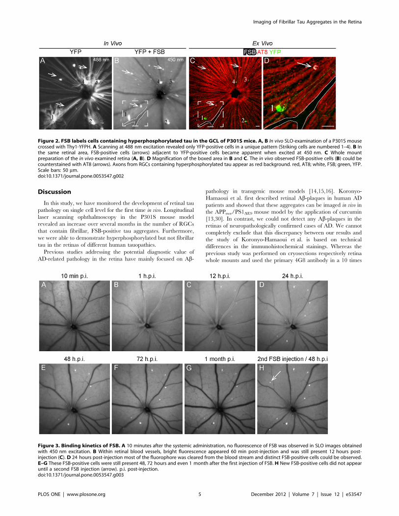
Discussion
In this study, we have monitored the development of retinal tau
pathology on single cell level for the first time in vivo. Longitudinal
laser scanning ophthalmoscopy in the P301S mouse model
revealed an increase over several months in the number of RGCs
that contain fibrillar, FSB-positive tau aggregates. Furthermore,
we were able to demonstrate hyperphosphorylated but not fibrillar
tau in the retinas of different human tauopathies.
Previous studies addressing the potential diagnostic value of
AD-related pathology in the retina have mainly focused on Ab-
pathology in transgenic mouse models [14,15,16]. Koronyo-
Hamaoui et al. first described retinal Ab-plaques in human AD
patients and showed that these aggregates can be imaged in vivo in
the APPswe/PS1DE9 mouse model by the application of curcumin
[13,30]. In contrast, we could not detect any Ab-plaques in the
retinas of neuropathologically confirmed cases of AD. We cannot
completely exclude that this discrepancy between our results and
the study of Koronyo-Hamaoui et al. is based on technical
differences in the immunohistochemical stainings. Whereas the
previous study was performed on cryosections respectively retina
whole mounts and used the primary 4G8 antibody in a 10 times
Figure 2. FSB labels cells containing hyperphosphorylated tau in the GCL of P301S mice. A, B In vivo SLO-examination of a P301S mousecrossed with Thy1-YFPH. A Scanning at 488 nm excitation revealed only YFP-positive cells in a unique pattern (Striking cells are numbered 1–4). B Inthe same retinal area, FSB-positive cells (arrows) adjacent to YFP-positive cells became apparent when excited at 450 nm. C Whole mountpreparation of the in vivo examined retina (A, B). D Magnification of the boxed area in B and C. The in vivo observed FSB-positive cells (B) could becounterstained with AT8 (arrows). Axons from RGCs containing hyperphosphorylated tau appear as red background. red, AT8; white, FSB; green, YFP.Scale bars: 50 mm.doi:10.1371/journal.pone.0053547.g002
Figure 3. Binding kinetics of FSB. A 10 minutes after the systemic administration, no fluorescence of FSB was observed in SLO images obtainedwith 450 nm excitation. B Within retinal blood vessels, bright fluorescence appeared 60 min post-injection and was still present 12 hours post-injection (C). D 24 hours post-injection most of the fluorophore was cleared from the blood stream and distinct FSB-positive cells could be observed.E–G These FSB-positive cells were still present 48, 72 hours and even 1 month after the first injection of FSB. H New FSB-positive cells did not appearuntil a second FSB injection (arrow). p.i. post-injection.doi:10.1371/journal.pone.0053547.g003
Imaging of Fibrillar Tau Aggregates in the Retina
PLOS ONE | www.plosone.org 5 December 2012 | Volume 7 | Issue 12 | e53547
Figure 4. In vivo long term imaging of FSB-positive cells displays the disease progression in P301S mice. A–D Representative retinaimaged between 2 and 5 months of age in an interval of 1 month. F–I Second retina imaged between 5 and 6.5 months of age in an interval of2 weeks. A’–I’ Enlarged images of the boxed areas in A and F demonstrating the appearance of new FSB-positive cells over the time (arrows). FSBwas administrated systemically before each imaging session and images were acquired at 450 nm excitation. E, J Quantification of the increasingnumber of FSB-positive cells in the age from 2 to 5 months and 5 to 6.5 months. The curve displays the mean from 10 different retinas (n = 5 mice).Error bars show SD. **P,0.01; ***P,0.001.doi:10.1371/journal.pone.0053547.g004
Imaging of Fibrillar Tau Aggregates in the Retina
PLOS ONE | www.plosone.org 6 December 2012 | Volume 7 | Issue 12 | e53547
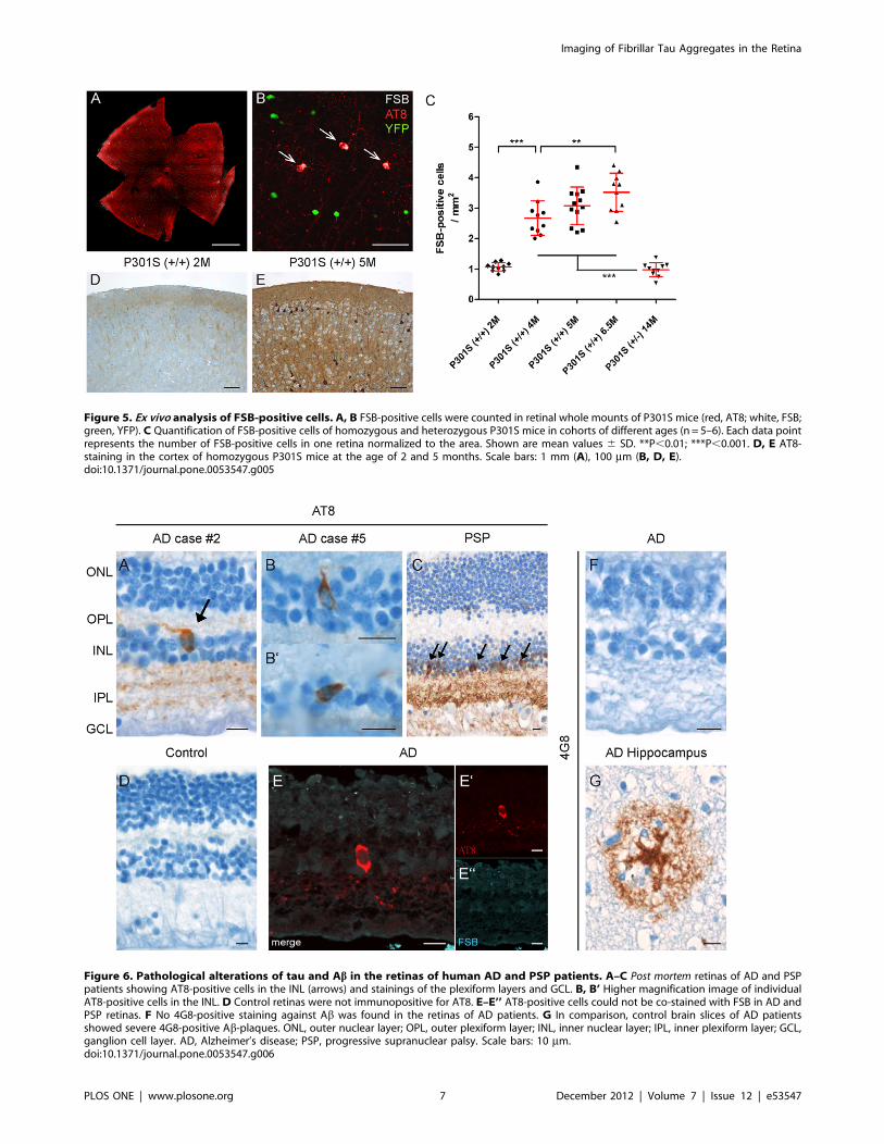
Figure 5. Ex vivo analysis of FSB-positive cells. A, B FSB-positive cells were counted in retinal whole mounts of P301S mice (red, AT8; white, FSB;green, YFP). C Quantification of FSB-positive cells of homozygous and heterozygous P301S mice in cohorts of different ages (n = 5–6). Each data pointrepresents the number of FSB-positive cells in one retina normalized to the area. Shown are mean values 6 SD. **P,0.01; ***P,0.001. D, E AT8-staining in the cortex of homozygous P301S mice at the age of 2 and 5 months. Scale bars: 1 mm (A), 100 mm (B, D, E).doi:10.1371/journal.pone.0053547.g005
Figure 6. Pathological alterations of tau and Ab in the retinas of human AD and PSP patients. A–C Post mortem retinas of AD and PSPpatients showing AT8-positive cells in the INL (arrows) and stainings of the plexiform layers and GCL. B, B’ Higher magnification image of individualAT8-positive cells in the INL. D Control retinas were not immunopositive for AT8. E–E’’ AT8-positive cells could not be co-stained with FSB in AD andPSP retinas. F No 4G8-positive staining against Ab was found in the retinas of AD patients. G In comparison, control brain slices of AD patientsshowed severe 4G8-positive Ab-plaques. ONL, outer nuclear layer; OPL, outer plexiform layer; INL, inner nuclear layer; IPL, inner plexiform layer; GCL,ganglion cell layer. AD, Alzheimer’s disease; PSP, progressive supranuclear palsy. Scale bars: 10 mm.doi:10.1371/journal.pone.0053547.g006
Imaging of Fibrillar Tau Aggregates in the Retina
PLOS ONE | www.plosone.org 7 December 2012 | Volume 7 | Issue 12 | e53547
higher concentration, we applied conventional immunohistochem-
ical procedures on paraffin embedded material as used in the
routine neuropathological diagnosis of AD. However, our findings
are in accordance with previous studies that failed to detect Ab-plaques within the retinas of neuropathologically confirmed AD
cases [31,32] and patients with a clinical presentation of AD [33].
Furthermore, we could not detect retinal Ab-plaques in vivo in
APPswe/PS1DE9 (n = 7, 16–24 months) and Tg2576 (n = 5, 16–
22 months) mice by using the fluorophores FSB and BSc4090,
both shown to bind to Ab-plaques in the brain of these mice
[34,35]. As demonstrated in this paper as well as in a previous
retina imaging study defining the nature of spots of increased
autofluorescence in the mouse retina [27], any fluorescent signal in
the retina has to be analysed in depth for specificity. Therefore, we
always examined the retinas before the application of fluorophores
like FSB or BSc4090, in order to be able to subtract unspecific
signals. Ex vivo immunohistochemical studies using various Ab-antibodies (4G8, 6E10, NAB228) on retinal serial sections of these
mice and other AD mouse models (3xTg-AD [36], APPPS1 [37],
TgCRND8 [38]) confirmed our in vivo observation that Ab-plaques cannot be detected in the retina of these transgenic models
even at very old age (unpublished observations). These negative
findings are in accordance to similar attempts at the University of
Pittsburgh (Dr. C.M. Mathis, personal communication).
In comparison, hyperphosphorylated tau could clearly be
detected in the retinas of AD and PSP cases. Therefore, we
focused our in vivo study on the detection of tau pathology in the
mouse retina. We chose a transgenic mouse model expressing
human tau with the P301S mutation, which develops abundant
FSB-positive tau inclusions in the brain, spinal cord, and the retina
[17,18,24].
We monitored the formation of fibrillar tau aggregates in vivo for
the first time by non-invasive means. In future studies, this
technique will be useful for the screening of drugs aimed at
reducing the formation of fibrillar tau aggregates. Since individual
animals can be studied longitudinally over several months, errors
due to variations in the strength of the pathology between single
mice can be reduced. Interestingly, RGCs harbouring FSB-
positive inclusions survived over several months fitting to the
observation that the overall ganglion cell number in the retina of
P301S mice was not found to be reduced [18]. This indicates,
similar to observations obtained in the cerebral cortex of other tau
transgenic mouse lines [39,40], that the formation of fibrillar tau
alone may not be sufficient to cause neuronal death. On the other
hand, tau hyperphosphorylation and aggregation result in reduced
axonal transport in the optic nerve and an increased loss of RGCs
after mild excitotoxic injury in P301S mice [20]. Observing the
formation of fibrillar tau aggregates in the retina could be useful to
further investigate the kinetic of RGCs loss after similar injuries in
vivo. Furthermore, the herein described long-term binding stability
of FSB could allow an additional, high-resolution analysis of
fibrillar tau formation and its effect on cellular environment by
time-stamp technology as already shown for cerebral Ab-plaquesin transgenic mice [41].
Despite the limited number of human cases in our study, we
provide for the first time evidence of the presence of hyperpho-
sphorylated tau in the retinas of AD and PSP patients. The used
antibody AT8 is a well-accepted marker in routine diagnosis of
AD and recognizes tau doubly phosphorylated at Ser202 and
Thr205 [21]. Remarkably, other tau epitopes were not found to be
phosphorylated as confirmed by the use of different antibodies
(AT100: Ser212/Thr214; AT180: Thr231/Thr235; AT270:
Thr181; PHF1: Ser396/404). This might explain why to date no
other study has described hyperphosphorylated tau in the retina of
different tauopathies. Only one out of 6 examined AD cases with
the most prolonged post mortem interval (72 h) showed no AT8-
positive staining. Therefore, we speculate that the post mortem
interval may affect the stability of hyperphosphorylated tau, as has
already been shown for other phosphoproteins [42]. It remains to
be seen, if hyperphosphorylated tau occurs in the retina of other
types of tauopathies (e.g. frontotemporal dementia) and whether
this pathological alteration can be observed at a presymptomatic
stage of the diseases. Moreover, retinal pathogenesis in different
types of dementias could be influenced by the aggregation of other
proteins like indicated by changes in the distribution pattern of
synucleins in AD or dementia with Lewy bodies [43,44].
Interestingly, accumulations of hyperphosphorylated tau were
also found in the retina of glaucoma patients [45]. These findings
tend to support a possible link between glaucoma and AD like
discussed in the literature [46].
The presence of AT8-positive, hyperphosphorylated tau in the
human retina led us to the hypothesis that our approach could be
applicable in the diagnosis of human tauopathies. Histological
examinations of the human retinas however revealed no evidence
for fibrillar tau aggregates or Ab-plaques. Molecular probes like
FSB or Thioflavin-S bind to tau and Ab in their fibrillar
conformation only. Therefore, an ophthalmic detection of
hyperphosphorylated tau based on the currently available
fluorophores won’t be applicable to human diagnosis. Our findings
challenge the results of an earlier study that described the potential
diagnostic value of Ab-plaques in the retina of human AD cases
[13,30].
In conclusion, we developed a method which potentially could
be used for the monitoring of compounds inhibiting the formation
of fibrillar tau. We present hyperphosphorylated tau in the retinas
of human tauopathies, but found no evidence for Ab-plaques orfibrillar tau aggregates. According to these findings, Ab and tau in
the retina are of limited value for the proposed ophthalmic
diagnosis of AD. A feasible diagnostic system based on the retinal
tau-pathology would depend on the development of novel
fluorescent probes against soluble, hyperphosphorylated tau.
Supporting Information
Figure S1 FSB-positive cells are vital. A–A’’’’ In P301S
mice crossed with Thy1-YFPH, 1.9% of FSB-positive cells also
contained YFP. No pathological alterations in the dendritic arbors
of these cells could be observed. Shown is one co-labelled cell
(FSB, AT8, and YFP) next to cells containing either YFP only or
FSB and AT8 only. A x–y projection, A’’’’ x–z projection. B–B’’Cells containing fibrillar tau in the retina of aged P301S mice
(6.5 months) are positive for NeuN (green, NeuN; white, FSB).
Scale bars: 20 mm.
(TIF)
Acknowledgments
The authors would like to thank the research group of Mathias Seeliger for
introducing them to the SLO examinations of rodents and Dr. Peter
Davies (Albert Einstein College of Medicine, New York, NY) for the
generous gift of the PHF-1 antibody. We also want to thank our animal
facility and the technical assistants from our histological lab for their
invaluable work.
Author Contributions
Conceived and designed the experiments: CS JH. Performed the
experiments: CS NAH SMO SS. Analyzed the data: CS SB. Contributed
reagents/materials/analysis tools: MWS TA MG HAK BS JH. Wrote the
paper: CS NAH SF MG JH.
Imaging of Fibrillar Tau Aggregates in the Retina
PLOS ONE | www.plosone.org 8 December 2012 | Volume 7 | Issue 12 | e53547
References
1. Goedert M (2004) Tau protein and neurodegeneration. Semin Cell Dev Biol 15:
45–49.2. Hasegawa M, Smith MJ, Goedert M (1998) Tau proteins with FTDP-17
mutations have a reduced ability to promote microtubule assembly. FEBS Lett437: 207–210.
3. Hong M, Zhukareva V, Vogelsberg-Ragaglia V, Wszolek Z, Reed L, et al.
(1998) Mutation-specific functional impairments in distinct tau isoforms ofhereditary FTDP-17. Science 282: 1914–1917.
4. Goedert M, Jakes R (2005) Mutations causing neurodegenerative tauopathies.Biochim Biophys Acta 1739: 240–250.
5. Ballard C, Gauthier S, Corbett A, Brayne C, Aarsland D, et al. (2011)
Alzheimer’s disease. Lancet 377: 1019–1031.6. Fox NC, Freeborough PA, Rossor MN (1996) Visualisation and quantification of
rates of atrophy in Alzheimer’s disease. Lancet 348: 94–97.7. Klunk WE, Engler H, Nordberg A, Wang Y, Blomqvist G, et al. (2004) Imaging
brain amyloid in Alzheimer’s disease with Pittsburgh Compound-B. Ann Neurol55: 306–319.
8. Nordberg A (2004) PET imaging of amyloid in Alzheimer’s disease. Lancet
Neurol 3: 519–527.9. Iseri PK, Altinas O, Tokay T, Yuksel N (2006) Relationship between cognitive
impairment and retinal morphological and visual functional abnormalities inAlzheimer disease. J Neuroophthalmol 26: 18–24.
10. Lu Y, Li Z, Zhang X, Ming B, Jia J, et al. (2010) Retinal nerve fiber layer
structure abnormalities in early Alzheimer’s disease: evidence in opticalcoherence tomography. Neurosci Lett 480: 69–72.
11. Paquet C, Boissonnot M, Roger F, Dighiero P, Gil R, et al. (2007) Abnormalretinal thickness in patients with mild cognitive impairment and Alzheimer’s
disease. Neurosci Lett 420: 97–99.12. Berisha F, Feke GT, Trempe CL, McMeel JW, Schepens CL (2007) Retinal
abnormalities in early Alzheimer’s disease. Invest Ophthalmol Vis Sci 48: 2285–
2289.13. Koronyo-Hamaoui M, Koronyo Y, Ljubimov AV, Miller CA, Ko MK, et al.
(2011) Identification of amyloid plaques in retinas from Alzheimer’s patients andnoninvasive in vivo optical imaging of retinal plaques in a mouse model.
Neuroimage 54 Suppl 1: 204–217.
14. Liu B, Rasool S, Yang Z, Glabe CG, Schreiber SS, et al. (2009) Amyloid-peptidevaccinations reduce {beta}-amyloid plaques but exacerbate vascular deposition
and inflammation in the retina of Alzheimer’s transgenic mice. Am J Pathol 175:2099–2110.
15. Ning A, Cui J, To E, Ashe KH, Matsubara J (2008) Amyloid-beta deposits leadto retinal degeneration in a mouse model of Alzheimer disease. Invest
Ophthalmol Vis Sci 49: 5136–5143.
16. Perez SE, Lumayag S, Kovacs B, Mufson EJ, Xu S (2009) Beta-amyloiddeposition and functional impairment in the retina of the APPswe/PS1DeltaE9
transgenic mouse model of Alzheimer’s disease. Invest Ophthalmol Vis Sci 50:793–800.
17. Allen B, Ingram E, Takao M, Smith MJ, Jakes R, et al. (2002) Abundant tau
filaments and nonapoptotic neurodegeneration in transgenic mice expressinghuman P301S tau protein. J Neurosci 22: 9340–9351.
18. Gasparini L, Crowther RA, Martin KR, Berg N, Coleman M, et al. (2011) Tauinclusions in retinal ganglion cells of human P301S tau transgenic mice: effects
on axonal viability. Neurobiol Aging 32: 419–433.19. Magnani E, Fan J, Gasparini L, Golding M, Williams M, et al. (2007)
Interaction of tau protein with the dynactin complex. EMBO J 26: 4546–4554.
20. Bull ND, Guidi A, Goedert M, Martin KR, Spillantini MG (2012) Reducedaxonal transport and increased excitotoxic retinal ganglion cell degeneration in
mice transgenic for human mutant P301S tau. PLoS One 7: e34724.21. Braak H, Alafuzoff I, Arzberger T, Kretzschmar H, Del Tredici K (2006)
Staging of Alzheimer disease-associated neurofibrillary pathology using paraffin
sections and immunocytochemistry. Acta Neuropathol 112: 389–404.22. Mirra SS, Heyman A, McKeel D, Sumi SM, Crain BJ, et al. (1991) The
Consortium to Establish a Registry for Alzheimer’s Disease (CERAD). Part II.Standardization of the neuropathologic assessment of Alzheimer’s disease.
Neurology 41: 479–486.
23. Seeliger MW, Beck SC, Pereyra-Munoz N, Dangel S, Tsai JY, et al. (2005) In
vivo confocal imaging of the retina in animal models using scanning laserophthalmoscopy. Vision Res 45: 3512–3519.
24. Velasco A, Fraser G, Delobel P, Ghetti B, Lavenir I, et al. (2008) Detection offilamentous tau inclusions by the fluorescent Congo red derivative FSB [(trans,
trans)-1-fluoro-2,5-bis(3-hydroxycarbonyl-4-hydroxy)styrylbenzene]. FEBS Lett
582: 901–906.
25. Otvos L Jr., Feiner L, Lang E, Szendrei GI, Goedert M, et al. (1994)
Monoclonal antibody PHF-1 recognizes tau protein phosphorylated at serineresidues 396 and 404. J Neurosci Res 39: 669–673.
26. Charbel Issa P, Singh MS, Lipinski DM, Chong NV, Delori FC, et al. (2012)Optimization of in vivo confocal autofluorescence imaging of the ocular fundus
in mice and its application to models of human retinal degeneration. Invest
Ophthalmol Vis Sci 53: 1066–1075.
27. Xu H, Chen M, Manivannan A, Lois N, Forrester JV (2008) Age-dependent
accumulation of lipofuscin in perivascular and subretinal microglia inexperimental mice. Aging Cell 7: 58–68.
28. Feng G, Mellor RH, Bernstein M, Keller-Peck C, Nguyen QT, et al. (2000)Imaging neuronal subsets in transgenic mice expressing multiple spectral
variants of GFP. Neuron 28: 41–51.
29. Walsh MK, Quigley HA (2008) In vivo time-lapse fluorescence imaging ofindividual retinal ganglion cells in mice. J Neurosci Methods 169: 214–221.
30. Koronyo Y, Salumbides BC, Black KL, Koronyo-Hamaoui M (2012)Alzheimer’s disease in the retina: imaging retinal abeta plaques for early
diagnosis and therapy assessment. Neurodegener Dis 10: 285–293.
31. Blanks JC, Hinton DR, Sadun AA, Miller CA (1989) Retinal ganglion celldegeneration in Alzheimer’s disease. Brain Res 501: 364–372.
32. Hinton DR, Sadun AA, Blanks JC, Miller CA (1986) Optic-nerve degenerationin Alzheimer’s disease. N Engl J Med 315: 485–487.
33. Leger F, Fernagut PO, Canron MH, Leoni S, Vital C, et al. (2011) Proteinaggregation in the aging retina. J Neuropathol Exp Neurol 70: 63–68.
34. Bolander A, Kieser D, Voss C, Bauer S, Schon C, et al. (2012) Bis(arylvinyl)pyr-
azines, -pyrimidines, and -pyridazines As Imaging Agents for Tau Fibrils andbeta-Amyloid Plaques in Alzheimer’s Disease Models. J Med Chem.
35. Higuchi M, Iwata N, Matsuba Y, Sato K, Sasamoto K, et al. (2005) 19F and 1HMRI detection of amyloid beta plaques in vivo. Nat Neurosci 8: 527–533.
36. Oddo S, Caccamo A, Kitazawa M, Tseng BP, LaFerla FM (2003) Amyloiddeposition precedes tangle formation in a triple transgenic model of Alzheimer’s
disease. Neurobiol Aging 24: 1063–1070.
37. Radde R, Bolmont T, Kaeser SA, Coomaraswamy J, Lindau D, et al. (2006)Abeta42-driven cerebral amyloidosis in transgenic mice reveals early and robust
pathology. EMBO Rep 7: 940–946.
38. Chishti MA, Yang DS, Janus C, Phinney AL, Horne P, et al. (2001) Early-onset
amyloid deposition and cognitive deficits in transgenic mice expressing a double
mutant form of amyloid precursor protein 695. J Biol Chem 276: 21562–21570.
39. de Calignon A, Fox LM, Pitstick R, Carlson GA, Bacskai BJ, et al. (2010)
Caspase activation precedes and leads to tangles. Nature 464: 1201–1204.
40. Santacruz K, Lewis J, Spires T, Paulson J, Kotilinek L, et al. (2005) Tau
suppression in a neurodegenerative mouse model improves memory function.Science 309: 476–481.
41. Condello C, Schain A, Grutzendler J (2011) Multicolor time-stamp reveals the
dynamics and toxicity of amyloid deposition. Sci Rep 1: 19.
42. Oka T, Tagawa K, Ito H, Okazawa H (2011) Dynamic changes of the
phosphoproteome in postmortem mouse brains. PLoS One 6: e21405.
43. Maurage CA, Ruchoux MM, de Vos R, Surguchov A, Destee A (2003) Retinal
involvement in dementia with Lewy bodies: a clue to hallucinations? Ann Neurol54: 542–547.
44. Surguchov A, McMahan B, Masliah E, Surgucheva I (2001) Synucleins in ocular
tissues. J Neurosci Res 65: 68–77.
45. Gupta N, Fong J, Ang LC, Yucel YH (2008) Retinal tau pathology in human
glaucomas. Can J Ophthalmol 43: 53–60.
46. Wostyn P, Audenaert K, De Deyn PP (2009) Alzheimer’s disease and glaucoma:
is there a causal relationship? Br J Ophthalmol 93: 1557–1559.
Imaging of Fibrillar Tau Aggregates in the Retina
PLOS ONE | www.plosone.org 9 December 2012 | Volume 7 | Issue 12 | e53547