Histone deacetylase 6 inhibition improves memory and reduces total tau levels in a mouse model of...
12
RESEARCH Open Access Histone deacetylase 6 inhibition improves memory and reduces total tau levels in a mouse model of tau deposition Maj-Linda Selenica 1,2 , Leif Benner 1 , Steven B Housley 1 , Barbara Manchec 1 , Daniel C Lee 1,2 , Kevin R Nash 1,3 , Jay Kalin 4 , Joel A Bergman 5 , Alan Kozikowski 5 , Marcia N Gordon 1,3 and Dave Morgan 1,3* Abstract Introduction: Tau pathology is associated with a number of age-related neurodegenerative disorders. Few treatments have been demonstrated to diminish the impact of tau pathology in mouse models and none are yet effective in humans. Histone deacetylase 6 (HDAC6) is an enzyme that removes acetyl groups from cytoplasmic proteins, rather than nuclear histones. Its substrates include tubulin, heat shock protein 90 and cortactin. Tubastatin A is a selective inhibitor of HDAC6. Modification of tau pathology by specific inhibition of HDAC6 presents a potential therapeutic approach in tauopathy. Methods: We treated rTg4510 mouse models of tau deposition and non-transgenic mice with tubastatin (25 mg/kg) or saline (0.9%) from 5 to 7 months of age. Cognitive behavior analysis, histology and biochemical analysis were applied to access the effect of tubastatin on memory, tau pathology and neurodegeneration (hippocampal volume). Results: We present data showing that tubastatin restored memory function in rTg4510 mice and reversed a hyperactivity phenotype. We further found that tubastatin reduced the levels of total tau, both histologically and by western analysis. Reduction in total tau levels was positively correlated with memory improvement in these mice. However, there was no impact on phosphorylated forms of tau, either by histology or western analysis, nor was there an impact on silver positive inclusions histologically. Conclusion: Potential mechanisms by which HDAC6 inhibitors might benefit the rTg4510 mouse include stabilization of microtubules secondary to increased tubulin acetylation, increased degradation of tau secondary to increased acetylation of HSP90 or both. These data support the use of HDAC6 inhibitors as potential therapeutic agents against tau pathology. Introduction Tauopathies are neurodegenerative disorders for which there are no effective treatments. Some disorders are caused by mutations in tau that increase the probability of tau aggregate formation, leading to intracellular neuro- fibrillary tangles [1]. These disorders are typically referred to as fronto-temporal dementias. Other tauopathies occur in different brain regions (corticobasal syndrome, progres- sive supranuclear palsy, and so forth) [2]. The most common disorder demonstrating tau inclusions is Alzhei- mer’ s disease, where the tau pathology correlates better than amyloid pathology with cognitive impairment [3]. Histone deacetylases (HDACs) are a family of proteins that remove acetyl moieties attached covalently to lysine residues in proteins. In the cell nucleus, HDACs catalyze the deacetylation of histones and, in general, promote chromatin condensation and repression of gene expres- sion. In transformed cells, these enzymes are thought to suppress proapoptotic programs, leading to unregulated proliferation. As such, HDAC inhibitors are widely explored as treatments for cancer [4]. There are at least 18 iso- enzymes in the HDAC family, divided into four homology classes. Classes I, II and IV are zinc dependent, while class * Correspondence: [email protected] 1 Byrd Alzheimer’s Institute, University of South Florida, Tampa, FL 33613, USA 3 College of Medicine, Department of Molecular Pharmacology and Physiology, University of South Florida, Tampa, FL 33612, USA Full list of author information is available at the end of the article © 2014 Selenica et al.; licensee BioMed Central Ltd. This is an Open Access article distributed under the terms of the Creative Commons Attribution License (http://creativecommons.org/licenses/by/2.0), which permits unrestricted use, distribution, and reproduction in any medium, provided the original work is properly credited. Selenica et al. Alzheimer's Research & Therapy 2014, 6:12 http://alzres.com/content/6/1/12
Transcript of Histone deacetylase 6 inhibition improves memory and reduces total tau levels in a mouse model of...

Selenica et al. Alzheimer's Research & Therapy 2014, 6:12http://alzres.com/content/6/1/12
RESEARCH Open Access
Histone deacetylase 6 inhibition improves memoryand reduces total tau levels in a mouse model oftau depositionMaj-Linda Selenica1,2, Leif Benner1, Steven B Housley1, Barbara Manchec1, Daniel C Lee1,2, Kevin R Nash1,3,Jay Kalin4, Joel A Bergman5, Alan Kozikowski5, Marcia N Gordon1,3 and Dave Morgan1,3*
Abstract
Introduction: Tau pathology is associated with a number of age-related neurodegenerative disorders. Few treatmentshave been demonstrated to diminish the impact of tau pathology in mouse models and none are yet effective inhumans. Histone deacetylase 6 (HDAC6) is an enzyme that removes acetyl groups from cytoplasmic proteins, ratherthan nuclear histones. Its substrates include tubulin, heat shock protein 90 and cortactin. Tubastatin A is a selectiveinhibitor of HDAC6. Modification of tau pathology by specific inhibition of HDAC6 presents a potential therapeuticapproach in tauopathy.
Methods: We treated rTg4510 mouse models of tau deposition and non-transgenic mice with tubastatin(25 mg/kg) or saline (0.9%) from 5 to 7 months of age. Cognitive behavior analysis, histology and biochemicalanalysis were applied to access the effect of tubastatin on memory, tau pathology and neurodegeneration(hippocampal volume).
Results: We present data showing that tubastatin restored memory function in rTg4510 mice and reverseda hyperactivity phenotype. We further found that tubastatin reduced the levels of total tau, bothhistologically and by western analysis. Reduction in total tau levels was positively correlated with memoryimprovement in these mice. However, there was no impact on phosphorylated forms of tau, either byhistology or western analysis, nor was there an impact on silver positive inclusions histologically.
Conclusion: Potential mechanisms by which HDAC6 inhibitors might benefit the rTg4510 mouse includestabilization of microtubules secondary to increased tubulin acetylation, increased degradation of tausecondary to increased acetylation of HSP90 or both. These data support the use of HDAC6 inhibitors aspotential therapeutic agents against tau pathology.
IntroductionTauopathies are neurodegenerative disorders for whichthere are no effective treatments. Some disorders arecaused by mutations in tau that increase the probability oftau aggregate formation, leading to intracellular neuro-fibrillary tangles [1]. These disorders are typically referredto as fronto-temporal dementias. Other tauopathies occurin different brain regions (corticobasal syndrome, progres-sive supranuclear palsy, and so forth) [2]. The most
* Correspondence: [email protected] Alzheimer’s Institute, University of South Florida, Tampa, FL 33613, USA3College of Medicine, Department of Molecular Pharmacology andPhysiology, University of South Florida, Tampa, FL 33612, USAFull list of author information is available at the end of the article
© 2014 Selenica et al.; licensee BioMed CentraCommons Attribution License (http://creativecreproduction in any medium, provided the or
common disorder demonstrating tau inclusions is Alzhei-mer’s disease, where the tau pathology correlates betterthan amyloid pathology with cognitive impairment [3].Histone deacetylases (HDACs) are a family of proteins
that remove acetyl moieties attached covalently to lysineresidues in proteins. In the cell nucleus, HDACs catalyzethe deacetylation of histones and, in general, promotechromatin condensation and repression of gene expres-sion. In transformed cells, these enzymes are thought tosuppress proapoptotic programs, leading to unregulatedproliferation. As such, HDAC inhibitors are widely exploredas treatments for cancer [4]. There are at least 18 iso-enzymes in the HDAC family, divided into four homologyclasses. Classes I, II and IV are zinc dependent, while class
l Ltd. This is an Open Access article distributed under the terms of the Creativeommons.org/licenses/by/2.0), which permits unrestricted use, distribution, andiginal work is properly credited.

Selenica et al. Alzheimer's Research & Therapy 2014, 6:12 Page 2 of 12http://alzres.com/content/6/1/12
III, also known as sirtuins, are NAD+ dependent fortheir enzyme activity. Class I HDACs (HDAC1, HDAC2,HDAC3 and HDAC8) are nuclear enzymes and are themajor focus of research for anti-tumor agents. Class IIenzymes are often tissue specific, divided into class IIaenzymes (HDAC4, HDAC5, HDAC7, HDAC9) that shuttlebetween cytoplasmic and nuclear compartments and classIIb enzymes (HDAC6 and HDAC10) that are primarilycytoplasmic and deacetylate nonhistone proteins. HDAC6has been shown to act upon tubulin, cortactin and HSP90.Tubulin acetylation is associated with increased micro-tubule stabilization [5].The Kozikowski laboratory has synthesized a number
of compounds focusing on HDAC6. One such agent istubastatin A (tubastatin). This molecule was found to havenanomolar potency in inhibiting HDAC6, but requiresmicromolar or greater concentrations to inhibit mostother HDACs (>1,000× selectivity for all but HDAC8, at50-fold selectivity). This agent was found to increase theacetylation of tubulin in cells, but not histone H4 proteins.Moreover, tubastatin treatment was found to be protectiveagainst homocysteic acid-induced oxidative stress [6]. Thisagent reduced the phenotype in a model of Charcot-Marie-Tooth disease [7]. This disorder is caused by mutationsin the 27 kDa small heat shock protein HSBP1, leadingto decreased tubulin acetylation and axonal atrophy.Tubastatin treatment prevented both the loss of acety-lated tubulin and axonopathy. Recent observations alsodemonstrate that decreases in HDAC6 activity or expres-sion promote tau clearance [8], while HDAC6 mutationsrescued tau-induced microtubule defects in a Drosophilamodel of tau pathology [9]. Given the observation thatphosphorylation of tau results in dissociation from tubulinand decreased stabilization of microtubules [10], we hy-pothesized that stabilizing microtubules by acetylationmight counteract the phenotype found in the rTg4510mouse model of tauopathy. Moreover, we chose to startinjecting mice at an age when there was already memoryloss and considerable tau pathology because, even at theearliest stages, Alzheimer patients have substantial existingtau deposition [11]. We have thus treated 5-month-oldrTg4510 mice with the HDAC6 selective drug tubastatinfor 2 months, and monitored its effect on the behavioraland pathological phenotype of this mouse.
Materials and methodsDrug preparationTubastatin was synthesized and provided by Dr AKozikowski [6] (University of Illinois at Chicago, Chicago,IL, USA). The compound was dissolved in 0.9% saline toreach a dose of 25 mg/kg body weight per animal. Thisdose has been shown to efficiently inhibit HDAC6 in vivo[7]. The solution was sonicated in the water bath for5 minutes at room temperature until is fully solubilized.
Animals and injection procedureThe production of rTg4510 mice, bred from parentalP301L tau and tTA lines, was maintained as originallydescribed by Santacruz and colleagues [12]. We admin-istered experimental agents using daily intraperitonealinjections into rTg4510 mice and nontransgenic littermatesfrom 5 to 7 months of age. Each group consisted of sixanimals per genotype. Mice were weighed, and injectedwith tubastatin (25 mg/kg) or 0.9% normal saline solution(vehicle). Animal procedures were consistent with the rec-ommendations of the National Research Council’s Guidefor the Care and Use of Laboratory Animals and were ap-proved by the University of South Florida Animal Careand Use Committee (IACUC).
Behavior methodsBehavioral analysis was carried out as described previously[13-17]. For 3 days before testing, mice were weighed andhandled in order to habituate the mice to contact with theexperimenter. The sequence of tests over a 2-week periodwere Y-maze, open field, rotarod, radial arm water maze,water maze reversal, open pool, and fear conditioning.
Y-mazeEach animal was placed in a Y-maze for a single 5-minutetrial, during which the sequence and total number of armchoices were recorded. Spontaneous alternation, expressedas a percentage of total alternations, was calculated asdescribed [18]. If an animal made the following sequence ofarm selections (1,2,3,2,1,3,1,2), the total alternation oppor-tunities will be six (total entries minus two) and the per-centage alternation would be 67% (four out of six). Chanceperformance is 50%
Open fieldThe open field is used as a standard test of general activity.Animals were monitored for 15 minutes in a 40 cm squareopen field with video tracking software, under moderatelighting. General activity levels were evaluated by mea-surements of horizontal activity and vertical activity. Theopen field test can also be used to measure anxiety levels,as assessed by the ratio of center distance traveled to totaldistance traveled. As the natural tendency of the animal isto avoid open areas, animals that show greater tendencyto explore in the center of the field show a lower tendencyfor anxiety, or the reverse situation can indicate a higheranxiety level.
RotarodMotor performance was assessed by placing the miceonto the round portion of a circular rod that was thenaccelerated slowly. Mice need to walk at the speed ofrod rotation to keep from falling. The time until falling

Selenica et al. Alzheimer's Research & Therapy 2014, 6:12 Page 3 of 12http://alzres.com/content/6/1/12
was recorded for each mouse. Mice were given four trialseach day for 2 consecutive days.
Two-day radial arm water mazeThe 2-day radial arm water maze has been previously de-scribed in detail [19]. Briefly, the radial arm water mazeconsists of a 1 m pool with six swim paths (arms) radiatingout of an open central area, with a hidden escape platformlocated at the end of one of the arms. On each trial, themouse was allowed to swim in the pool for up to 60 secondsto find the escape platform. Incorrect arm entries or failureto select an arm for 15 seconds were counted as errors. Fora given mouse, the platform was located in the same armon each trial, but the start arms were varied for each trialso that mice rely upon spatial cues to solve the task insteadof learning motor rules (that is, second arm on the right).On day 1, mice were given 15 trials where each block con-sist of three trials (five blocks in total) alternating between avisible platform (above the water) and a hidden platform(below the water). The following day, mice were given 15additional trials (five blocks), all using a hidden platform.The goal arm location for sequential mice was different toavoid odor cues from revealing the goal arm.
Radial arm water maze reversalThis test was completed on the day after the 2-day radialarm water maze and was performed under the sameconditions. The only difference was that the goal armwas located 180º from the original location. This reversaltask tests the ability of the mice to extinguish a learnedmemory to create a novel one. Fifteen trials were given ina single day, all with a hidden platform.
Visible platformThis was conducted in an open pool (no swim arms) to en-sure mice can see and perform a platform task [20]. The vi-sible platform was placed 0.8 cm above the water surfaceand had an attached 10 cm× 40 cm ensign. For the test day,the animal was placed into the pool facing the wall in eachof the four quadrants of the pool. Latency to find and ascendthe visible platform was recorded (maximum 60 seconds).
Associative fear conditioningFear conditioning was used to access both contextualand cued memory formation. For these experiments, anaversive stimulus (in this case, a mild foot shock, 0.5 mA)was paired with an auditory conditioned stimulus (whitenoise) within a novel environment. These tests were givenas follows: animals were placed in the fear conditioningapparatus for 2.5 minutes, then a 30-second acoustic con-ditioned stimulus (white noise, 70 dB) was coupled with a0.5 mA shock (unconditioned stimulus) applied to thefloor grid during the last 2 seconds of the conditionedstimulus. Two minutes later the conditioned stimulus and
unconditioned stimulus were again administered, andthe mice remained in the conditioning chamber for anadditional 2 minutes. The mice were placed in the samechamber and monitored for freezing to the context 24 hourslater (no shocks or auditory cue given). Immediately afterthe contextual test, mice were placed into a novel contextand exposed to the conditioned stimulus for 3 minutes(cued fear conditioning). Both the contextual and the cuedrecall tests were performed the next day ~24 hours follow-ing training. Learning was assessed by measuring freezingbehavior (that is, motionless position).
Tissue collectionTwo months post intraperitoneal injections, mice wereweighed, overdosed with pentobarbital (200 mg/kg) andperfused with 25 ml of 0.9% normal saline solution. Micewere placed on an isothermal pad after anesthesia andduring perfusion to avoid artifactual tau phosphorylationcaused by reductions in body temperature [21]. Brainswere collected from the animals 1 hour after the lasttubastatin injection, following saline perfusion, andwere hemisected down the sagittal midline. One hemi-sphere was dissected and frozen with dry ice for biochem-ical studies. The second hemisphere was immersion fixedin 4% paraformaldehyde for 24 hours, and cryoprotectedin successive incubations of 10%, 20% and 30% solutionsof sucrose for 24 hours in each solution. Subsequently,the fixed hemispheres were frozen on a cold stage andsectioned in the horizontal plane (25 μm thickness) ona sliding microtome and stored in Dulbecco’s phosphate-buffered saline with 10 mM sodium azide solution at 4°C.
Immunohistochemical/histological procedure and analysisEight sections, 200 μm apart, were chosen for analysisusing immunohistochemical procedural methods describedpreviously [22]. For each marker, floating sections from allanimals were placed in multi-sample staining trays, andendogenous peroxidase was blocked (10% methanol, 10%H2O2 in phosphate-buffered saline; 30 minutes). Tissuesamples were permeabilized (with 0.2% lysine, 1% TritonX-100 in phosphate-buffered saline solution), and incubatedovernight in appropriate primary antibody (clonal namesin parentheses). Anti-N-terminus of human tau antibodies(H150; Santa Cruz Biotechnology, Dallas, Texas, USA),anti-phospho-tau at serine 396 antibodies (pSer396; Ana-spec, Fremont, CA, USA), anti-phospho-tau at serine199/202 antibodies (pSer199/202; Anaspec, Fremont,CA, US), and anti-phospho-tau at serine 202 and threo-nine 205 antibodies (biotinylated AT8; ThermoScienti-fic, Waltham, MA, USA) were used in this study.Sections were washed in phosphate-buffered saline andthen incubated in corresponding biotinylated secondaryantibody, except for AT8 (pSer199/Thr205; Vector La-boratories, Burlingame, CA, USA). The tissue was again

Selenica et al. Alzheimer's Research & Therapy 2014, 6:12 Page 4 of 12http://alzres.com/content/6/1/12
washed after 2 hours, and incubated with the Vectastain®Elite® ABC kit (Vector Laboratories, Burlingame, CA, USA)for enzyme conjugation. Finally, sections were developedusing 0.05% diaminobenzidine, 0.5% Ni2+ and 0.03% H2O2.Tissue sections were then mounted onto slides, dehydrated,and cover slipped. Each immunochemical assay omittedsome sections from primary antibody incubation toevaluate nonspecific reaction of the secondary antibody.Prior work with nontransgenic mice confirmed the speci-ficity of the anti-tau antibodies for tau in rTg4510 mice.Gallyas histology was performed as described previously
[23], using sections that were premounted on slides and thenair-dried for a minimum of 24 hours. The sections were rehy-drated for 30 seconds prior to proceeding with the Gallyasprotocol. Slides were treated with 5% periodic acid for 5 mi-nutes, washed with water, and incubated sequentially in silveriodide (1 minute) and 0.5% acetic acid (10 minutes) solutionsprior to being placed in developer solution (2.5% sodium car-bonate, 0.1% ammonium nitrate, 0.1% silver nitrate, 1% tung-stosilicic acid, 0.7% formaldehyde). Slides were treated with0.5% acetic acid to stop the reaction, incubated with 0.1% goldchloride, placed in 1% sodium thiosulfate, and counterstainedwith 0.1% nuclear fast red in 2.5% aqueous aluminum sulfate,with each step separated by washes in water. Following a finalwash, slides were dehydrated and cover slipped.Stained sections were imaged using a Zeiss Mirax150
digital scanning microscope (Carl Zeiss MicroImagingGmbH Clinical 07740 Jena, Germany). The area of positivestain in each hippocampal section was analyzed. The soft-ware used hue, saturation and intensity to segment the imagefields. Thresholds for object segmentation were establishedwith images of high and low levels of staining to identifypositive staining over all intensity levels within the study.These limits were held constant for the analysis of every sec-tion in each study according to Gordon and colleagues [24].
Western blot analysisTissues for western analysis were prepared as described byCarroll and colleagues [25]. Briefly, the dissected anteriorcortex tissue was weighed and resuspended in RIPA buffer(50 mM Tris, 140 mM NaCl, 10% NP40, 10% sodium deox-ycholate, 10% SDS), with protease inhibitor cocktail (Sigma-Aldrich, St. Louis, MO, USA) and phosphatase inhibitorcocktails I and II (Sigma-Aldrich, St. Louis, MO, USA). Tis-sue was homogenized with a motorized pestle followed by abrief (10 seconds) sonication pulse. Aliquots were collectedat this point for biochemical analysis and were referred toas brain homogenate. Pierce BCA protein assay (Thermo-Scientific, Waltham, MA, USA) was used to determine pro-tein concentrations. For western analysis, 1 μg protein wasloaded for each sample. Antibodies against tau epitopespS396 and pS199/202 (AT8) were obtained from Anaspec,while H150 antibody was obtain from Santa Cruz Biotech-nologies. PHF1 antibody was kindly provided by Dr
Peter Davies. A monoclonal antibody against acety-lated α-tubulin at Lys40 was used to measure α-tubulinacetylation following treatment (clone 6-11B-1; Sigma-Aldrich, St. Louis, MO, USA). Total α-tubulin levels weremeasured using monoclonal anti α-tubulin antibody (cloneTU-01; ThermoScientific, Waltham, MA, USA).
StereologyMethods of design-based (unbiased) stereology were usedto quantify hippocampal volumes with assistance fromthe Stereologer system (Stereologer, St Petersburg, FL,USA). For this study a complete series of sectionsthrough the hippocampus was cut at a thickness set-ting of 50 μm and stained with cresyl violet. From thissample of sections, a systematic random sample of everyseventh section was used to estimate the total hippocampalvolume using the Cavalieri-point counting method [26].
Statistical analysisStatistical analysis and regression plots of studies includingboth transgenic and nontransgenic mice (behavior) wasperformed using two-way analysis of variance followedby Fisher’s protected least significant difference (PLSD)post hoc means comparison test using StatView version5.0.1 (SAS Institute Inc, Cary, NC, USA). Measures ex-cluding nontransgenic mice (tau levels) were analyzedby Student’s t test.
ResultsTo determine the therapeutic effect of HDAC6 inhibitionon tau pathology we administered tubastatin, which showspotency and selectivity towards HDAC6 inhibition in vitroand in vivo (IC50 = 15 nM for HDAC6) [6]. An equalnumber of 5-month-old rTg4510 mice and nontrans-genic littermates were treated with tubastatin or thesaline vehicle by daily intraperitoneal injection for2 months. At week 6 mice were started on a 2-week be-havioral test battery, and tissues were collected at the be-ginning of week 9 (Figure 1A). Measurements of α-tubulin acetylation were used to determine the efficacy ofthe treatment in modifying the acetylation state of α-tubulin at the Lys40 residue. Western blot analyses re-vealed a greater than twofold rise in acetylated α-tubulinto total α-tubulin in tubastatin-treated rTg4510 mice,while a 1.5-fold rise was observed in nontransgenic miceas compared with saline-treated littermates (Figure 1B,C).Interestingly, we observed a reduction in the levels ofα-tubulin following tubastatin treatment of rTg4510 butnot nontransgenic mice (Figure 1B). Data analysis (nor-malized to glyceraldehyde 3-phosphate dehydrogenase)demonstrated that tubastatin administration significantlyinduced acetylated α-tubulin levels in both nontransgenicand transgenic animals compared with saline-treated litter-mates (Figure 1C). These findings are indirectly indicative
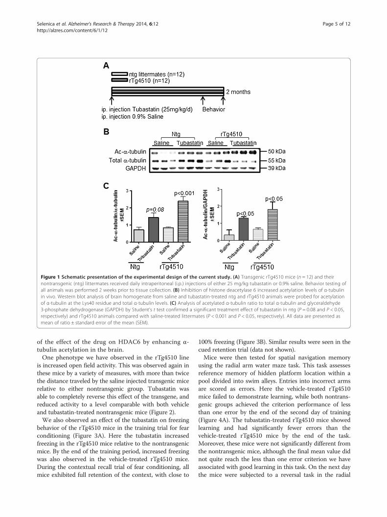
Figure 1 Schematic presentation of the experimental design of the current study. (A) Transgenic rTg4510 mice (n = 12) and theirnontransgenic (ntg) littermates received daily intraperitoneal (i.p.) injections of either 25 mg/kg tubastatin or 0.9% saline. Behavior testing ofall animals was performed 2 weeks prior to tissue collection. (B) Inhibition of histone deacetylase 6 increased acetylation levels of α-tubulinin vivo. Western blot analysis of brain homogenate from saline and tubastatin-treated ntg and rTg4510 animals were probed for acetylationof α-tubulin at the Lys40 residue and total α-tubulin levels. (C) Analysis of acetylated α-tubulin ratio to total α-tubulin and glyceraldehyde3-phosphate dehydrogenase (GAPDH) by Student’s t test confirmed a significant treatment effect of tubastatin in ntg (P = 0.08 and P < 0.05,respectively) and rTg4510 animals compared with saline-treated littermates (P < 0.001 and P < 0.05, respectively). All data are presented asmean of ratio ± standard error of the mean (SEM).
Selenica et al. Alzheimer's Research & Therapy 2014, 6:12 Page 5 of 12http://alzres.com/content/6/1/12
of the effect of the drug on HDAC6 by enhancing α-tubulin acetylation in the brain.One phenotype we have observed in the rTg4510 line
is increased open field activity. This was observed again inthese mice by a variety of measures, with more than twicethe distance traveled by the saline injected transgenic micerelative to either nontransgenic group. Tubastatin wasable to completely reverse this effect of the transgene, andreduced activity to a level comparable with both vehicleand tubastatin-treated nontransgenic mice (Figure 2).We also observed an effect of the tubastatin on freezing
behavior of the rTg4510 mice in the training trial for fearconditioning (Figure 3A). Here the tubastatin increasedfreezing in the rTg4510 mice relative to the nontransgenicmice. By the end of the training period, increased freezingwas also observed in the vehicle-treated rTg4510 mice.During the contextual recall trial of fear conditioning, allmice exhibited full retention of the context, with close to
100% freezing (Figure 3B). Similar results were seen in thecued retention trial (data not shown).Mice were then tested for spatial navigation memory
using the radial arm water maze task. This task assessesreference memory of hidden platform location within apool divided into swim alleys. Entries into incorrect armsare scored as errors. Here the vehicle-treated rTg4510mice failed to demonstrate learning, while both nontrans-genic groups achieved the criterion performance of lessthan one error by the end of the second day of training(Figure 4A). The tubastatin-treated rTg4510 mice showedlearning and had significantly fewer errors than thevehicle-treated rTg4510 mice by the end of the task.Moreover, these mice were not significantly different fromthe nontransgenic mice, although the final mean value didnot quite reach the less than one error criterion we haveassociated with good learning in this task. On the next daythe mice were subjected to a reversal task in the radial

Figure 2 Rescue of rTg4510 hyperactivity by tubastatin treatment.Treatment of rTg4510 animals with tubastatin significantly decreased theopen field test distance travelled compared with the saline-treatedlittermates (distance ± standard error of the mean, n= 6). Two-wayanalysis of variance revealed an overall effect of genotype (P= 0.03) andof treatment in rTg4510 mice (P= 0.03). Ntg, nontransgenic *P< 0.05.
Figure 3 Increased freezing in tubastatin-treated rTg4510 mice during thsignificantly increases the percent freezing ability of the rTg4510 mice compareof the fear conditioning training paradigm. Two-way analysis of variance followtransgenic (tg; P < 0.0001) and nontransgenic (ntg; P < 0.038) animal groups cexhibited high levels of freezing during the retention phase of the test, indica
Selenica et al. Alzheimer's Research & Therapy 2014, 6:12 Page 6 of 12http://alzres.com/content/6/1/12
arm water maze, with the platform moved to a new lo-cation. This requires unlearning the prior location andrelearning the new platform location. Here the vehicle-treated rTg4510 mice failed to learn the platform location,while both groups of nontransgenic mice rapidly acquiredthe new platform location. The tubastatin-treated rTg4510mice were intermediate and significantly better than thevehicle-treated rTg4510 mice, yet statistically worse thanthe nontransgenic mice (Figure 4B).Tissue sections were stained for a variety of tau isoforms.
Stains for total human tau levels with antibody H150 re-vealed significant reductions in tubastatin-treated rTg4510mice (nontransgenic mice have no detectable staining ineither group). This antibody stains both neurons (darkly)and neuropil (lightly; Figure 5A,B,C,D,E,F, arrowhead).The neuropil staining appears to be specific, as it is notobserved in brain regions lacking darkly stained neurons(thalamus, brain stem) or found in nontransgenic mice(not shown). Using the digital scanning microscope, weperformed image analysis measures separately in the
e acquisition phase of fear conditioning. (A) Treatment with tubastatind with the saline-treated littermates (n= 6/group) in the acquisition phaseed by Fisher’s PLSD test revealed a significant treatment effect in bothompared with saline-treated mice of the same genotype. (B) All miceting recall of the context. *P < 0.05, ***P < 0.001.

Figure 4 Rescue of rTg4510 memory impairment by tubastatin treatment. Number of errors made (average ± standard error of the mean(SEM)) while seeking the hidden platform in the radial arm water maze (RAWM). (A) Tubastatin-treated rTg4510 mice performed better in theinitial training (5 blocks of three trials) than vehicle-treated littermates (P = 0.002) and were not significantly different from the tubastatin-treatednontransgenic (ntg) mice. (B) Reversal testing on day 3 (blocks 1 to 5) showed rescue of the memory impairment of rTg4510 mice followinghistone deacetylase 6 inhibition compared with vehicle-treated littermates (two-way ANOVA and Fisher’s PLSD, P < 0.03). No significant differencein either test was detected between vehicle-treated and tubastatin-treated nontransgenic animals (P > 0.05). *P < 0.05, **P < 0.01, *** P < 0.001.
Selenica et al. Alzheimer's Research & Therapy 2014, 6:12 Page 7 of 12http://alzres.com/content/6/1/12
cerebral cortex, hippocampus and the entire hemisphere.There was a roughly 50% reduction in total tau stainingin the tubastatin-treated mice, which was statisticallysignificant in the hippocampus and whole section mea-surements (Figure 5G,H,I). We also stained sections fora variety of forms of phosphorylated tau, and per-formed Gallyas silver staining for aggregated tau. Noneof these markers were modified by the tubastatin treat-ment (Table 1). Given the positive correlation of taupathology with cognitive impairment in AD patients [3],we examined how HDAC6 inhibition in vivo correlateswith behavior and pathophysiological changes measuredin treated rTg4510 mice (Figure 5J and Additional file 1).The average number of errors made in the radial armwater maze was plotted against the percent tau load(H150) measured in the tissue of treated rTg4510 animals(Figure 5J). Mice treated with tubastatin significantly
reduced the total tau levels, which is positively corre-lated with the number of errors made during thememory performance test (linear regression, r = 0.690,P = 0.0130).In addition, tissue stained with cresyl violet was used
to estimate the total hippocampal volume as describedpreviously [26]. Stereological analysis showed no effectof tubastatin on the hippocampal volume of treated trans-genic mice (Figure 5K), although a clear reduction in thehippocampal volume was observed between rTg4510 miceand nontransgenic littermates. Further, human tau load(percent area) and the number of errors measured inradial arm water maze testing were compared with thehippocampal volume in all subsets of mice (Additionalfile 1A,B). These data demonstrate that total tau levelsand memory performance was negatively correlated withhippocampal volume.

Saline Tubastatin Saline Tubastatin0.0
0.9
1.8
2.7
3.6
4.5 **
Ntg rTg4510
HP
C V
olu
me
(mm
3 )±
SE
M
R=0.690P<0.05
0.0 1.0 2.0 3.0 4.00
50
100
150
200
250 TubastatinSaline
Total tau load (% area)
Err
ors
(± S
EM
)Saline Tubastatin
0.0
0.7
1.4
2.1
2.8
3.5
To
tal T
au/ C
ort
ex%
Are
a±
SE
M
Saline Tubastatin0.0
0.7
1.4
2.1
2.8
3.5
*
To
tal T
au/ H
PC
% A
rea
± S
EM
Saline Tubastatin0.0
0.5
1.0
1.5
2.0
**
Tota
l Tau
/ who
le b
rain
% A
rea
± S
EMTu
bast
atin
Sal
ine
H
I
G
J K
A B C
D E F
200µm 20µm
50µm
Figure 5 Tubastatin treatment decreases total human tau levels in the mouse brains with no changes in hippocampal atrophy.Representative images of brain tissue from (A, B, C) saline-treated and (D, E, F) tubastatin-treated rTg4510 mice stained for total human tau levels(H150): (A), (D) cortex, and (B), (C), (E), (F) hippocampus (HPC) at two different magnifications (original magnifications 5×, 10× and 65×). (B, E)Arrowhead, neuropil staining. Scale bar: 200 μm, 50 μm and 20 μm. (G, H, I) Quantification of immunostaining confirmed reduction of tau levelsfollowing treatment: (G) cortex (P = 0.055), (H) HPC (*P < 0.05), and (I) entire hemisphere (**P < 0.001); Student’s t test, n = 6. Nontransgenic (ntg)mice showed no immunoreactivity against human tau and were thus excluded from the statistical analyses. (J) Average number of errors madein the radial arm water maze was plotted against the tau load (H150) measured as the percent positive area in tissue, n = 5/6, P = 0.0130, r = 0.690.(K) HPC volume was measured by stereology. The reduced hippocampal volume observed in rTg4510 mice was not affected by the tubastatintreatment (two-way ANOVA, **P < 0.001). Data presented as average +/- SEM.
Selenica et al. Alzheimer's Research & Therapy 2014, 6:12 Page 8 of 12http://alzres.com/content/6/1/12
Next, we utilized the contralateral hemisphere andprepared tissue from the anterior cortex for western blotanalysis. Once again we found a significant reduction inthe total human tau measurement in the rTg4510 micetreated with tubastatin (Figure 6A, lanes 11 to 15) ascompared with the saline-treated littermates (Figure 6A,B,
Table 1 Histone deacetylase 6 inhibition decreases total tau l
Phosphorylation site Saline Tubastatin
Ser202/Thr205 (AT8) 0.8 ± 0.1 0.7 ± 0.1
Ser396 0.5 ± 0.1 0. 6 ± 0.1
Ser199/202 0.4 ± 0.1 0.5 ± 0.1
Gallyas stain (NFTs) 1.3 ± 0.2 1.7 ± 0.4
Total tau (H150) 2.2 ± 0.4 1.0 ± 0.2
Mean ± SEM and P values are indicated for immunopositive p-tau stain in the rTg45Student’s t test, n = 6. NFT, neurofibrillary tangle.
lanes 6 to 10). However, the western blot signals for phos-phorylated tau at various epitopes were unaffected by thedrug treatment, consistent with the histological results.Since tau antibodies recognize anti-human tau, no signalwas detected in nontransgenic animals in either group(Figure 6A, lanes 1 to 5).
evels but not pathological tau
P value Source
0.46 Ana Spec, EGT group, Fremont, CA, USA
0.34 Ana Spec, EGT group, Fremont, CA, USA
0.25 Ana Spec, EGT group, Fremont, CA, USA
0.21 Fischer scientific, Waltham, MA, USA
0.03 Santa Cruz Biotechnology, Inc. Dallas, Texas, USA
10 saline-treated versus tubastatin-treated animals.

Figure 6 Tubastatin treatment promotes reduction of total tau levels but not phosphorylated tau. (A) Levels of total human tau wereexamined by immunoblotting with H150 antibody (1:1,000 in 1 μg protein) in saline-treated (lanes 1 and 2) or tubastatin-treated (lanes 3 to 5)nontransgenic (ntg) mice, and in saline-treated (lanes 6 to 10) or tubastatin-treated (lanes 11 to 15) rTg4510 mice littermates. Levels of phosphorylatedtau present in the brain homogenate were examined using Ser396, Ser199/202 and PHF1 antibodies (1:1,000 dilution). No positive immunoreactionwas measured in saline-treated or tubastatin-treated ntg mice. (B) Quantification of immunoreactivity normalized to glyceraldehyde 3-phosphatedehydrogenase (GAPDH) revealed a significant decrease in levels of total tau in rTg4510 mice treated with tubastatin compared with saline-treatedrTg4510 mice (*P < 0.05). All data are presented as average SEM.
Selenica et al. Alzheimer's Research & Therapy 2014, 6:12 Page 9 of 12http://alzres.com/content/6/1/12
DiscussionThe primary result of this study is the rescue of the memoryimpairment found in the rTg4510 mouse line by a 2-monthtreatment with tubastatin. This reversed both the hyper-activity in these mice and the impaired spatial navigationperformance in the initial learning of the radial arm watermaze. The nontransgenic mice treated with the drug wereno different from the vehicle-treated mice on any of thebehavioral tests administered. Rescue of hyperactivity byloss of HDAC6 function has also been recently reportedin animal models of amyloid-beta deposition [27]. Inter-estingly, APPPS1-21 mice crossed with HDAC6−/− miceimproved their cognitive deficits without changing theamyloid-beta plaque load. This qualitatively resembles theeffects of breeding amyloid-beta depositing mice onto thetau null background (behavioral rescue without impactingamyloid pathology). In our study, the rescue of the spatial
navigation should be considered partial, as the mice weredeficient compared with nontransgenic mice on the rever-sal task (albeit improved relative to untreated transgenicmice). One should note that at the initiation of treatment(5 months) rTg4510 mice are already deficient on spatialnavigation tasks [12,28]. Moreover, they have already de-veloped considerable pathology, measured histologically[29]. Hence the residual impairment may reflect the im-pact of pathology that developed prior to the treatment.Several studies have demonstrated that neurodegenerationand associated memory impairments can be rescued bytreatment with various HDAC inhibitors treatment (forreview see [30]). For instance, treatment of CK-p25 mousewith the nonselective HDAC inhibitor sodium butyrate(class I inhibitor) promoted learning ability and the re-covery of long-term memory even after massive neuronalloss [31]. Using a series of elegant experiments, Guan and

Selenica et al. Alzheimer's Research & Therapy 2014, 6:12 Page 10 of 12http://alzres.com/content/6/1/12
colleagues demonstrated that chronic treatment of wild-type mice with suberoylanilide hydroxamic acid, a clinicallyapproved inhibitor of class I and class II HDACs, enhancedmemory formation more potently than sodium butyrate incontextual fear conditioning training and memory testingparadigm. The authors also provide evidence that gain ofHDAC2 function, but not HDAC1, impaired hippocampus-dependent memory formation as well as working memory,implicating HDAC2 in negatively regulating memoryformation in mice [32]. Interestingly, levels of HDAC2were found to be increased by neurotoxic insults in vitroin two mouse models of neurodegeneration, and in patientswith Alzheimer’s disease [33]. Other studies also demon-strated that treatment of APP/PS1 mice with valproic acid(class I inhibitor [34]) or trichostatin A (class I and class IIinhibitor [35]) rescued memory deficits in this model.These studies support our findings that inhibition of
HDAC6 rescued both the hyperactivity and the impairedspatial navigation performance of rTg4510 mice. The litera-ture is less clear as to whether rescue of the behavior isdue to reduction of pathology (amyloid-beta or tau). Forinstance, Ricobaraza and colleagues demonstrated thattreatment with sodium 4-phenylbutyrate (class I inhibitor)reversed spatial memory deficits independent of changesin amyloid-beta levels and senile plaque deposits, butdid reduce levels of pS202/Thr205 tau in 16-month-oldTg2576 mice [36]. However, chronic administration ofsodium 4-phenylbutyrate in 6-month-old Tg2576 mice(prior the onset of disease) prevented memory deficitsin these mice and demonstrated decreased levels ofamyloid-beta pathology [37]. In our study, behavior im-provements following tubastatin treatment were associ-ated with changes in the levels of total tau, not thephosphorylated tau isoforms or the aggregated tau form-ing argyrophilic deposits detected by Gallyas (argued to beneurofibrillary tangles). This roughly 50% reduction wasfound throughout the brain both histologically and bywestern analysis, and is positively correlated with thenumber of errors made in the radial arm water mazeby the mice treated with tubastatin. Superficially, thiswould imply that the memory impairments on spatialnavigation tasks are associated more with the highlevels of total tau, rather than with any specific isoformor aggregated tau. This is consistent with the observationof Santacruz and colleagues that inhibition of transgeneexpression in this mouse led to rescue of the memory im-pairment, but had no effect on the continued accumula-tion of abnormally phosphorylated or aggregated tau [12].This would be consistent with reports that tau aggregationappears to be self-propagating once initiated, and can bespread to adjacent neurons [38,39].In addition to modification of histones, cytosolic proteins
are also substrates for HDACs. Post-translational proteinacetylation is important in regulation of several cellular
events including microtubule stabilization and intracellu-lar transport [40,41]. In our study, we took advantage ofthe fact that α-tubulin is one of the main cytosolic sub-strates of HDAC6. Inhibition of HDAC6 is known toincrease acetylation of α-tubulin on Lys40 [40], which wasconfirmed by our data demonstrating that intraperitonealadministration of tubastatin, a selective inhibitor of HDAC6(Change back to IC50=15nM) [6], resulted in hypera-cetylation of α-tubulin in the brain of the treated mice.Although the mechanism by which tubastatin affectsmicrotubule stability was not investigated here, severalstudies support this notion [41]. For instance, inCharcot-Marie-Tooth disease it is suggested that theaxonopathy is due to destabilization of microtubules andimpaired axonal transport [7]. The rescue of this pheno-type by tubastatin is suspected to be caused by improvedmicrotubule function associated with increased acetylation.Consistent with this argument, a recent report found thattubastatin restored mitochondrial transport in a cell modelof amyloid toxicity [42], while another recent study sug-gested that inhibition of HDAC6 enzymatic activity bytubastatin increases its direct binding to microtubules andpossibly enhanced microtubule stability in human breastcancer cells (MCF-7) [43]. Improved axonal transport maythus be one mechanism by which tubastatin is achieving itsbenefits in this model.Another interaction of HDAC6 was reported with tau
itself in a cell-based overexpression system [44]. Hereaggregates of HDAC6 and tau could be detected withassociation occurring at the microtubule binding domainof tau (using immunoprecipitation studies). Moreover,these aggregates formed perinuclear structures resemblingaggresomes. HDAC6 inhibitors also reduced phosphoryl-ation of tau at position 202/205 (AT180), but not at pos-ition 396/404 (PHF-1). Given that the HDAC6 inhibitorsused by Ding and colleagues reduced phosphorylated taubut had no apparent effect on total tau, it is uncertainwhat the relevance of this association might be to thepresent study. One should also note that Ding and col-leagues reported that HDAC6 inhibitors did not blockthe association between HDAC6 and tau [44]. A similarinteraction between tau and HDAC6 was observed byPerez and colleagues using immunoprecipitation [45].These authors also found in neuronal cultures that taucould inhibit the deacetylation activity of HDAC6 in adose-dependent manner. They also argued that HDAC6inhibitors were less effective in increasing tubulin acetyl-ation in the presence of tau. However, given the twofoldelevation of tubulin acetylation with tubastatin in therTg4510 mouse, which expresses many fold more tau thanwild-type mice, this action of tau seems to be less import-ant in vivo than in cell systems. After we initiated thesestudies, a second mechanism by which HDAC6 inhibitionmight affect tauopathy was described. Cook and colleagues
Selenica et al. Alzheimer's Research & Therapy 2014, 6:12 Page 11 of 12http://alzres.com/content/6/1/12
showed that, through acetylation of HSP90, HDAC6regulates the degradation of tau in cell models [8]. WhenHDAC6 activity is decreased in vitro, there is an in-creased clearance of tau. Elevation of HDAC6 in cellspromotes accumulation of tau. These actions appear me-diated through inhibition of p23 binding to acetylatedHSP90, reducing its affinity for client proteins and enhan-cing their clearance. HDAC6 inhibition will thus bias theactions of HSP90 towards degradation of its client pro-teins, reducing the levels of tau.Our data presented here, with a lowering of total tau,
are consistent with this mechanism of action. Notably,these data provide multiple, related mechanisms by whichHDAC6 inhibition might have benefits in tauopathies.One interesting observation is the finding that HDAC6expression is increased by 52% in the Alzheimer’s diseasecortex and by 91% in the Alzheimer’s disease hippocam-pus, when compared with young normal brains [46], astau pathology progresses from entorhinal to hippocampalto neocortical regions [47]. Although the mechanism ofaction by which tubastatin reduces total tau and improvesthe behavior deficits of rTg4510 mouse models was notinvestigated in this study, our data are the first to dem-onstrate rescue of behavior phenotype and reduction oftotal tau levels in vivo. Future experiments will exploitthe mechanism behind such events.
ConclusionsUnanswered questions include whether initiating HDAC6inhibition earlier would not only prevent the behavioralchanges, but could also reduce the pathological changesfound in the rTg4510 mouse. A second question is whetherthe benefits would be present in the absence of elevatedtau expression, such as after treating rTg4510 mice withdoxycycline. Benefits found in the presence of doxycyclinewould imply that effects in stabilizing microtubules wouldbe a major mechanism, while loss of additional benefit oftubastatin would suggest that the major effect of tubasta-tin is due to lowering levels of total tau. In any case, thesedata strongly support development of HDAC6 inhibitors,such as tubastatin, as possible treatments for Alzheimer’sdisease and other tauopathies.
Additional file
Additional file 1: Is a figure showing hippocampal volume isnegatively correlated with total tau levels and memory performance.(A) Total tau load measured in tissue (H150, % area) was plotted against thehippocampal volume of rTg4510 and nontransgenic (ntg) animals, followingtreatment (n = 5/6, P = 0.0001, r = 0.714). (B) The average number of errorsmade in the radial arm water maze was plotted against the hippocampalvolume of rTg4510 and ntg animals (n = 5/6, P = 0.001, r = 0.657).
AbbreviationHDAC: histone deacetylase.
Competing interestsAK and JK have intellectual property through the University of Illinoisregarding the compound tubastatin. Patent number: US2013281484 (A1).
Authors’ contributionsM-LBS led the study and was involved in all aspects from injections ofanimals to data analysis, figure generation, data interpretation andmanuscript preparation. LB participated in daily injections, behavior testing,stereological procedures, and data acquisition. SBH participated in tissuepreparation, biochemical analysis as well as data analysis. BM assisted withimmunohistochemical staining, and data acquisition. DCL and KRNparticipated in data analysis and interpretation, and manuscript revisioncontributing to its intellectual content. JK and JAB participated in drugdesign and synthesis. AK supervised and designed drug synthesis as wellas contributed to the manuscript’s intellectual content and final approvals.DM and MNG were involved in overall conception, experimental design,data interpretation, manuscript preparation and final approval of the versionto be published, as well as securing funding for the project. All authors read,revised and approved the final manuscript.
AcknowledgementsThis work was supported by the Thome Foundation and Byrd Alzheimer’sInstitute. The authors wish to thank Dr Peter Mouton for great scientificadvice on performing the stereology methods (University of South Floridaand Byrd Alzheimer’s Institute).
Author details1Byrd Alzheimer’s Institute, University of South Florida, Tampa, FL 33613, USA.2College of Pharmacy, Department of Pharmaceutical Sciences, University ofSouth Florida, Tampa, FL 33612, USA. 3College of Medicine, Department ofMolecular Pharmacology and Physiology, University of South Florida, Tampa,FL 33612, USA. 4Department of Pharmacology and Molecular Science, TheJohns Hopkins University, School of Medicine, Baltimore, MD 21205, USA.5Drug Discovery Program, Department of Medicinal Chemistry andPharmacognosy, College of Pharmacy, University of Illinois at Chicago,Chicago, IL 60612, USA.
Received: 17 September 2013 Accepted: 24 February 2014Published: 27 February 2014
References1. Boeve BF, Hutton M: Refining frontotemporal dementia with
parkinsonism linked to chromosome 17: introducing FTDP-17 (MAPT)and FTDP-17 (PGRN). Arch Neurol 2008, 65:460–464.
2. Himmelstein DS, Ward SM, Lancia JK, Patterson KR, Binder LI: Tau as atherapeutic target in neurodegenerative disease. Pharmacol Ther 2012,136:8–22.
3. Nelson PT, Alafuzoff I, Bigio EH, Bouras C, Braak H, Cairns NJ, Castellani RJ,Crain BJ, Davies P, Del Tredici K, Duyckaerts C, Frosch MP, Haroutunian V,Hof PR, Hulette CM, Hyman BT, Iwatsubo T, Jellinger KA, Jicha GA, Kovari E,Kukull WA, Leverenz JB, Love S, Mackenzie IR, Mann DM, Masliah E, McKeeAC, Montine TJ, Morris JC, Schneider JA, et al: Correlation of Alzheimerdisease neuropathologic changes with cognitive status: a review of theliterature. J Neuropathol Exp Neurol 2012, 71:362–381.
4. Hoshino I, Matsubara H: Recent advances in histone deacetylase targetedcancer therapy. Surg Today 2010, 40:809–815.
5. Dallavalle S, Pisano C, Zunino F: Development and therapeutic impact ofHDAC6-selective inhibitors. Biochem Pharmacol 2012, 84:756–765.
6. Butler KV, Kalin J, Brochier C, Vistoli G, Langley B, Kozikowski AP: Rationaldesign and simple chemistry yield a superior, neuroprotective HDAC6inhibitor, tubastatin A. J Am Chem Soc 2010, 132:10842–10846.
7. d’ydewalle C, Van Den Bosch L, Robberecht W, Timmerman V, VandenBerghe P, Kozikowski AP, Irobi J, Van Damme P, Chiheb DM, Krishnan J:HDAC6 inhibitors reverse axonal loss in a mouse model of mutantHSPB1-induced Charcot-Marie-Tooth disease. Nat Med 2011, 17:968–974.
8. Cook C, Gendron TF, Scheffel K, Carlomagno Y, Dunmore J, DeTure M,Petrucelli L: Loss of HDAC6, a novel CHIP substrate, alleviates abnormaltau accumulation. Hum Mol Genet 2012, 21:2936–2945.
9. Xiong Y, Zhao K, Wu J, Xu Z, Jin S, Zhang YQ: HDAC6 mutations rescuehuman tau-induced microtubule defects in Drosophila. Proc Natl Acad SciU S A 2013, 110:4604–4609.

Selenica et al. Alzheimer's Research & Therapy 2014, 6:12 Page 12 of 12http://alzres.com/content/6/1/12
10. Mandelkow EM, Mandelkow E: Biochemistry and cell biology of tau protein inneurofibrillary degeneration. Cold Spring Harbor Perspect Med 2012, 2:a006247.
11. Morris JC, Price AL: Pathologic correlates of nondemented aging, mildcognitive impairment, and early-stage Alzheimer’s disease. J Mol Neurosci2001, 17:101–118.
12. Santacruz K, Lewis J, Spires T, Paulson J, Kotilinek L, Ingelsson M, GuimaraesA, DeTure M, Ramsden M, McGowan E, Forster C, Yue M, Orne J, Janus C,Mariash A, Kuskowski M, Hyman B, Hutton M, Ashe KH: Tau suppression ina neurodegenerative mouse model improves memory function. Science2005, 309:476–481.
13. Gordon MN, King DL, Diamond DM, Jantzen PT, Boyett KL, Hope CE,Hatcher JM, DiCarlo G, Gottschal P, Morgan D, Arendash GW: Correlationbetween cognitive deficits and Aá deposits in transgenic APP + PS1mice. Neurobiol Aging 2001, 22:377–385.
14. Morgan D, Diamond DM, Gottschall PE, Ugen KE, Dickey C, Hardy J, Duff K,Jantzen P, DiCarlo G, Wilcock D, Connor K, Hatcher J, Hope C, Gordon M,Arendash GW: A beta peptide vaccination prevents memory loss in ananimal model of Alzheimer’s disease. Nature 2000, 408:982–985.
15. Holcomb LA, Gordon MN, Jantzen P, Hsiao K, Duff K, Morgan D: Behavioralchanges in transgenic mice expressing both amyloid precursor proteinand presenilin-1 mutations: lack of association with amyloid deposits.Behav Gen 1999, 29:177–185.
16. Holcomb LA, Gordon MN, McGowan E, Yu X, Benkovic S, Jantzen P, Wright K,Saad I, Mueller R, Morgan D, Sanders S, Zehr C, O’Campo K, Hardy J, Prada CM,Eckman C, Younkin S, Hsiao K, Duff K: Accelerated Alzheimer-type phenotypein transgenic mice carrying both mutant amyloid precursor protein andpresenilin 1 transgenes. Nat Med 1998, 4:97–100.
17. Morgan D, Holcomb L, Saad I, Gordon M, Mahin M: Impaired spatialnavigation learning in transgenic mice over-expressing hemeoxygenase-1. Brain Res 1998, 808:110–112.
18. Anisman H: Time-dependent variations in aversively motivatedbehaviors: nonassociative effects of cholinergic and catecholaminergicactivity. Psychol Rev 1975, 82:359–385.
19. Alamed J, Wilcock DM, Diamond DM, Gordon MN, Morgan D: Two-dayradial-arm water maze learning and memory task; robust resolution ofamyloid-related memory deficits in transgenic mice. Nat Protoc 2006,1:1671–1679.
20. Garcia MF, Gordon MN, Hutton M, Lewis J, McGowan E, Dickey CA, MorganD, Arendash GW: The retinal degeneration (rd) gene seriously impairsspatial cognitive performance in normal and Alzheimer’s transgenicmice. NeuroReport 2004, 15:73–77.
21. Planel E, Richter KE, Nolan CE, Finley JE, Liu L, Wen Y, Krishnamurthy P,Herman M, Wang L, Schachter JB, Nelson RB, Lau LF, Duff KE: Anesthesialeads to tau hyperphosphorylation through inhibition of phosphataseactivity by hypothermia. J Neurosci 2007, 27:3090–3097.
22. Selenica ML, Alvarez JA, Nash KR, Lee DC, Cao C, Lin X, Reid P, Mouton PR,Morgan D, Gordon MN: Diverse activation of microglia by chemokine(C-C motif) ligand 2 overexpression in brain. J Neuroinflammation 2013, 10:86.
23. Lee DC, Rizer J, Selenica ML, Reid P, Kraft C, Johnson A, Blair L, Gordon MN,Dickey CA, Morgan D: LPS-induced inflammation exacerbates phospho-taupathology in rTg4510 mice. J Neuroinflamm 2010, 7:56.
24. Gordon MN, Holcomb LA, Jantzen PT, DiCarlo G, Wilcock D, Boyett KW,Connor K, Melachrino J, O’Callaghan JP, Morgan D: Time course of thedevelopment of Alzheimer-like pathology in the doubly transgenic PS1+ APP mouse. Exp Neurol 2002, 173:183–195.
25. Carroll JC, Iba M, Bangasser DA, Valentino RJ, James MJ, Brunden KR, Lee VM,Trojanowski JQ: Chronic stress exacerbates tau pathology, neurodegeneration,and cognitive performance through a corticotropin-releasing factorreceptor-dependent mechanism in a transgenic mouse model oftauopathy. J Neurosci 2011, 31:14436–14449.
26. Courchesne E, Mouton PR, Calhoun ME, Semendeferi K, Ahrens-Barbeau C,Hallet MJ, Barnes CC, Pierce K: Neuron number and size in prefrontalcortex of children with autism. JAMA 2011, 306:2001-2010.
27. Govindarajan N, Rao P, Burkhardt S, Sananbenesi F, Schluter OM, Bradke F,Lu J, Fischer A: Reducing HDAC6 ameliorates cognitive deficits in amouse model for Alzheimer’s disease. EMBO Mol Med 2013, 5:52–63.
28. O’Leary JC 3rd, Li Q, Marinec P, Blair LJ, Congdon EE, Johnson AG, JinwalUK, Koren J 3rd, Jones JR, Kraft C, Peters M, Abisambra JF, Duff KE, WeeberEJ, Gestwicki JE, Dickey CA: Phenothiazine-mediated rescue of cognitionin tau transgenic mice requires neuroprotection and reduced solubletau burden. Mol Neurodegener 2010, 5:45.
29. Dickey C, Kraft C, Jinwal U, Koren J, Johnson A, Anderson L, Lebson L, LeeD, Dickson D, de Silva R, Binder LI, Morgan D, Lewis J: Aging analysisreveals slowed tau turnover and enhanced stress response in a mousemodel of tauopathy. Am J Pathol 2009, 174:228–238.
30. Graff J, Tsai LH: Histone acetylation: molecular mnemonics on thechromatin. Nat Rev Neurosci 2013, 14:97–111.
31. Fischer A, Sananbenesi F, Wang X, Dobbin M, Tsai LH: Recovery of learningand memory is associated with chromatin remodelling. Nature 2007,447:178–182.
32. Guan JS, Haggarty SJ, Giacometti E, Dannenberg JH, Joseph N, Gao J,Nieland TJ, Zhou Y, Wang X, Mazitschek R, Bradner JE, DePinho RA, JaenischR, Tsai LH: HDAC2 negatively regulates memory formation and synapticplasticity. Nature 2009, 459:55–60.
33. Graff J, Rei D, Guan JS, Wang WY, Seo J, Hennig KM, Nieland TJ, Fass DM,Kao PF, Kahn M, Su SC, Samiei A, Joseph N, Haggarty SJ, Delalle I, Tsai LH:An epigenetic blockade of cognitive functions in the neurodegeneratingbrain. Nature 2012, 483:222–226.
34. Kilgore M, Miller CA, Fass DM, Hennig KM, Haggarty SJ, Sweatt JD,Rumbaugh G: Inhibitors of class 1 histone deacetylases reversecontextual memory deficits in a mouse model of Alzheimer’s disease.Neuropsychopharmacology 2010, 35:870–880.
35. Francis YI, Fa M, Ashraf H, Zhang H, Staniszewski A, Latchman DS, ArancioO: Dysregulation of histone acetylation in the APP/PS1 mouse model ofAlzheimer’s disease. J Alzheimers Dis 2009, 18:131–139.
36. Ricobaraza A, Cuadrado-Tejedor M, Perez-Mediavilla A, Frechilla D, Del Rio J,Garcia-Osta A: Phenylbutyrate ameliorates cognitive deficit and reduces taupathology in an Alzheimer’s disease mouse model. Neuropsychopharmacology2009, 34:1721–1732.
37. Ricobaraza A, Cuadrado-Tejedor M, Garcia-Osta A: Long-term phenylbutyrateadministration prevents memory deficits in Tg2576 mice by decreasing Aβ.Front Biosci (Elite Ed) 2011, 3:1375–1384.
38. de Calignon A, Polydoro M, Suarez-Calvet M, William C, Adamowicz DH,Kopeikina KJ, Pitstick R, Sahara N, Ashe KH, Carlson GA, Spires-Jones TL,Hyman BT: Propagation of tau pathology in a model of early Alzheimer’sdisease. Neuron 2012, 73:685–697.
39. Liu L, Drouet V, Wu JW, Witter MP, Small SA, Clelland C, Duff K: Trans-synapticspread of tau pathology in vivo. PLoS One 2012, 7:e31302.
40. Hubbert C, Guardiola A, Shao R, Kawaguchi Y, Ito A, Nixon A, Yoshida M,Wang XF, Yao TP: HDAC6 is a microtubule-associated deacetylase. Nature2002, 417:455–458.
41. Matsuyama A, Shimazu T, Sumida Y, Saito A, Yoshimatsu Y, Seigneurin-BernyD, Osada H, Komatsu Y, Nishino N, Khochbin S, Horinouchi S, Yoshida M: Invivo destabilization of dynamic microtubules by HDAC6-mediateddeacetylation. EMBO J 2002, 21:6820–6831.
42. Kim C, Choi H, Jung ES, Lee W, Oh S, Jeon NL, Mook-Jung I: HDAC6 inhibitorblocks amyloid beta-induced impairment of mitochondrial transport inhippocampal neurons. PLoS One 2012, 7:e42983.
43. Asthana J, Kapoor S, Mohan R, Panda D: Inhibition of HDAC6 deacetylaseactivity increases its binding with microtubules and suppressesmicrotubule dynamic instability in MCF-7 cells. J Biol Chem 2013,288:22516–22526.
44. Ding H, Dolan PJ, Johnson GV: Histone deacetylase 6 interacts with themicrotubule-associated protein tau. J Neurochem 2008, 106:2119–2130.
45. Perez M, Santa-Maria I, Gomez de Barreda E, Zhu X, Cuadros R, Cabrero JR,Sanchez-Madrid F, Dawson HN, Vitek MP, Perry G, Smith MA, Avila J:Tau – an inhibitor of deacetylase HDAC6 function. J Neurochem 2009,109:1756–1766.
46. Simoes-Pires C, Zwick V, Nurisso A, Schenker E, Carrupt PA, Cuendet M:HDAC6 as a target for neurodegenerative diseases: what makes itdifferent from the other HDACs? Mol Neurodegener 2013, 8:7.
47. Braak H, Braak E: Neuropathological stageing of Alzheimer-relatedchanges. Acta Neuropathol 1991, 82:239–259.
doi:10.1186/alzrt241Cite this article as: Selenica et al.: Histone deacetylase 6 inhibitionimproves memory and reduces total tau levels in a mouse model of taudeposition. Alzheimer's Research & Therapy 2014 6:12.