Lipoprotein inflammatory properties and serum amyloid A levels but not cholesterol levels predict...
10
Lipoprotein inflammatory properties and serum amyloid A levels but not cholesterol levels predict lesion area in cholesterol-fed rabbits Brian J. Van Lenten, 1, * Alan C. Wagner, * Mohamad Navab, 2, * G. M. Anantharamaiah, 2,† Susan Hama, 2, * Srinivasa T. Reddy, 2 , * ,§ and Alan M. Fogelman 2, * Department of Medicine* and Molecular and Medical Pharmacology, § David Geffen School of Medicine at the University of California at Los Angeles, Los Angeles, CA 90095-1679; and the Atherosclerosis Research Unit, Department of Medicine, † University of Alabama at Birmingham, Birmingham, AL 35294 Abstract Rabbits on a 1% cholesterol diet received injec- tions of vehicle with or without D-4F or L-4F. After 1 month, the percent of aorta with atherosclerotic lesions was 24 6 15% (vehicle), 10 6 6% (D-4F) (P , 0.01 vs. vehicle), and 13 6 9% (L-4F) (P , 0.05 vs. vehicle). Inflammatory indexes for HDL and LDL were determined by measuring monocyte chemotactic activity after adding rabbit lipoproteins to hu- man endothelial cells. HDL-inflammatory index (HII) and LDL-inflammatory index (LII), respectively, were 1.39 6 0.24; 1.35 6 0.29 (vehicle), 0.67 6 0.26; 0.63 6 0.38 (D-4F) (P , 0.001 vs. vehicle), and 0.67 6 0.2; 0.68 6 0.32 (L-4F) (P , 0.01 vs. vehicle). Serum amyloid A (SAA) levels were 95 6 39, 8 6 22, and 7 6 19 mg/ml, respectively, for vehicle, D-4F, and L-4F (P , 0.001 vs. vehicle). There was no correlation between lesion area and total plasma or HDL-cholesterol levels. In contrast, there was a positive correlation with HII, LII, and SAA (P 5 0.002, P 5 0.0026, P 5 0.0079, respec- tively). HII correlated closely with SAA levels (r 5 0.6616; r 2 5 0.4377, P , 0.0001). Thus, HII, LII, and SAA are better predictors of lesion area than are total plasma or HDL- cholesterol levels in cholesterol-fed rabbits.—Van Lenten, B. J., A. C. Wagner, M. Navab, G. M. Anantharamaiah, S. Hama, S. T. Reddy, and A. M. Fogelman. Lipoprotein in- flammatory properties and serum amyloid A levels but not cholesterol levels predict lesion area in cholesterol-fed rab- bits. J. Lipid Res. 2007. 48: 2344–2353. Supplementary key words atherosclerosis & LDL & HDL & apolipoprotein A-I mimetic peptides & inflammation Recent reports have suggested that the inflammatory properties of HDL may predict susceptibility to athero- sclerosis in mice and humans better than HDL-cholesterol levels (1–7). Although lesion area can be measured in in- dividual mice, lipoprotein inflammatory properties must be determined on pooled samples (four to five mice per pool) because of the amount of plasma required for these assays. The conclusion that lipoprotein inflammatory prop- erties predicted atherosclerosis was inferred after treatment with apolipoprotein mimetic peptides, because there was a change in the same direction in both lipoprotein inflam- matory properties and lesion area without a change in plasma cholesterol levels (1). This conclusion was not di- rectly determined from a mathematical correlation of lipo- protein inflammatory properties and lesions in the same animal because of the inability to obtain sufficient plasma from mice for these assays. Additionally, to date, there has been no correlation between a well-established marker of inflammation such as serum amyloid A (SAA) and assays that measure the inflammatory properties of lipoproteins by their ability to induce endothelial cells to produce mono- cyte chemoattractant protein-1 (MCP-1) or to inhibit them from doing so. The rabbit is large enough to allow the mea- surement of lipoprotein inflammatory properties and lesion areas in the same animal. Therefore, to directly test the importance of lipoprotein cholesterol levels versus their inflammatory properties in an animal model of athero- sclerosis and to compare these assays with a well-established marker of inflammation (SAA), we studied cholesterol-fed rabbits that were sham-treated or were treated with an apolipoprotein A-I (apoA-I) mimetic peptide (4F) that had previously been shown to have potent anti-atherosclerotic and vasoprotective properties in mouse models of athero- sclerosis (8–15) and in a rat model of diabetes and vascu- lar disease (16). Manuscript received 21 March 2007 and in revised form 6 July 2007 and in re-revised form 7 August 2007. Published, JLR Papers in Press, August 10, 2007. DOI 10.1194/jlr.M700138-JLR200 Abbreviations: apoA-I, apolipoprotein A-I; FPLC, fast protein liquid chromatography; HII, HDL-inflammatory index; LII, LDL-inflammatory index; MCP-1, monocyte chemoattractant protein-1; SAA, serum amy- loid A. 1 To whom correspondence should be addressed. e-mail: [email protected] 2 M. Navab, S. T. Reddy, S. Hama, G. M. Anantharamaiah, and A. M. Fogelman are principals in Bruin Pharma, and A. M. Fogelman is an officer in Bruin Pharma. Copyright D 2007 by the American Society for Biochemistry and Molecular Biology, Inc. This article is available online at http://www.jlr.org 2344 Journal of Lipid Research Volume 48, 2007 by guest, on June 18, 2014 www.jlr.org Downloaded from
-
Upload
independent -
Category
Documents
-
view
1 -
download
0
Transcript of Lipoprotein inflammatory properties and serum amyloid A levels but not cholesterol levels predict...
Lipoprotein inflammatory properties and serum
amyloid A levels but not cholesterol levels predict
lesion area in cholesterol-fed rabbits
Brian J. Van Lenten,1,* Alan C. Wagner,* Mohamad Navab,2,* G. M. Anantharamaiah,2,†
Susan Hama,2,* Srinivasa T. Reddy,2,*,§ and Alan M. Fogelman2,*
Department of Medicine* and Molecular and Medical Pharmacology,§ David Geffen School of Medicineat the University of California at Los Angeles, Los Angeles, CA 90095-1679; and the AtherosclerosisResearch Unit, Department of Medicine,† University of Alabama at Birmingham, Birmingham, AL 35294
Abstract Rabbits on a 1% cholesterol diet received injec-tions of vehicle with or without D-4F or L-4F. After 1 month,the percent of aorta with atherosclerotic lesions was 24 615% (vehicle), 10 6 6% (D-4F) (P , 0.01 vs. vehicle), and13 6 9% (L-4F) (P , 0.05 vs. vehicle). Inflammatory indexesfor HDL and LDL were determined by measuring monocytechemotactic activity after adding rabbit lipoproteins to hu-man endothelial cells. HDL-inflammatory index (HII) andLDL-inflammatory index (LII), respectively, were 1.39 6 0.24;1.35 6 0.29 (vehicle), 0.67 6 0.26; 0.63 6 0.38 (D-4F) (P ,0.001 vs. vehicle), and 0.67 6 0.2; 0.68 6 0.32 (L-4F) (P ,0.01 vs. vehicle). Serum amyloid A (SAA) levels were 95 639, 8 6 22, and 7 6 19 mg/ml, respectively, for vehicle, D-4F,and L-4F (P , 0.001 vs. vehicle). There was no correlationbetween lesion area and total plasma or HDL-cholesterollevels. In contrast, there was a positive correlation with HII,LII, and SAA (P 5 0.002, P 5 0.0026, P 5 0.0079, respec-tively). HII correlated closely with SAA levels (r 5 0.6616;r2 5 0.4377, P , 0.0001). Thus, HII, LII, and SAA arebetter predictors of lesion area than are total plasma or HDL-cholesterol levels in cholesterol-fed rabbits.—Van Lenten,B. J., A. C. Wagner, M. Navab, G. M. Anantharamaiah, S.Hama, S. T. Reddy, and A. M. Fogelman. Lipoprotein in-flammatory properties and serum amyloid A levels but notcholesterol levels predict lesion area in cholesterol-fed rab-bits. J. Lipid Res. 2007. 48: 2344–2353.
Supplementary key words atherosclerosis &LDL &HDL & apolipoproteinA-I mimetic peptides & inflammation
Recent reports have suggested that the inflammatoryproperties of HDL may predict susceptibility to athero-sclerosis in mice and humans better than HDL-cholesterollevels (1–7). Although lesion area can be measured in in-dividual mice, lipoprotein inflammatory properties mustbe determined on pooled samples (four to five mice per
pool) because of the amount of plasma required for theseassays. The conclusion that lipoprotein inflammatory prop-erties predicted atherosclerosis was inferred after treatmentwith apolipoprotein mimetic peptides, because there was achange in the same direction in both lipoprotein inflam-matory properties and lesion area without a change inplasma cholesterol levels (1). This conclusion was not di-rectly determined from a mathematical correlation of lipo-protein inflammatory properties and lesions in the sameanimal because of the inability to obtain sufficient plasmafrom mice for these assays. Additionally, to date, there hasbeen no correlation between a well-established marker ofinflammation such as serum amyloid A (SAA) and assaysthat measure the inflammatory properties of lipoproteinsby their ability to induce endothelial cells to produce mono-cyte chemoattractant protein-1 (MCP-1) or to inhibit themfrom doing so. The rabbit is large enough to allow the mea-surement of lipoprotein inflammatory properties and lesionareas in the same animal. Therefore, to directly test theimportance of lipoprotein cholesterol levels versus theirinflammatory properties in an animal model of athero-sclerosis and to compare these assays with a well-establishedmarker of inflammation (SAA), we studied cholesterol-fedrabbits that were sham-treated or were treated with anapolipoprotein A-I (apoA-I) mimetic peptide (4F) that hadpreviously been shown to have potent anti-atheroscleroticand vasoprotective properties in mouse models of athero-sclerosis (8–15) and in a rat model of diabetes and vascu-lar disease (16).
Manuscript received 21 March 2007 and in revised form 6 July 2007 and inre-revised form 7 August 2007.
Published, JLR Papers in Press, August 10, 2007.DOI 10.1194/jlr.M700138-JLR200
Abbreviations: apoA-I, apolipoprotein A-I; FPLC, fast protein liquidchromatography; HII, HDL-inflammatory index; LII, LDL-inflammatoryindex; MCP-1, monocyte chemoattractant protein-1; SAA, serum amy-loid A.
1 To whom correspondence should be addressed.e-mail: [email protected]
2 M. Navab, S. T. Reddy, S. Hama, G. M. Anantharamaiah, and A. M.Fogelman are principals in Bruin Pharma, and A. M. Fogelman is anofficer in Bruin Pharma.
Copyright D 2007 by the American Society for Biochemistry and Molecular Biology, Inc.
This article is available online at http://www.jlr.org2344 Journal of Lipid Research Volume 48, 2007
by guest, on June 18, 2014w
ww
.jlr.orgD
ownloaded from
MATERIALS AND METHODS
Peptides
The peptide Ac-D-W-F-K-A-F-Y-D-K-V-A-E-K-F-K-E-A-F-NH2 wassynthesized from all D-amino acids (D-4F) or all L-amino acids(L-4F) as previously described (8). Additionally, a scrambled pep-tide with the same amino acids as in 4F but in a sequence (Ac-D-W-F-A-K-D-Y-F-K-K-A-F-V-E-E-F-A-K-NH2) that does not promote a
helical formation was synthesized from L-amino acids as previ-ously described (11) as a control peptide for some studies.
Rabbits
Forty-five female New Zealand White rabbits weighing 2.2–2.6 kgwere purchased from Charles River Laboratories at 3 monthsof age and maintained on normal rabbit chow (Harlan Teklad;catalog # TD 0533). At 7 months of age, the rabbits weighed3.83 6 0.3 kg, and after an overnight fast, blood was collectedin tubes containing heparin (2.6 USP units per milliliter ofblood) for determination of plasma lipid levels, and they werestarted on a 1% cholesterol diet (Harlan Teklad; catalog # TD89109). After 1 month on the 1% cholesterol diet, the rabbitswere bled after an overnight fast. Plasma cholesterol levels wereagain determined, and groups of three rabbits were selectedwith similar plasma cholesterol levels. Each rabbit from the triowas then randomly assigned to one of three treatment groups.The 1% cholesterol diet was continued, and the rabbits receiveddaily subcutaneous injections in the loose skin (the nape) of theneck of 2 ml of vehicle alone (50 mM ammonium bicarbonate,pH 7.0, containing 0.1 mg/ml Tween-20) or vehicle containingD-4F or L-4F at 20 mg/ml and administered at a dose of 10 mg/kg/day. After a month of daily subcutaneous injections, therabbits weighed 3.75 6 0.3 kg, and there was no significant dif-ference in weight in any group, comparing the weight at the endof the study with the weight at the start of the study. There wasno significant difference in water or food consumption amonggroups. The rabbits were bled after a 16 h fast and euthanizedat the end of the study. The UCLA Animal Research Committeeapproved all aspects of the protocol.
Determination of aortic lesions
An en face method was used to determine rabbit aortic lesionsas previously described (17). Briefly, after removing any adven-titial fat, the heart was excised from each rabbit, with the at-tached and complete aortic tree, to just beyond the bifurcation tothe common iliac arteries. The segment, containing short lengthsof the innominate artery, the left common carotid, and the leftsubclavian artery, was bifurcated to expose the lumen for its en-tire length and through the aortic valve and left ventricle. Thetissue was pinned flat on Styrofoam blocks and inverted into traysof 10% buffered formalin for overnight fixation and subsequentstorage in sealed pouches for later analysis. The tissue was trans-ferred to black wax trays and repinned and stained with SudanIV. Photography was accomplished with a Canon 35 mm digitalEOS SLR camera suspended beneath a tripod also mountingdual synchronized stroboscopic flash units. The area of the entireaorta and the area of Sudan IV staining were determined fromthe photographs in a blinded fashion and in triplicate using theImage Pro Plus software program (Media Cybernetics, Inc.; SilverSpring, MD). Triplicate measurements yielded a mean coeffi-cient of variation of 17.34%. The average value for each specimenfrom the three determinations was used for group analysis.
Lipoprotein inflammatory index
Rabbit plasma was obtained from heparinized blood and wassucrose-cyropreserved by adding 20% by volume of a 53 stock
of filter-sterilized 50% w/v sucrose, 150 mM NaCl, 0.24 mMEDTA, pH 7.4, in pyrogen-free water. The plasma was frozenand stored at 280jC until it was separated by fast protein liquidchromatography (FPLC) as previously described (11). Briefly, thecolumn was eluted with an isocratic buffer containing 154 mmol/l NaCl and 0.02% sodium azide, pH 8.2, at a flow rate of 0.5 ml/min, pumped by a nonmetallic Beckman HPLC pump. Forty-eight1 ml fractions were collected. Cholesterol concentrations weredetermined in fractions 13–36 to get a complete profile of thevoid volume through the post-HDL fractions. The cholesterolprofile was used to identify the LDL and HDL regions. For LDL,fractions 18–23 or fractions 19–24 were pooled, depending onthe FPLC cholesterol profile. For HDL, fractions 25–31, 25–32,or 26–32 were pooled, depending on the cholesterol profile.ApoB was not found in the HDL fractions but was found in theLDL fractions, as determined by Western blot, using a goat poly-clonal antibody to rabbit apoB (Abcam, Inc.; Cambridge, MA),which was detected with an anti-goat biotinylated IgG secondaryantibody (Vector Laboratories; Burlingame, CA), and ECL1 wasused according to the manufacturer’s instructions (AmershamBiosciences/GE Health Care; Piscataway, NJ) for visualization ona Typhoon 9410 Variable Mode Imager (Molecular Dynamics/GE Health Care). The LDL and HDL fractions were collectedand tested in cultures of human aortic endothelial cells as de-scribed previously (11). Briefly, a standard control human LDL,prepared by ultracentrifugation of the plasma of a healthy vol-unteer, was added as an internal standard to all cultures at aconcentration of 100 mg/ml cholesterol. After 8 h, the super-natants were collected and the monocyte chemotactic activity(which is largely due to the activity of MCP-1) in the supernatantwas determined as previously described (8, 18). The values forthe standard control LDL were normalized to 1.0. For determi-nation of the HDL-inflammatory index (HII), a standard controlhuman HDL, prepared by ultracentrifugation of the plasma ofa healthy volunteer, or the test rabbit HDL, prepared by FPLC,was added at 50 mg/ml cholesterol, together with the standardcontrol human LDL at 100 mg/ml cholesterol. Monocyte che-motactic activity was measured as migrated monocytes per high-powered field, in triplicates in nine separate fields, after incubationof the endothelial cells with the lipoproteins. The value obtainedby addition of the standard control human LDL with the testHDL was divided by the monocyte chemotactic activity obtainedafter adding the standard control human LDL to the endothelialcells without HDL. In this assay, anti-inflammatory HDL resultsin HII values ,1.0 and pro-inflammatory HDL results in HIIvalues .1.0. For determination of the LDL-inflammatory index(LII), the test rabbit LDL was added to the cells at 100 mg/mlcholesterol without added HDL, and the resulting monocytechemotactic activity was divided by the monocyte chemotacticactivity obtained after addition of the standard control humanLDL at 100 mg/ml cholesterol without added HDL. In this as-say, if the test LDL induces more monocyte chemotactic activitythan the standard control human LDL, the LII value will be .1.0.Conversely, if the test LDL produces less monocyte chemotacticactivity than the standard control human LDL, the LII valuewill be ,1.0.
SAA
SAA levels were determined using a non-species-specific SAAELISA kit (Tridelta Development Limited, Bray, County Wicklow,Ireland; obtained through Tri-Delta Diagnostics, Inc., MorrisPlains, NJ). Briefly, samples, including standards of known SAAcontent, were added to precoated microwells containing a cap-tured monoclonal antibody specific for SAA, together with bio-tinylated anti-SAA monoclonal antibody. SAA present in the well
Lipoprotein inflammatory properties 2345
by guest, on June 18, 2014w
ww
.jlr.orgD
ownloaded from
was both captured on the plate by the immobilized antibodyand labeled with the conjugate antibody in a one-step proce-dure. After washing to remove all of the unbound material,streptavidin-horse radish peroxidase conjugate was added to thewells and incubated. Following the second incubation, tetramethylbenzidine substrate solution was added for 30 min at room tem-perature, at which time an acidic stop-solution provided by themanufacturer was added, and the intensity of the resulting colorwas determined in a spectrophotometer at 450 nm.
In vitro incubations with peptides
Peptide activity on apoB-containing lipoproteins in vitro wastested by incubating D-4F, L-4F, or scrambled L-4F at 10 mg/ml(the predicted Cmax for the doses used in vivo) with pooled plasmasamples from fasted rabbits that had been on the 1% cholesteroldiet for 1 month but prior to the initiation of the treatment regi-mens. The samples were incubated for 1 h at 37jC on Nutator(providing gentle mixing). Four treatment conditions were com-pared: a no-treatment sham addition (vehicle alone), scrambledL-4F (10 mg/ml), D-4F (10 mg/ml), or L-4F (10 mg/ml). Tripli-cate incubations were performed. FPLC fractionation of 200 mlaliquots of the incubated plasma were performed on three setsof dual Superose 6 columns. Each treatment condition was frac-tionated on each set of columns and their respective pumps soas not to bias the study by system differences. Cholesterol deter-minations were performed on the resultant fractions. Internalcontrols were utilized to normalize the cholesterol data obtainedfrom the twelve 96-well plates, each with its own standard curve.Normalized data were graphed as individual profiles. The stan-dard deviations were calculated and plotted as best-fit smoothlines in Excel.
Cholesteryl ester transfer protein activity
Plasma cholesteryl ester transfer protein (CETP) activity wasmeasured using a kit (KT-149) from Kamiya Biomedical Co. (Seat-tle, WA), following the manufacturer’s instructions. Briefly, theassay uses a donor molecule containing fluorescent self-quenchedneutral lipid that is transferred to an acceptor molecule in thepresence of CETP. CETP-mediated transfer of the fluorescentneutral lipid to the acceptor molecule results in an increase influorescence (excitation, 465 nm; emission, 535 nm). The spe-cific CETP activity of the sample is calculated as pmol/ml/h.
Other methods, including statistical analyses
Plasma lipid and lipoprotein concentrations were determinedas previously described (12, 18), and statistical analyses were per-formed by ANOVA or unpaired two-tail t-test. Regression andcorrelation calculations were all made using GraphPad InStat,version 3.05, 32 bit for Windows 95/NT (GraphPad Software; SanDiego, CA).
RESULTS
Plasma lipid, lipoprotein levels, and CETP activity
Table 1 contains the plasma cholesterol levels for thethree groups prior to starting the experiment, after 1 monthon the 1% cholesterol diet (i.e., just prior to treatment),and after 2 months on the cholesterol diet (i.e., at theconclusion of the treatment period). As shown in Table 1,the three groups were well matched for plasma choles-terol levels prior to the treatment period (i.e., there wereno significant differences among groups prior to treat-
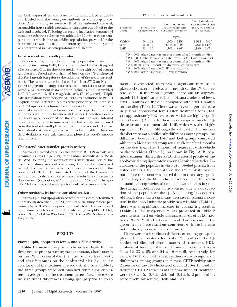
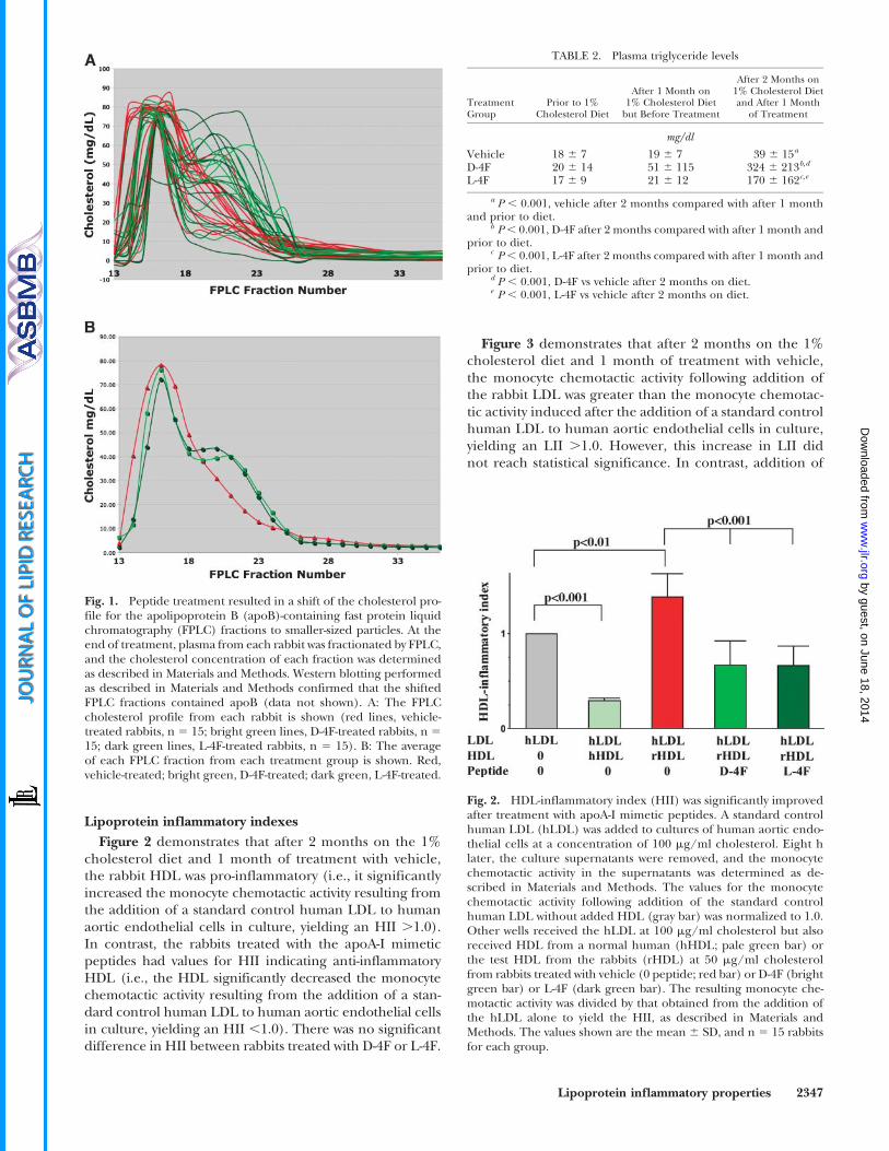
ment). As expected, there was a significant increase inplasma cholesterol levels after 1 month on the 1% choles-terol diet. In the vehicle group, there was an approxi-mately 19% significant decline in plasma cholesterol levelsafter 2 months on the diet, compared with after 1 monthon the diet (Table 1). There was an even larger decreasein plasma cholesterol levels after treatment with D-4F(an approximately 36% decrease), which was highly signifi-cant (Table 1). Similarly, there was an approximately 33%decrease after treatment with L-4F, which was also highlysignificant (Table 1). Although the values after 1 month onthe diet were not significantly different among groups, thedifference between the D-4F and L-4F groups comparedwith the vehicle-treated group was significant after 2 monthson the diet (i.e., after 1 month of treatment with vehicleor the peptides) (Table 1). As shown in Fig. 1A, B, pep-tide treatment shifted the FPLC cholesterol profile of theapoB-containing lipoproteins to smaller-sized particles. In-cubation of the peptides in vitro with pooled plasma fromfasted rabbits after 1 month on the 1% cholesterol dietbut before treatment was started did not cause any signifi-cant changes in the FPLC cholesterol profile of the apoB-containing lipoproteins (data not shown), suggesting thatthe change in profile seen in vivo was not due to a direct ac-tion of the peptides on the apoB-containing lipoproteins.Although there was a significant decrease in plasma choles-terol in the apoA-I mimetic peptide-treated rabbits (Table 1),there was a significant increase in plasma triglycerides(Table 2). The triglyceride values presented in Table 2were determined on whole plasma. Analysis of FPLC frac-tions 13–18 (VLDL fractions) revealed an increase in tri-glycerides in these fractions consistent with the increasein the whole plasma (data not shown).
There were no significant differences among groups inplasma HDL-cholesterol levels after 2 months on the 1%cholesterol diet and after 1 month of treatment. HDL-cholesterol levels at the conclusion of treatment were34 6 12, 33 6 25, and 24 6 10 mg/dl, respectively, forvehicle, D-4F, and L-4F. Similarly, there were no significantdifferences among groups in plasma CETP activity after2 months on the 1% cholesterol diet and after 1 month oftreatment. CETP activities at the conclusion of treatmentwere 17.8 6 6.2, 18.7 6 12.8, and 18.4 6 5.12 pmol/ml/h,respectively, for vehicle, D-4F, and L-4F.
TABLE 1. Plasma cholesterol levels
TreatmentGroup
Prior to 1%Cholesterol Diet
After 1 Month on1% Cholesterol Diet
but Before Treatment
After 2 Months on1% Cholesterol Dietand After 1 Month
of Treatment
mg/dl
Vehicle 60 6 14 1,963 6 515d 1,591 6 382a
D-4F 65 6 19 2,018 6 790d 1,292 6 317b,e
L-4F 53 6 15 1,701 6 626d 1,148 6 247c,f
a P , 0.05, after 2 months on diet versus after 1 month on diet.b P , 0.001, after 2 months on diet versus after 1 month on diet.c P , 0.01, after 2 months on diet versus after 1 month on diet.d P , 0.001, after 1 month on diet versus prior to diet.e P , 0.05, after 2 months D-4F versus vehicle.f P , 0.01, after 2 months L-4F versus vehicle.
2346 Journal of Lipid Research Volume 48, 2007
by guest, on June 18, 2014w
ww
.jlr.orgD
ownloaded from
Lipoprotein inflammatory indexes
Figure 2 demonstrates that after 2 months on the 1%cholesterol diet and 1 month of treatment with vehicle,the rabbit HDL was pro-inflammatory (i.e., it significantlyincreased the monocyte chemotactic activity resulting fromthe addition of a standard control human LDL to humanaortic endothelial cells in culture, yielding an HII .1.0).In contrast, the rabbits treated with the apoA-I mimeticpeptides had values for HII indicating anti-inflammatoryHDL (i.e., the HDL significantly decreased the monocytechemotactic activity resulting from the addition of a stan-dard control human LDL to human aortic endothelial cellsin culture, yielding an HII ,1.0). There was no significantdifference in HII between rabbits treated with D-4F or L-4F.
Figure 3 demonstrates that after 2 months on the 1%cholesterol diet and 1 month of treatment with vehicle,the monocyte chemotactic activity following addition ofthe rabbit LDL was greater than the monocyte chemotac-tic activity induced after the addition of a standard controlhuman LDL to human aortic endothelial cells in culture,yielding an LII .1.0. However, this increase in LII didnot reach statistical significance. In contrast, addition of
Fig. 1. Peptide treatment resulted in a shift of the cholesterol pro-file for the apolipoprotein B (apoB)-containing fast protein liquidchromatography (FPLC) fractions to smaller-sized particles. At theend of treatment, plasma from each rabbit was fractionated by FPLC,and the cholesterol concentration of each fraction was determinedas described in Materials and Methods. Western blotting performedas described in Materials and Methods confirmed that the shiftedFPLC fractions contained apoB (data not shown). A: The FPLCcholesterol profile from each rabbit is shown (red lines, vehicle-treated rabbits, n 5 15; bright green lines, D-4F-treated rabbits, n 5
15; dark green lines, L-4F-treated rabbits, n 5 15). B: The averageof each FPLC fraction from each treatment group is shown. Red,vehicle-treated; bright green, D-4F-treated; dark green, L-4F-treated.
TABLE 2. Plasma triglyceride levels
TreatmentGroup
Prior to 1%Cholesterol Diet
After 1 Month on1% Cholesterol Diet
but Before Treatment
After 2 Months on1% Cholesterol Dietand After 1 Month
of Treatment
mg/dl
Vehicle 18 6 7 19 6 7 39 6 15a
D-4F 20 6 14 51 6 115 324 6 213b,d
L-4F 17 6 9 21 6 12 170 6 162c,e
a P , 0.001, vehicle after 2 months compared with after 1 monthand prior to diet.
b P , 0.001, D-4F after 2 months compared with after 1 month andprior to diet.
c P , 0.001, L-4F after 2 months compared with after 1 month andprior to diet.
d P , 0.001, D-4F vs vehicle after 2 months on diet.e P , 0.001, L-4F vs vehicle after 2 months on diet.
Fig. 2. HDL-inflammatory index (HII) was significantly improvedafter treatment with apoA-I mimetic peptides. A standard controlhuman LDL (hLDL) was added to cultures of human aortic endo-thelial cells at a concentration of 100 mg/ml cholesterol. Eight hlater, the culture supernatants were removed, and the monocytechemotactic activity in the supernatants was determined as de-scribed in Materials and Methods. The values for the monocytechemotactic activity following addition of the standard controlhuman LDL without added HDL (gray bar) was normalized to 1.0.Other wells received the hLDL at 100 mg/ml cholesterol but alsoreceived HDL from a normal human (hHDL; pale green bar) orthe test HDL from the rabbits (rHDL) at 50 mg/ml cholesterolfrom rabbits treated with vehicle (0 peptide; red bar) or D-4F (brightgreen bar) or L-4F (dark green bar). The resulting monocyte che-motactic activity was divided by that obtained from the addition ofthe hLDL alone to yield the HII, as described in Materials andMethods. The values shown are the mean 6 SD, and n 5 15 rabbitsfor each group.
Lipoprotein inflammatory properties 2347
by guest, on June 18, 2014w
ww
.jlr.orgD
ownloaded from
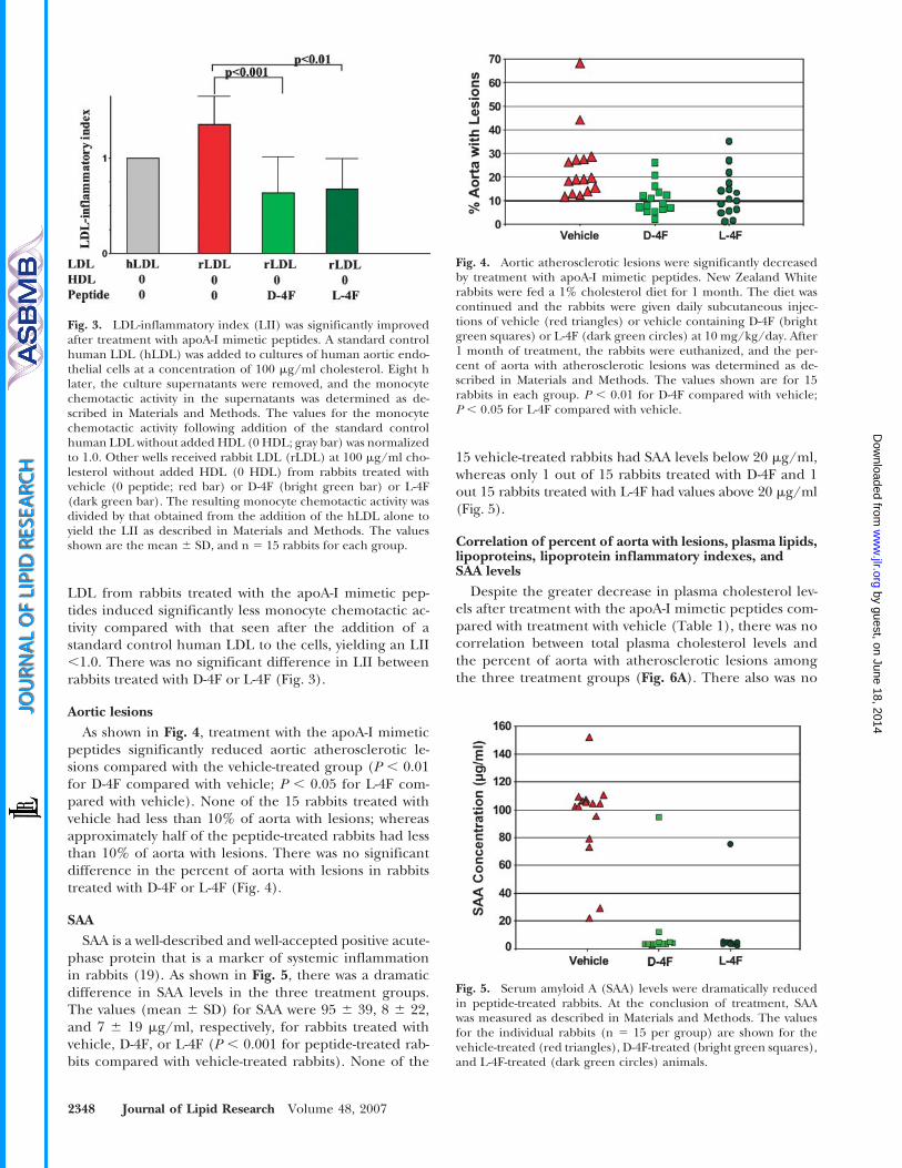
LDL from rabbits treated with the apoA-I mimetic pep-tides induced significantly less monocyte chemotactic ac-tivity compared with that seen after the addition of astandard control human LDL to the cells, yielding an LII,1.0. There was no significant difference in LII betweenrabbits treated with D-4F or L-4F (Fig. 3).
Aortic lesions
As shown in Fig. 4, treatment with the apoA-I mimeticpeptides significantly reduced aortic atherosclerotic le-sions compared with the vehicle-treated group (P , 0.01for D-4F compared with vehicle; P , 0.05 for L-4F com-pared with vehicle). None of the 15 rabbits treated withvehicle had less than 10% of aorta with lesions; whereasapproximately half of the peptide-treated rabbits had lessthan 10% of aorta with lesions. There was no significantdifference in the percent of aorta with lesions in rabbitstreated with D-4F or L-4F (Fig. 4).
SAA
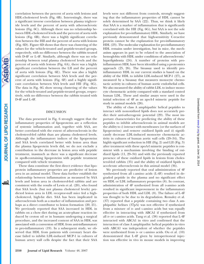
SAA is a well-described and well-accepted positive acute-phase protein that is a marker of systemic inflammationin rabbits (19). As shown in Fig. 5, there was a dramaticdifference in SAA levels in the three treatment groups.The values (mean 6 SD) for SAA were 95 6 39, 8 6 22,and 7 6 19 mg/ml, respectively, for rabbits treated withvehicle, D-4F, or L-4F (P , 0.001 for peptide-treated rab-bits compared with vehicle-treated rabbits). None of the
15 vehicle-treated rabbits had SAA levels below 20 mg/ml,whereas only 1 out of 15 rabbits treated with D-4F and 1out 15 rabbits treated with L-4F had values above 20 mg/ml(Fig. 5).
Correlation of percent of aorta with lesions, plasma lipids,lipoproteins, lipoprotein inflammatory indexes, andSAA levels
Despite the greater decrease in plasma cholesterol lev-els after treatment with the apoA-I mimetic peptides com-pared with treatment with vehicle (Table 1), there was nocorrelation between total plasma cholesterol levels andthe percent of aorta with atherosclerotic lesions amongthe three treatment groups (Fig. 6A). There also was no
Fig. 3. LDL-inflammatory index (LII) was significantly improvedafter treatment with apoA-I mimetic peptides. A standard controlhuman LDL (hLDL) was added to cultures of human aortic endo-thelial cells at a concentration of 100 mg/ml cholesterol. Eight hlater, the culture supernatants were removed, and the monocytechemotactic activity in the supernatants was determined as de-scribed in Materials and Methods. The values for the monocytechemotactic activity following addition of the standard controlhuman LDL without added HDL (0 HDL; gray bar) was normalizedto 1.0. Other wells received rabbit LDL (rLDL) at 100 mg/ml cho-lesterol without added HDL (0 HDL) from rabbits treated withvehicle (0 peptide; red bar) or D-4F (bright green bar) or L-4F(dark green bar). The resulting monocyte chemotactic activity wasdivided by that obtained from the addition of the hLDL alone toyield the LII as described in Materials and Methods. The valuesshown are the mean 6 SD, and n 5 15 rabbits for each group.
Fig. 4. Aortic atherosclerotic lesions were significantly decreasedby treatment with apoA-I mimetic peptides. New Zealand Whiterabbits were fed a 1% cholesterol diet for 1 month. The diet wascontinued and the rabbits were given daily subcutaneous injec-tions of vehicle (red triangles) or vehicle containing D-4F (brightgreen squares) or L-4F (dark green circles) at 10 mg/kg/day. After1 month of treatment, the rabbits were euthanized, and the per-cent of aorta with atherosclerotic lesions was determined as de-scribed in Materials and Methods. The values shown are for 15rabbits in each group. P , 0.01 for D-4F compared with vehicle;P , 0.05 for L-4F compared with vehicle.
Fig. 5. Serum amyloid A (SAA) levels were dramatically reducedin peptide-treated rabbits. At the conclusion of treatment, SAAwas measured as described in Materials and Methods. The valuesfor the individual rabbits (n 5 15 per group) are shown for thevehicle-treated (red triangles), D-4F-treated (bright green squares),and L-4F-treated (dark green circles) animals.
2348 Journal of Lipid Research Volume 48, 2007
by guest, on June 18, 2014w
ww
.jlr.orgD
ownloaded from
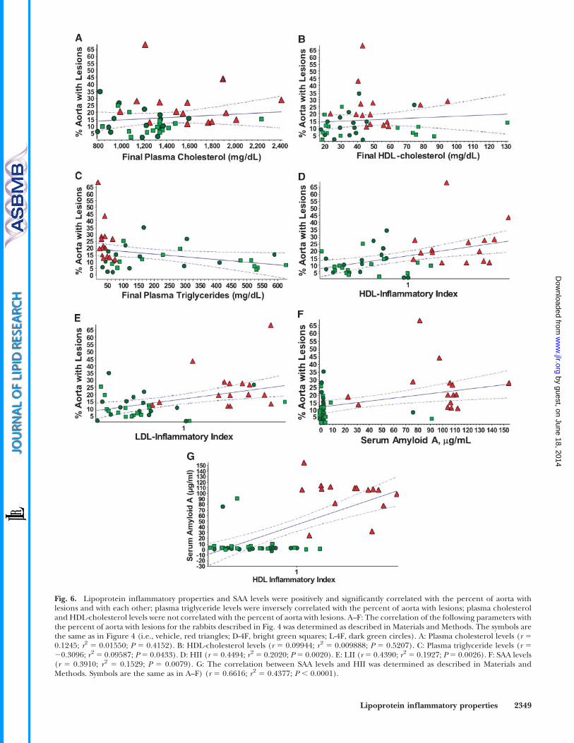
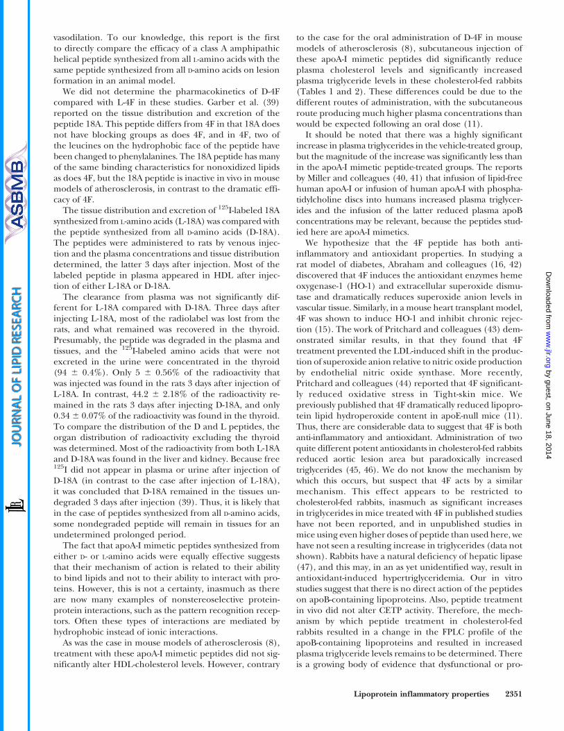
Fig. 6. Lipoprotein inflammatory properties and SAA levels were positively and significantly correlated with the percent of aorta withlesions and with each other; plasma triglyceride levels were inversely correlated with the percent of aorta with lesions; plasma cholesteroland HDL-cholesterol levels were not correlated with the percent of aorta with lesions. A–F: The correlation of the following parameters withthe percent of aorta with lesions for the rabbits described in Fig. 4 was determined as described in Materials and Methods. The symbols arethe same as in Figure 4 (i.e., vehicle, red triangles; D-4F, bright green squares; L-4F, dark green circles). A: Plasma cholesterol levels (r 50.1245; r2 5 0.01550; P 5 0.4152). B: HDL-cholesterol levels (r 5 0.09944; r2 5 0.009888; P 5 0.5207). C: Plasma triglyceride levels (r 520.3096; r2 5 0.09587; P 5 0.0433). D: HII (r 5 0.4494; r2 5 0.2020; P 5 0.0020). E: LII (r 5 0.4390; r2 5 0.1927; P 5 0.0026). F: SAA levels(r 5 0.3910; r2 5 0.1529; P 5 0.0079). G: The correlation between SAA levels and HII was determined as described in Materials andMethods. Symbols are the same as in A–F) (r 5 0.6616; r2 5 0.4377; P , 0.0001).
Lipoprotein inflammatory properties 2349
by guest, on June 18, 2014w
ww
.jlr.orgD
ownloaded from
correlation between the percent of aorta with lesions andHDL-cholesterol levels (Fig. 6B). Interestingly, there wasa significant inverse correlation between plasma triglycer-ide levels and the percent of aorta with atheroscleroticlesions (Fig. 6C). Although there was no relationship be-tween HDL-cholesterol levels and the percent of aorta withlesions (Fig. 6B), there was a highly significant correla-tion between the HII and the percent of aorta with lesions(Fig. 6D). Figure 6D shows that there was clustering of thevalues for the vehicle-treated and peptide-treated groups,respectively, with no difference between the rabbits treatedwith D-4F or L-4F. Similarly, although there was no rela-tionship between total plasma cholesterol levels and thepercent of aorta with lesions (Fig. 6A), there was a highlysignificant correlation between the LII and the percentof aorta with lesions (Fig. 6E). There was also a highlysignificant correlation between SAA levels and the per-cent of aorta with lesions (Fig. 6F) and a highly signifi-cant correlation between SAA levels and HII (Fig. 6G).The data in Fig. 6G show strong clustering of the valuesfor the vehicle-treated and peptide-treated groups, respec-tively, with no difference between the rabbits treated withD-4F and L-4F.
DISCUSSION
The data presented in Fig. 6 strongly suggest that theinflammatory properties of lipoproteins are a reflectionof systemic inflammation and, together with SAA, arebetter correlated with the extent of atherosclerosis in thecholesterol-fed rabbit than are plasma cholesterol levels.Although the inflammatory indices of the lipoproteinsand SAA levels correlated better with lesion area thanthe plasma lipoprotein levels did, we do not exclude acontribution to the decrease in lesion area with peptidetreatment, inasmuch as there was a significant decreasein apoB-containing lipoproteins with peptide treatmentcompared with vehicle treatment.
These data constitute the first direct evidence that lipo-protein inflammatory properties are predictive of lesionarea in an animal model. These data further establish therelationship between inflammation as measured by SAAlevels and lesion area in cholesterol-fed rabbits and areconsistent with the results of Lewis et al. (20), who foundthat SAA levels (but not plasma cholesterol levels) pre-dicted lesion area in LDL receptor-null mice fed a high-cholesterol, high-fat diet. SAA has been implicated inatherosclerosis both as a marker of inflammation and per-haps as a direct contributor to lesion formation (20, 21).We previously reported that SAA increased in HDL inrabbits on a chow diet during an acute-phase reaction in-duced by croton oil or in humans undergoing a surgicalprocedure, and the increased content of SAA in HDL wasassociated with a conversion of HDL from anti-inflammatoryto pro-inflammatory (19). In a subsequent study, we ob-served that HDL from patients with coronary heart dis-ease failed to inhibit LDL-induced MCP-1 in cultures ofhuman artery wall cells despite the fact that their SAA
levels were not different from controls, strongly suggest-ing that the inflammatory properties of HDL cannot besolely determined by SAA (22). Thus, we think it likelythat SAA is a marker of inflammation that is significantlycorrelated with the HII (Fig. 6G), but SAA is not the soleexplanation for pro-inflammatory HDL. Similarly, we havepreviously demonstrated that high-sensitivity C-reactiveprotein cannot be the explanation for pro-inflammatoryHDL (23). The molecular explanation for pro-inflammatoryHDL remains under investigation, but in mice, the mech-anism appears in part to be related to the association ofhemoglobin with HDL under conditions of atherosclerosis/hyperlipidemia (24). A number of proteins with pro-inflammatory HDL have been identified using a proteomicsapproach (25, 26). The bioassay used to identify pro-inflammatory HDL in our current studies is based on theability of the HDL to inhibit LDL-induced MCP-1 (27), asmeasured by a bioassay that measures monocyte chemo-tactic activity in cultures of human aortic endothelial cells.We also measured the ability of rabbit LDL to induce mono-cyte chemotactic activity compared with a standard controlhuman LDL. These and similar assays were used in theinitial selection of 4F as an apoA-I mimetic peptide forstudy in animal models (28).
The ability of class A amphipathic helical peptides tointeract with nonoxidized lipids does not accurately pre-dict their anti-atherogenic potential (29). The most im-portant characteristics for predicting the ability of thesepeptides to inhibit atherosclerosis in animal models arethe ability to i) interact with membranes (or the surfaces oflipoproteins) and remove oxidized lipids and ii) signifi-cantly decrease LDL-induced monoctye chemotactic ac-tivity in cultures of human aortic wall cells (29–34). Thehighly significant reduction in HII (Fig. 2) and LII (Fig. 3)after treatment with these apoA-I mimetic peptides is con-sistent with a mechanism involving the binding of oxi-dized lipids (11, 29–34) and with the previously reportedpresence of these oxidized lipids in lesions from choles-terol-fed rabbits (35) and the ability of oxidized lipids toaccelerate atherosclerosis in this animal model (36).
We previously reported that oral administration of 4Fsynthesized from all L-amino acids (L-4F) resulted in de-graded peptide in the plasma and no significant effecton HDL or LDL inflammatory properties (8). In contrast,administration of 4F synthesized from all D-amino acidsresulted in significant improvement in the inflammatoryproperties of both HDL and LDL (8). The failure of L-4Fwas thought to be due to its degradation. Remaley et al.(37) reported that a peptide containing two class A am-phipathic helixes (37pA) was not effective if synthesizedfrom a mixture of D- and L-amino acids but was equallyeffective in interacting with ABCA1 if synthesized fromall D- or L-amino acids. Tang et al. (38) reported that L-4Finteracted with ABCA1 in vitro and confirmed that theinteraction of class A amphipathic helical peptides in vitrowith ABCA1 was independent of whether the peptideswere synthesized from D- or L-amino acids. Ou et al. (10)demonstrated that L-4F given by intraperitoneal injec-tion was effective in vivo in mouse models in improving
2350 Journal of Lipid Research Volume 48, 2007
by guest, on June 18, 2014w
ww
.jlr.orgD
ownloaded from
vasodilation. To our knowledge, this report is the firstto directly compare the efficacy of a class A amphipathichelical peptide synthesized from all L-amino acids with thesame peptide synthesized from all D-amino acids on lesionformation in an animal model.
We did not determine the pharmacokinetics of D-4Fcompared with L-4F in these studies. Garber et al. (39)reported on the tissue distribution and excretion of thepeptide 18A. This peptide differs from 4F in that 18A doesnot have blocking groups as does 4F, and in 4F, two ofthe leucines on the hydrophobic face of the peptide havebeen changed to phenylalanines. The 18A peptide has manyof the same binding characteristics for nonoxidized lipidsas does 4F, but the 18A peptide is inactive in vivo in mousemodels of atherosclerosis, in contrast to the dramatic effi-cacy of 4F.
The tissue distribution and excretion of 125I-labeled 18Asynthesized from L-amino acids (L-18A) was compared withthe peptide synthesized from all D-amino acids (D-18A).The peptides were administered to rats by venous injec-tion and the plasma concentrations and tissue distributiondetermined, the latter 3 days after injection. Most of thelabeled peptide in plasma appeared in HDL after injec-tion of either L-18A or D-18A.
The clearance from plasma was not significantly dif-ferent for L-18A compared with D-18A. Three days afterinjecting L-18A, most of the radiolabel was lost from therats, and what remained was recovered in the thyroid.Presumably, the peptide was degraded in the plasma andtissues, and the 125I-labeled amino acids that were notexcreted in the urine were concentrated in the thyroid(94 6 0.4%). Only 5 6 0.56% of the radioactivity thatwas injected was found in the rats 3 days after injection ofL-18A. In contrast, 44.2 6 2.18% of the radioactivity re-mained in the rats 3 days after injecting D-18A, and only0.34 6 0.07% of the radioactivity was found in the thyroid.To compare the distribution of the D and L peptides, theorgan distribution of radioactivity excluding the thyroidwas determined. Most of the radioactivity from both L-18Aand D-18A was found in the liver and kidney. Because free125I did not appear in plasma or urine after injection ofD-18A (in contrast to the case after injection of L-18A),it was concluded that D-18A remained in the tissues un-degraded 3 days after injection (39). Thus, it is likely thatin the case of peptides synthesized from all D-amino acids,some nondegraded peptide will remain in tissues for anundetermined prolonged period.
The fact that apoA-I mimetic peptides synthesized fromeither D- or L-amino acids were equally effective suggeststhat their mechanism of action is related to their abilityto bind lipids and not to their ability to interact with pro-teins. However, this is not a certainty, inasmuch as thereare now many examples of nonstereoselective protein-protein interactions, such as the pattern recognition recep-tors. Often these types of interactions are mediated byhydrophobic instead of ionic interactions.
As was the case in mouse models of atherosclerosis (8),treatment with these apoA-I mimetic peptides did not sig-nificantly alter HDL-cholesterol levels. However, contrary
to the case for the oral administration of D-4F in mousemodels of atherosclerosis (8), subcutaneous injection ofthese apoA-I mimetic peptides did significantly reduceplasma cholesterol levels and significantly increasedplasma triglyceride levels in these cholesterol-fed rabbits(Tables 1 and 2). These differences could be due to thedifferent routes of administration, with the subcutaneousroute producing much higher plasma concentrations thanwould be expected following an oral dose (11).
It should be noted that there was a highly significantincrease in plasma triglycerides in the vehicle-treated group,but the magnitude of the increase was significantly less thanin the apoA-I mimetic peptide-treated groups. The reportsby Miller and colleagues (40, 41) that infusion of lipid-freehuman apoA-I or infusion of human apoA-I with phospha-tidylcholine discs into humans increased plasma triglycer-ides and the infusion of the latter reduced plasma apoBconcentrations may be relevant, because the peptides stud-ied here are apoA-I mimetics.
We hypothesize that the 4F peptide has both anti-inflammatory and antioxidant properties. In studying arat model of diabetes, Abraham and colleagues (16, 42)discovered that 4F induces the antioxidant enzymes hemeoxygenase-1 (HO-1) and extracellular superoxide dismu-tase and dramatically reduces superoxide anion levels invascular tissue. Similarly, in a mouse heart transplant model,4F was shown to induce HO-1 and inhibit chronic rejec-tion (15). The work of Pritchard and colleagues (43) dem-onstrated similar results, in that they found that 4Ftreatment prevented the LDL-induced shift in the produc-tion of superoxide anion relative to nitric oxide productionby endothelial nitric oxide synthase. More recently,Pritchard and colleagues (44) reported that 4F significant-ly reduced oxidative stress in Tight-skin mice. Wepreviously published that 4F dramatically reduced lipopro-tein lipid hydroperoxide content in apoE-null mice (11).Thus, there are considerable data to suggest that 4F is bothanti-inflammatory and antioxidant. Administration of twoquite different potent antioxidants in cholesterol-fed rabbitsreduced aortic lesion area but paradoxically increasedtriglycerides (45, 46). We do not know the mechanism bywhich this occurs, but suspect that 4F acts by a similarmechanism. This effect appears to be restricted tocholesterol-fed rabbits, inasmuch as significant increasesin triglycerides in mice treated with 4F in published studieshave not been reported, and in unpublished studies inmice using even higher doses of peptide than used here, wehave not seen a resulting increase in triglycerides (data notshown). Rabbits have a natural deficiency of hepatic lipase(47), and this may, in an as yet unidentified way, result inantioxidant-induced hypertriglyceridemia. Our in vitrostudies suggest that there is no direct action of the peptideson apoB-containing lipoproteins. Also, peptide treatmentin vivo did not alter CETP activity. Therefore, the mech-anism by which peptide treatment in cholesterol-fedrabbits resulted in a change in the FPLC profile of theapoB-containing lipoproteins and resulted in increasedplasma triglyceride levels remains to be determined. Thereis a growing body of evidence that dysfunctional or pro-
Lipoprotein inflammatory properties 2351
by guest, on June 18, 2014w
ww
.jlr.orgD
ownloaded from
inflammatory HDL may be predictive of chronic inflam-matory diseases in general (1–7, 18, 23, 44, 48–50).
The work presented here also adds to a growing bodyof evidence that plasma and HDL cholesterol levels arepoorly predictive of lesion area in animal models of ath-erosclerosis compared with measurements of inflammatorymarkers such as SAA and determinations of lipoproteininflammatory properties. This work suggests that follow-ing lipoprotein inflammatory properties and markers ofchronic inflammation during therapy potentially may im-prove outcomes in a variety of chronic inflammatory con-ditions. Prospective studies in humans will be required totest this hypothesis. Much is already known about themechanism of action of the peptides used in the studiesreported here (34). However, much is still unknown andwill require considerable further work before the mecha-nism of action of these peptides is truly understood.
This work was supported in part by US Public Health ServiceGrants HL-30568 and HL-34343 and the Laubisch, Castera, andM. K. Grey Funds at the University of California at Los Angeles.
REFERENCES
1. Navab, M., G. M. Anantharamaiah, S. T. Reddy, B. J. Van Lenten,and A. M. Fogelman. 2006. Mechanisms of disease: proatherogenicHDL—an evolving field. Nat. Clin. Pract. Endocrinol. Metab. 2: 504–511.
2. Kontush, A., and M. J. Chapman. 2006. Functionally defective high-density lipoprotein: a new therapeutic target at the crossroads ofdyslipidemia, inflammation, and atherosclerosis. Pharmacol. Rev. 58:342–374.
3. Roberts, C. K., C. Ng, S. Hama, A. J. Eliseo, and R. J. Barnard. 2006.Effect of a short-term diet and exercise intervention on inflammatory/anti-inflammatory properties of HDL in overweight/obese men withcardiovascular risk factors. J. Appl. Physiol. 101: 1727–1732.
4. Nicholls, S. J., P. Lundman, J. A. Harmer, B. Cutri, K. A. Griffiths,K. A. Rye, P. J. Barter, and D. S. Celermajer. 2006. Consumptionof saturated fat impairs the anti-inflammatory properties of high-density lipoproteins and endothelial function. J. Am. Coll. Cardiol.48: 715–720.
5. Shao, B., M. N. Oda, J. F. Oram, and J. W. Heinecke. 2006. Myelo-peroxidase: an inflammatory enzyme for generating dysfunctionalhigh density lipoprotein. Curr. Opin. Cardiol. 21: 322–328.
6. Navab, M., G. M. Anantharamaiah, S. T. Reddy, B. J. Van Lenten,B. J. Ansell, S. Hama, G. Hough, E. Bachini, V. R. Grijalva, A. C.Wagner, et al. 2005. The double jeopardy of HDL. Ann. Med. 37:173–178.
7. Nicholls, S. J., L. Zheng, and S. L. Hazen. 2005. Formation of dys-functional high-density lipoprotein by myeloperoxidase. TrendsCardiovasc. Med. 15: 212–219.
8. Navab, M., G. M. Anantharamaiah, S. Hama, D. W. Garber, M.Chaddha, G. Hough, R. Lallone, and A. M. Fogelman. 2002. Oraladministration of an apo A-I mimetic peptide synthesized fromD-amino acids dramatically reduces atherosclerosis in mice inde-pendent of plasma cholesterol. Circulation. 105: 290–292.
9. Van Lenten, B. J., A. C. Wagner, G. M. Anantharamaiah, D. W.Garber, M. C. Fishbein, L. Adhikary, D. P. Nayak, S. Hama, M.Navab, and A. M. Fogelman. 2002. Influenza infection promotesmacrophage traffic into arteries of mice that is prevented by D-4F, anapolipoprotein A-I mimetic peptide. Circulation. 106: 1127–1132.
10. Ou, J., Z. Ou, D. W. Jones, S. Holzhauer, O. A. Hatoum, A. W.Ackerman, D. W. Weihrauch, D. D. Gutterman, K. Guice, K. T.Oldham, et al. 2003. L-4F, an apolipoprotein A-I mimetic, dramati-cally improves vasodilation in hypercholesterolemia and sickle celldisease. Circulation. 107: 2337–2341.
11. Navab, M., G. M. Anantharamaiah, S. T. Reddy, S. Hama, G.Hough, V. R. Grijalva, A. C. Wagner, J. S. Frank, G. Datta, D.Garber, et al. 2004. Oral D-4F causes formation of pre-b high-
density lipoprotein and improves high-density lipoprotein-mediatedcholesterol efflux and reverse cholesterol transport from macro-phages in apolipoprotein E-null mice. Circulation. 109: 3215–3220.
12. Navab, M., G. M. Anantharamaiah, S. Hama, G. Hough, S. T.Reddy, J. S. Frank, D. W. Garber, S. Handattu, and A. M. Fogelman.2005. D-4F and statins synergize to render HDL anti-inflammatoryin mice and monkeys and cause lesion regression in old apolipo-protein E-null mice. Arterioscler. Thromb. Vasc. Biol. 25: 1426–1432.
13. Ou, J., J. Wang, H. Xu, Z. Ou, M. G. Sorci-Thomas, D. W. Jones,P. Signorino, J. C. Densmore, S. Kaul, K. T. Oldham, et al. 2005.Effects of D- 4F on vasodilation and vessel wall thickness in hypercho-lesterolemic LDL receptor-null and LDL receptor/apolipoprotein A-Idouble-knockout mice on Western diet. Circ. Res. 97: 1190–1197.
14. Buga, G. M., J. S. Frank, G. A. Mottino, M. Hendizadeh, A. Hakhamian,J. H. Tillisch, S. T. Reddy, M. Navab, G. M. Anantharamaiah, L. J.Ignarro, et al. 2006. D-4F decreases brain arteriole inflammationand improves cognitive performance in LDL receptor-null mice ona Western diet. J. Lipid Res. 47: 2148–2160.
15. Schnickel, G. T., G. R. Hsieh, E. L. Kachikwu, C. Garcia, A.Shefizadeh, M. C. Fishbein, and A. Ardehli. 2006. Cytoprotectivegene HO-1 and chronic rejection in heart transplantation. Trans-plant Proc. 38: 3259–3262.
16. Kruger, A. L., S. Peterson, S. Turkseven, P. M. Kaminski, F. F.Zhang, S. Quan, M. S. Wolin, and N. G. Abraham. 2005. D-4F in-duces heme oxygenase-1 and extracellular superoxide dismutase,decreases endothelial cell sloughing, and improves vascular reac-tivity in rat model of diabetes. Circulation. 111: 3126–3134.
17. Rittershaus, C. W., D. P. Miller, L. J. Thomas, M. D. Picard, C. M.Honan, C. D. Emmett, C. L. Pettey, H. Adari, R. A. Hammond, D. T.Beattie, et al. 2000. Vaccine-induced antibodies inhibit CETP ac-tivity in vivo and reduce aortic lesions in a rabbit model of athero-sclerosis. Arterioslcer. Thromb. Vasc. Biol. 20: 2106–2112.
18. Navab, M., S. Y. Hama, G. P. Hough, G. Subbanagounder, S. T.Reddy, and A. M. Fogelman. 2001. A cell-free assay for detectingHDL that is dysfunctional in preventing the formation of or inac-tivating oxidized phospholipids. J. Lipid Res. 42: 1308–1317.
19. Van Lenten, B. J., S. Hama, F. C. de Beer, D. M. Stafforini, T. M.McIntyre, S. M. Prescott, B. N. La Du, A. M. Fogelman, and M.Navab. 1995. Anti-inflammatory HDL becomes pro-inflammatoryduring the acute phase response. Loss of protective effect of HDLagainst LDL oxidation in aortic wall cell cocultures. J. Clin. Invest.96: 2758–2767.
20. Lewis, K. E., E. A. Kirk, T. O. McDonald, S. Wang, T. N. Wight, K. D.O’Brien, and A. Chait. 2004. Increase in serum amyloid A evokedby dietary cholesterol is associated with increased atherosclerosisin mice. Circulation. 110: 540–545.
21. Chait, A., C. Y. Han, J. F. Oram, and J. W. Heinecke. 2005.Lipoprotein-associated inflammatory proteins: markers or media-tors of cardiovascular disease? J. Lipid Res. 46: 389–403.
22. Navab, M., S. Hama-Levy, B. J. Van Lenten, G. C. Fonarow, C. J.Cardinez, L. W. Castellani, M-L. Brennan, A. J. Lusis, and A. M.Fogelman. 1997. Mildly oxidized LDL induces an increased apo-lipoprotein J/paraoxonase ratio. J. Clin. Invest. 99: 2005–2019.
23. Ansell, B. J., M. Navab, S. Hama, N. Kamranpour, G. Fonarow, G.Hough, S. Rahmani, R. Mottahedeh, R. Dave, S. T. Reddy, et al.2003. Inflammatory/anti-inflammatory properties of high-densitylipoprotein distinguish patients from control subjects better thanhigh-density lipoprotein cholesterol levels and are favorably affectedby simvastatin treatment. Circulation. 108: 2751–2756.
24. Watanabe, J., K. J. Chou, J. C. Liao, Y. Miao, H. H. Meng, H. Ge,V. Grijalva, S. Hama, K. Kozak, G. Buga, et al. 2007. Differentialassociation of hemoglobin with proinflammatory high density lipo-proteins in atherogenic/hyperlipidemic mice: a novel biomarkerof atherosclerosis. J. Biol. Chem. 282: 23698–23707.
25. Vaisar, T., S. Pennnathur, P. S. Green, S. A. Gharib, A. N. Hoofnagle,M. C. Cheung, J. Byun, S. Vuletic, S. Kassim, P. Singh, et al. 2007.Shotgun proteomics implicates protease inhibition and complementactivation in the anti-inflammatory properties of HDL. J. Clin. Invest.117: 746–756.
26. Reilly, M. P., and A. R. Tall. 2007. HDL proteomics: pot of goldor Pandora’s box? J. Clin. Invest. 117: 595–598.
27. Navab, M., S. S. Imes, S. Y. Hama, G. P. Hough, L. A. Ross, R. W.Bork, A. J. Valente, J. A. Berliner, D. C. Drinkwater, H. Laks, et al.1991. Monocyte transmigration induced by modification of lowdensity lipoprotein in cocultures of human aortic wall cells is dueto induction of monocyte chemotactic protein 1 synthesis and isabolished by high density lipoprotein. J. Clin. Invest. 88: 2039–2046.
2352 Journal of Lipid Research Volume 48, 2007
by guest, on June 18, 2014w
ww
.jlr.orgD
ownloaded from
28. Datta, G., M. Chaddha, S. Hama, M. Navab, A. M. Fogelman, D. W.Garber, V. K. Mishra, R. M. Epand, R. F. Epand, S. Lund-Katz, et al.2001. Effects of increasing hydrophobicity on the physical-chemicaland biological properties of a class A amphipathic helical peptide.J. Lipid Res. 42: 1096–1104.
29. Navab, M., G. M. Anantharamaiah, S. T. Reddy, S. Hama, G.Hough, V. R. Grijalva, N. Yu, B. J. Ansell, G. Datta, D. W. Garber,et al. 2005. Apolipoprotein A-I mimetic peptides. Arterioscler. Thromb.Vasc. Biol. 25: 1325–1331.
30. Datta, G., R. F. Epand, R. M. Epand, M. Chaddha, M. A. Kirksey,D. W. Garber, S. Lund-Katz, M. C. Phillips, S. Hama, M. Navab,et al. 2004. Aromatic residue position on the nonpolar face ofclass A amphipathic helical peptides determines biological activity.J. Biol. Chem. 279: 26509–26517.
31. Handattu, S. P., D. W. Garber, D. C. Horn, D. W. Hughes, B.Berno, A. D. Bain, V. K. Mishra, M. N. Palgunachari, G. Datta,G. M. Anantharamaiah, et al. 2007. ApoA-I mimetic peptides withdiffering ability to inhibit atherosclerosis also exhibit differencesin their interactions with membrane bilayers. J. Biol. Chem. 282:1980–1988.
32. Navab, M., S. Hama, C. J. Cooke, G. M. Anantharamaiah, M.Chaddha, L. Jin, G. Subbanagounder, K. F. Faull, S. T. Reddy,N. E. Miller, et al. 2000. Normal high density lipoprotein inhibitsthree steps in the formation of mildly oxidized low density lipo-protein: step 1. J. Lipid Res. 41: 1481–1494.
33. Navab, M., S. Y. Hama, G. M. Anantharamaiah, K. Hassan, G. P.Hough, A. D. Watson, S. T. Reddy, A. Sevanian, G. C. Fonarow, andA. M. Fogelman. 2000. Normal high density lipoprotein inhibitsthree steps in the formation of mildly oxidized low density lipo-protein: steps 2 and 3. J. Lipid Res. 41: 1495–1508.
34. Anantharamaiah, G. M., V. K. Mishra, D. W. Garber, G. Datta,S. P. Handattu, M. N. Palgunachari, M. Chaddha, M. Navab, S. T.Reddy, J. P. Segrest, et al. 2007. Structural requirements for anti-oxidative and anti-inflammatory properties of apo A-I mimeticpeptides. J. Lipid Res. Epub ahead of print.
35. Subbanagounder, G., N. Leitinger, D. C. Schwenke, J. W. Wong,H. Lee, C. Rizza, A. D. Watson, K. F. Faull, A. M. Fogelman, andJ. A. Berliner. 2000. Determinants of bioactivity of oxidized phos-pholipids. Specific oxidized fatty acyl groups at the sn-2 position.Arterioscler. Thromb. Vasc. Biol. 20: 2248–2254.
36. Staprans, I., J. H. Rapp, X-M. Pan, D. A. Hardman, and K. R.Finegold. 1996. Oxidized lipids in the diet accelerate the develop-ment of fatty streaks in cholesterol-fed rabbits. Arterioscler. Thromb.Vasc. Biol. 16: 533–538.
37. Remaley, A. T., F. Thomas, J. A. Stonik, S. J. Demonsky, S. E. Bark,E. B. Neufeld, A. V. Bocharov, T. G. Vishnyakova, A. P. Patterson,T. L. Eggerman, et al. 2003. Synthetic amphipathic helical peptidespromote lipid efflux from cells by an ABCA1-dependent and anABCA1-independent pathway. J. Lipid Res. 44: 828–836.
38. Tang, C., A. M. Vaughan, G. M. Anantharamaiah, and J. F. Oram.2006. Janus kinase 2 modulates the lipid-removing but not protein-stabilizing interactions of amphipathic helices with ABCA1. J. LipidRes. 47: 107–114.
39. Garber, D. W., Y. V. Venkatachalapathi, K. B. Gupta, J. Ibdah, M. C.
Phillips, J. B. Hazelrig, J. P. Segrest, and G. M. Anantharamaiah.1992. Turnover of synthetic class A amphipathic peptide analoguesof exchangeable apolipoproteins in rats. Correlation with physicalproperties. Arterioscler. Thromb. 12: 886–894.
40. Nanjee, M. N., J. R. Crouse, J. M. King, R. Hovorka, S. E. Rees,E. R. Carson, J-J. Morgenthaler, P. Lerch, and N. E. Miller. 1996.Effects of intravenous infusion of lipid-free apo A-I in humans.Arterioscler. Thromb. Vasc. Biol. 16: 1203–1214.
41. Nanjee, M. N., J. E. Doran, P. G. Lerch, and N. E. Miller. 1999.Acute effects of intravenous infusion of apoA1/phosphatidylcholinediscs on plasma lipoproteins in humans. Arterioscler. Thromb. Vasc. Biol.19: 979–989.
42. Peterson, S. J., D. Husney, A. L. Kruger, R. Olszanecki, F. Ricci,L. F. Rodella, A. Stachiotti, R. Rezzani, J. A. McClung, W. S.Aronow, et al. 2007. Long-term treatment with apolipoprotein A1mimetic peptide increases antioxidants and vascular repair in type Idiabetic rats. J. Pharmacol. Exp. Ther. 322: 514–520.
43. Ou, Z., J. Ou, A. W. Ackerman, K. T. Oldham, and K. A. Pritchard,Jr. 2003. L-4F, an apolipoprotein A-1 mimetic, restores nitric oxideand superoxide anion balance in low density lipoprotein-treatedendothelial cells. Circulation. 107: 1520–1524.
44. Weihrauch, D., H. Xu, Y. Shi, J. Wang, J. Brien, D. W. Jones, S.Kaul, R. A. Komorowski, M. E. Scuka, K. T. Oldham, et al. 2007.Effects of D-4F on vasodilation, oxidative stress, angiostatin, myo-cardial inflammation and angiogenic potential in Tight-skin mice.Am. J. Physiol. Heart Circ. Physiol. Epub ahead of print. May 11, 2007.doi:10.1152/ajpheart.00038.2007
45. Sparrow, C. P., T. W. Doebber, J. Olszewski, M. S. Wu, J. Ventre,K. A. Stevens, and Y. Chao. 1992. Low density lipoprotein is pro-tected from oxidation and the progression of atherosclerosis isslowed in cholesterol-fed rabbits by the antioxidant N, N’-DiphenylPhenylenediamine. J. Clin. Invest. 89: 1885–1891.
46. Freyschuss, A., A. Al-Schurbaji, I. Bjorkhem, A. Babiker, U.Diczfalusy, L. Berglund, and P. Henriksson. 2001. On the anti-atherogenic effect of the antioxidant BHT in cholesterol-fed rab-bits: inverse relation between serum triglycerides and atheroma-tous lesions. Biochim. Biophys. Acta. 1534: 129–138.
47. Clay, M. A., G. J. Hopkins, C. P. Ehnholm, and P. J. Barter. 1989.The rabbit as an animal model of hepatic lipase deficiency. Biochim.Biophys. Acta. 1002: 173–181.
48. McMahon, M., J. Grossman, J. FitzGerald, E. Dahlin-Lee, D. J.Wallace, B. Y. Thong, H. Badsha, K. Kalunian, C. Charles, M. Navab,et al. 2006. Pro-inflammatory high-density lipoprotein as a bio-marker for atherosclerosis in patients with systemic lupus erythe-matosus and rheumatoid arthritis. Arthritis Rheum. 54: 2541–2549.
49. Charles-Schoeman, C., D. Hhanna, D. E. Furst, M. McMahon, S. T.Reddy, A. M. Fogelman, H. E. Paulus, G. S. Park, T. Gong, and B. J.Ansell. 2007. Effects of high-dose atorvastatin on anti-inflammatoryproperties of high density lipoprotein in patients with rheumatoidarthritis: a pilot study. J. Rheum. 34: 1459–1464.
50. Kalantar-Zadeh, K., J. D. Kopple, N. Kamranpour, A. M. Fogelman,and M. Navab. 2007. Novel HDL-inflammatory index correlateswith body composition, quality of life and survival in hemodialysispatients. Kidney Int. In press.
Lipoprotein inflammatory properties 2353
by guest, on June 18, 2014w
ww
.jlr.orgD
ownloaded from