Genetic Variation at the Proprotein Convertase Subtilisin/Kexin Type 5 Gene Modulates High-Density...
19
Genetic Variation at the Proprotein Convertase Subtilisin/Kexin Type 5 Gene Modulates HDL Cholesterol Levels Iulia Iatan, MSc 1 , Zari Dastani, MD, PhD 2 , Ron Do, MSc 2 , Daphna Weissglas-Volkov, PhD 3 , Isabelle Ruel, PhD 1,2 , Jenny C. Lee, PhD 3 , Adriana Huertas-Vazquez, PhD 3 , Marja-Riitta Taskinen, MD, PhD 4 , Annik Prat, PhD 5 , Nabil G. Seidah, PhD 5 , Päivi Pajukanta, MD, PhD 3 , James C. Engert, PhD 2 , and Jacques Genest, MD 1,2 1 Department of Biochemistry, Cardiovascular Research Laboratories, Cardiology Division, Faculty of Medicine, McGill University Health Centre/Royal Victoria Hospital, Montreal, Quebec, H3A 1A1, Canada 2 Department of Human Genetics, Cardiovascular Research Laboratories, Cardiology Division, Faculty of Medicine, McGill University Health Centre/Royal Victoria Hospital, Montreal, Quebec, H3A 1A1, Canada 3 Department of Human Genetics, David Geffen School of Medicine at UCLA, Los Angeles, CA 90095, USA 4 Department of Medicine, Helsinki University Central Hospital, HUS-00029, Helsinki, Finland 5 Laboratory of Biochemical Neuroendocrinology, Clinical Research Institute of Montreal, Montreal, Quebec, H2W 1R7, Canada Abstract Background—A low level of plasma high-density lipoprotein cholesterol (HDL-C) is a risk factor for cardiovascular disease. HDL particles are modulated by a variety of lipases, including endothelial lipase (EL), a phospholipase present on vascular endothelial cells. The proprotein convertase subtilisin/kexin type 5 (PCSK5) gene product is known to directly inactivate EL, and, indirectly, cleave and activate angiopoetin-like protein 3, a natural inhibitor of EL. We therefore investigated the effect of human PCSK5 genetic variants on plasma HDL-C levels. Methods and Results—Haplotypes at the PCSK5 locus were examined in nine multi-generational families that included 60 individuals with HDL-C<10 th percentile. Segregation with low HDL-C in one family was found. Sequencing of the PCSK5 gene in 12 probands with HDL-C<5 th percentile identified seven novel variants. Using a two-stage design, we first genotyped these single-nucleotide polymorphisms (SNPs) along with 163 tagSNPs and 12 additional SNPs (n=182 total) in 457 individuals with documented coronary artery disease. We identified nine SNPs associated with HDL- C (P<0.05), with the strongest results for rs11144782 and rs11144766 (P=0.002 and P=0.005 respectively). The SNP rs11144782 was also associated with very low-density lipoprotein (P=0.039), triglycerides (P=0.049) and total apolipoprotein B levels (P=0.022). In stage 2, we replicated the association of rs11144766 with HDL-C (P=0.014) in an independent sample of Finnish low HDL- C families. In a combined analysis of both stages (n=883), region-wide significance of rs11144766 and low HDL-C was observed (unadjusted P=1.86×10 −4 and Bonferroni adjusted P=0.031). Conclusions—We conclude that variability at the PCSK5 locus influences HDL-C levels, possibly through the inactivation of EL activity and consequently, atherosclerotic cardiovascular disease risk. Corresponding author: Jacques Genest, MD, FRCP(C), FACC, Professor, Faculty of Medicine, McGill/Novartis Chair in Medicine, Director, Division of Cardiology, McGill University Health Center/Royal Victoria Hospital, 687 Pine Avenue West Rm. M4/76, Montreal, QC, Canada H3A 1A1, Tel: (514)934-1934 ext.34642, Fax: (514)843-2813, [email protected]. DISCLOSURES No conflict of interest to be declared. NIH Public Access Author Manuscript Circ Cardiovasc Genet. Author manuscript; available in PMC 2010 October 1. Published in final edited form as: Circ Cardiovasc Genet. 2009 October ; 2(5): 467–475. doi:10.1161/CIRCGENETICS.109.877811. NIH-PA Author Manuscript NIH-PA Author Manuscript NIH-PA Author Manuscript
Transcript of Genetic Variation at the Proprotein Convertase Subtilisin/Kexin Type 5 Gene Modulates High-Density...

Genetic Variation at the Proprotein Convertase Subtilisin/KexinType 5 Gene Modulates HDL Cholesterol Levels
Iulia Iatan, MSc1, Zari Dastani, MD, PhD2, Ron Do, MSc2, Daphna Weissglas-Volkov, PhD3,Isabelle Ruel, PhD1,2, Jenny C. Lee, PhD3, Adriana Huertas-Vazquez, PhD3, Marja-RiittaTaskinen, MD, PhD4, Annik Prat, PhD5, Nabil G. Seidah, PhD5, Päivi Pajukanta, MD, PhD3,James C. Engert, PhD2, and Jacques Genest, MD1,2
1 Department of Biochemistry, Cardiovascular Research Laboratories, Cardiology Division, Facultyof Medicine, McGill University Health Centre/Royal Victoria Hospital, Montreal, Quebec, H3A 1A1,Canada 2 Department of Human Genetics, Cardiovascular Research Laboratories, CardiologyDivision, Faculty of Medicine, McGill University Health Centre/Royal Victoria Hospital, Montreal,Quebec, H3A 1A1, Canada 3 Department of Human Genetics, David Geffen School of Medicine atUCLA, Los Angeles, CA 90095, USA 4 Department of Medicine, Helsinki University Central Hospital,HUS-00029, Helsinki, Finland 5 Laboratory of Biochemical Neuroendocrinology, Clinical ResearchInstitute of Montreal, Montreal, Quebec, H2W 1R7, Canada
AbstractBackground—A low level of plasma high-density lipoprotein cholesterol (HDL-C) is a risk factorfor cardiovascular disease. HDL particles are modulated by a variety of lipases, including endotheliallipase (EL), a phospholipase present on vascular endothelial cells. The proprotein convertasesubtilisin/kexin type 5 (PCSK5) gene product is known to directly inactivate EL, and, indirectly,cleave and activate angiopoetin-like protein 3, a natural inhibitor of EL. We therefore investigatedthe effect of human PCSK5 genetic variants on plasma HDL-C levels.
Methods and Results—Haplotypes at the PCSK5 locus were examined in nine multi-generationalfamilies that included 60 individuals with HDL-C<10th percentile. Segregation with low HDL-C inone family was found. Sequencing of the PCSK5 gene in 12 probands with HDL-C<5th percentileidentified seven novel variants. Using a two-stage design, we first genotyped these single-nucleotidepolymorphisms (SNPs) along with 163 tagSNPs and 12 additional SNPs (n=182 total) in 457individuals with documented coronary artery disease. We identified nine SNPs associated with HDL-C (P<0.05), with the strongest results for rs11144782 and rs11144766 (P=0.002 and P=0.005respectively). The SNP rs11144782 was also associated with very low-density lipoprotein (P=0.039),triglycerides (P=0.049) and total apolipoprotein B levels (P=0.022). In stage 2, we replicated theassociation of rs11144766 with HDL-C (P=0.014) in an independent sample of Finnish low HDL-C families. In a combined analysis of both stages (n=883), region-wide significance of rs11144766and low HDL-C was observed (unadjusted P=1.86×10⁻⁴ and Bonferroni adjusted P=0.031).
Conclusions—We conclude that variability at the PCSK5 locus influences HDL-C levels, possiblythrough the inactivation of EL activity and consequently, atherosclerotic cardiovascular disease risk.
Corresponding author: Jacques Genest, MD, FRCP(C), FACC, Professor, Faculty of Medicine, McGill/Novartis Chair in Medicine,Director, Division of Cardiology, McGill University Health Center/Royal Victoria Hospital, 687 Pine Avenue West Rm. M4/76,Montreal, QC, Canada H3A 1A1, Tel: (514)934-1934 ext.34642, Fax: (514)843-2813, [email protected] conflict of interest to be declared.
NIH Public AccessAuthor ManuscriptCirc Cardiovasc Genet. Author manuscript; available in PMC 2010 October 1.
Published in final edited form as:Circ Cardiovasc Genet. 2009 October ; 2(5): 467–475. doi:10.1161/CIRCGENETICS.109.877811.
NIH
-PA Author Manuscript
NIH
-PA Author Manuscript
NIH
-PA Author Manuscript

Keywordscholesterol; coronary disease; genetics; lipids; lipoproteins
INTRODUCTIONLow levels of high-density lipoprotein cholesterol (HDL-C) constitute a major risk factor forcoronary artery disease (CAD)1. Genetic factors account for a large component of the plasmavariability of HDL-C levels with heritability being approximately 0.502. Numerous humangenetic studies have identified loci contributing to HDL-C, including both linkage analysesand association studies, explaining some these variations. Despite these, the genetic andmetabolic factors that regulate HDL metabolism remain incompletely understood.
The metabolism of HDL is complex and involves a carefully orchestrated interplay betweenthe biogenesis of nascent HDL particles3, the continual exchange of lipid and protein moietiesof HDL in plasma and the modulation of HDL particles by a variety of enzymes, especiallylipases4. Endothelial lipase (EL), discovered by Rader and colleagues5, is a phospholipasepresent on vascular endothelial cells. It can be inactivated by angiopoetin-like protein 3(ANGPTL3)6 or by secretory proprotein convertases of the subtilisin/kexin type, such as Furin,PC5/6, and PACE47. The mammalian proprotein convertases comprise a family of ninemembers related to bacterial subtilisin and yeast kexin-like serine proteinases, criticallyinvolved in the activation/inactivation of various physiological and pathological processes,including those implicated in regulation of vascular events. These include PC1/3, PC2, Furin,PC4, PC5/6, PACE4, PC7, SKI-1/S1P and PCSK9, encoded by the genes PCSK1 to PCSK9.
While PCSK9 has been found to play a critical role in regulating lipid levels by enhancing low-density lipoprotein (LDL) receptor degradation9, proof of in vivo functions of the proproteinconvertase subtilisin/kexin type 5 (PCSK5) (OMIM:600488) and its protein, PC5/6, indyslipidemia and cardiovascular pathologies, has yet to be established. Murine Pcsk5 islocalized on chromosome 19 and encodes two alternatively spliced isoforms, soluble PC5A(915 amino acids (aa); 21 exons) and membrane bound PC5B (1877 aa; 38 exons)10. Althoughdevoid of a transmembrane domain, PC5A can exert its proteolytic action at the cell surface,as it is retained at the plasma membrane as a complex with tissue inhibitors of metalloproteases(TIMPs) and heparin sulfate proteoglycans (HSPG)11. The essential role of Pcsk5 has beenhighlighted by Essalmani et al. who observed death at birth in the knock-out (KO) mice, whileheterozygotes were healthy and fertile12. Except in the liver where both isoforms are equallyexpressed, PC5A is the major isoform in most tissues (87–100%), and only the intestine andkidney show a predominance of PC5B (74–92%)12,13. In humans, PCSK5 is located onchromosome 9q21.13, and while two alternatively spliced transcripts are described for thisgene14,15, only one, generating a 21-exon isoform and typically referred to as PCSK5, has itsfull length nature known (NM_006200.3).
There is strong biological plausibility for the involvement of PCSK5 in lipoprotein metabolism.Jin et al7 showed that PC5A inactivates ex vivo EL and lipoprotein lipase (LPL), with bothlipases being present at the vascular endothelial surface. High expression of PC5/6 inenterocytes also suggests a possible role in processing protein substrates that could regulatefood and/or sterol/lipid absorption10. Additionally, recent data from a genome-wide scanreports a region close to PCSK5 on chromosome 9, implicated in lipid regulation16, but its linkto HDL deficiency remains to be explored. In light of this, there is compelling evidence thatsuggests that PC5/6 may be a good candidate proteinase implicated in the control of circulatingHDL-C levels and mutations affecting its function may influence lipoprotein metabolism andHDL subspecies more specifically. Therefore, we undertook a study to determine whether
Iatan et al. Page 2
Circ Cardiovasc Genet. Author manuscript; available in PMC 2010 October 1.
NIH
-PA Author Manuscript
NIH
-PA Author Manuscript
NIH
-PA Author Manuscript

genetic variation within the PCSK5 gene contributes to variation in HDL-C levels, whichconsequently, could influence atherosclerotic cardiovascular disease risk.
METHODSFrench Canadian family subjects
A total of nine multigenerational French Canadian families consisting of 175 genotypedmembers were examined and sampled in the Preventive Cardiology/Lipid Clinic of the McGillUniversity Health Centre (MUHC). The selection criterion for probands was HDL-C<5th
percentile (age and gender-matched), based on the Lipid Research Clinics Population StudiesData Book17. All subjects provided informed consent for plasma and DNA sampling, isolation,and storage. The research protocol was reviewed and approved by the ResearchEthics Boardof the MUHC.
Stage 1 and 2 association study samplesFrench Canadian subjects—For stage 1 analysis, unrelated patients (n=457) of FrenchCanadian descent were selected from the Cardiology Clinic of the Clinical Research Instituteof Montreal that were <60 years of age and had angiographically documented CAD18.Individuals had HDL-C values ranging from HDL-C<5th percentile to HDL-C>95th percentile(age and gender-matched)17. We excluded patients with known causes of HDL deficiency(severe hypertriglyceridemia defined as plasma triglycerides (TG)>10 mmol/L, cellularphospholipid or cholesterol efflux defect or previously identified mutations in genes associatedwith HDL deficiency)19,20. Demographic and clinical information, medications, andlipoprotein profiles were determined on all participating subjects as previously described20.The research protocol was approved by the Research Ethics Board of the MUHC.
Finnish family subjects—The stage 2 study sample consisted of 39 Finnish low HDL-Cfamilies (426 genotyped individuals) recruited at the Helsinki and Turku central hospitals aspreviously described21. Each subject involved in this study provided written informed consent.The study design was approved by the ethics committees of the participating institutions.Inclusion criteria for the probands were age 30–60 years, at least 50% stenosis in one or morecoronary arteries, HDL-C level below the Finnish age- and sex-specific 10th populationpercentile (subjects coded as affected), total cholesterol (TC)<6.3 mmol/L for males and <6.0mmol/L for females, and TG<2.3 mmol/L for both genders21. Serum lipid and glucoseparameters were measured as previously22. Exclusion criteria for the probands were type 1and 2 diabetes mellitus, severe hepatic or renal disease, or body mass index >3021.
HaplotypingMicrosatellite genotypes were determined by deCODE Genetics (Reykjavik, Iceland) atmarkers D9S1777, D91876, D9S175, D9S1843 spanning 11.3 Mb and flanking the PCSK5gene on chromosome 9q21.13. Haplotypes were constructed using Cyrillic version 2.1.3(Cherwell Scientific Publishing Ltd) to examine the segregation of the PCSK5 locus with thelow HDL-C trait in nine families with HDL-C deficiency.
SequencingSequencing of the 21 exons and exon-intron boundries of the PCSK5 gene was performed in12 unrelated individuals with HDL-C<5th percentile. Exon-specific oligonucleotides weresynthesized (Integrated DNA Technologies, Coralville, Iowa, USA) and designed using thePrimer3 software (http://frodo.wi.mit.edu/)23 to include at least 22 bp of intronic sequence ateach intro-exon boundary. Polymerase chain reaction (PCR)-amplified fragments werepurified using the Millipore purification plate (Multiscreen PCR) and directly sequenced at the
Iatan et al. Page 3
Circ Cardiovasc Genet. Author manuscript; available in PMC 2010 October 1.
NIH
-PA Author Manuscript
NIH
-PA Author Manuscript
NIH
-PA Author Manuscript

McGill University and Genome Québec Innovation Centre Sequencing Platform using theApplied Biosystems 3730-xl DNA Analyzer system. The data were analyzed by SequencingAnalysis version 5.2 and Mutation Surveyor v2.41 (SoftGenetics, USA).
Single nucleotide polymorphisms (SNPs) selectionTo select the most informative SNPs for the first-stage genotyping of PCSK5, we utilized atagSNP strategy using HapMap Utah Residents with Northern and Western European Ancestry(CEU) spanning 304714bp of genomic DNA and including 2kb upstream of PCSK5 (HapMapRel27 PhaseII+III; Haploview v.4.024). We used a minor allele frequency (MAF)>0.05 andr2 threshold of 0.80. In addition, we included novel variants identified through sequencing(n=7), as well as SNPs selected from public genetic databases [NCBI, UCSC, SeattleSNPs,and Human SNP (Broad Institute)] (n=12), for a total of 182 SNPs to be genotyped in stage 1.
In the second stage, out of 9 SNPs that provided significant evidence of association withP<0.05, we selected 2 SNPs with P≤0.01 for stage 2 genotyping.
GenotypingStage 1 genotyping of 182 SNPs was performed using the Sequenom iPLEX Gold Assay(Sequenom, Cambridge, MA). Locus-specific PCR primers and allele-specificdetectionprimers were designed using Mass ARRAY Assay Design 3.1 software. DNA was amplifiedin a multiplex PCR and labeled using a single base extension reaction. The products weredesalted and transferred to a 384-element SpectroCHIP array. Allele detection was performedusing Matrix-Assisted Laser Desorption/Ionization Time-of-Flight Mass Spectrometry. Massspectrograms and clusters were analyzed by the TYPER 3.4 software. Ehrich et al.25 havepreviously provided details of the procedure. Prior to association analysis, quality control-check was performed by assessing integrity of genotypic data. We obtained a 96% success ratefor the SNPs and 99% of subjects (451 individuals) were successfully genotyped. For theremaining SNPs, a genotyping call rate >98% was obtained. After frequency and genotypepruning, there were 169 SNPs that were analyzed.
Genotyping of the 2 second-stage replication SNPs was performed in the Finnish low HDLfamilies using the pyrosequencing technique on the PSQHS96A platform with a >94%genotyping call rate. Both SNPs were in Hardy-Weinberg Equilibrium in the unrelatedfounders (P>0.5). The Pedcheck program was used to detect Mendelian errors in thefamilies26.
Statistical analysesStatistical analyses for the French Canadian association study were performed with PLINKv1.04 software (http://pngu.mgh.harvard.edu/purcell/plink/)27 and the SAS package v9.1.3(SAS Institute Inc., NC, USA). Quantitative association analysis for HDL-C was performedusing linear regression, after adjustments for age and sex. The additive model was tested in thestage 1 analysis as it has been shown to be robust for detecting association even when the truegenetic model is not additive28. We estimated the effect of significant SNPs on the basis of thelinear regression coefficient (β). Conditional analyses were performed using stepwise linearregression. Significance was set at P<0.05.
Association analysis in the Finnish family cohort was performed using the quantitativetransmission disequilibrium test (QTDT)29 implemented in the genetic analysis packageSOLAR30. The QTDT approach is robust to population stratification30 and has beenrecognized as a powerful method that utilizes data from all available relatives. The orthogonalmodel of association within a variance component framework that included age and sex as
Iatan et al. Page 4
Circ Cardiovasc Genet. Author manuscript; available in PMC 2010 October 1.
NIH
-PA Author Manuscript
NIH
-PA Author Manuscript
NIH
-PA Author Manuscript
covariates29 was used in our analyses, where the total association was partitioned intoorthogonal within and between family components.
We also performed a combined analysis of both stages for rs11144766 and rs11144782, usingthe Z-method to combine statistics. Test statistics from the French Canadian cohort and theFinish family-based study were weighted by the square-root of the sample size to calculate thecorresponding combined P value31. To correct for multiple testing, we adjusted for 169 SNPstested in stage 1 as well as for the 2 SNPs tested in stage 2, resulting in a Bonferroni correctionfor 171 independent tests in the overall combined analysis.
RESULTSFamilial Segregation Analyses
We first investigated whether genetic variability at the PCSK5 locus was associated with HDL-C in a Mendelian fashion. We examined the segregation of PCSK5 haplotypes with a severeHDL-C deficiency trait (HDL-C<10th percentile) in nine unrelated multigenerational familiesof French Canadian descent (175 subjects, mean number per kindred 17). Our study included71 men and 104 women, of which 29 and 31 were affected (HDL-C<10th percentile),respectively. Mean age for males was 50±16 years and 48±19 years for women; the mean HDL-C level in affected men was 0.64±0.08 mmol/L, compared with 1.12±0.22 mmol/L in non-affected men. Similarly, HDL-C levels in affected women were 0.87±0.15 mmol/L versus 1.39±0.26 mmol/L in non-affected. Four microsatellites, D9S1777, D9S1876, D9S175 andD9S1843, located upstream and downstream of the PCSK5 locus and spanning 11.3 Mb withinthe 9q21.13 region, were used to construct haplotypes at the PCSK5 gene. Only one small (n=7subjects) kindred was observed to display perfect segregation with HDL-C levels (data notshown). Thus, cumulatively, we did not find unambiguous segregation of this locus with thelow HDL-C trait using a dominant model of inheritance, suggesting that PCSK5 does not exerta Mendelian monogenic effect on HDL-C in these families.
SequencingWe undertook a thorough examination of the PCSK5 gene locus for coding and noncodingvariants. Sequencing at the PCSK5 locus was performed on all 21 exons and exon-intronboundaries using 22 pairs of primers for a total of 7455 bps in 12 unrelated individuals withlow HDL-C levels (<5th percentile). We identified a total of 19 polymorphisms, 7 of whichwere novel non-coding variants (Table 1, Figure 1). Of the 12 previously characterized SNPs,we observed 4 synonymous polymorphisms (rs7040769, rs7020560, rs2297342, rs10521468),one SNP in the 5′ untranslated region (rs12005073), and 7 intronic. Two of the novel variantswere insertions: one in intron 19 (IVS19-71insTAAAA) and the other in the 5′ untranslatedregion (c.385insGAGCTGCGGCGGCCCGGGGCTGC). We also found a deletion in intron20 (IVS20-50delTACTTTCAGGACTAAT), a variant in intron 4 (IVS4-3016T>A), and threepolymorphisms in the 5′ and 3′ untranslated regions (c.125C>A, c.72C>T; c.323G>Arespectively) (Table 1).
Association studiesTo investigate whether common genetic variants at the PCSK5 locus influence HDL-C levelsand therefore explain some of the interindividual variation of HDL-C plasma concentrations,we conducted a quantitative association analysis utilizing a two-stage approach. In stage 1, wegenotyped 169 tagSNPs in 457 unrelated subjects of French Canadian descent to screen forassociations. In stage 2, we genotyped significant signals (P<0.01) in Finnish low HDLfamilies, and subsequently performed a combined analysis of the two stages to identify variantsof region-wide significance. Skol et al.31 originally introduced this approach to reduce the costof genotyping in stage 1 while maintaining the overall power of the study.
Iatan et al. Page 5
Circ Cardiovasc Genet. Author manuscript; available in PMC 2010 October 1.
NIH
-PA Author Manuscript
NIH
-PA Author Manuscript
NIH
-PA Author Manuscript

In stage 1, a total of 169 SNPs (Figure 2) were tested for association in 457 French Canadianindividuals18. Using an additive model and adjusting for age and sex as covariates, weidentified 9 SNPs significantly associated with HDL-C (P<0.05), with the strongest resultsbeing rs11144782 and rs11144766 (MAF 0.164, β=−0.076 mmol/L, P=0.002; MAF 0.197, β=−0.063 mmol/L, P=0.005 respectively; Table 2, Figure 1). The rare G-allele of rs11144782decreased HDL-C levels by 0.076 mmol/L on average per allele, while the A risk allele ofrs11144766 decreased plasma HDL-C levels by 0.063 mmol/L. The effect of these minoralleles on HDL-C are displayed in Table 3. In addition, of the 9 polymorphisms identified,three other SNPs were shown to be significantly associated with decreased plasma HDL-Clevels (rs11144688, rs11144690, and rs1338746) while four others were associated with anincrease in HDL-C (rs1339246, rs1331384, rs4745522, rs2050833) (Table 2). These 9 variantswere all found in non-coding regions and were not in linkage disequilibrium (LD).
We next tested our two most significant polymorphisms (P<0.01), rs11144782 andrs11144766, for association with other lipoprotein traits and, adjusting for age and sex underan additive model, observed a significant positive effect on plasma TG (P=0.049), very low-density lipoprotein cholesterol (VLDL-C; P=0.039), and apolipoprotein B (apoB P=0.022)levels (Table 4) for rs11144782, suggesting that it modulates several aspects of lipidmetabolism. We also conducted a stepwise conditional regression analysis in the presence ofrs11144782 and age and sex as covariates. Using this approach, we re-identified rs11144766(P<0.001) and two other non-redundant SNPs (rs2050833, P<0.036; rs4745488, P<0.038) thatcontributed to the variability of HDL-C (Table 5), independently of one another, providingfurther evidence for the role of PCSK5 in HDL-C metabolism.
In stage 2, we followed up rs11144782 and rs11144766 which provided the most significantassociations (P≤0.01) in the French Canadian cohort, for replication in an independent sampleof 39 low HDL-C Finnish dyslipidemic families (n=426)21. We tested for association betweenthese variants and low HDL-C using QTDT with age and sex as covariates. While we did notobserve an association with rs11144782, rs11144766 was found to be significantly associatedwith HDL-C in the same direction as in the French Canadians (P=0.014).
Next, we performed a combined analysis of the stage 1 and 2 unrelated and family samples(n=883) for the two SNPs by combining the Z statistics, as described in Materials and Methods.We observed a strong association between rs11144766 and low HDL-C (P=1.86×10⁻⁴) for theadditive model and the same A risk allele. This result is region-wide significant: it surpassesthe Bonferroni correction for all 171 tests performed [169 SNPs tested in stage 1 and 2 SNPsin stage 2 (Bonferroni adjusted P=0.031)]. Thus, the association between rs11144766 andHDL-C in these Finnish dyslipidemic families is consistent with the results from the FrenchCanadian unrelated individuals, and provides strong evidence for the influence of rs11144766on HDL-C levels.
DISCUSSIONThe investigation of the molecular genetics and pathophysiology of HDL-C deficiency hasbeen an area of fertile research. Despite a large body of information identifying HDL-C as apotent anti-atherosclerosis lipoprotein, the fundamental mechanisms underlying the geneticregulation of the HDL-C metabolic pathway remain complex and poorly understood.
In the present study, we have demonstrated that genetic variability at the PCSK5 genemodulates HDL-C levels. By sequencing the gene, we identified seven novel non-codingvariants in patients with HDL-C deficiency (Figure 1). Although none of these newly identifiedvariants represent a missense, frameshift or non-sense polymorphism with obvious functionalconsequences, the possibility of their regulatory role cannot be excluded and further studies to
Iatan et al. Page 6
Circ Cardiovasc Genet. Author manuscript; available in PMC 2010 October 1.
NIH
-PA Author Manuscript
NIH
-PA Author Manuscript
NIH
-PA Author Manuscript

delineate their mechanistic effects are needed. We next performed an association study ofPCSK5 SNPs with HDL-C through a two-stage study design. This approach was an efficientway to optimize the power to detect true associations, while minimizing the overall amount ofgenotypes required for sufficient regional coverage31. In stage 1, we investigated theassociation of PCSK5 SNPs with HDL-C levels in 457 unrelated subjects of French Canadiandescent, using quantitative analyses. We identified nine significant SNPs (P<0.05, Figure 1,Table 1), five of them being associated with a decrease and four with an increase in HDL-Cplasma concentration. The strongest signals, rs11144782 and rs11144766 (MAF 0.164, β=−0.076, P=0.002; MAF 0.197, β=−0.63, P=0.005 respectively), were found to be negativemodulators of HDL-C levels, displaying an allele dosage-effect (Table 3): the minor allele (G)of rs11144782 was observed to contribute to a decrease of 8% in plasma HDL-C levels, whilethe minor allele (A) for rs11144766 lowered HDL-C serum concentration by 6% (Table 2 and3). Interestingly, the MAF of rs11144782 and rs11144766 in the HapMap-CEU samples (MAF0.156 and 0.142) was in concordance with both the French Canadian (MAF 0.164 and 0.197)and the Finnish study cohorts (MAF 0.168 and 0.197). In stage 2, we followed up significantmarkers in the previously mentioned independent study sample consisting of low HDL Finnishfamilies to confirm the observed associations. By means of a combined analysis of both stages(n=883), region-wide significance between rs11144766 and low HDL-C was observed(unadjusted P=1.86×10⁻⁴). Using the Bonferroni correction, rs11144766 remained significant(P=0.031) after adjusting for multiple testing (n=171), providing sound and consistent evidencefor its role in HDL-C metabolism.
Furthermore, in conditional regression analysis, we observed two additional SNPs putativelycontributing to HDL-C (P<0.05), independent of the effects of rs11144782 and rs11144766(Table 5). As a result, in the present study, we identified four signals at the PCSK5 locus,independent of one another. Interestingly, in stage 1 analysis, the rs11144782 variant was alsoassociated with other lipid traits including VLDL-C (P=0.039), TG (P=0.049) and total apoB(P=0.022) levels (Table 4), translating in an absolute increase in VLDL-C (0.169 mmol/L),TG (0.502 mmol/L), and apoB (10.72 g/L) per allele respectively, suggesting that PCSK5genetic variability may influence other aspects of lipoprotein metabolism and not solely HDL-C.
The two polymorphisms identified in this study, rs11144766 and rs11144782, are both locatedin introns of PCSK5. rs11144766 is found in the 10th intron, between exons coding for a regionof the catalytic domain of PC5/6, while rs11144782 occurs in the 15th intron, between exonsencoding the cysteine-rich motif (CRM) of PC5/6 (Figure 1). Despite their intronic localization,we show here that both of these SNPs are important regulators of HDL-C levels, potentiallymediating PC5/6 activity.
There are several possible explanations for such effects. These SNPs may be involved inregulating splicing of the PCSK5 transcript, to either enhance or suppress proper intron-removal. To explore this possibility, we used the ACESCAN2 Web Server(http://genes.mit.edu/acescan2/index.html) to scan for possible sites that may affect splicing.We identified a specific GTGTGG sequence present in the rare A-allele of rs11144766 as anIntronic Splicing Enhancer (ISE). This suggests that individuals carrying this variant may havemodified splicing of the PCSK5 gene which could impact PC5/6 activity. The catalytic domainof PCSK5 is crucial for its proteolytic convertase function, necessary for EL cleavage. Indeed,deleting exon 4 resulted in embryonic lethality12,13, stressing the importance of this domain.In contrast, while no ISEs were found to be associated with rs11144782, it may have othereffects on splicing which could alter PC5/6 function. The CRM of the latter is involved inregulating PC5/6A localization, which, in combination with TIMPs, binds to HSPG at the cellsurface32. Data reveals that the CRM confers protein-protein interactions, cell surface tetheringand is essential for the efficacious processing of the human proEL precursor, likely due to a
Iatan et al. Page 7
Circ Cardiovasc Genet. Author manuscript; available in PMC 2010 October 1.
NIH
-PA Author Manuscript
NIH
-PA Author Manuscript
NIH
-PA Author Manuscript

proximity effect resulting from close juxtaposition of the convertase and EL throughinteractions with cell-surface HSPGs8,11,33. Therefore, altering either the CRM or the catalyticdomain would likely impair PC5/6 activity and, indirectly, overexpress EL. Subsequently, thismight explain the observed decrease in HDL-C levels in individuals carrying these SNPs.Beyond the possibility of modulating splicing, these variants may be located in importantbinding sites for unknown factors, such as microRNAs. Furthermore, it is also possible thatthey are in LD with other un-genotyped SNPs.
Though we have highlighted the importance of rs11144782 and rs11144766 in this study, muchwork remains to be done to better elucidate their functional mechanisms and effects on PC5/6activity. Despite this, our findings suggest an in vivo conceptual mechanism for howrs11144766 and rs11144782 could potentially affect HDL-C metabolism (Figure 3). Wepropose that by altering PC5/6 function, these variants could prevent the internal cleavage ofthe HSPG-bound EL by PC5/6 or its inhibition by activated ANGPTL3. As a result, EL willbe fully active and its effect on HDL unopposed, resulting in a pronounced phospholipaseactivity and consequently, hydrolysis of HDL phospholipids that will produce smaller HDLparticles. This will reduce plasma HDL-C levels, in concordance with studies in which miceoverexpressing human EL revealed a marked depletion in HDL-C levels5. The EL-mediatedreduction of HDL-phospholipids can also alter the lipid composition and physical propertiesof HDL, resulting in a diminished ability of HDL to mediate scavenger receptor class B typeI-dependent cholesterol efflux34. Additionally, association of rs11144782 with increasedVLDL-C, TG and apoB levels implicates the effects of other lipases, such as LPL and hepaticlipase. Further studies are however needed to elucidate their exact physiologic role on plasmalipoprotein metabolism in the presence of PCSK5 variants affecting PC5/6 function. Indeed, aPC5/6 loss of activity could allow the activation of the heparin-like glycosaminoglycans-boundLPL and its triglyceride hydrolase function on chylomicrons and VLDL, subsequentlydecreasing them. Similarly, ANGPTL3, a liver-derived member of the vascular endothelialgrowth factor family, shown to be an endogenous inhibitor of EL6 in cell-free systems, playsa potential role in regulating HDL levels35. In line with the study done by Shimizugawa etal.36 in which overexpression of ANGPTL3 in KK/San mice resulted in a marked increase ofTG-enriched VLDL levels through the inhibition of LPL activity, loss of hepatic PC5/6 activityunder pathophysiological, genetic or diseased conditions could increase EL and LPL activities,resulting in reduction of plasma HDL-C, VLDL-C and TG concentrations. Thus, there areseveral venues by which PCSK5 variants could impact lipoprotein metabolism. The relativecontribution of these pathways upon PC5/6 inactivation is still unknown, and further work isrequired to determine the overall importance of each component in the system.
Given the significance of our results, a clearer understanding of the molecular interactionsbetween the PC5/6-EL system and HDL structure, as well as the direct impact of HDLremodeling by PC5/6-EL on the reverse cholesterol transport process and endothelial function,should be the focus of future scientific studies. It would also be essential to replicate ourfindings in larger and more diverse study samples37 and functionally characterize thesevariants. Accordingly, unravelling the in vivo and in vitro effects of the PC5/6-EL system couldrefine our comprehension of the complex HDL metabolic pathway and provide novel insightsinto the human atheroprotective system in health and disease.
In conclusion, we observed an association of region-wide significance between rs11144766and HDL-C under an additive model in an unrelated and family-based sample (n=883). Theseresults support the contribution of PCSK5 to HDL-C levels and its pivotal role in HDL-Cmetabolism. While previous work by Cao et al.38 identified 2 silent SNPs in PCSK5 varyingin frequency among ethnic groups, no other studies thus far have analyzed the geneticvariability at the PCSK5 gene locus and its contribution to HDL-C levels. This report istherefore the first comprehensive examination of such genetic variation, implicating PCSK5
Iatan et al. Page 8
Circ Cardiovasc Genet. Author manuscript; available in PMC 2010 October 1.
NIH
-PA Author Manuscript
NIH
-PA Author Manuscript
NIH
-PA Author Manuscript

as an important and influential modulator of HDL-C serum levels in humans. These findingscan thus firmly place PCSK5 on the list of genes associated with HDL-C and emphasize theneed to investigate PC5/6 and its related substrates for identification of specific therapeutictargets for treatment of cardiovascular disease.
AcknowledgmentsWe thank the French Canadian and Finnish families who participated in this study. Leena Peltonen is thanked forsample collection, as well as E. Nikkola and M. Lupsakko for their laboratory technical assistance. We gratefullyacknowledge the Montreal Genome Centre staff for their sequencing and genotyping services.
Sources of Funding
This work was supported by the Canadian Institutes of Health Research [MOP 62834, MOP 44363 and CTP 82946];and the Heart and Stroke Foundation of Quebec. J.G. holds the McGill Novartis Chair in Cardiology at McGillUniversity. I.I. is a recipient of a Doctoral Research Award from the Fonds de la Recherche en Santé du Québec anda Frederick Banting and Charles Best Canada Graduate Scholarships Doctoral Award from the Canadian Institutes ofHealth Research. Z.D. is a recipient of a Doctoral Research Award from the Heart and Stroke Foundation of Canadaand R.D. holds a Canada Graduate Scholarship Doctoral Award from Canadian Institutes of Health Research. Thisstudy was also funded by NIH [HL-28481 and HL082762 to P. P.]; the Clinical Research Institute Helsinki UniversityCentral Hospital Ltd [to M.-R.T.]; and the Finnish Heart Foundation [to M-R.T.]. D.W.-V. and A.H.-V. are supportedby the National Human Genome Research Institute [T32 HG02536] and the American Heart Association [0725232Y]respectively.
References1. Iatan I, Alrasadi K, Ruel I, Alwaili K, Genest J. Effect of ABCA1 mutations on risk for myocardial
infarction. Curr Atheroscler Rep 2008;10:413–426. [PubMed: 18706283]2. Dastani Z, Engert JC, Genest J, Marcil M. Genetics of high-density lipoproteins. Curr Opin Cardiol
2006;21:329–335. [PubMed: 16755202]3. Hassan HH, Bailey D, Lee DY, Iatan I, Hafiane A, Ruel I, Krimbou L, Genest J. Quantitative analysis
of ABCA1-dependent compartmentalization and trafficking of apolipoprotein A-I: implications fordetermining cellular kinetics of nascent high density lipoprotein biogenesis. J Biol Chem2008;283:11164–11175. [PubMed: 18218626]
4. Lewis GF, Rader DJ. New insights into the regulation of HDL metabolism and reverse cholesteroltransport. Circ Res 2005;96:1221–1232. [PubMed: 15976321]
5. Jaye M, Lynch KJ, Krawiec J, Marchadier D, Maugeais C, Doan K, South V, Amin D, Perrone M,Rader DJ. A novel endothelial-derived lipase that modulates HDL metabolism. Nat Genet1999;21:424–428. [PubMed: 10192396]
6. Shimamura M, Matsuda M, Yasumo H, Okazaki M, Fujimoto K, Kono K, Shimizugawa T, Ando Y,Koishi R, Kohama T, Sakai N, Kotani K, Komuro R, Ishida T, Hirata K, Yamashita S, Furukawa H,Shimomura I. Angiopoietin-like protein3 regulates plasma HDL cholesterol through suppression ofendothelial lipase. Arterioscler Thromb Vasc Biol 2007;27:366–372. [PubMed: 17110602]
7. Jin W, Wang X, Millar JS, Quertermous T, Rothblat GH, Glick JM, Rader DJ. Hepatic proproteinconvertases modulate HDL metabolism. Cell Metab 2007;6:129–136. [PubMed: 17681148]
8. Seidah NG, Khatib AM, Prat A. The proprotein convertases and their implication in sterol and/or lipidmetabolism. Biol Chem 2006;387:871–877. [PubMed: 16913836]
9. Maxwell KN, Breslow JL. Adenoviral-mediated expression of Pcsk9 in mice results in a low-densitylipoprotein receptor knockout phenotype. Proc Natl Acad Sci U S A 2004;101:7100–7105. [PubMed:15118091]
10. Lusson J, Vieau D, Hamelin J, Day R, Chretien M, Seidah NG. cDNA structure of the mouse and ratsubtilisin/kexin-like PC5: a candidate proprotein convertase expressed in endocrine andnonendocrine cells. Proc Natl Acad Sci U S A 1993;90:6691–6695. [PubMed: 8341687]
11. Nour N, Mayer G, Mort JS, Salvas A, Mbikay M, Morrison CJ, Overall CM, Seidah NG. The cysteine-rich domain of the secreted proprotein convertases PC5A and PACE4 functions as a cell surfaceanchor and interacts with tissue inhibitors of metalloproteinases. Mol Biol Cell 2005;16:5215–5226.[PubMed: 16135528]
Iatan et al. Page 9
Circ Cardiovasc Genet. Author manuscript; available in PMC 2010 October 1.
NIH
-PA Author Manuscript
NIH
-PA Author Manuscript
NIH
-PA Author Manuscript

12. Essalmani R, Zaid A, Marcinkiewicz J, Chamberland A, Pasquato A, Seidah NG, Prat A. In vivofunctions of the proprotein convertase PC5/6 during mouse development: Gdf11 is a likely substrate.Proc Natl Acad Sci U S A 2008;105:5750–5755. [PubMed: 18378898]
13. Essalmani R, Hamelin J, Marcinkiewicz J, Chamberland A, Mbikay M, Chretien M, Seidah NG, PratA. Deletion of the gene encoding proprotein convertase 5/6 causes early embryonic lethality in themouse. Mol Cell Biol 2006;26:354–361. [PubMed: 16354705]
14. Miranda L, Wolf J, Pichuantes S, Duke R, Franzusoff A. Isolation of the human PC6 gene encodingthe putative host protease for HIV-1 gp160 processing in CD4+ T lymphocytes. Proc Natl Acad SciU S A 1996;93:7695–7700. [PubMed: 8755538]
15. Nakagawa T, Hosaka M, Torii S, Watanabe T, Murakami K, Nakayama K. Identification andfunctional expression of a new member of the mammalian Kex2-like processing endoprotease family:its striking structural similarity to PACE4. J Biochem 1993;113:132–135. [PubMed: 8468318]
16. Falchi M, Andrew T, Snieder H, Swaminathan R, Surdulescu GL, Spector TD. Identification of QTLsfor serum lipid levels in a female sib-pair cohort: a novel application to improve the power of two-locus linkage analysis. Hum Mol Genet 2005;14:2971–2979. [PubMed: 16135557]
17. NIH Washington DC. Lipid Research Clinics Population Studies Databook. Department of healthand human services, Public Health Service; Washington DC: 1980. NIH publication 80–1527
18. Weber M, McNicoll S, Marcil M, Connelly P, Lussier-Cacan S, Davignon J, Latour Y, Genest J Jr.Metabolic factors clustering, lipoprotein cholesterol, apolipoprotein B, lipoprotein (a) andapolipoprotein E phenotypes in premature coronary artery disease in French Canadians. Can J Cardiol1997;13:253–260. [PubMed: 9117913]
19. Alrasadi K, Ruel IL, Marcil M, Genest J. Functional mutations of the ABCA1 gene in subjects ofFrench-Canadian descent with HDL deficiency. Atherosclerosis 2006;188:281–291. [PubMed:16343503]
20. Marcil M, Yu L, Krimbou L, Boucher B, Oram JF, Cohn JS, Genest J Jr. Cellular cholesterol transportand efflux in fibroblasts are abnormal in subjects with familial HDL deficiency. Arterioscler ThrombVasc Biol 1999;19:159–169. [PubMed: 9888879]
21. Pajukanta P, Allayee H, Krass KL, Kuraishy A, Soro A, Lilja HE, Mar R, Taskinen MR, Nuotio I,Laakso M, Rotter JI, de Bruin TW, Cantor RM, Lusis AJ, Peltonen L. Combined analysis of genomescans of dutch and finnish families reveals a susceptibility locus for high-density lipoproteincholesterol on chromosome 16q. Am J Hum Genet 2003;72:903–917. [PubMed: 12638083]
22. Pajukanta P, Nuotio I, Terwilliger JD, Porkka KV, Ylitalo K, Pihlajamaki J, Suomalainen AJ, SyvanenAC, Lehtimaki T, Viikari JS, Laakso M, Taskinen MR, Ehnholm C, Peltonen L. Linkage of familialcombined hyperlipidaemia to chromosome 1q21-q23. Nat Genet 1998;18:369–373. [PubMed:9537421]
23. Rozen S, Skaletsky H. Primer3 on the WWW for general users and for biologist programmers.Methods Mol Biol 2000;132:365–386. [PubMed: 10547847]
24. Barrett JC. FBMJDMJ. Haploview: analysis and visualization of LD and haplotype maps.Bioinformatics 21:263–265. 15 A.D. [PubMed: 15297300]
25. Ehrich M, Bocker S, van den BD. Multiplexed discovery of sequence polymorphisms using base-specific cleavage and MALDI-TOF MS. Nucleic Acids Res 2005;33:e38. [PubMed: 15731331]
26. O’Connell JR, Weeks DE. PedCheck: a program for identification of genotype incompatibilities inlinkage analysis. Am J Hum Genet 1998;63:259–266. [PubMed: 9634505]
27. Purcell S, Neale B, Todd-Brown K, Thomas L, Ferreira MA, Bender D, Maller J, Sklar P, de BakkerPI, Daly MJ, Sham PC. PLINK: a tool set for whole-genome association and population-based linkageanalyses. Am J Hum Genet 2007;81:559–575. [PubMed: 17701901]
28. Tu IP, Balise RR, Whittemore AS. Detection of disease genes by use of family data. II. Applicationto nuclear families. Am J Hum Genet 2000;66:1341–1350. [PubMed: 10739759]
29. Abecasis GR, Cardon LR, Cookson WO. A general test of association for quantitative traits in nuclearfamilies. Am J Hum Genet 2000;66:279–292. [PubMed: 10631157]
30. Havill LM, Dyer TD, Richardson DK, Mahaney MC, Blangero J. The quantitative trait linkagedisequilibrium test: a more powerful alternative to the quantitative transmission disequilibrium testfor use in the absence of population stratification. BMC Genet 2005;6 (Suppl 1):S91. [PubMed:16451707]
Iatan et al. Page 10
Circ Cardiovasc Genet. Author manuscript; available in PMC 2010 October 1.
NIH
-PA Author Manuscript
NIH
-PA Author Manuscript
NIH
-PA Author Manuscript

31. Skol AD, Scott LJ, Abecasis GR, Boehnke M. Joint analysis is more efficient than replication-basedanalysis for two-stage genome-wide association studies. Nat Genet 2006;38:209–213. [PubMed:16415888]
32. Seidah NG, Prat A. The proprotein convertases are potential targets in the treatment of dyslipidemia.J Mol Med 2007;85:685–696. [PubMed: 17351764]
33. Broedl UC, Jin W, Rader DJ. Endothelial lipase: a modulator of lipoprotein metabolism upregulatedby inflammation. Trends Cardiovasc Med 2004;14:202–206. [PubMed: 15261893]
34. Yancey PG, Kawashiri MA, Moore R, Glick JM, Williams DL, Connelly MA, Rader DJ, RothblatGH. In vivo modulation of HDL phospholipid has opposing effects on SR-BI-and ABCA1-mediatedcholesterol efflux. J Lipid Res 2004;45:337–346. [PubMed: 14594995]
35. Koishi R, Ando Y, Ono M, Shimamura M, Yasumo H, Fujiwara T, Horikoshi H, Furukawa H. Angptl3regulates lipid metabolism in mice. Nat Genet 2002;30:151–157. [PubMed: 11788823]
36. Shimizugawa T, Ono M, Shimamura M, Yoshida K, Ando Y, Koishi R, Ueda K, Inaba T, MinekuraH, Kohama T, Furukawa H. ANGPTL3 decreases very low density lipoprotein triglyceride clearanceby inhibition of lipoprotein lipase. J Biol Chem 2002;277:33742–33748. [PubMed: 12097324]
37. Kathiresan S, Melander O, Anevski D, Guiducci C, Burtt NP, Roos C, Hirschhorn JN, Berglund G,Hedblad B, Groop L, Altshuler DM, Newton-Cheh C, Orho-Melander M. Polymorphisms associatedwith cholesterol and risk of cardiovascular events. N Engl J Med 2008;358:1240–1249. [PubMed:18354102]
38. Cao H, Mok A, Miskie B, Hegele RA. Single-nucleotide polymorphisms of the proprotein convertasesubtilisin/kexin type 5 (PCSK5) gene. J Hum Genet 2001;46:730–732. [PubMed: 11776387]
Iatan et al. Page 11
Circ Cardiovasc Genet. Author manuscript; available in PMC 2010 October 1.
NIH
-PA Author Manuscript
NIH
-PA Author Manuscript
NIH
-PA Author Manuscript

Figure 1. SNP locations in the PCSK5 geneSchematic representation of the human PCSK5 gene locus showing the exon structure and thelocation of the 19 polymorphisms (bottom panel) discovered through sequencing and the 9genetic variants associated with HDL-C (upper panel) identified by genotyping. SNPs in boldare associated with HDL-C with P<0.01. Locations are based on RefSeq NM_006200.3.
Iatan et al. Page 12
Circ Cardiovasc Genet. Author manuscript; available in PMC 2010 October 1.
NIH
-PA Author Manuscript
NIH
-PA Author Manuscript
NIH
-PA Author Manuscript

Figure 2. LD map of SNPs investigated in the PCSK5 geneThe LD map was generated using Haploview24. Numbers and white to black shading indicater2 values (black=high, white=low).
Iatan et al. Page 13
Circ Cardiovasc Genet. Author manuscript; available in PMC 2010 October 1.
NIH
-PA Author Manuscript
NIH
-PA Author Manuscript
NIH
-PA Author Manuscript
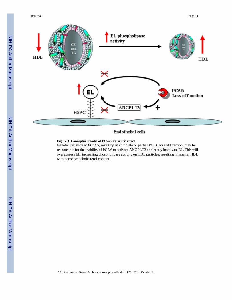
Figure 3. Conceptual model of PCSK5 variants’ effect.Genetic variation at PCSK5, resulting in complete or partial PC5/6 loss of function, may beresponsible for the inability of PC5/6 to activate ANGPLT3 or directly inactivate EL. This willoverexpress EL, increasing phospholipase activity on HDL particles, resulting in smaller HDLwith decreased cholesterol content.
Iatan et al. Page 14
Circ Cardiovasc Genet. Author manuscript; available in PMC 2010 October 1.
NIH
-PA Author Manuscript
NIH
-PA Author Manuscript
NIH
-PA Author Manuscript

NIH
-PA Author Manuscript
NIH
-PA Author Manuscript
NIH
-PA Author Manuscript
Iatan et al. Page 15
Table 1
PCSK5 Polymorphisms Identified by Sequencing
Functional Class Location Validated SNPs* Novel Variants
Exonic
Synonymous
Exon 1 rs7040769
Exon 1 rs7020560
Exon 12 rs2297342 _
Exon 18 rs10521468
5′UTR
rs12005073 c.125C>A
c.72C>T
c.385insGAGCTGCGGCGGCCCGGGGCTGC
3′UTR c.323G>A
Intronic
Int.4 IVS4-3016T>A
Int.7 rs2297344
Int.8 rs1416547
Int.11 rs3824474
Int.16 rs2270570
Int.17 rs1537183
Int.19 rs3830384 IVS19-71insTAAAA
Int.20 rs10869726 IVS20-50delTACTTTCAGGACTAAT
*Validated SNPs correspond to variants identifying by sequencing, but previously characterized.
UTR, untranslated region; Int, intron.
Circ Cardiovasc Genet. Author manuscript; available in PMC 2010 October 1.

NIH
-PA Author Manuscript
NIH
-PA Author Manuscript
NIH
-PA Author Manuscript
Iatan et al. Page 16
Table 2
Association Results of Stage 1 Analysis for the HDL-C Trait
SNP Effect (β) P MAF
rs11144782 −0.076 0.002 0.164
rs11144766 −0.063 0.005 0.197
rs1339246 0.056 0.018 0.176
rs1331384 0.037 0.038 0.485
rs11144688 −0.053 0.039 0.137
rs11144690 −0.093 0.040 0.040
rs1338746 −0.036 0.044 0.424
rs4745522 0.051 0.045 0.143
rs2050833 0.045 0.045 0.199
Quantitative association analysis for HDL-C in the French Canadians was performed using linear regression, after adjustments for age and sex, underan additive model. β is the linear regression coefficient corresponding to the effect size per copy of the minor allele. Bolded SNPs represent significantvariants (P≤0.01) selected for Stage 2 analysis. MAF, minor allele frequency.
Circ Cardiovasc Genet. Author manuscript; available in PMC 2010 October 1.

NIH
-PA Author Manuscript
NIH
-PA Author Manuscript
NIH
-PA Author Manuscript
Iatan et al. Page 17
Tabl
e 3
Dis
tribu
tion
of G
enot
ypic
Cla
sses
for r
s111
4478
2 an
d rs
1114
4766
in th
e St
age
1 Fr
ench
Can
adia
n Su
bjec
ts
rs11
1447
82rs
1114
4766
Gen
otyp
es (n
)C
C (n
=301
)G
C (n
=123
)G
G (n
=10)
GG
(n=2
75)
AG
(n=1
28)
AA
(n=1
9)
Freq
uenc
y0.
690.
280.
020.
650.
300.
04
Mea
n H
DL
-C*
(mm
ol/L
)0.
98±0
.29
0.89
±0.2
40.
8±0.
200.
97±0
.30
0.91
±0.2
30.
85±0
.18
Ana
lyse
s wer
e pe
rfor
med
usi
ng P
LIN
K v
1.04
with
age
and
sex
as c
ovar
iate
s, un
der a
n ad
ditiv
e m
odel
. HD
L-C
, hig
h-de
nsity
lipo
prot
ein
chol
este
rol.
* The
mea
n±SD
of H
DL-
C a
re st
ratif
ied
by g
enot
ype,
and
wei
ghte
d by
the
freq
uenc
ies o
f the
ir ge
noty
pic
clas
s.
Circ Cardiovasc Genet. Author manuscript; available in PMC 2010 October 1.

NIH
-PA Author Manuscript
NIH
-PA Author Manuscript
NIH
-PA Author Manuscript
Iatan et al. Page 18
Table 4
Lipid Traits Associated with rs11144782
SNP Trait Effect (β) P
HDL (mmol/L) −0.076 0.002
rs11144782 TG (mmol/L) 0.502 0.049
VLDL (mmol/L) 0.169 0.039
apoBtot (mg/dL) 10.720 0.022
Analyses were performed using PLINK v1.04 with age and sex as covariates, under an additive model. β is the linear regression coefficient,corresponding to the effect size per copy of the minor allele. HDL-C, high-density lipoprotein cholesterol; TG, triglycerides; VLDL, very low-densitylipoprotein; apoBtot, total apolipoprotein B.
Circ Cardiovasc Genet. Author manuscript; available in PMC 2010 October 1.

NIH
-PA Author Manuscript
NIH
-PA Author Manuscript
NIH
-PA Author Manuscript
Iatan et al. Page 19
Table 5
Independent Signals Found at the PCSK5 Locus for the HDL-C Trait
SNP Effect (β) P
rs11144782 −0.076 0.002
rs11144766 −0.072 0.001
rs2050833 0.047 0.036
rs4745488 −0.039 0.038
Conditional analyses were performed using stepwise linear regression adjusting for age, sex, rs11144782, rs11144766 and rs2050833, respectively.β is the linear regression coefficient, corresponding to the effect size per copy of the minor allele.
Circ Cardiovasc Genet. Author manuscript; available in PMC 2010 October 1.