Diurnal cortisol amplitude and fronto-limbic activity in response to stressful stimuli
Linear Age-Correlated Functional Development ofRight Inferior Fronto-Striato-Cerebellar NetworksDuring Response Inhibition and Anterior Cingulate
During Error-Related Processes
Katya Rubia,* Anna B. Smith, Eric Taylor, and Michael Brammer
Department of Child Psychiatry, Institute of Psychiatry, London SE5 8AF, United Kingdom
Abstract: Inhibitory and performance-monitoring functions have been shown to develop throughout ado-lescence. The developmental functional magnetic resonance imaging (fMRI) literature on inhibitory control,however, has been relatively inconsistent with respect to functional development of prefrontal cortex in theprogression from childhood to adulthood. Age-related performance differences between adults and chil-dren have been shown to be a confound and may explain inconsistencies in findings. The development oferror-related processes has not been studied so far using fMRI. The aim of this study was to investigate theneural substrates of the development of inhibitory control and error-related functions by use of an individu-ally adjusted task design that forced subjects to fail on 50% of trials, and therefore controlled for differencesin task difficulty and performance between different age groups. Event-related fMRI was used to comparebrain activation between 21 adults and 26 children/adolescents during successful motor inhibition andinhibition failure. Adults compared with children/adolescents showed increased brain activation in rightinferior prefrontal cortex during successful inhibition and in anterior cingulate during inhibition failure. Awhole-brain age-regression analysis between 10 and 42 years showed progressive age-related changes inactivation in these two brain regions, with additional changes in thalamus, striatum, and cerebellum. Age-correlated brain regions correlated with each other and with inhibitory performance, suggesting they formdeveloping fronto-striato-thalamic and fronto-cerebellar neural pathways for inhibitory control. This studyshows developmental specialization of the integrated function of right inferior prefrontal cortex, basalganglia, thalamus, and cerebellum for inhibitory control and of anterior cingulate gyrus for error-relatedprocesses. Hum Brain Mapp 28:1163–1177, 2007. VVC 2007Wiley-Liss, Inc.
Key words: development; cognitive neuroscience; cognitive control; executive functions; fMRI; motorinhibition
INTRODUCTION
Basic cognitive functions are established in childhood;however, more complex self-regulatory functions such asinhibitory control, interference inhibition, performancemonitoring, including conflict and error detection, andcognitive flexibility have been shown to develop through-out adolescence [Levin et al., 1991]. Rudimentary forms ofinhibitory control and error monitoring can be observed asearly as about the age of 4 [Jones et al., 2003], but the de-velopment of these self-regulatory and performance-moni-toring functions appears not to be fully mature until mid
Contract grant sponsor: Medical Research Council; Contract grantnumber: G9900839; Contract grant sponsor: The Wellcome Trust;Contract grant number: 053272/Z/98/Z/JRS/JP/JAT.
*Correspondence to: Dr. Katya Rubia, Department of Child Psychi-atry, Institute of Psychiatry, 16 De Crespigny Park, London SE58AF, United Kingdom. E-mail: [email protected]
Received for publication 20 June 2006; Revised 10 August 2006;Accepted 14 August 2006
DOI: 10.1002/hbm.20347Published online 30 May 2007 in Wiley InterScience (www.interscience.wiley.com).
VVC 2007 Wiley-Liss, Inc.
r Human Brain Mapping 28:1163–1177 (2007) r
to late adolescence. The development of selective motorresponse inhibition in the go/no-go task has thus shownto peak at about 12 years [Levin et al., 1991], whereaswithholding of an already triggered motor response in themore difficult stop task has been shown to peak between13 and 17 years [Williams et al., 1999]. Performance- anderror-monitoring functions appear to develop well intopuberty [Davies et al., 2004a,b]. The late development ofinhibitory control processes have been related to the rela-tively late anatomical maturation of the frontal lobes [Bjor-klund and Harnishfeger, 1990; Dempster, 1992], which hasbeen confirmed by functional imaging findings of prefron-tal mediation of inhibitory control [Durston et al., 2002a;Garavan et al., 1999; Menon et al., 2001; Rubia et al., 2000,2001, 2003].Important brain maturational changes continue well into
late adolescence and early adulthood such as synapticpruning and reorganization, programmed cell death, anddendritic and axonal arborization [Changeux and Danchin,1976; Huttenlocher and Dabholkar, 1997]. These matura-tional changes have been related to progressive morpho-logical changes in white to grey matter ratio in most brainareas from early childhood to late adolescence, presumablyreflecting myelination [Giedd et al., 1999; Gogtay et al.,2004; Paus et al., 1999; Sowell et al., 2004]. The myelinationprocesses appear to develop in a posterior-to-anterior-shift,with frontal brain regions myelinating last. Myelinationprocesses in some frontal brain regions peak as late as inmidadulthood [Gogtay et al., 2004; Sowell et al., 2004]. Thelate development of frontal lobe structures that mediate in-hibition has made inhibitory control an important target ofdevelopmental functional imaging studies.Functional imaging has the advantage of elucidating the
dynamic nature of cognitive development [Rubia, 2002].The existing functional imaging literature on the develop-ment of inhibitory control functions, however, has beenrelatively inconsistent with respect to the maturation ofprefrontal function. Some developmental fMRI studieshave reported increased frontal activation in adults com-pared with children during tasks of inhibitory controlfunctions [Bunge et al., 2002; Luna et al., 2001; Rubia et al.,2000, 2007; Tamm et al., 2002], supporting a maturationalhypothesis of brain function where brain regions thatmature latest are also brain regions that increase in func-tion with age. Some of the same studies also reported therecruitment of different prefrontal brain regions in chil-dren compared with adults, which has been interpreted asa reflection of alternative strategies for task management[Luna et al., 2001; Rubia et al., 2000; Tamm et al., 2002].Other developmental imaging studies on inhibitory con-trol, however, have found increased activation in frontaland parietal lobes in children and adolescents comparedwith adults, which has been interpreted as a reflection of amore diffuse and less specialized prefrontal function inchildren, related to the need for greater effort in childrento manage task performance [Booth et al., 2003; Caseyet al., 1997; Durston et al., 2002b].
The majority of developmental fMRI studies on inhibi-tory control were based on relatively small sample sizesand have used block design fMRI (with the exception ofthe studies by Bunge et al. [2002] and Durston et al.,[2002b, 2006]), which may partly explain the inconsisten-cies in findings. Performance differences in block andevent-related fMRI studies have been shown to be an im-portant confound in the comparison of functional imagingdata of different age groups and to contaminate real find-ings of age-related brain activation differences in geriatric[Grossman et al., 2002; Murphy and Garavan, 2004] anddevelopmental populations [Brown et al., 2005; Schlaggaret al., 2002]. Performance mismatch in developmental datahas been shown to lead to a false attribution of such differ-ences between age groups to effects of maturation per se[Brown et al., 2005; Schlaggar et al., 2002]. Differentapproaches have been used to disentangle performance-related effects from age-related effects in developmentalimaging data. One approach is to examine correlationsbetween neural activity, performance, and age or to covaryfor performance [Casey et al., 1997, 2002; Thomas et al.,2004]. Another is to compare subgroups from different agegroups that are matched in performance [Brown et al.,2005; Bunge et al., 2002; Schlaggar et al., 2002]. A thirdapproach is to incorporate different difficulty levels in thetask design, with the aim to post-hoc compare betweenmatched (easy trials) and nonmatched (difficult trials)performance [Durston et al., 2002b]. These approaches,however, do not address strategy differences that mayexist between children and adults and the fact that tasksmay be inherently more difficult and effortful for children,even when task performance is matched by external taskscores.The aim of this study was to apply an alternative, novel
approach to avoid the problem of differences in difficultylevels between different age groups by the use of a cogni-tive paradigm that is individually adjusted to control fordifferences in task difficulty and therefore performance lev-els between different age groups. We thus aimed to clarifythe relationship between the development of inhibitory con-trol and prefrontal brain function in a study design thatcontrolled for potential confounds of task difficulty and per-formance mismatch between children and adults. For thispurpose, we combined event-related fMRI with a cognitiveparadigm that adjusted for individual performance. ThefMRI adaptation of the individually adjusted tracking stoptask [Rubia et al., 2003] requires withdrawal of a motorresponse when a go-signal is shortly followed by a stop sig-nal. The time interval between go and stop signal is alteredaccording to each subjects’ performance, making sure thateach subject succeeds and fails to 50% of the stop trialsthroughout the task. Each subject is therefore working at theedge of his/her own inhibitory capacity, providing homoge-nous difficulty levels between subjects and groups [Rubiaet al., 2003].Another advantage of the tracking stop task is that it
also measures neural networks related to error-related
r Rubia et al. r
r 1164 r
functions such as error detection and performance moni-toring during the 50% trials of stop failure. Developmentalimaging studies using electroencephalography (EEG) haveshown that the error related negative potential (ERN), pre-sumably originating in anterior cingulate gyrus, maturesrelatively late during adolescence [Davies et al., 2004a;Ladouceur et al., 2004] reaching into mid adulthood[Davies et al., 2004b]. No fMRI studies, however, exist sofar on the development of error-related functions.In healthy adults, the visual tracking stop task has been
shown to elicit brain activation in right inferior prefrontalcortex during successful stop trials and in right mesial pre-frontal cortex, anterior cingulate gyrus, and parietal lobesduring stop failures [Rubia et al., 2003]. Based on the evi-dence for development of inhibitory and error detectionfunctions in midadolescence, for anatomical maturation ofthe prefrontal cortex until midadulthood and for postado-lescent functional maturation of inferior and mesial pre-frontal brain regions during self-control functions [Bungeet al., 2002; Rubia et al., 2000; Tamm et al., 2002], wehypothesized that when performance was matchedbetween groups, children, and adolescents compared withadults would show reduced brain activation in rightinferior prefrontal cortex during stop trials and in mesialprefrontal cortex and anterior cingulate gyrus during stopfailures. We furthermore hypothesized that there would beprogressive linear changes in these two brain regionsacross all subjects in the age range of 10–42 years.
METHOD
Subjects
Forty seven male right-handed children and adults par-ticipated, 21 adults and 26 children and adolescents. Theadults were in the age range of 20–42 years (mean age ¼28(5)) and the children and adolescents in the age range of10–17 years (mean age ¼ 15(2)). Exclusion criteria werehistory of substance abuse, head injury, mental retardation,or mental, clinical or neurological disorder.
fMRI Activation Task Design
Rapid, mixed trial, randomized presentation, and event-related fMRI design was used. Interstimulus-intervals (ISI)were randomly jittered between 1.6 and 2 s, and theappearance of target events was randomized to optimizestatistical efficiency (Burrock et al., 1998; Dale, 1999; Daleand Buckner, 1997].Subjects were practiced once on the stop task prior to
scanning to ensure correct understanding of the taskinstructions. The stop task was presented on a mirrorwithin the MRI scanner during the scan and response datawere recorded on a PC.
Tracking Stop Task
The tracking stop task requires withholding of a motorresponse to a go stimulus when it is followed unexpect-edly and unpredictably by a stop signal [Logan et al.,1997]. In our visual fMRI adaptation of this task [Rubiaet al., 2003], arrows (of 500 ms duration) pointing either tothe left or right side appeared on the middle of the screenwith a mean ISI of 1.8 s. Subjects were instructed torespond to the arrow direction by making a buttonresponse with their left or right thumb. On the unpredict-able, infrequent stop trials (20% of trials), the arrows point-ing left or right were followed (about 250 ms later) byarrows pointing upwards, and subjects had to inhibit theirmotor responses. The time interval of 250 ms between go-signal and stop signal onsets changes according to eachsubject’s performance and is calculated based on the sub-ject’s overall probability of inhibition on all previous trials,which is recalculated after each stop trial. If the overallprobability of inhibition on previous trials was over 50%,then the stop signal delay would increase in steps of 50ms, thus making it harder to inhibit. If the overall proba-bility of inhibition of all previous trials reaches below 50%after any given stop trial, the stop signal delay woulddecrease in steps of 50 ms, making it easier to inhibit. Thetracking algorithm makes sure the task is equally challeng-ing and difficult for each individual, providing 50% suc-cessful and 50% unsuccessful inhibition trials. Forty stoptrials were pseudorandomly interspersed with 156 go trials(78 left and 78 right pointing arrows) and at least threerepetition times apart for adequate separation of the hemo-dynamic response. Since the algorithm of the task designmakes sure that subjects fail to half of all stop events, suc-cessful and unsuccessful stop events control each otherperfectly for low frequency (i.e. 20 each).In the event-related fMRI analysis, brain activation to
the 50% successful stop trials is contrasted with that of the50% unsuccessful stop trials (i.e. successful stop trials–unsuccessful stop trials). Brain activation to unsuccessfulstop trials are thus subtracted from brain activation to thesuccessful stop trials to control for attentional effects of thelow frequency appearance of stop trials. Activation to go-response trials is subtracted from activation to unsuccess-ful stop trials to control for brain activation related tomotor response execution (i.e. unsuccessful stop–go trials)[for details of the task see Rubia et al., 2003, 2005b].
Analysis of Performance Data
Univariate ANOVAs with IQ as covariate was used tocompare groups in their task performance using the fol-lowing variables: mean reaction time (MRT) to go trials;stop signal reaction time (SSRT), calculated by subtractingthe mean stop signal delay (SSD: the average time betweengo and stop signal at which the subject managed to inhibitto 50% of trials) from the mean reaction time (MRT) to gotrials, i.e. MRT � SSD [Logan et al., 1997]; and probability
r Development of Inhibition and Error Detection in fMRI r
r 1165 r
of inhibition (PI) to stop trials. The SSRT is an indicator ofthe speed of the inhibitory process. P-values were adjustedfor multiple testing using the false discovery rate [FDR;Benjamini and Hochberg, 1995]. Pearson correlations wereperformed to examine linear correlations between age andperformance measures across all subjects; significance lev-els were adjusted using the FDR.
fMRI Image Acquisition
Gradient-echo echoplanar MR imaging (EPI) data wereacquired on a GE Signa 1.5T Horizon LX System (General;Electric, Milwaukee, WI) at the Maudsley Hospital, Lon-don. Consistent image quality was ensured by a semiauto-mated quality control procedure. A quadrature birdcagehead coil was used for RF transmission and reception. Ineach of 16 noncontiguous planes parallel to the anterior–posterior commissural, 196 T2*-weighted MR imagesdepicting BOLD (Blood Oxygen Level Dependent) contrastcovering the whole brain were acquired with TE ¼ 40 ms,TR ¼ 1.8 s, flip angle ¼ 908, in-plane resolution ¼ 3.1 mm,slice thickness ¼ 7 mm, and slice-skip ¼ 0.7 mm. This EPIdataset provided almost complete brain coverage.
fMRI Individual Analysis
The data were first realigned [Bullmore et al., 1999] tominimize motion related artifacts and smoothed using aGaussian filter (FWHM 7.2 mm). Time series analysis wasthen carried out by first convolving each experimental con-dition (i.e. stop–go trials; unsuccessful stop–go trials) with gvariate functions, modeling delays of 4 and 8 s, respectively(to allow variability within this range). Go trials were usedas implicit baseline trials and subtracted from the two mainexperimental conditions (successful and nonsuccessful stoptrials). The third contrast of interest (successful stop–unsuc-cessful stop) is a direct subtraction of the two primary con-trasts. The weighted sum of these two convolutions thatgave the best fit (least-squares) to the time series at eachvoxel was then computed and a goodness of fit statistic wascomputed at each voxel, consisting of the ratio of the sum ofsquares of deviations from the mean intensity value due tothe model (fitted time series) divided by the sum of squaresdue to the residuals (original time series minus model timeseries). This statistic is called the SSQ-ratio.Significance of the SSQ ratio volumes obtained was
tested using the wavelet-based time series permutationmethod described in detail by Bullmore et al. [2001].
fMRI Group Mapping
Statistical maps for each contrast of interest were trans-formed into standard space, as previously described byBrammer et al. [1997] and tested for significance using per-mutation methods at voxel and cluster levels [Brammeret al., 1997; Bullmore et al., 2001]. Zero false positive
activated clusters were expected at a P-value of <0.05 atvoxel and of <0.0025 at cluster-levels.A two-tailed t-test was performed to test for between-
group differences in 3D motion for the x, y, z rotation andx, y, z translation during task performance.
ANCOVA for Group Comparisons in Brain
Activation
Analysis of variance with IQ as covariate was carried outon the SSQ ratio maps in standard space by first computingthe difference in median SSR ratio between groups at eachvoxel. Subsequent inference of the probability of this differ-ence under the null hypothesis was made by reference to thenull distribution obtained by repeated random permutation(1,000 times) of group membership and recomputation of thedifference in median SSR ratios between the two groupsobtained from the resampling process. Cluster-level mapswere then obtained as described above [Bullmore et al.,1999]. For this particular group comparison, less than onefalse activated cluster was expected at a P-value of P < 0.05for voxel-comparison and P < 0.025 for cluster comparison.
Phase Analysis
For any given experimental contrast, information can beobtained not only about the size but also the direction ofthe activation from the general linear model fit to the timeseries of activation. The sign of BOLD response withrespect to baseline can be either positive (in ‘‘phase’’ withthe activation condition) or negative (in ‘‘phase’’ with thebaseline condition). This estimation can then be taken intoaccount to determine whether a group difference reflects adifference between positive or negative BOLD responses.This analysis was performed at a group level, and wasbased on the positive or negative average SSQ ratios foreach group in any of the between-group activation clus-ters. For all between-group activation differences for allcontrasts, only BOLD responses were considered in bothgroups that were positive-based on the average SSQ ratioof each group relative to the activation condition of inter-est. This also applies to the correlation analyses.
Whole-Brain Correlation Analysis Between Age
and Brain Activation
A regression analysis was conducted between brain activa-tion and age with IQ as covariate across all subjects, inde-pendent of their group status. For this purpose, the Pearsonproduct-moment correlation coefficient was first computed ateach intracerebral voxel in standard [Talairach and Tour-noux, 1988] space between the age data and the BOLDresponse (% change in signal) over all subjects. Random per-mutation testing (of the age data, 50 times) was used to assessthe probability of any particular correlation coefficient underthe null hypothesis. Statistical analysis was extended to clus-ter level as described by Bullmore et al. [1999]. A P-value of
r Rubia et al. r
r 1166 r
P < 0.05 at voxel and P < 0.025 at cluster-levels was chosen,resulting in less than one error cluster.To investigate whether age-correlated brain activation
clusters during successful inhibition were correlated withinhibitory performance, the SSQ ratios were extracted foreach subject for every voxel in each of the age-correlatedactivation clusters, and the mean values of these SSQratios for all voxels in the chosen clusters calculated foreach subject. Pearson correlations were then performedbetween SSRT/PI and the mean SSQ ratios for each subjectfor all age-correlated clusters.
RESULTS
Task Performance
There were group differences on the intelligence scoreson the Raven’s Standard Progressive Matrices Intelligence
Questionnaire (IQ) [Raven, 1960] (adults: mean convertednonverbal IQ: 114 (11); children/adolescents mean IQ: 106(13), t ¼ 2.7, df ¼ 45, P < 0.034). Consequently, all analy-ses were performed with IQ as covariate.Univariate ANCOVAs showed no group differences in
performance. Probability of inhibition to stop trials wasclose to 50% in all subjects, suggesting that the trackingstop mechanism was successful (PI (standard deviation(SD)) adults: 54% (7%); children/adolescents: 50% (4%),F ¼ 1.8, df ¼ 2, P ¼ 0.07). No significant group differenceswere observed in SSRT (SD): adults 225 (277); children: 273(254), F ¼ 0.3, df ¼ 2, n.s. No significant differences wereobserved for MRT to go trials (SD): adults: 863 (167); Chil-dren 763 (188), F ¼ 0.2, df ¼ 2, P < 0.08).Across all subjects, no significant linear correlations
were observed between age and performance measures(PI: r ¼ 0.3, P ¼ n.s.; SSRT:r ¼ �0.1, P ¼ n.s.; MRT go: r ¼�0.1, P ¼ n.s.).
TABLE I. Group brain activation foci for adults and children/adolescents on
the stop task and brain activation differences
Brain regions of activation (BA) Tal. coord N voxels
Successful stop–unsuccessful stop trialsAdults R inferior frontal gyrus (BA 45/47) 41, 26, 11 113
L inferior frontal gyrus (BA 45) �44, 29, 6 31R medial frontal gyrus (BA 9) 9, 40, 22 21L medial temporal gyrus (BA 37) �45, �59, �7 27R cerebellum/occipital gyrus (BA 19) 33, �73, �11 75L cerebellum �22, �40, �19 29L posterior cingulate (BA 30) �10, �53, 9 43
Children/adolescents R inferior frontal gyrus (BA 47/45) 46, 15, �4 48R medial frontal gyrus (BA 10/46) 29, 54, 8 48L medial frontal gyrus (BA 9) �22, 13, 40 29L medial prefrontal gyrus (BA 9) �40, 1, 41 24R premotor cortex (BA 6) 37, �9, 40 22L insula/superior temporal gyrus (BA 22) �44, 4, 4 81R superior temporal gyrus (BA 21/38) 37, 5, �28 15R precuneus (BA 7) 4, �56, 46 21R cerebellum 18, �79, �22 28
Adults > children/adolescents R inferior prefrontal gyrus (BA 45) 43, 33, 9 50
Unsuccessful stop–go trialsAdults Mesial frontal/anterior cingulate (BA 10/32/24) 1, 52, 17 217
L inferior frontal gyrus (BA 45) �49, 29, 5 17R superior/middle temporal gyrus (BA 22/38) 48, �9, 3 173L superior temporal gyrus (BA 22) �49, �29, 29 111L inferior parietal lobe (BA 39) �57, �28, 20 10R posterior cingulate gyrus (BA 31) 2, �48, 27 24R middle cingulate gyrus (BA 23) 2, �18, 29 24
Children/adolescents Mesial frontal/anterior cingulate (BA 9/32) �5, 44, 31 293L middle temporal gyrus (BA 21) �54, �26, �3 125L middle temporal gyrus (BA 39) �40, �60, 21 49R middle temporal gyrus (BA 21) 47, �18, �1 122R inf. parietal gyrus (BA 39) 42, �59, 24 30Posterior cingulate/precuneus (BA 23/30/31) 2, �54, 20 134
Adults > children/adolescents Anterior cingulate gyrus (BA 24/32) 0, 30, 26 31
Group Brain Activation maps were conducted at P < 0.05 at voxel and P < 0.0025 at cluster-levels. ANOVAs were conducted at P <
0.05 at voxel and P < 0.01 at cluster level for the comparison between adults and children/adolescents.Tal. coord., Talairach coordinates, shown for the peak of the 3D activation cluster; BA, Brodman area; N voxels, number of voxels.Only clusters larger than 10 voxels are reported.
r Development of Inhibition and Error Detection in fMRI r
r 1167 r
Within-Group Brain Activation
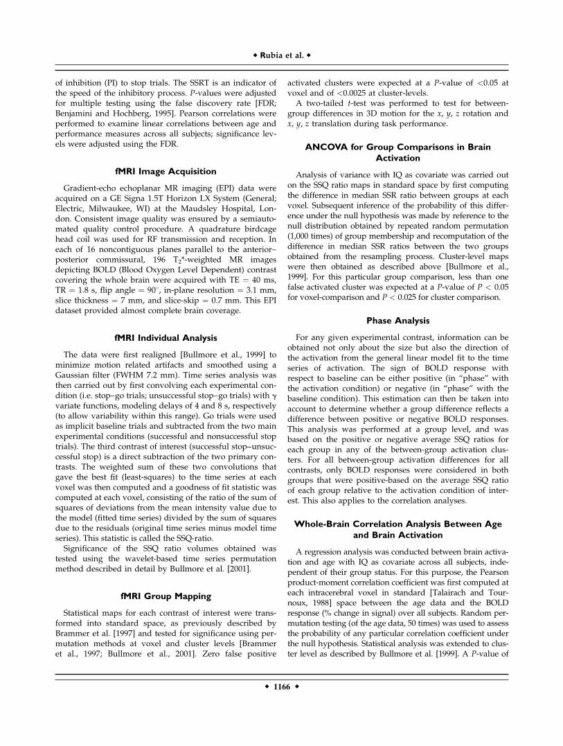
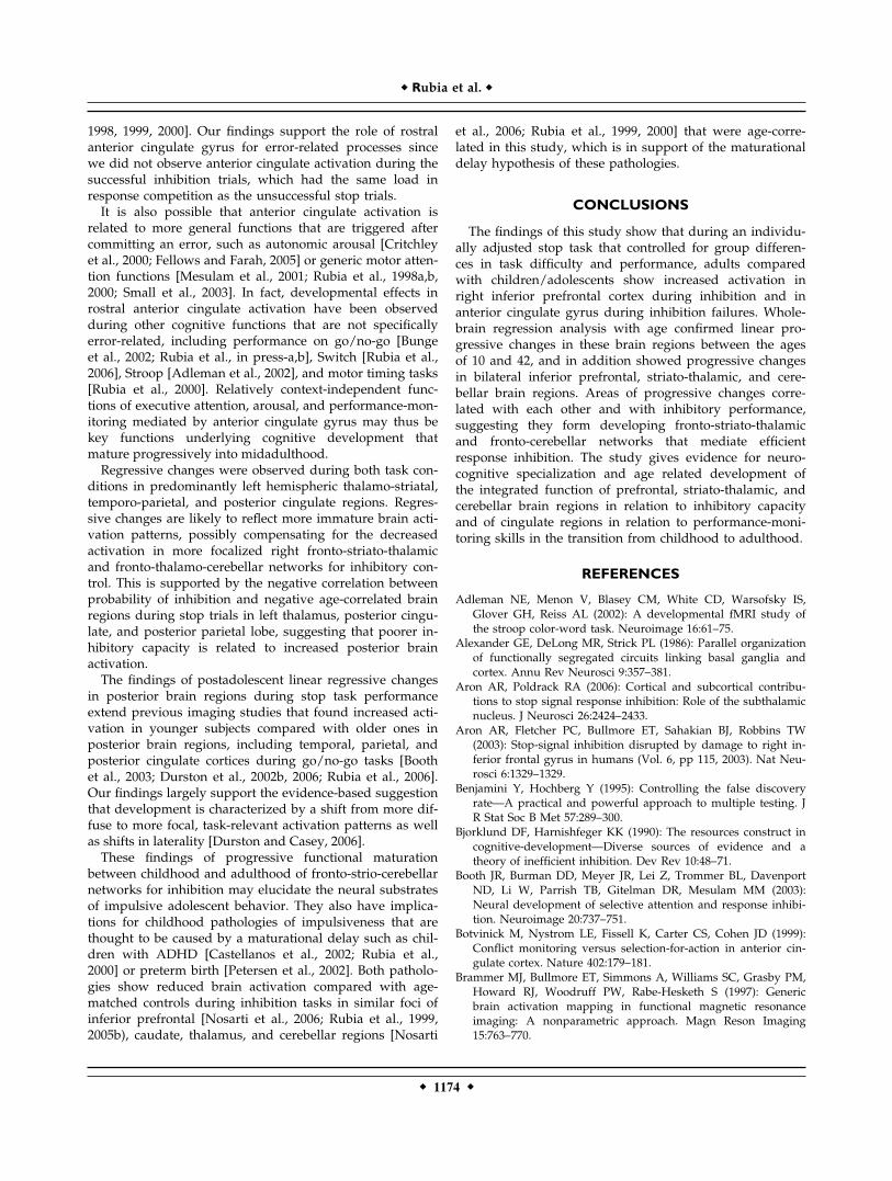
During successful stop trials (contrasted with unsuccess-ful stop trials), adults and children/adolescents showedpredominantly right but also left inferior prefrontal andbilateral medial prefrontal activation (see Table I, Fig. 1a).In adults, activation in right (r ¼ �0.4, P < 0.05) and leftinferior prefrontal cortex (r ¼ �0.4, P < 0.59) correlatednegatively with SSRT (suggesting that subjects with fasterinhibitory speed showed greater activation in this area). Inadolescents, no activation correlated with SSRT; probability
of inhibition was correlated with activation in superiortemporal gyrus (r ¼ 0.6, P < 0.007).During unsuccessful stop trials (contrasted with go-tri-
als), adults and children/adolescents showed similar acti-vation foci in mainly mesial prefrontal cortex, anterior,and posterior cingulate gyrus (see Table I, Fig. 1b).
Group Differences in Motion
All subjects were within acceptable limits of headmotion. No significant group differences were observed in
Figure 1.
Clusters of brain regions of within-group activation are shown in
horizontal slices in adults and in children/adolescents for (a): the
contrast of successful stop trials–unsuccessful stop trials and (b):
the contrast of unsuccessful stop trials–go trials. Group activa-
tion maps are thresholded at P < 0.05 at voxel and at P <
0.0025 at cluster levels. For the horizontal sections, the z-coor-
dinate is indicated in distance (mm) from the anterior-posterior-
commissure. The left side of the brain corresponds to the left
side of the image. [Color figure can be viewed in the online
issue, which is available at www.interscience.wiley.com.]
r Rubia et al. r
r 1168 r
the extent of 3D motion for the x, y, z rotation and x, y, ztranslation during task performance.
Between-Group Differences in Activation
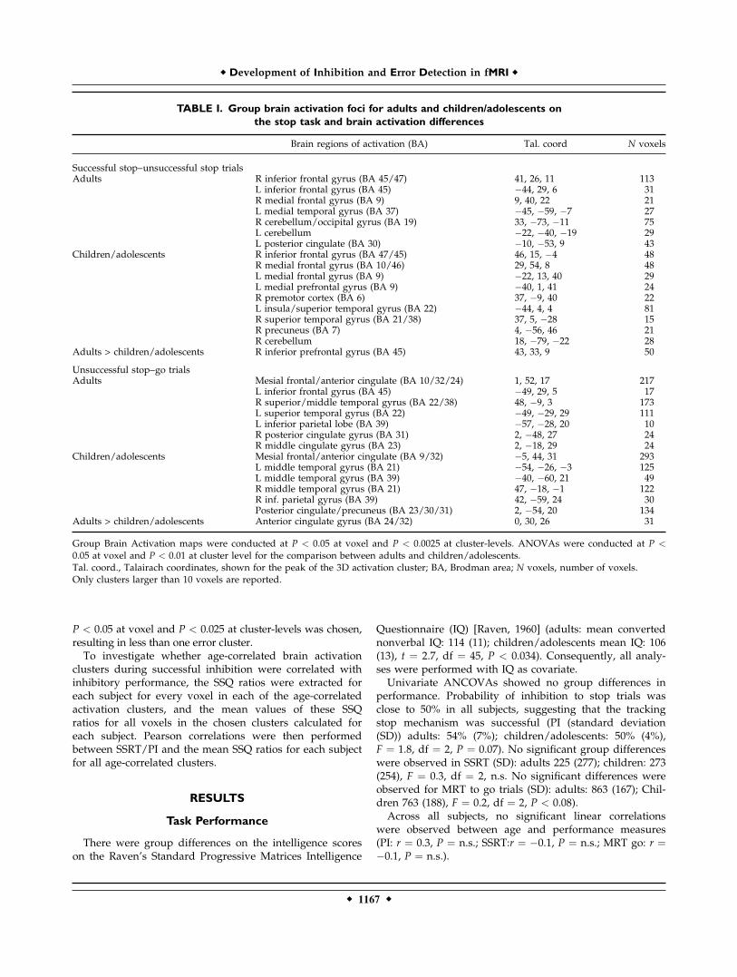
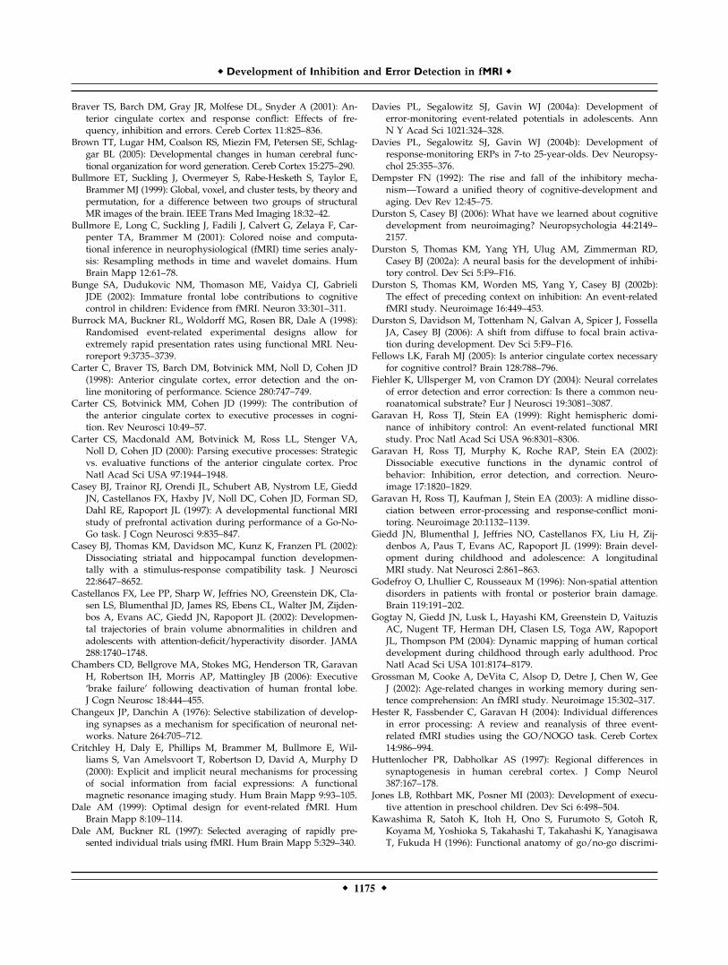
ANCOVA (with IQ as covariate) for between-group dif-ferences in brain activation for the successful stop trialscontrasted with unsuccessful stop trials showed that adultscompared with children/adolescents showed a significantincrease of activation in right inferior prefrontal cortex,reaching from orbitofrontal to inferior prefrontal gyrus,bordering premotor cortex (see Table I, Fig. 2a). No brainregions were significantly more activated in children/ado-lescents compared to adults.ANCOVA for between-group differences in brain activa-
tion for the contrast of unsuccessful stop trials with go tri-als showed that there was increased brain activation inadults compared with children/adolescents in pregenual,rostral anterior cingulate gyrus (see Table I, Fig. 2b). Nobrain regions were significantly more activated in chil-dren/adolescents compared with adults.
Whole-Brain Correlation Analysis Between
Age and Brain Activation
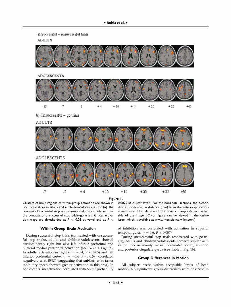
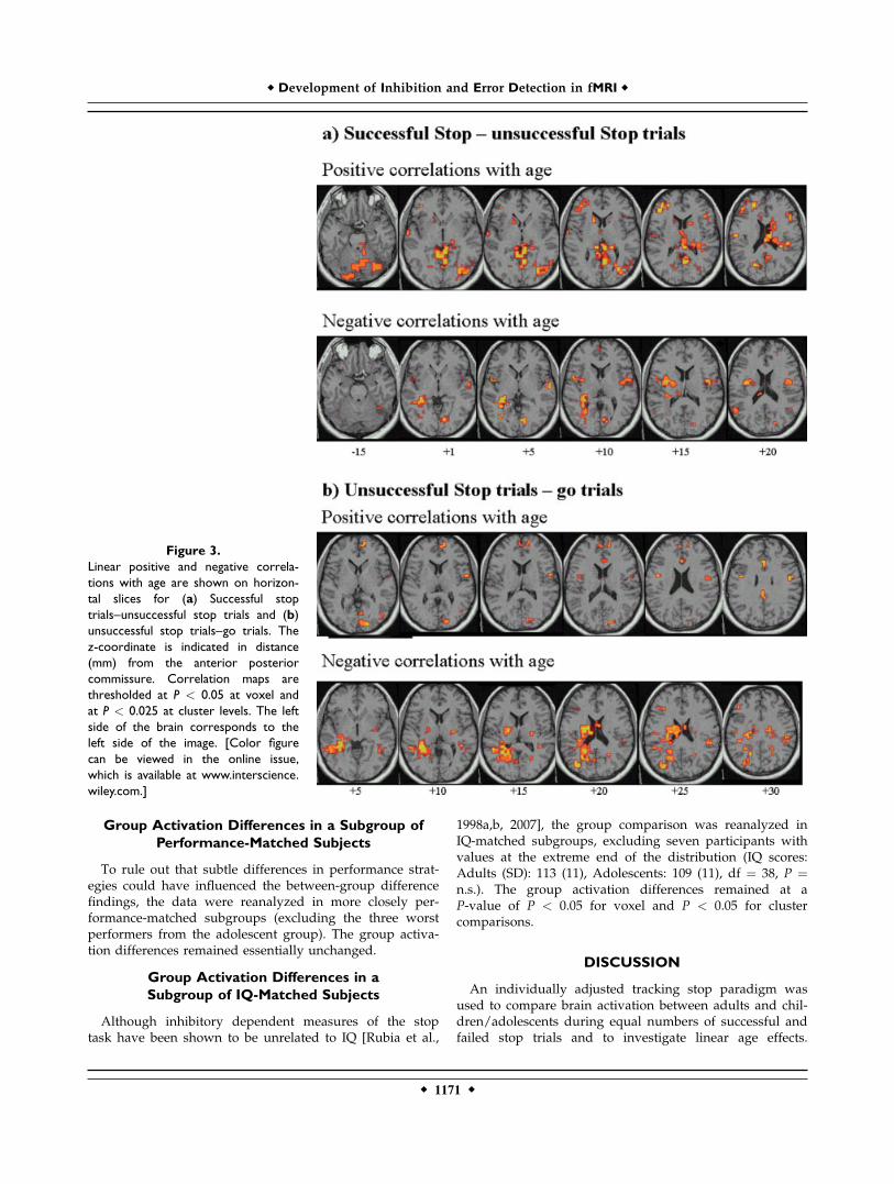
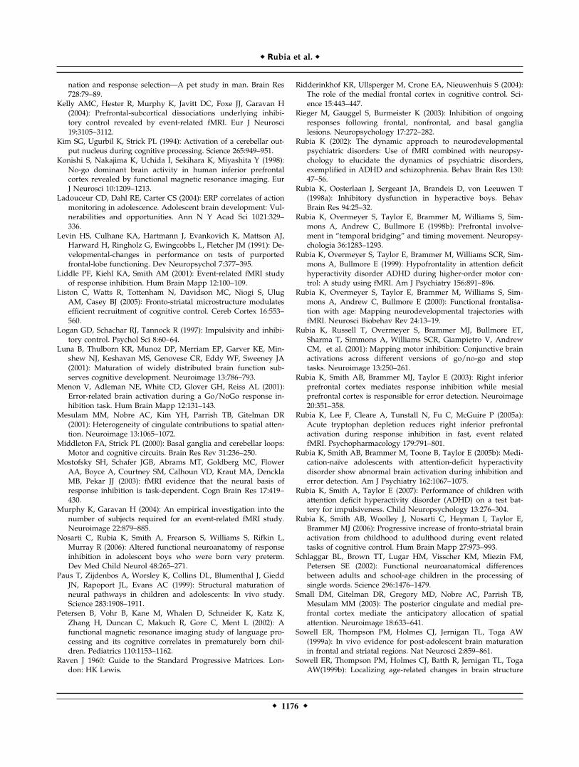
For the successful stop trials compared with unsuccess-ful stop trials, the whole-brain linear regression analysiswith age of all subjects and brain regions showed apositive linear correlation between age and activation inbilateral inferior prefrontal cortex in a large cluster in pre-dominantly right thalamus, insula, caudate, and in the cer-ebellum. SSRT correlated negatively with activation inbilateral thalamus and caudate and the vermis of the cere-bellum. Probability of inhibition correlated positively withbilateral inferior prefrontal gyri, caudate, thalamus, andcerebellum (see Table II, Fig. 3a).Negative correlations with age were observed in bilateral
insula and in predominantly left thalamus, putamen, andposterior cingulate gyrus. SSRT did not correlate with any ofthe activation clusters, but probability of inhibition corre-lated negatively with activation in posterior cingulate gyrus,left parietal lobe, and left thalamus (see Table II, Fig. 3a).For the unsuccessful stop trials compared with go trials,
positive correlations with age were observed mostly infrontal pole and anterior and posterior cingulate. Negativecorrelations with age were observed in predominantly leftthalamus, basal ganglia, and posterior cingulate and parie-tal cortices (see Table II, Fig. 3b).
Correlation Between Areas of Linear
Progressive Changes During Stop Trials
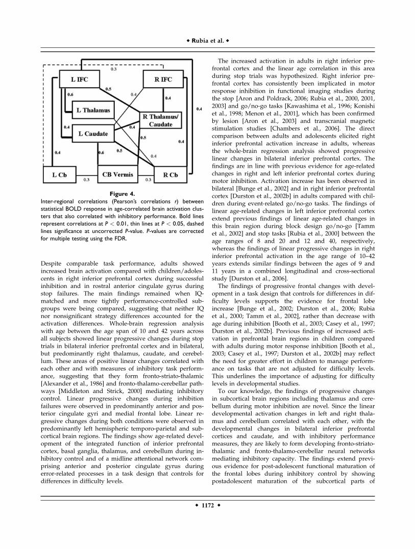
If brain areas of positive linear changes in right and leftinferior prefrontal cortices, thalamus, caudate, and cerebel-lum constitute fronto-strio-thalamic and fronto-cerebellarpathways, they should be intercorrelated. To test for this
hypothesis, the mean values of the SSQ ratios for all voxelsin the age-correlated clusters were calculated for each sub-ject, and Pearson correlations (corrected for multiple test-
Figure 2.
Brain regions of increased activation in adults compared to chil-
dren/adolescents (P < 0.01) during (a): successful stop trials
contrasted with unsuccessful stop trials. Shown is increased acti-
vation in right inferior prefrontal cortex (BA 47/45/44) in adults
compared with children/adolescents in 3D and in the horizontal
sections. (b) Unsuccessful stop trials contrasted with go-trials.
Shown is increased activation in adults compared with children/
adolescents in rostral anterior cingulate gyrus (BA 24) in 3D and
in the horizontal sections. For the horizontal sections, the z-
coordinate is indicated in distance (mm) from the anterior-pos-
terior-commissure. The left side of the brain corresponds to the
left side of the image. [Color figure can be viewed in the online
issue, which is available at www.interscience.wiley.com.]
r Development of Inhibition and Error Detection in fMRI r
r 1169 r
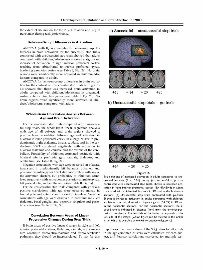
ing using the FDR) were then performed between thesemean SSQ ratios for each subject for each cluster ofinterest. Figure 4 shows the inter-regional intercorrelationfindings between bilateral age-correlated activation areasof inferior prefrontal cortex, caudate, thalamus, and cere-
bellum. Both right and left inferior prefrontal cortices cor-related with bilateral caudate, thalamus, and lateral cere-bellum. Right and left caudate and thalamus correlatedwith each other and with the vermis of the cerebellum (seeFig. 4).
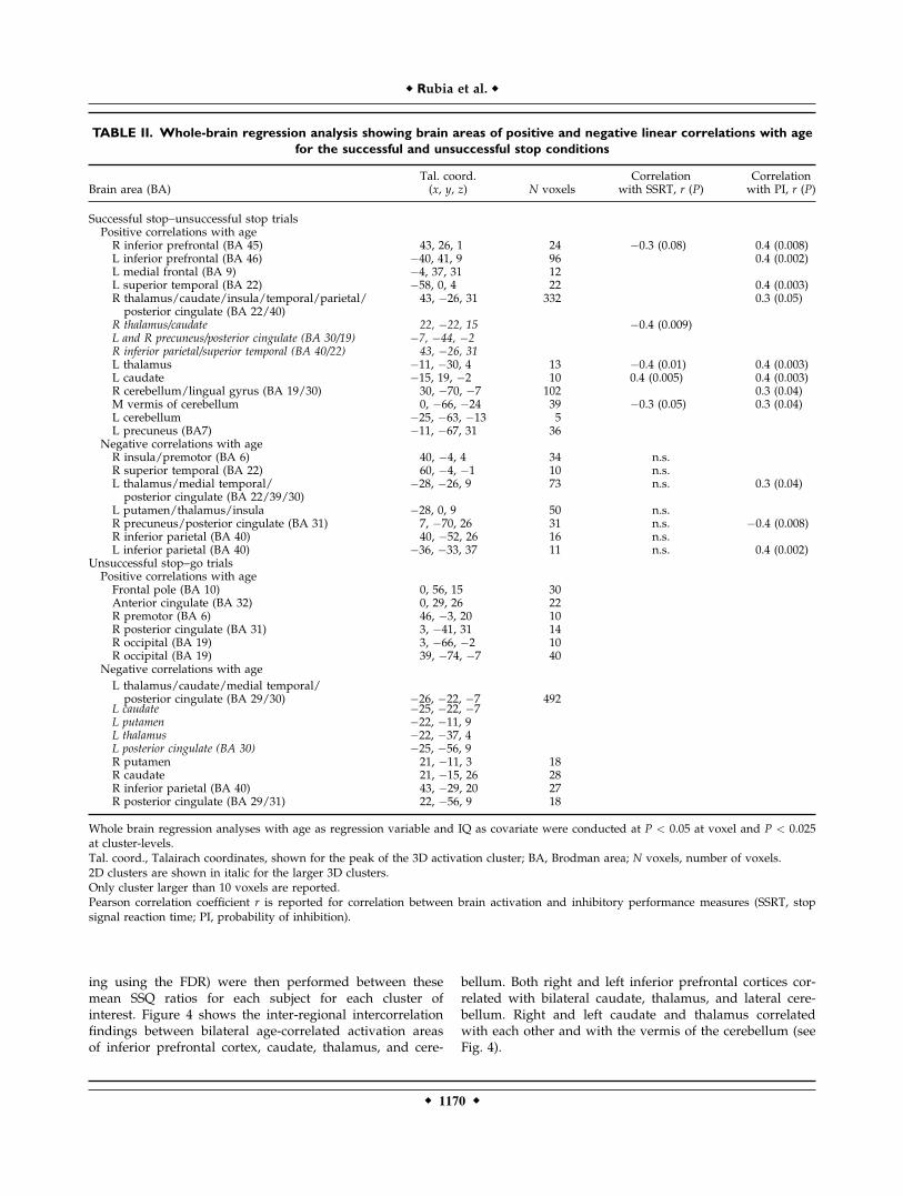
TABLE II. Whole-brain regression analysis showing brain areas of positive and negative linear correlations with age
for the successful and unsuccessful stop conditions
Brain area (BA)Tal. coord.(x, y, z) N voxels
Correlationwith SSRT, r (P)
Correlationwith PI, r (P)
Successful stop–unsuccessful stop trialsPositive correlations with ageR inferior prefrontal (BA 45) 43, 26, 1 24 �0.3 (0.08) 0.4 (0.008)L inferior prefrontal (BA 46) �40, 41, 9 96 0.4 (0.002)L medial frontal (BA 9) �4, 37, 31 12L superior temporal (BA 22) �58, 0, 4 22 0.4 (0.003)R thalamus/caudate/insula/temporal/parietal/posterior cingulate (BA 22/40)
43, �26, 31 332 0.3 (0.05)
R thalamus/caudate 22, �22, 15 �0.4 (0.009)L and R precuneus/posterior cingulate (BA 30/19) �7, �44, �2R inferior parietal/superior temporal (BA 40/22) 43, �26, 31L thalamus �11, �30, 4 13 �0.4 (0.01) 0.4 (0.003)L caudate �15, 19, �2 10 0.4 (0.005) 0.4 (0.003)R cerebellum/lingual gyrus (BA 19/30) 30, �70, �7 102 0.3 (0.04)M vermis of cerebellum 0, �66, �24 39 �0.3 (0.05) 0.3 (0.04)L cerebellum �25, �63, �13 5L precuneus (BA7) �11, �67, 31 36
Negative correlations with ageR insula/premotor (BA 6) 40, �4, 4 34 n.s.R superior temporal (BA 22) 60, �4, �1 10 n.s.L thalamus/medial temporal/posterior cingulate (BA 22/39/30)
�28, �26, 9 73 n.s. 0.3 (0.04)
L putamen/thalamus/insula �28, 0, 9 50 n.s.R precuneus/posterior cingulate (BA 31) 7, �70, 26 31 n.s. �0.4 (0.008)R inferior parietal (BA 40) 40, �52, 26 16 n.s.L inferior parietal (BA 40) �36, �33, 37 11 n.s. 0.4 (0.002)
Unsuccessful stop–go trialsPositive correlations with ageFrontal pole (BA 10) 0, 56, 15 30Anterior cingulate (BA 32) 0, 29, 26 22R premotor (BA 6) 46, �3, 20 10R posterior cingulate (BA 31) 3, �41, 31 14R occipital (BA 19) 3, �66, �2 10R occipital (BA 19) 39, �74, �7 40
Negative correlations with age
L thalamus/caudate/medial temporal/posterior cingulate (BA 29/30) �26, �22, �7 492
L caudate �25, �22, �7L putamen �22, �11, 9L thalamus �22, �37, 4L posterior cingulate (BA 30) �25, �56, 9R putamen 21, �11, 3 18R caudate 21, �15, 26 28R inferior parietal (BA 40) 43, �29, 20 27R posterior cingulate (BA 29/31) 22, �56, 9 18
Whole brain regression analyses with age as regression variable and IQ as covariate were conducted at P < 0.05 at voxel and P < 0.025at cluster-levels.Tal. coord., Talairach coordinates, shown for the peak of the 3D activation cluster; BA, Brodman area; N voxels, number of voxels.2D clusters are shown in italic for the larger 3D clusters.Only cluster larger than 10 voxels are reported.Pearson correlation coefficient r is reported for correlation between brain activation and inhibitory performance measures (SSRT, stopsignal reaction time; PI, probability of inhibition).
r Rubia et al. r
r 1170 r
Group Activation Differences in a Subgroup of
Performance-Matched Subjects
To rule out that subtle differences in performance strat-egies could have influenced the between-group differencefindings, the data were reanalyzed in more closely per-formance-matched subgroups (excluding the three worstperformers from the adolescent group). The group activa-tion differences remained essentially unchanged.
Group Activation Differences in a
Subgroup of IQ-Matched Subjects
Although inhibitory dependent measures of the stoptask have been shown to be unrelated to IQ [Rubia et al.,
1998a,b, 2007], the group comparison was reanalyzed inIQ-matched subgroups, excluding seven participants withvalues at the extreme end of the distribution (IQ scores:Adults (SD): 113 (11), Adolescents: 109 (11), df ¼ 38, P ¼n.s.). The group activation differences remained at aP-value of P < 0.05 for voxel and P < 0.05 for clustercomparisons.
DISCUSSION
An individually adjusted tracking stop paradigm wasused to compare brain activation between adults and chil-dren/adolescents during equal numbers of successful andfailed stop trials and to investigate linear age effects.
Figure 3.
Linear positive and negative correla-
tions with age are shown on horizon-
tal slices for (a) Successful stop
trials–unsuccessful stop trials and (b)
unsuccessful stop trials–go trials. The
z-coordinate is indicated in distance
(mm) from the anterior posterior
commissure. Correlation maps are
thresholded at P < 0.05 at voxel and
at P < 0.025 at cluster levels. The left
side of the brain corresponds to the
left side of the image. [Color figure
can be viewed in the online issue,
which is available at www.interscience.
wiley.com.]
r Development of Inhibition and Error Detection in fMRI r
r 1171 r
Despite comparable task performance, adults showedincreased brain activation compared with children/adoles-cents in right inferior prefrontal cortex during successfulinhibition and in rostral anterior cingulate gyrus duringstop failures. The main findings remained when IQ-matched and more tightly performance-controlled sub-groups were being compared, suggesting that neither IQnor nonsignificant strategy differences accounted for theactivation differences. Whole-brain regression analysiswith age between the age span of 10 and 42 years acrossall subjects showed linear progressive changes during stoptrials in bilateral inferior prefrontal cortex and in bilateral,but predominantly right thalamus, caudate, and cerebel-lum. These areas of positive linear changes correlated witheach other and with measures of inhibitory task perform-ance, suggesting that they form fronto-striato-thalamic[Alexander et al., 1986] and fronto-thalamo-cerebellar path-ways [Middleton and Strick, 2000] mediating inhibitorycontrol. Linear progressive changes during inhibitionfailures were observed in predominantly anterior and pos-terior cingulate gyri and medial frontal lobe. Linear re-gressive changes during both conditions were observed inpredominantly left hemispheric temporo-parietal and sub-cortical brain regions. The findings show age-related devel-opment of the integrated function of inferior prefrontalcortex, basal ganglia, thalamus, and cerebellum during in-hibitory control and of a midline attentional network com-prising anterior and posterior cingulate gyrus duringerror-related processes in a task design that controls fordifferences in difficulty levels.
The increased activation in adults in right inferior pre-frontal cortex and the linear age correlation in this areaduring stop trials was hypothesized. Right inferior pre-frontal cortex has consistently been implicated in motorresponse inhibition in functional imaging studies duringthe stop [Aron and Poldrack, 2006; Rubia et al., 2000, 2001,2003] and go/no-go tasks [Kawashima et al., 1996; Konishiet al., 1998; Menon et al., 2001], which has been confirmedby lesion [Aron et al., 2003] and transcranial magneticstimulation studies [Chambers et al., 2006]. The directcomparison between adults and adolescents elicited rightinferior prefrontal activation increase in adults, whereasthe whole-brain regression analysis showed progressivelinear changes in bilateral inferior prefrontal cortex. Thefindings are in line with previous evidence for age-relatedchanges in right and left inferior prefrontal cortex duringmotor inhibition. Activation increase has been observed inbilateral [Bunge et al., 2002] and in right inferior prefrontalcortex [Durston et al., 2002b] in adults compared with chil-dren during event-related go/no-go tasks. The findings oflinear age-related changes in left inferior prefrontal cortexextend previous findings of linear age-related changes inthis brain region during block design go/no-go [Tammet al., 2002] and stop tasks [Rubia et al., 2000] between theage ranges of 8 and 20 and 12 and 40, respectively,whereas the findings of linear progressive changes in rightinferior prefrontal activation in the age range of 10–42years extends similar findings between the ages of 9 and11 years in a combined longitudinal and cross-sectionalstudy [Durston et al., 2006].The findings of progressive frontal changes with devel-
opment in a task design that controls for differences in dif-ficulty levels supports the evidence for frontal lobeincrease [Bunge et al., 2002; Durston et al., 2006; Rubiaet al., 2000; Tamm et al., 2002], rather than decrease withage during inhibition [Booth et al., 2003; Casey et al., 1997;Durston et al., 2002b]. Previous findings of increased acti-vation in prefrontal brain regions in children comparedwith adults during motor response inhibition [Booth et al.,2003; Casey et al., 1997; Durston et al., 2002b] may reflectthe need for greater effort in children to manage perform-ance on tasks that are not adjusted for difficulty levels.This underlines the importance of adjusting for difficultylevels in developmental studies.To our knowledge, the findings of progressive changes
in subcortical brain regions including thalamus and cere-bellum during motor inhibition are novel. Since the lineardevelopmental activation changes in left and right thala-mus and cerebellum correlated with each other, with thedevelopmental changes in bilateral inferior prefrontalcortices and caudate, and with inhibitory performancemeasures, they are likely to form developing fronto-striato-thalamic and fronto-thalamo-cerebellar neural networksmediating inhibitory capacity. The findings extend previ-ous evidence for post-adolescent functional maturation ofthe frontal lobes during inhibitory control by showingpostadolescent maturation of the subcortical parts of
Figure 4.
Inter-regional correlations (Pearson’s correlations r) between
statistical BOLD response in age-correlated brain activation clus-
ters that also correlated with inhibitory performance. Bold lines
represent correlations at P < 0.01, thin lines at P < 0.05, dashed
lines significance at uncorrected P-value. P-values are corrected
for multiple testing using the FDR.
r Rubia et al. r
r 1172 r
fronto-stiato-thalamic and fronto-thalamo-cerebellar path-ways of motor inhibition.Inhibitory motor response in go/no-go and stop tasks
has been suggested to be mediated by fronto-striato-tha-lamic pathways, based on evidence from lesion [Godefroyet al., 1996; Rieger et al., 2003] and imaging studies impli-cating the basal ganglia [Kelly et al., 2004; Rubia et al.,1999, 2000, 2006] and the thalamus [Garavan et al.,2002; Liddle et al., 2001] in motor inhibition tasks. In stud-ies using the stop task in healthy adults, the basal ganglia[Vink et al., 2005] and the thalamus [Aron and Poldrack,2006] have been found to correlate with SSRT and to bemore strongly activated in better performers. Althoughthere is thus evidence from adult studies for the involve-ment of frontal, striatal, and thalamic brain regions for in-hibitory control, the evidence for functional developmentof the striatal and thalamic parts of fronto-striato-thalamicnetworks for inhibitory control, however, has been incon-clusive so far. Two previous studies have shown activationincrease in left and right caudate, respectively, in adultscompared with children during a go/no-go task [Durstonet al., 2002b; Rubia et al., in press-a,b]. Other studies, how-ever, found left caudate to be decreased in adults com-pared with adolescents during go/no-go [Booth et al.,2003] and stop tasks [Rubia et al., 2000], or not to differbetween children and adults during a go/no-go task[Bunge et al., 2002]. The use of relatively small subjectnumbers in previous developmental imaging studies of in-hibition may be responsible for these relatively inconsis-tent findings of caudate maturation, which may only beobservable with larger detective power in fMRI. In thisstudy, we observed progressive changes in predominantlyright basal ganglia and thalamus with regressive changesin predominantly left hemispheric homologue areas. Shiftsin laterality of basal ganglia activation from left to rightwith increasing age would reconcile apparent inconsisten-cies in previous findings. Although no previous studieshave reported progressive changes in thalamus, the regres-sive changes in left thalamus are in line with findings ofBooth et al. [2003] of increased activation in left thalamusin children. The progressive age-related shift in right-later-alization of basal ganglia and thalamus is also in line withevidence for a prominent role of right hemispheric fronto-striato-thalamic networks for inhibitory control in adults[Aron and Poldrack, 2006; Aron et al., 2003; Durston et al.,2002a; Rieger et al., 2003; Rubia et al., 2001, 2003, 2006].Shifts in laterality have previously been observed in devel-opmental imaging studies of cognitive control in frontalbrain regions [Bunge et al., 2002; Rubia et al., 2000] andmay reflect progressive focalization of lateralized neuralnetworks for task performance.Developmental functional changes in fronto-striato-tha-
lamic neural networks parallel structural developmentalchanges in striatal and thalamic brain regions alongsideprefrontal lobe changes between childhood and midadult-hood [Castellanos et al., 2002; Giedd et al., 1999; Sowellet al., 1999b; Sowell et al., 1999a]. They are also in line
with recent evidence for progressive structural maturationof fronto-striatal fiber tracts between the age range of 7–31years, which correlated with inhibitory performance on ago/no-go task [Liston et al., 2005].The cerebellum has rarely been measured in older fMRI
studies because of technical limitations of whole braincoverage. In more recent imaging studies, however, thecerebellum has been shown to be activated in adults dur-ing inhibitory control in the go/no-go task [Bunge et al.,2002; Garavan et al., 2003; Liddle et al., 2001; Mostofskiet al., 2003; Rubia et al., 2006]. In this study, the lateralcerebellum was activated in both children and adults dur-ing stop trials. The lateral cerebellum projects via the thal-amus to motor, but also prefrontal cortical areas [Kimet al., 1994; Middleton and Strick, 2000] and is likely toassist in both the preparation and inhibition of movement[Luna et al., 2001; Mostofski et al., 2003]. Related to ourfindings of progressive changes in cerebellum duringmotor response inhibition are findings of linear changes inleft cerebellum between the ages of 8 and 30 during reflexinhibition in the antisaccade task [Luna et al., 2001]. Thefindings of age-correlated progressive recruitment of thelateral cerebellum and vermis concomitantly and in corre-lation with right inferior prefrontal cortex and thalamussuggest a developmental functional enhancement, possiblyreflecting fine-tuning of the projections between frontalcortex, thalamus, basal ganglia, and cerebellum that medi-ate inhibitory control.The finding of increased brain activation in adults in an-
terior cingulate gyrus and of progressive age-relatedchanges in this area and medial frontal cortex during inhi-bition failure is also in line with the apriori hypothesis. Asfar as we are aware, this is the first developmental fMRIstudy of error-related brain activation. Subjects receivedimplicit feedback when they made an error to stop trialsby seeing the stop signal appear after they pressed theresponse button. Although enhanced in adults, anteriorcingulate and medial frontal activation was observed inboth age groups, in line with the postulated role of thisbrain region in error detection during no-go [Garavanet al., 2002; Hester et al., 2004; Liddle et al., 2001; Menonet al., 2001] and stop trials [Rubia et al., 2003]. These firstdevelopmental fMRI findings of a positive age-correlationin anterior cingulate are in line with EEG studies showingdiscrete and positive linear changes in the error-relatednegative amplitudes—shown to be originating in anteriorcingulate—between early childhood to late adolescence[Davies et al., 2004a; Ladouceur et al., 2004] and into mida-dulthood [Davies et al., 2004b].It has been debated whether anterior cingulate activa-
tion, in particular the rostral part, in tasks of response con-flict is specifically error-related [Fiehler et al., 2004; Gara-van et al., 2002, 2003; Hester et al., 2004; Ridderinkhofet al., 2004; Ullsperger and von Cramon, 2001, 2004a,b) orwhether it has a more general role of a conflict detector,independent of whether errors are being committed or not[Botvinick et al., 1999; Braver et al., 2001; Carter et al.,
r Development of Inhibition and Error Detection in fMRI r
r 1173 r
1998, 1999, 2000]. Our findings support the role of rostralanterior cingulate gyrus for error-related processes sincewe did not observe anterior cingulate activation during thesuccessful inhibition trials, which had the same load inresponse competition as the unsuccessful stop trials.It is also possible that anterior cingulate activation is
related to more general functions that are triggered aftercommitting an error, such as autonomic arousal [Critchleyet al., 2000; Fellows and Farah, 2005] or generic motor atten-tion functions [Mesulam et al., 2001; Rubia et al., 1998a,b,2000; Small et al., 2003]. In fact, developmental effects inrostral anterior cingulate activation have been observedduring other cognitive functions that are not specificallyerror-related, including performance on go/no-go [Bungeet al., 2002; Rubia et al., in press-a,b], Switch [Rubia et al.,2006], Stroop [Adleman et al., 2002], and motor timing tasks[Rubia et al., 2000]. Relatively context-independent func-tions of executive attention, arousal, and performance-mon-itoring mediated by anterior cingulate gyrus may thus bekey functions underlying cognitive development thatmature progressively into midadulthood.Regressive changes were observed during both task con-
ditions in predominantly left hemispheric thalamo-striatal,temporo-parietal, and posterior cingulate regions. Regres-sive changes are likely to reflect more immature brain acti-vation patterns, possibly compensating for the decreasedactivation in more focalized right fronto-striato-thalamicand fronto-thalamo-cerebellar networks for inhibitory con-trol. This is supported by the negative correlation betweenprobability of inhibition and negative age-correlated brainregions during stop trials in left thalamus, posterior cingu-late, and posterior parietal lobe, suggesting that poorer in-hibitory capacity is related to increased posterior brainactivation.The findings of postadolescent linear regressive changes
in posterior brain regions during stop task performanceextend previous imaging studies that found increased acti-vation in younger subjects compared with older ones inposterior brain regions, including temporal, parietal, andposterior cingulate cortices during go/no-go tasks [Boothet al., 2003; Durston et al., 2002b, 2006; Rubia et al., 2006].Our findings largely support the evidence-based suggestionthat development is characterized by a shift from more dif-fuse to more focal, task-relevant activation patterns as wellas shifts in laterality [Durston and Casey, 2006].These findings of progressive functional maturation
between childhood and adulthood of fronto-strio-cerebellarnetworks for inhibition may elucidate the neural substratesof impulsive adolescent behavior. They also have implica-tions for childhood pathologies of impulsiveness that arethought to be caused by a maturational delay such as chil-dren with ADHD [Castellanos et al., 2002; Rubia et al.,2000] or preterm birth [Petersen et al., 2002]. Both patholo-gies show reduced brain activation compared with age-matched controls during inhibition tasks in similar foci ofinferior prefrontal [Nosarti et al., 2006; Rubia et al., 1999,2005b), caudate, thalamus, and cerebellar regions [Nosarti
et al., 2006; Rubia et al., 1999, 2000] that were age-corre-lated in this study, which is in support of the maturationaldelay hypothesis of these pathologies.
CONCLUSIONS
The findings of this study show that during an individu-ally adjusted stop task that controlled for group differen-ces in task difficulty and performance, adults comparedwith children/adolescents show increased activation inright inferior prefrontal cortex during inhibition and inanterior cingulate gyrus during inhibition failures. Whole-brain regression analysis with age confirmed linear pro-gressive changes in these brain regions between the agesof 10 and 42, and in addition showed progressive changesin bilateral inferior prefrontal, striato-thalamic, and cere-bellar brain regions. Areas of progressive changes corre-lated with each other and with inhibitory performance,suggesting they form developing fronto-striato-thalamicand fronto-cerebellar networks that mediate efficientresponse inhibition. The study gives evidence for neuro-cognitive specialization and age related development ofthe integrated function of prefrontal, striato-thalamic, andcerebellar brain regions in relation to inhibitory capacityand of cingulate regions in relation to performance-moni-toring skills in the transition from childhood to adulthood.
REFERENCES
Adleman NE, Menon V, Blasey CM, White CD, Warsofsky IS,Glover GH, Reiss AL (2002): A developmental fMRI study ofthe stroop color-word task. Neuroimage 16:61–75.
Alexander GE, DeLong MR, Strick PL (1986): Parallel organizationof functionally segregated circuits linking basal ganglia andcortex. Annu Rev Neurosci 9:357–381.
Aron AR, Poldrack RA (2006): Cortical and subcortical contribu-tions to stop signal response inhibition: Role of the subthalamicnucleus. J Neurosci 26:2424–2433.
Aron AR, Fletcher PC, Bullmore ET, Sahakian BJ, Robbins TW(2003): Stop-signal inhibition disrupted by damage to right in-ferior frontal gyrus in humans (Vol. 6, pp 115, 2003). Nat Neu-rosci 6:1329–1329.
Benjamini Y, Hochberg Y (1995): Controlling the false discoveryrate—A practical and powerful approach to multiple testing. JR Stat Soc B Met 57:289–300.
Bjorklund DF, Harnishfeger KK (1990): The resources construct incognitive-development—Diverse sources of evidence and atheory of inefficient inhibition. Dev Rev 10:48–71.
Booth JR, Burman DD, Meyer JR, Lei Z, Trommer BL, DavenportND, Li W, Parrish TB, Gitelman DR, Mesulam MM (2003):Neural development of selective attention and response inhibi-tion. Neuroimage 20:737–751.
Botvinick M, Nystrom LE, Fissell K, Carter CS, Cohen JD (1999):Conflict monitoring versus selection-for-action in anterior cin-gulate cortex. Nature 402:179–181.
Brammer MJ, Bullmore ET, Simmons A, Williams SC, Grasby PM,Howard RJ, Woodruff PW, Rabe-Hesketh S (1997): Genericbrain activation mapping in functional magnetic resonanceimaging: A nonparametric approach. Magn Reson Imaging15:763–770.
r Rubia et al. r
r 1174 r
Braver TS, Barch DM, Gray JR, Molfese DL, Snyder A (2001): An-terior cingulate cortex and response conflict: Effects of fre-quency, inhibition and errors. Cereb Cortex 11:825–836.
Brown TT, Lugar HM, Coalson RS, Miezin FM, Petersen SE, Schlag-gar BL (2005): Developmental changes in human cerebral func-tional organization for word generation. Cereb Cortex 15:275–290.
Bullmore ET, Suckling J, Overmeyer S, Rabe-Hesketh S, Taylor E,Brammer MJ (1999): Global, voxel, and cluster tests, by theory andpermutation, for a difference between two groups of structuralMR images of the brain. IEEE Trans Med Imaging 18:32–42.
Bullmore E, Long C, Suckling J, Fadili J, Calvert G, Zelaya F, Car-penter TA, Brammer M (2001): Colored noise and computa-tional inference in neurophysiological (fMRI) time series analy-sis: Resampling methods in time and wavelet domains. HumBrain Mapp 12:61–78.
Bunge SA, Dudukovic NM, Thomason ME, Vaidya CJ, GabrieliJDE (2002): Immature frontal lobe contributions to cognitivecontrol in children: Evidence from fMRI. Neuron 33:301–311.
Burrock MA, Buckner RL, Woldorff MG, Rosen BR, Dale A (1998):Randomised event-related experimental designs allow forextremely rapid presentation rates using functional MRI. Neu-roreport 9:3735–3739.
Carter C, Braver TS, Barch DM, Botvinick MM, Noll D, Cohen JD(1998): Anterior cingulate cortex, error detection and the on-line monitoring of performance. Science 280:747–749.
Carter CS, Botvinick MM, Cohen JD (1999): The contribution ofthe anterior cingulate cortex to executive processes in cogni-tion. Rev Neurosci 10:49–57.
Carter CS, Macdonald AM, Botvinick M, Ross LL, Stenger VA,Noll D, Cohen JD (2000): Parsing executive processes: Strategicvs. evaluative functions of the anterior cingulate cortex. ProcNatl Acad Sci USA 97:1944–1948.
Casey BJ, Trainor RJ, Orendi JL, Schubert AB, Nystrom LE, GieddJN, Castellanos FX, Haxby JV, Noll DC, Cohen JD, Forman SD,Dahl RE, Rapoport JL (1997): A developmental functional MRIstudy of prefrontal activation during performance of a Go-No-Go task. J Cogn Neurosci 9:835–847.
Casey BJ, Thomas KM, Davidson MC, Kunz K, Franzen PL (2002):Dissociating striatal and hippocampal function developmen-tally with a stimulus-response compatibility task. J Neurosci22:8647–8652.
Castellanos FX, Lee PP, Sharp W, Jeffries NO, Greenstein DK, Cla-sen LS, Blumenthal JD, James RS, Ebens CL, Walter JM, Zijden-bos A, Evans AC, Giedd JN, Rapoport JL (2002): Developmen-tal trajectories of brain volume abnormalities in children andadolescents with attention-deficit/hyperactivity disorder. JAMA288:1740–1748.
Chambers CD, Bellgrove MA, Stokes MG, Henderson TR, GaravanH, Robertson IH, Morris AP, Mattingley JB (2006): Executive‘brake failure’ following deactivation of human frontal lobe.J Cogn Neurosc 18:444–455.
Changeux JP, Danchin A (1976): Selective stabilization of develop-ing synapses as a mechanism for specification of neuronal net-works. Nature 264:705–712.
Critchley H, Daly E, Phillips M, Brammer M, Bullmore E, Wil-liams S, Van Amelsvoort T, Robertson D, David A, Murphy D(2000): Explicit and implicit neural mechanisms for processingof social information from facial expressions: A functionalmagnetic resonance imaging study. Hum Brain Mapp 9:93–105.
Dale AM (1999): Optimal design for event-related fMRI. HumBrain Mapp 8:109–114.
Dale AM, Buckner RL (1997): Selected averaging of rapidly pre-sented individual trials using fMRI. Hum Brain Mapp 5:329–340.
Davies PL, Segalowitz SJ, Gavin WJ (2004a): Development oferror-monitoring event-related potentials in adolescents. AnnN Y Acad Sci 1021:324–328.
Davies PL, Segalowitz SJ, Gavin WJ (2004b): Development ofresponse-monitoring ERPs in 7-to 25-year-olds. Dev Neuropsy-chol 25:355–376.
Dempster FN (1992): The rise and fall of the inhibitory mecha-nism—Toward a unified theory of cognitive-development andaging. Dev Rev 12:45–75.
Durston S, Casey BJ (2006): What have we learned about cognitivedevelopment from neuroimaging? Neuropsychologia 44:2149–2157.
Durston S, Thomas KM, Yang YH, Ulug AM, Zimmerman RD,Casey BJ (2002a): A neural basis for the development of inhibi-tory control. Dev Sci 5:F9–F16.
Durston S, Thomas KM, Worden MS, Yang Y, Casey BJ (2002b):The effect of preceding context on inhibition: An event-relatedfMRI study. Neuroimage 16:449–453.
Durston S, Davidson M, Tottenham N, Galvan A, Spicer J, FossellaJA, Casey BJ (2006): A shift from diffuse to focal brain activa-tion during development. Dev Sci 5:F9–F16.
Fellows LK, Farah MJ (2005): Is anterior cingulate cortex necessaryfor cognitive control? Brain 128:788–796.
Fiehler K, Ullsperger M, von Cramon DY (2004): Neural correlatesof error detection and error correction: Is there a common neu-roanatomical substrate? Eur J Neurosci 19:3081–3087.
Garavan H, Ross TJ, Stein EA (1999): Right hemispheric domi-nance of inhibitory control: An event-related functional MRIstudy. Proc Natl Acad Sci USA 96:8301–8306.
Garavan H, Ross TJ, Murphy K, Roche RAP, Stein EA (2002):Dissociable executive functions in the dynamic control ofbehavior: Inhibition, error detection, and correction. Neuro-image 17:1820–1829.
Garavan H, Ross TJ, Kaufman J, Stein EA (2003): A midline disso-ciation between error-processing and response-conflict moni-toring. Neuroimage 20:1132–1139.
Giedd JN, Blumenthal J, Jeffries NO, Castellanos FX, Liu H, Zij-denbos A, Paus T, Evans AC, Rapoport JL (1999): Brain devel-opment during childhood and adolescence: A longitudinalMRI study. Nat Neurosci 2:861–863.
Godefroy O, Lhullier C, Rousseaux M (1996): Non-spatial attentiondisorders in patients with frontal or posterior brain damage.Brain 119:191–202.
Gogtay N, Giedd JN, Lusk L, Hayashi KM, Greenstein D, VaituzisAC, Nugent TF, Herman DH, Clasen LS, Toga AW, RapoportJL, Thompson PM (2004): Dynamic mapping of human corticaldevelopment during childhood through early adulthood. ProcNatl Acad Sci USA 101:8174–8179.
Grossman M, Cooke A, DeVita C, Alsop D, Detre J, Chen W, GeeJ (2002): Age-related changes in working memory during sen-tence comprehension: An fMRI study. Neuroimage 15:302–317.
Hester R, Fassbender C, Garavan H (2004): Individual differencesin error processing: A review and reanalysis of three event-related fMRI studies using the GO/NOGO task. Cereb Cortex14:986–994.
Huttenlocher PR, Dabholkar AS (1997): Regional differences insynaptogenesis in human cerebral cortex. J Comp Neurol387:167–178.
Jones LB, Rothbart MK, Posner MI (2003): Development of execu-tive attention in preschool children. Dev Sci 6:498–504.
Kawashima R, Satoh K, Itoh H, Ono S, Furumoto S, Gotoh R,Koyama M, Yoshioka S, Takahashi T, Takahashi K, YanagisawaT, Fukuda H (1996): Functional anatomy of go/no-go discrimi-
r Development of Inhibition and Error Detection in fMRI r
r 1175 r
nation and response selection—A pet study in man. Brain Res728:79–89.
Kelly AMC, Hester R, Murphy K, Javitt DC, Foxe JJ, Garavan H(2004): Prefrontal-subcortical dissociations underlying inhibi-tory control revealed by event-related fMRI. Eur J Neurosci19:3105–3112.
Kim SG, Ugurbil K, Strick PL (1994): Activation of a cerebellar out-put nucleus during cognitive processing. Science 265:949–951.
Konishi S, Nakajima K, Uchida I, Sekihara K, Miyashita Y (1998):No-go dominant brain activity in human inferior prefrontalcortex revealed by functional magnetic resonance imaging. EurJ Neurosci 10:1209–1213.
Ladouceur CD, Dahl RE, Carter CS (2004): ERP correlates of actionmonitoring in adolescence. Adolescent brain development: Vul-nerabilities and opportunities. Ann N Y Acad Sci 1021:329–336.
Levin HS, Culhane KA, Hartmann J, Evankovich K, Mattson AJ,Harward H, Ringholz G, Ewingcobbs L, Fletcher JM (1991): De-velopmental-changes in performance on tests of purportedfrontal-lobe functioning. Dev Neuropsychol 7:377–395.
Liddle PF, Kiehl KA, Smith AM (2001): Event-related fMRI studyof response inhibition. Hum Brain Mapp 12:100–109.
Liston C, Watts R, Tottenham N, Davidson MC, Niogi S, UlugAM, Casey BJ (2005): Fronto-striatal microstructure modulatesefficient recruitment of cognitive control. Cereb Cortex 16:553–560.
Logan GD, Schachar RJ, Tannock R (1997): Impulsivity and inhibi-tory control. Psychol Sci 8:60–64.
Luna B, Thulborn KR, Munoz DP, Merriam EP, Garver KE, Min-shew NJ, Keshavan MS, Genovese CR, Eddy WF, Sweeney JA(2001): Maturation of widely distributed brain function sub-serves cognitive development. Neuroimage 13:786–793.
Menon V, Adleman NE, White CD, Glover GH, Reiss AL (2001):Error-related brain activation during a Go/NoGo response in-hibition task. Hum Brain Mapp 12:131–143.
Mesulam MM, Nobre AC, Kim YH, Parrish TB, Gitelman DR(2001): Heterogeneity of cingulate contributions to spatial atten-tion. Neuroimage 13:1065–1072.
Middleton FA, Strick PL (2000): Basal ganglia and cerebellar loops:Motor and cognitive circuits. Brain Res Rev 31:236–250.
Mostofsky SH, Schafer JGB, Abrams MT, Goldberg MC, FlowerAA, Boyce A, Courtney SM, Calhoun VD, Kraut MA, DencklaMB, Pekar JJ (2003): fMRI evidence that the neural basis ofresponse inhibition is task-dependent. Cogn Brain Res 17:419–430.
Murphy K, Garavan H (2004): An empirical investigation into thenumber of subjects required for an event-related fMRI study.Neuroimage 22:879–885.
Nosarti C, Rubia K, Smith A, Frearson S, Williams S, Rifkin L,Murray R (2006): Altered functional neuroanatomy of responseinhibition in adolescent boys who were born very preterm.Dev Med Child Neurol 48:265–271.
Paus T, Zijdenbos A, Worsley K, Collins DL, Blumenthal J, GieddJN, Rapoport JL, Evans AC (1999): Structural maturation ofneural pathways in children and adolescents: In vivo study.Science 283:1908–1911.
Petersen B, Vohr B, Kane M, Whalen D, Schneider K, Katz K,Zhang H, Duncan C, Makuch R, Gore C, Ment L (2002): Afunctional magnetic resonance imaging study of language pro-cessing and its cognitive correlates in prematurely born chil-dren. Pediatrics 110:1153–1162.
Raven J 1960: Guide to the Standard Progressive Matrices. Lon-don: HK Lewis.
Ridderinkhof KR, Ullsperger M, Crone EA, Nieuwenhuis S (2004):The role of the medial frontal cortex in cognitive control. Sci-ence 15:443–447.
Rieger M, Gauggel S, Burmeister K (2003): Inhibition of ongoingresponses following frontal, nonfrontal, and basal ganglialesions. Neuropsychology 17:272–282.
Rubia K (2002): The dynamic approach to neurodevelopmentalpsychiatric disorders: Use of fMRI combined with neuropsy-chology to elucidate the dynamics of psychiatric disorders,exemplified in ADHD and schizophrenia. Behav Brain Res 130:47–56.
Rubia K, Oosterlaan J, Sergeant JA, Brandeis D, von Leeuwen T(1998a): Inhibitory dysfunction in hyperactive boys. BehavBrain Res 94:25–32.
Rubia K, Overmeyer S, Taylor E, Brammer M, Williams S, Sim-mons A, Andrew C, Bullmore E (1998b): Prefrontal involve-ment in ‘‘temporal bridging’’ and timing movement. Neuropsy-chologia 36:1283–1293.
Rubia K, Overmeyer S, Taylor E, Brammer M, Williams SCR, Sim-mons A, Bullmore E (1999): Hypofrontality in attention deficithyperactivity disorder ADHD during higher-order motor con-trol: A study using fMRI. Am J Psychiatry 156:891–896.
Rubia K, Overmeyer S, Taylor E, Brammer M, Williams S, Sim-mons A, Andrew C, Bullmore E (2000): Functional frontalisa-tion with age: Mapping neurodevelopmental trajectories withfMRI. Neurosci Biobehav Rev 24:13–19.
Rubia K, Russell T, Overmeyer S, Brammer MJ, Bullmore ET,Sharma T, Simmons A, Williams SCR, Giampietro V, AndrewCM, et al. (2001): Mapping motor inhibition: Conjunctive brainactivations across different versions of go/no-go and stoptasks. Neuroimage 13:250–261.
Rubia K, Smith AB, Brammer MJ, Taylor E (2003): Right inferiorprefrontal cortex mediates response inhibition while mesialprefrontal cortex is responsible for error detection. Neuroimage20:351–358.
Rubia K, Lee F, Cleare A, Tunstall N, Fu C, McGuire P (2005a):Acute tryptophan depletion reduces right inferior prefrontalactivation during response inhibition in fast, event relatedfMRI. Psychopharmacology 179:791–801.
Rubia K, Smith AB, Brammer M, Toone B, Taylor E (2005b): Medi-cation-naı̈ve adolescents with attention-deficit hyperactivitydisorder show abnormal brain activation during inhibition anderror detection. Am J Psychiatry 162:1067–1075.
Rubia K, Smith A, Taylor E (2007): Performance of children withattention deficit hyperactivity disorder (ADHD) on a test bat-tery for impulsiveness. Child Neuropsychology 13:276–304.
Rubia K, Smith AB, Woolley J, Nosarti C, Heyman I, Taylor E,Brammer MJ (2006): Progressive increase of fronto-striatal brainactivation from childhood to adulthood during event relatedtasks of cognitive control. Hum Brain Mapp 27:973–993.
Schlaggar BL, Brown TT, Lugar HM, Visscher KM, Miezin FM,Petersen SE (2002): Functional neuroanatomical differencesbetween adults and school-age children in the processing ofsingle words. Science 296:1476–1479.
Small DM, Gitelman DR, Gregory MD, Nobre AC, Parrish TB,Mesulam MM (2003): The posterior cingulate and medial pre-frontal cortex mediate the anticipatory allocation of spatialattention. Neuroimage 18:633–641.
Sowell ER, Thompson PM, Holmes CJ, Jernigan TL, Toga AW(1999a): In vivo evidence for post-adolescent brain maturationin frontal and striatal regions. Nat Neurosci 2:859–861.
Sowell ER, Thompson PM, Holmes CJ, Batth R, Jernigan TL, TogaAW(1999b): Localizing age-related changes in brain structure
r Rubia et al. r
r 1176 r

between childhood and adolescence using statistical parametricmapping. Neuroimage 9:587–597.
Sowell ER, Thompson PM, Toga AW(2004): Mapping changes in thehuman cortex throughout the span of life. Neuroscientist 10:372–392.
Talairach J, Tournoux P (1988): Co-planar Stereotaxic Atlas of theBrain. New York: Thieme.
Tamm L, Menon V, Reiss AL (2002): Maturation of brain functionassociated with response inhibition. J Am Acad Child AdolescPsychiatry 41:1231–1238.
Thomas KM, Hunt RH, Vizueta N, Sommer T, Durston S, YangYH, Worden MS (2004): Evidence of developmental differencesin implicit sequence learning: An fMRI study of children andadults. J Cogn Neurosci 16:1339–1351.
Ullsperger M, von Cramon DY (2001): Subprocesses of perform-ance monitoring: A dissociation of error processing and re-
sponse competition revealed by event-related fMRI and ERPs.Neuroimage 14:1387–1401.
Ullsperger M, von Cramon DY (2004a): Decision making, perform-ance and outcome monitoring in frontal cortical areas. NatNeurosci 7:1173–1174.
Ullsperger M, von Cramon DY (2004b): Neuroimaging of perform-ance monitoring: Error detection and beyond. Cortex 40:593–604.
Vink M, Kahn RS, Raemakers M, van den Heuvel M, BoersmaM, Ramsey NF (2005): Function of striatum beyond inhibitionand execution of motor responses. Hum Brain Mapp 25:336–344.
Williams BR, Ponesse JS, Schachar RJ, Logan GD, Tannock R(1999): Development of inhibitory control across the life span.Dev Psychol 35:205–213.
r Development of Inhibition and Error Detection in fMRI r
r 1177 r
Copyright © 2022 FDOKUMEN