Cementless Total Hip Arthroplasty with Medial Wall Osteotomy ...
A
(fmca©
K
Mti“mobtam(fo
am
gH
GT
0d
Neuroscience Letters 421 (2007) 16–21
Differential engagement of anterior cingulate and adjacent medialfrontal cortex in adept meditators and non-meditators�
Britta K. Holzel ∗, Ulrich Ott, Hannes Hempel, Andrea Hackl,Katharina Wolf, Rudolf Stark, Dieter Vaitl
Bender Institute of Neuroimaging, Justus-Liebig-University, Giessen, Germany
Received 27 October 2006; received in revised form 9 March 2007; accepted 12 April 2007
bstract
This study investigated differences in brain activation during meditation between meditators and non-meditators. Fifteen Vipassana meditatorsmean practice: 7.9 years, 2 h daily) and fifteen non-meditators, matched for sex, age, education, and handedness, participated in a block-designMRI study that included mindfulness of breathing and mental arithmetic conditions. For the meditation condition (contrasted to arithmetic),
editators showed stronger activations in the rostral anterior cingulate cortex and the dorsal medial prefrontal cortex bilaterally, compared toontrols. Greater rostral anterior cingulate cortex activation in meditators may reflect stronger processing of distracting events. The increasedctivation in the medial prefrontal cortex may reflect that meditators are stronger engaged in emotional processing.
2007 Elsevier Ireland Ltd. All rights reserved.
tal co
t[opaIot
tpfb
eywords: Meditation; Mindfulness; Anterior cingulate cortex; Medial prefron
editation has recently gained increasing interest as a way ofraining the mind [2]. However, considering the diversity of med-tation techniques, there is no simple answer to the questionWhat is being trained while meditating?”. During self-guidededitation, concentration is typically focused on a specific
bject (e.g., a mantra, an image, body sensations, or one’s ownreath) while distracting events are disregarded. Meditation ishereby a state of intense concentration or heightened awarenessnd differs from daydream-like resting [6]. Therefore, the task ofeditation is characterized by two general cognitive conditions:
a) the disregard of distracting events and memories that inter-ere with the task and thus create conflict, and (b) the absencef mind wandering.
A review of neuroimaging studies on meditation by Newbergnd Iversen [13] points out that volitional, self-guided types ofeditation usually involve an activation of the prefrontal cor-
� This study was supported by the Institut fur Grenzgebiete der Psycholo-ie und Psychohygiene (Institute for Frontier Areas of Psychology and Mentalealth) in Freiburg, Germany.∗ Corresponding author at: Bender Institute of Neuroimaging, University ofiessen, Otto-Behaghel-Str. 10H, 35394 Giessen, Germany.el.: +49 641 99 26334; fax: +49 641 99 26309.
E-mail address: [email protected] (B.K. Holzel).
taApoda(Ane
304-3940/$ – see front matter © 2007 Elsevier Ireland Ltd. All rights reserved.oi:10.1016/j.neulet.2007.04.074
rtex; Emotion regulation; Attention regulation
ex (e.g., [10]), as well as the anterior cingulate cortex (ACC)1,10]. The ACC is generally involved in detecting the presencef conflicts emerging from incompatible streams of informationrocessing [18]. During meditation, distracting external eventss well as memories represent conflicting events to task goals.n this case, ACC activation may contribute to the maintenancef attention by alerting those systems directly implementingop-down regulation to resolve this conflict [18].
Meditators report that regular meditation practice enableshem to focus their attention on a single object for an extendederiod of time [2], and that distractions disturb this focus lessrequently. Consequently, brain activation patterns should differetween experienced meditators and persons without meditationraining. On one hand, it seems plausible that the repeated ACCctivation during meditation training might lead to an increasedCC activation for experienced meditators compared to peo-le who are not trained in disregarding distractions. On thether hand, it is equally possible that the increased ability toisregard distractions might cause a diminished need for ACCctivation. As presented at the 2004 Society for Neuroscience
SfN) meeting, Brefczynski-Lewis et al. [3] found increasedCC activation in meditation only for novice meditators, butot for experienced meditators. The authors assume that experi-nced meditators are typically less distracted and better enabled
ience
trmbictaiaittaismsait
miaois
oWwttttaeMwasm
sa3mscMwItita
mpo
tttfs(cpatbpiratt
kictIfhtA
etttbm
prs(1T
Sirelease 12), which is based on the general linear model (GLM)approach. Preprocessing consisted of realignment, slice-timecorrection, normalization to the standard space of the Mon-
B.K. Holzel et al. / Neurosc
o sustain attentional focus. Greater ACC activation in controlseflects “greater error proneness (i.e., distraction) and conflictonitoring in the controls than in the adepts; the conflict would
e between the instruction to focus and the difficulty of comply-ng with such instructions” [11]. While trained meditators areontinuously able to keep their attention on the object of medi-ation, non-meditators presumably switch between the task anddaydreaming-like resting, or default state. The “default mode”
s considered an organized mode of brain function that is presents a baseline and attenuates during specific goal-directed behav-ors [7]. It is characterized by activity in midline areas withinhe medial prefrontal cortex (MPFC), the posterior cingulate cor-ex (PCC), and the precuneus [16]. Activity in these regions isssumed to indicate spontaneous self-generated mental activity,.e., streams of thoughts and episodic memories. Resting statetructures are thought to be functionally affected by long-termeditative practices [11]. The study by Pagnoni et al. [14], pre-
ented at the SfN meeting in 2005, found a marked reduction ofctivation in the default mode network during a meditative taskn meditators compared to controls, suggesting that meditationraining may reduce the activity in the default mode network.
One technique used for cultivating concentration in Buddhistindfulness meditation (Vipassana) is mindfulness of breath-
ng. Sensations around the nostrils and above the upper lip thatrise due to breathing are observed with a mindful attitude; with-ut judging or analyzing [8]. Whenever the focus of attentions lost, trainees are required to redirect their attention to theseensations.
The aim of the present study was to investigate the effectsf the training of cognitive functions by meditation practice.e therefore compared cerebral hemodynamic responses ofell-trained meditators and participants without prior medita-
ion experience during mindfulness of breathing. Meditators arerained to disregard distractions during meditation. Followinghe results of Brefczynski-Lewis et al. [3], we predicted themo show less ACC activation than non-meditators. In addition,s meditators are trained to refrain from mind wandering, wexpected less activation in the default mode structures dorsalPFC, PCC, and precuneus. Our sample included meditators,ho might habitually practice meditation when not engaged indifferent task. Therefore, a mental arithmetic task was cho-
en as the control condition to ensure the participants would noteditate.Thirty healthy, right-handed subjects participated in the
tudy; fifteen meditators (mean age: 33.8 years; standard devi-tion (S.D.): 4.6 years) and fifteen non-meditators (mean age:3.4 years; S.D.: 5.6 years). Participants in the two groups wereatched for sex, age, and level of education. Each group con-
isted of twelve male and three female participants. Writtenonsent was obtained after the experiment had been explained.editators were recruited in a Vipassana Center in Germany,here meditation is taught in the tradition of S.N. Goenka [8].
nclusion criteria were a regular meditation practice for at least
wo years, a daily meditation practice of 2 h, and the participationn a minimum of four 10-day courses (see [8]). The actual dura-ion of meditation practice ranged between 2 and 16 years withmean of 7.9 years (S.D.: 5.1 years). All meditators have imple-wtra
Letters 421 (2007) 16–21 17
ented mindfulness of breathing (anapana sati) into their dailyractice. Controls had no previous experience with meditationr similar practices.
In the block design, participants performed three differentasks: (a) In the mindfulness of breathing (mindfulness) condi-ion, they had to observe the sensations evoked by breathing inhe area below the nostrils and above the upper lip. Whenever theocus was lost, the attention should patiently be redirected to theensations in the relevant face area. (b) For the mental arithmeticarithmetic) condition, ten numbers between 0 and 20 were suc-essively presented on a screen; these had to be added up by thearticipants. A screen with three different results was presentednd participants had to choose the correct answer by pressinghe appropriate button. (c) The so-called button condition resem-led the mindfulness condition; additionally, participants had toress a button with every first sensation they encountered whennhaling. It is mentioned here for the sake of completeness only;esults will not be presented in this paper.1 Participants weresked to keep their eyes closed during the mindfulness condi-ion. A sound marked the end of each phase and information onhe next phase could be read on screen.
In order to apply a sensitive high-pass filter, the blocks wereept as short as possible while still allowing participants to getnto a meditation-like state, which requires a certain length ofoncentration without interruption. Thus, mindfulness and but-on condition lasted 1 min, the condition arithmetic lasted 30 s.n one half of the experiment, six blocks of the conditions mind-ulness and six blocks of arithmetic alternated, while in the otheralf, button and arithmetic took turns (six blocks each). To coun-eract serial effects, the order of the two halves was balanced.ltogether, the duration of the three conditions was 6 min each.Following the scanning, participants rated their subjective
xperiences during the mindfulness condition. They were askedo give the percentage of time they had kept their attention on theask. Additionally, they rated their subjective difficulties withhe task, tiredness, boredom, relaxation, happiness, and well-eing on a Likert-type scale from zero (“not at all”) to ten (“veryuch”).A total of 481 scans were obtained using a Siemens Sym-
hony 1.5 T scanner with a standard head coil. Volumes wereegistered using a T2*-weighted gradient echo-planar imagingequence (EPI) with 30 axial slices covering the whole brainfield of view 192 mm, flip angle 90◦, thickness 4 mm, gapmm, descending, 64 × 64 pixels, TR = 3 s, echo time 50 ms).he orientation of the axial slices was parallel to the AC-PC line.
Preprocessing and statistical analyses were conducted usingPM2 (Wellcome Department of Cognitive Neurology, London)
mplemented in Matlab (Mathworks Inc., Natick, MA, USA,
1 We included the button condition as a further control condition hoping itould facilitate the maintenance of attention by giving an external task. Under
he button condition, however, subjects reported very diverse subjective expe-iences. We, therefore, found data not comparable and excluded it from furthernalyses.

1 ience
t(wc
icsst
prw[v
fwth
wMtmgMdhMtScMwM
bgnsisxxxy−z
oAvr
a
s(c
gndfwtA
ndorctmtvtptTc
htmroai
Lfgttpgpmnrrbmtmlt
8 B.K. Holzel et al. / Neurosc
real Neurological Institute brain (MNI-brain), and smoothing9 mm). The voxel-based time series were high-pass filteredith a cutoff of 256 s. Movement parameters were considered
ovariates.The conditions were individually modeled using the canon-
cal hemodynamic response function and contrasts werealculated in the first level analysis. These were then used inecond level random effects analyses. Exploratory voxel inten-ity analyses were inspected at an � level of 0.05, corrected forhe entire volume (correction according to family wise error).
For definition of the regions of interest (ROI), the anatomicalarcellation of the normalized brain (single-subject high-esolution T1 volume of the Montreal Neurological Institute)as used [17]. Masks were created using the MARINA software
20] based on the assignment between anatomical structures andoxel coordinates.
ROI analyses (threshold: p = 0.001, uncorrected; � = 0.05,amily wise error corrected for ROI; cluster size >10 voxels)ere calculated for the ACC, the dorsal MPFC (medial part of
he superior frontal gyrus), the precuneus, and the PCC in bothemispheres for all contrasts.
A significant group difference in the percentage of time inhich participants kept their attention on the task (meditators:= 83.5%; S.D. = 13.1%; controls: M = 57.7%; S.D. = 24.5%;
= 3.6; df = 28; p = .001) indicated that meditators experiencedore concentration during mindfulness than controls. The
roup difference was also significant for boredom (meditators:= 0.13; S.D. = 0.35; controls: M = 2.77; S.D. = 2.18; t = −4.62;
f = 14.7; p < .001). Though not significant, controls reported toave encountered more difficulties with the task (meditators:= 2.23; S.D. = 1.94; controls: M = 3.77; S.D. = 2.53), more
iredness (meditators: M = 1.3; S.D. = 2.05; controls: M = 2.83;.D. = 2.28), less happiness (meditators: M = 3.97; S.D. = 2.97;ontrols: M = 3.37; S.D. = 2.32) and less well-being (meditators:
= 6.00; S.D. = 2.39; controls: M = 5.87; S.D. = 2.42). Thereas no difference in the experienced relaxation (meditators:= 6.47; S.D. = 2.50; controls: M = 6.40; S.D. = 2.32).First, exploratory analyses are reported for the contrasts
etween the conditions for the whole sample. Activationsreater in mindfulness than in arithmetic (contrast mindful-ess > arithmetic) are displayed in Table 1. Default modetructures and ACC failed significance. However, when choos-ng a lower threshold (uncorrected, p = .001) and correcting formall volume, the left precuneus (18 voxels; t = 3.88; p = 0.069;, y, z: −6, −54, 27), the left PCC (5 voxels; t = 3.74; p = 0.016;, y, z: −3, −54, 27), right MPFC (88 voxels; t = 7.45; p < 0.001;, y, z: −9, 63, 6), left MPFC (56 voxels; t = 6.84; p < 0.001; x,, z: 12, 66, 6), left ACC (37 voxels; t = 4.81; p = 0.004; x, y, z:3, 30, −9), and right ACC (20 voxels; t = 4.75; p = 0.004; x, y,
: 3, 27, −9) showed activations.The contrast arithmetic > mindfulness showed a large cluster
f activation (18827 voxels), covering almost the entire brain.ctivation peaks occurred in the occipital lobe. Regions of acti-
ation within the cluster and respective percentages for thoseegions are displayed in Table 2.We also inspected the contrast mindfulness > arithmetic sep-rately for the two sample groups. For either of the groups, one
sgwn
Letters 421 (2007) 16–21
ignificant cluster was found in the left inferior temporal lobemeditators: 51 voxels; p = .004; t = 9.76; x, y, z: −48, −21, −30;ontrols: 13 voxels; p < .001; t = 11.73; x, y, z: −51, −6, −39).
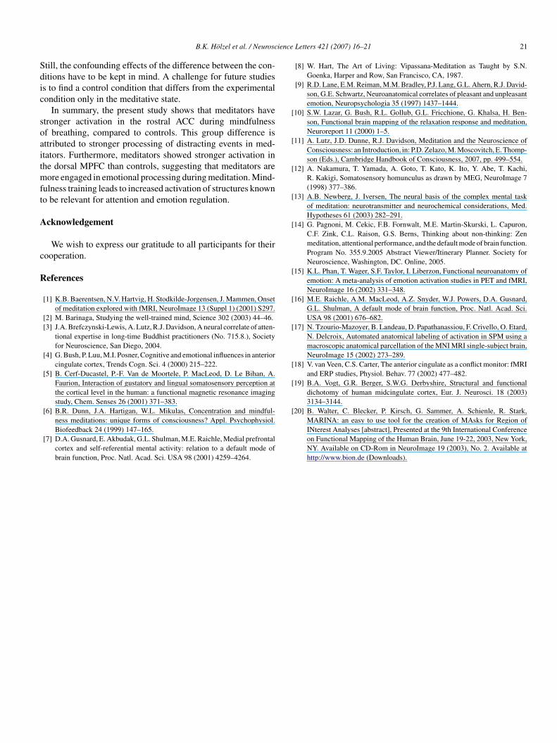
For testing the postulated hypotheses, we compared theroups of meditators and controls in the contrast mindful-ess > arithmetic. Neither in the exploratory nor in ROI analysesid controls show enhanced activations compared to meditatorsor the contrast mindfulness > arithmetic. Yet, greater activationas observed for meditators than for controls in ROI analyses in
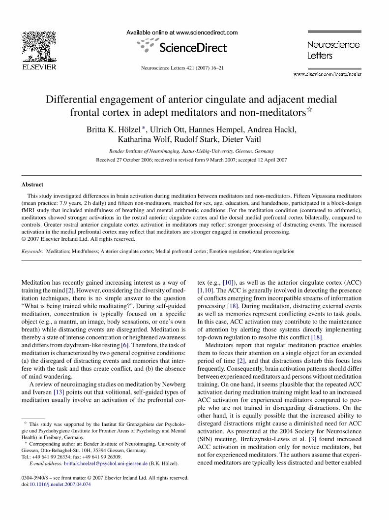
he bilateral ACC and dorsal MPFC (Table 1; Fig. 1). EnhancedCC activation was located at rostral sites.
Additionally, activation patterns are reported for the mindful-ess condition for the whole sample in Table 3. In order to betteretect activations, the threshold was set to an uncorrected p-valuef 0.0001 (cluster size >20 voxels). Activations occurred in theight rolandic operculum, bilateral parahippocampal region, lefterebellum, left inferior temporal gyrus, and bilateral postcen-ral gyri. When contrasting activations of the two groups during
indfulness, no significant activations were found in either ofhe contrasts. However, when lowering the threshold to a p-alue of 0.001 and correcting for small volume, there was aendency for significance at the left MPFC (16 voxels; t = 4.345;= 0.084; x, y, z: −27, 36, 27) and right MPFC (47 voxels;= 4.45; p = 0.071; x, y, z: 24, 39, 24) for meditators > controls.here were no group differences during the mental arithmeticondition.
To investigate the effect of meditation training, the cerebralemodynamic response during mindfulness of breathing, con-rasted to mental arithmetic, was compared between experienced
editators and participants without previous meditation expe-ience. In contrast to our expectations, greater activation wasbserved for meditators than for controls in the bilateral ACCnd the dorsal MPFC in both hemispheres. We did not find effectsn the PCC and the precuneus.
Contrary to the results of the present study, Brefczynski-ewis et al. [3] found greater ACC activation for novices than
or experienced meditators. This difference was attributed toreater error proneness and conflict monitoring in the noviceshan in the adepts [11]. The exact location of ACC activation inhe study by Brefczynski-Lewis et al. was not reported. In theresent study, the difference in the activation between the tworoups has its peak at rostral ACC sites. Within the ACC a majorarcellation between dorsal and rostral-ventral regions can beade based upon cytoarchitecture and connectivity [4]. Cog-
itive tasks consistently activate the dorsal division, while theostral-ventral division is activated by affect-related tasks, whichequire emotional processing [4]. The significant difference inrain activation in the rostral ACC in our study suggests thateditation training leads to more cortical processing of emo-
ional conflict during mindfulness. Self-report data indicates thateditators have kept more continuous attention on the task, felt
ess bored, and have encountered less difficulties with the taskhan non-meditators. This suggests that meditators were more
uccessful in attention regulation. Consequently, it seems thatreater subjective expertise in attention regulation goes alongith greater ACC activation, rather than supporting the expla-ation of compensatory ACC activation in novices. Vogt et al.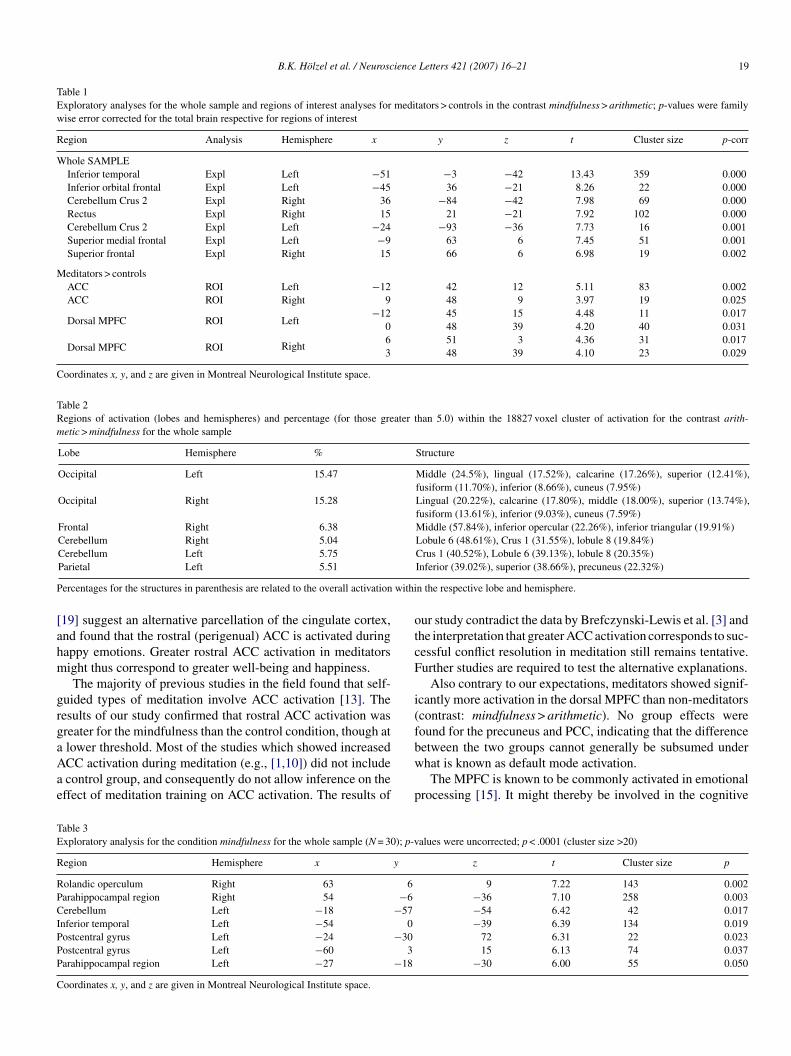
B.K. Holzel et al. / Neuroscience Letters 421 (2007) 16–21 19
Table 1Exploratory analyses for the whole sample and regions of interest analyses for meditators > controls in the contrast mindfulness > arithmetic; p-values were familywise error corrected for the total brain respective for regions of interest
Region Analysis Hemisphere x y z t Cluster size p-corr
Whole SAMPLEInferior temporal Expl Left −51 −3 −42 13.43 359 0.000Inferior orbital frontal Expl Left −45 36 −21 8.26 22 0.000Cerebellum Crus 2 Expl Right 36 −84 −42 7.98 69 0.000Rectus Expl Right 15 21 −21 7.92 102 0.000Cerebellum Crus 2 Expl Left −24 −93 −36 7.73 16 0.001Superior medial frontal Expl Left −9 63 6 7.45 51 0.001Superior frontal Expl Right 15 66 6 6.98 19 0.002
Meditators > controlsACC ROI Left −12 42 12 5.11 83 0.002ACC ROI Right 9 48 9 3.97 19 0.025
Dorsal MPFC ROI Left−12 45 15 4.48 11 0.017
0 48 39 4.20 40 0.031
Dorsal MPFC ROI Right6 51 3 4.36 31 0.0173 48 39 4.10 23 0.029
Coordinates x, y, and z are given in Montreal Neurological Institute space.
Table 2Regions of activation (lobes and hemispheres) and percentage (for those greater than 5.0) within the 18827 voxel cluster of activation for the contrast arith-metic > mindfulness for the whole sample
Lobe Hemisphere % Structure
Occipital Left 15.47 Middle (24.5%), lingual (17.52%), calcarine (17.26%), superior (12.41%),fusiform (11.70%), inferior (8.66%), cuneus (7.95%)
Occipital Right 15.28 Lingual (20.22%), calcarine (17.80%), middle (18.00%), superior (13.74%),fusiform (13.61%), inferior (9.03%), cuneus (7.59%)
Frontal Right 6.38 Middle (57.84%), inferior opercular (22.26%), inferior triangular (19.91%)Cerebellum Right 5.04 Lobule 6 (48.61%), Crus 1 (31.55%), lobule 8 (19.84%)
P withi
[ahm
grgaAae
otcF
i(f
TE
R
RPCIPPP
C
Cerebellum Left 5.75Parietal Left 5.51
ercentages for the structures in parenthesis are related to the overall activation
19] suggest an alternative parcellation of the cingulate cortex,nd found that the rostral (perigenual) ACC is activated duringappy emotions. Greater rostral ACC activation in meditatorsight thus correspond to greater well-being and happiness.The majority of previous studies in the field found that self-
uided types of meditation involve ACC activation [13]. Theesults of our study confirmed that rostral ACC activation wasreater for the mindfulness than the control condition, though at
lower threshold. Most of the studies which showed increasedCC activation during meditation (e.g., [1,10]) did not includecontrol group, and consequently do not allow inference on theffect of meditation training on ACC activation. The results ofbw
p
able 3xploratory analysis for the condition mindfulness for the whole sample (N = 30); p-v
egion Hemisphere x y
olandic operculum Right 63 6arahippocampal region Right 54 −6erebellum Left −18 −57
nferior temporal Left −54 0ostcentral gyrus Left −24 −30ostcentral gyrus Left −60 3arahippocampal region Left −27 −18
oordinates x, y, and z are given in Montreal Neurological Institute space.
Crus 1 (40.52%), Lobule 6 (39.13%), lobule 8 (20.35%)Inferior (39.02%), superior (38.66%), precuneus (22.32%)
n the respective lobe and hemisphere.
ur study contradict the data by Brefczynski-Lewis et al. [3] andhe interpretation that greater ACC activation corresponds to suc-essful conflict resolution in meditation still remains tentative.urther studies are required to test the alternative explanations.
Also contrary to our expectations, meditators showed signif-cantly more activation in the dorsal MPFC than non-meditatorscontrast: mindfulness > arithmetic). No group effects wereound for the precuneus and PCC, indicating that the difference
etween the two groups cannot generally be subsumed underhat is known as default mode activation.The MPFC is known to be commonly activated in emotionalrocessing [15]. It might thereby be involved in the cognitive
alues were uncorrected; p < .0001 (cluster size >20)
z t Cluster size p
9 7.22 143 0.002−36 7.10 258 0.003−54 6.42 42 0.017−39 6.39 134 0.019
72 6.31 22 0.02315 6.13 74 0.037
−30 6.00 55 0.050

20 B.K. Holzel et al. / Neuroscience Letters 421 (2007) 16–21
F prefr( 8; z =
aewatr
ipArcrgats
eitt
iaeaom1
tasrtttmti
ig. 1. Stronger brain activation in the rostral anterior cigulate cortex and medialthreshold: p = 0.01, extend threshold >5 voxels; MNI coordinates: x = −6; y = 4
spects of emotional processing, such as paying attention tomotion or the identification of emotions. Thus, it is engagedhen subjects internally attend to their emotional state [9]. We
ssume that stronger MPFC activation in meditators implieshat they are more strongly engaged in emotional processing,eflecting their improved ability for emotion regulation.
Aside from testing the hypotheses of the present study, wenvestigated the activation during mindfulness of breathing (ana-ana sati) across the whole sample on an exploratory basis.ctivations occurred in the postcentral gyri, extending into the
olandic operculum, as well as temporal, parahippocampal anderebellar regions. Neuroimaging studies localized the sensoryepresentation of the lips at the base of the pre- and postcentralyri [12]. The adjacent rolandic operculum was shown to bective in oro-pharyngeal somatosensory sensitivity [5]. Activa-ion here obviously corresponds to the sensory aspect of anapanaati.
In the present study, we responded to the shortcomings of
arlier studies on meditation by using a rather large sample andnvestigating a well-defined and precisely described meditationechnique [8]. The participants were not randomly assigned tohe groups, but rather self-selected by their meditation train-mtnf
ontal cortex in meditators than controls for the contrast mindfulness > arithmetic6; color bar indicates T-values).
ng. Therefore, differences between the groups cannot be clearlyttributed to the meditation training alone, but may have alreadyxisted beforehand. Longitudinal studies are required to clearlyttribute the differences to the training in meditation itself. Inrder to apply a sensitive high-pass filter, we kept periods ofeditation as short as possible. However, meditation phases ofmin do not allow meditators to reach deep meditation.
The control task, mental arithmetic, differs from the medi-ation condition not only in regard to the meditative state, butlso in respect to several other aspects, such as the external (ver-us internal) focus, open (versus closed) eyes, preparation foresponse (versus maintaining quiet). Thus, differences betweenhe conditions are attributable to factors other than the medita-ive state. However, we did not investigate the difference betweenhe conditions, but group differences within this contrast. As the
ental arithmetic task should be comparable for both groups,he group comparison on the contrast mindfulness > arithmetics considered an appropriate way to test the training effect of
editation. Differential MPFC activation, though at a lowerhreshold, was observable for the mindfulness condition, butot for the arithmetic condition, confirming that the group dif-erence at the contrast can strongly be attributed to mindfulness.
ience
Sdic
soaitmft
A
c
R
[
[
[
[
[
[
[
[
[
[
[
B.K. Holzel et al. / Neurosc
till, the confounding effects of the difference between the con-itions have to be kept in mind. A challenge for future studiess to find a control condition that differs from the experimentalondition only in the meditative state.
In summary, the present study shows that meditators havetronger activation in the rostral ACC during mindfulnessf breathing, compared to controls. This group difference isttributed to stronger processing of distracting events in med-tators. Furthermore, meditators showed stronger activation inhe dorsal MPFC than controls, suggesting that meditators areore engaged in emotional processing during meditation. Mind-
ulness training leads to increased activation of structures knowno be relevant for attention and emotion regulation.
cknowledgement
We wish to express our gratitude to all participants for theirooperation.
eferences
[1] K.B. Baerentsen, N.V. Hartvig, H. Stodkilde-Jorgensen, J. Mammen, Onsetof meditation explored with fMRI, NeuroImage 13 (Suppl 1) (2001) S297.
[2] M. Barinaga, Studying the well-trained mind, Science 302 (2003) 44–46.[3] J.A. Brefczynski-Lewis, A. Lutz, R.J. Davidson, A neural correlate of atten-
tional expertise in long-time Buddhist practitioners (No. 715.8.), Societyfor Neuroscience, San Diego, 2004.
[4] G. Bush, P. Luu, M.I. Posner, Cognitive and emotional influences in anteriorcingulate cortex, Trends Cogn. Sci. 4 (2000) 215–222.
[5] B. Cerf-Ducastel, P.-F. Van de Moortele, P. MacLeod, D. Le Bihan, A.Faurion, Interaction of gustatory and lingual somatosensory perception atthe cortical level in the human: a functional magnetic resonance imagingstudy, Chem. Senses 26 (2001) 371–383.
[6] B.R. Dunn, J.A. Hartigan, W.L. Mikulas, Concentration and mindful-
ness meditations: unique forms of consciousness? Appl. Psychophysiol.Biofeedback 24 (1999) 147–165.[7] D.A. Gusnard, E. Akbudak, G.L. Shulman, M.E. Raichle, Medial prefrontalcortex and self-referential mental activity: relation to a default mode ofbrain function, Proc. Natl. Acad. Sci. USA 98 (2001) 4259–4264.
Letters 421 (2007) 16–21 21
[8] W. Hart, The Art of Living: Vipassana-Meditation as Taught by S.N.Goenka, Harper and Row, San Francisco, CA, 1987.
[9] R.D. Lane, E.M. Reiman, M.M. Bradley, P.J. Lang, G.L. Ahern, R.J. David-son, G.E. Schwartz, Neuroanatomical correlates of pleasant and unpleasantemotion, Neuropsychologia 35 (1997) 1437–1444.
10] S.W. Lazar, G. Bush, R.L. Gollub, G.L. Fricchione, G. Khalsa, H. Ben-son, Functional brain mapping of the relaxation response and meditation,Neuroreport 11 (2000) 1–5.
11] A. Lutz, J.D. Dunne, R.J. Davidson, Meditation and the Neuroscience ofConsciousness: an Introduction, in: P.D. Zelazo, M. Moscovitch, E. Thomp-son (Eds.), Cambridge Handbook of Consciousness, 2007, pp. 499–554.
12] A. Nakamura, T. Yamada, A. Goto, T. Kato, K. Ito, Y. Abe, T. Kachi,R. Kakigi, Somatosensory homunculus as drawn by MEG, NeuroImage 7(1998) 377–386.
13] A.B. Newberg, J. Iversen, The neural basis of the complex mental taskof meditation: neurotransmitter and neurochemical considerations, Med.Hypotheses 61 (2003) 282–291.
14] G. Pagnoni, M. Cekic, F.B. Fornwalt, M.E. Martin-Skurski, L. Capuron,C.F. Zink, C.L. Raison, G.S. Berns, Thinking about non-thinking: Zenmeditation, attentional performance, and the default mode of brain function.Program No. 355.9.2005 Abstract Viewer/Itinerary Planner. Society forNeuroscience, Washington, DC. Online, 2005.
15] K.L. Phan, T. Wager, S.F. Taylor, I. Liberzon, Functional neuroanatomy ofemotion: A meta-analysis of emotion activation studies in PET and fMRI,NeuroImage 16 (2002) 331–348.
16] M.E. Raichle, A.M. MacLeod, A.Z. Snyder, W.J. Powers, D.A. Gusnard,G.L. Shulman, A default mode of brain function, Proc. Natl. Acad. Sci.USA 98 (2001) 676–682.
17] N. Tzourio-Mazoyer, B. Landeau, D. Papathanassiou, F. Crivello, O. Etard,N. Delcroix, Automated anatomical labeling of activation in SPM using amacroscopic anatomical parcellation of the MNI MRI single-subject brain,NeuroImage 15 (2002) 273–289.
18] V. van Veen, C.S. Carter, The anterior cingulate as a conflict monitor: fMRIand ERP studies, Physiol. Behav. 77 (2002) 477–482.
19] B.A. Vogt, G.R. Berger, S.W.G. Derbyshire, Structural and functionaldichotomy of human midcingulate cortex, Eur. J. Neurosci. 18 (2003)3134–3144.
20] B. Walter, C. Blecker, P. Kirsch, G. Sammer, A. Schienle, R. Stark,
MARINA: an easy to use tool for the creation of MAsks for Region ofINterest Analyses [abstract], Presented at the 9th International Conferenceon Functional Mapping of the Human Brain, June 19-22, 2003, New York,NY. Available on CD-Rom in NeuroImage 19 (2003), No. 2. Available athttp://www.bion.de (Downloads).Copyright © 2022 FDOKUMEN