Association of Nicotine Addiction and Nicotine's Actions With Separate Cingulate Cortex Functional...
21
Nicotine Addiction and Nicotine's Actions Are Associated with Separate Cingulate Cortex Functional Circuits L. Elliot Hong 1,3,* , Hong Gu 2,3 , Yihong Yang 2,3 , Thomas J. Ross 2 , Betty Jo Salmeron 2 , Brittany Buchholz 1 , Gunvant K. Thaker 1 , and Elliot A. Stein 2 1 Maryland Psychiatric Research Center, Department of Psychiatry, University of Maryland School of Medicine, Baltimore, Maryland 21228 2 Neuroimaging Research Branch, National Institute on Drug Abuse, NIH, Baltimore, Maryland 21224 Abstract Context—Understanding the mechanisms underlying nicotine addiction in order to develop more effective treatment is a public health priority. Research consistently shows that nicotine transiently improves multiple cognitive functions. However, using nicotine replacement to treat nicotine addiction yields generally inconsistent results. While this dichotomy is well known, the reasons are unclear. Imaging studies showed that nicotine challenges almost always involve cingulate cortex, suggesting that this loci may be a key region associated with nicotine addiction and its treatment. Objective—To identify cingulate functional circuits that are associated with the severity of nicotine addiction and to study how nicotine affects them. Design—Using region-specific resting-state fMRI signals to extract resting-state cingulate functional connectivity, and to study how nicotine addiction and acute nicotine administration modulate these functional pathways, in a double-blind, placebo-controlled design. Setting—Outpatient clinics. Participants—Nineteen healthy smokers. Intervention(s)—Single dose (21/35mg) nicotine patch. Main Outcome Measure(s)—Correlation of nicotine addiction severity and cingulate resting state functional connectivity, and effects of acute nicotine on connectivity strength. Results—Clearly separated pathways that correlated with nicotine addiction vs. nicotinic action were found. The severity of nicotine addiction was associated with the strength of dorsal anterior cingulate cortex (dACC)-striatal circuits, which were not modified by nicotine patch administration. In contrast, acute nicotine enhanced cingulate-neocortical functional connectivity patterns. Conclusions—Nicotine addiction was strongly associated with functional circuits interconnecting dACC and the striatum. Acute nicotine administration had no significant effect on these circuits. Rather, nicotine enhanced several cingulate-neocortical functional connectivity circuits that were not associated with the severity of nicotine addiction, but may play a role in nicotine's cognitive enhancing properties. Resting state dACC-striatum functional connectivity may serve as a circuit- level biomarker for nicotine addiction, and the development of new therapeutics aiming to enhance the dACC-striatum functional pathways may be effective for nicotine addiction treatment. * Corresponding to Dr. Hong, Maryland Psychiatric Research Center, P.O. Box 21247, Baltimore, MD 21228. Tel: 410 402 6828. Fax: 410 402 6023. Email: [email protected].. 3 These authors contributed equally to this work NIH Public Access Author Manuscript Arch Gen Psychiatry. Author manuscript; available in PMC 2010 April 1. Published in final edited form as: Arch Gen Psychiatry. 2009 April ; 66(4): 431–441. doi:10.1001/archgenpsychiatry.2009.2. NIH-PA Author Manuscript NIH-PA Author Manuscript NIH-PA Author Manuscript
Transcript of Association of Nicotine Addiction and Nicotine's Actions With Separate Cingulate Cortex Functional...

Nicotine Addiction and Nicotine's Actions Are Associated withSeparate Cingulate Cortex Functional Circuits
L. Elliot Hong1,3,*, Hong Gu2,3, Yihong Yang2,3, Thomas J. Ross2, Betty Jo Salmeron2,Brittany Buchholz1, Gunvant K. Thaker1, and Elliot A. Stein21 Maryland Psychiatric Research Center, Department of Psychiatry, University of Maryland Schoolof Medicine, Baltimore, Maryland 212282 Neuroimaging Research Branch, National Institute on Drug Abuse, NIH, Baltimore, Maryland21224
AbstractContext—Understanding the mechanisms underlying nicotine addiction in order to develop moreeffective treatment is a public health priority. Research consistently shows that nicotine transientlyimproves multiple cognitive functions. However, using nicotine replacement to treat nicotineaddiction yields generally inconsistent results. While this dichotomy is well known, the reasons areunclear. Imaging studies showed that nicotine challenges almost always involve cingulate cortex,suggesting that this loci may be a key region associated with nicotine addiction and its treatment.
Objective—To identify cingulate functional circuits that are associated with the severity of nicotineaddiction and to study how nicotine affects them.
Design—Using region-specific resting-state fMRI signals to extract resting-state cingulatefunctional connectivity, and to study how nicotine addiction and acute nicotine administrationmodulate these functional pathways, in a double-blind, placebo-controlled design.
Setting—Outpatient clinics.
Participants—Nineteen healthy smokers.
Intervention(s)—Single dose (21/35mg) nicotine patch.
Main Outcome Measure(s)—Correlation of nicotine addiction severity and cingulate restingstate functional connectivity, and effects of acute nicotine on connectivity strength.
Results—Clearly separated pathways that correlated with nicotine addiction vs. nicotinic actionwere found. The severity of nicotine addiction was associated with the strength of dorsal anteriorcingulate cortex (dACC)-striatal circuits, which were not modified by nicotine patch administration.In contrast, acute nicotine enhanced cingulate-neocortical functional connectivity patterns.
Conclusions—Nicotine addiction was strongly associated with functional circuits interconnectingdACC and the striatum. Acute nicotine administration had no significant effect on these circuits.Rather, nicotine enhanced several cingulate-neocortical functional connectivity circuits that were notassociated with the severity of nicotine addiction, but may play a role in nicotine's cognitiveenhancing properties. Resting state dACC-striatum functional connectivity may serve as a circuit-level biomarker for nicotine addiction, and the development of new therapeutics aiming to enhancethe dACC-striatum functional pathways may be effective for nicotine addiction treatment.
* Corresponding to Dr. Hong, Maryland Psychiatric Research Center, P.O. Box 21247, Baltimore, MD 21228. Tel: 410 402 6828. Fax:410 402 6023. Email: [email protected] authors contributed equally to this work
NIH Public AccessAuthor ManuscriptArch Gen Psychiatry. Author manuscript; available in PMC 2010 April 1.
Published in final edited form as:Arch Gen Psychiatry. 2009 April ; 66(4): 431–441. doi:10.1001/archgenpsychiatry.2009.2.
NIH
-PA Author Manuscript
NIH
-PA Author Manuscript
NIH
-PA Author Manuscript

IntroductionNicotine, the addictive component in tobacco, has been shown to transiently improveperformance on a wide range of cognitive and neurophysiological tasks in humans, includingattention 1;2, visual information processing 3, computational abilities 4, prepulse inhibition 5,vigilance 6 and working memory 7. What remains unclear is how to explain nicotine's effecton such diverse activities, which are controlled, at least in part, by different brain circuits. Theseemingly pervasive behavioral effects of nicotine and the localization of nicotinic receptorson neurons of different neurotransmitter systems 8;9 suggest the interesting possibility thatnicotine may exert a modulatory effect on multiple functional brain circuits independent ofspecific task performance. New MRI approaches to identify functional network connectivity10;11 present the opportunity to test this hypothesis in human subjects.
A common thread running through the imaging literature is the association of the cingulate innicotine's acute effects and nicotine addiction 9;12-15. Early autoradiographic imaging showedthat the cingulate alone had increased glucose utilization following nicotine administration inrats 12. Human PET studies identified high concentrations of nicotinic binding sites in thecingulate, insula, thalamus, basal ganglia, and the frontal lobe 9. Nicotine dose-dependentlyincreased activity in the nucleus accumbens, amygdala, cingulate, and frontal lobes in the task-free state 13. Nicotine improved sustained attention by increasing activation in posteriorcingulate, bilateral parietal and occipital cortex, thalamus and caudate 3, and improved workingmemory by either enhancing 16 or reducing 14 activation in the cingulate and other areas. Morerecently, nicotine was found to improve attention by deactivating anterior and posteriorcingulate and other brain regions 2, while activation in the posterior cingulate predicted thebehavioral effects of nicotine 15. To summarize, of regions associated with nicotine's actions,the cingulate was the most frequently detected, suggesting that this region may be a convergentstructure pivotal for the diverse CNS nicotinic effects. However, the exact location(s) ofnicotine's actions within the cingulate varies across studies. Perhaps more importantly, theapparent contradiction on the valence of nicotine's action on the cingulate needs to bereconciled (both significant activation and deactivation have been reported).
Recent advances in understanding the resting-state functional connectivity of the brain 10;11;17 provide a framework to test the hypothesis that specific cingulate functional circuitry mayvary both as a function of nicotine dependence and a function of acute nicotinic action,independent of any particular behavioral activation condition. Resting-state functionalconnectivity is task-independent, low frequency synchronized activities across brain regions10;11. Its neural bases are under intense studies. The slow coherent fluctuations in resting-statefMRI BOLD signals may reflect suppression of neuronal activity 10;18-21. The stable spatialorganization of resting-state functional connectivity maps suggests that it can be constrainedby anatomical connectivity 22;23. Based upon prior knowledge of the functional andhistological subdivisions of the cingulate cortex 24-29, we partitioned the entire humancingulate into anatomic subregions. The study is designed to first characterize the resting-statefunctional connectivity associated with each cingulate subregion, then to identify cingulatecircuits that are associated with nicotine addiction and finally to determine how acute nicotineadministration affects circuits that are associated with nicotine addiction.
MethodsSubject
Nineteen (5 female) healthy smokers (10 or more cigarettes per day) participated in the study.Subjects gave written informed consent approved by both University of Maryland and NationalInstitute on Drug Abuse IRB panels. Subjects were 18−50 years of age, right handed, and wererecruited through media advertisements. Major medical and neurological conditions were
Hong et al. Page 2
Arch Gen Psychiatry. Author manuscript; available in PMC 2010 April 1.
NIH
-PA Author Manuscript
NIH
-PA Author Manuscript
NIH
-PA Author Manuscript

exclusionary criteria. Subjects have no Axis I diagnoses other than nicotine dependence asevaluated by the Structured Clinical Interview for DSM-IV (SCID) 30. The severity of nicotineaddiction was assessed by the Fagerstrom Test for Nicotine Dependence (FTND) 31. FTNDincludes 6 items and produces a score from 0 to 10 with higher scores indicating more severenicotine addiction. FTND marks the genetic liability of nicotine addiction 32;33. The cumulativeexposure to smoking was calculated as pack-year.
DesignThis was a double-blind, placebo controlled, cross-over, randomized fMRI study comparingnicotine vs. placebo patch effect on resting-state functional connectivity. Subjects werescanned on two occasions approximately one week apart, within 5−14 days. They receivedeither a nicotine patch (Nicoderm CQ, SmithKline Beecham) or an identical placebo patch oneach occasion. We used a two-dose strategy to better match the regular nicotine intakes: 21mgfor individuals who smoked 10−15 cigarettes/day (n=6) and 35 mg for individuals who smokedmore than 15 cigarettes/day (n=13).
Subjects were instructed to maintain their normal smoking routine prior to patch application,and then abstain for about 4.5 hours following patch administration, including 2.5 hours priorto scanning, and 2 hours inside the scanner. The 4.5 hour window was chosen to avoidconfounds induced by withdrawal, which generally begins 6−12 hours after abstinence 34. Inthe scanner, each subject underwent 1 hour of smooth pursuit eye movement/fixation tasks(data not shown), followed by a 5-minute resting scan and an 8-minute structural scan. Restingscan data were therefore collected about 3.5 hours after patch administration, which is withinthe window of steady plasma nicotine levels 6. A self-reported symptom checklist and a self-reported mood change questionnaire 35 were administered before patch application and afterscan. Breath CO was measured immediately prior to patch application. Plasma nicotine levelswere measured at the end of each scan session.
MRI acquisitionData were collected on a 3 Tesla Siemens Allegra scanner equipped with a quadrature volumehead coil. Subjects were given a simple instruction to rest and keep their eyes open. A staticneutral image (the projector's logo) was presented on the screen during the resting scan. A bitebar was used to minimize head motion. Resting-state fMRI were acquired over 39, 4 mm axial,interleaving slices using a gradient-echo EPI sequence (150 volumes, TE/TR = 27/2000 ms;FA = 80°; FOV = 220×220 mm2; image matrix = 64×64). High-resolution (1×1×1 mm3) T1-weighted MPRAGE images were acquired after each resting scan.
Data ProcessingData were analyzed in AFNI 36 and MATLAB (The MathWorks, Inc., Natick, MA). Volumeswere slice-timing aligned and motion corrected to the base volume that minimally deviatedfrom other volumes using an AFNI built-in algorithm 37. After linear detrending of the timecourse of each voxel, volumes were spatially normalized and resampled to Talairach space at3×3×3 mm3, spatially smoothed (FWHM 6 mm), and temporally low-pass filtered (fcutoff= 0.1Hz) 38-40. Correlation analyses were performed by calculating the cross-correlation coefficient(CC) between each voxel's time course and the template time course extracted by averagingtime courses of all the voxels in the defined regions of interest (“seed ROI”), including the sixrigid head-motion parameter time courses and the average time course in white matter asnuisance covariates 41;42. The white matter mask was generated by segmenting the high-resolution anatomical images in SPM5 43 and down-gridding the obtained white matter masksto the same resolution as the functional data. These nuisance covariates regress out fluctuationsunlikely to be relevant to neuronal activities 41. CC maps were then converted to z-score mapsusing an AFNI built-in function.
Hong et al. Page 3
Arch Gen Psychiatry. Author manuscript; available in PMC 2010 April 1.
NIH
-PA Author Manuscript
NIH
-PA Author Manuscript
NIH
-PA Author Manuscript
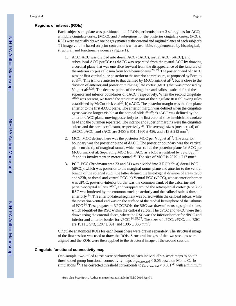
Regions of interest (ROIs)Each subject's cingulate was partitioned into 7 ROIs per hemisphere: 3 subregions for ACC;a middle cingulate cortex (MCC); and 3 subregions for the posterior cingulate cortex (PCC).ROIs were manually drawn on the grey matter at the coronal and sagittal planes of each subject'sT1 image volume based on prior conventions when available, supplemented by histological,structural, and functional evidence (Figure 1):
1. ACC. ACC was divided into dorsal ACC (dACC), rostral ACC (oACC), andsubcallosal ACC (sACC): a) dACC was separated from the rostral ACC by drawinga coronal plane that was one slice forward from the disappearance of the juncture ofthe anterior corpus callosum from both hemispheres 28;29. The posterior end of dACCwas the first vertical slice posterior to the anterior commissure, as proposed by Fornitoet al28. This is more anterior to that defined by McCormick et al29, but is close to thedivision of anterior and posterior mid-cingulate cortex (MCC) that was proposed byVogt et al25;26. The deepest points of the cingulate and callosal sulci defined thesuperior and inferior boundaries of dACC, respectively. When the second cingulate28;29 was present, we traced the structure as part of the cingulate ROI following rulesestablished by McCormick et al29; b) oACC. The posterior margin was the first planeanterior to the first dACC plane. The anterior margin was defined when the cingulategyrus was no longer visible at the coronal slide 28;29; c) sACC was defined by theanterior dACC plane, moving posteriorly to the first coronal slice in which the caudatehead and the putamen separated. The interior and superior margins were the cingulatesulcus and the corpus callosum, respectively 28. The average sizes (mean ± s.d) ofdACC, oACC, and sACC are 3455 ± 851, 1360 ± 456, and 813 ± 212 mm3.
2. MCC. MCC defined here was the posterior MCC per Vogt et al25. The anteriorboundary was the posterior plane of dACC. The posterior boundary was the verticalplane on the tip of marginal ramus, which was called the posterior plane for ACC perMcCormick et al. Separating MCC from ACC as a ROI is justified by cytology 25;26 and its involvement in motor control 44. The size of MCC is 2679 ± 717 mm3.
3. PCC. PCC (Brodmann area 23 and 31) was divided into 3 ROIs 27: a) dorsal PCC(dPCC), which was posterior to the marginal ramus plane and anterior to the ventralbranch of the splenial sulci; the latter defined the histological division of areas d23band v23b, or dorsal and ventral PCC; b) Ventral PCC (vPCC), whose anterior borderwas dPCC, posterior-inferior border was the common trunk of the calcarine andparieto-occipital sulcus 24;27, and wrapped around the retrosplenial cortex (RSC). c)RSC was bordered by the common truck posteriorly and the callosal sulcus dorso-anteriorly 24. The anterior-lateral segment was buried within the callosal sulcus; whilethe posterior-ventral end was on the surface of the medial hemisphere of the isthmusof PCC 24. To segregate the 3 PCC ROIs, the RSC was drawn first using sagittal slices,which identified the RSC within the callosal sulcus. The dPCC and vPCC were thendrawn using the coronal slices, where the RSC was the inferior border for dPCC andinferior and anterior border for vPCC 24;25;27. The sizes of dPCC, vPCC, and RSCare 1911 ± 573, 1207 ± 391, and 1395 ± 366 mm3.
Cingulate anatomical ROIs for each hemisphere were drawn separately. The structural imageof the first session was used to draw the ROIs. Structural images of the two sessions werealigned and the ROIs were then applied to the structural image of the second session.
Cingulate functional connectivity mapOne-sample, two-tailed t-tests were performed on each individual's z-score maps to obtainthresholded group functional connectivity maps at pcorrected < 0.05 based on Monte Carlosimulations 45. The corrected threshold corresponds to puncorrected < 0.001 46 with a minimum
Hong et al. Page 4
Arch Gen Psychiatry. Author manuscript; available in PMC 2010 April 1.
NIH
-PA Author Manuscript
NIH
-PA Author Manuscript
NIH
-PA Author Manuscript
cluster size of 810 mm3 in both the nicotine and placebo conditions. For each seed ROI,thresholded group functional connectivity maps from both nicotine and placebo conditionswere combined using “OR” operation to generate a mask, which was used to constrain thesubsequent analyses.
Nicotine addiction severity by drug interaction on cingulate connectivityWe first investigated interactions between addiction severity (FTND) and nicotinic effect onfunctional connectivity using the regression model: V'i(diff) = ß'0 + ß'X + ε', where V' was thez score of the arithmetic difference between nicotine vs. placebo condition on the ith voxel, Xwas the centered FTND score, plus a random error term ε'. The t-statistics of ß' tested the effectof FTND on nicotine vs. placebo differences, i.e., drug × FTND interaction. Statisticalsignificance of the t-statistic for this and the subsequent analyses were thresholded aftercorrection for multiple comparisons using Monte Carlo simulations to obtain pcorrected < 0.05,corresponding to puncorrected < 0.001 with a minimum cluster size of 243 mm3 – 405 mm3. Thedifferent cluster size thresholds reflect different numbers of comparisons to be corrected forconnectivity maps of different seed ROIs.
Main effect of nicotine addiction severity on cingulate connectivityIn the event of no significant interaction, we applied a second regression model: Vi(mean) =ß0 + ßX + ε, where V was the z score of the mean of nicotine and placebo conditions on theith voxel. The t-statistics of ß tested the main effect of FTND (X). To investigate whether theeffect of FTND on cingulate connectivity was secondary to covariates such as chronic smokingexposure, nicotine level, and blood pressure, exploratory regression analyses were performedto examine the relative contribution of addiction severity and each covariate. The β of FTNDrepresented the independent contributions of FTND to the functional connectivity aftercontrolling for the covariate.
Nicotine Effects on Cingulate ConnectivityTo identify nicotinic effects on cingulate functional connectivity, paired sample t-tests wereperformed on z-score maps to assess placebo vs. nicotine differences for each ROI.
For clinical data, paired t-tests were used to examine nicotine vs. placebo effect on side effects,withdrawal symptoms, and nicotine and CO measures. Pearson's correlations were used toexamine relationships between clinical and nicotine addiction parameters.
ResultsNicotine and demographic information
Participants were 35.7 ± 11.1 (mean ± S.D.) years of age, had 13.0 ± 1.8 years of education,started smoking at 16.9 ± 5.7 and became regular smokers at 18.9 ± 5.7 years of age. Theirnicotine addiction severity, as measured by FTND, was 4.3 ± 2.4. Lifetime exposure to cigarettesmoking was 15.6 ± 10.9 pack-year. Changes in withdrawal, side effect symptoms and moodwere not statistically different between placebo vs. nicotine conditions (Table 1). Withdrawalsymptoms assessed using time (before and after scan) and drug (nicotine vs. placebo) asrepeated measures also did not show significant time × drug interaction (F1,18=0.42, p=0.52)or main effect of drug (p=0.21) or time (p=0.09). However, change in systolic blood pressurewas significantly greater in the nicotine as compared to placebo condition (p=0.05). The FTNDscore significantly correlated to CO level prior to placebo (r=0.53, p=0.02) or nicotine patch(r=0.61, p=0.006), but not to nicotine plasma level in placebo (r=0.24, p=0.35) or nicotine(r=0.29, p=0.28) conditions, confirming that nicotine level by itself is a state measure and isnot directly related to addiction severity.
Hong et al. Page 5
Arch Gen Psychiatry. Author manuscript; available in PMC 2010 April 1.
NIH
-PA Author Manuscript
NIH
-PA Author Manuscript
NIH
-PA Author Manuscript
Cingulate functional connectivityThe connectivity maps of each cingulate “seed ROI” (pcorrected < 0.05, corresponding topuncorrected < 0.001 and a cluster size of 810 mm3) are presented in Figure 2. In general, thestrongest connectivity of each cingulate ROI was adjacent to its seed region. Non-adjacentregions also showed significant connectivity associated with each cingulate subregion. Adescriptive summary of the statistically corrected functional connectivity maps of eachcingulate ROI is given in the legend.
Cingulate connectivity and nicotine addiction severityVoxel-wise regression analyses showed no significant interaction between FTND score anddrug condition in any cingulate functional circuit. In contrast, voxel-wise regression analysesfor the main effect of FTND score showed that FTND was significantly (pcorrected < 0.05,corresponding to puncorrected < 0.001 and a cluster size of 243 mm3 – 405 mm3) and negativelycorrelated with three co-activated circuits: between left dACC and bilateral striatum (Figure3), and between right dACC and right striatum (Figure 4). The extent and coordinates of thesefindings are listed in Table 2. Note that these correlations were present and statisticallysignificant in both placebo and nicotine conditions, indicating that nicotine did not remove thenegative correlations.
To investigate whether the above effect of FTND on the dACC-striatal connectivity could besecondary to chronic exposure to smoking, rather than addiction per se, exploratory regressionanalyses were performed to examine the relative contribution of chronic exposure (pack-year).Analyses were carried out in placebo and nicotine conditions separately for each circuit. FTNDcorrelations remained significant in all three dACC-striatal functional connectivity paths in theplacebo (n = 19, β = −0.88 to −0.95, t = −6.14 to −7.27, all p ≤ 0.001) and nicotine (all β =−0.56 to −0.70, t = −2.50 to −3.56, p = 0.024 to 0.003) conditions, after controlling for chronicexposure.
Nicotine plasma concentration during imaging also could have influenced the contribution ofFTND to the ACC-striatal path. We repeated the above analyses using nicotine level as apredictor. FTND remained negatively correlated to all three dACC-striatal functionalconnectivity scores in both placebo (n = 17, all β ≤ −0.78, all t ≤ −4.65, all p < 0.001) andnicotine (n = 16, all β ≤ −0.61, all t ≤ −2.84, all p ≤ 0.01) conditions after controlling for nicotinelevels. FTND also remained significantly correlated to the corresponding dACC-striatalconnectivity after controlling for age and gender (data not shown). Taken together, thesefindings do not support the conclusion that the negative correlations between addiction severityand the dACC-striatal connectivity were substantially biased by chronic smoking exposure,nicotine level during scanning, age, or gender.
Finally, we conducted exploratory regression analyses using the seven DSM-IV criteria fornicotine dependence as predictors. The DSM-IV criteria together significantly contributed tothe FTND-derived dACC-striatum functional connectivities (R2 change 0.58 − 0.67 for the 3connectivities, all p<0.001). Within the models, the functional connectivities were most closelyassociated with DSM-defined tolerance (standardized coefficients beta = −0.69 to −0.79 forthe 3 connectivities, all p< 0.001) and to a lesser extent withdrawal (beta = −0.34 for the rightdACC-striatum connectivity, p=0.02). These findings are consistent with the symptoms(tolerance and withdrawal) typically contributing to FTND scores in tobacco smokers.
Nicotinic effects on cingulate connectivityVoxel-wise paired t-tests showed that acute nicotine administration significantly enhanced thecoherence strength of several cingulate functional connectivity paths compared to the placebocondition (pcorrected < 0.05). The coordinates having significant nicotine effects are presented
Hong et al. Page 6
Arch Gen Psychiatry. Author manuscript; available in PMC 2010 April 1.
NIH
-PA Author Manuscript
NIH
-PA Author Manuscript
NIH
-PA Author Manuscript
in Table 3. Figure 5 illustrates the increased connectivity following nicotine administrationbetween seed ROIs and these locations. The z scores of these seven circuits were significantlydifferent between nicotine vs. placebo conditions (paired t-tests, t= 4.1 − 5.2, p ≤ 0.001) afterBonferroni correction for fourteen ROI comparisons (0.05/14=0.003). A consistent pattern ofnicotine's effect was seen in the functional connectivity between PCC ROIs and frontal midlinestructures, including orbitofrontal, medial superior frontal, and anterior cingulate regions(Figure 5b, 5f, and 5g). In addition, left subcallosal ACC showed significantly enhancedconnectivity with medial frontal cortex (Figure 5d). Another common pattern was the cingulateconnectivity with parietal regions, including connectivity between left dACC and superiorparietal lobule and the postcentral gyrus, and between the right MCC and the inferior parietallobule and the postcentral gyrus (Figure 5a and 5e). Nicotine also enhanced a relatively “local”circuit between the dPCC and vPCC (Figure 5c). Notably, and in contrast to the above, therewas no indication that nicotine significantly enhanced any of the three ACC-striatal functionalconnectivity circuits, even with paired t-tests. Finally, all nicotine effects were in the positive(enhanced) direction; nicotine never significantly reduced synchronized activity between thecingulate and any other region of the brain.
DiscussionThis study identified non-task driven, resting-state functional networks associated with discretecingulate subregions and examined the relationship between an established marker for nicotineaddiction and each cingulate circuit. We report here, for the first time, that severity of nicotineaddiction was inversely associated with the strength of the coherent activity between dACCand striatum, i.e., the more severe the addiction, the weaker the functional connectivity.Critically, acute nicotine challenge did not abolish these correlations. In contrast, as part of adouble dissociation, acute nicotine did enhance the coherence of other cingulate-corticalcircuits that were not correlated with nicotine addiction.
The dACC-striatal functional connectivity paths coincide with known “hard-wired” pathways47-49. Baleydier and Mauguiere noted that the anterior cingulate gyrus (area 24) is connectedwith “the caudate nucleus, the claustrum, the lateral frontal and the posterior parietal (area 7)cortices” 47-49. Others also showed that ACC projects to the ventromedial regions of thecaudate nucleus and putamen 50. These fibers are also described as the striatal fibers that arebranches from the cingulate bundle 48;51; all of which provide a putative anatomical basis forthe functional connectivity observations.
FTND is an established clinical and genetic trait marker of nicotine addiction and has a highheritability around 0.72 to 0.75 32;33. Its validity in marking nicotine addiction is furthersupported by association studies using FTND as a primary phenotype, which have identifiednicotinic acetylcholine receptor variants contributing to nicotine addiction 52;53. By showingsignificant correlations with FTND, we can hypothesize that resting-state synchronized activityin the dACC-striatum circuits may serve as a circuitry level endophenotypic marker for nicotineaddiction. Anatomically, the identified striatal clusters mainly include the putamen and itstransition to the nucleus accumbens, encompassing roughly a transitional zone between dorsaland ventral striatum. The progression of addiction is thought to be associated with initial medialfrontal/anterior cingulate cortex executive control over ventral striatal reinforcementmechanisms which, as the addictive processes continue, are replaced by more habit driven,dorsal striatal activity 54;55. The finding that more severe nicotine addiction is associated withweaker functional connectivity between ACC and striatum may be tied to an underlyingdysfunction associated with addictive behaviors that are modulated by this circuit.
We observed that nicotine, even at high plasma levels (34.3 ± 13.1 ng/ml), did not substantiallyalter the negative correlations between FTND score and the ACC-striatal coherence. While
Hong et al. Page 7
Arch Gen Psychiatry. Author manuscript; available in PMC 2010 April 1.
NIH
-PA Author Manuscript
NIH
-PA Author Manuscript
NIH
-PA Author Manuscript

there were non-significant numerical reductions in the strength of the negative correlationsduring nicotine administration in the post-hoc exploratory analyses, the significant negativecorrelations were not abolished. In other words, nicotine administration alone, at least over theshort duration used in this study, did not robustly influence the circuit level abnormalitiesassociated with nicotine addiction severity. In contrast, nicotine did significantly enhance thefunctional connectivity of several cingulate-cortical networks, which also seem to follow thepresence of known fiber tracks interconnecting these regions. For example, one suchconnectivity path is between PCCs and the orbitofrontal cortex/ACC. Anatomically, PCC islinked with the medial orbito-prefrontal cortex, the medial extension of the prefrontal cortex,and the ACC by the cingulate bundle 25;47;49;51. Another example, the ACC/MCC - parietallobe network, is a circuit with known reciprocal fiber connections 49;51. Notably, all of theobserved nicotine effects were to enhance the higher-order, isocortical functional connections.Whether this cortico-cortical enhancement of functional connectivity is channeled directly viathese structures cannot be determined with the present technology, although this may besuggested by the well-established cortico-cortical fibers linking these regions. That restingstate connectivity strength has been shown related to performance on a working memory taskalso suggests the physiological relevance of our observations 56.
The apparent contrast between the effects of acute nicotine on cingulate-neocortical but notcingulate-striatal functional connectivity may be clinically relevant. While many availabletreatments have helped to reduce the prevalence of smoking, the efficacy of nicotinereplacement therapy (NRT) has been generally unsatisfactory; for example, nicotine patchtreatment yields only about a 10−15% long-term quit rate (e.g., 57). However, nicotine, evenin a single dose, can induce robust, transient enhancement of diverse neurophysiological andcognitive functions (see Introduction). The mechanisms responsible for this dichotomy areunknown.
Based on the current data, we hypothesize that nicotine may improve behavioral tasks througha transient enhancement of distributed cingulate-neocortical or other cortico-cortical functionalnetworks, regardless of task demands. The observed nicotine-related behavioral and imagingsignals in a given experiment may represent the circuits taxed by the specific behavioral taskperformed and may be reflected by the enhanced cingulate-neocortical connectivity strengthsreported herein. Depending on the behavioral tasks during imaging, cingulate activity wasshown to have increased (e.g., 3;12;13;16) or decreased (e.g., 2;14;58) following nicotineadministration. The present resting-state connectivity data offer a new perspective towardsresolving these inconsistencies by suggesting that the cingulate involvement in nicotinicactions may not be simply local increases or decreases of activity within the cingulate proper,but may be related to enhancement in synchronized activity between cingulate and itsfunctionally connected cortical regions. On the other hand, the FTND correlation data suggestthat nicotine addiction is not primarily associated with the coherent activity of the neocorticalconnections, but rather is associated with a more specific alteration in dACC-striatalconnectivity. Acute administration of nicotine itself does not substantially affect the strengthof this connectivity circuit, which may in part help explain the low rate of smoking cessationby NRT. Our findings offer potential brain circuitry level mechanisms to explain theincongruent data of clear nicotinic effects on cognitive enhancement vs. its limited effect ontreating nicotine addiction.
Task-independent, functionally coherent resting state networks are thought to representintrinsically synchronized activity of the brain and has been termed default mode network 11;17. The most consistent regions associated with this network are PCC, ACC, and medial frontalregions 10;11, along with several frequently detected areas such as superior and inferior parietallobule and precuneus 10;11;17;59. The default mode network is reliably detected regardless ofwhether a person keeps their eyes closed or open during the resting state11;17. The default mode
Hong et al. Page 8
Arch Gen Psychiatry. Author manuscript; available in PMC 2010 April 1.
NIH
-PA Author Manuscript
NIH
-PA Author Manuscript
NIH
-PA Author Manuscript
maps identified from the PCC and ACC subregions in this study were remarkably similar tofindings of the corresponding cingulate seed ROIs from previous studies 11;60. As illustratedby our data, the potential functional role of the default mode connectivity is intriguing. Thedefault state connectivity was initially proposed to represent a “sentinel” role of the resting butawake brain to broadly evaluate information from external and internal milieu 10. Our datashow that this resting-state network is also subject to pharmacological manipulations.
The steady state nicotine effect following patch administration on neocortical connectivity doesnot necessarily imply a similar effect during more rapid nicotine delivery as provided bycigarette smoking. The current patch design identifies the pharmacological action of nicotineon brain circuit dynamics. Replication studies using faster nicotine delivery systems, such asnasal spray or smoking itself, is warranted.
Although we did find mild elevated systolic pressure in the nicotine condition, it is likely thatour findings were due to nicotine induced neuronal activity rather than secondary to nonspecificcardiovascular changes. Blood pressure changes do not correlate with cortical brain activation61. A recent paper demonstrated no effect of nicotine on finger-tapping induced BOLDactivation 62. The correlations seen between FTND and regional connectivity were similarbetween the nicotine and placebo conditions, which also argue against a nonspecific vasculareffect.
In conclusion, this study demonstrated that nicotine increases cingulate-neocortical functionalconnectivity coherence strength during the ‘resting-state’. However, this short-term nicotinechallenge does not significantly alter the cingulate-striatal circuitry that was associated withthe severity of nicotine addiction, suggesting that nicotine replacement does not necessarilycorrect the network abnormalities associated with nicotine addiction. The abnormal resting-state cingulate-striatal functional connectivity may serve as an in vivo biomarker for testingnew, potentially more effective, nicotine addiction therapeutics. Our study further suggeststhat the non-task dependent resting-state imaging approach might also be useful to characterizepharmacologically induced changes associated with nicotine and perhaps otherpharmacological-imaging studies.
AcknowledgmentsSupport was received from National Institute on Health grants MH70644, 79172, 49826, 77852, 68580, and N01-DA-5-9909, the National Institute on Drug Abuse Intramural Research Program, Neurophysiology Core of theUniversity of Maryland General Clinical Research Center (# M01-RR16500), and the Maryland Cigarette RestitutionFund Program – Other Tobacco-Related Diseases Research Grant.
Reference List1. Levin ED, Rezvani AH. Development of nicotinic drug therapy for cognitive disorders. Eur J
Pharmacol 2000;393:141–146. [PubMed: 10771007]2. Hahn B, Ross TJ, Yang Y, Kim I, Huestis MA, Stein EA. Nicotine enhances visuospatial attention by
deactivating areas of the resting brain default network. J Neurosci 2007;27:3477–3489. [PubMed:17392464]
3. Lawrence NS, Ross TJ, Stein EA. Cognitive mechanisms of nicotine on visual attention. Neuron2002;36:539–548. [PubMed: 12408855]
4. Myers CS, Taylor RC, Moolchan ET, Heishman SJ. Dose-Related Enhancement of Mood andCognition in Smokers Administered Nicotine Nasal Spray. Neuropsychopharmacology. 2007
5. Kumari V, Gray JA. Smoking withdrawal, nicotine dependence and prepulse inhibition of the acousticstartle reflex. Psychopharmacology (Berl) 1999;141:11–15. [PubMed: 9952059]
Hong et al. Page 9
Arch Gen Psychiatry. Author manuscript; available in PMC 2010 April 1.
NIH
-PA Author Manuscript
NIH
-PA Author Manuscript
NIH
-PA Author Manuscript
6. Mancuso G, Andres P, Ansseau M, Tirelli E. Effects of nicotine administered via a transdermal deliverysystem on vigilance: a repeated measure study. Psychopharmacology (Berl) 1999;142:18–23.[PubMed: 10102778]
7. McClernon FJ, Gilbert DG, Radtke R. Effects of transdermal nicotine on lateralized identification andmemory interference. Hum Psychopharmacol 2003;18:339–343. [PubMed: 12858319]
8. Swanson LW, Simmons DM, Whiting PJ, Lindstrom J. Immunohistochemical localization of neuronalnicotinic receptors in the rodent central nervous system. J Neurosci 1987;7:3334–3342. [PubMed:2822866]
9. Nyback H, Nordberg A, Langstrom B, Halldin C, Hartvig P, Ahlin A, Swahn CG, Sedvall G. Attemptsto visualize nicotinic receptors in the brain of monkey and man by positron emission tomography.Prog Brain Res 1989;79:313–319. [PubMed: 2587749]
10. Raichle ME, MacLeod AM, Snyder AZ, Powers WJ, Gusnard DA, Shulman GL. A default mode ofbrain function. Proc Natl Acad Sci U S A 2001;98:676–682. [PubMed: 11209064]
11. Greicius MD, Krasnow B, Reiss AL, Menon V. Functional connectivity in the resting brain: a networkanalysis of the default mode hypothesis. Proc Natl Acad Sci U S A 2003;100:253–258. [PubMed:12506194]
12. Grunwald F, Schrock H, Kuschinsky W. The effect of an acute nicotine infusion on the local cerebralglucose utilization of the awake rat. Brain Res 1987;400:232–238. [PubMed: 3815071]
13. Stein EA, Pankiewicz J, Harsch HH, Cho JK, Fuller SA, Hoffmann RG, Hawkins M, Rao SM,Bandettini PA, Bloom AS. Nicotine-induced limbic cortical activation in the human brain: afunctional MRI study. Am J Psychiatry 1998;155:1009–1015. [PubMed: 9699686]
14. Ernst M, Matochik JA, Heishman SJ, Van Horn JD, Jons PH, Henningfield JE, London ED. Effectof nicotine on brain activation during performance of a working memory task. Proc Natl Acad SciU S A 2001;98:4728–4733. [PubMed: 11274349]
15. Giessing C, Fink GR, Rosler F, Thiel CM. fMRI data predict individual differences of behavioraleffects of nicotine: a partial least square analysis. J Cogn Neurosci 2007;19:658–670. [PubMed:17381256]
16. Kumari V, Gray JA, ffytche DH, Mitterschiffthaler MT, Das M, Zachariah E, Vythelingum GN,Williams SC, Simmons A, Sharma T. Cognitive effects of nicotine in humans: an fMRI study.NeuroImage 2003;19:1002–1013. [PubMed: 12880828]
17. Fransson P. Spontaneous low-frequency BOLD signal fluctuations: an fMRI investigation of theresting-state default mode of brain function hypothesis. Hum Brain Mapp 2005;26:15–29. [PubMed:15852468]
18. Shulman GL, Fiez JA, Corbetta M, Buckner RL, Miezin FM, Raichle ME, Petersen SE. CommonBlood Flow Changes across Visual Tasks: II. Decreases in Cerebral Cortex. J Cogn Neruosci1997;9:648–663.
19. Gusnard DA, Raichle ME, Raichle ME. Searching for a baseline: functional imaging and the restinghuman brain. Nat Rev Neurosci 2001;2:685–694. [PubMed: 11584306]
20. Smith AT, Williams AL, Singh KD. Negative BOLD in the visual cortex: evidence against bloodstealing. Hum Brain Mapp 2004;21:213–220. [PubMed: 15038003]
21. Shmuel A, Augath M, Oeltermann A, Logothetis NK. Negative functional MRI response correlateswith decreases in neuronal activity in monkey visual area V1. Nat Neurosci 2006;9:569–577.[PubMed: 16547508]
22. Vincent JL, Patel GH, Fox MD, Snyder AZ, Baker JT, Van Essen DC, Zempel JM, Snyder LH,Corbetta M, Raichle ME. Intrinsic functional architecture in the anaesthetized monkey brain. Nature2007;447:83–86. [PubMed: 17476267]
23. Johnston JM, Vaishnavi SN, Smyth MD, Zhang D, He BJ, Zempel JM, Shimony JS, Snyder AZ,Raichle ME. Loss of resting interhemispheric functional connectivity after complete section of thecorpus callosum. J Neurosci 2008;28:6453–6458. [PubMed: 18562616]
24. Morris R, Paxinos G, Petrides M. Architectonic analysis of the human retrosplenial cortex. J CompNeurol 2000;421:14–28. [PubMed: 10813770]
25. Vogt BA, Vogt LJ, Perl DP, Hof PR. Cytology of human caudomedial cingulate, retrosplenial, andcaudal parahippocampal cortices. J Comp Neurol 2001;438:353–376. [PubMed: 11550177]
Hong et al. Page 10
Arch Gen Psychiatry. Author manuscript; available in PMC 2010 April 1.
NIH
-PA Author Manuscript
NIH
-PA Author Manuscript
NIH
-PA Author Manuscript
26. Vogt BA, Vogt L. Cytology of human dorsal midcingulate and supplementary motor cortices. J ChemNeuroanat 2003;26:301–309. [PubMed: 14729132]
27. Vogt BA, Vogt L, Laureys S. Cytology and functionally correlated circuits of human posteriorcingulate areas. NeuroImage 2006;29:452–466. [PubMed: 16140550]
28. Fornito A, Whittle S, Wood SJ, Velakoulis D, Pantelis C, Yucel M. The influence of sulcal variabilityon morphometry of the human anterior cingulate and paracingulate cortex. NeuroImage2006;33:843–854. [PubMed: 16996751]
29. McCormick LM, Ziebell S, Nopoulos P, Cassell M, Andreasen NC, Brumm M. Anterior cingulatecortex: an MRI-based parcellation method. NeuroImage 2006;32:1167–1175. [PubMed: 16859929]
30. First, MB.; Spitzer, RL.; Gibbon, M.; Williams, JBW. Structured Clinical Interview for DSMIV AxisI Disorders. American Psychiatric Publishing, Inc; Arlington: 1997.
31. Heatherton TF, Kozlowski LT, Frecker RC, Fagerstrom KO. The Fagerstrom Test for NicotineDependence: a revision of the Fagerstrom Tolerance Questionnaire. Br J Addict 1991;86:1119–1127.[PubMed: 1932883]
32. Kendler KS, Neale MC, Sullivan P, Corey LA, Gardner CO, Prescott CA. A population-based twinstudy in women of smoking initiation and nicotine dependence. Psychol Med 1999;29:299–308.[PubMed: 10218922]
33. Vink JM, Willemsen G, Boomsma DI. Heritability of smoking initiation and nicotine dependence.Behav Genet 2005;35:397–406. [PubMed: 15971021]
34. Hughes JR, Higgins ST, Bickel WK. Nicotine withdrawal versus other drug withdrawal syndromes:similarities and dissimilarities. Addiction 1994;89:1461–1470. [PubMed: 7841857]
35. Parrott AC, Garnham NJ, Wesnes K, Pincock C. Cigarette smoking and abstinence: comparativeeffects upon cognitive task performance and mood state over 24 hours. Hum Psychopharmacol1996;11:391–400.
36. Cox RW. Software for analysis and visualization of functional magnetic resonance neuroimages.Computers and Biomedical Research 1996;29:162–173. [PubMed: 8812068]
37. Oakes TR, Johnstone T, Ores Walsh KS, Greischar LL, Alexander AL, Fox AS, Davidson RJ.Comparison of fMRI motion correction software tools. NeuroImage 2005;28:529–543. [PubMed:16099178]
38. Biswal B, Yetkin FZ, Haughton VM, Hyde JS. Functional connectivity in the motor cortex of restinghuman brain using echo-planar MRI. Magn Reson Med 1995;34:537–541. [PubMed: 8524021]
39. Lowe MJ, Mock BJ, Sorenson JA. Functional connectivity in single and multislice echoplanar imagingusing resting-state fluctuations. NeuroImage 1998;7:119–132. [PubMed: 9558644]
40. Cordes D, Haughton VM, Arfanakis K, Carew JD, Turski PA, Moritz CH, Quigley MA, MeyerandME. Frequencies contributing to functional connectivity in the cerebral cortex in “resting-state” data.AJNR Am J Neuroradiol 2001;22:1326–1333. [PubMed: 11498421]
41. Fox MD, Snyder AZ, Vincent JL, Corbetta M, Van Essen DC, Raichle ME. The human brain isintrinsically organized into dynamic, anticorrelated functional networks. Proc Natl Acad Sci U S A2005;102:9673–9678. [PubMed: 15976020]
42. Lund TE, Madsen KH, Sidaros K, Luo WL, Nichols TE. Non-white noise in fMRI: does modellinghave an impact? NeuroImage 2006;29:54–66. [PubMed: 16099175]
43. Ashburner J, Friston KJ. Unified segmentation. NeuroImage 2005;26:839–851. [PubMed: 15955494]44. Turken AU, Swick D. Response selection in the human anterior cingulate cortex. Nat Neurosci
1999;2:920–924. [PubMed: 10491614]45. Ward, BD. Simultaneous Inference for FMRI Data.. AFNI manual. 2000.
http://afni.nimh.nih.gov/afni/doc/manual/AlphaSim46. Thirion B, Pinel P, Meriaux S, Roche A, Dehaene S, Poline JB. Analysis of a large fMRI cohort:
Statistical and methodological issues for group analyses. NeuroImage 2007;35:105–120. [PubMed:17239619]
47. Baleydier C, Mauguiere F. The duality of the cingulate gyrus in monkey. Neuroanatomical study andfunctional hypothesis. Brain 1980;103:525–554. [PubMed: 6774795]
48. Schmahmann, JD.; Pandya, DN. Fiber pathways of the brain. Oxford University Press; New York:2006.
Hong et al. Page 11
Arch Gen Psychiatry. Author manuscript; available in PMC 2010 April 1.
NIH
-PA Author Manuscript
NIH
-PA Author Manuscript
NIH
-PA Author Manuscript
49. Vogt BA, Pandya DN. Cingulate cortex of the rhesus monkey: II. Cortical afferents. J Comp Neurol1987;262:271–289. [PubMed: 3624555]
50. Selemon LD, Goldman-Rakic PS. Longitudinal topography and interdigitation of corticostriatalprojections in the rhesus monkey. J Neurosci 1985;5:776–794. [PubMed: 2983048]
51. Mufson EJ, Pandya DN. Some observations on the course and composition of the cingulum bundlein the rhesus monkey. J Comp Neurol 1984;225:31–43. [PubMed: 6725639]
52. Saccone SF, Hinrichs AL, Saccone NL, Chase GA, Konvicka K, Madden PA, Breslau N, JohnsonEO, Hatsukami D, Pomerleau O, Swan GE, Goate AM, Rutter J, Bertelsen S, Fox L, Fugman D,Martin NG, Montgomery GW, Wang JC, Ballinger DG, Rice JP, Bierut LJ. Cholinergic nicotinicreceptor genes implicated in a nicotine dependence association study targeting 348 candidate geneswith 3713 SNPs. Hum Mol Genet 2007;16:36–49. [PubMed: 17135278]
53. Thorgeirsson TE, Geller F, Sulem P, Rafnar T, Wiste A, Magnusson KP, Manolescu A, ThorleifssonG, Stefansson H, Ingason A, Stacey SN, Bergthorsson JT, Thorlacius S, Gudmundsson J, Jonsson T,Jakobsdottir M, Saemundsdottir J, Olafsdottir O, Gudmundsson LJ, Bjornsdottir G, Kristjansson K,Skuladottir H, Isaksson HJ, Gudbjartsson T, Jones GT, Mueller T, Gottsater A, Flex A, Aben KK,de Vegt F, Mulders PF, Isla D, Vidal MJ, Asin L, Saez B, Murillo L, Blondal T, Kolbeinsson H,Stefansson JG, Hansdottir I, Runarsdottir V, Pola R, Lindblad B, van Rij AM, Dieplinger B,Haltmayer M, Mayordomo JI, Kiemeney LA, Matthiasson SE, Oskarsson H, Tyrfingsson T,Gudbjartsson DF, Gulcher JR, Jonsson S, Thorsteinsdottir U, Kong A, Stefansson K. A variantassociated with nicotine dependence, lung cancer and peripheral arterial disease. Nature2008;452:638–642. [PubMed: 18385739]
54. Everitt BJ, Robbins TW. Neural systems of reinforcement for drug addiction: from actions to habitsto compulsion. Nat Neurosci 2005;8:1481–1489. [PubMed: 16251991]
55. Kalivas PW, O'Brien C. Drug addiction as a pathology of staged neuroplasticity.Neuropsychopharmacology 2008;33:166–180. [PubMed: 17805308]
56. Hampson M, Driesen NR, Skudlarski P, Gore JC, Constable RT. Brain connectivity related to workingmemory performance. J Neurosci 2006;26:13338–13343. [PubMed: 17182784]
57. Hyland A, Rezaishiraz H, Giovino G, Bauer JE, Michael CK. Over-the-counter availability of nicotinereplacement therapy and smoking cessation. Nicotine Tob Res 2005;7:547–555. [PubMed:16085526]
58. Stapleton JM, Gilson SF, Wong DF, Villemagne VL, Dannals RF, Grayson RF, Henningfield JE,London ED. Intravenous nicotine reduces cerebral glucose metabolism: a preliminary study.Neuropsychopharmacology 2003;28:765–772. [PubMed: 12655323]
59. Shulman GL, Corbetta M, Buckner RL, Raichle ME, Fiez JA, Miezin FM, Petersen SE. Top-downmodulation of early sensory cortex. Cereb Cortex 1997;7:193–206. [PubMed: 9143441]
60. Margulies DS, Kelly AM, Uddin LQ, Biswal BB, Castellanos FX, Milham MP. Mapping thefunctional connectivity of anterior cingulate cortex. NeuroImage 2007;37:579–588. [PubMed:17604651]
61. Grunwald F, Schrock H, Kuschinsky W. The influence of nicotine on local cerebral blood flow inrats. Neurosci Lett 1991;124:108–110. [PubMed: 1857536]
62. Murphy K, Dixon V, LaGrave K, Kaufman J, Risinger R, Bloom A, Garavan H. A validation of event-related FMRI comparisons between users of cocaine, nicotine, or cannabis and control subjects. AmJ Psychiatry 2006;163:1245–1251. [PubMed: 16816231]
Hong et al. Page 12
Arch Gen Psychiatry. Author manuscript; available in PMC 2010 April 1.
NIH
-PA Author Manuscript
NIH
-PA Author Manuscript
NIH
-PA Author Manuscript
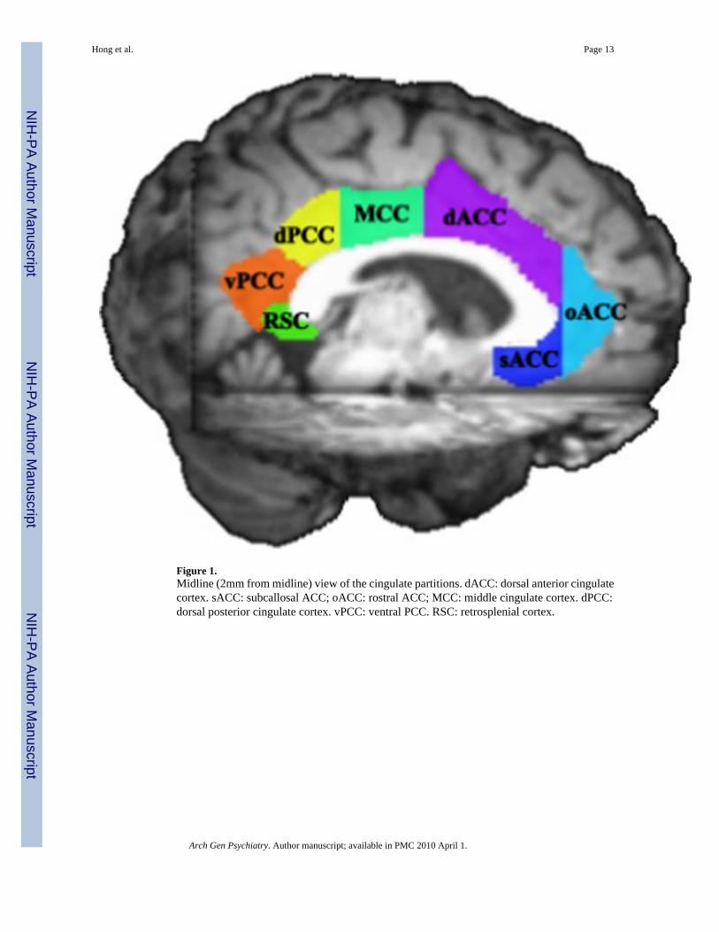
Figure 1.Midline (2mm from midline) view of the cingulate partitions. dACC: dorsal anterior cingulatecortex. sACC: subcallosal ACC; oACC: rostral ACC; MCC: middle cingulate cortex. dPCC:dorsal posterior cingulate cortex. vPCC: ventral PCC. RSC: retrosplenial cortex.
Hong et al. Page 13
Arch Gen Psychiatry. Author manuscript; available in PMC 2010 April 1.
NIH
-PA Author Manuscript
NIH
-PA Author Manuscript
NIH
-PA Author Manuscript
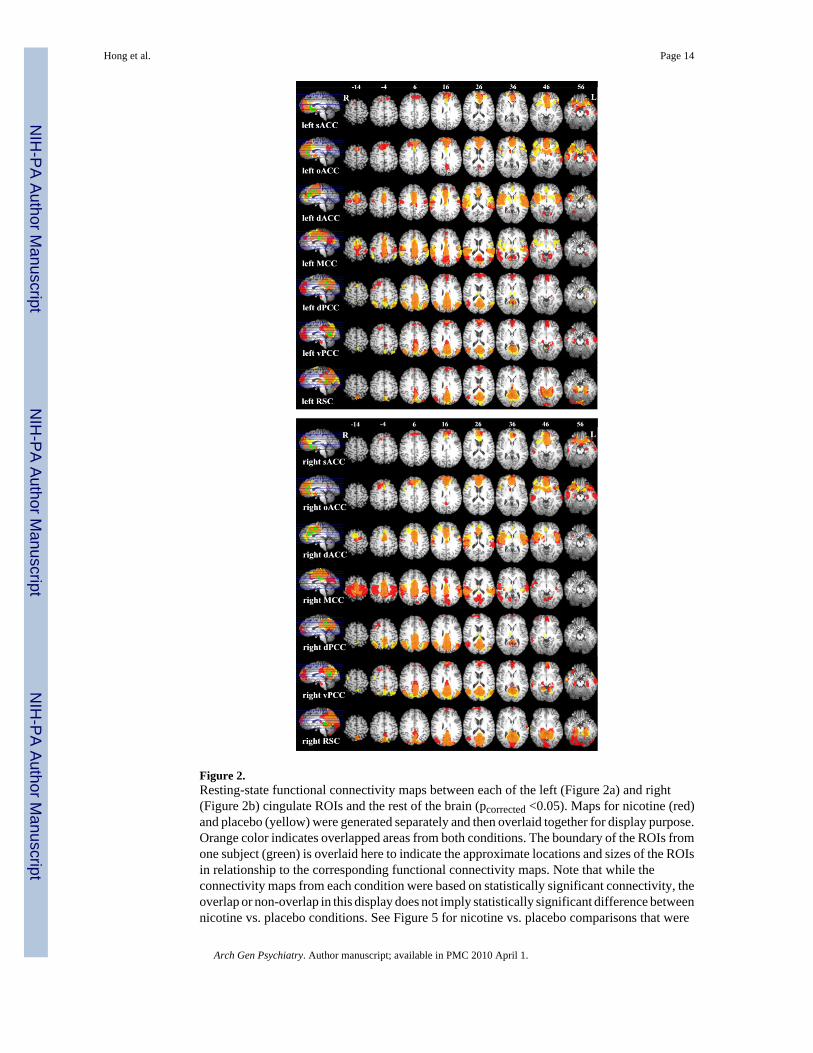
Figure 2.Resting-state functional connectivity maps between each of the left (Figure 2a) and right(Figure 2b) cingulate ROIs and the rest of the brain (pcorrected <0.05). Maps for nicotine (red)and placebo (yellow) were generated separately and then overlaid together for display purpose.Orange color indicates overlapped areas from both conditions. The boundary of the ROIs fromone subject (green) is overlaid here to indicate the approximate locations and sizes of the ROIsin relationship to the corresponding functional connectivity maps. Note that while theconnectivity maps from each condition were based on statistically significant connectivity, theoverlap or non-overlap in this display does not imply statistically significant difference betweennicotine vs. placebo conditions. See Figure 5 for nicotine vs. placebo comparisons that were
Hong et al. Page 14
Arch Gen Psychiatry. Author manuscript; available in PMC 2010 April 1.
NIH
-PA Author Manuscript
NIH
-PA Author Manuscript
NIH
-PA Author Manuscript
statistically significant. Subcallosal ACC: This ROI showed significant BOLD synchronywith dACC, medial wall of the superior frontal and orbitofrontal cortex, caudate, middletemporal cortex, precuneus, and vPCC. Rostral ACC: This ROI showed connectivity withsACC, dACC, superior frontal cortex. middle temporal cortex, insula, caudate, ventral striatum,precentral cortex, and precuneus. Dorsal ACC: sACC, oACC, superior frontal, orbitofrontal,and dorsolateral prefrontal cortex, dorsal and ventral striatum, and thalamus. MCC: dACC,superior and middle frontal cortex, PCC, insula, superior temporal cortex, inferior parietalcortex, thalamus, precuneus/ cuneus, and precentral cortex. Dorsal PCC: PCC, precuneus,cuneus, middle frontal and orbitofrontal gyrus, and thalamus (left side only). Ventral PCC:PCC, precuneus, cuneus, lingual gyrus, angular gyrus, middle and superior temporal gyrus,and in inferior parietal gyrus, medial superior and middle frontal gyrus, and parahippocampalgyrus (right side only). RSC: PCC, postcentral cortex, precuneus, cuneus, parahippocampal /fusiform gyrus, thalamus, superior frontal gyrus (left side only), medial precentral area, leftcerebellum, and superior temporal gyrus.
Hong et al. Page 15
Arch Gen Psychiatry. Author manuscript; available in PMC 2010 April 1.
NIH
-PA Author Manuscript
NIH
-PA Author Manuscript
NIH
-PA Author Manuscript

Figure 3.Negative correlations between FTND and left dACC-bilateral striatum functional connectivity.Two discrete clusters (blue) had significant main effect of FTND. Scatter plot data were basedon mean z values of the significant clusters. Fit lines indicate correlations from combinednicotine and placebo trials (no significant interaction): a) r= −0.76, p <0.001; b) r= −0.69,p<0.001. See Table 2 for description of the corresponding anatomic coordinates, the extent ofthe significant cluster (pcorrected <0.05), and the correlation coefficients of nicotine and placeboconditions. Dotted lines on the axial image are the corresponding planes of the coronal images(I & II).
Hong et al. Page 16
Arch Gen Psychiatry. Author manuscript; available in PMC 2010 April 1.
NIH
-PA Author Manuscript
NIH
-PA Author Manuscript
NIH
-PA Author Manuscript
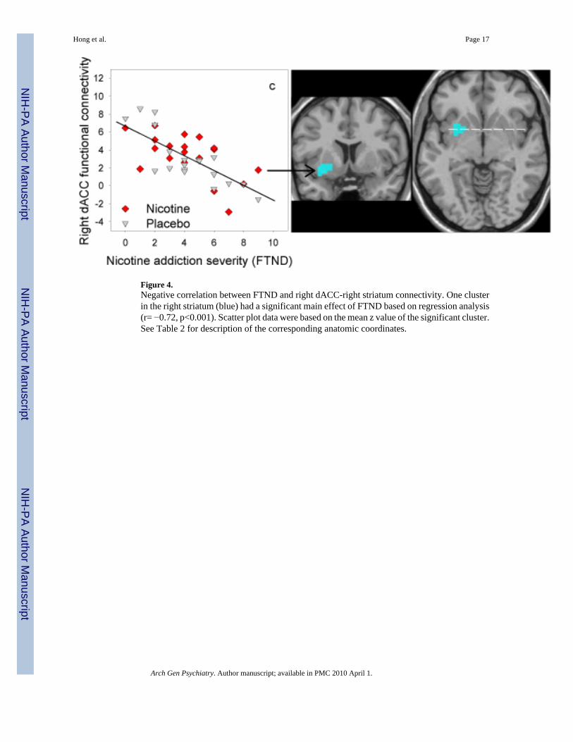
Figure 4.Negative correlation between FTND and right dACC-right striatum connectivity. One clusterin the right striatum (blue) had a significant main effect of FTND based on regression analysis(r= −0.72, p<0.001). Scatter plot data were based on the mean z value of the significant cluster.See Table 2 for description of the corresponding anatomic coordinates.
Hong et al. Page 17
Arch Gen Psychiatry. Author manuscript; available in PMC 2010 April 1.
NIH
-PA Author Manuscript
NIH
-PA Author Manuscript
NIH
-PA Author Manuscript

Figure 5.Significant main effect of drug on cingulate circuitry based on paired t-tests. Seven resting-state functional connectivity pathways were statistically enhanced by nicotine administration(red) when compared to placebo. See Table 3 for the coordinates and the extents of eachsignificant cluster. There was never a statistically significant reduction in functionalconnectivity by nicotine. Ovals indicate the seed ROIs. Arrows infer the relationship betweenthe seed ROI and the areas that were affected by nicotine and do not imply directionality ofthe functional connectivity path. These figures represent the nicotine-placebo differences inFigure 2 that were statistically significant.
Hong et al. Page 18
Arch Gen Psychiatry. Author manuscript; available in PMC 2010 April 1.
NIH
-PA Author Manuscript
NIH
-PA Author Manuscript
NIH
-PA Author Manuscript

NIH
-PA Author Manuscript
NIH
-PA Author Manuscript
NIH
-PA Author Manuscript
Hong et al. Page 19Ta
ble
1
A c
ompa
rison
of n
icot
ine
vs. p
lace
bo re
late
d m
easu
rem
ents
. The
re w
as n
o si
gnifi
cant
bia
s on
smok
ing
leve
l prio
r to
each
con
ditio
n ba
sed
on si
mila
r CO
leve
ls p
rior t
o pl
aceb
o vs
. nic
otin
e pa
tch
appl
icat
ions
. Pos
t-sca
n ni
cotin
e le
vels
con
firm
ed ro
bust
diff
eren
ces i
n dr
ug le
vels
dur
ing
plac
ebo
vs. n
icot
ine
scan
ning
.
Plac
ebo
patc
h co
nditi
on(n
=19)
Nic
otin
e pa
tch
cond
ition
(n=1
9)t v
alue
adf
p va
lue
CO
leve
l prio
r to
patc
hap
plic
atio
n27
.2 ±
13.
0b25
.0 ±
11.
51.
3418
0.20
Nic
otin
e le
vel a
t the
end
of s
can
(ng/
ml)
3.0
± 4.
734
.3 ±
13.
19.
3214
c<0
.001
*
Cha
nged in
pul
se ra
te−4
.9 ±
8.5
−2.9
± 1
1.3
0.78
180.
44C
hang
e in
syst
olic
blo
odpr
essu
re3.
9 ±
13.4
9.6
± 12
.32.
1218
0.05
*
Cha
nge
in d
iast
olic
blo
odpr
essu
re5.
8 ±
6.3
6.4
± 10
.30.
2718
0.79
Cha
nge
in w
ithdr
awal
/sid
eef
fect
sym
ptom
s0.
7 ±
1.4
0.8
± 1.
30.
0017
1.00
Cha
nge
in se
lf-re
porte
d m
ood
0.8
± 2.
91.
5 ±
4.8
0.90
180.
38
a Paire
d t-t
est.
b Mea
n ±
stan
dard
dev
iatio
n.
c Nic
otin
e le
vels
wer
e av
aila
ble
in 1
5 su
bjec
ts fo
r bot
h ni
cotin
e an
d pl
aceb
o pa
tch
cond
ition
s. N
icot
ine
leve
ls w
ere
not a
vaila
ble
in o
ne su
bjec
t in
plac
ebo,
two
subj
ects
in n
icot
ine,
1 su
bjec
t in
both
plac
ebo
and
nico
tine
cond
ition
s due
to la
bora
tory
err
ors (
bloo
d sa
mpl
e he
mol
ysis
or e
quip
men
t pro
blem
).
d Cha
nges
from
pre
-pat
ch a
pplic
atio
n to
imm
edia
tely
prio
r to
patc
h re
mov
al, a
bout
4.5
hou
r int
erva
l.
* Stat
istic
ally
sign
ifica
nt
Arch Gen Psychiatry. Author manuscript; available in PMC 2010 April 1.

NIH
-PA Author Manuscript
NIH
-PA Author Manuscript
NIH
-PA Author Manuscript
Hong et al. Page 20Ta
ble
2
Effe
cts o
f nic
otin
e ad
dict
ion
seve
rity
(FTN
D) o
n ci
ngul
ate
func
tiona
l con
nect
ivity
. Mai
n ef
fect
s of F
TND
wer
e fo
und
in 3
func
tiona
lly c
oher
ent c
ircui
ts:
betw
een
left
dAC
C a
nd b
ilate
ral s
triat
a (a
righ
t ant
erov
entra
l clu
ster
and
a le
ft po
ster
iodo
rsal
clu
ster
); an
d be
twee
n rig
ht d
AC
C a
nd a
righ
t ant
erov
entra
lst
riata
l clu
ster
. The
FTN
D e
ffec
ts o
n th
ese
circ
uits
wer
e in
the
nega
tive
dire
ctio
n in
bot
h pl
aceb
o an
d ni
cotin
e co
nditi
ons,
as il
lust
rate
d by
thei
r cor
rela
tion
coef
ficie
nts.
Cor
rela
tion
coef
ficie
nts f
or e
ach
cond
ition
wer
e ca
lcul
ated
by
extra
ctin
g th
e m
ean
z sc
ore
of e
ach
sign
ifica
nt c
lust
er a
nd c
orre
latin
g th
em w
ithFT
ND
scor
es. N
ote
that
all
3 ci
ngul
ate-
stria
tal c
orre
latio
ns re
mai
ned
sign
ifica
nt e
ven
afte
r a B
onfe
rron
i cor
rect
ion
for 1
4 co
mpa
rison
s (p<
0.00
36),
in th
em
ain
anal
ysis
whe
re n
icot
ine
cond
ition
and
pla
cebo
con
ditio
n w
ere
com
bine
d (s
ince
ther
e w
as n
o in
tera
ctio
n). H
owev
er, i
f we
sepa
rate
nic
otin
e co
nditi
onan
d pl
aceb
o co
nditi
on a
s an
expl
orat
ory
anal
ysis
, we
foun
d al
l 3 c
onne
ctiv
ities
rem
ain
sign
ifica
nt in
pla
cebo
con
ditio
n, b
ut o
nly
one
is si
gnifi
cant
in th
eni
cotin
e co
nditi
on. T
he a
nato
mic
bou
ndar
ies o
f the
sign
ifica
nt c
lust
ers a
re g
iven
bel
ow. A
lso
see
Figu
re 3
and
4.a
. Rig
ht st
riatu
m, a
mor
e an
tero
vent
ral
clus
ter c
ompa
red
to b
; it i
nclu
des p
arts
of p
utam
en, v
entra
l stri
atum
, and
cla
ustru
m. T
he e
dge
of th
e cl
uste
r als
o ov
erla
ps w
ith th
e in
sula
, the
am
ygda
le a
ndth
e pa
rahi
ppoc
ampa
l gyr
usb.
Lef
t stri
atum
, inc
ludi
ng p
art o
f put
amen
, cau
date
tail,
cla
ustru
m a
nd in
sula
.c. R
ight
stria
tum
, ess
entia
lly th
e sa
me
loca
tion
asa,
an
ante
rove
ntra
l clu
ster
that
ext
ends
into
par
ts o
f put
amen
, ven
tral s
triat
um, a
nd c
laus
trum
. The
edg
e of
the
clus
ter o
verla
ps w
ith th
e am
ygda
la a
nd th
epa
rahi
ppoc
ampa
l gyr
us.*
Not
sign
ifica
nt if
app
lyin
g B
onfe
rron
i cor
rect
ion
for 1
4 co
mpa
rison
s
Seed
RO
IsC
o-ac
tivat
ing
Loc
atio
nsV
olum
e (m
m3 )
Tal
arai
ch C
oodi
nate
s of M
axim
aPl
aceb
o an
d N
icot
ine
Tri
als
Com
bine
dPl
aceb
o T
rial
s Alo
neN
icot
ine
Tri
als A
lone
xy
zr
valu
ep
valu
er
valu
ep
valu
er
valu
ep
valu
e
Left
dAC
Ca.
Rig
ht S
triat
um12
4231
−4−5
−0.7
6<0
.001
−0.8
4<0
.001
−0.6
8<0
.001
b. L
eft S
triat
um70
2−3
5−1
3−2
−0.6
9<0
.001
−0.8
3<0
.001
−0.5
70.
01*
Rig
ht d
AC
Cc.
Rig
ht st
riatu
m94
527
−3−4
−0.7
2<0
.001
−0.8
2<0
.001
−0.6
00.
007*
Arch Gen Psychiatry. Author manuscript; available in PMC 2010 April 1.

NIH
-PA Author Manuscript
NIH
-PA Author Manuscript
NIH
-PA Author Manuscript
Hong et al. Page 21Ta
ble
3
Cin
gula
te fu
nctio
nal c
onne
ctiv
ity c
ircui
ts e
nhan
ced
by n
icot
ine
Seed
RO
IsFu
nctio
nally
Con
nect
ed R
egio
ns E
nhan
ced
by N
icot
ine
Loc
atio
nL
ater
ality
Bro
dman
n A
rea
Vol
ume
Tal
arai
ch C
oodi
nate
s of M
axim
a (x
, y, z
)
Left
dAC
Ca.
Sup
erio
r par
ieta
l lob
ule
and
post
cent
ral g
yrus
Con
trala
tera
lB
A 5
/799
919
−44
62
Left
vPC
Cb.
Med
ial f
ront
al g
yrus
and
AC
CB
ilate
ral
BA
10/
3212
960
4612
Left
dPC
Cc.
Pos
terio
r cin
gula
teB
ilate
ral
BA
23/
3064
8−1
−54
19Le
ft sA
CC
d. M
edia
l fro
ntal
gyr
usIp
sila
tera
lB
A 8
459
−11
3844
Rig
ht M
CC
e. P
ostc
entra
l gyr
us a
nd In
ferio
rpa
rieta
l lob
ule
Con
trala
tera
lB
A 5
/40
1107
−30
−37
56
Rig
ht v
PCC
f. M
edia
l fro
ntal
gyr
usB
ilate
ral
BA
9/1
037
8−1
4713
Rig
ht d
PCC
g. M
edia
l fro
ntal
gyr
usB
ilate
ral
BA
9/1
045
93
5919
Arch Gen Psychiatry. Author manuscript; available in PMC 2010 April 1.