Improvement of Handoff Latency by GPS based Handoff Technique
Upload
independentCategory
view
5download
0
This article was published in an Elsevier journal. The attached copyis furnished to the author for non-commercial research and
education use, including for instruction at the author’s institution,sharing with colleagues and providing to institution administration.
Other uses, including reproduction and distribution, or selling orlicensing copies, or posting to personal, institutional or third party
websites are prohibited.
In most cases authors are permitted to post their version of thearticle (e.g. in Word or Tex form) to their personal website orinstitutional repository. Authors requiring further information
regarding Elsevier’s archiving and manuscript policies areencouraged to visit:
http://www.elsevier.com/copyright

Author's personal copy
Reduced microstructural integrity of the white matter underlyinganterior cingulate cortex is associated with increased saccadiclatency in schizophrenia
Dara S. Manoach,a,c,⁎ G. Avinash Ketwaroo,d Frida E. Polli,a,e Katharine N. Thakkar,a
Jason J.S. Barton,f Donald C. Goff,a Bruce Fischl,b,c,g
Mark Vangel,b,c and David S. Tuchb,c,d,1
aDepartment of Psychiatry, Massachusetts General Hospital, Harvard Medical School, Boston, MA 02215, USAbDepartment of Radiology, Massachusetts General Hospital, Harvard Medical School, Boston, MA 02215, USAcAthinoula A. Martinos Center for Biomedical Imaging, Charlestown, MA 02129, USAdDivision of Health Sciences and Technology, Harvard Medical School, and Massachusetts Institute of Technology, Cambridge, MA 02139, USAeDepartment of Psychology, Suffolk University, Boston, MA 02114, USAfDepartments of Neurology, Ophthalmology and Visual Sciences, University of British Columbia, Vancouver, BC, CanadagComputer Science and Artificial Intelligence Laboratory, Massachusetts Institute of Technology, Cambridge, MA 02139, USA
Received 6 February 2007; revised 28 March 2007; accepted 28 April 2007Available online 21 May 2007
The anterior cingulate cortex (ACC) is a key component of a networkthat directs both spatial attention and saccadic eye movements, whichare tightly linked. Diffusion tensor imaging (DTI) has demonstratedreduced microstructural integrity of the anterior cingulum bundle asindexed by fractional anisotropy (FA) in schizophrenia, but thefunctional significance of these abnormalities is unclear. Using DTI, weexamined the white matter underlying anterior cingulate cortex inschizophrenia to determine whether reduced FA is associated withprolonged latencies of volitional saccades. Seventeen chronic, medi-cated schizophrenia outpatients and nineteen healthy controls hadhigh-resolution DTI scans. FA maps were registered to structural scansand mapped across participants using a surface-based coordinatesystem. Cingulate white matter was divided into rostral and dorsalanterior regions and a posterior region. Patients showed reduced FA incingulate white matter of the right hemisphere. Reduced FA in thewhite matter underlying anterior cingulate cortex, frontal eye field,and posterior parietal cortex of the right hemisphere was associatedwith longer saccadic latencies in schizophrenia, though given therelatively small sample size, these relations warrant replication. Thesefindings demonstrate that in schizophrenia, increased latency ofvolitional saccades is associated with reduced microstructural integrityof the white matter underlying key cortical components of a right-hemisphere dominant network for visuospatial attention and ocularmotor control. Moreover, they suggest that anterior cingulate whitematter abnormalities contribute to slower performance of volitional
saccades and to inter-individual variability of saccadic latency inchronic, medicated schizophrenia.© 2007 Published by Elsevier Inc.
Keywords: Anterior cingulate cortex; Antisaccade; Cingulum bundle;Diffusion tensor imaging; Schizophrenia; White matter
The anterior cingulate cortex (ACC) is a key participant in thedirection of both spatial attention (Mesulam, 1981) and eye gaze,which are tightly linked (Hunt and Kingstone, 2003; Klein andMcCormick, 1989). In neuroimaging studies, the ACC showsincreased activation when ocular motor control is required,including on prosaccade and antisaccade tasks. Prosaccades are aprepotent response of looking towards a suddenly appearing visualtarget. Antisaccades require inhibition of the prepotent prosaccadeand the generation of the novel behavior of looking away from atarget. The ACC shows increased activation during prosaccadescompared to fixation (Brown et al., 2006), greater activation forantisaccades than prosaccades (e.g., Brown et al., 2006; Doricchi etal., 1997; Ford et al., 2005; Manoach et al., 2007; Paus et al.,1993), and reduced activity in association with deficient anti-saccade performance in schizophrenia (Crawford et al., 1996).Specifically, the posterior part of the dorsal ACC has been labeledthe ‘cingulate eye field’ on the basis of its involvement in tasksrequiring volitional, but not reflexive saccadic control (Gaymard etal., 1998; Paus et al., 1993; Pierrot-Deseilligny et al., 2004).Lesions of the posterior dorsal ACC increase antisaccade errors(Milea et al., 2003) and prolong the latencies of both pro- andantisaccades (Gaymard et al., 1998). Schizophrenia is characterized
www.elsevier.com/locate/ynimgNeuroImage 37 (2007) 599–610
⁎ Corresponding author. Massachusetts General Hospital, 36 First Avenue,Room 420, Charlestown, MA 02129, USA. Fax: +1 617 726 0504.
E-mail address: [email protected] (D.S. Manoach).1 Current address: Novartis Pharma AG, Basel, Switzerland.Available online on ScienceDirect (www.sciencedirect.com).
1053-8119/$ - see front matter © 2007 Published by Elsevier Inc.doi:10.1016/j.neuroimage.2007.04.062

Author's personal copy
by abnormal performance of volitional saccades (e.g., Calkins etal., 2003; Harris et al., 2006; Radant et al., 2007) and bothfunctional and structural abnormalities of the ACC (for reviews,see Benes, 2000; Holcomb, 2004). In a previous study wereported significant slowing of correct prosaccade and antisac-cade trials, demonstrating that in schizophrenia, even correctvolitional saccades are abnormal (Manoach et al., 2002). In thepresent study, using the same saccadic paradigm, we investigatedwhether abnormalities of the white matter (WM) underlyingdorsal ACC might contribute to the slowing of correct volitionalsaccades.
We focus on the ACC because unlike other key corticalcomponents of the ocular motor network (i.e., frontal, parietal, andsupplementary eye fields) (McDowell and Clementz, 2001), there isabundant evidence of both structural and functional abnormalities ofthe ACC in schizophrenia. Moreover, functional MRI evidencesuggests that the ACC contributes to performance of our saccadictask in healthy individuals (Polli et al., 2005a), while showingabnormal activity in schizophrenia (Polli et al., 2005b). In additionto reports of ACC gray matter reductions in schizophrenia (e.g.,Goldstein et al., 1999; Ha et al., 2004; Kuperberg et al., 2003;Mitelman et al., 2005; Ohnuma et al., 1997; Sigmundsson et al.,2001; Suzuki et al., 2002; Yamasue et al., 2004), there is evidence ofvolume reductions in the underlying WM (McDonald et al., 2005;Mitelman et al., 2005), and histopathological evidence of dis-turbances in micro- and macrocircuitry that might alter commu-nication between the ACC and connected regions (for reviews, seeBenes, 1993, 2000).
We examined the microstructural integrity of the WM under-lying ACC using diffusion tensor imaging (DTI). DTI is an MRItechnique that can detect white matter pathology in vivo. DTI wasused to index the fractional anisotropy (FA) of water diffusion,which reflects the degree of directional coherence of water diffusionin tissue. While FA correlates with axon myelination (Harsan et al.,2006), not all of the biophysical determinants of WM diffusionanisotropy are fully understood (Beaulieu, 2002). Consistent withother evidence of ACC WM abnormalities in schizophrenia,previous DTI studies have found reduced FA of the cingulumbundle, the WM tract that underlies cingulate cortex (Ardekani etal., 2003; Hao et al., 2006; Kubicki et al., 2003; Sun et al., 2003;Wang et al., 2004), although there are also several negative reports(Agartz et al., 2001; Buchsbaum et al., 1998; Burns et al., 2003;Foong et al., 2002). Various factors may contribute to thesediscrepancies, such as differences in sample composition withregard to course of illness and/or medication regimen. Given thelack of standard methods for DTI acquisition and analysis,methodological differences likely play an important role (Jones etal., 2005; Kanaan et al., 2005).
Some previous DTI studies of the cingulum bundle inschizophrenia employed voxel-based analyses that depend onregistering brains to a particular atlas, averaging across partici-pants, and then testing for group differences at each voxel (e.g.,Agartz et al., 2001; Ardekani et al., 2003; Buchsbaum et al., 1998;Foong et al., 2002). Healthy individuals show a high degree ofinter-subject variability of brain morphology, particularly inregions involved in higher cognitive function (Brett et al., 2002;Rajkowska and Goldman-Rakic, 1995), and in schizophreniavariability is even greater (Park et al., 2004a). Morphologicvariability can confound inter-group comparisons of registered dataas it may result in the averaging of disparate brain regions, whichcould artifactually lower and/or lead to greater variability of FA
measurements, which depend on the organizational coherence ofWM. Defining regions of interest (ROIs) in the unregisteredimages of individual participants avoids the problems of inter-subject registration, but results may differ depending on whichportion of the cingulum is sampled. Several studies used geometric(e.g., spherical or rectangular) ROIs placed on slices in the anteriorand/or posterior cingulum (Burns et al., 2003; Sun et al., 2003;Wang et al., 2004) while another study sampled only the middlecingulum since the most anterior and posterior portions are affectedby a high degree of curvature that can confound the measurementof FA (Kubicki et al., 2003). If cingulum abnormalities areconfined to specific subregions, selective sampling may miss groupdifferences.
The present study employed high-resolution DTI and corticalsurface-based analyses (Dale et al., 1999; Fischl et al., 1999a) toinvestigate the microstructural integrity of cingulate white matter inschizophrenia. High-resolution DTI acquisition enabled accuratemeasurements of FA in the WM underlying the entire cingulategyrus in spite of the high degree of curvature. This avoids biasesinherent in sampling only from selected regions. The primaryanalyses involved defining ROIs based on anatomical landmarks inindividual participants, measuring FA in the WM underlying eachvertex (surface equivalent of voxel), averaging FA values acrossvertices in each ROI for each participant, and comparing groups.
We complemented the ROI analyses with vertex-wise analyses.This involved surface-based inter-subject registration and groupcomparisons of FA at every vertex of the registered data. Unlikevolumetric approaches that rely only on intensity information,surface-based registration employs a non-rigid alignment algorithmto explicitly align cortical folding patterns and should be relativelyrobust to inter-individual differences in the gyral and sulcal anatomyof cingulate cortex (Dale et al., 1999; Fischl et al., 1999a). Whilesurface-based procedures have been extensively applied in func-tional magnetic resonance imaging studies and to measure corticalthickness (e.g., Kuperberg et al., 2003; Rosas et al., 2002), this is thefirst application to DTI.
We divided the cingulate cortex into anterior and posteriorsegments and the anterior segment was further divided into rostraland dorsal regions. This division is based on differences incytoarchitecture, function, and connectivity (Bush et al., 1998,2000; Devinsky et al., 1995; Vogt et al., 1979; Whalen et al., 1998).Although previous work primarily implicates anterior cingulate inschizophrenia (Benes, 2000; Holcomb, 2004; Kerns et al., 2005;Laurens et al., 2003; Tamminga et al., 2000), we also investigatedthe WM of the posterior cingulate to determine the regionalspecificity of FA reductions and because several recent studies reportposterior cingulate cortex abnormalities (Mitelman et al., 2005;Shimizu et al., 2007; Suzuki et al., 2005).
We expected to find FA reductions in anterior cingulate WM inschizophrenia and wanted to determine whether reduced FA,specifically in the WM underlying dorsal ACC, was associatedwith prolonged latencies of correct volitional saccades. Individualdifferences in cognitive processing speed have been proposed toreflect WM physiology, particularly myelination (e.g., Luciano etal., 2004), based on the well-established role of WM myelinthickness and axon diameter in determining conduction velocity.Recent reports of relations between FA (which reflects myelination(Harsan et al., 2006) and other WM microstructural properties(Beaulieu, 2002)) and cognitive reaction time lend indirect supportto this proposal (Madden et al., 2004; Nestor et al., 2007; Tuch et al.,2005). On this basis, we reasoned that dorsal ACC WM micro-
600 D.S. Manoach et al. / NeuroImage 37 (2007) 599–610

Author's personal copy
structure might contribute to volitional saccadic latency, and thatmicrostructural abnormalities, by impairing communication in theocular motor network, could contribute to prolonged latencies inschizophrenia. Because prosaccade and antisaccade trials werepresented in a pseudorandom sequence, both trial types requiredvigilance to instructional cues and task switching. Thus, theprosaccade trials of the present study were not purely ‘reflexive’,rather they were cognitively demanding volitional saccades. Insummary, we expected that reduced FA in theWMunderlying dorsalACC would be related to prolonged latencies of volitionalprosaccades and antisaccades in schizophrenia, as might be expectedbased on a previous lesion study (Gaymard et al., 1998).
Methods
Participants
(Table 1 provides demographic information.) The schizophreniasample was comprised of 17 chronic outpatients recruited from anurban community mental health center, who had been maintained onstable doses of a variety of atypical antipsychotic medications for atleast 6 weeks. Diagnoses were confirmed with Structured ClinicalInterviews for DSM-IV (First et al., 1997). Clinical status wascharacterized with the Brief Psychiatric Rating Scale (BPRS)(Overall and Gorham, 1962), the Positive and Negative SyndromeScale (PANSS) (Kay et al., 1987), and the Scale for the Assessmentof Negative Symptoms (SANS) (Andreasen, 1983). Nineteenhealthy control participants, without a personal history of psychiatricillness or a family history of schizophrenia spectrum disorders, wererecruited from the community with poster advertisements.
Participants were screened to exclude substance abuse or depen-dence within the past 6 months, a history of head injury resulting in asustained loss of consciousness and/or cognitive sequelae, neuro-logical illness, and any disorder affecting cerebral metabolism. Allparticipants endorsed strong right-hand preference as determinedby a laterality score of 70 or above on the modified EdinburghHandedness Inventory (White and Ashton, 1976). The groups didnot differ with regard to age, sex, handedness, or parental socio-economic status (Hollingshead, 1965). Participants gave written in-formed consent. The study was approved by institutional review
boards at Massachusetts General Hospital and the MassachusettsDepartment of Mental Health.
Image acquisitionHead stabilization was achieved with cushioning and partici-
pants wore earplugs to attenuate scanner noise. Images werecollected using a 3.0-T Siemens Trio MRI scanner (SiemensMedical System, Iselin, NJ). Automated shimming procedures wereperformed and scout images were obtained. Two high-resolutionstructural images were acquired in the sagittal plane for sliceprescription, spatial normalization (spherical and Talairach), andcortical surface reconstruction using a high-resolution 3D magne-tization prepared rapid gradient echo (MPRAGE) sequence(repetition time (TR), 2530 ms; echo spacing, 7.25 ms; echo time(TE), 3 ms; flip angle 7°) with an in-plane resolution of 1 mm and1.3 mm slice thickness. Single-shot EPI DTI was acquired using atwice refocused spin echo sequence (Reese et al., 2003) with thefollowing sequence parameters: TR/TE=8400/82 ms; b=700s/mm2; NEX=1; ten T2 images acquired with b=0; 72 diffusiondirections; 128×128 matrix; 2×2 mm in-plane resolution; 64 axialoblique (AC–PC) slices; 2-mm (0 mm gap) slice thickness; scanduration 12′44″. The n=72 diffusion directions were obtained usingthe electrostatic shell algorithm (Jones, 2004).
Acquisition of behavioral dataSaccadic latency and directional accuracy measurements were
acquired during magnetoencephalography scanning using elec-trooculography. Two bipolar pairs of EOG electrodes wereplaced to capture vertical and horizontal eye movements andblinks. The signals were sampled at 600 Hz. The saccadic taskstimuli were generated using the Vision Shell programmingplatform (www.visionshell.com) and presented with a DigitalLight Processing (DLP Infocus 350) projector onto a back-projection screen placed 102 cm in front of the subject. The taskconsisted of a pseudorandom series of prosaccade and anti-saccade trials with randomly interspersed fixation intervals of 2,4, or 6 s. Saccadic trials were balanced for right and leftwardmovements. Fig. 1 provides a graphic depiction of the task and adescription of task parameters. Participants performed eight runsof the task, with short rests between runs. Each run lasted 5 min22 s. The total experiment lasted about 1 h and generated a totalof 278 prosaccade 285 antisaccade trials and 107 fixationintervals.
Scoring and analysis of eye movement dataEOG data were scored in MATLAB (Mathworks, Natick, MA)
using a partially automated program that determined the direc-tional accuracy of each saccade with respect to the requiredresponse and the latency from target onset. For each trial, saccadiconset was defined as the point preceding peak velocity at whichthe horizontal eye-position trace deviated from fixation (Fig. 1).To determine this point an automated algorithm started at the pointof peak velocity and searched the eye- position trace backwards tofixation. Fixation was defined as the time point at which the slopeof the eye-position trace was zero as determined by evaluating theslope in relation to the two preceding time points, a movingwindow of 5 ms. Algorithm results were visually inspected toensure accuracy. Only trials with saccades in the desired directionand latencies between 130 and 800 ms were considered correct,and only correct saccades were included in the latency analyses.The cutoff of 130 ms excluded anticipatory saccades, which are
Table 1Means, standard deviations, and group comparisons of demographic dataand rating scale scores
Subject characteristics Healthysubjects(n=19)
Schizophreniasubjects(n=17)
t p
Age 36±13 41±12 1.27 0.21Sex 12M/7F 13M/4F ϕ=0.14 0.39Handedness
(Edinburgh)92±10 86±19 1.27 0.21
Parental SES a 2.1±1.2 2.6±1.0 z=1.4 0.17Age of onset 24.5±7.0Length of illness (years) 16.7±10.2
Level of severityBPRS 15.5±8.2 MinimalPANSS positive 13.0±5.4 MildPANSS negative 16.8±6.2 MildSANS 32.7±16.2 Questionablea A lower score denotes higher status.
601D.S. Manoach et al. / NeuroImage 37 (2007) 599–610

Author's personal copy
executed too quickly to be a valid response to the appearance ofthe target (Doricchi et al., 1997; Fischer and Breitmeyer, 1987;Straube et al., 1999). Trials with eye blinks (defined as verticalpeak-to-peak EOG amplitude exceeding 200 μV) prior to saccadicresponse were rejected from further analysis.
DTI analysisThe objective of the DTI analysis was to measure and conduct
group comparisons of FA in the WM underlying specific regions.We relied primarily on ROI analyses that were based on individualanatomy. We complemented ROI analyses with a vertex-wiseanalysis of registered group data. For both analyses, FA wasmeasured in the undeformed WM 2 mm below the WM/gray matterboundary. This strategy minimizes partial volume contributionsfrom cortical gray matter and preserves regional specificity.
DTI data were analyzed using the followingmulti-step procedure(detailed below): (1) motion/eddy current distortion correction; (2)tensor reconstruction; (3) registration of FA and T1 volumes; (4)projection of subcortical FA values onto the WM/gray matterinterface; (5) and group comparisons of average FA values in ROIsbased on the unregistered data of each participant (ROI analysis).The vertex-wise analysis involved the following additional steps: (6)
registration of surface FA maps across participants and (7) groupcomparisons of FAvalues of the WM underlying each vertex on thecortical surface.
(1) The raw diffusion data were corrected for head motion andresidual eddy current distortion by registering all images to the firstT2 image, which was acquired with b=0. The registration wasperformed using the FLIRT tool (Jenkinson and Smith, 2001) fromthe FSL software library (http://www.fmrib.ox.ac.uk/fsl). The re-gistration used a global affine (12 degrees of freedom) transforma-tion, a mutual information cost function, and sinc resampling. (2)The diffusion tensor and FAvolumes were then reconstructed usingthe standard least squares fit to the log diffusion signal (Basser et al.,1994).
(3) FA volumes were registered to the high-resolution structural(T1) volumes for each participant using the T2 volume as anintermediary. The T2 volume was taken from the b=0 image in theDTI acquisition and was therefore in register with the FA volume.The T2 intermediary was employed (a) to avoid potential bias thatmight arise from using the FAvolume in the registration, since FA isthe variable of interest and (b) because the gray/white boundary inthe T1 volume has better correspondence with the T2 volume thanthe FA volume, allowing for a more accurate registration.
Fig. 1. Saccadic paradigm. Saccadic trials lasted 4000 ms and began with an instructional cue at screen center. For half of the participants, an orange ring was thecue for a PS trial and a blue X the cue for an AS trial. These cues were reversed for the rest of the participants. The cue was flanked horizontally by two smallgreen squares of 0.2° side that marked the potential locations of stimulus appearance, 10° left and right of center. These squares remained on the screen for theduration of each run. At 300 ms the instructional cue was replaced by a green fixation ring at screen center with a diameter of 0.4° and luminance of 20 cd/m2.After 1700 ms the ring shifted to one of the two stimulus locations, right or left, with equal probability. This ring was the stimulus to which participantsresponded. The green ring remained in the peripheral location for 1000 ms and then returned to the center where participants were instructed to return their gazefor 1000 ms. Fixation intervals were simply a continuation of the fixation display that constituted the final second of the previous saccadic trial.
602 D.S. Manoach et al. / NeuroImage 37 (2007) 599–610

Author's personal copy
Each participant's averaged T2 volume was manually registeredto the T1 volume with Tkregister2 (http://surfer.nmr.mgh.harvard.edu) using 9 degrees of freedom including translation, rotation, andscaling. Manual registration was used to (a) maximize anatomicagreement along the medial wall and (b) because echo planarimaging susceptibility distortions in DTI can confound globalregistration procedures. The resulting spatial transformation wasapplied to the FA volume, thus bringing the FA and T1 volumesinto register.
(4) For each participant, a surface representation of the WM/gray matter boundary was derived from the T1 volume using apreviously described segmentation, surface reconstruction, andinflation algorithm (Dale et al., 1999; Fischl et al., 1999a). FA wassampled in the WM 2 mm below the WM/gray matter boundary foreach vertex on the surface and then projected onto the WM/grayinterface. FA values were smoothed using n=50 iterations ofreplacement by nearest neighbors. This corresponds to smoothingby approximately 10-mm FWHM on the surface.
(5) Cingulate cortex was divided into rostral and dorsal ACCand posterior cingulate cortex in each hemisphere for eachparticipant by an investigator (GAK) who was blind to groupmembership. To differentiate between anterior and posteriorcingulate cortex we employed an automated surface-basedparcellation system (Fischl et al., 2004). The ACC was furtherdivided into dorsal and rostral regions by drawing a line per-pendicular to the intercommisural plane at the anterior boundary ofthe genu of the corpus callosum (Devinsky et al., 1995). FA valuesof the WM underlying each vertex were averaged for each ROI –dorsal and rostral anterior cingulate cortex (dACC, rACC) andposterior cingulate cortex (PCC) – for each hemisphere and werecompared between groups using a repeated measures analysis ofvariance with group, region, and hemisphere as factors. As acontrol analysis, the surface areas of the ROIs were compared todetermine if there were group differences in the size of the regionssampled. Group differences in hemispheric asymmetry of FA wereexamined using a difference score that controls for overall dif-ferences in FA values: (LH−RH) / (LH+RH), where LH and RHare the FA values in the left and right hemispheres for a particularROI.
(6) For the vertex-wise analysis, FA maps were registeredacross participants. This was achieved by registering the T1 surfacemaps across participants based on gyral folding patterns (Fischl etal., 1999b) and then applying these transformations to the FAmaps, which were in register with T1 maps (see steps 3 and 4).
(7) To test for significant group differences in FA in theregistered group data, a t-test was performed in the WM un-derlying each vertex. A false discovery rate (FDR) threshold ofq=0.05 was applied to control for multiple comparisons(Genovese et al., 2002). For the purposes of calculating the FDRsignificance threshold, the hypothesis space was restricted tocingulate regions, which were defined in the registered group datausing the same anatomical definitions as in the ROI analysis (seestep 5). Statistical maps were displayed on a template brainconsisting of the averaged cortical surface of an independentsample of 40 adults from the Buckner laboratory at WashingtonUniversity.
Saccadic regressionsThe association between cingulate WM FA values and saccadic
latency, adjusting both variables for age, was investigated usingmultiple regression analyses for each group separately since the
groups differed in variability for both latency and FA. For theseregressions FAwas the dependent variable and saccadic latency andage were covariates. The mean latency of correct prosaccades orantisaccades was the covariate of interest. Age was regarded as apotential confound given the documented relation of increasing agewith decreasing FA (Pfefferbaum et al., 2000; Salat et al., 2005) andlonger antisaccade and prosaccade latencies (Munoz et al., 1998).Using age as a covariate, we also examined the relation of FA toantipsychotic dose as indexed by chlorpromazine equivalent(Woods, 2003). The ROI regressions used the mean FA value foreach cingulum ROI of each subject. A vertex-wise regression wasalso performed at each vertex of the cortical surface for the registeredgroup data using a nominal threshold of p≤0.005 since none of thep-values were significant at FDR q=0.05.
Results
Saccadic performance
One control participant was excluded from all analyses involvingbehavioral data as an outlier since his error rates for antisaccade andprosaccade trials were, respectively, 4.8 and 3.0 standard deviationsabove the group mean. Schizophrenia participants showed a trend tolonger response latencies for both prosaccades (t(33)=1.82,p=0.08; control: 231±41 ms; schizophrenia: 268±75 ms) andantisaccades (t(33)=1.82, p=0.08; control: 280±52 ms; schizo-phrenia: 320±77 ms). Error rates were logit transformed priorto analysis. Patients made more antisaccade errors than controls(t(33)=2.37, p=0.02; control: 11±9%; schizophrenia: 17±9%),but did not differ in their prosaccade error rate (t(33)=0.71,p=0.48; control: 4±3%; schizophrenia: 6±7%).
Group differences in FA
ROI analysesPatients showed reduced FA in cingulate WM compared to
controls (F(1,34)=9.36, p=0.004), and there was a significantgroup by hemisphere interaction (F(1,34)=10.20, p=0.003) (Fig.2). This was due to greater FA reductions in right vs. left cingulateWM in patients. Compared to controls, patients showed signi-ficantly decreased FA in right cingulate WM (F(1,34)=14.57,p=0.0005), and there was a trend to a group by region interaction(F(1,34)=3.01, p=0.06) indicating larger differences in anteriorthan posterior cingulate WM regions (rACB: t(34)=3.42, p=0.002;dACB: t(34)=3.39, p=0.002; PCB: t(34)=2.49, p=0.02). In theleft hemisphere there was only a trend to reduced cingulate WM FAin patients (F(1,34)=3.05, p=0.09), no group by region interaction(F(1,34)=0.86, p=0.43), and only the group difference in rACCWM approached significance (rACC: t(34)=1.84, p=0.08; dACC:t(34)=1.42, p=0.17; PCC: t(34)=0.84, p=0.41). In summary, therewas significantly reduced FA in the WM of all three right cingulateROIs in schizophrenia.
Schizophrenia participants also showed greater asymmetry ofcingulateWMFA (leftN right) than controls as indexed by differencescores (F(1,34)=9.74, p=0.004) and although region did notinteract with group (F(1,34)=1.16, p=0.32), patients showedsignificantly greater leftward asymmetry than controls in dACCWM (t(34)=3.11, p=0.004), but not in rACC WM (t(34)=0.51,p=0.62) or PCC WM (t(34)=1.52, p=0.14). Within the patientgroup, FAwas significantly lower in the right than left hemisphere indACC WM (t(16)=4.45, p=0.0004) and PCC WM (t(16)=3.04,
603D.S. Manoach et al. / NeuroImage 37 (2007) 599–610

Author's personal copy
p=0.008), but not rACC WM (t(16)=0.96, p=0.35). Controlparticipants did not show significant FA asymmetries in cingulateWM (rACC: t(18)=−1.65, p=0.12; dACC: t(18)=0.40, p=0.70;PCC: t(18)=1.49, p=0.15).
The surface areas of the individual ROIs did not differ betweengroups (pN0.35 for all ROIs).
Vertex-wise analysisThis analysis reproduced the findings of significantly reduced
FA in the WM underlying rACC, dACC, and PCC of the right, butnot the left hemisphere (Fig. 3). Although it was not included in thehypothesis space for FDR correction, reduced FA was alsoobserved bilaterally in the corpus callosum, consistent with otherstudies (e.g., Agartz et al., 2001; Ardekani et al., 2003; Foong etal., 2000). Table 2 provides approximate Talairach coordinates forthe surface locations of peak differences within the cingulate.(Approximate Talairach coordinates were derived by mappingsurface-based coordinates back to the original structural volume foreach participant, registering the volumes to the MontrealNeurological Institute (MNI305) atlas (Collins et al., 1994), andaveraging the MNI305 coordinates that corresponded to the surfacepeak across participants. The resulting coordinates were trans-formed to standard Talairach space using an algorithm developedby Matthew Brett (http://imaging.mrc-cbu.cam.ac.uk/imaging/MniTalairach)).
Within-group regressions of FA and saccadic latency
ROI analysisThere were significant relations between reduced FA in the WM
of right dACC and rACC and longer latencies for both pro- andantisaccades in schizophrenia (Table 3 and Fig. 4). Controls showeda significant relation between reduced FA in right rACC WM andprosaccade latency. There were no correlations between saccadiclatency and FA in the WM of right PCC or in any of the lefthemisphere ROIs in either group. A comparison of the coefficientsbetween patients and controls revealed that group differences in the
strength of the relations between latency and FA met or approachedsignificance only in right dACC WM (antisaccades: z(34)=2.04,p=0.04; prosaccades: z(34)=1.67, p=0.10). No relations wereobserved between antipsychotic dose and FA (pN0.35 in all ROIs)or saccadic latency (pN0.25 for pro- and antisaccades). AdjustingFA and latency values for years of illness instead of age did notsubstantially change the findings.
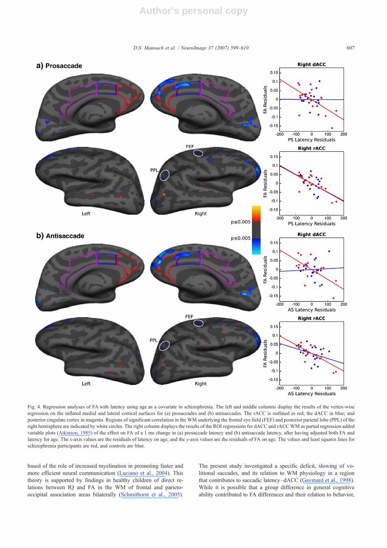
Vertex-wise analysisNo significant relations were seen using a FDR threshold that
corrected for multiple comparisons. With a nominal threshold ofp≤ .005 we reproduced the ROI findings of significant relationsbetween reduced FA in right rACC and dACC WM and prolongedlatencies of both pro- and antisaccades in schizophrenia (Fig. 4;Table 2 provides approximate Talairach coordinates and exact p-values). The right dACC area showing a relation overlaps with thearea of significant FA reduction in schizophrenia. There were alsorelations in the WM of other regions including the frontal eye field(Paus, 1996) and posterior parietal lobe of the right hemisphere forboth pro- and antisaccades. In left dACC WM there was a smallarea where greater FA was associated with longer prosaccadelatencies in the schizophrenia group. In controls, saccadic latencywas not associated with FA in cingulate WM of either hemisphereor in the WM underlying the frontal eye field or posterior parietallobe.
Discussion
Relative to controls, patients with schizophrenia showed reducedFA in cingulate WM of the right hemisphere. Moreover, lower FA inthe WM underlying anterior cingulate cortex, frontal eye field andposterior parietal cortex of the right hemisphere was associated withlonger latencies of volitional saccades. These three densely inter-connected regions comprise the key cortical components of a right-hemisphere dominant network for the spatial distribution ofattention (Gitelman et al., 1999; Mesulam, 1981, 1990) and alsoplay a critical role in ocular motor control (Pierrot-Deseilligny et al.,
Fig. 2. Bar graphs of mean FAvalues with standard error bars for each ROI in control and schizophrenia participants. The brackets indicate comparisons betweenthe bars indicated. The left vs. right comparisons within the schizophrenia and control groups are based on difference scores. C=control, L=left hemisphere,R=right hemisphere, Sz=schizophrenia.
604 D.S. Manoach et al. / NeuroImage 37 (2007) 599–610

Author's personal copy
2004). Thus, the present findings demonstrate that WM micro-structural integrity in a network for spatially directed attention andthe execution of volitional saccades is associated with saccadiclatency in schizophrenia.
The present findings complement a recent report of a relationbetween increased reaction time on a task measuring selectiveattention and decreased volume of the right cingulum bundle inschizophrenia (Nestor et al., 2007). More generally, findings ofrelations between reaction time and FA in other regions support thehypothesis that WM microstructure contributes to individualdifferences in the speed of cognitive processing (Madden et al.,2004; Tuch et al., 2005). This leads us to hypothesize that reducedintegrity of anterior cingulate WM impairs communication in theocular motor network and by so doing contributes to slower latenciesof correct volitional saccades and to inter-individual variability ofsaccadic latency in schizophrenia.
Frontal eye field, posterior parietal cortex, and dorsal ACC are allthought to contribute to saccadic latency. Pre-target activity in thefrontal eye field has been shown to predict the latency of both
prosaccades and antisaccades based on single unit recordings inmonkeys (Everling and Munoz, 2000) and functional MRI inhumans (Connolly et al., 2005). In humans, damage to frontal eyefield, posterior parietal cortex, or to the WM underlying theseregions is associated with increased saccadic latency (Pierrot-Deseilligny et al., 1987). Right dorsal ACC lesions have also beenfound to increase the latencies of pro- and antisaccades (Gaymard etal., 1998).
One may question why, if the same network is implicated insaccadic latency in both patients and controls, was the relationbetween FA in right dACC WM and saccadic latency stronger inschizophrenia? While this may reflect the greater range of saccadiclatency in schizophrenia, a more intriguing possibility is that theabnormally reduced microstructural integrity of right dACCWM inschizophrenia contributes to slower saccadic responses. Theobservation that the dACC WM region, in which reduced FA issignificantly related to longer saccadic latencies, overlaps with theregion that shows significant FA reductions in schizophreniasupports this hypothesis (compare Figs. 3c and 4). In controls,
Fig. 3. Vertex-wise group measurements of FA in the registered group data displayed on the inflated medial cortical surface of the template brain. (a) Averagedgroup FA in healthy participants; (b) averaged group FA in schizophrenia participants; (c) p-value map for group differences in FA at an FDR threshold ofq≤0.05. The rACC is outlined in red; the dACC in blue; and posterior cingulate cortex in magenta.
605D.S. Manoach et al. / NeuroImage 37 (2007) 599–610

Author's personal copy
dACCWM integrity may be adequate for saccades, and if this werethe case, variability in WM microstructure would play a less sig-nificant role in determining saccadic latency.
Neuroimaging, electrophysiological, and lesion studies haveimplicated the dorsal ACC in volitional saccades, but we also,unexpectedly, found a relation of saccadic latency to FA in the WMunderlying right rostral ACC in both patients and controls. Werecently reported differential activity in the rostral ACC for errorvs. correct antisaccades in healthy participants (Polli et al., 2005a).The timing of activation suggested that rACC contributed both toresponse preparation and evaluation, an interpretation that isconsistent with previous findings (e.g., Drevets and Raichle, 1998;van Veen and Carter, 2002). The role of the rACC in performanceoptimization may account for the relation of greater integrity ofrACC WM to faster saccadic responses.
In the present study, saccadic latencies were associated withlower FA primarily in the right hemisphere. In humans, the righthemisphere is thought to play a dominant role in directing bothspatial attention (Heilman and Valenstein, 1979; Mesulam, 1981)and eye gaze (De Renzi et al., 1982; Meador et al., 1989). Weinterpret the asymmetric relations we observed as being compatiblewith evidence of right-hemisphere specialization for spatial attentionand volitional ocular motor control.
Moreover, relative to controls, patients showed significantlyreduced FA in cingulate WM of the right hemisphere only, andsignificantly greater leftN right asymmetry of FA in dACC WM.Within the patient group, FA inWMunderlying dACC and PCCwassignificantly lower in the right than the left hemisphere. Controls didnot show any significant asymmetries of FA in cingulate WM.Previous studies have reported either right (Hao et al., 2006) orbilateral (Ardekani et al., 2003; Kubicki et al., 2003; Wang et al.,2004) cingulum bundle FA reductions in schizophrenia, andleftN right asymmetry of FA in the cingulum bundle in healthyparticipants (Gong et al., 2005) and in both schizophrenia patientsand controls (Kubicki et al., 2003; Park et al., 2004b; Wang et al.,2004). In two of three studies, asymmetry, though present, wasreduced in schizophrenia (Park et al., 2004b;Wang et al., 2004). Thisdiffers from our finding of significant asymmetry only inschizophrenia, which was not predicted and therefore warrantsreplication. We interpret our findings of abnormally asymmetricaland reduced FA in right dACC WM in schizophrenia, and therelation of this reduction to longer saccadic latencies, to suggest apathological process affecting WM rather than normal develop-mental causes of asymmetry that are associated with functionallateralization.
Several potential limitations of the present study are consideredbelow. First, it is important to emphasize that FA is an indirectmeasureof WM microstructure and because its biophysical mechanisms arenot completely understood, it is not possible to attribute the relationswe observed to a particular WM property. WM density and volumemay have contributed to group differences in FA and its relationswith saccadic latency, a possibility that requires further study.
Second, group differences in general cognitive ability maycontribute to our findings. In this and many other studies of schi-zophrenia, participants are matched for parental socioeconomicstatus rather than participant socioeconomic status or IQ. The pri-mary rationale for this strategy is that reduced IQ in schizophrenia,which predates the onset of illness and then further declines(Seidman et al., 2006), may reflect cognitive deficits that are coreto schizophrenic pathology, and not an artifact that should becontrolled (Meehl, 1970). Obtaining valid estimates of premorbidIQ in schizophrenia is also problematic since amotivation, psy-chosis, and related disruptions of attention may prevent optimalengagement in cognitive testing. Tests that are relatively robust tothese effects, such as single word reading and spelling (Dalby andWilliams, 1986), are sensitive to educational attainment, whichmay also be compromised by schizophrenia and its prodrome. Arelationship between WM physiology and IQ has been theorized
Table 2Approximate Talairach coordinates corresponding to surface locations ofpeak group differences and latency–FA correlations in cingulate whitematter in the vertex-wise analyses
Location Talairachcoordinates
p-value
x y z
Group FAdifferences
rACC 11 52 5 0.000076 37 15 0.00009
dACC 12 13 27 0.0001rACC/dACC border 12 27 27 0.0002PCC 3 −27 11 0.0003
PS latency–FA correlation
rACC 11 51 18 0.0001dACC 12 9 30 0.00007rACC/dACC border 12 26 25 0.0003
AS latency–FA correlation
rACC 11 51 18 0.00001dACC 12 9 30 0.0003rACC/dACC border 12 26 25 0.0004
PS=prosaccades, AS=antisaccades, rACC=rostral anterior cingulate whitematter, dACC=dorsal anterior cingulate white matter, PCC=posteriorcingulate white matter.
Table 3Change in FA due to a 1s change in latency, from a regression model that adjusts both FA and latency for the effect of age
Right Left
rACC dACC PCC rACC dACC PCC
Controls(n=18)
PS Estimate −0.51±0.24 −0.007±0.28 −0.09±0.19 −0.08±0.30 −0.34±0.20 −0.09±0.26p 0.05 * 0.98 0.64 0.81 0.11 0.73
AS Estimate −0.28±0.20 0.05±0.22 −0.07±0.15 0.05±0.24 −0.20±0.17 0.10±0.21p 0.18 0.81 0.65 0.84 0.24 0.64
Schizophrenia(n=17)
PS Estimate −0.49±0.12 −0.58±0.20 −0.12±0.13 −0.23±0.17 −0.11±0.17 −0.15±0.12p 0.001 * 0.01 * 0.34 0.20 0.53 0.25
AS Estimate −0.50±0.12 −0.56±0.20 −0.14±0.12 −0.21±0.17 −0.16±0.17 −0.16±0.12p 0.001 * 0.02 * 0.27 0.23 0.35 0.21
The results are presented as changes, corresponding standard errors, and p-values.* Indicates a significant relation.
606 D.S. Manoach et al. / NeuroImage 37 (2007) 599–610
Author's personal copy
based of the role of increased myelination in promoting faster andmore efficient neural communication (Luciano et al., 2004). Thistheory is supported by findings in healthy children of direct re-lations between IQ and FA in the WM of frontal and parieto-occipital association areas bilaterally (Schmithorst et al., 2005).
The present study investigated a specific deficit, slowing of vo-litional saccades, and its relation to WM physiology in a regionthat contributes to saccadic latency–dACC (Gaymard et al., 1998).While it is possible that a group difference in general cognitiveability contributed to FA differences and their relation to behavior,
Fig. 4. Regression analyses of FA with latency using age as a covariate in schizophrenia. The left and middle columns display the results of the vertex-wiseregression on the inflated medial and lateral cortical surfaces for (a) prosaccades and (b) antisaccades. The rACC is outlined in red; the dACC in blue; andposterior cingulate cortex in magenta. Regions of significant correlation in the WM underlying the frontal eye field (FEF) and posterior parietal lobe (PPL) of theright hemisphere are indicated by white circles. The right column displays the results of the ROI regressions for dACC and rACCWM as partial regression addedvariable plots (Atkinson, 1985) of the effect on FA of a 1 ms change in (a) prosaccade latency and (b) antisaccade latency, after having adjusted both FA andlatency for age. The x-axis values are the residuals of latency on age, and the y-axis values are the residuals of FA on age. The values and least squares lines forschizophrenia participants are red, and controls are blue.
607D.S. Manoach et al. / NeuroImage 37 (2007) 599–610

Author's personal copy
if it were the primary factor, one would expect to see widespreadchanges and correlations, rather than the fairly circumscribed onesthat we observed.
Third, this studymay have suffered from a lack of power due to therelatively small sample size, particularly with regard to groupdifferences in saccadic latency and its associations with FA. Whileour primary (ROI) analysis showed the predicted significant relationsbetween FA in dACC WM and saccadic latency in patients, in thevertex-wise analysis the significance of these relations did not passcorrection for multiple comparisons. In addition, unlike our previousstudy of this task, group differences in saccadic latency approached,but did not meet, statistical significance (Manoach et al., 2002). Othergroups have also reported longer latencies for correct prosaccadesand/or antisaccades (e.g., Fukushima et al., 1994; Harris et al., 2006;Hutton et al., 1998) but these findings are not consistent (e.g.,Clementz et al., 1994). These inconsistencies likely stem fromvariations in task design that determine which cognitive processes arerequired, but also reflect differences in statistical power. This isillustrated by a recent multi-site study of antisaccades that showedsignificantly increased latency of correct antisaccades in schizo-phrenia (n=143 patients, 195 controls, pb0.0001) (Radant et al.,2007). The magnitude of the group difference for antisaccade latencywas comparable to that of the present study (37 and 40 ms res-pectively). Although latencies for patients were longer at all sevensites, the difference reached statistical significance only at one site.(The probability of all seven sites having the same direction of groupdifference by chance is 0.016). Along with the present data, thissuggests that correct volitional saccades have longer latencies inschizophrenia, but that larger samples are required to reliablydemonstrate this effect due to its size and to measurement variabilityin schizophrenia.
Finally, in the present study, the schizophrenia sample waslimited to chronic, medicated patients. Antipsychotic dose was notrelated to saccadic latency or to FA in the cingulum ROIs. While itwould be difficult to account for the findings on the basis ofmedications, particularly with regard to their lateralization, theeffects of antipsychotic medications on WM microstructure areunknown and cannot be ruled out as a factor in the current findings.It is also possible that reduced WM microstructure might be relatedto the progression of schizophrenia and would not be seen inpatients early in the course of illness.
In summary, this study documented FA reductions in the WMunderlying cingulate cortex in chronic, medicated schizophreniaand related these abnormalities to saccadic latency. These findingsdemonstrate an association between deficits in volitional ocularmotor control and reduced microstructural integrity of the WMunderlying key cortical components of a network for saccadicexecution. They suggest that ACC WM abnormalities contribute toslower performance of volitional saccades and to inter-individualvariability of saccadic latency in schizophrenia.
Acknowledgments
The authors gratefully acknowledge technical assistance fromDavid Salat, Josh Snyder, Thomas Benner, and Szymon Mikulskiand thoughtful comments from Emi Takahashi. Support was pro-vided by the National Institute for Mental Health (R01 67720, R0131340, PO1 31154), National Center for Research Resources (P41-RR14075, R01 RR16594-01A1, BIRN002, U24 RR021382),GlaxoSmithKline, Athinoula A. Martinos Foundation, MentalIllness and Neuroscience Discovery (MIND) Institute (DOE-DE-
FG02-99ER62764), National Institute for Biomedical Imaging andBioengineering (R01 EB001550; National Alliance for MedicalImage Computing (NAMIC) U54 EB005149 funded through theNIH Roadmap for Medical Research).
References
Agartz, I., Andersson, J.L., Skare, S., 2001. Abnormal brain white matter inschizophrenia: a diffusion tensor imaging study. NeuroReport 12,2251–2254.
Andreasen, N.C., 1983. Scale for the Assessment of Negative Symptoms(SANS). University of Iowa, Iowa City.
Ardekani, B.A., Nierenberg, J., Hoptman, M.J., Javitt, D.C., Lim, K.O.,2003. MRI study of white matter diffusion anisotropy in schizophrenia.NeuroReport 14, 2025–2029.
Atkinson, A.C., 1985. Plots, Transformations and Regression: AnIntroduction to Graphical Methods of Diagnostic Regression Analysis.Oxford Univ. Press, New York.
Basser, P.J., Mattiello, J., LeBihan, D., 1994. MR diffusion tensor spec-troscopy and imaging. Biophys. J. 66, 259–267.
Beaulieu, C., 2002. The basis of anisotropic water diffusion in the nervoussystem—A technical review. NMR Biomed. 15, 435–455.
Benes, F.M., 1993. Neurobiological investigations in cingulate cortex ofschizophrenic brain. Schizophr. Bull. 19, 537–549.
Benes, F.M., 2000. Emerging principles of altered neural circuitry inschizophrenia. Brain Res. Brain Res. Rev. 31, 251–269.
Brett, M., Johnsrude, I.S., Owern, A.M., 2002. The problem of functionallocalization in the human brain. Nat. Rev., Neurosci. 3, 243–249.
Brown, M.R., Goltz, H.C., Vilis, T., Ford, K.A., Everling, S., 2006.Inhibition and generation of saccades: rapid event-related fMRI ofprosaccades, antisaccades, and nogo trials. NeuroImage 33, 644–659.
Buchsbaum, M.S., Tang, C.Y., Peled, S., Gudbjartsson, H., Lu, D., Hazlett,E.A., et al., 1998. MRI white matter diffusion anisotropy and PETmetabolic rate in schizophrenia. NeuroReport 9, 425–430.
Burns, J., Job, D., Bastin, M.E., Whalley, H., Macgillivray, T., Johnstone,E.C., Lawrie, S.M., 2003. Structural disconnectivity in schizophrenia: adiffusion tensor magnetic resonance imaging study. Br. J. Psychiatry182, 439–443.
Bush, G., Whalen, P.J., Rosen, B.R., Jenike, M.A., McInerney, S.C., Rauch,S.L., 1998. The counting stroop: an interference task specialized forfunctional neuroimaging—validation study with functional MRI. Hum.Brain Mapp. 6, 270–282.
Bush, G., Luu, P., Posner, M.I., 2000. Cognitive and emotional influences inanterior cingulate cortex. Trends Cogn. Sci. 4, 215–222.
Calkins, M.E., Iacono, W.G., Curtis, C.E., 2003. Smooth pursuit andantisaccade performance evidence trait stability in schizophrenia patientsand their relatives. Int. J. Psychophysiol. 49, 139–146.
Clementz, B.A., McDowell, J.E., Zisook, S., 1994. Saccadic system func-tioning among schizophrenia patients and their first-degree biologicalrelatives. J. Abnorm. Psychology 103, 277–287.
Collins, D.L., Neelin, P., Peters, T.M., Evans, A.C., 1994. Automatic 3Dintersubject registration of MR volumetric data in standardized Talairachspace. J. Comput. Assist. Tomogr. 18, 192–205.
Connolly, J.D., Goodale, M.A., Goltz, H.C., Munoz, D.P., 2005. fMRIactivation in the human frontal eye field is correlated with saccadicreaction time. J. Neurophysiol. 94, 605–611.
Crawford, T.J., Puri, B.K., Nijran, K.S., Jones, B., Kennard, C., Lewis, S.W.,1996. Abnormal saccadic distractibility in patients with schizophrenia: a99mTc-HMPAO SPET study. Psychol. Med. 26, 265–277.
Dalby, J.T., Williams, R., 1986. Preserved reading and spelling ability inpsychotic disorders. Psychol. Med. 16, 171–175.
Dale, A.M., Fischl, B., Sereno, M.I., 1999. Cortical surface-based analysis:I. Segmentation and surface reconstruction. NeuroImage 9, 179–194.
De Renzi, E., Colombo, A., Faglioni, P., Gibertoni, M., 1982. Conjugategaze paresis in stroke patients with unilateral damage: an unexpectedinstance of hemispheric asymmetry. Arch. Neurol. 39, 482–486.
608 D.S. Manoach et al. / NeuroImage 37 (2007) 599–610

Author's personal copy
Devinsky, O., Morrell, M.J., Vogt, B.A., 1995. Contributions of anteriorcingulate cortex to behaviour. Brain 118 (Pt 1), 279–306.
Doricchi, F., Perani, D., Inoccia, C., Grassi, F., Cappa, S.F., Bettinardi, V.,et al., 1997. Neural control of fast-regular saccades and antisaccades: aninvestigation using positron emission tomography. Exp. Brain Res. 116,50–62.
Drevets, W.C., Raichle, M.E., 1998. Reciprocal suppression of regionalcerebral blood flow during emotional versus higher cognitive processes:implications for interactions between emotion and cognition. Cogn.Emot. 12, 353–385.
Everling, S., Munoz, D.P., 2000. Neuronal correlates for preparatory setassociated with pro-saccades and anti-saccades in the primate frontal eyefield. J. Neurosci. 20, 387–400.
First, M.B., Spitzer, R.L., Gibbon, M., Williams, J.B.W., 1997. Struc-tured Clinical Interview for DSM-IV Axis I Disorders, ResearchVersion, Patient Edition with Psychotic Screen (SCID-I/P W/PSYSCREEN). Biometrics Research, New York State Psychiatric Institute,New York.
Fischer, B., Breitmeyer, B., 1987. Mechanisms of visual attention revealedby saccadic eye movements. Neuropsychologia 25, 73–83.
Fischl, B., Sereno, M.I., Dale, A.M., 1999a. Cortical surface-based analysis:II. Inflation, flattening, and a surface-based coordinate system. Neuro-Image 9, 195–207.
Fischl, B., Sereno, M.I., Tootell, R.B.H., Dale, A.M., 1999b. High-resolution intersubject averaging and a coordinate system for the corticalsurface. Hum. Brain Mapp. 8, 272–284.
Fischl, B., van der Kouwe, A., Destrieux, C., Halgren, E., Segonne, F., Salat,D.H., 2004. Automatically parcellating the human cerebral cortex.Cereb. Cortex 14, 11–22.
Foong, J., Maier, M., Clark, C.A., Barker, G.J., Miller, D.H., Ron, M.A.,2000. Neuropathological abnormalities of the corpus callosum inschizophrenia: a diffusion tensor imaging study. J. Neurol. Neurosurg.Psychiatry 68, 242–244.
Foong, J., Symms, M.R., Barker, G.J., Maier, M., Miller, D.H., Ron, M.A.,2002. Investigating regional white matter in schizophrenia usingdiffusion tensor imaging. NeuroReport 13, 333–336.
Ford, K.A., Goltz, H.C., Brown, M.R., Everling, S., 2005. Neural processesassociated with antisaccade task performance investigated with event-related fMRI. J. Neurophysiol. 94, 429–440.
Fukushima, J., Fukushima, K., Miyasaka, K., Yamashita, I., 1994. Voluntarycontrol of saccadic eye movement in patients with frontal cortical lesionsand parkinsonian patients in comparison with that in schizophrenics.Biol. Psychiatry 36, 21–30.
Gaymard, B., Rivaud, S., Cassarini, J.F., Dubard, T., Rancure, G., Agid, Y.,Pierrot-Deseilligny, C., 1998. Effects of anterior cingulate cortex lesionson ocular saccades in humans. Exp. Brain Res. 120, 173–183.
Genovese, C.R., Lazar, N.A., Nichols, T., 2002. Thresholding of statisticalmaps in functional neuroimaging using the false discovery rate.NeuroImage 15, 870–878.
Gitelman, D.R., Nobre, A.C., Parrish, T.B., LaBar, K.S., Kim, Y.H., Meyer,J.R., Mesulam, M., 1999. A large-scale distributed network for covertspatial attention: further anatomical delineation based on stringentbehavioural and cognitive controls. Brain 122, 1093–1106.
Goldstein, J.M., Goodman, J.M., Seidman, L.J., Kennedy, D.N., Makris, N.,Lee, H., et al., 1999. Cortical abnormalities in schizophrenia identifiedby structural magnetic resonance imaging. Arch. Gen. Psychiatry 56,537–547.
Gong, G., Jiang, T., Zhu, C., Zang, Y., Wang, F., Xie, S., et al., 2005.Asymmetry analysis of cingulum based on scale-invariant parameteriza-tion by diffusion tensor imaging. Hum. Brain Mapp. 24, 92–98.
Ha, T.H., Youn, T., Ha, K.S., Rho, K.S., Lee, J.M., Kim, I.Y., et al., 2004.Gray matter abnormalities in paranoid schizophrenia and their clinicalcorrelations. Psychiatry Res. 132, 251–260.
Hao, Y., Liu, Z., Jiang, T., Gong, G., Liu, H., Tan, L., et al., 2006. Whitematter integrity of the whole brain is disrupted in first-episodeschizophrenia. NeuroReport 17, 23–26.
Harris, M.S., Reilly, J.L., Keshavan, M.S., Sweeney, J.A., 2006. Longi-
tudinal studies of antisaccades in antipsychotic-naive first-episodeschizophrenia. Psychol. Med. 36, 485–494.
Harsan, L.A., Poulet, P., Guignard, B., Steibel, J., Parizel, N., de Sousa, P.L.,2006. Brain dysmyelination and recovery assessment by noninvasive invivo diffusion tensor magnetic resonance imaging. J. Neurosci. Res. 83,392–402.
Heilman, K.M., Valenstein, E., 1979. Mechanisms underlying hemispatialneglect. Ann. Neurol. 5, 166–170.
Holcomb, H.H., 2004. Practice, learning, and the likelihood of making anerror: how task experience shapes physiological response in patientswith schizophrenia. Psychopharmacology (Berlin) 174, 136–142.
Hollingshead, A.B., 1965. Two Factor Index of Social Position. YaleUniversity Press, New Haven, CT.
Hunt, A.R., Kingstone, A., 2003. Covert and overt voluntary attention:linked or independent? Brain Res. Cogn. Brain Res. 18, 102–105.
Hutton, S.B., Crawford, T.J., Puri, B.K., Duncan, L.J., Chapman, M.,Kennard, C., et al., 1998. Smooth pursuit and saccadic abnormalities infirst-episode schizophrenia. Psychol. Med. 28, 685–692.
Jenkinson, M., Smith, S., 2001. A global optimisation method for robustaffine registration of brain images. Med. Image Anal. 5, 143–156.
Jones, D.K., 2004. The effect of gradient sampling schemes on measuresderived from diffusion tensor MRI: a Monte Carlo study. Magn. Reson.Med. 51, 807–815.
Jones, D.K., Symms, M.R., Cercignani, M., Howard, R.J., 2005. The effectof filter size on VBM analyses of DT-MRI data. NeuroImage 26,546–554.
Kanaan, R.A., Kim, J.S., Kaufmann, W.E., Pearlson, G.D., Barker, G.J.,McGuire, P.K., 2005. Diffusion tensor imaging in schizophrenia. Biol.Psychiatry 58, 921–929.
Kay, S.R., Fiszbein, A., Opler, L.A., 1987. The positive and negative syn-drome scale (PANSS) for schizophrenia. Schizophr. Bull. 13, 261–276.
Kerns, J.G., Cohen, J.D., MacDonald III, A.W., Johnson, M.K., Stenger,V.A., Aizenstein, H., Carter, C.S., 2005. Decreased conflict- and error-related activity in the anterior cingulate cortex in subjects withschizophrenia. Am. J. Psychiatry 162, 1833–1839.
Klein, R., McCormick, P., 1989. Covert visual orienting: hemifield-activation can be mimicked by zoom lens and midlocation placementstrategies. Acta Psychol. (Amst) 70, 235–250.
Kubicki, M., Westin, C.F., Nestor, P.G., Wible, C.G., Frumin, M., Maier,S.E., 2003. Cingulate fasciculus integrity disruption in schizophrenia:a magnetic resonance diffusion tensor imaging study. Biol. Psychiatry54, 1171–1180.
Kuperberg, G.R., Broome, M.R., McGuire, P.K., David, A.S., Eddy, M.,Ozawa, F., 2003. Regionally localized thinning of the cerebral cortex inschizophrenia. Arch. Gen. Psychiatry 60, 878–888.
Laurens, K.R., Ngan, E.T., Bates, A.T., Kiehl, K.A., Liddle, P.F., 2003.Rostral anterior cingulate cortex dysfunction during error processing inschizophrenia. Brain 126, 610–622.
Luciano, M., Wright, M.J., Geffen, G.M., Geffen, L.B., Smith, G.A., Martin,N.G., 2004. A genetic investigation of the covariation among inspectiontime, choice reaction time, and IQ subtest scores. Behav. Genet. 34, 41–50.
Madden, D.J., Whiting, W.L., Huettel, S.A., White, L.E., MacFall, J.R.,Provenzale, J.M., 2004. Diffusion tensor imaging of adult agedifferences in cerebral white matter: relation to response time. Neuro-Image 21, 1174–1181.
Manoach, D.S., Lindgren, K.A., Cherkasova, M.V., Goff, D.C., Halpern,E.F., Intriligator, J., Barton, J.J.S., 2002. Schizophrenic subjects showdeficient inhibition but intact task-switching on saccadic tasks. Biol.Psychiatry 51, 816–826.
Manoach, D.S., Thakkar, K.N., Cain, M.S., Polli, F.E., Edelman, J.A.,Fischl, B., Barton, J.J.S., 2007. Neural activity is modulated by trialhistory: a functional magnetic resonance imaging study of the effects of aprevious antisaccade. J. Nuerosci. 27.
McDonald, C., Bullmore, E., Sham, P., Chitnis, X., Suckling, J., MacCabe,J., et al., 2005. Regional volume deviations of brain structure inschizophrenia and psychotic bipolar disorder: computational morpho-metry study. Br. J. Psychiatry 186, 369–377.
609D.S. Manoach et al. / NeuroImage 37 (2007) 599–610

Author's personal copy
McDowell, J.E., Clementz, B.A., 2001. Behavioral and brain imaging stu-dies of saccadic performance in schizophrenia. Biol. Psychol. 57, 5–22.
Meador, K.J., Loring, D.W., Lee, G.P., Brooks, B.S., Nichols, F.T.,Thompson, E.E., et al., 1989. Hemisphere asymmetry for eye gazemechanisms. Brain 112, 103–111.
Meehl, P.E., 1970. Nuisance variables and the ex post facto design. In:Radner, M., Winokur, S. (Eds.), Minnesota Studies in the Philosophy ofScience. University of Minnesota Press, Minneapolis, pp. 373–402.
Mesulam, M.M., 1981. A cortical network for directed attention andunilateral neglect. Ann. Neurol. 10, 309–325.
Mesulam, M.M., 1990. Large-scale neurocognitive networks and distributedprocessing for attention, language, andmemory. Ann.Neurol. 28, 597–613.
Milea, D., Lehericy, S., Rivaud-Pechoux, S., Duffau, H., Lobel, E., Capelle,L., et al., 2003. Antisaccade deficit after anterior cingulate cortexresection. NeuroReport 14, 283–287.
Mitelman, S.A., Shihabuddin, L., Brickman, A.M., Hazlett, E.A.,Buchsbaum, M.S., 2005. Volume of the cingulate and outcome inschizophrenia. Schizophr. Res. 72, 91–108.
Munoz, D.P., Broughton, J.R., Goldring, J.E., Armstrong, I.T., 1998. Age-related performance of human subjects on saccadic eye movement tasks.Exp. Brain Res. 121, 391–400.
Nestor, P.G., Kubicki, M., Spencer, K.M., Niznikiewicz, M., McCarley,R.W., Shenton, M.E., 2007. Attentional networks and cingulumbundle in chronic schizophrenia. Schizophr. Res. 90, 308–315.
Ohnuma, T., Kimura, M., Takahashi, T., Iwamoto, N., Arai, H., 1997. Amagnetic resonance imaging study in first-episode disorganized-typepatients with schizophrenia. Psychiatry Clin. Neurosci. 51, 9–15.
Overall, J.E., Gorham, D.R., 1962. The brief psychiatric rating scale.Psychol. Rep. 10, 799–812.
Park, H.J., Levitt, J., Shenton, M.E., Salisbury, D.F., Kubicki, M., Kikinis,R., et al., 2004a. An MRI study of spatial probability brain mapdifferences between first-episode schizophrenia and normal controls.NeuroImage 22, 1231–1246.
Park, H.J., Westin, C.F., Kubicki, M., Maier, S.E., Niznikiewicz, M., Baer,A., et al., 2004b. White matter hemisphere asymmetries in healthysubjects and in schizophrenia: a diffusion tensor MRI study. NeuroImage23, 213–223.
Paus, T., 1996. Location and function of the human frontal eye-field: aselective review. Neuropsychologia 34, 475–483.
Paus, T., Petrides, M., Evans, A.C., Meyer, E., 1993. Role of the human anteriorcingulate cortex in the control of oculomotor, manual, and speech responses:a positron emission tomography study. J. Neurophysiol. 70, 453–469.
Pfefferbaum, A., Sullivan, E.V., Hedehus, M., Lim, K.O., Adalsteinsson, E.,Moseley, M., 2000. Age-related decline in brain white matter anisotropymeasured with spatially corrected echo-planar diffusion tensor imaging.Magn. Reson. Med. 44, 259–268.
Pierrot-Deseilligny, C., Rivaud, S., Penet, C., Rigolet, M.H., 1987. Latenciesof visually guided saccades in unilateral hemispheric cerebral lesions.Ann. Neurol. 21, 138–148.
Pierrot-Deseilligny, C., Milea, D., Muri, R.M., 2004. Eye movement controlby the cerebral cortex. Curr. Opin. Neurol. 17, 17–25.
Polli, F.E., Barton, J.J., Cain, M.S., Thakkar, K.N., Rauch, S.L., Manoach,D.S., 2005a. Rostral and dorsal anterior cingulate cortex makedissociable contributions during antisaccade error commission. Proc.Natl. Acad. Sci. U. S. A. 102, 15700–15705.
Polli, F.E., Cain, M.S., Thakkar, K.N., Barton, J.J.S., Manoach, D.S.,2005b. Intact task-induced deactivation but decreased performanceevaluation during antisaccade errors in schizophrenia. Soc. Neurosci.(Washington, DC) (Program No. 876.12).
Radant, A.D., Dobie, D.J., Calkins, M.E., Olincy, A., Braff, D.L., Cadenhead,K.S., et al., 2007. Successful multi-site measurement of antisaccadeperformance deficits in schizophrenia. Schizophr. Res. 89, 320–329.
Rajkowska, G., Goldman-Rakic, P.S., 1995. Cytoarchitectonic definition ofprefrontal areas in the normal human cortex: II. Variability in locations ofareas 9 and 46 and relationship to the Talairach coordinate system.Cereb. Cortex 5, 323–337.
Reese, T.G., Heid, O., Weisskoff, R.M., Wedeen, V.J., 2003. Reduction of
eddy-current-induced distortion in diffusion MRI using a twice-refocused spin echo. Magn. Reson. Med. 49, 177–182.
Rosas, H.D., Liu, A.K., Hersch, S., Glessner, M., Ferrante, R.J., Salat, D.H.,et al., 2002. Regional and progressive thinning of the cortical ribbon inHuntington's disease. Neurology 58, 695–701.
Salat, D.H., Tuch, D.S., Greve, D.N., van der Kouwe, A.J., Hevelone, N.D.,Zaleta, A.K., et al., 2005. Age-related alterations in white matter micro-structure measured by diffusion tensor imaging. Neurobiol. Aging 26,1215–1227.
Schmithorst, V.J., Wilke, M., Dardzinski, B.J., Holland, S.K., 2005.Cognitive functions correlate with white matter architecture in a normalpediatric population: a diffusion tensor MRI study. Hum. Brain Mapp.26, 139–147.
Seidman, L.J., Buka, S.L., Goldstein, J.M., Tsuang, M.T., 2006. Intellectualdecline in schizophrenia: evidence from a prospective birth cohort 28year follow-up study. J. Clin. Exp. Neuropsychol. 28, 225–242.
Shimizu, E., Hashimoto,K.,Ochi, S., Fukami,G., Fujisaki,M., Koike, K., et al.,2007. Posterior cingulate gyrusmetabolic changes in chronic schizophreniawith generalized cognitive deficits. J. Psychiatr. Res. 41, 49–56.
Sigmundsson, T., Suckling, J., Maier, M., Williams, S., Bullmore, E.,Greenwood, K., et al., 2001. Structural abnormalities in frontal,temporal, and limbic regions and interconnecting white matter tracts inschizophrenic patients with prominent negative symptoms. Am. J.Psychiatry 158, 234–243.
Straube, A., Riedel, M., Eggert, T., Müller, N., 1999. Internally and ex-ternally guided voluntary saccades in unmedicated and medicatedschizophrenic patients: Part I. Saccadic velocity. Eur. Arch. PsychiatryClin. Neurosci. 249, 1–6.
Sun, Z., Wang, F., Cui, L., Breeze, J., Du, X., Wang, X., et al., 2003.Abnormal anterior cingulum in patients with schizophrenia: a diffusiontensor imaging study. NeuroReport 14, 1833–1836.
Suzuki, M., Nohara, S., Hagino, H., Kurokawa, K., Yotsutsuji, T., Kawasaki,Y., et al., 2002. Regional changes in brain gray and white matter inpatients with schizophrenia demonstrated with voxel-based analysis ofMRI. Schizophr. Res. 55, 41–54.
Suzuki, M., Zhou, S.Y., Hagino, H., Niu, L., Takahashi, T., Kawasaki, Y.,et al., 2005. Morphological brain changes associated with Schneider'sfirst-rank symptoms in schizophrenia: a MRI study. Psychol. Med. 35,549–560.
Tamminga, C.A., Vogel, M., Gao, X., Lahti, A.C., Holcomb, H.H., 2000.The limbic cortex in schizophrenia: focus on the anterior cingulate. BrainRes. Brain Res. Rev. 31, 364–370.
Tuch, D.S., Salat, D.H., Wisco, J.J., Zaleta, A.K., Hevelone, N.D., Rosas,H.D., 2005. Choice reaction time performance correlates with diffusionanisotropy in white matter pathways supporting visuospatial attention.Proc. Natl. Acad. Sci. U. S. A. 102, 12212–12217.
van Veen, V., Carter, C.S., 2002. The timing of action-monitoring processesin the anterior cingulate cortex. J. Cogn. Neurosci. 14, 593–602.
Vogt, B.A., Rosene, D.L., Pandya, D.N., 1979. Thalamic and corticalafferents differentiate anterior from posterior cingulate cortex in themonkey. Science 204, 205–207.
Wang, F., Sun, Z., Cui, L., Du, X., Wang, X., Zhang, H., et al., 2004.Anterior cingulum abnormalities in male patients with schizophreniadetermined through diffusion tensor imaging. Am. J. Psychiatry 161,573–575.
Whalen, P.J., Bush, G., McNally, R.J., Wilhelm, S., McInerney, S.C., Jenike,M.A., Rauch, S.L., 1998. The emotional counting Stroop paradigm: afunctional magnetic resonance imaging probe of the anterior cingulateaffective division. Biol. Psychiatry 44, 1219–1228.
White, K., Ashton, R., 1976. Handedness assessment inventory. Neuro-psychologia 14, 261–264.
Woods, S.W., 2003. Chlorpromazine equivalent doses for the newer atypicalantipsychotics. J. Clin. Psychiatry 64, 663–667.
Yamasue, H., Iwanami, A., Hirayasu, Y., Yamada, H., Abe, O., Kuroki, N.,et al., 2004. Localized volume reduction in prefrontal, temporolimbic,and paralimbic regions in schizophrenia: an MRI parcellation study.Psychiatry Res. 131, 195–207.
610 D.S. Manoach et al. / NeuroImage 37 (2007) 599–610
Copyright © 2022 FDOKUMEN