Diurnal cortisol amplitude and fronto-limbic activity in response to stressful stimuli
11
Diurnal cortisol amplitude and fronto-limbic activity in response to stressful stimuli Amy C. Cunningham-Bussel a,1, * , James C. Root a,1 , Tracy Butler a , Oliver Tuescher a,g , Hong Pan a,b , Jane Epstein a,b , Daniel S. Weisholtz a , Michelle Pavony a , Michael E. Silverman c , Martin S. Goldstein c , Margaret Altemus a , Marylene Cloitre d , Joseph LeDoux e , Bruce McEwen f , Emily Stern a,b , David Silbersweig a,b a Functional Neuroimaging Laboratory, Department of Psychiatry, Box 140, Rm 1302, Weill Cornell Medical College, 1300 York Avenue, New York, NY 10065, United States b Department of Psychiatry, Brigham and Women’s/Faulkner Hospitals, Harvard Medical School, United States c Mt. Sinai School of Medicine, One Gustave L. Levy Place, Box 1230, New York, NY 10029, United States d New York University School of Medicine, 550 First Avenue, New York, NY 10016, United States e Center for Neural Science, New York University, 4 Washington Place, Room 809, New York, NY 10003-6621, United States f The Rockefeller University, 1230 York Avenue, New York, NY 10021, United States g Department of Neurology, University of Freiburg, United States Received 15 September 2007; received in revised form 25 November 2008; accepted 25 November 2008 1. Introduction Animal research and recent human neuroimaging studies (Wang et al., 2005; Urry et al., 2006; Liberzon et al., Psychoneuroendocrinology (2009) 34, 694—704 KEYWORDS Amygdala; Medial prefrontal cortex; Hippocampus; Cortisol; Neuroimaging; Stress Summary The development and exacerbation of many psychiatric and neurologic conditions are associated with dysregulation of the hypothalamic pituitary adrenal (HPA) axis as measured by aberrant levels of cortisol secretion. Here we report on the relationship between the amplitude of diurnal cortisol secretion, measured across 3 typical days in 18 healthy individuals, and blood oxygen level dependant (BOLD) response in limbic fear/stress circuits, elicited by in-scanner presentation of emotionally negative stimuli, specifically, images of the World Trade Center (WTC) attack. Results indicate that subjects who secrete a greater amplitude of cortisol diurnally demonstrate less brain activation in limbic regions, including the amygdala and hippocampus/ parahippocampus, and hypothalamus during exposure to traumatic WTC-related images. Such initial findings can begin to link our understanding, in humans, of the relationship between the diurnal amplitude of a hormone integral to the stress response, and those neuroanatomical regions that are implicated as both modulating and being modulated by that response. # 2008 Elsevier Ltd. All rights reserved. * Corresponding author. Tel.: +1 212 746 8025; fax: +1 212 746 5818. E-mail address: [email protected] (A.C. Cunningham-Bussel). 1 Both authors contributed equally to this work. available at www.sciencedirect.com journal homepage: www.elsevier.com/locate/psyneuen 0306-4530/$ — see front matter # 2008 Elsevier Ltd. All rights reserved. doi:10.1016/j.psyneuen.2008.11.011
-
Upload
transnationalsupport -
Category
Documents
-
view
2 -
download
0
Transcript of Diurnal cortisol amplitude and fronto-limbic activity in response to stressful stimuli
Diurnal cortisol amplitude and fronto-limbic activityin response to stressful stimuli
Amy C. Cunningham-Bussel a,1,*, James C. Root a,1, Tracy Butler a,Oliver Tuescher a,g, Hong Pan a,b, Jane Epstein a,b, Daniel S. Weisholtz a,Michelle Pavony a, Michael E. Silverman c, Martin S. Goldstein c,Margaret Altemus a, Marylene Cloitre d, Joseph LeDoux e, Bruce McEwen f,Emily Stern a,b, David Silbersweig a,b
a Functional Neuroimaging Laboratory, Department of Psychiatry, Box 140, Rm 1302, Weill Cornell Medical College,1300 York Avenue, New York, NY 10065, United StatesbDepartment of Psychiatry, Brigham and Women’s/Faulkner Hospitals, Harvard Medical School, United StatescMt. Sinai School of Medicine, One Gustave L. Levy Place, Box 1230, New York, NY 10029, United StatesdNew York University School of Medicine, 550 First Avenue, New York, NY 10016, United StateseCenter for Neural Science, New York University, 4 Washington Place, Room 809, New York, NY 10003-6621, United StatesfThe Rockefeller University, 1230 York Avenue, New York, NY 10021, United StatesgDepartment of Neurology, University of Freiburg, United States
Received 15 September 2007; received in revised form 25 November 2008; accepted 25 November 2008
Psychoneuroendocrinology (2009) 34, 694—704
KEYWORDSAmygdala;Medial prefrontal cortex;Hippocampus;Cortisol;Neuroimaging;Stress
Summary The development and exacerbation of many psychiatric and neurologic conditionsare associated with dysregulation of the hypothalamic pituitary adrenal (HPA) axis as measured byaberrant levels of cortisol secretion. Herewe report on the relationship between the amplitude ofdiurnal cortisol secretion, measured across 3 typical days in 18 healthy individuals, and bloodoxygen level dependant (BOLD) response in limbic fear/stress circuits, elicited by in-scannerpresentation of emotionally negative stimuli, specifically, images of the World Trade Center(WTC) attack. Results indicate that subjects who secrete a greater amplitude of cortisol diurnallydemonstrate less brain activation in limbic regions, including the amygdala and hippocampus/parahippocampus, and hypothalamus during exposure to traumatic WTC-related images. Suchinitial findings can begin to link our understanding, in humans, of the relationship between thediurnal amplitude of a hormone integral to the stress response, and those neuroanatomicalregions that are implicated as both modulating and being modulated by that response.# 2008 Elsevier Ltd. All rights reserved.
ava i lab le at www.sc ienced i rect .com
journa l homepage: www.el sev ier.com/locate/psyneuen
* Corresponding author. Tel.: +1 212 746 8025; fax: +1 212 746 5818.E-mail address: [email protected]
(A.C. Cunningham-Bussel).1 Both authors contributed equally to this work.
0306-4530/$ — see front matter # 2008 Elsevier Ltd. All rights reservedoi:10.1016/j.psyneuen.2008.11.011
1. Introduction
Animal research and recent human neuroimaging studies(Wang et al., 2005; Urry et al., 2006; Liberzon et al.,
d.

Diurnal cortisol amplitude and fronto-limbic activity 695
2007) have drawn attention to the role of the amygdala,ventral medial prefrontal cortex (vmPFC) and hippocampusas regions critically involved in both emotional/fear proces-sing and regulation of the hypothalamic pituitary adrenal(HPA) axis (Herman et al., 2005; Urry et al., 2006). Activationof the HPA axis in response to stressors (physiological orpsychological disturbances perceived to threaten an organ-ism’s homeostasis) leads to a cascade of molecular eventsresulting in the production of glucocorticoids: cortisol inhumans and corticosterone in rodents (Herman and Seroogy,2006; Bremner, 2006). The HPA axis maintains a strong diurnalglucocorticoid rhythm, which peaks in the first 30 min afterawakening and falls throughout the remainder of the day tonegligible amounts during the evening nadir (Clow et al.,2004; Herman and Seroogy, 2006). The amount of cortisolreleased at the peak of the diurnal cycle can be similar to theamount released during a stress response (Herman and Sero-ogy, 2006).
Cortisol is involved in modulating many beneficial pro-cesses relevant to stress-related situations (Charney, 2004).However, prolonged or repeated stress has been associatedwith cognitive impairment and structural brain changes(McEwen, 2002; Lupien et al., 2002; Charney, 2004; McEwen,2005; Radley and Morrison, 2005; Oei et al., 2006).
Both stress and fear processes are interrelated as theyare modulated both by overlapping neuroanatomicalregions, such as the amygdala, hippocampus and prefrontalcortex and secreted products including cortisol. Indeed,prolonged exposure to stress hormones and limbic activityelicited under conditions of chronic stress may result inincreased arousal to novel stimuli, and the generation ofmemories of fear (Miller and McEwen, 2006). Extensiveanimal literature has indicated that the influence ofparticular limbic centers on HPA axis functioning is‘‘region and stressor specific’’ (Herman et al., 2005). Inhumans, physical stressors such as electric shock andpsychological stressors (e.g. mental arithmetic) can acti-vate the HPA axis, eliciting a cortisol response; howeverreliable associations between specific psychological stres-sors and HPA axis activation remains unclear, particularly inregards to emotion inducing stimuli (Kemeny and Dicker-son, 2004).
Psychiatric illnesses including post-traumatic stressdisorder (PTSD) and depression are frequently associatedwith dysregulated diurnal cortisol rhythm (McEwen, 2002;Sotres-Bayon et al., 2004; Clow et al., 2004; Neylan et al.,2005; Risbrough and Stein, 2006; Herman and Seroogy,2006; Rauch et al., 2006; Mackenzie et al., 2007). Withexceptions, many PTSD patients exhibit both hypocortiso-lemia and increased suppression of cortisol following dex-amethasone administration as compared to healthycontrols (Yehuda et al., 1991; Young and Breslau, 2004;Olff et al., 2006; Bierer et al., 2006). In contrast, depressedpatients often demonstrate both chronic diurnal hypercor-tisolemia and less dexamethasone suppression of cortisolrelative to healthy subjects (Kathol et al., 1989; Holsboer,2000; Yehuda, 2002; McEwen, 2005).
In addition to and not unrelated to their regulation of theHPA axis, the amygdala, mPFC and hippocampus are known toplay key roles in the processing of emotional (including fear)stimuli in the present study and in others (Lane et al., 1997;Gur et al., 2002; Taylor et al., 2003; Phan et al., 2004; Britton
et al., 2006; Dannlowski et al., 2007). Furthermore, patientswith psychiatric disorders, and anxiety disorders specifically,demonstrate aberrant fronto-limbic neural activity as docu-mented by a large number of neuroimaging studies (Rauchet al., 2006). To date, however, only a few studies have begunlinking these endocrine and neuroimaging findings in patient(Bonne et al., 2003; Liberzon et al., 2007) and healthy (Wanget al., 2005; Urry et al., 2006) populations. Urry et al. (2006)demonstrated in older adults an inverse relationship betweenthe diurnal cortisol rhythm (sampled for 1 week prior toscanning) and BOLD activation in regions such as the amyg-dala in response to emotionally negative pictures. The pre-sent study therefore, though distinct, builds upon theprevious work by Urry et al. (2006) to assess the relationshipbetween the amplitude of daily cortisol release and activa-tion in limbic brain regions (amygdala, mPFC, hippocampus/parahippocampus and hypothalamus) in response to emo-tionally negative, stress-related images of theWTC attacks inhealthy participants. In accordance with prior findings inhumans (Urry et al., 2006) and animals (Diamond et al.,1992; Herman et al., 2005; Herman and Mueller, 2006) wehypothesized that the hippocampus and vmPFC will demon-strate a negative correlation with an index of diurnal salivarycortisol and that the amygdala will demonstrate a positivecorrelation.
2. Materials and methods
2.1. Subjects
18 healthy right-handed subjects, 11 male and 7 female,average age of 31 (S.D. 8.8) participated in fMRI scanningas part of this research study. All subjects were present inNew York City at the time of the World Trade Centerattacks, but were not directly exposed to the disaster(all subjects were greater than 2 miles from the WTC siteon 9/11/2001.) No subject had a current or prior diagnosisof PTSD or any other psychiatric disorder, as assessed byStructured Clinical Interview for DSM-IV (SCID). Addition-ally, no subject was taking any medication. Subjects wererecruited through advertisements placed in newspapersand flyers distributed throughout New York. All subjectsgave informed consent prior to scanning. This studywas approved by the New York Presbyterian Hospital–—Weill/Cornell Institutional Review Board. Subjects werescanned an average of 939 days (S.D. 209) after the 9/11 attacks.
2.2. Study design and stimuli
Subjects participated in a single scanning session consistingof five runs. Each run included six blocks. During eachblock, eight full-color images corresponding to one (of six)experimental condition were individually presented. Eachimage was presented only once. Image stimulus conditionswere as follows: the World Trade Center (WTC) attack; NewYork City (NYC) unrelated to the attack; the Vietnam War(VIE); suburbs (SUB); countryside (COU); and scrambledimages (SCR) (Fig. 1). Scrambled images were created frommeaningful images using a MATLAB algorithm that rear-ranged groups of pixels, preventing recognition while pre-
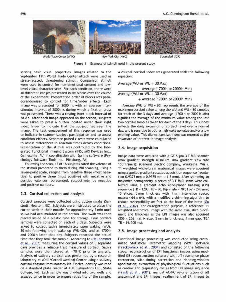
Figure 1 Example of stimuli used in the present study.
696 A.C. Cunningham-Bussel et al.
serving basic visual properties. Images related to theSeptember 11th World Trade Center attack were used asstress-related, threatening stimuli. Comparison stimuliwere used to control for non-emotional content and low-level visual characteristics. For each condition, there were40 different images presented in six blocks over the courseof the experiment. Presentation order of blocks was pseu-dorandomized to control for time/order effects. Eachimage was presented for 2000 ms with an average inter-stimulus interval of 2800 ms during which a fixation crosswas presented. There was a resting inter-block interval of28.8 s. After each image appeared on the screen, subjectswere asked to press a button located under their rightindex finger to indicate that the subject had seen theimage. The task engagement of this response was usedto indicate in scanner subject participation and to assesscondition effects. Separate paired t-tests were calculatedto assess differences in reaction times across conditions.Presentation of the stimuli was controlled by the Inte-grated Functional imaging System (IFIS; MRI Devices Inc.,Gainesville, FL) in coordination with Eprime software (Psy-chology Software Tools Inc., Pittsburg, PA).
Following the scan, 17 of 18 subjects rated the valence ofthe stimuli presented to them during MRI scanning, using aseven-point scale, ranging from negative three (most nega-tive) to positive three (most positive) with negative andpositive valences represented, respectively, by negativeand positive numbers.
2.3. Cortisol collection and analysis
Cortisol samples were collected using cotton swabs (Sar-stedt, Newton, NC). Subjects were instructed to place thecotton swab in their mouths for approximately 2 min untilsaliva had accumulated in the cotton. The swab was thenplaced inside of a plastic tube for storage. Four cortisolsamples were collected on each of 3 days. Subjects wereasked to collect saliva immediately upon waking (WU),30 min following their wake up (WU+30), and at 1700 hand 2000 h later that day. Subjects recorded the actualtime that they took the sample. According to (Hellhammeret al., 2007) measuring the cortisol values on 3 separatedays provides a reliable trait measure of cortisol. Salivasamples were then stored at �70 8C prior to analysis.Analysis of salivary cortisol was performed by a researchlaboratory at Weill/Cornell Medical Center using a salivarycortisol enzyme immunoassay kit. Optical density was readon a standard plate reader at 450 (Salimetrics LLC, StateCollege, PA). Each sample was divided into two wells andassayed twice in order to ensure reliability of the sample.
A diurnal cortisol index was generated with the followingequation:
AverageðWU or WUþ 30MaxÞ� Averageð1700 h or 2000 h MinÞ
AverageðWU or WUþ 30MaxÞþ Averageð1700 h or 2000 h MinÞ
Average (WU or WU + 30) represents the average of themaximum cortisol value among the WU and WU + 30 samplesfor each of the 3 days and Average (1700 h or 2000 h Min)signifies the average of the minimum value among the lasttwo cortisol samples taken for each of the 3 days. This indexreflects the daily excursion of cortisol level over a normalday, and is sensitive to both a high wake up value and/or a lowevening value. This diurnal cortisol index was entered as thecovariate of interest in image analysis.
2.4. Image acquisition
Image data were acquired with a GE Signa 3 T MRI-scanner(max gradient strength 40 mT/m, max gradient slew rate150 T/(m/s)) (General Electric Company, Waukesha, Wis.).T1 weighted whole-brain anatomical images were acquiredusing a spoiled gradient recalled acquisition sequence (resolu-tion 0.9375 mm � 0.9375 mm � 1.5 mm). After shimming tomaximize homogeneity, a series of 3 T fMRI scans were col-lected using a gradient echo echo-planar imaging (EPI)sequence (TR = 1200; TE = 30; flip angle = 708; FoV = 240 mm;15 slices; 5 mm thickness with 1 mm inter-slice space;matrix = 64 � 64), with a modified z-shimming algorithm toreduce susceptibility artifact at the base of the brain (Guet al., 2002). For co-registration purpose, a reference T1weighted anatomical image with the same axial slice place-ment and thickness as the EPI images was also acquired(256 � 256 matrix size, 5 mm in thickness, 1 mm gap, TE/TR = 14/500 ms).
2.5. Image processing and analysis
Functional image processing was conducted using custo-mized Statistical Parametric Mapping (SPM) software(Frackowiack et al., 2004) and consisted of the followingsteps: reconstruction of EPI functional images using mod-ified GE reconstruction software with off-resonance phasecorrection, slice-timing correction and Hanning-windowapodization; extraction of physiological fluctuations suchas cardiac and respiratory cycles from EPI image sequence(Frank et al., 2001); manual AC-PC re-orientation of allanatomical and EPI images; realignment of EPI images to

Figure 2 This image depicts the range of each subject’s diurnalcortisol rhythm. The morning maximum value, is the average ofthe maximum cortisol value (either the wake up or wake up plus30 min samples) for each of the 3 days. The evening minimum isthe average of either 5 p.m. or 8 p.m. sample (whichever had thelowest cortisol level) from each of the 3 days. This plot demon-strates the expected decline in the level of salivary cortisolacross the day.
Diurnal cortisol amplitude and fronto-limbic activity 697
correct for slight head movement between scans based onintracranial voxels (data sets with movement of greaterthan 1/3 voxel over the study session were excluded); co-registration of functional EPI images to the correspondinghigh-resolution anatomical image, based on rigid bodytransformation parameters of the reference T1 weightedanatomical image to the latter for each individual subject;stereotactic normalization to a standardized coordinatespace (Montreal Neurological Institute 152 brain averageT1-template [ICBM152_T1]) based on the high-resolutionanatomical image; and spatial smoothing with an isotropicGaussian kernel (FWHM = 7.5 mm) to increase signal-to-noise ratio.
A two-state voxel-wise correlational analysis (Kherifet al., 2002) was performed to evaluate the relationshipbetween the relative cortisol level change and the averageBOLD response per condition. First, a voxel-wise multiplelinear regression model was employed at the individualsubject level. This was comprised of the regressor of inter-est (stimulus onset times convolved with a prototypicalhemodynamic response function) and covariates of nointerest, the temporal first-order derivative of the principalregressors, global fluctuations, physiological fluctuations,realignment parameters, and scanning periods. The tem-poral global fluctuation estimated as the mean intensitywithin brain region of each volume was removed throughproportional scaling, and an AR(1) model of the time coursewas used to accommodate temporal correlation in conse-cutive scans. Effects at every brain voxel were estimatedusing the EM (expectation maximization) algorithm, whichresulted in a set of contrast images of condition-specificeffects for each subject (including the effect of each con-dition vs. rest and the effects of between-condition com-parisons), which were entered into the second stage grouplevel fixed-effects correlational analysis to assess thewithin-group effect sizes of the key hypotheses. Second,a voxel-wise correlational analysis using a multiple regres-sion model in an ANCOVA setting at the group level wasperformed, with the condition-specific BOLD response asthe dependent variable, the diurnal cortisol index as theindependent variable, and specific demographic factors(age, gender, days from September 11th, 2001) incorpo-rated as covariates of no interest.
A voxel-wise inference at the group level was thendrawn according to Gaussian random field theory, focusingon the following a priori regions of interest (ROIs): bilat-eral amygdala, hippocampus/parahippocampus, vmPFCand hypothalamus. Except for the hypothalamus mask(which was generated in-house: see supplementary mate-rials) ROI’s were defined based on standard AutomatedAnatomical Labeling (AAL) masks created by the Neuro-functional Imaging Group (Tzourio-Mazoyer et al., 2002)with the vmPFC mask incorporating bilateral ventromedial,orbitofrontal, and anterior cingulate AAL regions. Forthese a priori ROIs, correlations were considered signifi-cant if their initial voxel-wise p-values were less than0.001 and family-wise error corrected p-values of thepeaks within the ROIs were less than 0.05. Forregions outside the above ROIs, the correlations wereconsidered significant if both the uncorrected voxel-wisep-values and the corrected p-values of the peaks were lessthan 0.001.
3. Results
3.1. Behavioral results
3.1.1. Valence ratingWTC images were rated as significantly more negative thanall other image types ( p < 0.0001) except VIE. WTC mean,�2.27 (S.D. 0.81); NYC mean, .8529 (S.D. 0.89); COU mean,1.485 (S.D. 0.92) VIE, mean �1.88 (S.D. 0.77) (Supplemen-tary Fig. 1).
3.1.2. Reaction timeSubjects responded significantly more slowly to WTC imagesthan to all other image types. WTC mean 992 s (S.D. 271);NYC mean 888.5 s (S.D. 210); COU mean 845 s (201); VIEmean, 934 s (S.D. 269). In particular, WTCvNYC was signifi-cant p < 0.0095, WTCvVIE p < 0.0344, WTCvSUB p < 0.0046,WTCvCOU p < 0.0008 and WTCvSCR p < 0.0009 (Supplemen-tary Fig. 2).
3.1.3. Diurnal cortisol indexEach subject’s diurnal cortisol index, reflecting his or heraverage diurnal cortisol excursion, ranged from .038 to1.696 mg/dL, mean 0.63 (S.D.: 0.22). The average valuefollowing waking (consisting of the awakening and awakeningplus 30 min) was 0.55 mg/dL (S.D.: 0.38). The average eve-ning value (consisting of the last two samples taken at 1700 hand 2000 h) was: 0.10 (S.D.: 0.05) (Fig. 2). Peak values in themorning and nadir in the evening are in accord with the well-established normal pattern (Smyth et al., 1997; Clow et al.,2004). A plot of the four collection times averaged across allparticipants demonstrates that the collection times capturedthe morning cortisol peak as well as the fall throughout theremainder of the day (Supplementary Fig. 3).

Table 1 Brain regions that demonstrate a significant correlation with the diurnal measure of salivary cortisol are listed above.
Contrast Region x y z Peak z Voxel-wise p(corrected)
Cluster(mm3)
WTCvSCR Negative correlationL Amygdala (amygdalar/hippocampal
transition zone)�24 �9 �12 �5.26 <0.0001 540
L Amygdala �21 0 �18 �3.51 0.0142R Amygdala (amygdalar/hippocampaltransition zone)
24 �9 �15 �5.48 <0.0001 1431
R Amygdala 27 3 �21 �5.21 <0.0001L Hippocampus (ventral) �27 �12 �12 �5.62 <0.0001 4698
L Hippocampus (dorsal) �27 �42 �3 �5.38 <0.0001L Parahippocampus �18 6 �21 �4.72 0.0008 351R Hippocampus (dorsal) 18 �33 �3 �6.59 <0.0001 7263
R Hippocampus (ventral) 24 �12 �15 �5.53 <0.0001
NYCvSCR Negative correlationL Hippocampus (dorsal) �24 �39 �3 �6.11 <0.0001 1053L Hippocampus (ventral) �21 �15 �15 �3.73 0.0485 351L Parahippocampus/precuneus �18 �42 3 �3.75 0.0464 108R Hippocampus (dorsal) 18 �33 �3 �5.16 0.0001 945R Hippocampus (ventral) 21 �15 �15 �4.7 0.0009 891
WTCvNYC Negative correlationR Parahippocampus/fusiform 24 �27 �27 �4.09 0.0124 162R Amygdala 30 0 �21 �3.12 0.0341 27
WTC Negative correlationVentral medial PFC 9 15 �21 �4.56 0.0022 567Ventral medial PFC �15 9 �21 �4.08 0.0190 27R Parahippocampus 18 0 �21 �3.96 0.0024 108L Hippocampus/amygdala �27 �9 �12 �3.85 0.0037 108R Amygdala 27 0 �18 �4.72 0.0001 1053L Parahippocampus/fusiform �24 �12 �36 5.47 <0.0001 594L Parahippocampus �18 3 �21 �4.75 0.0007 675L Hippocampus �27 �12 �12 �3.84 0.0408 297R Parahippocampus/fusiform 30 �30 �18 �4.35 0.0048 540R Hippocampus (dorsal) 18 �33 �3 �5.31 <0.0001 729R Parahippocampus 21 9 �24 �4.41 0.0037 837Hypothalamus �3 3 �15 �3.3 0.0323 54
NYC Negative correlationVentral medial PFC 12 18 �18 �4.71 0.0010 783L Parahippocampus/amygdala �15 3 �21 �4.01 0.0199 135R Parahippocampus 21 �33 �12 4.32 0.0056 189R Hippocampus (dorsal) 18 �33 �3 �3.99 0.0238 297Hypothalamus 3 3 �12 �3.36 0.0236 216
SCR Positive correlationL Parahippocampus/hippocampus �24 �39 �3 6.32 <0.0001 4266L Hippocampus (ventral) �30 �27 �9 4.58 0.0005R Parahippocampus/fusiform 30 �9 �33 5.48 <0.0001 4779R Hippocampus (ventral) 33 �21 �15 4.56 0.0018R Amygdala 14 �9 �15 3.84 <0.011 135
Negative correlationHypothalamus �6 0 �3 �3.26 0.0240 27
A priori regions of interest including: bilateral amygdala; bilateral ventral hippocampus; vmPFC; and hypothalamus, were corrected formultiple comparisons over the volume of the particular brain area in selected conditions/contrasts of interest and were statisticallysignificant with an initial threshold of p uncorrected<0.001 followed by a family-wise error less than .05. All coordinates are reported in MNIspace.
698 A.C. Cunningham-Bussel et al.

Table 2 Brain regions corrected for multiple comparisons over the whole-brain volume are statistically significant following afamily-wise error corrected p-value of less than 0.0001, and a spatial extent threshold of 1/4 cc are listed below regions of interest.
Contrast Region x y z Peak z Voxel-wise
p (corrected)
Cluster
(mm3)
WTCvSCR Positive correlation
R Cerebellum �27 �90 �18 5.58 0.0004 324
Negative correlation
R Hippocampus/lingual gyrus 18 �33 �3 �6.59 <0.0001 9747
R Amygdala 27 3 �21 �5.07 0.0009
L Hippocampus (ventral) �30 �24 �6 �5.72 0.0002 8046
L Superior occipital gyrus �21 �78 39 �6.65 <0.0001 3024
L Middle occipital gyrus �45 �75 12 �5.45 0.0008 2052
NYCvSCR Negative correlation
R Middle temporal gyrus 54 �63 18 �5.72 0.0002 3429
L Middle temporal gyrus �45 �66 24 �6.13 <0.0001 2592
L Parahippocampus �24 �39 �3 �6.11 <0.0001 1053
WTC Positive correlation
R Superior temporal gyrus 45 �27 0 6.18 <0.0001 17010
R Supramarginal gyrus 51 �39 30 5.61 0.0003
R Middle temporal gyrus 72 �18 �3 5.45 0.0008
R Precuneus 9 �66 42 6.41 <0.0001 3726
R Calcarine 6 �90 9 5.9 0.0001 567
R Inferior frontal pars-triangularis 42 36 0 5.48 0.0007 1755
L Middle temporal gyrus �45 �12 �15 6.07 <0.0001 1242
Retro-splenial cingulate/posterior thalamus 0 �33 9 5.89 0.0001 2403
R Superior frontal gyrus 21 54 39 5.42 0.0009 702
L Fusiform �24 �12 �36 5.47 0.0007 459
L Cerebellum �36 �84 �21 5.88 0.0001 324
Negative correlation
L Fusiform �33 �57 �15 <�8 <0.0001 44145
L Superior occipital gyrus �18 �75 33 <�8 <0.0001
R Fusiform 36 �48 �15 �7.58 <0.0001
R Cuneus 9 �81 33 <�8 <0.0001 3078
L Inferior temporal gyrus �39 �36 �27 �6.02 <0.0001 1512
L Superior medial frontal gyrus �9 78 18 �6.23 <0.0001 6399
L Lingual gyrus �15 �84 �15 �6.1 <0.0001 999
R Thalamus 21 �27 0 �6.41 <0.0001 1674
L Thalamus �27 �24 0 �5.59 0.0004 783
Superior coliculus 0 �24 �3 �6.77 <0.0001 729
R Precentral gyrus 54 6 39 �6.72 <0.0001 1053
R Cerebellum 12 �63 �45 �7 <0.0001 594
SCR Positive correlation
L hippocampus/parahippocampus �24 �39 �3 6.32 <0.0001 3213
R Fusiform 30 �9 �36 5.8 0.0001 2025
Negative correlation
R Calcarine gyurs 9 �87 0 <�8 <0.0001 7182
L Superior occipital gyrus �12 �90 12 �7.37 <0.0001 7074
R Cerebellum 18 �48 �18 <�8 <0.0001 4401
R Cuneus 9 �84 33 �6.7 <0.0001 1593
R Middle frontal gyrus 36 33 24 �5.8 0.0001 6615
L Middle cingulate gyrus �9 9 36 �5.57 0.0004 2727
L Inferior frontal operculum �63 15 24 �6.41 <0.0001 945
R Precentral gyrus 51 9 36 �5.76 0.0002 1053
NYC Negative correlation
L Fusiform �33 �57 �15 <�8 <0.0001 30807
R Lingual gyrus 18 �72 �3 �7.83 <0.0001
R Calcarine 6 �84 0 �7.18 <0.0001
R Inferior temporal gyrus 48 �45 �18 �6.51 <0.0001
R Middle temporal gyrus 48 �72 15 <�8 <0.0001 7128
L Inferior frontal pars-triangularis �48 21 27 �5.82 0.0001 3780
R Precentral gyrus 57 9 39 �6.65 <0.0001 945
R Cuneus 9 �81 36 �5.67 0.0003 621
R Cerebellum 18 �66 �45 �6.07 <0.0001 432
Diurnal cortisol amplitude and fronto-limbic activity 699

700 A.C. Cunningham-Bussel et al.
3.1.4. Correlation of BOLD and cortisolSignificant findings within a priori ROIs are listed in Table 1.Significant findings outside these ROIs are listed in Table 2.
3.2. Amygdala and hippocampus/parahippocampus
There was a negative correlation between bilateral amygda-lar activity and the diurnal cortisol index most notable in theWTCvSCR contrast. Specifically, the WTCvSCR contrast wasstatistically more negative than all other conditions versusSCR in the right amygdala statistically more negative than theCOUvSCR contrast in all ROI’s within the medial temporallobe and statistically more negative than the VIEvSCR con-trast in the left hippocampus/parahippocampus (Fig. 3).
This negative correlation was also present bilaterally inthe amygdala and hippocampus/parahippocampus in theWTC stand-alone (vs. rest) condition. It was present in theVIEvSCR contrast (bilaterally) and VIE stand-alone (on theleft). Of all the stand-alone conditions (SUB, COU, VIE, WTC,NYC and SCR) onlyWTC and VIE — the two conditions designedto be emotionally negative — demonstrated a significantnegative correlation between amygdalar activity and thediurnal cortisol index.
With regard to the hippocampus, the WTCvSCR andNYCvSCR contrasts demonstrated significant inverse correla-tions between bilateral ventral hippocampi and the diurnalcortisol index. Of note, these findings are driven in part by asignificant positive correlation between diurnal cortisolindex and the SCR stand-alone condition.
Figure 3 Medial temporal lobe BOLD activity correlations with deriparametric map ( puncorrected < 0.001; overlaid onto T1 canonical anegative (blue) correlations in BOLD activity between the WTC (vs.significant negative correlations in bilateral amygdala and hippocamcortisol index for each medial temporal lobe ROI maximumz given forsignificantly ( pcorrected < 0.05) from the WTC condition in that ROI.derived cortisol index is significantly stronger for WTC than for all otemporal lobe ROI’s seen above. zPeak voxel within [R or L] amy[coordinates] technically within the ROI, but located in the amygda
3.2.1. vmPFCThe stand-alone conditions, WTC and NYC, demonstratednegative correlations between activity in vmPFC (specificallythe subgenual cingulate) and the diurnal cortisol index.
3.2.2. HypothalamusThe stand-alone conditions, WTC, NYC, SCR, COU and SUB alldemonstrated significant negative correlations between acti-vation in hypothalamus and the diurnal cortisol index.
3.2.3. Non-correlational fMRI activationsAlthough the focus of this paper is on the relation betweencortisol and brain activity, it is important to note thatbilateral amygdala and hippocampus demonstratedincreased BOLD activity in response to WTC imagery ascompared to rest.
4. Discussion
4.1. Behavioral results
As expected, participants rated WTC images as significantlymore negative than all other image type except VIE images.Participants also responded significantly more slowly to WTCimages than to all other image types. Previous studies havedemonstrated slowed reaction time when contrasting nega-tive and neutral images, which may reflect a greater cogni-tive load due to both visualization of complex scenes and theassociated cognitive demand and interference associatedwith increasing emotional content (Britton et al., 2006;
ved cortisol index for each experimental condition. (I) Statisticalxial sections z = �21 to z = �9) showing positive (yellow) andSCR) contrast and the derived diurnal cortisol index. Note thepus. (II) Effect size plots show the correlation with the diurnaleach experimental condition (vs. SCR). *Conditions, which differNote that in the right amygdala, the negative correlation withther conditions; this general pattern holds true in other medialgdala proper was plotted, rather than another local maximale/hippocampal border zone.

Diurnal cortisol amplitude and fronto-limbic activity 701
Glascher et al., 2007). Taken together, these behavioralfindings indicate that participants perceived the WTC imagesas emotionally negative.
4.2. Correlational data
4.2.1. Amygdala and hippocampus/parahippocampusThe primary result of this study consists of the negativecorrelation between bilateral both amygdalar and hippocam-pal/parahippocampal brain activity induced by WTC imagesand the diurnal cortisol index, a measure of HPA axis func-tioning. This indicates that subjects who typically demon-strate a greater amplitude of diurnal cortisol have less brainactivation within the amygdala and hippocampus in responseto emotionally negative, stress-related images. This negativecorrelation in bilateral medial temporal lobe limbic regionswas consistently present when the WTC images were con-trasted with the resting state, with scrambled images (a low-level visual control condition), and in contrast to a high-levelcontrol condition, COU, consisting of images judged mostpleasant by subjects. When contrasted with other high-levelcontrol conditions (NYC, VIE and SUB), findings were signifi-cant unilaterally.
Examining the pattern of amygdalar and hippocampal/parahippocampal activity during each condition (vs. SCR) asshown in Fig. 3, it can be seen that the statistically mostnegative medial temporal lobe correlations with the diurnalcortisol index occurred as expected during the WTC condi-tion, with the correlation weakening in association withprogression from more aversive control conditions (VIE andNYC, which for reasons discussed below likely had contextual‘‘bleed-over’’ effects from the WTC condition) to less aver-sive or pleasant control conditions (SCR, SUB, COU). Theretention of significant findings in the right amygdala and lefthippocampus/parahippocampus in theWTCvVIE contrast sug-gests that indeed the WTC images likely had greater emo-tional significance to study participants (none of whom wereVietnam veterans) because of their temporal and spatialproximity to the WTC attacks. In the case of the WTCvNYCcontrast, that only the right amygdala demonstrated statis-tical significance suggests that images of New York City,especially when presented in close temporal proximity toWTC images, may have had negative emotional associations,reminding subjects (all of whom were New Yorkers) of theWorld Trade Center attacks, and potential future New Yorktargets. This may relate to a more general process by whichemotional meaning (WTC images) can generalize into neutralstimuli that are related to negative stimuli, as occurs inanimals during aversive contextual conditioning (Siegmundand Wotjak, 2007) and in humans with anxiety disordersincluding PTSD (Felmingham et al., 2003).
Our findings of an inverse relationship between dailycortisol excursion and medial temporal lobe activity canbe understood with reference to previous animal and humanneuroimaging studies examining the relationship betweencortisol and brain activation. In accordance with the findingsof a reciprocal relationship between hippocampal activationand cortisol level de Leon et al. (1997) reported that admin-istration of an exogenous bolus of hydrocortisone caused abilateral reduction in hippocampal glucose uptake in healthycontrols, as measured by [18F]-2-fluoro-deoxy-D-glucosepositron emission tomography (FDG-PET). Electrical stimula-
tion of the subiculum, CA1, CA2 and anterior regions of thehippocampus in humans has been shown to suppress plasmacorticosteroid levels (Rubin et al., 1966). It can also be seenin the context of other work (Diamond et al., 1992) whichdocuments that in mice, at high levels of glucocorticoids,there is a decrease in the number of primed burst potentia-tion in the CA1 region of the hippocampus. In particular,despite methodological differences, the present results areconcordant with a prior study by Urry et al. (2006) in whichsubjects were asked to modulate felt emotion in response tonegative images. An inverse relationship was found betweenthe slope of diurnal cortisol and activation in limbic regionsincluding the amygdala. These findings were interpreted asindicating that in healthy subjects, steeper daily cortisolslopes — considered to be the healthy and normativeresponse — were associated with less amygdalar activity inresponse to negative stimuli. Similarly, we interpret thefindings of the present study as reflecting the healthyresponse profile of the participants to emotional stimuli.
Studies of other populations, though not completely ana-logous, may support this model. In humans, administration ofexogenous cortisol was preventative of the later develop-ment of PTSD symptoms in patients treated in an intensivecare unit. (Schelling et al., 2004; Aerni et al., 2004; Soraviaet al., 2006) Although it is not clear to what extent long-termcortisol administration mimics physiologically increasedlevels of diurnal salivary cortisol, this study adds an impor-tant perspective to behavioral changes that result from aprolonged increase in the level of glucocorticoids.
In another study Liberzon et al. (2007), identified a rela-tionship between plasma cortisol level sampled before andafter each of ten scans and brain activation elicited byemotional imagery. In particular, the rostral anterior cingu-late, posterior cingulate and superior temporal gyrus demon-strated a relationship with pre-scan cortisol levels in healthytrauma-exposed individuals. An earlier single photon emis-sion computed tomography (SPECT) study demonstrated aninverse correlation between bilateral vmPFC activity (atrest) and morning cortisol levels in healthy trauma-exposedparticipants (Bonne et al., 2003).
The somewhat paradoxical finding that increased amyg-dala activity correlates with a decreased amplitude of sali-vary cortisol may relate to the highly coupled connectionbetween the amygdala and hippocampus, both at the neu-ronal level and functionally in their regulation of the HPAaxis, which may allow for the activity of one to influencefiring in the other in the context of greater amplitudes of thediurnal cortisol secretion.
4.2.2. Hypothalamus and vmPFCCorrelation of diurnal cortisol index and activation of thehypothalamus and vmPFC (subgenual cingulate) was signifi-cant in the WTC and NYC stand-alone conditions, but was notpresent after correction for low-level visual processing(WTCvSCR and NYCvSCR contrasts). This indicates that thedaily amplitude of cortisol may not be specifically related toresponse to emotional probes in healthy individuals as oper-ationalized in this study, in these regions. It should be noted,however, that there was a trend towards a positive correla-tion between daily cortisol index and a different region ofvmPFC (15, 33, �18) ( p < 0.145) for the WTCvSCR contrast.Despite not reaching statistical significance, this trend is of
702 A.C. Cunningham-Bussel et al.
interest in the context of previous neuroimaging studiesrevealing a reciprocal relationship between the medialPFC and meso-temporal limbic cortices (Rauch et al.,2006). In relation to the current findings, this reciprocalrelationship between the ventral medial PFC and meso-tem-poral limbic cortices may represent a greater top-downmodulation of meso-temporal reactivity to stress-relatedstimuli in individuals who have greater amplitudes of cortisolover the course of the day. This finding is anatomically andperhaps functionally distinct from the other mPFC site iden-tified by the stand-alone WTC and NYC conditions.
4.2.3. Potential mechanism relating the diurnalcortisol response and brain activation from theperspective of molecular and behavioral neuroscienceThe central nervous system both regulates and is affected byactivity of the HPA axis. In addition to mediating negativefeedback effects on the HPA axis (de Leon et al., 1997), thesereceptors are present within the medial prefrontal cortex(mPFC), hippocampus, and amygdala–—three limbic centersknown to modulate the stress response (Reul and de Kloet,1985; Fuxe et al., 1985; Ahima and Harlan, 1990; Herman,1993; Herman et al., 2005). The differing affinities andlocalizations of the MR and GR suggests a mechanism bywhich the usual day cortisol level might have a differenteffect on regulation of the HPA axis and associated limbiccenters than cortisol secreted in response to a stressor(Sapolsky et al., 1984; Herman et al., 1995; De Kloetet al., 1998; Herman and Mueller, 2006). In addition thesereceptors may become downregulated, be differentiallyexpressed, or contain polymorphisms further altering therelationship between plasma cortisol levels and brain activity(Meyer et al., 2001). The interaction of genetic polymorph-isms with an individual’s experience of stressful life eventscan predispose an individual to psychiatric illness (Caspiet al., 2003; DeRijk and de Kloet, 2005).
As to the mechanisms of action of glucocorticoids on thebrain, cortisol would be expected both to modulate genetranscription and to initiate signaling cascades at theplasma membrane as recently identified (Johnson et al.,2005). In relation to emotional processing, the diurnalrhythm of cortisol may be involved in the developmentand maintenance of a physiological set point, from whichstress-induced limbic activity and cortisol changes modu-late phasic neurobehavioral responses. In this model, thediurnal cortisol rhythm could act centrally to prime theneural circuits that terminate the HPA stress response, ashas been shown previously in animal models (Akana et al.,1988; Jacobson et al., 1988).
4.3. Limitations
As discussed above, the relationship between cortisol andassociated brain regions that contribute to HPA axis regulationis bi-directional. Underlying molecular and genetic processesmay also contribute to the complex association betweencortisol secretion and regional brain responses. The temporaland spatial resolution of fMRI, and the cortisol measurementsemployed (meant to address the usual diurnal range) precludeinterpretations aboutdirect interactions or causality. In regardto the methods, it is a limitation of this manuscript that two
awakening cortisol sampleswerecollectedasopposed to threeas indicated by (Hellhammer et al., 2007). This was notrequired of the study participants in order to increase subjectcompliance and decrease burden.
4.4. Conclusions
This study’s findings contribute to an understanding of theneuro-physiological stress response profile associated withthe amplitude of the diurnal cortisol secretion. The primaryfocus of this manuscript is to determine the healthy relation-ship between diurnal cortisol rhythmicity and the BOLDresponse.
The present results suggest that a greater daily cortisolexcursion is associated with less responsiveness of limbicregions associated with both the stress and fear response,perhaps mediated by enhanced ventral medial PFC modula-tion. The specific interrelation between cortisol and brainactivity in these limbic centers can be examined in follow-upstudies, and may be important in increasing our understand-ing of the role and consequences of diurnal cortisol variationin both healthy adaptation and psychiatric disorders. Knowl-edge of diurnal cortisol secretion and associated brainresponse profile may distinguish subjects who could be atincreased risk of developing a future psychiatric disorder,PTSD in particular, through its relationship to fear/stresscircuit activity.
Role of the funding source
Funding for this study was provided by NIMH Grant P50MH58911-S1; the NIMH had no further role in study.
Conflicts of interest statement
None declared.
Acknowledgements
We would like to thank Wolfgang Engelien our computerengineer and Judith Allen our clinical coordinator for theirvalued contributions.
Appendix A. Supplementary data
Supplementary data associated with this article can befound, in the online version, at doi:10.1016/j.psy-neuen.2008.11.011.
References
Aerni, A., Traber, R., Hock, C., Roozendaal, B., Schelling, G., Papas-sotiropoulos, A., Nitsch, R.M., Schnyder, U., de Quervain, D.J.,2004. Low-dose cortisol for symptoms of posttraumatic stressdisorder. Am. J. Psychiatry 161, 1488—1490.
Ahima, R.S., Harlan, R.E., 1990. Charting of type II glucocorticoidreceptor-like immunoreactivity in the rat central nervous system.Neuroscience 39, 579—604.
Akana, S.F., Jacobson, L., Cascio, C.S., Shinsako, J., Dallman, M.F.,1988. Constant corticosterone replacement normalizes basal

Diurnal cortisol amplitude and fronto-limbic activity 703
adrenocorticotropin (ACTH) but permits sustained ACTH hyper-secretion after stress in adrenalectomized rats. Endocrinology122, 1337—1342.
Bierer, L.M., Tischler, L., Labinsky, E., Cahill, S., Foa, E., Yehuda, R.,2006. Clinical correlates of 24-h cortisol and norepinephrineexcretion among subjects seeking treatment following the worldtrade center attacks on 9/11. Ann. N YAcad. Sci. 1071, 514—520.
Bonne, O., Gilboa, A., Louzoun, Y., Brandes, D., Yona, I., Lester, H.,Barkai, G., Freedman, N., Chisin, R., Shalev, A.Y., 2003. Restingregional cerebral perfusion in recent posttraumatic stress dis-order. Biol. Psychiatry 54, 1077—1086.
Bremner, J.D., 2006. Traumatic stress: effects on the brain. DialoguesClin. Neurosci. 8, 445—461.
Britton, J.C., Taylor, S.F., Sudheimer, K.D., Liberzon, I., 2006. Facialexpressions and complex IAPS pictures: common and differentialnetworks. NeuroImage 31, 906—919.
Caspi, A., Sugden, K., Moffitt, T.E., Taylor, A., Craig, I.W., Harring-ton, H., McClay, J., Mill, J., Martin, J., Braithwaite, A., Poulton,R., 2003. Influence of life stress on depression: moderation by apolymorphism in the 5-HTT gene. Science (New York, N.Y). 301,386—389.
Charney, D.S., 2004. Psychobiological mechanisms of resilience andvulnerability: implications for successful adaptation to extremestress. Am. J. Psychiatry 161, 195—216.
Clow, A., Thorn, L., Evans, P., Hucklebridge, F., 2004. The awakeningcortisol response: methodological issues and significance. Stress7, 29—37.
Dannlowski, U., Ohrmann, P., Bauer, J., Kugel, H., Arolt, V., Heindel,W., Suslow, T., 2007. Amygdala reactivity predicts automaticnegative evaluations for facial emotions. Psychiatry Res. 154,13—20.
De Kloet, E.R., Vreugdenhil, E., Oitzl, M.S., Joels, M., 1998. Braincorticosteroid receptor balance in health and disease. Endocr.Rev. 19, 269—301.
de Leon, M.J., McRae, T., Rusinek, H., Convit, A., De Santi, S.,Tarshish, C., Golomb, J., Volkow, N., Daisley, K., Orentreich,N., McEwen, B., 1997. Cortisol reduces hippocampal glucosemetabolism in normal elderly, but not in Alzheimer’s disease.J. Clin. Endocrinol. Metab. 82, 3251—3259.
DeRijk, R., de Kloet, E.R., 2005. Corticosteroid receptor geneticpolymorphisms and stress responsivity. Endocrine 28, 263—270.
Diamond, D.M., Bennett, M.C., Fleshner, M., Rose, G.M., 1992.Inverted-U relationship between the level of peripheral corticos-terone and the magnitude of hippocampal primed burst potentia-tion. Hippocampus 2, 421—430.
Felmingham, K.L., Bryant, R.A., Gordon, E., 2003. Processing angryand neutral faces in post-traumatic stress disorder: an event-related potentials study. Neuroreport 14, 777—780.
Frackowiack, R.S.J., Friston, K.J., Frith, C., Dolan, R., Price, C.J.,Zeki, S., 2004. In: Ashburner, J., Penny, W. (Eds.), Human BrainFunction. Elsevier/Academic Press.
Frank, L.R., Buxton, R.B., Wong, E.C., 2001. Estimation of respira-tion-induced noise fluctuations from undersampled multislicefMRI data. Magn. Reson. Med. 45, 635—644.
Fuxe, K., Wikstrom, A.C., Okret, S., Agnati, L.F., Harfstrand, A., Yu,Z.Y., Granholm, L., Zoli, M., Vale, W., Gustafsson, J.A., 1985.Mapping of glucocorticoid receptor immunoreactive neurons inthe rat tel- and diencephalon using amonoclonal antibody againstrat liver glucocorticoid receptor. Endocrinology 117, 1803—1812.
Glascher, J., Rose, M., Buchel, C., 2007. Independent effects ofemotion and working memory load on visual activation in thelateral occipital complex. J. Neurosci. 27, 4366—4373.
Gu, H., Feng, H., Zhan, W., Xu, S., Silbersweig, D.A., Stern, E., Yang,Y., 2002. Single-shot interleaved z-shim EPI with optimized com-pensation for signal losses due to susceptibility-induced fieldinhomogeneity at 3 T. NeuroImage 17, 1358—1364.
Gur, R.C., Schroeder, L., Turner, T., McGrath, C., Chan, R.M., Tur-etsky, B.I., Alsop, D., Maldjian, J., Gur, R.E., 2002. Brain
activation during facial emotion processing. NeuroImage 16,651—662.
Hellhammer, J., Fries, E., Schweisthal, O.W., Schlotz,W., Stone, A.A.,Hagemann, D., 2007. Several daily measurements are necessary toreliably assess the cortisol rise after awakening: state- and traitcomponents. Psychoneuroendocrinology 32, 80—86.
Herman, J.P., 1993. Regulation of adrenocorticosteroid receptormRNA expression in the central nervous system. Cell Mol. Neu-robiol. 13, 349—372.
Herman, J.P., Adams, D., Prewitt, C., 1995. Regulatory changes inneuroendocrine stress-integrative circuitry produced by a vari-able stress paradigm. Neuroendocrinology 61, 180—190.
Herman, J.P., Mueller, N.K., 2006. Role of the ventral subiculum instress integration. Behav. Brain Res. 174, 215—224.
Herman, J.P., Ostrander, M.M., Mueller, N.K., Figueiredo, H., 2005.Limbic system mechanisms of stress regulation: hypothalamo-pituitary-adrenocortical axis. Prog. Neuropsychopharmacol. Biol.Psychiatry 29, 1201—1213.
Herman, J.P., Seroogy, K., 2006. Hypothalamic—pituitary—adrenalaxis, glucocorticoids, and neurologic disease. Neurol. Clin. 24,461—481 vi.
Holsboer, F., 2000. The corticosteroid receptor hypothesis of depres-sion. Neuropsychopharmacology 23, 477—501.
Jacobson, L., Akana, S.F., Cascio, C.S., Shinsako, J., Dallman, M.F.,1988. Circadian variations in plasma corticosterone permit nor-mal termination of adrenocorticotropin responses to stress. Endo-crinology 122, 1343—1348.
Johnson, L.R., Farb, C., Morrison, J.H., McEwen, B.S., LeDoux, J.E.,2005. Localization of glucocorticoid receptors at postsynapticmembranes in the lateral amygdala. Neuroscience 136, 289—299.
Kathol, R.G., Jaeckle, R.S., Lopez, J.F., Meller, W.H., 1989. Patho-physiology of HPA axis abnormalities in patients with majordepression: an update. Am. J. Psychiatry 146, 311—317.
Kemeny, M.E., Dickerson, S.S., 2004. Acute stressors and cortisolresponses: a theoretical integration and synthesis of laboratoryresearch. Psychol. Bull. 130, 355—391.
Kherif, F., Poline, J.B., Flandin, G., Benali, H., Simon, O., Dehaene,S., Worsley, K.J., 2002. Neuroimage 16 (4) 1068—1083PMID:12202094.
Lane, R.D., Reiman, E.M., Bradley, M.M., Lang, P.J., Ahern, G.L.,Davidson, R.J., Schwartz, G.E., 1997. Neuroanatomical corre-lates of pleasant and unpleasant emotion. Neuropsychologia 35,1437—1444.
Liberzon, I., Britton, J.C., King, A.P., Phan, K.L., Abelson, J.L.,Taylor, S.F., 2007. Paralimbic and medial prefrontal corticalinvolvement in neuroendocrine responses to traumatic stimuli.Am. J. Psychiatry 8, 1250—1258.
Lupien, S.J., Wilkinson, C.W., Briere, S., Menard, C., Ng Ying Kin,N.M., Nair, N.P., 2002. The modulatory effects of corticosteroidson cognition: studies in young human populations. Psychoneur-oendocrinology 27, 401—416.
Mackenzie, E.M., Odontiadis, J., Le Melledo, J.M., Prior, T.I., Baker,G.B., 2007. The relevance of neuroactive steroids in schizophre-nia, depression, and anxiety disorders. Cell Mol. Neurobiol..
McEwen, B.S., 2002. The neurobiology and neuroendocrinology ofstress. Implications for post-traumatic stress disorder from abasic science perspective. Psychiatr. Clin. North Am. 25, 469—494 ix.
McEwen, B.S., 2005. Glucocorticoids, depression, and mood disor-ders: structural remodeling in the brain. Metabolism 54, 20—23.
Miller, M.M., McEwen, B.S., 2006. Establishing an agenda for transla-tional research on PTSD. Ann. N Y Acad. Sci. 1071, 294—312.
Meyer, U., van Kampen, M., Isovich, E., Flugge, G., Fuchs, E., 2001.Chronic psychosocial stress regulates the expression of both GRand MR mRNA in the hippocampal formation of tree shrews.Hippocampus 11, 329—336.
Neylan, T.C., Brunet, A., Pole, N., Best, S.R., Metzler, T.J., Yehuda,R., Marmar, C.R., 2005. PTSD symptoms predict waking salivary

704 A.C. Cunningham-Bussel et al.
cortisol levels in police officers. Psychoneuroendocrinology 30,373—381.
Oei, N.Y., Everaerd, W.T., Elzinga, B.M., van Well, S., Bermond, B.,2006. Psychosocial stress impairs working memory at high loads:an association with cortisol levels and memory retrieval. Stress 9,133—141.
Olff, M., Guzelcan, Y., de Vries, G.J., Assies, J., Gersons, B.P., 2006.HPA- and HPT-axis alterations in chronic posttraumatic stressdisorder. Psychoneuroendocrinology 31, 1220—1230.
Phan, K.L., Taylor, S.F.,Welsh, R.C., Ho, S.H., Britton, J.C., Liberzon,I., 2004. Neural correlates of individual ratings of emotionalsalience: a trial-related fMRI study. NeuroImage 21, 768—780.
Radley, J.J., Morrison, J.H., 2005. Repeated stress and structuralplasticity in the brain. Ageing Res. Rev. 4, 271—287.
Rauch, S.L., Shin, L.M., Phelps, E.A., 2006. Neurocircuitry models ofposttraumatic stress disorder and extinction: human neuroima-ging research–—past, present, and future. Biol. Psychiatry 60,376—382.
Reul, J.M., de Kloet, E.R., 1985. Two receptor systems for corticos-terone in rat brain:microdistribution and differential occupation.Endocrinology 117, 2505—2511.
Risbrough, V.B., Stein, M.B., 2006. Role of corticotropin releasingfactor in anxiety disorders: a translational research perspective.Horm. Behav. 50, 550—561.
Rubin, R.T., Mandell, A.J., Crandall, P.H., 1966. Corticosteroidresponses to limbic stimulation in man: localization of stimulussites. Science (New York, N. Y.) 153, 767—768.
Sapolsky, R.M., Krey, L.C., McEwen, B.S., 1984. Glucocorticoid-sen-sitive hippocampal neurons are involved in terminating the adre-nocortical stress response. Proc. Natl. Acad. Sci. U.S.A. 81, 6174—6177.
Schelling, G., Roozendaal, B., De Quervain, D.J., 2004. Can post-traumatic stress disorder be prevented with glucocorticoids?Ann. N Y Acad. Sci. 1032, 158—166.
Siegmund, A., Wotjak, C.T., 2007. Hyperarousal does not depend ontrauma-related contextual memory in an animal model of Post-traumatic Stress Disorder. Physiol. Behav. 90, 103—107.
Smyth, J.M., Ockenfels, M.C., Gorin, A.A., Catley, D., Porter, L.S.,Kirschbaum, C., Hellhammer, D.H., Stone, A.A., 1997. Individualdifferences in the diurnal cycle of cortisol. Psychoneuroendocri-nology 22, 89—105.
Soravia, L.M., Heinrichs, M., Aerni, A., Maroni, C., Schelling, G.,Ehlert, U., Roozendaal, B., de Quervain, D.J., 2006. Glucocorti-coids reduce phobic fear in humans. Proc. Natl. Acad. Sci. U.S.A.103, 5585—5590.
Sotres-Bayon, F., Bush, D.E., LeDoux, J.E., 2004. Emotional perse-veration: an update on prefrontal-amygdala interactions in fearextinction. Learn. Mem. 11, 525—535.
Taylor, S.F., Phan, K.L., Decker, L.R., Liberzon, I., 2003. Subjectiverating of emotionally salient stimuli modulates neural activity.NeuroImage 18, 650—659.
Tzourio-Mazoyer, N., Landeau, B., Papathanassiou, D., Crivello, F.,Etard, O., Delcroix, N., Mazoyer, B., Joliot, M., 2002. Automatedanatomical labeling of activations in SPM using a macroscopicanatomical parcellation of the MNI MRI single-subject brain.NeuroImage 15, 273—289.
Urry, H.L., van Reekum, C.M., Johnstone, T., Kalin, N.H., Thurow,M.E., Schaefer, H.S., Jackson, C.A., Frye, C.J., Greischar, L.L.,Alexander, A.L., Davidson, R.J., 2006. Amygdala and ventrome-dial prefrontal cortex are inversely coupled during regulation ofnegative affect and predict the diurnal pattern of cortisol secre-tion among older adults. J. Neurosci. 26, 4415—4425.
Wang, J., Rao, H., Wetmore, G.S., Furlan, P.M., Korczykowski, M.,Dinges, D.F., Detre, J.A., 2005. Perfusion functional MRI revealscerebral blood flow pattern under psychological stress. Proc.Natl. Acad. Sci. U.S.A. 102, 17804—17809.
Yehuda, R., 2002. Current status of cortisol findings in post-traumaticstress disorder. Psychiatr. Clin. North Am. 25, 341—368 vii.
Yehuda, R., Giller, E.L., Southwick, S.M., Lowy, M.T., Mason, J.W.,1991. Hypothalamic—pituitary—adrenal dysfunction in posttrau-matic stress disorder. Biol. Psychiatry 30, 1031—1048.
Young, E.A., Breslau, N., 2004. Saliva cortisol in posttraumatic stressdisorder: a community epidemiologic study. Biol. Psychiatry 56,205—209.