Effects of Circadian Regulation and Rest-Activity State on Spontaneous Seizures in a Rat Model of...
8
Epilepsia, 41(.5):502-509, 2000 Lippincott Williams & Wilkins, Inc., Philadelphia 0 International League Against Epilcpsy Laboratory Research Effects of Circadian Regulation and Rest-Activity State on Spontaneous Seizures in a Rat Model of Limbic Epilepsy *-fMark Quigg, *Hope Clayburn, ?$Martin Straume, ?$Michael Menaker, and *Edward H. Bertram 111 "Comprehensive Epilepsy Program, Department oj Neurology, ?National Science Foundation Center ,for Biological Timing, $Department of Biology, and $Division of Endocrinology, Department of Medicine, University oj Virginia, Charlottesvilb, Virginia, U.S.A. Summary: Purpose: Circadian regulation via the suprachias- matic nuclei and rest-activity state may influence expression of limbic seizures. Methods: Male rats (n = 14) were made epileptic by elec- trical stimulation of the hippocampus, causing limbic status epilepticus and subsequent seizures. We monitored seizures with intrahippocampal electrodes in 12-12-h lighvdark (LD) cycles and in continuous dark (DD). We used radiotelemetry monitoring of activity to measure state and body temperature to determine circadian phase. Cosinor analysis and x2 tests deter- mined whether seizures occurred rhythmically when plotted by phase. State was defined as inactive or active in 10-min epochs based on whether activity count was below or above a cut-off value validated from video observation. Results: In LD, the peak seizure occurrence was 14:59 h after circadian temperature peak (95% confidence limit, l3:37- 16:19). Phasic seizure occurrence persisted in DD for 14:05 (12:31-15:38), p < 0.0001, against uniform mean distribution. In LD, 14,787 epochs contained 1,268 seizures; seizures pref- erentially occurred during inactive epochs (965 observed, 878 expected in proportion to the overall distribution of inactive versus active epochs; p < 0.001). In DD, 20,664 epochs con- tained 1,609 seizures; seizures had no preferential occurrence by state (999 observed, 1,025 expected; p = 0.16). Conclusions: Limbic seizures occurred with an endogenous circadian rhythm. Seizures preferentially struck during inactiv- ity during entrainment to the light-dark cycle. Key Words: Chronobiology-Circadian rhythms-Epilepsy-Hypothal- amus-Thermoregulation-Mesial temporal lobe epilepsy. The daily periodicity of seizures in patients with epi- lepsy was reported in the early literature of neurology (1,2). More recent findings in animal and human studies have concentrated on effects of state (sleep-wakefulness or rest-activity) on occurrence of spontaneous seizures or on interictal epileptiform discharges (3-5). Con- versely, recent findings both in animal models (6-8) and in human partial epilepsy (8) suggest a direct circadian influence on seizures of limbic origin. There are several limitations to earlier studies' appli- cability in evaluating the role of circadian regulation in epilepsy. First, many studies were performed by using clinical observation without electrographic confirmation, and seizures may have occurred unobserved or may not - Accepted December 20, 1999. Address correspondence and reprint requests to Dr. M. Quigg at University of Virginia, Department of Neurology, Box 394, Health Sciences Center, Charlottesville, VA 22908, U.S.A. E-mail: quigg @virginia.edu have been epileptic. Second, older studies on humans were conducted among a variety of epileptic syndromes with different pathophysiologic substrates. Third, studies often concentrated on sleep-wake stage or rest-activity state. Circadian and sleep-wake systems, although re- lated, are separate and may affect seizure expression in- dependently. For example, a comparison of the distribu- tion of seizures in patients with mesial temporal lobe epilepsy (MTLE) and a rat model of MTLE showed that daily distributions of seizures were in phase for both species with peak times of seizure occurrence in the late hours of the light phase. Nocturnal rats and diurnal hu- inans, however, maintain rest-activity cycles that are out of phase, suggesting that some other factor besides state may influence daily seizure occurrence (8). Finally, many studies have demonstrated the ultradian or sleep- stage-dependent occurrence of interictal epileptiform discharges (4,9,10), but the frequency of interictal dis- charges is a poor predictor of seizure occurrence (I 1). 502
Transcript of Effects of Circadian Regulation and Rest-Activity State on Spontaneous Seizures in a Rat Model of...

Epilepsia, 41(.5):502-509, 2000 Lippincott Williams & Wilkins, Inc., Philadelphia 0 International League Against Epilcpsy
Laboratory Research
Effects of Circadian Regulation and Rest-Activity State on Spontaneous Seizures in a Rat Model of Limbic Epilepsy
*-fMark Quigg, *Hope Clayburn, ?$Martin Straume, ?$Michael Menaker, and *Edward H. Bertram 111
"Comprehensive Epilepsy Program, Department o j Neurology, ?National Science Foundation Center ,for Biological Timing, $Department of Biology, and $Division of Endocrinology, Department of Medicine, University o j Virginia,
Charlottesvilb, Virginia, U.S.A.
Summary: Purpose: Circadian regulation via the suprachias- matic nuclei and rest-activity state may influence expression of limbic seizures.
Methods: Male rats (n = 14) were made epileptic by elec- trical stimulation of the hippocampus, causing limbic status epilepticus and subsequent seizures. We monitored seizures with intrahippocampal electrodes in 12-12-h lighvdark (LD) cycles and in continuous dark (DD). We used radiotelemetry monitoring of activity to measure state and body temperature to determine circadian phase. Cosinor analysis and x2 tests deter- mined whether seizures occurred rhythmically when plotted by phase. State was defined as inactive or active in 10-min epochs based on whether activity count was below or above a cut-off value validated from video observation.
Results: In LD, the peak seizure occurrence was 14:59 h after
circadian temperature peak (95% confidence limit, l3:37- 16:19). Phasic seizure occurrence persisted in DD for 14:05 (12:31-15:38), p < 0.0001, against uniform mean distribution. In LD, 14,787 epochs contained 1,268 seizures; seizures pref- erentially occurred during inactive epochs (965 observed, 878 expected in proportion to the overall distribution of inactive versus active epochs; p < 0.001). In DD, 20,664 epochs con- tained 1,609 seizures; seizures had no preferential occurrence by state (999 observed, 1,025 expected; p = 0.16).
Conclusions: Limbic seizures occurred with an endogenous circadian rhythm. Seizures preferentially struck during inactiv- ity during entrainment to the light-dark cycle. Key Words: Chronobiology-Circadian rhythms-Epilepsy-Hypothal- amus-Thermoregulation-Mesial temporal lobe epilepsy.
The daily periodicity of seizures in patients with epi- lepsy was reported in the early literature of neurology (1,2). More recent findings in animal and human studies have concentrated on effects of state (sleep-wakefulness or rest-activity) on occurrence of spontaneous seizures or on interictal epileptiform discharges (3-5). Con- versely, recent findings both in animal models (6-8) and in human partial epilepsy (8) suggest a direct circadian influence on seizures of limbic origin.
There are several limitations to earlier studies' appli- cability in evaluating the role of circadian regulation in epilepsy. First, many studies were performed by using clinical observation without electrographic confirmation, and seizures may have occurred unobserved or may not -
Accepted December 20, 1999. Address correspondence and reprint requests to Dr. M. Quigg
at University of Virginia, Department of Neurology, Box 394, Health Sciences Center, Charlottesville, VA 22908, U.S.A. E-mail: quigg @virginia.edu
have been epileptic. Second, older studies on humans were conducted among a variety of epileptic syndromes with different pathophysiologic substrates. Third, studies often concentrated on sleep-wake stage or rest-activity state. Circadian and sleep-wake systems, although re- lated, are separate and may affect seizure expression in- dependently. For example, a comparison of the distribu- tion of seizures in patients with mesial temporal lobe epilepsy (MTLE) and a rat model of MTLE showed that daily distributions of seizures were in phase for both species with peak times of seizure occurrence in the late hours of the light phase. Nocturnal rats and diurnal hu- inans, however, maintain rest-activity cycles that are out of phase, suggesting that some other factor besides state may influence daily seizure occurrence (8). Finally, many studies have demonstrated the ultradian or sleep- stage-dependent occurrence of interictal epileptiform discharges (4,9,10), but the frequency of interictal dis- charges is a poor predictor of seizure occurrence ( I 1).
502

CIRCADIAN RHYTHM OF LIMBIC SEIZURES 503
The distinction between circadian rhythms and other rhythmic phenomena is important. Any behavioral or physiologic activity that recurs cyclically with a period of 24 h can be either daily or circadian (12). Daily rhythms are exogenously imposed and cease after with- drawal of time cues (zeitgebers). Circadian rhythms, on the other hand, persist in environments lacking zeit- gebers and are mediated by biologic clocks (12).
Therefore, a definition of a circadian rhythm is any activity that regularly oscillates, has a period of oscilla- tion of -24 h, and persists in an environment lacking external time cues (12). Although some studies suggest that epileptic seizures occur in a daily pattern ( 6 4 , no study to date has demonstrated that epileptic seizures fulfill these criteria. We used a well-characterized rat model of electrically induced limbic epilepsy (self- sustained limbic status epilepticus) that shares clinical, electrographic, and histologic features with human MTLE (6, I3,14) to test the hypothesis that spontaneous limbic seizures occur in an endogenously regulated, cir- cadian pattern. We also evaluated the role of rest-activity state in spontaneous seizure occurrence.
METHODS
Animal preparation Studies were performed with approval of the Univer-
sity of Virginia Animal Welfare Committee. All animals were anesthetized with ketamine and xylazine. Young adult male Sprague-Dawley rats (200-250 g) were im- planted with electrodes in the left posterior hippocampus and left anterior piriform cortex according to a standard protocol (1 5). Ground and indifferent monopolar elec- trodes were placed over the cerebellum. All stimulations were performed 2 1 week after electrode implantation.
Rats were electrically stimulated by using a standard protocol of continuous hippocampal stimulation (CHS) (15). After CHS, most animals experienced self- sustained limbic status epilepticus (SSLSE) (16). After SSLSE, a majority of animals developed spontaneous limbic seizures after a maturational period of 2 months.
Two to 6 months after SE when animals had devel- oped stable, chronic epilepsy (6), animals were electro- graphically monitored via electrical swivel connectors to a 16-channel amplifier (Grass) (1 7). Electrographic sei-
zure detections were determined manually by continuous fast review (Stellate) (6,14,15,18). Seizures appeared as discharges -100 pV greater than baseline activity, fol- lowed by a brief suppression, emergence of rhythmic fast spike activity admixed with periodic epileptiform dis- charges, and terminated in a gradual decrease in the fre- quency of interictal spikes to suppression of activity in- terspersed with periodic discharges (Fig. 1).
To ensure that we collected enough seizures to de- scribe a meaningful distribution, animals with less than one seizure in an initial 48-h trial period were rejected for further study. Once enrolled in the study, however, ani- mals were not excluded by subsequent seizure rate to prevent possible selection bias.
Qualifying animals were implanted with radiotelemet- ric transmitters to provide continuous core body tem- perature and locomotor activity data (VM-FH disc, Mini- mitter). Transmitters were inserted into the peritoneal cavity via a small incision through the linea alba under halothane anesthesia.
Physiologic monitoring To ensure that spontaneous seizures occurred without
the influence of external stimuli, animals were housed in a common, light-proof vivarium in separate cages that were designed to minimize animal handling (17). Food and water were provided ad lib. Maintenance times were irregularly distributed to prevent nonphotic entrainment or masking.
Epileptic seizures were recorded during one of two 10- to 14-day monitoring conditions: a 12-12 h light-dark cycle (LD) with lights on at 0700, during which animals were entrained to a common, imposed rhythm, and a constant dark condition (DD) augmented with continu- ous dim red light, during which rhythms were allowed to free-run without external time cues. EEG and radiotele- metric temperature and activity were monitored continu- ously for each animal. The clock time of each electro- graphic seizure for each animal was recorded. Tempera- ture and activity data were sampled once a minute by a computerized acquisition system (Vital View, Minimit- ter) ,
Circadian rhythm analysis The external light-dark cycle is a powerful entraining
rhythm, and animals kept in LD conditions stay entrained
Epilepsia, Vol. 41, No. 5, 2000

504 M . QUIGG ET AL.
to the 24-h zeitgeber. In DD, once entraining stimuli are removed, circadian rhythms become dependent on the endogenous rhythm established by the suprachiasmatic nuclei (SCN) (12). Because each animal in a constant environment may have a unique circadian period, sei- zures can not be tabulated by time of day. Instead times of seizure occurrence must be normalized to circadian phase determined by each animal’s circadian rhythm of body temperature (CRT). CRT is a traditional marker of circadian regulation (12,19) and has been validated in this animal model in previous experiments (20).
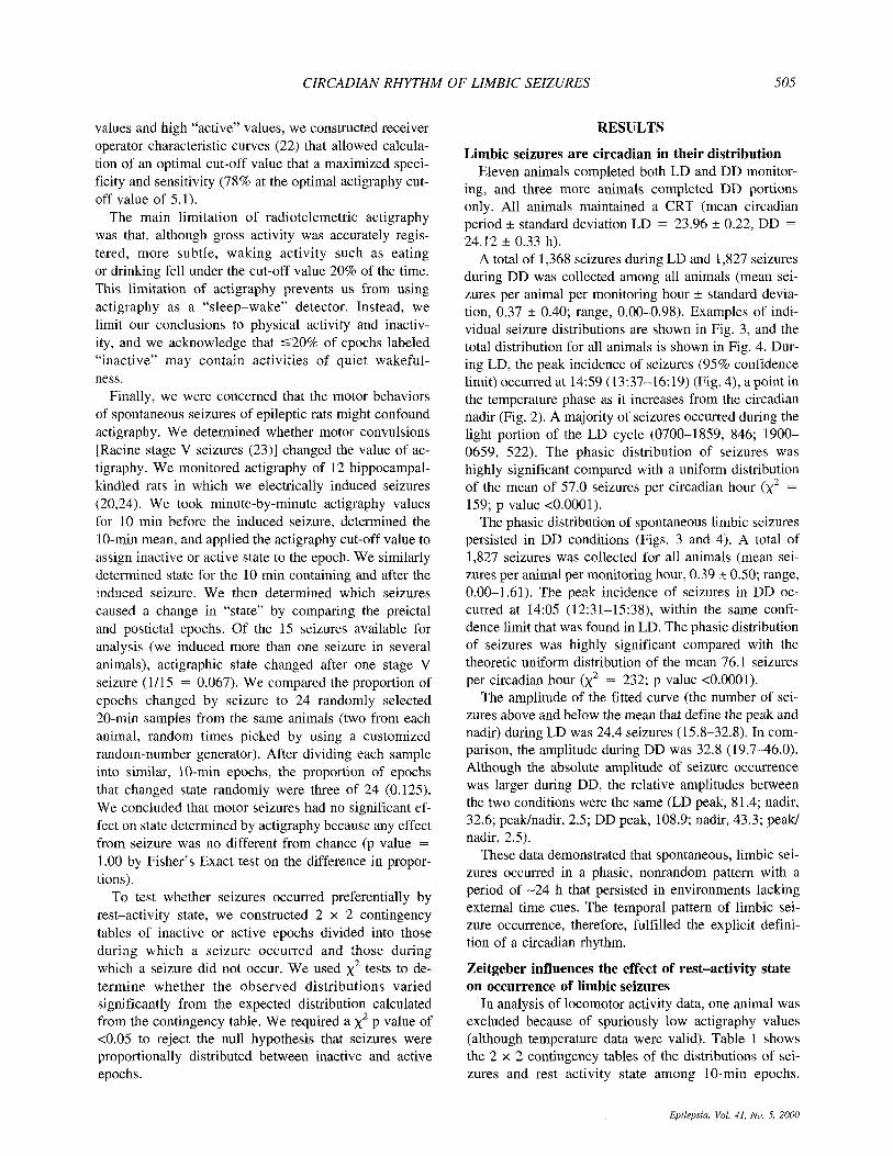
Temperature and activity points were reduced by av- eraging blocks of 10 points to form 10-min epochs. As shown in Fig. 2A, we used cosinor-nonlinear least squares analysis to resolve rhythmic temperature data into a best-fit cosine function with parameters of mean (average temperature), amplitude (maxima and minima of temperature about the mean), period (the duration of a complete cycle), and phase (position of maxima in rela- tion to a time reference). Figure 2B shows an example of temperature and locomotor activity data with best-fit co- sinor waveforms from an epileptic rat. A modified Gauss-Newton nonlinear least squares minimization al- gorithm provided 95th percentile confidence limits for parameter estimation (21).
To demonstrate the circadian distribution of seizures, we first constructed a normalized, 24-h circadian clock for each animal during each LD and DD trial based on phase and period of the CRT. We set physiologic “mid- night” (0000 and 2400) at the temperature peak, and “noon” (1200) at the temperature nadir (Fig. 2A). We then summed seizures among all animals and plotted a
FIG. 2. Examples of core body temperature and locomotor activ- ity monitored continuously by us- ing intraperitoneal radio telemetry in an epileptic rat. A: Excerpt of the middle 48 h of circadian tem- perature data and activityhnactiv- ity data from the -1 1 days shown in (B). Raw data points are re- solved into parameters of ampli- tude (A), mean (M), period (T), and phase (@) by cosinor-nonlinear least squares analysis by the equation f(t) = M + A x cos [24t + @)/TI. The calculated temperature peak is set to a circadian phase time of “0000 and the nadir to “1200.” UD, light-dark cycle; All, activity (black bands)/inactivity (white bands) determined from cut-off value of actigraphy.
1200 1800
frequency distribution by circadian phase. Cosinor- nonlinear least squares analysis was used again to pro- vide a best-fit summary of the 24 data points. To test whether resulting distributions were uniformly distrib- uted, we calculated the mean seizure occurrence per hour (total seizured24 h) and calculated the x2 of observed values compared with the expected mean. We rejected the null hypothesis of uniform seizure distribution for p values <0.05.
Locomotor activity analysis We used actigraphy to determine the mean level of
activity in a 10-min epoch that contained a seizure. We performed experiments to validate radiotelemetric actig- raphy to determine rest-activity state.
We adapted observational criteria of “behavioral inac- tivity” and “behavioral activity” used in a previous study of rest-activity state and epilepsy (5) to define objec- tively an activity level that could be used to divide ac- tigraphy data into “inactive” and “active” epochs. Be- havior in 48-h samples from four epileptic animals was recorded on video tape. Epochs were defined as behav- iorally active if gross, volitional movement (e.g., groom- ing, walking) was seen within the first 30 s of a 10-min epoch. We scored an epoch as inactive if no gross move- ment was apparent within the 30-s sample. If slight head movement, breathing, or eye opening were the only movements discernible during the 30-s sample, we scored the epoch as inactive. Each behavioral sample was matched with its mean activity value for the con-e- sponding 10 min. By using the entire range of actigraphy values to separate them iteratively into low, “inactive”
CIRCADIAN PHASE TIME 0000 0600 1200 1800 0000 0600
A
LID All 0
TIME OF DAY
40
39
38
B 0
0 3 - E ? 37
LID 11 12 13 14 15 16 17 18 19 20 21 22 23
DAY OF MONTH
CIRCADIAN RHYTHM OF LIMBIC SEIZURES 505
values and high “active” values, we constructed receiver operator characteristic curves (22) that allowed calcula- tion of an optimal cut-off value that a maximized speci- ficity and sensitivity (78% at the optimal actigraphy cut- off value of 5.1).
The main limitation of radiotelemetric actigraphy was that, although gross activity was accurately regis- tered, more subtle, waking activity such as eating or drinking fell under the cut-off value 20% of the time. This limitation of actigraphy prevents us from using actigraphy as a “sleep-wake’’ detector. Instead, we limit our conclusions to physical activity and inactiv- ity, and we acknowledge that 520% of epochs labeled “inactive” may contain activities of quiet wakeful- ness.
Finally, we were concerned that the motor behaviors of spontaneous seizures of epileptic rats might confound actigraphy. We determined whether motor convulsions [Racine stage V seizures (23)] changed the value of ac- tigraphy. We monitored actigraphy of 12 hippocampal- kindled rats in which we electrically induced seizures (20,24). We took minute-by-minute actigraphy values for 10 min before the induced seizure, determined the 10-min mean, and applied the actigraphy cut-off value to assign inactive or active state to the epoch. We similarly determined state for the 10 min containing and after the induced seizure. We then determined which seizures caused a change in “state” by comparing the preictal and postictal epochs. Of the 15 seizures available for analysis (we induced more than one seizure in several animals), actigraphic state changed after one stage V seizure (]/I5 = 0.067). We compared the proportion of epochs changed by seizure to 24 randomly selected 20-min samples from the same animals (two from each animal, random times picked by using a customized random-number generator). After dividing each sample into similar, 10-min epochs, the proportion of epochs that changed state randomly were three of 24 (0.125). We concluded that motor seizures had no significant ef- fect on state determined by actigraphy because any effect from seizure was no different from chance (p value = 1 .OO by Fisher’s Exact test on the difference in propor- tions).
To test whether seizures occurred preferentially by rest-activity state, we constructed 2 x 2 contingency tables of inactive or active epochs divided into those during which a seizure occurred and those during which a seizure did not occur. We used x2 tests to de- termine whether the observed distributions varied significantly from the expected distribution calculated from the contingency table. We required a x2 p value of <0.05 to reject the null hypothesis that seizures were proportionally distributed between inactive and active epochs.
RESULTS
Limbic seizures are circadian in their distribution Eleven animals completed both LD and DD monitor-
ing, and three more animals completed DD portions only. All animals maintained a CRT (mean circadian period k standard deviation LD = 23.96 * 0.22, DD = 24.12 f 0.33 h).
A total of 1,368 seizures during LD and 1,827 seizures during DD was collected among all animals (mean sei- zures per animal per monitoring hour k standard devia- tion, 0.37 * 0.40; range, 0.00-0.98). Examples of indi- vidual seizure distributions are shown in Fig. 3, and the total distribution for all animals is shown in Fig. 4. Dur- ing LD, the peak incidence of seizures (95% confidence limit) occurred at 14:59 (13:37-16:19) (Fig. 4), a point in the temperature phase as it increases from the circadian nadir (Fig. 2). A majority of seizures occurred during the light portion of the LD cycle (0700-1859, 846; 1900- 0659, 522). The phasic distribution of seizures was highly significant compared with a uniform distribution of the mean of 57.0 seizures per circadian hour (x2 = 159; p value <0.0001).
The phasic distribution of spontaneous limbic seizures persisted in DD conditions (Figs. 3 and 4). A total of 1,827 seizures was collected for all animals (mean sei- zures per animal per monitoring hour, 0.39 rt_ 0.50; range, 0.00-1.61). The peak incidence of seizures in DD oc- curred at 14:05 (12:31-15:38), within the same confi- dence limit that was found in LD. The phasic distribution of seizures was highly significant compared with the theoretic uniform distribution of the mean 76.1 seizures per circadian hour (x2 = 232; p value <0.0001).
The amplitude of the fitted curve (the number of sei- zures above and below the mean that define the peak and nadir) during LD was 24.4 seizures (15.8-32.8). In com- parison, the amplitude during DD was 32.8 (19.7-46.0). Although the absolute amplitude of seizure occurrence was larger during DD, the relative amplitudes between the two conditions were the same (LD peak, 81.4; nadir, 32.6; peakhadir, 2.5; DD peak, 108.9; nadir, 43.3; peak/ nadir, 2.5).
These data demonstrated that spontaneous, limbic sei- zures occurred in a phasic, nonrandom pattern with a period of -24 h that persisted in environments lacking external time cues. The temporal pattern of limbic sei- zure occurrence, therefore, fulfilled the explicit defini- tion of a circadian rhythm.
Zeitgeber influences the effect of rest-activity state on occurrence of limbic seizures
In analysis of locomotor activity data, one animal was excluded because of spuriously low actigraphy values (although temperature data were valid). Table 1 shows the 2 x 2 contingency tables of the distributions of sei- zures and rest-activity state among 10-min epochs.
Epilepsia, Vol. 41, No. 5, 2000

506
20 -
M. QUIGG ET AL.
r44 DD,@=13:42(10:18-15:05)
FIG. 3. Examples of the distribution of spontaneous limbic seizures in three rats with high (rat, 44 and 13) and low (rat, 84) seizure rates. Although animals met a minimal seizure rate for further study, once enrolled, they were monitored re- gardless of rate. Bars at top show light exposure: LD (left) and DD (right). a, time (95th percentile confidence limit) of peak seizure occurrence calculated from cosinor-nonlinear least squares.
20-
15-
10-
5-
::r--- r44 LD,@ = I 3:07( 1 123-1 5:52)
r84 DD
10
0- ee,ee+ , - ,
0- v) 0 3 6 9 12 15 18 21 24
P x 10
E 3 5 z
0- 0 3 6 9 12 15 18 21 24
10 1 5 1
5 4
0 0 3 6 9 12 15 18 21 24
1 5 1 I
0 3 6 9 12 15 18 21 24
20] r13 DD,@=14:27(11:46-18:05) I I5i
0 3 6 9 12 15 1 8 21 2'4
Overall, 14,787 epochs in LD and 20,664 epochs in DD were analyzed. With the validated threshold value of actigraphy counts, inactive epochs comprised 69% of LD epochs and 64% of DD epochs. Thus removal of regular light exposure decreased the proportion of epochs spent inactive from 2.3: 1 (inactive/active) to 1.8: 1.
If state had no influence on seizure occurrence, then seizures should be distributed between inactive and ac- tive epochs in a proportion similar to the overall distri- bution of all inactive and active epochs. In LD, the pro- portion of inactive/active epochs containing seizures was 3.2:l compared with 2.3: 1 inactive/active overall. During regular entrainment to light, therefore, seizures preferen- tially occurred during inactive epochs (x2 p value <0.0001). The opposite trend was seen when light was removed as an entraining influence. In DD, the propor- tion of inactive to active epochs containing seizures was 1.6: 1 compared with 1.8: 1 overall; therefore, seizures tended to occur during active epochs (x2 p value = 0.16).
To confirm the differences in distributions of seizures by state, we directly compared the proportions of inac- tive with active epochs that contained seizures during LD (3.2: 1) versus DD (1.6: 1). The proportion of seizures that
Circadian time (temp peak = 0000, nadir = 1200)
occurred during inactive epochs in LD was twice that in DD (x2, 64.3; p value <0.0001).
These findings suggested that the inactive state sig- nificantly facilitated the spontaneous expression of lim- bic seizures. Its modulatory effect, however, required the entraining influence of the light-dark cycle. Once light exposure was removed, the expression of limbic seizures slightly favored the active state.
DISCUSSION The principal finding of this study was that spontane-
ous limbic seizures of a rat model of MTLE occurred with a circadian distribution. The primary test of whether a daily rhythm is circadian and susceptible to regulation by the SCN is whether the rhythm persists in the absence of rhythmic external stimuli (12). Limbic seizures of epi- leptic rats were circadian in distribution because they occurred in a phasic, 24-h pattern, and the pattern per- sisted in constant conditions. Furthermore, the inactive state had a significant positive association with seizure occurrence, but this positive influence was present only when the animals were exposed to a light-dark cycle.
Effects of the rest-activity state or of circadian regu- lation may vary with the pathophysiology or presumptive
Epilepsia, Vol. 41, No. 5, 2000

CIRCADIAN RHYTHM OF LIMBIC SEIZURES 507
E 100 - 3 ‘i 80 -
6 0 -
5 4 0 - z
20 -
+
a5 Q
I2O 1 LD I
m m dfY Peak 14:59 (13:37-16:19) I
LIGHT 0 0000 0600 1200 1800 2400
120
100
,z 80
f 60
n E 40
L
3
(0 Lc
m
Peak 14:05 (12:31-15:38) I 20-1
0000 0600 1200 1800 2400 Circadian time (temperature peak = 0000, nadir = 1200)
FIG. 4. Distribution of spontaneous limbic seizures in self- sustained limbic status epilepticus rats in a light-dark cycle (LD) and in continuous darkness (DD) by phase of the circadian rhythm of temperature. The phasic distribution of seizures is maintained during DD. Time of peak seizure occurrence (95th percentile confidence limit) calculated from cosinor-nonlinear least squares.
epileptogenic injury that underlies the epileptic syn- drome. The diurnal distribution of seizures found during LD was similar to that found in other limbic epilepsies. Seizures occurred most often in the late afternoon during the light phase in the self-sustained status epilepticus rat (6,8), in the pilocarpine-treated rat (7,2S), and in humans with MTLE (8). An exception is the kainate-treated rat ( 5 ) , which like other partial epilepsy models tended to have seizures in the light portion of the light-dark cycle, but which like humans with generalized epilepsies (26) or with nonlimbic, extratemporal lobe epilepsies (8), had most seizures during early light exposure.
Our findings indicate that the circadian occurrence of limbic seizures is not a passive reflection of the environ- ment; limbic seizures are susceptible to an endogenous circadian regulation. Future experiments can seek to de- termine the specific agents responsible for circadian modulation. Although we demonstrated the importance of endogenous factors on seizure occurrence, we directly tested the effect of light exposure, an exogenous factor, on activity state and on seizure occurrence. About 1.6 times as many seizures occurred during the light portion in LD than during the dark portion, and that pattern per- sisted during the corresponding circadian phase in the absence of light in DD. Although the amplitude of cir-
cadian seizure occurrence appeared relatively suppressed during LD, the relative amplitude adjusted for the greater number of seizures in DD was the same between LD and DD.
Regular light exposure increased the proportion of ep- ochs spent inactive; we expected the shift toward inac- tivity because light suppresses the active state in noctur- nal animals. The effect of light exposure on seizure oc- currence is less easily explained. In constant dark, seizures tended to occur disproportionately during active epochs. Regular light exposure, however, significantly reversed this distribution. Light exposure, or perhaps, the process of entrainment to periodic light exposure, shifted seizures from active to inactive epochs. Whether this shift was due to the change in distribution of activity state or due to the direct effect of light exposure itself is unclear. Light exposure, in the form of high-intensity flashing lights, is a well-known precipitant of epileptic seizures, especially in patients with juvenile myoclonic epilepsy and other forms of idiopathic epilepsy (27). The effects of prolonged light exposure, however, are incon- sistent. Constant light exposure facilitates epileptogenic discharges and seizures in the epileptic baboon (28). Most animal models of partial epilepsy (5-8,2S) and hu- mans with partial epilepsy (8) experience more seizures during the light portion of the diurnal cycle. Conversely, during the summer when prolonged light exposure in- creases, the incidence of epileptic discharges in patients with photosensitive epilepsy decreases (29). The rela- tions among long-term light exposure, activity state, and seizure threshold suggest that exogenous stimuli remain important factors in spontaneous seizure occurrence.
TABLE 1. 2 x 2 contingency table distribution of 10-min epochs of seizures and activity states in epileptic rats
Inactive Active Totals
LD No seizure
Seizure
Totals
X 2 p Value
No seizure
Seizure
Totals
DD
X 2 p Value
9,273
965
10,238
30.7 <0.0001
12,168
999
13,167
2.0 0.16
4,246 13,519
303 1,268
4549 14,787
(2.2: 1) (ratio)
(3.2:l) (ratio)
(2.3:l) (ratio)
6,887 19,055
610 1,609
7,497 20,664
(1.8:l) (ratio)
(1.6: 1) (ratio)
(1.8: 1) (ratio)
Activity state was determined from continuous locomotor activity measurements. A cut-off activity value was determined from correla- tion of activity to videotaped behavioral activity. Activity values below the cut-off value (3.1 counts/lO-min epochs) were designated “inac- tive,” and values greater than cut-off were designated “active.”
Epikpsia, Vol. 41, No. 5, 2000

508 M. QUIGG ET AL.
This study demonstrated that exogenous factors-light exposure-interacted with endogenous influences to modulate the pattern of limbic seizure occurrence. En- dogenous cycles, or interactions among endogenous cycles, also must influence circadian seizure onset. Mel- atonin has shown both anti- and proconvulsant effects (30,3 1). Noradrenaline and other adrenoreceptor-active agonists have anticonvulsant actions (32,33). Some agents of the hypothalamic-pituitary-adrenal axis have proconvulsant effects, in the case of corticotropin- releasing hormone (34), and others have anticonvulsant effects, such as corticotropin (35).
A direct effect of the SCN on limbic epilepsy is one possible influence. The SCN has direct and indirect con- nections with the limbic system (36). Our rat model of MTLE and human subjects demonstrated diurnal seizure distributions that are in phase. This finding is intriguing because electrical activity (37) and glucose metabolism (38) in the SCN are maximal during the light phase of the 24-h cycle regardless of whether the animal is diurnal or nocturnal. Thus the increased incidence of seizures dur- ing the light phase coincides with the phase of maximal SCN activity. Because the SCN is the primary circadian pacemaker in mammals (12), its influence on the tem- poral distribution of seizures may be direct. We also can speculate that other hypothalamic regions-the paraven- tricular nucleus (PVN), for example, that may function as a relay site between circadian and limbic functions (39)-may participate in the circadian pattern of limbic seizure occurrence.
Another potential modulator of limbic seizures is sleep-wake (40) or rest-activity state (5) . We used ra- dioteleinetric activity monitoring (actigraphy) to deter- mine rest-activity state. Although actigraphy provides an accurate, minute-to-minute assessment of locomotor ac- tivity, its accuracy in describing sleep-wake state is con- founded by the degree to which the animal moves during sleep and remains motionless during wakefulness. Sleep staging by EEG is potentially the most accurate method of state determination. Many laboratories use some form of polysomnography, dividing state into wakefulness, rapid eye movement (REM), and non-REM sleep (41,42). EEG methods, however, have shortcomings. Polysomnography is data and labor intensive, and semi- automated methods lack consensus of standards and are often proprietary from laboratory to laboratory. Further- more, our epileptic animals have another obstacle; in pilot studies, the interictal EEG of epileptic rats was abnormal and lacked reliable, state-related changes. Ac- tigraphy offered several advantages over EEG analysis in this model. First, it avoided the problem of abnormal EEG encountered in our epileptic animals. Second, ac- tigraphy was measured with the same transmitters and receivers that provided us with circadian data. Third, except for a manually scored calibration period, actigra-
phy was automated and required no subjective interpre- tation, thus lending itself to long durations of monitoring. With these advantages, both human and animal studies have validated actigraphy as useful markers of overall sleep time (43-45). Radiotelemetric actigraphy, com- pared with other methods, arrived at reasonable estima- tions of rest-activity per day. The proportion of time spent inactive versus active varies with the animal model, environmental conditions, and the method of measurement. Our permanently tethered, epileptic ani- mals spent 69% of a 24-h day inactive when measured by radiotelemetric actigraphy. Untethered, kainate-treated rats spend 75% inactive as determined by video obser- vation ( 5 ) . Nontelemetric actigraphy of nonepileptic rats determined that 40% of time was inactive (44). When sleep stage is scored by computer from cortical EEG and EMG, nonepileptic rats spend 57% of 24 h asleep (46).
State remains an important factor in seizure modula- tion. Early studies document that interictal epileptiform discharges follow a clear ultradian pattern of occurrence (9,10) that, in later studies, has been confirmed to cor- respond to non-REM sleep (4). Transitions from sleep to wakefulness (26), slow-wave sleep (47), and inactivity (5) have all been associated with an increased propensity for seizure occurrence. Our data are consistent with these studies in that seizure occurrence was associated with the inactive state, but the facilitating effect of inactivity was smaller than was noted in previous studies. Furthermore, the effect was significant only when reinforced by the external light-dark cycle and disappeared-even slightly reversed-in constant darkness. In a comparison of the daily distributions of partial seizures of humans and epi- leptic rats (8), the similarities in phase of seizures in humans with MTLE and in epileptic rats in spite of the differences in the two species’ rest-activity cycles im- plied that the rest-activity state may not be the major factor in regulation of limbic seizure onset. Our data suggest that although state is not the major determinant of seizure expression, it may have an influence that var- ies throughout the 24-h cycle. Transition states must be explored because a high number of seizures occurred in the rising phase of temperature corresponding to loco- motor activity onset. Clearly, further studies remain to be performed to examine transition states as periods par- ticularly liable for seizure expression (3) by use of de- finitive methods of sleep staging in experimental ani- mals.
Acknowledgment: This study was funded in part by a Junior Investigator Research Award from the Epilepsy Foundation of America and by NIH-NINDS K08-NS02021 (M.Q.). M.S. ac- knowledges support from NSF-DIR8920162 and NIH R01- DK51562, and E.H.B., from NS-25605.
Epikpsia, Vol. 41, Nu. 5, 2000

CIRCADIAN RHYTHM OF LIMBIC SEIZURES 509
REFERENCES
1. Langdon-Down M, Brain W. Time of day in relation to convul- sions in epilepsy. Lancet 1929;1:1029-32.
2. Gowers W. Course of epilepsy. In: Gowers W, ed. Epilepsy and other chronic convulsive diseases: their causes, symptoms and treatment. New York: William Wood, 1885: 157-64.
3. Shouse M, Martins da Silva A, Sammaritano M. Circadian rhythms, sleep and epilepsy. J Clin Neurophysiol 1996;13:32-50.
4. Malow B, Lin X, Kushwaha R, Aldrich M. Interictal spiking in- creases with sleep depth in temporal lobe epilepsy. Epilepsia 1998; 39: 1309-16.
5. Hellier J, Dudek F. Spontaneous motor seizures of kainate-induced epileptic rats: effect of time of day and activity state. Epilepsy Res 1999;35:47-57.
6. Bertram E, Cornett J. The evolution of a rat model of chronic spontaneous limbic seizures. Brain Res 1994;661: 157-62.
7. Cavalhiero E, Leite J, Bortolotto Z, Turski W, Ikonomidou C, Turski L. Long-term effects of pilocarpine in rats: structural dam- age of the brain triggers kindling and spontaneous recurrent sei- zures. Epilepsiu 1991 ;32:778-82.
8. Quigg M, Straume M, Menaker M, Bertram E. Temporal distribu- tion of partial seizures: comparison of an animal model with hu- man partial epilepsy. Ann Neurol 1998;43:748-55.
9. Kellaway P. Sleep and epilepsy. Epilepsia l985;26:S15-30. 10. Stevens J, Kodoma H, Lonsbury B, Mills L. Ultradian character-
istics of spontaneous seizure discharges recorded by radio telemetry in man. Electroencephalogr Clin Neurophysiol 197 1 ;3 1 :3 13-25.
11. Gotman J, Koffler D. Interictal spiking increases after seizures but does not after decreases in medication. Electroencephalogr Clin Neurophysiol 1989;72:7-15.
12. Murphy P, Campbell S. Physiology of the circadian system in animals and humans. J Clin Neurophysiol 1996;13:2-16.
13. Bertram E. Functional anatomy of spontaneous seizures in a rat model of limbic epilepsy. Epilepsia 1997;38:95-105.
14. Bertram E, Lothman E. Morphometric effects of intermittent kindled seizures and limbic status epilepticus in the dentate gyrus of the rat. Bruin Res 1993;603:25-31.
15. Lothman E, Bertram E, Beckenstein J, Perlin J. Self-sustaining limbic status epilepticus induced by “continuous” hippocampal stimulation: electrographic and behavioral characteristics. Epilepsy Res 1989;3:107-19.
16. Lothman E, Bertram E, Kapur J, Stringer J. Recurrent spontaneous hippocampal seizures in the rat as a chronic sequelae to limbic status epilepticus. Epilepsy Res 1990;6: 110-8.
17. Bertram EH, Williamson JM, Cornett JF, Spradlin S, Chen ZF. Design and construction of a long-term continuous video-EEG monitoring unit for simultaneous recording of multiple small ani- mals. Bruin Res Brain Res Protoc 1997;2:85-97.
18. Bertram E, Cornett J. The ontology of seizures in a rat model of limbic epilepsy: evidence for a kindling in development of chronic spontaneous seizures. Brain Res 1993;625:295-300.
19. Refinetti R, Menaker M. Circadian rhythm of body temperature. Physiol Behav 1992;51:613-37.
20. Quigg M, Clayburn H, Straume M, Menaker M, Bertram E. Hy- pothalamic neuronal loss and altered circadian rhythm of tempera- ture in a rat model of mesial temporal lobe epilepsy. Epilepsia 1999;40: 1688-93.
21. Straume M, Frasier S , Johnson M. Least squares analysis of fluo- rescence data. In: Lakowicz J, ed. Topics on fluorescent spectros- copy. New York: Plenum Press, 1991: 177-240.
22. Greiner M, Sohr D, Gobel P. A modified ROC analysis for the selection of cutoff values and the definition of intermediate results of diagnostic tests. J Zmmunol Methods 1995;185: 123-32.
23. Racine R. Modification of seizure activity by electrical stimula- tion: 11. Motor seizure. Electroencephalogr Clin Neurophysiol 1972;32:28 1-94.
24. Lothman E, Hatlelid J, Zorumski C, Conry J, Moon P, Perlin J. Kindling with rapidly recurring hippocampal seizures. Brain Res
25. Arida R, Fulvio A, Peres C, Cavalheiro E. Course of untreated seizures in the pilocarpine model of epilepsy. Epilepsy Res 1999; 34:99-107.
1985;360:83-Y 1.
26. Janz D. Epilepsy and the sleeping-waking cycle. In: Magnus 0, Lorentz De Haas A, eds. The epilepsies. Amsterdam: North Hol- land, 1974:457-90.
27. Niedermeyer E. Primary (idiopathic) generalized epilepsy and un- derlying mechanisms. Clin Electroencephalogr 1996;27: 1-2 I.
28. Ehlers CL, Killam EK. The effects of constant light on EEG and seizure activity in the epileptic baboon. Electroencephalogr Clin Neurophysiol 1982;54: 187-93.
29. Danesi M. Seasonal variations in the incidence of photoparoxys- ma1 response to stimulation among photosensitive epileptic pa- tients: evidence from repeated EEG recordings. J Neurol Neuro- surg Psychiatry 1988;561:875-7.
30. Albertson T, Peterson S, Starke L. The anticonvulsant properties of melatonin on kindled seizures in rats. Neurophurmacology 198 1 ;
31, Yehuda S, Mostofsky D. Circadian effects of beta-endorphin, mel- atonin, DSIP, and amphetamine on pentylenetetrazol-induced sei- zures. Peptides 1993;14:203-5.
32. Ferraro G, Sardo P, Sabatino M, La Grutta V. Locus coeruleus noradrenaline system and focal penicillin hippocampal epilepsy: neurophysiological study. Epilepsy Res 1995; 19:215-20.
33. Shouse M, Langer J, Bier M, et al. The alpha 2-adrenoreceptor agonist clonidine suppresses seizures, whereas the alpha 2-adreno- receptor antagonist idazoxan promotes seizures in amygdala- kindled kittens: a comparison of amygdala and pontine microinfu- sion effects. Epilepsia 1996;37:709-17.
34. Ehlers C, Henriksen S, Wang M, Rivier J, Vale W, Bloom F. Corticotropin releasing factor produces increases in brain excit- ability and convulsive seizures in rats. Brain Res 1983;278:332-6.
35. Holmes GL. Effect of non-sex hormones on neuronal excitability, seizures, and the electroencephalogram. Epilepsiu 1991;32:S11-8.
36. Buijs R. The anatomical basis for the expression of circadian rhythms: the efferent projections of the suprachiasmatic nucleus. In: Buijs R, Kalsheek A, Romijn H, Pennartz C, Mirmiran M, eds. Progress in brain research. Amsterdam: Elsevier, 1996:22940.
37. Inouye S-I. Circadian rhythms of neuropeptides in the suprachias- matic nucleus. In: Buijs R, Kalsbeek H, Romijn H, Pennartz C, Mirmiriran M, eds. Progress in brain research: hypothalamic in- tegration of circadian rhythms. Amsterdam: Elsevier, 1996:75-90.
38. Schwartz W, Reppert S, Eagan S, Moore-Ede M. In vivo metabolic activity of the SCN: a comparative study. Brain Res 1983;274: 184-7.
39. Peng Z, Grassi-Zucconi G, Bentivoglio M. Fos-related protein ex- pression in the midline paraventricular nucleus of the rat hypo- thalamus: basal oscillation and relationship with limbic efferents. Exp Brain Res 1995;104:21-9.
40. Shouse M, Langer J, Bier M, et al. Alpha 2 adrenoreceptor agonist clonidine suppresses seizures, whereas the alpha 2 adrenoreceptor antagonist idazoxan promotes seizures: pontine microinfusion studies of amygdala-kindled kittens. Bruin Res 1996;731:203-7.
41. Schwierin B, Borbely AA, Tobler I. Prolonged effects of 24-h total sleep deprivation on sleep and sleep EEG in the rat. Neuroscience Lett 1999;26 1 :6 1 4 .
42. Rechtschaffen A, Bergmann BM, Gilliland MA, Bauer K. Effects of method, duration, and sleep stage on rebounds from sleep de- privation in the rat. Sleep 1999;22:11-31.
43. Mullaney D, Kripke D, Messin S. Wrist-actigraphic estimation of sleep time. Sleep 1980;3:83-92.
44. Carroll D, Denenberg V, Thoman E. Reliability and validity of computer scoring of behavioral sleep-wake states in rats and rab- bits. Physiol Behuv 1993;54:269-73.
45. Zeidner L, Denenberg V, Thoman E, Weyand T. Comparisons of behavioral, motoric, and electrical criteria for assessment of sleep- wake states in the rabbit. Physiol Behav 1983;31:273-8.
46. Mistlberger R, Bergmann B, Waldenar W, Rechtscheffen A. Re- covery sleep following sleep deprivation in intact and suprachias- matically lesioned rats. Sleep 1983;6:217-33.
47. Bazil CW. Walczak TS. Effects of sleep and sleep stage on epi- leptic and nonepileptic seizures. Epilepsia 1997;38:56-62.
20~61-6.
Epilepsiu, Vol. 41, NO. 5, 2000