Mapping Structural Brain Alterations in Obsessive-Compulsive Disorder
Upload
independentCategory
view
0download
0
ORIGINAL ARTICLE
Different patterns of local field potentials from limbic DBStargets in patients with major depressive and obsessivecompulsive disorderW-J Neumann1, J Huebl1, C Brücke1, L Gabriëls2, M Bajbouj3, A Merkl1,3, G-H Schneider4, B Nuttin5, P Brown6 and AA Kühn1,7,8
The role of distinct limbic areas in emotion regulation has been largely inferred from neuroimaging studies. Recently, theopportunity for intracranial recordings from limbic areas has arisen in patients undergoing deep brain stimulation (DBS) forneuropsychiatric disorders including major depressive disorder (MDD) and obsessive compulsive disorder (OCD). Here we test thehypothesis that distinct temporal patterns of local field potential (LFP) activity in the human limbic system reflect disease state andsymptom severity in MDD and OCD patients. To this end, we recorded LFPs via implanted DBS electrodes from the bed nucleus ofstria terminalis (BNST area) in 12 patients (5 OCD, 7 MDD) and from the subgenual cingulate cortex in 7 MDD patients (CG25 area).We found a distinct pattern of oscillatory activity with significantly higher α-power in MDD compared with OCD in the BNST area(broad α-band 8–14 Hz; Po0.01) and a similar level of α-activity in the CG25 area as in the BNST area in MDD patients. The mean α-power correlated with severity of depressive symptoms as assessed by the Beck depression inventory in MDD (n= 14, r= 0.55,P= 0.042) but not with severity of obsessive compulsive symptoms in OCD. Here we show larger α-band activity in MDD patientscompared with OCD recorded from intracranial DBS targets. Our results suggest that α-activity in the limbic system may be asignature of symptom severity in MDD and may serve as a potential state biomarker for closed loop DBS in MDD.
Molecular Psychiatry (2014) 00, 000–000. doi:10.1038/mp.2014.2
Keywords: alpha oscillations; deep brain stimulation; depressive disorder; local field potential; obsessive compulsive disorder
INTRODUCTIONDeep brain stimulation (DBS) has recently gained attention as apromising therapeutic approach in therapy-resistant psychiatricdisorders and encouraging results have been obtained in patientswith Tourette syndrome, major depressive disorder (MDD) andobsessive compulsive disorder (OCD).1–4 Other psychiatric condi-tions, such as addiction, anorexia nervosa and schizophrenia, arealso under clinical evaluation.5–7 However, the mechanism ofaction of DBS is still not well understood. Target areas for DBS inpsychiatry and neurology have been adopted from empirical dataderived from neurosurgical lesion studies that reported beneficialclinical results rather than being driven by pathophysiologicalhypotheses.8 The opportunity to record local field potentials (LFPs)from DBS electrodes during and shortly after DBS surgery, whileDBS electrodes are still externalised, has contributed considerablyto our understanding of the underlying pathophysiology of circuitdisorders such as Parkinson’s disease (PD), and of the physiologyof the basal ganglia.9–12 Studies from patients undergoing DBS formovement disorders have pointed to abnormal patterning ofneuronal activity that differs between disease states.13,14 DBS maysuppress disruptive aberrant activity across different frequencybands such as increased β-band activity in PD,15,16 therebyoffering an explanation for how disparate clinical states may be
effectively treated by DBS in the same target area.13 Moreover,abnormal neuronal patterns may serve as biomarkers that arecurrently tested for optimised closed loop DBS in movementdisorders.17
Our understanding of the oscillatory neuronal circuits subser-ving psychiatric disorders is growing;18 however, little is knownabout the underlying nature of subcortical oscillatory neuronalactivity in the structures targeted by DBS. Thus, the optimal targetstructures for psychiatric indications are less well defined, asevident in the current investigation of five target areas for DBS inMDD (nucleus accumbens, medial forebrain bundle, anteriorsubgenual cingulate cortex (CG25), anterior limb of internalcapsule and the inferior thalamic peduncle)4,3,19–22 and four areasfor OCD (nucleus accumbens, ventral subthalamic nucleus,anterior limb of internal capsule and the inferior thalamicpeduncle).1,19,22–28 For MDD, two of these anatomical structureshave been targeted more frequently than others: the CG25 andthe anterior limb of the internal capsule with the bed nucleus ofstria terminalis (BNST) in the trajectory (subsequently referred toas the BNST area). Clinical case series of DBS in treatment-resistantMDD have reported favorable results from chronic high-frequencystimulation of the CG25.4,29,30 A series of positron emissiontomography studies have established hypermetabolism in this
1Department of Neurology, Charité–University Medicine Berlin, Berlin, Germany; 2Department of Psychiatry, University Hospital Leuven, Leuven, Belgium; 3Department ofPsychiatry and Psychotherapy, Charité–University Medicine Berlin, Berlin, Germany; 4Department of Neurosurgery, Charité–University Medicine Berlin, Berlin, Germany;5Department of Neurosurgery, University Hospital Leuven, Leuven, Belgium; 6Nuffield Department of Clinical Neurosciences, University of Oxford, and National Institute of HealthRelated Research Oxford Biomedical Research Centre, Oxford, UK; 7Berlin School of Mind and Brain, Charité–University Medicine Berlin, Berlin, Germany and 8NeuroCure,Charité–University Medicine Berlin, Berlin, Germany. Correspondence: Professor Dr AA Kühn, Department of Neurology, Charité–University Medicine Berlin, Campus VirchowKlinikum, Augustenburger Platz 1, Berlin 13353, Germany.E-mail: [email protected] 2 July 2013; revised 15 December 2013; accepted 6 January 2014
Molecular Psychiatry (2014), 1–7© 2014 Macmillan Publishers Limited All rights reserved 1359-4184/14
www.nature.com/mp
area as a correlate of negative mood and depression. Furthermore,DBS-related remission of MDD symptoms has been associatedwith a reduction of cerebral blood flow in the target area anddownstream limbic and cortical areas.4,31 The BNST area is thesecond major DBS target area investigated in MDD followingsignificant improvement of depression ratings in patientsoperated for OCD.28,32 The BNST is one of the main output nucleiof the extended amygdala33,34 and functionally related tosustained fear and anxiety as perceived in major depressive andanxiety disorders.35 A recent animal study demonstrated thatoptogenetic photostimulation of efferent subcortical γ-aminobu-tyric acid-containing (GABAergic) and glutamatergic networkprojections from BNST in freely behaving mice differentiallyelicited anxiolytic and anxiogenic behavioural phenotypes sup-porting the role of the BNST in the regulation of emotionalstates.36 Clinical studies are now examining the benefit of BNSTarea stimulation in both OCD and MDD.The aims of our study were twofold. First, we sought distinct
spatio-temporal patterns of synchronised neuronal networkactivity related to MDD and OCD in patients undergoingDBS of the same target, the BNST area. Second, we sought todetermine whether any such phenotype-dependent neuronalactivity was a local feature or also represented in other limbicstructures. To this end, we also recorded from MDD patientsundergoing surgery to the CG25. In all, we compared LFP activityrecorded directly from DBS electrodes in 14 patients sufferingfrom intractable MDD and five patients with OCD. We hypothe-sised that oscillatory patterning differs between disease statesproviding a neurophysiological signature of the disease that mayreflect symptom severity. Our results provide evidence that therelative level of synchronised α-oscillations in limbic structuresmay distinguish MDD and OCD and that these oscillationscorrelate with the patients’ self reported symptom severity asassessed by the Beck depression inventory (BDI) at the time ofrecording.
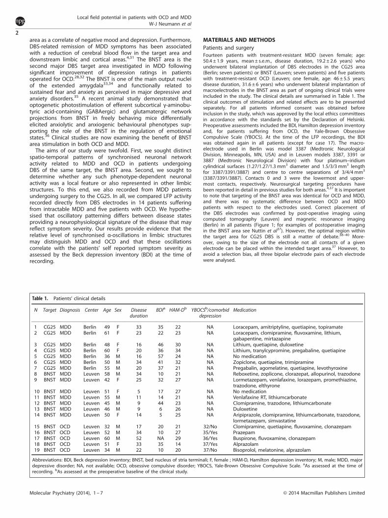
MATERIALS AND METHODSPatients and surgeryFourteen patients with treatment-resistant MDD (seven female; age:50.4 ± 1.9 years, mean± s.e.m., disease duration, 19.2 ± 2.6 years) whounderwent bilateral implantation of DBS electrodes in the CG25 area(Berlin; seven patients) or BNST (Leuven; seven patients) and five patientswith treatment-resistant OCD (Leuven; one female, age: 46 ± 5.5 years;disease duration, 31.6 ± 6 years) who underwent bilateral implantation ofmacroelectrodes in the BNST area as part of ongoing clinical trials wereincluded in the study. The clinical details are summarised in Table 1. Theclinical outcomes of stimulation and related effects are to be presentedseparately. For all patients informed consent was obtained beforeinclusion in the study, which was approved by the local ethics committeesin accordance with the standards set by the Declaration of Helsinki.Preoperative assessments included the BDI, Hamilton depression inventoryand, for patients suffering from OCD, the Yale-Brown ObsessiveCompulsive Scale (YBOCS). At the time of the LFP recordings, the BDIwas obtained again in all patients (except for case 17). The macro-electrode used in Berlin was model 3387 (Medtronic NeurologicalDivision, Minneapolis, MN, USA) and in Leuven models 3387, 3391 or3887 (Medtronic Neurological Division) with four platinum–iridiumcylindrical surfaces (1.27/1.27/1.3 mm3 diameter and 1.5/3/3mm3 lengthfor 3387/3391/3887) and centre to centre separations of 3/4/4 mm3
(3387/3391/3887). Contacts 0 and 3 were the lowermost and upper-most contacts, respectively. Neurosurgical targeting procedures havebeen reported in detail in previous studies for both areas.4,37 It is importantto note that targeting of the BNST area was identical for OCD and MDD,and there was no systematic difference between OCD and MDDpatients with respect to the electrodes used. Correct placement ofthe DBS electrodes was confirmed by post-operative imaging usingcomputed tomography (Leuven) and magnetic resonance imaging(Berlin) in all patients (Figure 1; for examples of postoperative imagingin the BNST area see Nuttin et al37). However, the optimal region withinthe target area for CG25 DBS is still a matter of debate.38–40 More-over, owing to the size of the electrode not all contacts of a givenelectrode can be placed within the intended target area.37 However, toavoid a selection bias, all three bipolar electrode pairs of each electrodewere analysed.
Table 1. Patients’ clinical details
N Target Diagnosis Center Age Sex Diseaseduration
BDIa HAM-Db YBOCSb/comorbiddepression
Medication
1 CG25 MDD Berlin 49 F 33 35 22 NA Loracepam, amitriptyline, quetiapine, topiramate2 CG25 MDD Berlin 61 F 23 22 23 NA Loracepam, clomipramine, fluvoxamine, lithium,
gabapentine, mirtazapine3 CG25 MDD Berlin 48 F 16 46 30 NA Lithium, quetiapine, duloxetine4 CG25 MDD Berlin 60 F 20 36 34 NA Lithium, tranylcypromine, pregabaline, quetiapine5 CG25 MDD Berlin 36 M 16 57 24 NA No medication6 CG25 MDD Berlin 50 M 34 41 32 NA Zopiclone, quetiapine, trimipramine7 CG25 MDD Berlin 55 M 20 37 21 NA Pregabalin, agomelatine, quetiapine, levothyroxine8 BNST MDD Leuven 58 M 34 10 21 NA Reboxetine, zoplicone, clorazepat, allopurinol, trazodone9 BNST MDD Leuven 42 F 25 32 27 NA Lormetazepam, venlafaxine, lorazepam, promethiazine,
trazodone, elthyrone10 BNST MDD Leuven 51 F 5 17 27 NA No medication11 BNST MDD Leuven 55 M 11 14 21 NA Venlafaxine RT, lithiumcarbonate12 BNST MDD Leuven 45 M 9 44 23 NA Clomipramine, trazodone, lithiumcarbonate13 BNST MDD Leuven 46 M 9 6 26 NA Duloxetine14 BNST MDD Leuven 50 F 14 5 25 NA Aripiprazole, clomipramine, lithiumcarbonate, trazodone,
lormetazepam, simvastatine15 BNST OCD Leuven 32 M 17 20 21 32/No Clomipramine, quetiapine, fluvoxamine, clonazepam16 BNST OCD Leuven 52 M 34 10 27 35/Yes Prazepam17 BNST OCD Leuven 60 M 52 NA 29 36/Yes Buspirone, fluvoxamine, clonazepam18 BNST OCD Leuven 51 F 33 35 14 37/Yes Alprazolam19 BNST OCD Leuven 34 M 22 10 20 37/No Bisoprolol, melatonine, alprazolam
Abbreviations: BDI, Beck depression inventory; BNST, bed nucleus of stria terminali; F, female ; HAM-D, Hamilton depression inventory; M, male; MDD, majordepressive disorder; NA, not available; OCD, obsessive compulsive disorder; YBOCS, Yale-Brown Obsessive Compulsive Scale. aAs assessed at the time ofrecording. bAs assessed at the preoperative baseline of the clinical study.
Local field potential in patients with OCD and MDDW-J Neumann et al
2
Molecular Psychiatry (2014), 1 – 7 © 2014 Macmillan Publishers Limited

Experiments and recordingsAll patients were studied within 2–7 days after initial DBS surgery, whilethe DBS leads were externalised. There was no difference in timing ofrecordings between OCD (5 ± 0.5 days) and MDD (4.9 ± 0.9 days) patients inLeuven (average timing for Berlin was 2.7 ± 0.3 days). LFPs were obtainedbipolarly from the adjacent contacts of the DBS macroelectrode (01, 12,23), amplified (x50 000) and filtered at 1–250 Hz. For patients 1–7 (Berlin), aD360 amplifier was used (Digitimer, Hertfordshire, UK) and signals wererecorded at a sampling rate of 1 kHz through a 1401 A-D converter (CED,Cambridge UK) onto a computer using Spike2 (CED) software. For patients8–19 (Leuven), we used a portable amplifier (Biopotential Analyzer Diana,St Petersburg, Russia) with sampling frequency 1.5 kHz (except for MDDcases 9 and 10, which were recorded with an older version of the recordingsystem and therefore sampled at 185 Hz). Overall, 42 CG25 and 72 BNSTcontact pairs were recorded from 38 electrodes in 19 patients. All patientscompleted rest recordings of at least 100 s duration (127 ± 1.5 s for CG25and 137± 8.8 s for BNST, mean± s.e.m.). During recordings, patients wereseated comfortably in an armchair and asked to relax with eyes open, andnot to speak.
Spectral analysisAll data were downsampled (or interpolated for cases 9 and 10) to 1 kHz,visually inspected for artefacts and analysed using custom MATLAB code(The Mathworks, Natick, MA, USA) based on SPM8 for magnetoencephalo-graphy/electroencephalography (Wellcome Department of CognitiveNeurology, London, UK; www.fil.ion.ucl.ac.uk/spm/) and FieldTrip (DondersCenter for Cognitive Neuroimaging, University Nijmegen, Nijmegen, theNetherlands; http://fieldtrip.fcdonders.nl/). All continuous rest recordingswere divided into arbitrary epochs of 1.024 s (1024 samples) andtransferred into the frequency domain using fast Fourier transform-based methods. This resulted in a frequency resolution of 0.98 Hz over 512frequency bins. Power-spectra were normalised to the mean s.d. of 3–47Hz and 53–97 Hz power and further expressed in arbitrary units (a.u.). The0–3 and 47–53 Hz ranges were omitted so as to avoid contamination bymovement artefact and mains noise, respectively. Relative rather thanabsolute power was analysed to allow comparison across subjects, asabsolute power is more likely to be dependent on proximity to the LFPsource than relative power and to vary with minor changes in recordingtechnique. As two patients were originally sampled at 185 Hz inaccordance with the Nyquist theorem, we only further analysed frequencybins up to 90 Hz for all patients. For visualisation purposes power spectrawere averaged over all three contact pairs of each electrode andsubsequently averaged across each patient group. In order to define thefocality of spectral activity, the contact pair with the highest band power inthe frequency range of interest (8–14 Hz) was selected and power at theremaining contact pairs per electrode was expressed as percentage ofmaximum band power.
Statistical analysisAveraged power spectra of MDD and OCD patients recorded from theBNST were compared using non-parametric Monte Carlo permutation tests
for each frequency bin.41 Therefore, the null hypothesis was tested thatpower spectra from both groups were interchangeable by comparingmean sample values from each frequency bin in MDD and OCD patients to5000 replications of the test statistic generated by randomly exchangingelements between distributions from MDD and OCD spectra. Thisprocedure is free from any assumptions about the distributions of powerand differences in power and is robust against small sample size.41
Multiple comparisons were corrected for all frequency bins by controllingthe false discovery rate for an α-level of α=0.05.42 To rule out undesiredeffects caused by differences in recording procedures, this directcomparison was only carried out between seven MDD patients (cases8–14) and five OCD patients (cases 15–19) with LFPs recorded from theBNST target area using the same recording set-up. Only P-values that weresmaller than the false discovery rate-corrected threshold of P= 0.0074 (forα= 0.05) and that extended over at least three frequency bins wereconsidered significant. After confirmation of significant α-power differ-ences between MDD-BNST and OCD-BNST, spectral power averages werecomputed for the significant α-range (8–14 Hz) for all patients. All α-bandaverages were normally distributed as confirmed by one-sampledKolmogorov–Smirnov tests. In a subsequent analysis, we compared onlycontact pairs with the highest α-band power for the MDD-BNST and OCD-BNST patients’ group with a parametric independent t-test to rule out apossible localisation bias introduced by averaging of contact pairs.Furthermore, a potential systematic influence of medication was testedby subgroup analyses of OCD and MDD patients treated by benzodiaze-pine (5/5 OCD patients; patients 15–19; 5/14 MDD patients; patients 1,2,6,8and 9) using non-parametric Wilcoxon’s rank-sum test across groups (OCDversus MDD) and within-group analysis for atypical neuroleptics, tricyclicantidepressants and lithium for MDD (atypical neuroleptic medication:5/14 MDD patients, patients 1,3,4,6 and 7; tricyclic antidepressanttreatment: 4/14 MDD patients, patients 1,6,12 and 14; lithium 6/14 MDDpatients, patients 2,3,4,11,12 and 14).In a next step, α-band power averages for each electrode were
submitted to a 3 × 2× 2 mixed analysis of variance. Between-subject factorswere patient group (MDD-CG25, MDD-BNST and OCD-BNST) and sex (male,female) and the within-subject factor was the electrode’s hemisphere totest for laterality effects. Post hoc significance levels were false discoveryrate corrected for multiple comparisons. In order to evaluate frequency-specific effects, the analysis of variance was repeated for mean power inthe β-frequency band (15–35 Hz). To test for an association of oscillatorypower with psychiatric symptom severity, bivariate two-tailed Spearman’scorrelations were carried out for averaged α- and β-power values perpatient and the individual perioperative BDI (MDD patients) and YBOCS(OCD patients).
RESULTSPower spectraThe relative power spectra averaged across all contact pairs,electrodes and subjects separately for each patient group and DBSarea, revealed a distinct peak in the α-frequency range in MDDpatients, irrespective of the DBS target area (Figures 2a and b). The
Figure 1. Postoperative magnetic resonance imaging (MRI). Sagittal (a) and coronal (b) slice of a postoperative T2-weighted structural MRIwith electrodes at CG25 target area (case 1).
Local field potential in patients with OCD and MDDW-J Neumann et al
3
© 2014 Macmillan Publishers Limited Molecular Psychiatry (2014), 1 – 7

α-power peak was not present in OCD patients. Individualexamples of the spectrogram for each group are shown inFigure 3. Comparison of MDD and OCD power spectra for theBNST area revealed significantly higher power in MDD patientsover a broad α-frequency band from 8 to 14 Hz (Po0.01;Figure 2b). Subsequent comparison of mean 8–14 Hz activityfrom the contact pairs with the highest relative α-power in eachelectrode confirmed significantly higher α-power in MDD BNST(3.1 ± 0.4 a.u.) as compared with OCD BNST (1.4 ± 0.12 a.u.;independent samples t-test, Po0.01). This was also the case ina subgroup analysis of five MDD BNST patients that excludedcases 9 and 10, which were sampled at 185 Hz (2.6 ± 0.46 a.u.) andthe five OCD BNST patients (1.4 ± 0.12 a.u.; Independent samplest-test, P= 0.007).Analysis of variance of averaged α-power for each electrode
confirmed significant main effects for the factor patient group(MDD-CG25 versus MDD-BNST versus OCD-BNST; F(2,13) = 9,008;P= 0.004, η2 = 0,581; Figure 4a). No significant main effects werefound for sex (F(1,13) = 0.1; P= 0.76, η2 = 0.008) or hemisphere(F(1,13) = 0.119; P= 0.736, η2 = 0,09), and none of the testedinteractions were significant. Post hoc t-tests indicated significanthigher α-power in the MDD-CG25 and MDD-BNST patient groupcompared with the OCD-BNST group (P= 0.01 and P= 0.03,respectively). Comparison of MDD-CG25 and MDD-BNST failed to
show a significant difference (P= 0.08). The effect of patient groupwas frequency specific to the α-band; there were no significantmain effects or interactions in a separate analysis of variance usingmean β-power (15–35 Hz). Additional subgroup analysis of MDDand OCD patients treated with benzodiazepines confirmed themain result with significantly larger α-power in the MDD subgroupas compared with OCD (Wilcoxon’s rank-sum test, Po0.01). Thus,the main result of larger α-power in MDD was irrespective ofbenzodiazepine treatment. Within the MDD group comparisonsfor atypical neuroleptics, tricyclic antidepressant therapy andlithium did not reveal significant differences (Wilcoxon’s rank-sumtest, P= 0.6, P= 0.1 and P= 0.059, respectively; see also Supple-mentary Figure). Interestingly, mean α-activity was lower in thesubgroup treated with tricyclics and lithium, which wouldonly mean that we may underestimate the α-activity in thoseMDD patients. Thus, medication is unlikely to explain thesystematic α-power differences observed between OCD andMDD patients.Alpha power maxima were arbitrarily distributed across
contact pairs (MDD-CG25/MDD-BNST/OCD-BNST: contact pair 01:n= 5/4/3; contact pair 12: 6/3/2; contact pair 23: 3/7/4) witha mean drop of 15.0 ± 2.3% for MDD-CG25, 36.4 ± 4.0% forMDD-BNST and 30.3 ± 4.5% for OCD-BNST at the remainingcontact pairs.
Figure 2. Grand mean of normalised power spectra for the major depressive disorder (MDD)-CG25 (a) and MDD-bed nucleus of stria terminalis(BNST), and obsessive compulsive disorder (OCD)-BNST patient groups (b) plotted for the frequency range of 2–40 Hz. Shaded areas of powerspectra indicate 95% confidence limits for each patient group and target area. Grey shaded area in panel b shows the significant differencebetween OCD-BNST (red line) and MDD-BNST (blue line) patient groups (8–14 Hz). The grey horizontal dotted line indicates the false discoveryrate (FDR)-corrected significance threshold of P= 0.0074, blue dots indicate the P-value per frequency bin (only uncorrected P-values belowP= 0.05 shown). Some of the individual power spectra in both BNST patient groups contained peaks in the β-frequency band (15–35 Hz; 37out of 78 contact pairs), but β-band activity did not differ between patient groups. No peaks were present above 40 Hz. Power spectra in thefigures are interpolated for the purpose of visualisation.
Figure 3. Individual examples of relative power spectra for each group visualised over the recording time. Note the consistent α-powerelevation in the major depressive disorder (MDD) patients (a), CG25, patient 7; (b), bed nucleus of stria terminalis (BNST), patient 10. Inobsessive compulsive disorder (OCD) patients the α-power peak was smaller and less consistent over time (c), BNST, patient 17.Time–frequency representations in the figure were interpolated for the purpose of visualisation. Colour bar indicates relative spectral power(a.u.).
Local field potential in patients with OCD and MDDW-J Neumann et al
4
Molecular Psychiatry (2014), 1 – 7 © 2014 Macmillan Publishers Limited

Correlation of oscillatory power and depressive symptomsRelative α-power averaged across hemispheres correlated sig-nificantly with the self-reported disease severity as measured bythe BDI at the time of LFP recordings in MDD patients (n= 14MDD, Spearman’s ρ= 0.55, P= 0.042; Figure 4b). The correlationremained significant when the two MDD patients sampled at 185Hz were rejected (Spearman’s ρ= 0.66; P= 0.026). No correlationwas found for YBOCS in OCD patients. Beta band activity did notcorrelate with BDI or YBOCS (results not shown).
DISCUSSIONWe have demonstrated that patterning of oscillatory neuronalpopulation activity in the human BNST area can distinguishpatients suffering from treatment-resistant depression andpatients with medically intractable OCD. Furthermore, our resultssuggest a link between enhanced LFP broad α-activity (8–14 Hz) inthe CG25 and BNST area in MDD and self-reported affective state.Although correlation does not prove causation, the demonstrationof a significant correlation can be interpreted as support for thehypothesis that increased α-band activity may be a state markerfor depressive symptoms in MDD. This raises the possibility thatDBS may act by suppression of aberrant α-activity in MDD. Beforewe proceed with a more detailed discussion of our findings, weshould bear in mind the potential limitations of our study.We present data from a relatively small cohort of patients, in
line with the sparseness of these experimental proceduresin psychiatric patients. Moreover, intracranial recordings in humansubjects suffer from the major limitation that electrode placementis not backed up by histological investigation and hence remainspresumptive. This was further compounded by the use of differentelectrode models across patients that span a variable distance ashave been used in our patients. Postoperative imaging wasconducted in all our patients and was consistent with electrodeplacement in the selected target area. Another confound is theaction of antidepressant medications, although these are not knownto alter cortical α-encephalographic activity in MDD;43,44 moreover,MDD as well as OCD patients received pharmacological treatmentfor their mood disorder. Moreover, subgroup analysis of patientsselected by medication classes within and across patient groups didnot indicate systematic drug effects. Variations in vigilance couldalso present a possible confound and this was not explicitly tested inour patients. Furthermore, our results cannot be compared withthose of healthy controls, an inherent limitation of intracranialrecordings in humans. One way to overcome this limitation is tocompare neuronal activity within the same target structure frompatients with different disease entities or define a consistent pattern
of oscillatory activity along the network nodes of a circuit for aspecific disorder. Here, we follow both approaches. First, wecompare our results from MDD patients with recordings frompatients suffering from OCD, two different psychiatric conditions,although depressive symptoms may also occur in OCD. Second, werevealed a disease-specific neuronal pattern for MDD from twotarget areas of the limbic network, the CG25 and BNST area. As theobserved difference in oscillatory activity between MDD and OCDdoes not allow us to define which neuronal pattern relates to anabnormal affective state, we therefore conducted correlativeanalyses for both, the OCD and MDD symptom severity scales withneurophysiological parameters. Significant results were onlyobtained for the BDI and not for the YBOCS. Correlations pointedto higher rating scores of depression with enhanced α-power andalso suggest that the symptomatology underlying the differences inthe MDD and OCD groups may relate to depression rather thanOCD. Interestingly, BDI scores were also high in some of the OCDpatients indicating concurrent depressive symptoms, but in thepresence of lower α-power. This might suggest a different basis fordepressive symptomatology in OCD.45 Thus, a previous PET studyhas demonstrated that depressive episodes occurring in OCD maybe partially mediated by different basal ganglia-thalamic loopscompared with primary MDD patients.46 Finally, correlations of α-power were performed with the self-rated BDI score in our patients.Future studies should also use patient-independent clinical scalessuch as the Hamilton depression inventory to assess depressivesymptoms in MDD at the time of LFP recordings.Notwithstanding the above-mentioned limitations, and in
addition to the evidence that oscillatory activity differs betweenthe two conditions, a significant correlation between perioperativeBDI and α-band power suggests that synchronous α-activity isrelated to current mood state across limbic target structures inMDD patients. Thus, it could be speculated that the observedα-activity is part of a limbic oscillatory network that is overly activein MDD. In line with the observations of a DBS-induced sup-pression of pathological network activity in PD patients that isparalleled by clinical improvement in motor symptoms,12,15,16,47–49
aberrant α-band activity in MDD may potentially be decreased byDBS of limbic targets. Although the focus on α-band activity in thisstudy is driven by the primary statistical comparison of OCD andMDD, it is further backed up by non-invasive electrophysiologicalstudies that establish increased α-network activity as an oscillatoryhallmark of the depressive state at the cortical level.50–56
Anterior electroencephalogram α-power asymmetry with increa-sed left frontal α-power in depressed patients has been related toabnormal emotional processing.57,58 However, this asymmetryindex is an inconsistent finding59 and we could not replicatehemispheric asymmetry in the subcortical BNST or CG25 areas.
Figure 4. Statistical analysis of averaged α-power (grand means shown in panel a) revealed significant differences between major depressivedisorder (MDD) and obsessive compulsive disorder (OCD) groups (*Po0.05; **P= 0.01), but no effect for laterality or sex. (b) Averagedα-power significantly correlated with the individual Beck depression inventory (BDI) for MDD patients (n= 14 MDD, Spearman’s ρ= 0.55,P= 0.042).
Local field potential in patients with OCD and MDDW-J Neumann et al
5
© 2014 Macmillan Publishers Limited Molecular Psychiatry (2014), 1 – 7

Modulation of cortical α-power occurs during passive viewing ofemotional pictures.60,61 Moreover, intracortical recordings fromthe human subthalamic nucleus show oscillatory α-band modula-tions during emotional processing that are linked to thedepressive symptoms in PD patients.62–64
Increases of α-activity have been related to inhibition andsuppression of task-irrelevant information and α-desynchronisa-tion may reflect active processing and excitation.65,66 Viewed inthis light, increased α-activity would indicate reduced activity ofthe respective brain areas in our MDD patients, which is incontrast to the bilateral CG25 hypermetabolism reported in MDDpatients using PET.4 Nevertheless, other recent studies also pointto enhanced α-band coherence and loss of selectivity in restingfunctional connectivity in the α-band, primarily between differentcortical sites and the prefrontal cortex.50,51 Our findings extendthese observations to subcortical areas, suggesting that enhancedα-band power may disturb normal processing of emotionalinformation in MDD (although cortical electroencephalogram forcoherence measures were not available in our patients owing tosurgical dressings). Corroboration and extension of our results inanimal studies is highly desirable, but electrophysiologicalrecordings in validated animal models for depression have beenunavailable up to now. In this regard, it is intriguing that long-termhigh-frequency stimulation in the wild-type rodent nucleusaccumbens leads to a significant decrease in oscillatory α-bandnetwork activity, whereas cessation of stimulation triggers anactivity rebound in the same frequency band,67 although thesefindings should be replicated in animal models for depression toallow inferences about the mechanism of DBS in MDD.We report the first evidence of disease-related oscillatory
activity recorded directly from human limbic structures implicatedin the pathophysiology of MDD. Our results suggest that locallygenerated α-oscillations in the human limbic system may relate todepressive symptoms and negative mood state in MDD.Pathological oscillatory patterns have been shown to be helpfulin intraoperative target selection for DBS surgery in movementdisorders68 and are now being evaluated as a basis for feedbackcontrol in closed loop stimulation.17,69 Our results suggest thatincreased α-band activity might be a candidate biomarker in MDDfor adaptive DBS, although further studies investigating cortico-subcortical interactions of α-activity and emotional processing inthe limbic system are needed to gain a comprehensive insightinto the underlying oscillatory network and its role in depression.
CONFLICT OF INTERESTThe authors declare no conflict of interest.
ACKNOWLEDGMENTSThis work was supported by the German Research Foundation (DFG, grant KFO 247).We thank Jens Kuhn and Sabine Aust for help with some of the patients operated inBerlin. Medtronic provided all implanted material for free for all patients.
DISCLOSUREWJN received travel grants from St Jude medical. JH received speaker’shonoraria from Merz Pharmaceuticals. BN and LG hold a chair ‘Neurosurgeryfor psychiatric disorders’, a donation from Medtronic. BN received grantsfor travel, research and education from Medtronic. PB received honorariesfrom Medtronic and Sapiens. AAK received honoraries from Medtronic, St JudeMedical, Boston Scientific, Novartis, Bayer AG and support for travel toconferences by Ipsen Pharma.
REFERENCES1 Goodman WK, Alterman RL. Deep brain stimulation for intractable psychiatric
disorders. Annu Rev Med 2012; 63: 511–524.
2 Ackermans L, Duits A, van der Linden C, Tijssen M, Schruers K, Temel Y et al.Double-blind clinical trial of thalamic stimulation in patients with Tourettesyndrome. Brain 2011; 134: 832–844.
3 Holtzheimer PE, Kelley ME, Gross RE, Filkowski MM, Garlow SJ, Barrocas A et al.Subcallosal cingulate deep brain stimulation for treatment-resistant unipolar andbipolar depression. Arch Gen Psychiatry 2012; 69: 150–158.
4 Mayberg HS, Lozano AM, Voon V, McNeely HE, Seminowicz D, Hamani C et al.Deep brain stimulation for treatment-resistant depression. Neuron 2005; 45:651–660.
5 Muller UJ, Voges J, Steiner J, Galazky I, Heinze HJ, Moller M et al. Deep brainstimulation of the nucleus accumbens for the treatment of addiction. Ann N YAcad Sci 2013; 1282: 119–128.
6 Wu H, Van Dyck-Lippens PJ, Santegoeds R, van Kuyck K, Gabriels L, Lin G et al.Deep-brain stimulation for anorexia nervosa. World Neurosurg 2012; 80: S29.
7 Lipsman N, Woodside DB, Giacobbe P, Hamani C, Carter JC, Norwood SJ et al.Subcallosal cingulate deep brain stimulation for treatment-refractory anorexia.Lancet 2013; 381: 1361–1370.
8 Wichmann T, Delong MR. Deep brain stimulation for neurologic and neuro-psychiatric disorders. Neuron 2006; 52: 197–204.
9 Brown P. Abnormal oscillatory synchronisation in the motor system leads toimpaired movement. Curr Opin Neurobiol 2007; 17: 656–664.
10 Brown P. Oscillatory nature of human basal ganglia activity: relationship to thepathophysiology of Parkinson's disease. Mov Disord 2003; 18: 357–363.
11 Brown P, Eusebio A. Paradoxes of functional neurosurgery: clues from basalganglia recordings. Mov Disord 2008; 23: 12–20, quiz 158.
12 Jenkinson N, Brown P. New insights into the relationship between dopamine,beta oscillations and motor function. Trends Neurosci 2011; 34: 611–618.
13 Eusebio A, Brown P. Synchronisation in the beta frequency-band---the bad boy ofparkinsonism or an innocent bystander? Exp Neurol 2009; 217: 1–3.
14 Silberstein P, Kuhn AA, Kupsch A, Trottenberg T, Krauss JK, Wohrle JC et al.Patterning of globus pallidus local field potentials differs between Parkinson'sdisease and dystonia. Brain 2003; 126: 2597–2608.
15 Kuhn AA, Kempf F, Brucke C, Gaynor Doyle L, Martinez-Torres I, Pogosyan A et al.High-frequency stimulation of the subthalamic nucleus suppresses oscillatorybeta activity in patients with Parkinson's disease in parallel with improvement inmotor performance. J Neurosci 2008; 28: 6165–6173.
16 Eusebio A, Thevathasan W, Doyle Gaynor L, Pogosyan A, Bye E, Foltynie T et al.Deep brain stimulation can suppress pathological synchronisation in parkinsonianpatients. J Neurol Neurosurg Psychiatry 2011; 82: 569–573.
17 Rosin B, Slovik M, Mitelman R, Rivlin-Etzion M, Haber SN, Israel Z et al. Closed-loopdeep brain stimulation is superior in ameliorating parkinsonism. Neuron 2011; 72:370–384.
18 Uhlhaas PJ, Singer W. Neuronal dynamics and neuropsychiatric disorders: towarda translational paradigm for dysfunctional large-scale networks. Neuron 2012; 75:963–980.
19 Greenberg BD, Malone DA, Friehs GM, Rezai AR, Kubu CS, Malloy PF et al. Three-year outcomes in deep brain stimulation for highly resistant obsessive-compulsive disorder. Neuropsychopharmacology 2006; 31: 2384–2393.
20 Bewernick BH, Hurlemann R, Matusch A, Kayser S, Grubert C, Hadrysiewicz B et al.Nucleus accumbens deep brain stimulation decreases ratings of depression andanxiety in treatment-resistant depression. Biol Psychiatry 2010; 67: 110–116.
21 Schlaepfer TE, Bewernick BH, Kayser S, Madler B, Coenen VA. Rapid effects of deepbrain stimulation for treatment-resistant major depression. Biol Psychiatry 2013;73: 1204–1212.
22 Jimenez F, Nicolini H, Lozano AM, Piedimonte F, Salin R, Velasco F.Electrical stimulation of the inferior thalamic peduncle in the treatmentof major depression and obsessive compulsive disorders. World Neurosurg 2012;80: S30.
23 Blomstedt P, Sjoberg RL, Hansson M, Bodlund O, Hariz MI. Deep brain stimulationin the treatment of obsessive compulsive disorder. World Neurosurg 2012; 80:e245–e253.
24 Goodman WK, Foote KD, Greenberg BD, Ricciuti N, Bauer R, Ward H et al.Deep brain stimulation for intractable obsessive compulsive disorder: pilotstudy using a blinded, staggered-onset design. Biol Psychiatry 2010; 67:535–542.
25 Haynes WI, Mallet L. High-frequency stimulation of deep brain structures inobsessive-compulsive disorder: the search for a valid circuit. Eur J Neurosci 2010;32: 1118–1127.
26 Mallet L, Polosan M, Jaafari N, Baup N, Welter ML, Fontaine D et al. Subthalamicnucleus stimulation in severe obsessive-compulsive disorder. N Engl J Med 2008;359: 2121–2134.
27 Jimenez-Ponce F, Velasco-Campos F, Castro-Farfan G, Nicolini H, Velasco AL,Salin-Pascual R et al. Preliminary study in patients with obsessive-compulsivedisorder treated with electrical stimulation in the inferior thalamic peduncle.Neurosurgery 2009; 65: 203–209, discussion 209.
Local field potential in patients with OCD and MDDW-J Neumann et al
6
Molecular Psychiatry (2014), 1 – 7 © 2014 Macmillan Publishers Limited

28 Greenberg BD, Gabriels LA, Malone DA Jr, Rezai AR, Friehs GM, Okun MS et al.Deep brain stimulation of the ventral internal capsule/ventral striatum forobsessive-compulsive disorder: worldwide experience. Mol Psychiatry 2010; 15:64–79.
29 Lozano AM, Giacobbe P, Hamani C, Rizvi SJ, Kennedy SH, Kolivakis TT et al.A multicenter pilot study of subcallosal cingulate area deep brain stimulation fortreatment-resistant depression. J Neurosurg 2012; 116: 315–322.
30 Kennedy SH, Giacobbe P, Rizvi SJ, Placenza FM, Nishikawa Y, Mayberg HS et al.Deep brain stimulation for treatment-resistant depression: follow-up after 3 to6 years. Am J Psychiatry 2011; 168: 502–510.
31 Van Laere K, Nuttin B, Gabriels L, Dupont P, Rasmussen S, Greenberg BD et al.Metabolic imaging of anterior capsular stimulation in refractory obsessive-compulsive disorder: a key role for the subgenual anterior cingulate and ventralstriatum. J Nucl Med 2006; 47: 740–747.
32 Nuttin B, Cosyns P, Demeulemeester H, Gybels J, Meyerson B. Electrical stimula-tion in anterior limbs of internal capsules in patients with obsessive-compulsivedisorder. Lancet 1999; 354: 1526.
33 Alheid GF, Heimer L. New perspectives in basal forebrain organization of specialrelevance for neuropsychiatric disorders: the striatopallidal, amygdaloid, andcorticopetal components of substantia innominata. Neuroscience 1988; 27:1–39.
34 Alheid GF, Beltramino CA, De Olmos JS, Forbes MS, Swanson DJ, Heimer L. Theneuronal organization of the supracapsular part of the stria terminalis in the rat:the dorsal component of the extended amygdala. Neuroscience 1998; 84:967–996.
35 Davis M, Walker DL, Miles L, Grillon C. Phasic vs sustained fear in rats and humans:role of the extended amygdala in fear vs anxiety. Neuropsychopharmacology 2010;35: 105–135.
36 Jennings JH, Sparta DR, Stamatakis AM, Ung RL, Pleil KE, Kash TL et al. Distinctextended amygdala circuits for divergent motivational states. Nature 2013; 496:224–228.
37 Nuttin B, Gielen F, van Kuyck K, Wu H, Luyten L, Welkenhuysen M et al. Targetingbed nucleus of the stria terminalis for severe obsessive-compulsive disorder: moreunexpected lead placement in obsessive-compulsive disorder than in surgery formovement disorders. World Neurosurg 2012; 80: S30.
38 Bhatia KD, Henderson L, Ramsey-Stewart G, May J. Diffusion tensor imaging to aidsubgenual cingulum target selection for deep brain stimulation in depression.Stereotact Funct Neurosurg 2012; 90: 225–232.
39 Hamani C, Mayberg H, Snyder B, Giacobbe P, Kennedy S, Lozano AM. Deep brainstimulation of the subcallosal cingulate gyrus for depression: anatomical locationof active contacts in clinical responders and a suggested guideline for targeting.J Neurosurg 2009; 111: 1209–1215.
40 Riva-Posse P, Holtzheimer PE, Garlow SJ, Mayberg HS. Practical considerations inthe development and refinement of subcallosal cingulate white matter deepbrain stimulation for the treatment resistant depression. World Neurosurg 2012;80: S27.
41 Good, P. Permutation, Parametric and Bootstrap Tests of Hypotheses. SpringerScience+Business Media, Incorporated: Berlin, Heidelberg, New York, 2005.
42 Benjamini Y, Krieger AM, Yekutieli D. Adaptive linear step-up procedures thatcontrol the false discovery rate. Biometrika 2006; 93 : 491–507.
43 Kwon JS, Youn T, Jung HY. Right hemisphere abnormalities in major depression:quantitative electroencephalographic findings before and after treatment. J AffectDisord 1996; 40: 169–173.
44 Shagass C, Roemer RA, Josiassen RC. Some quantitative EEG findings inunmedicated and medicated major depressives. Neuropsychobiology 1988; 19:169–175.
45 Bruder GE, Fong R, Tenke CE, Leite P, Towey JP, Stewart JE et al. Regional brainasymmetries in major depression with or without an anxiety disorder: a quanti-tative electroencephalographic study. Biol Psychiatry 1997; 41: 939–948.
46 Saxena S, Brody AL, Ho ML, Alborzian S, Ho MK, Maidment KM et al. Cerebralmetabolism in major depression and obsessive-compulsive disorder. Biol Psy-chiatry 2001; 50: 159–170.
47 Kuhn AA, Kupsch A, Schneider GH, Brown P. Reduction in subthalamic 8-35 Hzoscillatory activity correlates with clinical improvement in Parkinson's disease. EurJ Neurosci 2006; vol. 23, Francepp 1956–1960.
48 Little S, Pogosyan A, Kuhn AA, Brown P. Beta band stability over time correlateswith Parkinsonian rigidity and bradykinesia. Exp Neurol 2012; 236: 383–388.
49 McIntyre CC, Hahn PJ. Network perspectives on the mechanisms of deep brainstimulation. 2010; 38: 329–337.
50 Leuchter AF, Cook IA, Hunter AM, Cai C, Horvath S. Resting-state quantitativeelectroencephalography reveals increased neurophysiologic connectivity indepression. PLoS One 2012; 7: e32508.
51 Fingelkurts AA, Rytsala H, Suominen K, Isometsa E, Kahkonen S. Impaired func-tional connectivity at EEG alpha and theta frequency bands in major depression.Hum Brain Mapp 2007; 28: 247–261.
52 Pollock VE, Schneider LS. Quantitative, waking EEG research on depression. BiolPsychiatry 1990; 27: 757–780.
53 Roemer RA, Shagass C, Dubin W, Jaffe R, Siegal L. Quantitative EEG in elderlydepressives. Brain Topogr 1992; 4: 285–290.
54 Baehr E, Rosenfeld JP, Baehr R, Earnest C. Comparison of two EEG asymmetryindices in depressed patients vs normal. Int J Psychophysiol 1998; 31: 89–92.
55 Ricardo-Garcell J, Gonzalez-Olvera JJ, Miranda E, Harmony T, Reyes E, Almeida Let al. EEG sources in a group of patients with major depressive disorders. Int JPsychophysiol 2009; 71: 70–74.
56 Kemp AH, Griffiths K, Felmingham KL, Shankman SA, Drinkenburg W, Arns M et al.Disorder specificity despite comorbidity: resting EEG alpha asymmetry in majordepressive disorder and post-traumatic stress disorder. Biol Psychol 2010; 85:350–354.
57 Davidson RJ. Brain Asymmetry (Bradford Books). MIT Press: Cambridge, MA, USA,1996, pp 752.
58 Henriques JB, Davidson RJ. Regional brain electrical asymmetries discriminate bet-ween previously depressed and healthy control subjects. J Abnorm Psychol 1990;99: 22–31.
59 Debener S, Beauducel A, Nessler D, Brocke B, Heilemann H, Kayser J. Is restinganterior EEG alpha asymmetry a trait marker for depression? Findings for healthyadults and clinically depressed patients. Neuropsychobiology 2000; 41: 31–37.
60 Aftanas L, Varlamov A, Pavlov S, Makhnev V, Reva N. Event-related synchroniza-tion and desynchronization during affective processing: emergence of valence-related time-dependent hemispheric asymmetries in theta and upper alpha band.Int J Neurosci 2001; 110: 197–219.
61 Popov T, Steffen A, Weisz N, Miller GA, Rockstroh B. Cross-frequency dynamicsof neuromagnetic oscillatory activity: two mechanisms of emotion regulation.Psychophysiology 2012; 49: 1545–1557.
62 Kühn AA, Hariz MI, Silberstein P, Tisch S, Kupsch A, Schneider GH et al. Activationof the subthalamic region during emotional processing in Parkinson disease.Neurology 2005; 65: 707–713.
63 Brücke C, Kupsch A, Schneider GH, Hariz MI, Nuttin B, Kopp U et al. The sub-thalamic region is activated during valence-related emotional processing inpatients with Parkinson's disease. Eur J Neurosci 2007; 26: 767–774.
64 Huebl J, Schoenecker T, Siegert S, Brücke C, Schneider GH, Kupsch A et al.Modulation of subthalamic alpha activity to emotional stimuli correlates withdepressive symptoms in Parkinson's disease. Mov Disord 2011; 26: 477–483.
65 Klimesch W, Fellinger R, Freunberger R. Alpha oscillations and early stages ofvisual encoding. Front Psychol 2011; 2: 118.
66 Pfurtscheller G, Lopes da Silva FH. Event-related EEG/MEG synchronization anddesynchronization: basic principles. Clin Neurophysiol 1999; 110: 1842–1857.
67 Ewing SG, Grace AA. Long-term high frequency deep brain stimulation of thenucleus accumbens drives time-dependent changes in functional connectivity inthe rodent limbic system. Brain Stimul 2013; 6: 274–285.
68 Chen CC, Pogosyan A, Zrinzo LU, Tisch S, Limousin P, Ashkan K et al. Intra-operative recordings of local field potentials can help localize the subthalamicnucleus in Parkinson's disease surgery. Exp Neurol 2006; 198: 214–221.
69 Little S, Brown P. What brain signals are suitable for feedback control of deepbrain stimulation in Parkinson's disease? Ann N Y Acad Sci 2012; 1265: 9–24.
Supplementary Information accompanies the paper on the Molecular Psychiatry website (http://www.nature.com/mp)
Local field potential in patients with OCD and MDDW-J Neumann et al
7
© 2014 Macmillan Publishers Limited Molecular Psychiatry (2014), 1 – 7
Copyright © 2022 FDOKUMEN