Ligand Selectivity of D2 Dopamine Receptors Is Modulated by Changes in Local Dynamics Produced by...
15
Ligand Selectivity of D 2 Dopamine Receptors Is Modulated by Changes in Local Dynamics Produced by Sodium Binding Spencer S. Ericksen, David F. Cummings, Harel Weinstein, and John A. Schetz Department of Physiology and Biophysics, Weill Medical College of Cornell University, New York, New York (S.S.E., H.W.); and Department of Pharmacology and Neuroscience, University of North Texas Health Science Center, Fort Worth, Texas (D.F.C., J.A.S.) Received May 27, 2008; accepted October 8, 2008 ABSTRACT We have uncovered a significant allosteric response of the D 2 dopamine receptor to physiologically relevant concentrations of sodium (140 mM), characterized by a sodium-enhanced binding affinity for a D 4 -selective class of agonists and antag- onists. This enhancement is significantly more pronounced in a D 2 -V2.61(91)F mutant and cannot be mimicked by an equiva- lent concentration of the sodium replacement cation N-methyl- D-glucamine. This phenomenon was explored computationally at the molecular level by analyzing the effect of sodium binding on the dynamic properties of D 2 receptor model constructs. Normal mode analysis identified one mode (M 19 ), which is involved in the open/closed motions of the binding cleft as being particularly sensitive to the sodium effect. To examine the consequences for D 2 receptor ligand recognition, one of the ligands, L-745,870 [3-{[4-(4-chlorophenyl) piperazin-1-yl]- methyl}-1H-pyrrolo[2,3-b]pyridine or CPPMA, chlorophenyl- piperazinyl methylazaindole], was docked into conformers along the M 19 trajectory. Structurally and pharmacologically well estab- lished ligand-receptor interactions, including the ionic interaction with D3.32(114) and interactions between the ligand aryl moieties and V2.61(91)F, were achieved only in “open” phase conformers. The docking of ()-raclopride [3,5-dichloro-N-(1-ethylpyrrolidin-2- ylmethyl)-2-hydroxy-6-methoxybenzamide] suggests that the same binding cleft changes in response to sodium-binding perturbation account as well for the enhancements in binding affinity for substituted benzamides in the wild-type D 2 receptor. Our findings demonstrate how key interactions can be modu- lated by occupancy at an allosteric site and are consistent with a mechanism in which sodium binding enhances the affinity of selected ligands through dynamic changes that increase ac- cessibility of substituted benzamides and 1,4-DAP ligands to the orthosteric site and accessibility of 1,4-DAPs to V2.61(91)F. Sodium ions have been shown to modulate dopamine re- ceptors, and allosteric modulation by sodium ions has been shown to drive the conformational equilibrium of heterotri- meric G protein-coupled receptors (GPCRs) toward an ago- nist low-affinity state (for review, see Schetz, 2005). In dopa- mine receptors, like in other heterotrimeric GPCRs, the highly conserved and negatively charged aspartic acid at position 2.50 (the generic numbering system is defined in Ballesteros and Weinstein, 1995) has been identified as a sodium interaction site. For example, charge-neutralizing mutations in the D 2 or the D 4 receptor [e.g., D2.50(80)N or D2.50(80)A] make them sodium-insensitive, whereas a charge-sparing mutation [e.g., D2.50(80)E] retains much of the sodium sensitivity (Neve et al., 1991; Schetz and Sibley, This study was supported by the National Institutes of Health [Grant R01-MH063162], the Cofrin Center for Biomedical Information in the HRH Prince Alwaleed Bin Talal Bin Abdulaziz Alsaud Institute for Computational Biomedicine at Weill Medical College of Cornell University, and National Institutes of Health, National Institute on Drug Abuse [Postdoctoral Training Grant T32 DA007274]. Article, publication date, and citation information can be found at http://jpet.aspetjournals.org. doi:10.1124/jpet.108.141531. ABBREVIATIONS: 1,4-DAP, 1,4-disubstituted aromatic piperidine/piperazine; NMA, normal mode analysis; NET-856, [ 3 H]methylspiperone; ()-quinpirole, (4aR-trans)-4,4a,5,6,7,8,8a,9-octahydro-5-propyl-1H-pyrazolo[3,4-g]quinoline; TMS, transmembrane segment; HEK, human em- bryonic kidney; DMEM, Dulbecco’s modified Eagle’s media; MSP, methylspiperone, 8-[4-(4-fluorophenyl)-4-oxobutyl]-(3-methyl-1-phenyl)-1,3,8- triazaspiro[4,5]decan-4-one hydrochloride; Ro 20-1724, 4-[(3-butoxy-4-methoxyphenyl)-methyl]-2-imidazolidinone; ANOVA, analysis of variance; GROMACS, Groningen machine for chemical simulations; TM, transmembrane; NOMAD-Ref, normal mode analysis deformation and refinement; RMSD, root mean square deviation; L-745,870, 3-{[4-(4-chlorophenyl) piperazin-1-yl]methyl}-1H-pyrrolo[2,3-b]pyridine or CPPMA, chlorophenylpiperazinyl methylazaindole; L-750,667, 3-{[4-(4-iodophenyl) piperazin-1-yl]methyl}-1H-pyrrolo[2,3-b]pyridine; NGD 94-1, 2-phenyl-4(5)-[4 –92-pyrimidinyl)-piperazin-1-yl)-methyl]-imidazole; ()-raclopride, 3,5-dichloro-N-(1-ethylpyrrolidin-2-ylmethyl)-2- hydroxy-6-methoxybenzamide; RBI-257, 1-[4-iodobenzyl]-4-[ N-(3-isopropoxy-2-pyridinyl)-N-methyl]-aminopiperidine; PD168,077, N-[[4-(2-cyanophenyl)- 1-piperazinyl]methyl]-3-methylbenzamide; FAUC213, 2-[4-(4-chlorophenyl)piperazin-1-ylmethyl]pyrazolo[1,5-a]pyridine; Gpp(NH)p, 5-guanylyl- imidodiphosphate; Ro61-6270, 2-amino-benzoic acid 1-benzyl-piperidin-4-yl ester; UK14304, 5-bromo-N-(4,5-dihydro-1H-imidazol-2-yl)-6- quinoxalinamine; p-iodoclonidine, 2-[(2,6-dichloro-4-iodophenyl)imino]imidazoline. 0022-3565/09/3281-40–54$20.00 THE JOURNAL OF PHARMACOLOGY AND EXPERIMENTAL THERAPEUTICS Vol. 328, No. 1 Copyright © 2009 by The American Society for Pharmacology and Experimental Therapeutics 141531/3418523 JPET 328:40–54, 2009 Printed in U.S.A. 40
Transcript of Ligand Selectivity of D2 Dopamine Receptors Is Modulated by Changes in Local Dynamics Produced by...

Ligand Selectivity of D2 Dopamine Receptors Is Modulated byChanges in Local Dynamics Produced by Sodium Binding
Spencer S. Ericksen, David F. Cummings, Harel Weinstein, and John A. SchetzDepartment of Physiology and Biophysics, Weill Medical College of Cornell University, New York, New York (S.S.E., H.W.);and Department of Pharmacology and Neuroscience, University of North Texas Health Science Center, Fort Worth, Texas(D.F.C., J.A.S.)
Received May 27, 2008; accepted October 8, 2008
ABSTRACTWe have uncovered a significant allosteric response of the D2dopamine receptor to physiologically relevant concentrationsof sodium (140 mM), characterized by a sodium-enhancedbinding affinity for a D4-selective class of agonists and antag-onists. This enhancement is significantly more pronounced in aD2-V2.61(91)F mutant and cannot be mimicked by an equiva-lent concentration of the sodium replacement cation N-methyl-D-glucamine. This phenomenon was explored computationallyat the molecular level by analyzing the effect of sodium bindingon the dynamic properties of D2 receptor model constructs.Normal mode analysis identified one mode (M19), which isinvolved in the open/closed motions of the binding cleft asbeing particularly sensitive to the sodium effect. To examine theconsequences for D2 receptor ligand recognition, one of theligands, L-745,870 [3-{[4-(4-chlorophenyl) piperazin-1-yl]-methyl}-1H-pyrrolo[2,3-b]pyridine or CPPMA, chlorophenyl-
piperazinyl methylazaindole], was docked into conformers alongthe M19 trajectory. Structurally and pharmacologically well estab-lished ligand-receptor interactions, including the ionic interactionwith D3.32(114) and interactions between the ligand aryl moietiesand V2.61(91)F, were achieved only in “open” phase conformers.The docking of (�)-raclopride [3,5-dichloro-N-(1-ethylpyrrolidin-2-ylmethyl)-2-hydroxy-6-methoxybenzamide] suggests that thesame binding cleft changes in response to sodium-bindingperturbation account as well for the enhancements in bindingaffinity for substituted benzamides in the wild-type D2 receptor.Our findings demonstrate how key interactions can be modu-lated by occupancy at an allosteric site and are consistent witha mechanism in which sodium binding enhances the affinity ofselected ligands through dynamic changes that increase ac-cessibility of substituted benzamides and 1,4-DAP ligands tothe orthosteric site and accessibility of 1,4-DAPs to V2.61(91)F.
Sodium ions have been shown to modulate dopamine re-ceptors, and allosteric modulation by sodium ions has beenshown to drive the conformational equilibrium of heterotri-
meric G protein-coupled receptors (GPCRs) toward an ago-nist low-affinity state (for review, see Schetz, 2005). In dopa-mine receptors, like in other heterotrimeric GPCRs, thehighly conserved and negatively charged aspartic acid atposition 2.50 (the generic numbering system is defined inBallesteros and Weinstein, 1995) has been identified as asodium interaction site. For example, charge-neutralizingmutations in the D2 or the D4 receptor [e.g., D2.50(80)N orD2.50(80)A] make them sodium-insensitive, whereas acharge-sparing mutation [e.g., D2.50(80)E] retains much ofthe sodium sensitivity (Neve et al., 1991; Schetz and Sibley,
This study was supported by the National Institutes of Health [GrantR01-MH063162], the Cofrin Center for Biomedical Information in the HRHPrince Alwaleed Bin Talal Bin Abdulaziz Alsaud Institute for ComputationalBiomedicine at Weill Medical College of Cornell University, and NationalInstitutes of Health, National Institute on Drug Abuse [Postdoctoral TrainingGrant T32 DA007274].
Article, publication date, and citation information can be found athttp://jpet.aspetjournals.org.
doi:10.1124/jpet.108.141531.
ABBREVIATIONS: 1,4-DAP, 1,4-disubstituted aromatic piperidine/piperazine; NMA, normal mode analysis; NET-856, [3H]methylspiperone;(�)-quinpirole, (4aR-trans)-4,4a,5,6,7,8,8a,9-octahydro-5-propyl-1H-pyrazolo[3,4-g]quinoline; TMS, transmembrane segment; HEK, human em-bryonic kidney; DMEM, Dulbecco’s modified Eagle’s media; MSP, methylspiperone, 8-[4-(4-fluorophenyl)-4-oxobutyl]-(3-methyl-1-phenyl)-1,3,8-triazaspiro[4,5]decan-4-one hydrochloride; Ro 20-1724, 4-[(3-butoxy-4-methoxyphenyl)-methyl]-2-imidazolidinone; ANOVA, analysis ofvariance; GROMACS, Groningen machine for chemical simulations; TM, transmembrane; NOMAD-Ref, normal mode analysis deformationand refinement; RMSD, root mean square deviation; L-745,870, 3-{[4-(4-chlorophenyl) piperazin-1-yl]methyl}-1H-pyrrolo[2,3-b]pyridine orCPPMA, chlorophenylpiperazinyl methylazaindole; L-750,667, 3-{[4-(4-iodophenyl) piperazin-1-yl]methyl}-1H-pyrrolo[2,3-b]pyridine; NGD94-1, 2-phenyl-4(5)-[4 –92-pyrimidinyl)-piperazin-1-yl)-methyl]-imidazole; (�)-raclopride, 3,5-dichloro-N-(1-ethylpyrrolidin-2-ylmethyl)-2-hydroxy-6-methoxybenzamide; RBI-257, 1-[4-iodobenzyl]-4-[N-(3-isopropoxy-2-pyridinyl)-N-methyl]-aminopiperidine; PD168,077, N-[[4-(2-cyanophenyl)-1-piperazinyl]methyl]-3-methylbenzamide; FAUC213, 2-[4-(4-chlorophenyl)piperazin-1-ylmethyl]pyrazolo[1,5-a]pyridine; Gpp(NH)p, 5�-guanylyl-imidodiphosphate; Ro61-6270, 2-amino-benzoic acid 1-benzyl-piperidin-4-yl ester; UK14304, 5-bromo-N-(4,5-dihydro-1H-imidazol-2-yl)-6-quinoxalinamine; p-iodoclonidine, 2-[(2,6-dichloro-4-iodophenyl)imino]imidazoline.
0022-3565/09/3281-40–54$20.00THE JOURNAL OF PHARMACOLOGY AND EXPERIMENTAL THERAPEUTICS Vol. 328, No. 1Copyright © 2009 by The American Society for Pharmacology and Experimental Therapeutics 141531/3418523JPET 328:40–54, 2009 Printed in U.S.A.
40
2001). D2 receptor mutations at other positions [e.g.,S3.39(121)A and S7.46(391)A] also diminish sensitivity, pre-sumably by reducing the H-bonding capacity at the sodiumbinding site (Neve et al., 2001). The latter studies, in thestructural context of the high-resolution crystalline structureof bovine rhodopsin (Palczewski et al., 2000), led to a revisedmodel of the sodium binding site (Neve et al., 2001), in whichsodium is at the center of a square-pyramidal hydrogen-bonding network whose vertices are formed by D2.50(80),S3.39(121), N7.45(390), and S7.46(391); sodium binding isthought to neutralize the negative charge at D2.50(80). Allo-steric modulation of dopamine receptors by sodium has beenshown previously to reduce the affinity of endogenous ago-nists and zinc, increase the affinity or binding capacity(Bmax) of substituted benzamide antagonists, and alter therate of chemical modification (Neve, 1991; Schetz et al., 1999,2001; Vivo et al., 2006). However, despite the prevalence ofstudies across multiple GPCR families indicating allostericmodulation by sodium, an exhaustive search of the literaturefailed to identify a mechanism for the sodium-induced effects.
In the process of determining the source of large discrepan-cies in binding affinity reported for 1,4-disubstituted aromaticpiperidines/piperazines (1,4-DAPs) for the D2-V2.61(91)Fmutant (Simpson et al., 1999; Floresca et al., 2005), we dis-covered a change, elicited by sodium binding, in the dynamicproperties of the receptor that correlate with a dramaticincrease in sodium sensitivity for both agonists and antago-nists belonging to a similar structural class. This findingoffers an opportunity to understand the allosteric mechanismof the effect produced by sodium binding. To this end, wecarried out a normal mode analysis (NMA) of the dynamicproperties of various D2 receptor constructs using three-di-mensional molecular models of the receptor. Normal modesare calculated from the molecular structure model and pro-vide information about the component harmonic motions orvibrations of the molecule that characterize its dynamic fluc-tuation as it occupies a stable conformational state (e.g.,inactive state, etc.). The modes constitute a set of orthogonalvectors ranked by energy (or the corresponding frequency),which indicates the direction in which each particle (thecomponent atoms, or residues, or C�) is moving at that par-ticular level of energy (frequency). Thus, the superposition ofall the normal mode vectors describes the entire intrinsicmotion of the molecule based on its shape and molecularconnectivity, but often one or a few low-frequency (low en-ergy) modes contribute most significantly to this thermal“breathing” motion of the molecule. It was demonstrated formany proteins that the directions of the lowest frequencymodes also tend to indicate the path of molecular movementsassociated with functionally relevant conformational changes(Cui and Bahar, 2006). Here, the comparison of the normalmodes between sodium-bound and sodium-free structures ofthe receptor models allowed us to identify a specific sodium-responsive normal mode motion that indicates distinct dynamicchanges in the environment of position 2.61(91) and accountsfor the hypersensitivity in the mutant D2-V2.61(91)F. Thus,when backbone movements associated with this mode are ex-plored as an additional degree of freedom in the ligand dockingprocess, the “open” conformations produced within the trajec-tory of this normal mode are found to promote ligand bindingposes that are consistent with experimentally verified interac-tions. These findings connect the dynamic properties character-
ized by the NMA of the wild-type and highly sensitive mutantreceptor with experimentally observed sodium-dependent allo-steric effects and suggest a mechanism by which the presence ofsodium alters ligand affinity.
Materials and MethodsReagents. Cell culture media were purchased from HyClone Lab-
oratories (Logan, UT). For radioligand studies, NET-856 (70–80Ci/mmol) was purchased from PerkinElmer Life and Analytical Sci-ences (Waltham, MA), and wash buffer reagents were purchasedfrom United States Biological (Swampscott, MA). The source of(�)-quinpirole and forskolin was Sigma-Aldrich (St. Louis, MO).Other drugs were purchased from Tocris Bioscience (Ellisville, MO).
Site-Directed Mutagenesis. Mutagenesis was accomplished us-ing a QuikChange kit (Stratagene, La Jolla, CA). The integrity ofmutations and the lack of unwanted mutations were confirmed byfull-length sequencing at the University of Maine DNA sequencingfacility (Orono, ME). Mutant receptors are named employing thesystem created by Ballesteros and Weinstein (1995) and other no-menclature conventions. In brief, for each residue in a transmem-brane segment, the first digit denotes the transmembrane segment(TMS), followed by a period and a relative position index within thetransmembrane segment. The most conserved amino acid in a TMSis assigned the position index 50, and the other amino acids withinthis TMS are numbered relative to the conserved amino acid. Thenumber in parentheses is the residue number in the sequence of therat D2 dopamine receptor short isoform. Our naming system forthe mutants begins with a letter to designate the species (e.g., “r” forrat or “h” for human) followed by the receptor subtype abbreviation,e.g., rD2 for rat D2 dopamine receptor. Next, the single-letter abbre-viation for the amino acid is listed followed by its position and thenamino acid substitution. For example, rD2-V2.61(91)F denotes a ratD2 receptor with valine at position 2.61(91) being substituted forphenylalanine (Fig. 1).
Transfections. DNA constructs subcloned into the pcDNA3.1vector were transfected into human embryonic kidney (HEK) 293cells by CaPO4 precipitation. In brief, 20 �g of DNA was mixed with60 �l of 2 M CaCl2, and the mixture was added to an appropriatevolume of sterile water to make 500 �l of solution. This DNA-CaCl2solution was then added drop-wise to 500 �l of 2� HEPES-bufferedsaline while bubbling the HEPES-buffered saline with a 1-ml sero-logical pipet. The resultant final transfection mixture was allowed tosit for 30 min before drop-wise addition to 150-cm2 culture dishesseeded with an appropriate number of cells. For stable transfections,HEK293 cells were seeded at a density of 200,000 cells/150-cm2
culture dish. For transient transfections, COS7 cells were seeded ata density of 1.5 million cells/150-cm2 culture dish. Both cell lineswere allowed to grow overnight in 20 ml of sterile growth mediacontaining DMEM supplemented with 10% bovine calf serum, 100�M sodium pyruvate, and 1% penicillin/streptomycin (5000 units).This resulted in approximately 30% confluent COS-7 cells and lessthan 5% confluent HEK293 cells. The transfected cells were incu-bated overnight with the final transfection mixture, after which themedia were replaced. Two to 4 h before transfection, the media werereplaced with 20 ml of new sterile growth media. Again, the next day,the media were replaced. For the HEK293 cells, the media weresupplemented with 2 mg/ml G-418 to allow for clonal selection.Stable clones of HEK293 cells containing the mutant receptor weregenerated after several weeks of G-418-selective pressure and ex-panded for further use in radioligand binding and functional assays.
Membrane Preparation. Cell membranes were prepared by firstdetaching healthy cells with lifting buffer (Dulbecco’s phosphate-buff-ered saline without Ca2� and Mg2�; 5 mM EDTA) and then pelletingthe detached cells by centrifugation in a sterile conical tube for 10 minat 700g. After centrifugation, the supernatant was decanted from thepellet. The pellet was then resuspended in 10 ml of lysis buffer (5 mM
Sodium-Induced Dynamics Modulate D2 Receptor Function 41
Tris, 5 mM MgCl2, pH 7.4 at 4°C) and allowed to lyse on ice for 5 to 10min before transfer to an ice-cold Dounce homogenizer. Eight fullstrokes of the Dounce homogenizer were used to disrupt the whole cellsby glass-on-glass homogenization. The resulting homogenate waspoured into a centrifugation tube, balanced by the addition of coldbinding buffer (50 mM Tris, pH 7.4 at 4°C), and then centrifuged at28,000g for 45 min. The supernatant was decanted from the resultingmembrane pellet. This membrane pellet was then resuspended in coldbinding buffer and recentrifuged. The final membrane pellet was resus-pended in an appropriate amount of cold binding buffer for the exper-iment, rehomogenized by four strokes in an ice-cold Dounce homoge-nizer, and then stored on ice for same day use.
Radioligand Binding Studies. Both receptor saturation exper-iments and radiolabeled competition assays were used to character-ize the receptors in this study. In brief, the binding and wash buffersconsist of 50 mM Tris, pH 7.4, at 25°C, with 1 N KOH used for thefine pH adjustment. For sodium shift assays, the binding and washbuffers were supplemented with 140 mM NaCl. The membrane den-sity of the receptors and their affinity for the radioligand [3H]meth-ylspiperone ([3H]MSP) was assessed by saturation isotherm analy-sis. For this type of assay, the cell membranes were allowed toequilibrate with increasing nanomolar concentrations of [3H]MSP inthe presence or absence of 5 �M (�)-butaclamol, a dopamine receptorantagonist used to define the nonspecific interactions of [3H]MSP.After 90 min of equilibration at room temperature, the samples wererapidly filtrated and washed with ice-cold binding buffer (50 mMTris, pH 7.4, at 0°C) through GF/C filters pretreated for 10 min with0.3% polyethylenimine. The filters were allowed to dry before cuttingthem into vials. Vials were then filled with 3.5 ml of scintillationfluid and mixed before quantifying the amount of radioactivity in ascintillation counter. Radiolabeled competition assays were per-formed in a similar fashion to saturation assays, except that a fixed
concentration of 0.5 nM [3H]MSP was utilized in conjunction withincreasing concentrations of nonradiolabeled competitive ligand.Membrane protein concentration was determined by bicinchoninicacid assay (Pierce Chemical, Rockford, IL) according to the manu-facturer’s instructions.
cAMP Functional Assays. Intracellular cAMP concentrationswere determined using a PerkinElmer Fusion plate analyzer and acAMP Alphascreen detection kit (PerkinElmer Life and AnalyticalSciences). The assay was performed essentially according to themanufacturer’s specifications, except for adaptations we devised tomeasure cAMP levels in attached cells. In brief, HEK293 cells stablyexpressing mutant receptor were resuspended in sterile growth me-dia (DMEM with 10% bovine calf serum, 1% penicillin/streptomycin,100 �M sodium pyruvate) and then plated at a density of 50,000cells/well in 96-well microtiter plates coated with poly-L-lysine (Sig-ma-Aldrich). The next morning, stimulation buffer (DMEM, 20 mMHEPES, 100 �M sodium metabisulfite, 30 �M Ro 20-1724, pH 7.4 at25°C), cell lysis buffer (0.3% Tween 20, 20 mM HEPES, 1 �g/�lbovine serum albumin, pH 7.4 at 25°C), and bead buffer (20 mMHEPES, 30 �M Ro 20-1724, 1 �g/�l bovine serum albumin, 1�Hanks’ balanced salt solution, pH 7.4 at 25°C) were freshly preparedand pH adjusted to 7.4 with 1 N cell culture tested sodium hydroxide(Sigma-Aldrich). To examine the Gi protein-mediated inhibition ofadenylyl cyclase, the levels of cAMP were first raised with 6 �Mforskolin, a direct stimulator of adenylyl cyclase. Drug dilutions wereprepared in stimulation buffer, and 200 �l of dilution was added perwell in an empty 96-well microtiter plate and allowed to equilibratein the 37°C incubator for 30 min. Culture medium was removed fromthe cells and the temperature- and carbon dioxide-equilibrated drugdilutions were rapidly added to the cells using a multichannel pipet.Cells were then incubated the presence of the drug dilutions at 37°Cfor 20 min and then centrifuged at 1500g for 5 min. Drug dilutions
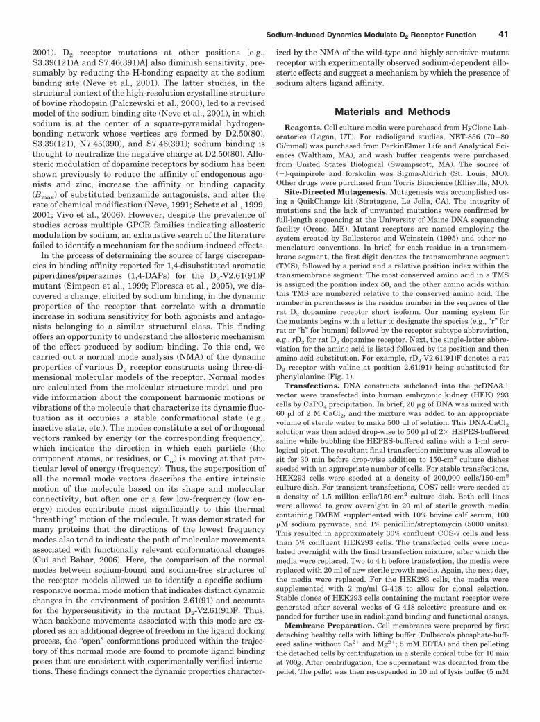
Fig. 1. Depiction of the D2-V2.61(91)F mutant do-pamine receptor as a monomer in a section of lipidbilayer. This figure represents the unfolded D2L re-ceptor showing the amino terminus (�NH2) on theextracellular side and the carboxyl terminus(�CO2H) on the intracellular side. Open circles (E)are used to indicate wild-type amino acids, whereasclosed circles (F and U) are used to represent spe-cific amino acids. As shown in the sequence, a va-line-to-phenylalanine mutation at amino acid resi-due 91 (F) results in the D2-V2.61(91)F mutantdopamine receptor. The purpose of the V2.61(91)Fmutation is to modify the binding pocket of the D2receptor with the corresponding residue of the D4receptor and make it more accommodating to D4-selective 1,4-DAPs (Simpson et al., 1999; Schetz etal., 2000; Kortagere et al., 2004; Floresca et al.,2005). Although most ligands bind an orthostericbinding site accessible from the extracellular face ofthe receptor (Floresca and Schetz, 2004), the sodiumion binds the receptor through an intracellular al-losteric binding site formed by the interactions oftransmembrane segments 2, 3, and 7 (Neve et al.,2001). Also shown in the diagram and the sequenceis the relative position of the conserved negativelycharged D2.50(80) that is critical for the interactionof sodium ions with the dopamine receptor (Neve etal., 1991, 2001).
42 Ericksen et al.
were carefully removed by pipetting, and 100 �l of cell lysis bufferwas added to each well. The cells were lysed by shaking at 600 rpmon a microtiter plate shaker for 1.5 h. A portion of the resultinglysate (30 �l) was transferred to an opaque 96-well Costar plate(catalog no. 07-200-309; Corning Inc., Corning, NY) and challengedwith 0.5 units of acceptor and donor beads (9.35 and 12.5 �g/ml,respectively) containing 5 units of biotinylated cAMP (3.76 nM).Before reading, this reaction was allowed to equilibrate for 1 h withshaking at 600 rpm, protected from light with aluminum foil.
Calculations and Data Analysis. Data points for each experi-ment were sampled in triplicate, and each experiment repeated threetimes, except where noted. The geometric mean and S.D. are re-ported for each experiment; however, the errors in the graphs areS.E.M. values. The equilibrium dissociation constant (KD) of[3H]MSP was determined from saturation isotherm analysis. Theinhibition constants (Ki) for all radioligand competition assays werecalculated with the Cheng-Prusoff equation: Ki � IC50/(1 � [radioli-gand]/KD), where KD is the equilibrium dissociation constant of theradioligand. A K0.5 value is reported in cases where the Hill slope issignificantly different from unity. All data were analyzed usingPrism version 4.0 (GraphPad Software Inc., San Diego, CA). For theinhibition assays, data from three or more assays were combined andthen interpreted by extrapolating all dose-response curves to zero togenerate IC50 values. These were subsequently converted to Ki val-ues before analysis by one-way ANOVA with a Dunnett’s post hocanalysis. For saturation isotherm binding assays, specific bindingcurves were obtained by subtracting nonspecific binding [defined asbinding in the presence of 5 �M (�)-butaclamol] from the totalbinding at each concentration of radioligand. Values for KD and Bmax
were determined from the specific binding curve. Sodium shift bind-ing assays were analyzed by comparing the receptor data with andwithout sodium in a paired two-tailed Student’s t test. cAMP func-tional assays were assessed by first quantifying the amount of cAMPgenerated per milligram of protein in each sample and then normal-izing this value as a percentage of the cAMP generated by unopposed6 �M forskolin. Efficacy was determined by subtracting the best fitvalues for the bottom of the curve (lowest horizontal asymptote) fromthe top of the curve (highest horizontal asymptote). Functional as-says are graphed as sigmoidal semilog dose-response curves. Statis-tical analyses of the curve fitting procedure included the run test, Ftest, and Pearson’s correlation coefficient. Potency and efficacy val-ues generated from three or more replicate curves were analyzed byone-way ANOVA with a Dunnett’s post hoc analysis. Significancewas established at the 95% confidence level (p � 0.05).
Construction of Receptor Homology Models. A wild-type D2
receptor model was constructed using Modeler 9v1 (Sali and Blun-dell, 1993) simultaneously using as templates the (1GZM) structureof bovine rhodopsin (Li et al., 2004) and the recently determinedstructure of the �2 adrenergic receptor (Cherezov et al., 2007)(2RH1). Initially, 1000 D2 receptor structures were generated andranked by Modeler’s objective function. The models were then struc-turally aligned and clustered using the GROMACS version 3.3 pack-age (van der Spoel et al., 2005) gcluster utility (cutoff � 0.20 Å). Themost representative (central) structure from the best scoring clusterwas selected for further analysis and mutated to V2.61F(91) beforeenergy minimization runs.
Building Sodium-Bound D2 Receptor Models. Sodium-boundmodels were constructed with the sodium cation placed at the puta-tive sodium binding pocket near D2.50(80) proposed by Neve et al.(2001). One negative control was constructed by positioning thesodium far from the TM region, at the intracellular carboxylateterminus (C415) where it would not affect the dynamic propertiesthrough direct interaction with the TM region. A second negativecontrol used in this study was the sodium-free “null” system. All thestructures were subjected to energy minimization runs in vacuo withthe low-memory Broyden-Fletcher-Goldfarb-Shanno (quasi-Newto-nian algorithm for energy minimization) method, in three stages,each carried out to convergence (Fmax 10 kJ/mol/nm) using GRO-
MACS version 3.3 (van der Spoel et al., 2005), with the molecularsystems parameterized according to the optimized potentials forliquid simulations all-atom force field (Jorgensen et al., 1996). Elec-trostatic interactions were treated by the particle-mesh Ewaldmethod (Essman et al., 1995). In the first stage, all heavy proteinatoms were restrained with half-harmonic force restraints (k � 1000kJ/mol/nm), with only the sodium ion unrestrained. In the secondminimization stage, sodium and all side chains of residues within 6Å of the sodium were unrestrained. Finally, sodium and all atoms ofresidues within 6 Å of the sodium position were unrestrained duringminimization, with residues outside of this region restrained.
Normal Mode Analysis. To examine the effect of sodium bindingon the dynamic properties of the receptor molecule, we used NMA,which determines a spectrum of independent harmonic (vibrational)motions available to a particular stable molecular conformationwithin a harmonic approximation (for further description, see Intro-duction). Our analysis focused on the lower frequency modes thatcomprise more facile motions (thus, higher amplitude) along direc-tions that coincide with the more shallow curvatures along the po-tential well (Tama and Sanejouand, 2001) and have been shown toindicate function-related dynamics of proteins (Cui and Bahar,2006). Each minimized structure was submitted to the NOMAD-RefWeb server for NMA (Lindahl et al., 2006). Elastic network models(Tirion, 1996) were built from C� positions, with the sodium ionrepresented as an additional C� at its optimized binding position.The first 106 normal modes (M1–106) were calculated for each elasticnetwork model using default parameters, with the exception of thedistance-weighting parameter in which a nondefault value of 3.0 Åwas applied as recommended for C�-only models.
To examine divergence in dynamical behavior between sodium-bound and control (null) structures, dot products were computed foreach sodium-bound normal mode vector against all computed modesfrom the null system. A window of the null spectrum was then chosenby centering it at the most analogous mode Mj
Null (highest dot prod-uct with Mi
Na�). The dot product squares were then summed over theselected null spectrum window to provide a Pij value (Pij � i,j(window)
�MiNa��Mj
Null�2). A range of vectors from the control structures must beincluded because what might appear as a unique sodium-boundmode can be recapitulated by a set of such vectors, when combined.This overcomes a potential pitfall in comparative NMA resultingfrom a comparison of only pairs of corresponding modes betweenstructures (Ming and Wall, 2005). Pij was then plotted as a functionof Mi
Na� using various window sizes to identify the difference innormal mode(s) between sodium-bound structures and control.
Ligand Docking into D2 Receptor Conformers from theM19
Na� Trajectory. The trajectory of the sodium-sensitive M19Na� was
selected to explore effects of sodium-related motions on ligand dock-ing because: 1) its Pij value demonstrates significant divergence fromthe conformational space of the other modes; 2) it was the lowestfrequency mode among those showing divergence and represents asofter and, thus, higher amplitude collective domain motion; and 3)visual inspection revealed that as part of its characteristic motion,an open phase appears to widen the binding pocket, increasingaccessibility (from the extracellular milieu) and its volume in theV2.61(91)F mutant. Therefore, we constructed a series of D2 receptormodels representing points in the trajectory of this particular modeby rebuilding all-atom structures on the C� frames from the M19
trajectory (output from NOMAD-Ref). Backbone and side chain at-oms were built onto the fixed C� template with Modeler 9v1, followedby minimization, a short 15-ps MD run, and minimization with C�
atoms fixed for each procedure. The structure was then minimized toconvergence with positional restraints on the C� carbons in GRO-MACS, as described under Materials and Methods.
Ligands were constructed in Discovery Studio (Accelrys, SanDiego, CA), and their geometries were optimized with ab initio quan-tum mechanical calculations using the HF6–31G** basis set inGaussian03 (Frisch et al., 2004). The partial charges were set ac-cording to the AutoDockTools 1.4.5 automated Gasteiger partial
Sodium-Induced Dynamics Modulate D2 Receptor Function 43
charge assignment. Ligands were then docked 50 times into eachM19-based D2 receptor frame using the Lamarckian genetic algo-rithm search routine in AutoDock 4.0 (Morris et al., 1998), withdefault parameters and a maximal number of energy evaluations of4.0 � 106. Selective side chain flexibility was allowed within thedocking routine; we explored the rotation of dihedrals in the sidechains of F2.61(91), F3.28(110), V3.29(111), D3.32(114), W6.48(358),F6.51(361), and H6.55(375), chosen because of their apparently crit-ical position in the ligand binding site. After docking into eachreceptor frame, poses were clustered using an RMSD tolerance of3.0 Å, and clusters were ranked by mean energies.
ResultsWhen transiently expressed in COS-7 cells, the D2-
V2.61(91)F mutant receptor had a level of expression and anaffinity for the moderately D2-selective 1,4-DAP [3H]MSPthat was similar to the wild-type D2 receptor (1.1- and 4.1-fold, respectively) (Table 1; Fig. 2). It also had an affinitycomparable with that of the wild-type receptor for the agonist
(�)-quinpirole, which lacks a 1,4-DAP structural motif (1.2-fold, Table 1). The 1,4-DAP structural motif consists of twoaromatic rings linked to positions 1 and 4 of a central six-membered piperidine or piperazine ring. L-745,870, for ex-ample, has two distinct aryl substituents that extend frompositions 1 and 4 of the central piperazine ring directly andvia a methylene spacer (Table 1). One of these ring nitrogensis likely protonated at physiological pH and interacts withthe acidic pocket residue D3.32(114). For all ligands testedwithin this structure class, the D2-V2.61(91)F mutant hadaffinities very similar to those measured for the wild-type D2
receptor, including L-745,870 and six other D4-selective 1,4-DAPs (1.1–3.3-fold changes, Table 1). All of these 1,4-DAPshave been tested in previous binding studies designed toinvestigate molecular determinants of ligand selectivity forD4 receptors versus D2 receptor subtypes (Schetz et al., 2000;Kortagere et al., 2004), but only one of them (L-745,870 orCPPMA) had been tested on the D2-V2.61(91)F mutant, and
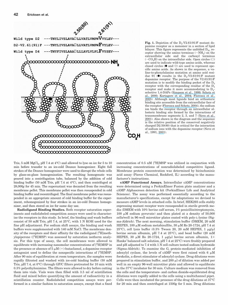
TABLE 1Affinity of D4-selective ligands for the wild-type D2 and D2-V2.61(91)F mutant receptors expressed in COS-7 cellsAffinities for the wild-type D4 receptor are shown for comparison. Affinity values (Ki or KD, nanomolar) are expressed as geometric averages of the mean of threeexperiments � S.D. The fold changes in affinity values relative to the wild-type D2 (D2-WT) are shown in parentheses with the direction of the change indicated by arrows:1, increase in Ki value corresponding to a decreased affinity; and 2, decrease in Ki value corresponding to an increased affinity.
Ligand Structure D2-WT D2-V2.61(91)F D4-WT
L-745,870 (CPPMA)N N
H
NN
Cl 656 � 227 (1) 482 � 251 (21.4) 0.32 � 0.14 (22050)
L-750,667c
N NH
NN
I 1400 � 950 (1) 1100 � 510 (21.3) 0.11 � 0.02a (213,400)
RBI-257
N
N
N
O
I85 � 12 (1) 78 � 3.3 (11.1) 0.27 � 0.10b (2315)
FAUC213cN
N
Cl
NN
1300 � 640 (1) 1200 � 730 (21.1) 1.1 � 0.22b (21030)
Ro61-6270N O
NH2
O655 � 274 (1) 1121 � 70* (11.7) 0.89 � 0.12b (2736)
NGD 94-1
N
N NN
N
HN
817 � 284 (1) 3358 � 266* (13.3) 0.3 � 0.04b (22720)
PD168,077 NN
NH
O
CN 1380 � 64 (1) 3601 � 474*(12.6) 1.5 � 0.41b (2540)
(�)-QuinpiroleNHN
NH
H
812 � 617 (1) 673 � 466 (21.2) N.D.
�3H MethylspiperoneN
FO
NN
O
0.016 � 0.0032 (1) 0.066 � 0.030* (14.1) 0.29 � 0.030a (118)
N.D., not determined.* Significant differences between values for wild-type D2 and D2-V2.61(91)F mutant receptors were determined at 95% confidence by Dunnett’s multiple comparison test.a Schetz et al. (2000).b Kortagere et al. (2004).c Floresca et al. (2005).
44 Ericksen et al.
it was reported to have a large improvement in bindingaffinity (Simpson et al., 1999).
In an effort to determine the source of the discrepancy be-tween the reported 97-fold increase in affinity for L-745,870 foran N- and C-terminally epitope-tagged human D2-V2.61(91)Fmutant (Simpson et al., 1999) and the lack of change for theidentical mutation in the rat receptor for the same ligand (Table1) and several other ligands belonging to the same structural
class (Table 1; Floresca et al., 2005), we systematically elimi-nated differences between the two experimental systems. Ini-tially, we expressed our rat D2-V2.61(91)F mutant in the sameHEK293 cell line used in the previous report. Changing the cellbackground had only a moderate effect on the relative affinitymeasured for L-745,870 at the rD2-V2.61(91)F mutant versusthe wild-type rD2 receptor that was difficult to accurately quan-tify because of low affinity and limited drug solubility under the
TABLE 2The affinities of L-745,870, L-750,667, and NGD 94-1 for the D2-V2.61(91)F mutant receptor are significantly enhanced in the presence of 140 mMNaClReceptors were stably expressed in the HEK293 cell line. Binding affinities (Ki) were calculated from the Cheng-Prusoff equation, Ki � IC50/(1��radioligand /KD, andexpressed as geometric mean values (nanomolar) � S.E.M. (n � 3). The D2-V2.61(91)F was run in three paired experiments, whereas wild-type D2 was run only in two pairedexperiments.
D2-WT* D2-V2.61(91)F
No NaCl 140 mM NaCl No NaCl 140 mM NaCl
L-745,870 5232 � 8419 (1) 778 � 89 (27) 943 � 328* (1) 27.0 � 4.34 (235)L-750,667 �10,000 (1) 1331 � 927 (�27) 1271 � 924* (1) 34 � 24 (237)NGD 94-1 �10,000 (1) 3827 � 3299 (�23) �10,000* (1) 616 � 128* (�216)
* These are approximate values based upon extrapolation. The fold changes in affinity values relative to the receptor in the absence of sodium are shown in parentheseswith the direction of the change indicated by arrows: 2, decrease in Ki value corresponding to an increased affinity.
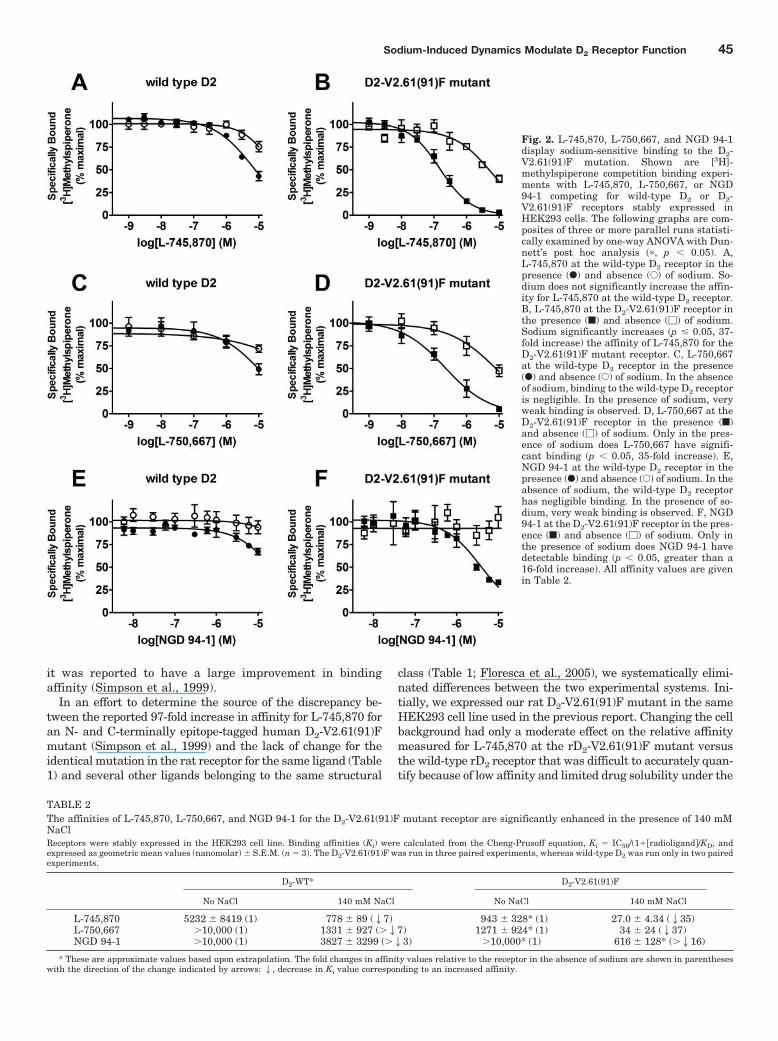
Fig. 2. L-745,870, L-750,667, and NGD 94-1display sodium-sensitive binding to the D2-V2.61(91)F mutation. Shown are [3H]-methylspiperone competition binding experi-ments with L-745,870, L-750,667, or NGD94-1 competing for wild-type D2 or D2-V2.61(91)F receptors stably expressed inHEK293 cells. The following graphs are com-posites of three or more parallel runs statisti-cally examined by one-way ANOVA with Dun-nett’s post hoc analysis (�, p 0.05). A,L-745,870 at the wild-type D2 receptor in thepresence (F) and absence (E) of sodium. So-dium does not significantly increase the affin-ity for L-745,870 at the wild-type D2 receptor.B, L-745,870 at the D2-V2.61(91)F receptor inthe presence (f) and absence (�) of sodium.Sodium significantly increases (p � 0.05, 37-fold increase) the affinity of L-745,870 for theD2-V2.61(91)F mutant receptor. C, L-750,667at the wild-type D2 receptor in the presence(F) and absence (E) of sodium. In the absenceof sodium, binding to the wild-type D2 receptoris negligible. In the presence of sodium, veryweak binding is observed. D, L-750,667 at theD2-V2.61(91)F receptor in the presence (f)and absence (�) of sodium. Only in the pres-ence of sodium does L-750,667 have signifi-cant binding (p 0.05, 35-fold increase). E,NGD 94-1 at the wild-type D2 receptor in thepresence (F) and absence (E) of sodium. In theabsence of sodium, the wild-type D2 receptorhas negligible binding. In the presence of so-dium, very weak binding is observed. F, NGD94-1 at the D2-V2.61(91)F receptor in the pres-ence (f) and absence (�) of sodium. Only inthe presence of sodium does NGD 94-1 havedetectable binding (p 0.05, greater than a16-fold increase). All affinity values are givenin Table 2.
Sodium-Induced Dynamics Modulate D2 Receptor Function 45
conditions tested (�5.5-fold increase based on estimates fromextrapolated values, Fig. 2, A and B; Table 2).
The stable expression of these receptors in the HEK293 cellline also allowed us to study D2-like dopamine receptor cAMPfunctional responses. Although useful for the initial bindingstudies, transient expression in a COS-7 cell line lacked asuitable functional response in our assay system (data notshown). The HEK293 cell line lacks endogenous receptors fordopamine but can mediate a cAMP functional response fortransfected dopamine receptors (data not shown). In prepa-ration for functional assays, the cell surface density of dopa-mine receptors expressed in several stable HEK293 cloneswas determined with [3H]MSP saturation isotherm analysis,after which selected clones were matched by receptor densityto avoid discrepancies because of spare receptors. HEK293cell lines expressing 8.6 � 3.4 pmol/mg protein of the wild-type D2 receptor and 11.6 � 0.23 pmol/mg protein of theD2-V2.61(91)F receptor were selected for use in all subse-quent experiments. The corresponding [3H]MSP affinity val-ues (KD) were 74 � 9.7 pM for the wild-type D2 receptor and95 � 18 pM for the D2-V2.61(91)F receptor. The absoluteaffinities of [3H]MSP for the D2-V2.61(91)F mutant receptorexpressed in COS-7 and HEK293 cells were similar (1.4-fold
different), although small differences were found for the wild-type receptor (4.6-fold different). For this same reason, therelative differences between wild-type and mutant D2 recep-tors within the same cell line are more pronounced in COS-7than in HEK293 cells (4.1- versus 1.3-fold, respectively).
Our next step was to assess whether differences in the bind-ing buffer could account for the large difference observed be-tween our initial COS-7 binding data (Table 1) and publishedHEK293 binding data from Simpson et al. (1999). The strikingfinding was that the addition of 140 mM sodium chloride to thebinding (and wash) buffer, to mimic the buffer conditions in theprevious report (Simpson et al., 1999), resulted in a large (35-fold) increase in L-745,870 affinity for the rD2-V2.61(91)F mu-tant receptor (Fig. 2B; Table 2), although the wild-type D2
receptor displayed only a very limited sodium sensitivity forthis ligand (�7-fold, Fig. 2A; Table 2). These affinity values inthe presence of sodium are consistent with those for hD2 wild-type and hD2-V2.61(91)F published in an earlier report (920 �200 and 9.5 � 4.0 nM, respectively, Simpson et al., 1999). Asimilarly strong pattern of sodium-sensitive binding is evidentfor L-750,667, the iodinated derivative of L-745,870 (Fig. 2, Cand D; Table 2); the affinity of L-750,667 for the rD2-V2.61(91)Fmutant was increased 37-fold in the presence of 140 mM so-
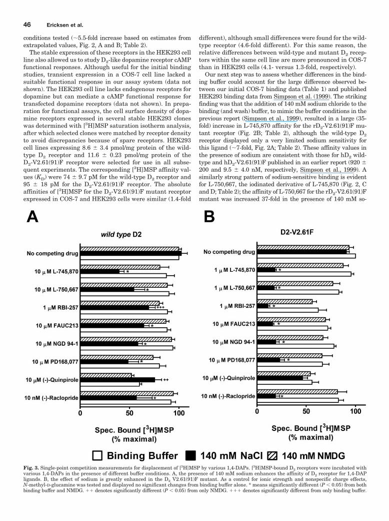
Fig. 3. Single-point competition measurements for displacement of [3H]MSP by various 1,4-DAPs. [3H]MSP-bound D2 receptors were incubated withvarious 1,4-DAPs in the presence of different buffer conditions. A, the presence of 140 mM sodium enhances the affinity of D2 receptor for 1,4-DAPligands. B, the effect of sodium is greatly enhanced in the D2 V2.61(91)F mutant. As a control for ionic strength and nonspecific charge effects,N-methyl-D-glucamine was tested and displayed no significant changes from binding buffer alone. * means significantly different (P 0.05) from bothbinding buffer and NMDG. �� denotes significantly different (P 0.05) from only NMDG. ��� denotes significantly different from only binding buffer.
46 Ericksen et al.
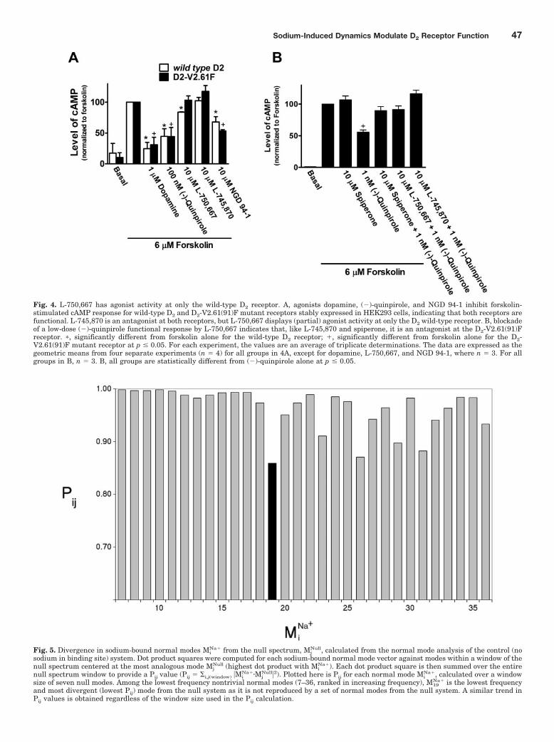
Fig. 4. L-750,667 has agonist activity at only the wild-type D2 receptor. A, agonists dopamine, (�)-quinpirole, and NGD 94-1 inhibit forskolin-stimulated cAMP response for wild-type D2 and D2-V2.61(91)F mutant receptors stably expressed in HEK293 cells, indicating that both receptors arefunctional. L-745,870 is an antagonist at both receptors, but L-750,667 displays (partial) agonist activity at only the D2 wild-type receptor. B, blockadeof a low-dose (�)-quinpirole functional response by L-750,667 indicates that, like L-745,870 and spiperone, it is an antagonist at the D2-V2.61(91)Freceptor. �, significantly different from forskolin alone for the wild-type D2 receptor; �, significantly different from forskolin alone for the D2-V2.61(91)F mutant receptor at p � 0.05. For each experiment, the values are an average of triplicate determinations. The data are expressed as thegeometric means from four separate experiments (n � 4) for all groups in 4A, except for dopamine, L-750,667, and NGD 94-1, where n � 3. For allgroups in B, n � 3. B, all groups are statistically different from (�)-quinpirole alone at p � 0.05.
Fig. 5. Divergence in sodium-bound normal modes MiNa� from the null spectrum, Mj
Null, calculated from the normal mode analysis of the control (nosodium in binding site) system. Dot product squares were computed for each sodium-bound normal mode vector against modes within a window of thenull spectrum centered at the most analogous mode Mj
Null (highest dot product with MiNa�). Each dot product square is then summed over the entire
null spectrum window to provide a Pij value (Pij � i,j(window) �MiNa��Mj
Null�2). Plotted here is Pij for each normal mode MiNa�, calculated over a window
size of seven null modes. Among the lowest frequency nontrivial normal modes (7–36, ranked in increasing frequency), M19Na� is the lowest frequency
and most divergent (lowest Pij) mode from the null system as it is not reproduced by a set of normal modes from the null system. A similar trend inPij values is obtained regardless of the window size used in the Pij calculation.
Sodium-Induced Dynamics Modulate D2 Receptor Function 47
dium, even though there is only a moderate sodium sensitivityfor this ligand at the wild-type receptor (��7-fold). AnotherD4-selective 1,4-DAP, NGD 94-1, chosen because its structure isless similar to L-745,870 and L-750,667 (Table 1), still displayedcomparably enhanced affinity for the D2-V2.61(91)F mutant inthe presence of sodium (Fig. 2, E and F; Table 2) but only a verysmall sodium-dependent increase in affinity for the wild-typereceptor (Fig. 2E; Table 1). Similar patterns of sodium sensitiv-ity were also observed for several other D4-selective 1,4-DAPsand the sodium-sensitive substituted benzamide antagonist,(�)-raclopride (Fig. 3), and these patterns were not mimickedby the same concentration of N-methyl-D-glucamine, a sodiumreplacement ion (Fig. 3). Note that differences in (in the absenceof sodium) affinities appear in Tables 1 and 2, but these are notcomparable. The affinities for expressed wild-type and mutantD2 receptors in Table 1 are from membranes isolated fromCOS7 cells, whereas those in Table 2 are from membranesisolated from HEK293 cells.
In whole-cell attached functional assays, the full agonistsdopamine and (�)-quinpirole both strongly reversed forsko-lin-stimulated increases in cAMP for both wild-type and D2-V2.61(91)F mutant receptors (Fig. 4A). Spiperone was able tofully reverse the (�)-quinpirole-stimulated inhibition ofcAMP (Fig. 4B), but neither (�)-quinpirole nor L-750,667were able to reduce forskolin-stimulated cAMP accumulationin untransfected HEK293 cells (data not shown).
It is notable that we found that L-750,667 has partialagonist properties at the wild-type D2 receptor (Fig. 4A) butacts as an antagonist at the D2-V2.61(91)F mutant receptor(Fig. 4B). However, the chlorinated derivative, L-745,870,has no agonist properties at either receptor, whereas NGD
94-1 has agonist properties at both receptors (Fig. 4A). Con-centrations of L-745,870 five times higher than that shown inFig. 4 did not change its functional profile (data not shown).Despite these drugs having enhanced affinity in the presenceof a high concentration of sodium (Fig. 2), no attempt wasmade to measure such sodium effects in the functionalassays.
Dynamic Properties of the Receptor Constructs. NMAperformed on molecular model constructs of the D2 receptor wasused to examine the dynamic response to sodium ion binding. Inan effort to characterize changes in the receptor’s harmonicmotions imparted by the perturbation of sodium ion, we com-pared the spectrum of normal modes calculated for: 1) thesodium-bound structure [near D2.50(80)], 2) a negative controlwith sodium ion bound outside the protein core (at the carbox-ylate terminus), and 3) a system without any sodium, hereafterreferred to as the null system. To ascertain which intrinsiclow-frequency (and relatively high-amplitude) motions are sen-sitive to the perturbation from sodium binding within the re-ceptor, we calculated the sum of dot product squares, Pij �i,j(window) �Mi
Na��MjNull�2 (see Fig. 5) for each sodium-bound
normal mode (MiNa�) over ranges (windows) of modes in the
null spectrum MjNull (for details, see Materials and Methods). A
plot of Pij as a function of sodium-bound normal mode vectors(Mi
Na�) indicates that sodium ion binding influences the char-acteristic movements of only a few low-frequency modes. Pij
values 0.9 are indicative of motions different from thosedescribed by the normal modes of the null system. Such valueswere observed for modes 19, 26, 29, and 31 in the sodium-boundstructure, hereafter referred to as M19
Na�, M26Na�, and so forth.
We explored different window sizes (window � 7, 15, 21, and
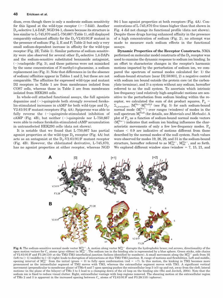
Fig. 6. The sodium-sensitive normal mode vector M19Na�. A, motion along vector M19
Na� disrupts the hydrophobic brace; red arrows, directionality of theopen motion vectors for C� atoms (gray ribbon) in M19
Na�. The sodium ion in the binding site is represented by a blue sphere. Green sticks, side chainsof V2.61(91)F and F3.28(110) at the TM2-TM3 interhelical junction (helices identified by numbers). A small movement along the M19
Na� path from [0](left) to [�1] (middle) to [�2] (right) leads to disruption of interactions at this TM2-TM3 junction. B, range of motions and flexibilities. Left and middle,opening interval of M19
Na� from the initial (green � 0) to fully open conformation (red � �7). In this motion, the kinking in TM2 becomes morepronounced as the intracellular segment of TM2 moves with TM3, whereas the extracellular segment moves with TM1. A significant verticalmovement (perpendicular to membrane plane) of TMs 6 and 7 upon opening pushes the extracellular loop 3 (e3) up and out, away from the cleft; lateralmotions (in the plane of the bilayer) of TMs 3 to 5 lead to a clamping down of the e2 loop on the binding site (Shi and Javitch, 2004). Note that thesodium ion is fixed to reduce visual clutter. Right, extracellular vantage with loop regions removed. The shearing motion at the extracellular regionof TMs 2 and 3 is apparent in the increased spacing between C� atoms of V2.61(91)F and F3.28(110) (spheres).
48 Ericksen et al.

106) in the null spectrum to see whether divergent MiNa� could
be recapitulated (Pij � 1.0) by comparison against larger win-dows of the null spectrum. M19
Na�, however, consistently pro-vided Pij values significantly lower than the other Mi
Na� modes.Pij calculations for the negative control spectrum (Mi
Control)against the null spectrum (not shown) do not yield uniquely lowPij values comparable with M19
Na�, which also suggests that the
M19Na� divergence is site specific in regards to sodium perturba-
tion. M19Na� had no clearly equivalent mode in the entire spec-
trum calculated for the null system. The modes M19Null and M16
Null
were most analogous, with PijNa�/Null values of 0.25 and 0.36,
respectively. Together, these findings suggest that the differentmotion described by M19
Na� is because of the effect of sodiumbinding on the dynamic properties of the receptor molecule.
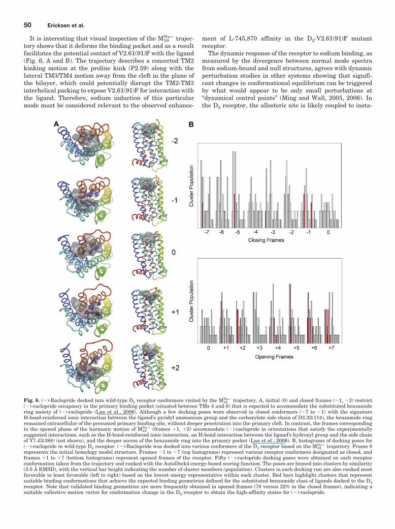
Fig. 7. Docking of L-745,870 into D2 V2.61(91)F receptor conformers visited by the M19Na� trajectory. A, frames shown from top to bottom correspond
to closed (negative) and open (positive) steps in the M19Na� trajectory, respectively. The D2 receptor structure is rendered as a ribbon, and the TM
segments are colored according to the rainbow spectrum: TM1, blue to TM7, red. The transparent gray blob indicates the cumulative van der Waalsspace occupied by the top five most favorable binding poses. Initial (0) and closed frames �1 and �2 prevent ligand access for direct interactions withV2.61(91)F/F3.28 and D3.32(114) and restrict occupancy to regions extracellular of the presumed binding site. In contrast, in the frames correspondingto the open phase of the harmonic motion of M19
Na� (frames �1, �2), L-745,870 is accommodated in orientations that support experimentally suggestedinteractions with V2.61(91)F, F3.28(110), and D3.32(114). Maroon stick, L-745,870 bound in experimentally validated poses with the p-chlorophenylring toward F2.61(91) and F3.28(110). B, histograms of docking pose clusters. L-745,870 was docked into various conformers of the D2 receptor basedon the M19
Na� trajectory. Frame 0 represents the initial homology model structure. Frames �1 to �7 (top histograms) represent various receptorconformers designated as closed, and frames �1 to �7 (bottom histograms) represent open frames of the receptor. The histogram collects results from50 poses obtained from each independent L-745,870 docking performed on each receptor conformation taken from the trajectory and ranked with theAutoDock4 energy-based scoring function. The poses are binned into clusters by similarity (3.0 Å RMSD), with the vertical bar height indicating thenumber of cluster members (population). Clusters in each docking run are also ranked most favorable to least favorable (left to right) based on thelowest energy representative within each cluster. Red bars highlight clusters that represent suitable binding conformations that achieve the expectedbinding geometries defined for the 1,4-DAP class of ligands docked to the D4 receptor (Kortagere et al., 2004). Note that proper binding geometriesare only obtained in opened frames, indicating the need for a conformation change in the D2 receptor to obtain the high-affinity state.
Sodium-Induced Dynamics Modulate D2 Receptor Function 49
It is interesting that visual inspection of the M19Na� trajec-
tory shows that it deforms the binding pocket and as a resultfacilitates the potential contact of V2.61(91)F with the ligand(Fig. 6, A and B). The trajectory describes a concerted TM2kinking motion at the proline kink (P2.59) along with thelateral TM3/TM4 motion away from the cleft in the plane ofthe bilayer, which could potentially disrupt the TM2-TM3interhelical packing to expose V2.61(91)F for interaction withthe ligand. Therefore, sodium induction of this particularmode must be considered relevant to the observed enhance-
ment of L-745,870 affinity in the D2-V2.61(91)F mutantreceptor.
The dynamic response of the receptor to sodium binding, asmeasured by the divergence between normal mode spectrafrom sodium-bound and null structures, agrees with dynamicperturbation studies in other systems showing that signifi-cant changes in conformational equilibrium can be triggeredby what would appear to be only small perturbations at“dynamical control points” (Ming and Wall, 2005, 2006). Inthe D2 receptor, the allosteric site is likely coupled to insta-
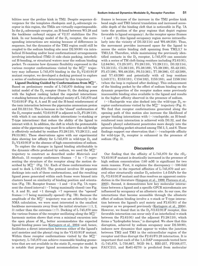
Fig. 8. (�)-Raclopride docked into wild-type D2 receptor conformers visited by the M19Na� trajectory. A, initial (0) and closed frames (�1, �2) restrict
(�)-raclopride occupancy in the primary binding pocket (situated between TMs 4 and 6) that is expected to accommodate the substituted benzamidering moiety of (�)-raclopride (Lan et al., 2006). Although a few docking poses were observed in closed conformers (�7 to �1) with the signatureH-bond-reinforced ionic interaction between the ligand’s pyridyl ammonium group and the carboxylate side chain of D3.32(114), the benzamide ringremained extracellular of the presumed primary binding site, without deeper penetration into the primary cleft. In contrast, the frames correspondingto the opened phase of the harmonic motion of M19
Na� (frames �1, �2) accommodate (�)-raclopride in orientations that satisfy the experimentallysuggested interactions, such as the H-bond-reinforced ionic interaction, an H-bond interaction between the ligand’s hydroxyl group and the side chainof Y7.43(388) (not shown), and the deeper access of the benzamide ring into the primary pocket (Lan et al., 2006). B, histograms of docking poses for(�)-raclopride in wild-type D2 receptor. (�)-Raclopride was docked into various conformers of the D2 receptor based on the M19
Na� trajectory. Frame 0represents the initial homology model structure. Frames �1 to �7 (top histograms) represent various receptor conformers designated as closed, andframes �1 to �7 (bottom histograms) represent opened frames of the receptor. Fifty (�)-raclopride docking poses were obtained on each receptorconformation taken from the trajectory and ranked with the AutoDock4 energy-based scoring function. The poses are binned into clusters by similarity(3.0 Å RMSD), with the vertical bar height indicating the number of cluster members (population). Clusters in each docking run are also ranked mostfavorable to least favorable (left to right) based on the lowest energy representative within each cluster. Red bars highlight clusters that representsuitable binding conformations that achieve the expected binding geometries defined for the substituted benzamide class of ligands docked to the D2receptor. Note that validated binding geometries are more frequently obtained in opened frames (78 versus 22% in the closed frames), indicating asuitable collective motion vector for conformation change in the D2 receptor to obtain the high-affinity states for (�)-raclopride.
50 Ericksen et al.
bilities near the proline kink in TM2. Despite sequence di-vergence for the templates rhodopsin and �2-adrenergic re-ceptor in this region, the TM2s are virtually superimposable.In the �2-adrenergic receptor, an H-bond between W3.28 andthe backbone carbonyl oxygen of V2.57 stabilizes the Prokink. In our homology model of the D2 receptor, the corre-sponding interactions are missing because of differences insequence, but the dynamics of the TM2 region could still becoupled to the sodium binding site near D2.50(80) via intra-helical H-bonding and/or local conformational arrangementsinvolving interhelical (TM2–3) side chain packing, interheli-cal H-bonding, or structural waters near the sodium bindingpocket. To examine how dynamic flexibility expressed in thevarious receptor conformations visited by the M19
Na� trajec-tory may influence L-745,870 binding in the D2-V2.61(91)Fmutant receptor, we developed a docking protocol to explorea series of conformations determined by this trajectory.
Ligand Docking Guided by Conformations from M19Na�.
Based on preliminary results of L-745,870 docking into ourinitial model of the D2 receptor (frame 0), the docking poseswith the highest ranking lacked the expected interactions:�-stacking between the p-chlorophenyl moiety of L-745,870 andV2.61(91)F (Fig. 6, A and B) and the H-bond reinforcement ofthe ionic interaction between the piperazine ammonium protonand D3.32(114). This is because in the “closed” conformation ofthe D2 receptor model, V2.61(91)F is adjacent to D2-F3.28(110),with which it can maintain stable interactions (�-stacking orT-type interactions) that reduce the ability of the ligand tointeract with it. In addition, the cleft in which the protonatableamine of the piperazine ring must fit to achieve this salt bridgeis effectively occluded by residues F3.28(110), V3.29(111), andF6.51(361). These observations agree with our experimentaldata showing low affinity for L-745,870 in wild-type D2 andD2-V2.61(91)F in the absence of high concentrations of sodium.
To explore the changes in ligand binding attributable tothe dynamic effects produced by sodium, we used the M19
Na�
trajectory to construct as described under Materials andMethods, 15 receptor conformers (frames �7 to �7) repre-senting the structure of the receptor along the motion de-scribed by M19
Na� (Fig. 7A). Each of these conformations wasused to dock L-745,870. The protocol involves 50 separatedockings into each of these conformations, and the resultingligand poses generated within each frame were binned intoclusters based on similarity of binding position and orienta-tion (Fig. 7B). Receptor frames �1 and �2 in Fig. 7A repre-sent the closed interval (�7 being maximally closed) (see Fig.6, A and B), and �1 through �7 represent the “opened”frames (�7 being maximally opened) (Fig. 7B). Because theamplitude of the M19
Na� trajectory was set arbitrarily in theNMA calculation, we were most interested in the smallestbackbone movements away from the initial structure (�1, 2,3) rather than the extremes (�7). Docking of L-745,870 intothe various frames of the receptor oscillating along the M19
Na�
harmonic motion shows that even a minimal excursion intothe open phase of M19 better accommodates the expectedligand binding poses (see Kortagere et al., 2004) and, hence,facilitates a direct interaction between either of the ligand’saryl moieties and the phenyl ring in the V2.61(91)F mutant.Within these receptor conformations visited by the M19
Na�
trajectory, L-745,870 can achieve reasonable binding geome-tries that are not available in the static D2 receptor model. Itis notable that proper ligand accommodation in the open
frames is because of the increase in the TM2 proline kinkbend angle and TM3 lateral translation and increased acces-sible depth of the binding cleft that is noticeable in Fig. 7A(note the position of the gray regions that depict regionsfavorable to ligand occupancy). As the receptor opens (frames�1 and �2), this ligand occupancy region moves intracellu-larly into the vicinity of D3.32(114) and W6.48(358). Thus,the movement provides increased space for the ligand toaccess the entire binding cleft spanning from TM2,3,7 toTM3,5,6. Therefore, while maintaining the previously iden-tified contacts in the D2 receptor, L-745,870 can associatewith a series of TM cleft-lining residues including F2.61(91),L2.64(94), C3.25(107), F3.28(110), V3.29(111), D3.32(114),V3.33(115), C3.36(118), F5.38(189), V5.39(190), S5.42(193),F5.47(198), W6.48(358), F6.51(361), T7.39(386), G7.42(389),and Y7.43(390) and potentially with e2 loop residuesL143(171), E153(181), C154(182), I155(183), and I156(184)(when the loop is replaced after docking). This enhancementof the binding pocket by the effect of sodium binding on thedynamic properties of the receptor makes some previouslyinaccessible binding sites available to the ligand and resultsin the higher affinity observed in the presence of sodium.
(�)-Raclopride was also docked into the wild-type D2 re-ceptor conformations visited by the M19
Na� trajectory (Fig. 8).We find that receptor conformations generated along theopening path of this motion also increase the likelihood ofproper binding interactions with (�)-raclopride; an H-bond-reinforced ionic interaction is achieved with D3.32, and theligand’s phenyl substituent penetrates more deeply into theprimary binding pocket situated among TM 3, 5, and 6. Thesefindings support our observation that (�)-raclopride affinityfor wild-type D2 receptor is enhanced in the presence ofsodium (Fig. 3).
DiscussionOur finding that the affinity of L-745,870 for the rD2-
V2.61(91)F mutant is drastically increased in the presence ofhigh sodium concentration (140 mM) is significant for twomain reasons. First, it explains the discrepancy (�100-folddifferences) in the reported affinities of L-745,870 and sev-eral other structurally similar D4-selective 1,4-DAPs for theD2-V2.61(91)F mutant and thus resolves an apparent contra-diction in the literature (Simpson et al., 1999; Floresca et al.,2005). Second, it demonstrates how key molecular interac-tions between a ligand and a specific GPCR microdomain areinfluenced by occupancy of an allosteric site. In our case, theinteractions that become accessible through the allostericeffect of sodium binding involve a �-stack or T-type interac-tion between the ligand’s aryl moiety and F2.61(91) of thereceptor as we proposed previously (Kortagere et al., 2004).However, we found that in the D2-V2.61(91)F receptor, thisfavorable interaction can occur only if an interhelical �-stackbetween the F2.61(91) and the adjacent F3.28(110), whichforms a “hydrophobic brace,” is disrupted. We show that thisdisruption, achieved by sodium occupancy near D2.50(80),induces new dynamics that appear to widen the junctionbetween TM2 and TM3 in the extracellular region of thereceptor and thus disrupt the F2.61/F3.28 interaction. It isimportant that each of the 1,4-DAP compounds tested here(L-745,870, L-750,667, NGD 94-1, RBI-257, PD168,077,FAUC213, and Ro61-6270) is predicted from molecular
Sodium-Induced Dynamics Modulate D2 Receptor Function 51
models to engage in similar interactions when phenylala-nine occupies position 2.61(91); in agreement, we foundexperimentally that all compounds tested display drasticsodium-dependent increases in affinity for the D2-V2.61(91)Fmutant.
Although previous studies have revealed the affinity rela-tionship between the discriminant structural features of 1,4-DAPs and the 1,4-DAP D4/D2 selectivity-conferring positions2.61(91), 3.28(110), and 3.29(111), the effects of mutations inthis microdomain on the activity of 1,4-DAP ligands had notbeen described previously (Simpson et al., 1999; Schetz et al.,2000; Kortagere et al., 2004; Floresca et al., 2005). Therefore,because the D2-V2.61(91)F receptor exhibits sodium-sensi-tive affinity changes to 1,4-DAPs, we examined here thefunctional properties of L-745,870, L-750,667, and NGD 94-1at the wild-type and D2-V2.61(91)F receptor. In contrast tothe reports showing reduction in agonist affinities as a resultof allosteric modulation by sodium, we report here sodium-dependent enhancement of affinities for the 1,4-DAP ago-nists (and antagonists) similar to that observed for the sub-stituted benzamide antagonists (Neve, 1991). Moreover, wereport here that although the D2-V2.61(91)F mutant can beactivated by dopamine and (�)-quinpirole, it cannot be acti-vated by L-750,667, which exhibits weak partial agonistproperties on the wild-type D2 receptor. Rather, L-750,667acts as an antagonist of the D2-V2.61(91)F mutant. The rel-atively small influence of the D2-V2.61(91)F mutation on thebinding affinity of (�)-quinpirole and methylspiperone dem-onstrated here suggests that neither ligand is likely to di-rectly contact 2.61(91). Furthermore, this mutation had littleeffect on the activation of the receptor by (�)-quinpirole andits reversal by methylspiperone. This suggests that the2.61(91)-3.28(110) hydrophobic brace can prevent the recep-tor from being activated by some agonists if their bindingbrings them near position 2.61(91). Because the affinities ofother 1,4-DAPs, which retained their agonist properties atthe D2-V2.61(91)F mutant receptor, were also enhanced inthe presence of sodium, we reviewed the literature concern-ing the connection between agonist high- and low-affinitystates, G protein coupling, and sensitivity to sodium andGTP.
In broken membrane preparations, high concentrations ofeither sodium ions (120 mM) or GTP and its related analogs(100 �M) decrease the affinity of agonists for catecholamin-ergic GPCRs (Paris et al., 1989; Neve, 1991; Neve et al., 2001;Schetz and Sibley, 2001). However, the similarities betweenthe agonist affinity shift for sodium and GTP are coincidentalbecause sodium binds an allosteric site on dopamine recep-tors accessible from the intracellular side (Neve, 1991; Neveet al., 2001; Schetz and Sibley, 2001), whereas GTP binds Gproteins that then complex with the receptor at a differentintracellular allosteric site. Under conditions (e.g., no GTP)in which a ternary complex is formed (receptor � G protein),kinetic studies of detergent-solubilized or broken membrane� adrenergic receptors demonstrate that both sodium ionsand Gpp(NH)p accelerate the rate of agonist dissociation,and a synergistic increase in the rate is observed in thepresence of both of these modulators (Limbird et al., 1982). Inbroken membrane equilibrium studies of � adrenergic recep-tors, agonist affinity decreases in the presence of either so-dium or Gpp(NH)p, and a further decrease in affinity isobserved in the presence of both of these modulators. These
results were cited as evidence that sodium ions do not com-pete with GTP for its binding to G proteins (Limbird et al.,1982). Later studies by the same group demonstrated thatsubstitution of the negative charge of the conserved asparticacid in TM2 [D2.50(79)] to a neutral asparagine in the �2adrenergic receptor resulted in a total loss of epinephrine’ssensitivity to sodium (Horstman et al., 1990). When testedfor its ability to stimulate GTPase activity, this same mutantreceptor had a 7.5-fold decrease in potency for the agonistUK14304 with no change in efficacy (Ceresa and Limbird,1994) and no change in affinity relative to the wild-typereceptor. Increasing concentrations of Gpp(NH)p decreasethe agonist p-[125I]iodoclonidine’s binding to the wild-typebut not the mutant receptor (Ceresa and Limbird, 1994).These data suggest that the sodium-insensitive D2.50(79)Nmutant � adrenergic receptor can still couple to G proteinsbut that the state of coupling (coupled or uncoupled) nolonger influences agonist high- and low-affinity states of thereceptor. Thus, it would appear that, at least in the case of �2adrenergic receptors, sodium ions control the agonist high-and low-affinity states of the receptor but do not uncouple Gproteins from their receptor.
Broken membrane equilibrium studies demonstrate thatdopamine’s affinity for the D2 receptor is decreased whenpertussis toxin or GTP uncouple G proteins from the receptorand that the addition of sodium ions further decreases do-pamine’s affinity (Neve et al., 1989). This was cited as evi-dence that sodium ions interact directly with dopamine re-ceptors. Later studies by the same group demonstrated thatsubstitution of the conserved aspartate in TM2 [D2.50(80)]for alanine results in a mutant receptor whose affinities foragonists and substituted benzamide antagonists are sodiuminsensitive (Neve et al., 1991); however, this same mutantreceptor is unable to efficiently couple to G proteins, makingit impossible to carry out the same types of studies as de-scribed above for �2 adrenergic receptors. Finally, the rate ofchemical modification of D2 dopamine receptors by the thiol-reactive agent N-ethylmaleimide is altered in the presence ofsodium (Neve, 1991), and this is consistent with our finding[utilizing the D2-V2.61(91)F mutant receptor as a molecularprobe of the 1,4-DAP binding pocket] that the binding ofsodium ions to the allosteric site [D2-D2.50(80)] causes sig-nificant conformation changes in the receptor in the region ofthe orthosteric binding pocket.
The changes in dynamic properties of the receptor that canexplain the observed pharmacological consequences of so-dium binding are illustrated by our findings from NMA.Thus, the presence of sodium was found from this analysis togive rise to a unique, high-amplitude (low-frequency) normalmode that is not observed for the wild-type structure.Following the motions represented by this unique, sodium-related mode shows that it produces a wider cleft openingin the region responsible for ligand binding. These “open-ing dynamics” allow larger ligands to be accommodated inthe pocket for ligand recognition and make room for betterinteraction of the ligand with the sites in the receptors atwhich it forms H-bonds, aromatic interactions, and hydro-phobic matches. This leads to the measured improvementin the ligand’s affinity for the receptor. Specifically, theeffects of sodium binding near D2.50(80) on the dynamicsin the microdomain surrounding the 2.61(91) position wereshown by NMA to involve an increase in the scissors-like
52 Ericksen et al.
helical movements of the extracellular portions of TMs 2and 3. This motion accounts for the enlarged opening and,consequently, for the large affinity changes observed at theD2-V2.61(91)F mutant, where these movements disruptthe hydrophobic brace formed by the interaction betweenV2.61(91)F and F3.28(110). The result of these local rear-rangements is a receptor conformation more suitable foraccommodating a ligand like L-745,870, which can nowmake stabilizing interactions as found in the optimal dock-ing poses. Similar sodium-enhanced motions are predictedto occur in the wild-type receptor, which lacks a hydropho-bic brace, but cleft opening would lead to better ligandaccess to the conserved aspartic acid at D3.32(114) for areinforced ionic bond interaction with the protonatablepiperizinyl amine portion of the docked 1,4-DAP. Thisprediction is supported experimentally by our findings forthe wild-type D2 receptor whose affinities for L-745,870,L-750,667, and NGD 94-1 are somewhat increased (�3–7-fold) in the presence of sodium. Moreover, the docking ofthe substituted benzamide (�)-raclopride, which belongsto a distinct structural class of D2 antagonists, into con-formations along the sodium-induced normal mode pro-duced very similar results. Again, suitable binding poseswere more readily achieved in wild-type D2 receptor con-formations visited along the open phase of the sodium-induced mode (Fig. 8), thus accounting for the enhancedaffinity for (�)-raclopride (Fig. 3) and other substitutedbenzamides at the D2 receptor when modulated by sodium(Neve, 1991).
How these dynamic changes affect ligand binding is clearlydemonstrated by exploring the binding poses of L-745,870 ina series of receptor conformers built along the characteristicvector of M19
Na�. It is notable that involving NMA in a dockingprotocol to provide a basis for exploring collective backbonemovements in ligand docking has also been used successfullyto reproduce known conformations from ligand-bound crystalstructures (Lindahl and Delarue, 2005). Here, we found thatdocking into conformations generated from the open intervalof M19
Na� accommodates L-745,870 in the expected bindingmodes for 1,4-DAPs in D4 dopamine receptors (Kortagere etal., 2004) because the opening motion disrupts contact be-tween F2.61(91) and F3.28(110), which in turn creates accessto these residues that appeared inaccessible to the p-chloro-phenyl (“mode 1”) or pyrrolopyridinyl group (“mode 2”) ofL-745,870 in the initial D2 receptor homology model. Theaccess is established by the dynamic changes and enablesfavorable aromatic-aromatic interactions between the ligandmoieties and the receptor sites. This result suggests, there-fore, that the specific perturbation propagated from the so-dium binding site influences ligand affinity by dynamicallychanging the interactions in the orthosteric site. The back-bone movement observed in our model might capture thephenomenon of sodium allostery by achieving the high-affin-ity receptor conformational state along the M19
Na� vector pathand/or by disrupting the brace that stabilizes the low-affinitystate without a significant, time-averaged, conformationalchange, i.e., through the sodium-induced amplitude increasealong this vector (Cooper and Dryden, 1984; Popovych et al.,2006). In any case, the presence and effect of endogenousallosteric modulators (e.g., sodium, G protein, GPCR dimerpartner, etc.) should be considered when selecting a receptorrepresentation for in silico library screening. It is also impor-
tant to point out that the use of target receptor conformationsbased on normal mode trajectories could inform the design ofdrugs working through allosteric mechanisms.
Acknowledgments
We thank Dr. Christina Z. Floresca and Shiuwei Chen for assis-tance with early phases of this work and the following people andtheir organizations for supplying samples of compounds that werenot commercially available: Dr. Claus Riemer (Hoffman-La Roche,Nutley, NJ) for Ro 62-6170 and Dr. Andrew Thurkauf (NeurogenCorporation, Branford, CT) for NGD 94-1. In addition, we thankXavier Rovira for providing NMA analysis scripts.
ReferencesBallesteros JA and Weinstein H (1995) Integrated methods for modeling G-protein
coupled receptors. Methods Neurosci 25:366–428.Ceresa BP and Limbird LE (1994) Mutation of an aspartate residue highly conserved
among G-protein-coupled receptors results in nonreciprocal disruption of alpha2-adrenergic receptor-G-protein interactions: A negative charge at amino acidresidue 79 forecasts alpha 2A-adrenergic receptor sensitivity to allosteric modu-lation by monovalent cations and fully effective receptor/G-protein coupling. J BiolChem 269:29557–29564.
Cherezov V, Rosenbaum DM, Hanson MA, Rasmussen SG, Thian FS, Kobilka TS,Choi HJ, Kuhn P, Weis WI, Kobilka BK, et al. (2007) High-resolution crystalstructure of an engineered human beta2-adrenergic G protein-coupled receptor.Science 318:1258–1265.
Cooper A and Dryden DT (1984) Allostery without conformational change: A plau-sible model. Eur Biophys J 11:103–109.
Cui Q and Bahar I (2006) Normal Mode Analysis: Theory and Applications toBiological and Chemical Systems, Chapman & Hall/CRC Press, New York.
Essman U, Perela L, Berkowitz ML, Darden T, Lee H, and Pedersen LG (1995) Asmooth particle mesh Ewald method. J Chem Phys 103:8577–8592.
Floresca CZ and Schetz JA (2004) Dopamine receptor microdomains involved inmolecular recognition and the regulation of drug affinity and function. J ReceptSignal Transduct Res 24:207–239.
Floresca CZ, Chen S, Kortagere S, and Schetz JA (2005) Reciprocal mutations inTM2/TM3 in a D2 dopamine receptor background confirms the importance of thismicrodomain as a selective determinant of para-halogenated 1,4-disubstitutedaromatic piperazines. Arch Pharm (Weinheim) 338:268–275.
Frisch MJ, Trucks GW, Schlegel HB, Scuseria GE, Robb MA, Cheeseman JR,Montgomery JA Jr, Vreven T, Kudin KN, Burant JC, et al. (2004) Gaussian 03,Revision C.02, Gaussian, Inc., Wallingford, CT.
Horstman DA, Brandon S, Wilson AL, Guyer CA, Cragoe EJ Jr, and Limbird LE(1990) An aspartate conserved among G-protein receptors confers allosteric regu-lation of alpha 2-adrenergic receptors by sodium. J Biol Chem 265:21590–21595.
Jorgensen WL, Maxwell DS, and Tirado-Rives J (1996) Development and testing ofthe OPLS all-atom force field on conformational energetics and properties oforganic liquids. J Am Chem Soc 118:11225–11236.
Kortagere S, Gmeiner P, Weinstein H, and Schetz JA (2004) Certain 1,4-disubstituted aromatic piperidines and piperazines with extreme selectivity forthe dopamine D4 receptor interact with a common receptor microdomain. MolPharmacol 66:1491–1499.
Lan H, DuRand CJ, Teeter MM, and Neve KA (2006) Structural determinants ofpharmacological specificity between D1 and D2 dopamine receptors. Mol Pharma-col 69:185–194.
Li J, Edwards PC, Burghammer M, Villa C, and Schertler GF (2004) Structure ofbovine rhodopsin in a trigonal crystal form. J Mol Biol 343:1409–1438.
Limbird LE, Speck JL, and Smith SK (1982) Sodium ion modulates agonist andantagonist interactions with the human platelet alpha 2-adrenergic receptor inmembrane and solubilized preparations. Mol Pharmacol 21:609–617.
Lindahl E, Azuara C, Koehl P, and Delarue M (2006) NOMAD-Ref: visualization,deformation and refinement of macromolecular structures based on all-atom nor-mal mode analysis. Nucleic Acids Res 34:W52–W56.
Lindahl E and Delarue M (2005) Refinement of docked protein-ligand and protein-DNA structures using low frequency normal mode amplitude optimization. NucleicAcids Res 33:4496–4506.
Ming D and Wall ME (2005) Quantifying allosteric effects in proteins. Proteins59:697–707.
Ming D and Wall ME (2006) Interactions in native binding sites cause a large changein protein dynamics. J Mol Biol 358:213–223.
Morris GM, Goodsell DS, Halliday RS, Huey R, Hart WE, Belew RK, and Olson AJ(1998) Automated docking using a lamarckian genetic algorithm and empiricalbinding free energy function. J Comp Chem 19:1639–1662.
Neve KA (1991) Regulation of dopamine D2 receptors by sodium and pH. MolPharmacol 39:570–578.
Neve KA, Cox BA, Henningsen RA, Spanoyannis A, and Neve RL (1991) Pivotal rolefor aspartate-80 in the regulation of dopamine D2 receptor affinity for drugs andinhibition of adenylyl cyclase. Mol Pharmacol 39:733–739.
Neve KA, Cumbay MG, Thompson KR, Yang R, Buck DC, Watts VJ, DuRand CJ, andTeeter MM (2001) Modeling and mutational analysis of a putative sodium-bindingpocket on the dopamine D2 receptor. Mol Pharmacol 60:373–381.
Neve KA, Henningsen RA, Bunzow JR, and Civelli O (1989) Functional character-ization of a rat dopamine D-2 receptor cDNA expressed in a mammalian cell line.Mol Pharmacol 36:446–451.
Palczewski K, Kumasaka T, Hori T, Behnke CA, Motoshima H, Fox BA, Le Tronq I,
Sodium-Induced Dynamics Modulate D2 Receptor Function 53
Teller DC, Okada T, Stenkamp RE, et al. (2000) Crystal structure of rhodopsin: aG protein-coupled receptor. Science 298:739–745.
Paris H, Galitzky J, and Senard JM (1989) Interactions of full and partial agonistswith HT29 cell alpha 2-adrenoceptor: comparative study of [3H]UK-14,304 and[3H]clonidine binding. Mol Pharmacol 35:345–354.
Popovych N, Sun S, Ebright RH, and Kalodimos CG (2006) Dynamically drivenprotein allostery. Nat Struct Mol Biol 13:831–838.
Sali A and Blundell TL (1993) Comparative protein modelling by satisfaction ofspatial restraints. J Mol Biol 234:779–815.
Schetz JA (2005) Allosteric modulation of dopamine receptors. Mini Rev Med Chem5:555–561.
Schetz JA, Benjamin PS, and Sibley DR (2000) Nonconserved residues in the secondtransmembrane-spanning domain of the D(4) dopamine receptor are moleculardeterminants of D(4)-selective pharmacology. Mol Pharmacol 57:144–152.
Schetz JA, Chu A, and Sibley DR (1999) Zinc modulates antagonist interactions withD2-like dopamine receptors through distinct molecular mechanisms. J PharmacolExp Ther 289:956–964.
Schetz JA and Sibley DR (2001) The binding-site crevice of the D4 dopamine receptoris coupled to three distinct sites of allosteric modulation. J Pharmacol Exp Ther296:359–363.
Shi L and Javitch JA (2004) The second extracellular loop of the dopamine D2receptor lines the binding-site crevice. Proc Natl Acad Sci U S A 101:440–445.
Simpson MM, Ballesteros JA, Chiappa V, Chen J, Suehiro M, Hartman DS, Godel T,Snyder LA, Sakmar TP, and Javitch JA (1999) Dopamine D4/D2 receptor selec-tivity is determined by a divergent aromatic microdomain contained within thesecond, third, and seventh membrane-spanning segments. Mol Pharmacol 56:1116–1126.
Tama F and Sanejouand YH (2001) Conformational change of proteins arising fromnormal mode calculations. Protein Eng 14:1–6.
Tirion MM (1996) Large amplitude elastic motions in proteins from a single-parameter, atomic analysis. Phys Rev Lett 77:1905–1908.
van der Spoel D, Lindahl E, Hess B, Groenhof G, Mark AE, and Berendsen HJC(2005) GROMACS: fast, flexible, and free. J Comp Chem 26:1701–1718.
Vivo M, Lin H, and Strange PG (2006) Investigation of cooperativity in the bindingof ligands to the D2 dopamine receptor. Mol Pharmacol 69:226–235.
Address correspondence to: Dr. John A. Schetz, Department of Pharma-cology and Neuroscience, University of North Texas Health Science Center,3500 Camp Bowie Blvd., Fort Worth, TX 76107. E-mail: [email protected]
54 Ericksen et al.