Mechanisms of Permeation and Selectivity in Calcium Channels

31 Mar 2005 10:0 AR AR243-BB34-07.tex XMLPublishSM(2004/02/24) P1: KUV10.1146/annurev.biophys.34.040204.144655
Annu. Rev. Biophys. Biomol. Struct. 2005. 34:153–71doi: 10.1146/annurev.biophys.34.040204.144655
Copyright c© 2005 by Annual Reviews. All rights reservedFirst published online as a Review in Advance on March 3, 2005
ION CONDUCTION AND SELECTIVITY
IN K+ CHANNELS
Benoıt RouxDepartment of Physiology and Biophysics, Weill Medical College of Cornell University,New York, NY 10021; email: [email protected]
Key Words molecular dynamic simulations, KcsA, free energy, potential of meanforce, crystallographic B-factors, gating
■ Abstract Potassium (K+) channels are tetrameric membrane-spanning proteinsthat provide a selective pore for the conductance of K+ across the cell membranes.These channels are most remarkable in their ability to discriminate K+ from Na+ bymore than a thousandfold and conduct at a throughput rate near diffusion limit. Therecent progress in the structural characterization of K+ channel provides us with aunique opportunity to understand their function at the atomic level. With their abil-ity to go beyond static structures, molecular dynamics simulations based on atomicmodels can play an important role in shaping our view of how ion channels carry outtheir function. The purpose of this review is to summarize the most important find-ings from experiments and computations and to highlight a number of fundamentalmechanistic questions about ion conduction and selectivity that will require furtherwork.
CONTENTS
PERSPECTIVE AND OVERVIEW . . . . . . . . . . . . . . . . . . . . . . . . . . . . . . . . . . . . . . . . 154KEY MECHANISTIC QUESTIONS . . . . . . . . . . . . . . . . . . . . . . . . . . . . . . . . . . . . . . 155ION CONDUCTION . . . . . . . . . . . . . . . . . . . . . . . . . . . . . . . . . . . . . . . . . . . . . . . . . . . 157
Transport Cycle . . . . . . . . . . . . . . . . . . . . . . . . . . . . . . . . . . . . . . . . . . . . . . . . . . . . . . 157ION SELECTIVITY . . . . . . . . . . . . . . . . . . . . . . . . . . . . . . . . . . . . . . . . . . . . . . . . . . . . 158
Importance of Carbonyl-Carbonyl Repulsion . . . . . . . . . . . . . . . . . . . . . . . . . . . . . . 158Control of Selectivity by Dynamical and Electrostatic Properties . . . . . . . . . . . . . . . 159
CATION BINDING SITES . . . . . . . . . . . . . . . . . . . . . . . . . . . . . . . . . . . . . . . . . . . . . . 160External S0 Binding Site . . . . . . . . . . . . . . . . . . . . . . . . . . . . . . . . . . . . . . . . . . . . . . . 160Hydrated Cation in Vestibular Cavity . . . . . . . . . . . . . . . . . . . . . . . . . . . . . . . . . . . . . 161Multi-Ion Energetics . . . . . . . . . . . . . . . . . . . . . . . . . . . . . . . . . . . . . . . . . . . . . . . . . . 161Selectivity of the Binding Sites . . . . . . . . . . . . . . . . . . . . . . . . . . . . . . . . . . . . . . . . . 162
PORE BLOCKERS AND INHIBITORS . . . . . . . . . . . . . . . . . . . . . . . . . . . . . . . . . . . . 163Quaternary Ammonium and Other Compounds . . . . . . . . . . . . . . . . . . . . . . . . . . . . . 163Toxin Peptide Inhibitors . . . . . . . . . . . . . . . . . . . . . . . . . . . . . . . . . . . . . . . . . . . . . . . 163
1056-8700/05/0609-0153$20.00 153
Ann
u. R
ev. B
ioph
ys. B
iom
ol. S
truc
t. 20
05.3
4:15
3-17
1. D
ownl
oade
d fr
om a
rjou
rnal
s.an
nual
revi
ews.
org
by C
OR
NE
LL
UN
IV M
ED
ICA
L C
OL
LE
GE
on
05/0
4/05
. For
per
sona
l use
onl
y.

31 Mar 2005 10:0 AR AR243-BB34-07.tex XMLPublishSM(2004/02/24) P1: KUV
154 ROUX
GATING . . . . . . . . . . . . . . . . . . . . . . . . . . . . . . . . . . . . . . . . . . . . . . . . . . . . . . . . . . . . . 164Intracellular Gating . . . . . . . . . . . . . . . . . . . . . . . . . . . . . . . . . . . . . . . . . . . . . . . . . . . 164Gating by the Selectivity Filter? . . . . . . . . . . . . . . . . . . . . . . . . . . . . . . . . . . . . . . . . . 164
CONCLUSION . . . . . . . . . . . . . . . . . . . . . . . . . . . . . . . . . . . . . . . . . . . . . . . . . . . . . . . . 165
PERSPECTIVE AND OVERVIEW
The determination of the structure of the bacterial KcsA channel from Streptomyceslividans in 1998 by MacKinnon and coworkers offered the first view of the generalarchitecture of these proteins (39) (Figure 1, see color insert). Because KcsA isstructurally similar to eukaryotic K+ channels, investigations of KcsA are expectedto help researchers understand a large class of biologically important channels. TheX-ray structure of KcsA triggered a large number of computational studies basedon molecular dynamics (MD) (4, 6, 7, 16, 17, 21, 25, 26, 29, 31, 36, 38, 54,55, 82, 83, 84, 103, 118, 119), Poisson-Boltzmann (PB) (108, 113, 114), andBrownian dynamics (BD) (5, 22, 23, 91). Because of the moderate resolution ofthe X-ray data (3.2 A), the computational studies initiated from the original 1998KcsA structure proceeded with a limited amount of information. In particular, theexact number and configurations of the ions and water molecules in the selectivityfilter were not known, and whether the channel was in an open or closed statein the crystal structure was unclear. The picture was rapidly refined during thefollowing few years as additional structural and functional information becameavailable. X-ray structures of the KcsA channel were obtained at higher resolu-tion (up to 2.0 A) (95, 132, 135), followed by structures for other K+ channels:MthK (3.4 A), a calcium-activated channel crystallized in an open state (68); KvAP(3.2 A), a voltage-gated channel (70); and KirBAC (3.6 A), an inward rectifier (77).Further information about the molecular movements of the transmembrane helicesof KcsA and their role in channel gating was obtained by electron spin resonance(ESR) (30, 32, 51, 52, 80) and mass spectroscopy (75). At the same time, the resultsof electrophysiological experiments on KcsA became available (59, 78, 79, 102).In retrospect, it is encouraging that many results from the computational studieshave been consistent with the rapidly emerging data, sometimes even in advanceof the fact (93). A number of factors have contributed to the recent success of thecomputational studies on biological membrane channels: clearly posed concep-tual challenges, suitability of simulation timescales, availability of high-resolutionstructures, and opportunities for experimental verification (115).
Several excellent reviews covering computational studies of ion channels havebeen published in the past few years. A number of them have provided timelyand critical summaries of the most important results from simulation studies (37,94, 111, 115, 116), structural modeling (49, 116), and bioinformatics sequenceanalysis approaches (24, 117). A few have focused specifically on the generalmethodologies used in computational studies of ion channels (28, 112, 123) (formore about simulation methodologies, see References 2 and 13). The purposeof this review is to summarize important findings and to highlight a number of
Ann
u. R
ev. B
ioph
ys. B
iom
ol. S
truc
t. 20
05.3
4:15
3-17
1. D
ownl
oade
d fr
om a
rjou
rnal
s.an
nual
revi
ews.
org
by C
OR
NE
LL
UN
IV M
ED
ICA
L C
OL
LE
GE
on
05/0
4/05
. For
per
sona
l use
onl
y.

31 Mar 2005 10:0 AR AR243-BB34-07.tex XMLPublishSM(2004/02/24) P1: KUV
POTASSIUM CHANNELS 155
fundamental mechanistic questions about ion conduction and selectivity that willrequire further work.
KEY MECHANISTIC QUESTIONS
One key mechanistic question about K+ channels is how are they able to achievea fast throughput rate and yet remain highly selective for K+ over Na+. More than50 years ago, Hodgkin & Keynes (66) used radioactive flux marker experiments toshow that the K+ channel was a narrow pore containing two to three K+ and thatpermeation involved single-file ion movements across the membrane. Accordingto the “knock-on” mechanism that they proposed, the approach of one ion from oneside of the pore is coupled to the simultaneous exit of another ion on the oppositeside of the pore,
thereby enabling fast conduction. Over the following years, the notion of selectiveion channels as narrow molecular “proteinaceous” pores and the steric constraintsthey put on permeation was refined, particularly through the work of Mullins (97,98). But steric constraint did not explain how a pore could discriminate K+ fromNa+, i.e., allow the passage of the larger cation and block the smaller one. A thermo-dynamic perspective on ion selectivity based on hydration free energy differences,elaborated by Eisenman (40), clarified these matters considerably. Fundamentally,ion selectivity is controlled by the relative free energy of K+ and Na+ in the poreand in the bulk solution. In the early 1970s, Bezanilla & Armstrong (19) expandedon this idea of a narrow molecular pore and proposed the “snug-fit” hypothesis toexplain the selectivity of K+ channels: Na+ ions do not enter the narrowest part ofthe pore because they are too small to fit well in the coordination cage provided bythe channel as replacement for the water molecules surrounding the ion in solution.A different point of view was proposed by Eisenman and coworkers (120), whoargued that the selectivity was more similar to the transfer process from water toan organic solvent. Detailed ion-flux experiments highlighted the high selectivityof K+ channels (63) but could be consistent with both explanations.
The X-ray structure of KcsA determined in 1998 (39) is strikingly consistentwith the classical views of a very selective, fast-conducting, multi-ion pore (Fig-ure 1). The pore comprises a wide, nonpolar aqueous cavity on the intracellularside, leading up, on the extracellular side, to a narrow pore that is 12 A long andlined exclusively by main chain carbonyl oxygens. Formed by the residues corre-sponding to the signature sequence TTVGYG, common to all K+ channels (58),this region of the pore acts as a selectivity filter by allowing only the passage ofnearly dehydrated K+ ions across the cell membrane. One rate-limiting step in theconduction mechanism is expected to be the translocation of nearly dehydrated K+
Ann
u. R
ev. B
ioph
ys. B
iom
ol. S
truc
t. 20
05.3
4:15
3-17
1. D
ownl
oade
d fr
om a
rjou
rnal
s.an
nual
revi
ews.
org
by C
OR
NE
LL
UN
IV M
ED
ICA
L C
OL
LE
GE
on
05/0
4/05
. For
per
sona
l use
onl
y.

31 Mar 2005 10:0 AR AR243-BB34-07.tex XMLPublishSM(2004/02/24) P1: KUV
156 ROUX
ions in single file through the narrowest region of the pore. The latter appears to bewell adapted to compensate for the dehydration of K+ but not Na+ ions. A singleisolated K+ ion would interact strongly with the carbonyl oxygens and remaintightly bound to the selectivity filter. But the presence of multiple K+ results inmutual electrostatic repulsion directed along the axis of the narrow pore, whichcontributes to yield a high-throughput rate via the knock-on mechanism.
However, how selective conduction of K+ ions takes place efficiently at theatomic level is complex. The knock-on mechanism assumes that ion-channel at-traction and ion-ion repulsion play compensating effects, as several ions movesimultaneously in single file through the narrow pore: The approach of one ionfrom one side of the (doubly occupied) selectivity filter is coupled to the simulta-neous exit of an other ion on the opposite side. Although plausible, this mechanismrelies on a strikingly delicate energy balance. To allow rapid ion conduction, thestrong attraction between the ions and the channel must be exquisitely counterbal-anced by the electrostatic repulsive forces between the ions. A simple calculationshows that the direct ion-ion coulombic repulsion could vary by several tens ofkcal per molecule during the conduction process. Somehow, the K+ channel ex-ploits such large energies in a productive manner to yield a flux of approximately108 ions per second. Fast conduction implies that there is no significant activa-tion free energy barrier opposing the concerted ion translocation. How can this bepossible?
Furthermore, how the channel achieves a high selectivity for K+ over Na+ is alsoa fascinating question. On the basis of the X-ray structure, it has been suggested thatthe filter is constrained in an optimal geometry by a network of aromatic residuessurrounding the selectivity filter so that a dehydrated K+ ion fits snugly with propercoordination by the backbone carbonyl oxygens, but that it cannot distort suffi-ciently to coordinate a smaller cation such as Na+ (39, 135). It is almost taken forgranted that the X-ray structure of KcsA confirmed the elegant mechanism of se-lectivity proposed by Bezanilla & Armstrong (19) (11, 12, 15, 22, 48, 65, 130, 135).However, this question is more complex than it appears to be. The atomic radiusof K+ and Na+ differs only by 0.38 A (106), implying that the snug-fit mechanism(19) relies on the ability of the selectivity filter to retain rigidly a remarkably precise(subangstrom) geometry to discriminate between these two cations. Proteins, likemost biological macromolecular assemblies, are soft materials that display signif-icant structural flexibility at room temperature (73). Despite some uncertainties,the B-factors of the KcsA channel indicate that the RMS fluctuations of the atomslining the selectivity filter are on the order of 0.75 to 1.0 A (Figure 2, see colorinsert), in general agreement with MD simulations of KcsA (4, 6, 7, 16, 17, 21, 29,38, 54, 55, 82, 83, 103, 118, 119). The magnitude of atomic thermal fluctuations isfundamentally related to the intrinsic flexibility of a protein, i.e., how it respondsstructurally to external perturbations (3). These considerations suggest that, at roomtemperature, the flexible/fluctuating channel should distort easily to cradle Na+
with little energetic cost; this is seen in simulations with Na+ in KcsA (21, 38, 54).The flexibility of the pore is further highlighted by the experimental observation
Ann
u. R
ev. B
ioph
ys. B
iom
ol. S
truc
t. 20
05.3
4:15
3-17
1. D
ownl
oade
d fr
om a
rjou
rnal
s.an
nual
revi
ews.
org
by C
OR
NE
LL
UN
IV M
ED
ICA
L C
OL
LE
GE
on
05/0
4/05
. For
per
sona
l use
onl
y.

31 Mar 2005 10:0 AR AR243-BB34-07.tex XMLPublishSM(2004/02/24) P1: KUV
POTASSIUM CHANNELS 157
that K+ is needed for the overall stability of the channel structure (76, 81, 92, 133,135). Therefore, although the snug-fit hypothesis is consistent with a static viewof the crystallographic structure, it does not jibe with the current knowledge ofproteins. But then, what is the microscopic mechanism of selectivity?
ION CONDUCTION
The most fundamental mechanistic insight from MD simulations about the ionconduction through the KcsA channel is the concept of a single file of K+ sep-arated by water molecules dynamically moving through the narrowest region ofthe pore in a highly correlated fashion (4, 6, 7, 16, 17, 29, 38, 54, 55, 118, 119).One water molecule per K+ is transported through the narrow pore (7, 16, 17),which is observed experimentally (1) and consistent with diffraction data (131).The selectivity filter is also where the driving force arising from the transmem-brane electrostatic potential is predominant (18, 69, 113). The maximum electricconductance of K+ through the selectivity filter of KcsA was calculated to be on theorder of 300 to 500 pS (18), in agreement with experimental measurements (79).
Transport Cycle
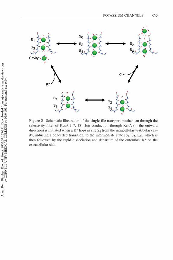
The elementary events underlying the ion conduction process are best visualizedwith a transport cycle between multi-ion states. The transport cycle based onthe computational studies is shown in Figure 3 (see color insert) (17, 18). Thecycle bears many similarities to the classical knock-on mechanism of Hodgkin &Keynes (66) shown in Scheme 1. The conduction process, with alternatively twoand three K+ in the narrowest part of the pore, is consistent with ion occupancydetermined by X-ray diffraction at high resolution (131, 134, 135). At physiologicalconcentrations the selectivity filter is occupied predominantly by two K+ ions, inthe configurations [S1, S3] or [S2, S4] (7, 39, 134, 135). At some point (e.g., duringan outward conduction event), a third ion hops from the intracellular vestibularcavity into site S4, while two ions in the selectivity filter are located in sites S1 andS3. The incoming ion induces a concerted transition to a state in which the threeions occupy sites S4, S2, and S0, which is then followed by the rapid dissociationand departure of the outermost ions in S0 on the extracellular side, yielding theconduction of one K+.
A transport cycle was also elaborated by Morais-Cabral et al. (95) to describe theion conduction mechanism. Like the ion conduction mechanism determined fromthe computation studies (Figure 3), this process is similar to the classical knock-on mechanism of Hodgkin & Keynes (66) shown in Scheme 1; it pictures ionconduction as a collision between an incoming K+ and the two ions located in theselectivity filter, resulting in the ejection of the ion at the opposite end of the pore.But a number of differences are also worth noting. Morais-Cabral et al. (95) stressedthe importance of the energy balance between the configurations with two K+, [S3,
Ann
u. R
ev. B
ioph
ys. B
iom
ol. S
truc
t. 20
05.3
4:15
3-17
1. D
ownl
oade
d fr
om a
rjou
rnal
s.an
nual
revi
ews.
org
by C
OR
NE
LL
UN
IV M
ED
ICA
L C
OL
LE
GE
on
05/0
4/05
. For
per
sona
l use
onl
y.

31 Mar 2005 10:0 AR AR243-BB34-07.tex XMLPublishSM(2004/02/24) P1: KUV
158 ROUX
S1] and [S4, S2]. They represented the rate-limiting step as an unfavorable activatedcomplex with three equidistant K+ located between the four cation binding sitesof the selectivity filter, S1, S2, and S4, and their transport cycle does not explicitlyinvolve other binding sites, such as the vestibular cavity or S0. This putative high-energy activated complex with three K+ is not observed in the computationalstudies (17). Instead, the key microscopic event underlying ion conduction is theconcerted transition between the two stable low-energy configurations with threeK+, [cavity, S3, S1] and [S4, S2, S0] (17). The two-ion configurations [S1, S3]and [S2, S4] have indeed roughly the same free energy (7), and the exchange ratebetween them is extremely rapid (4, 6, 7, 16, 17, 29, 38, 54, 55, 118, 119). But theback-and-forth shuttling of two K+ in the selectivity filter, in the absence of a thirdK+, does not control the throughput rate. The critical step leading to productiveconduction involves configurations with three K+ located within the vicinity ofthe pore, that is, the five main cation binding sites, S0, S1, S2, S3, and S4, and thevestibular cavity. According to the computations, efficient knock-on is apparentlymade possible because the approach of an incoming ion on the intracellular side isnot energetically prohibitive, whereas the dissociation of the outermost ion in S0 isaccelerated by ion-ion repulsion (17, 18). Although the computational results areconsistent with available data, they have yet to be verified directly by experiments.
ION SELECTIVITY
The key question about selectivity is straightforward: What is the microscopic basisfor the unfavorable free energy: ��G(Na+ → K+) = [
Gpore(Na+) − Gpore(K+)]
− [Gbulk(Na+) − Gbulk(K+)
]? By going beyond considerations based on static
structures, researchers can use alchemical free energy perturbation (FEP) com-putations based on atomic models (2, 13) to help clarify the (often hidden) mi-croscopic factors that govern thermodynamics. The results of FEP computationsshow that, indeed, occupancy by Na+ in the selectivity filter is thermodynamicallyunfavorable (4, 7, 17, 83, 103), which is in good agreement with ion-flux measure-ments (101, 102). But these results present a clear paradox: The calculated freeenergies are consistent with a selective channel, but the thermal fluctuations of thebackbone atoms lining the pore are obviously too large to be consistent with thesnug-fit mechanism (19, 39). An alternative explanation might be that selectivityarises locally, from the intrinsic properties of the microscopic interactions takingplace near the ion in the pore (40, 41, 120, 128).
Importance of Carbonyl-Carbonyl Repulsion
The most important interactions taking place in the first coordination shell aroundan ion in the pore are the very strong electrostatic attraction and core repulsionbetween the cation and the nearest carbonyl groups, and the moderate electrostaticrepulsion between the coordinating carbonyl groups. A useful strategy to clarify
Ann
u. R
ev. B
ioph
ys. B
iom
ol. S
truc
t. 20
05.3
4:15
3-17
1. D
ownl
oade
d fr
om a
rjou
rnal
s.an
nual
revi
ews.
org
by C
OR
NE
LL
UN
IV M
ED
ICA
L C
OL
LE
GE
on
05/0
4/05
. For
per
sona
l use
onl
y.
31 Mar 2005 10:0 AR AR243-BB34-07.tex XMLPublishSM(2004/02/24) P1: KUV
POTASSIUM CHANNELS 159
how selectivity arises is to carry out FEP calculations in which the electrostaticinteraction between the carbonyl groups located in different subunits of the channelis artificially turned off (103). The effect of turning off the carbonyl-carbonylinteraction on the calculated free energy is informative because it is intrinsicallyassociated with the local dynamics and fluctuations of the first coordination shellaround the cation. For instance, no significant impact on ��G is expected in thecase of a locally rigid structure. Conversely, a large change in ��G is indicativeof a flexible coordination structure. Remarkably, removing the carbonyl-carbonylinteraction in a fully flexible KcsA channel completely destroys the selectivityof site S2, which then becomes significantly favorable for Na+. The relative freeenergy, originally unfavorable for Na+ by 5.3 kcal mol−1, becomes favorableby 2.9 kcal mol−1, with a net loss of 8.2 kcal mol−1 in selectivity, when therepulsive interactions between the backbone carbonyl is turned off. To assessfurther the influence of the architectural rigidity of the protein relative to the localcarbonyl interactions, additional FEP calculations were performed, keeping allatoms fixed except those forming the selectivity filter (i.e., the backbone atomof residues Thr-74 to Asp-78). Remarkably, despite the frozen protein structuresurrounding the selectivity filter, the FEP calculations for S2 show that the relativefree energy decreases from 6.7 to 0.9 kcal mol−1 when the carbonyl repulsion inthe selectivity filter is removed. There is a loss of almost 6 kcal mol−1 of selectivityfor K+ over Na+ even if only the backbone of the selectivity filter is allowed tomove dynamically and fluctuate. The FEP calculations show that any architecturalsubangstrom rigidity of the protein conferred by the highly conserved residuessurrounding the selectivity filter cannot be a key factor in making the channelselective for K+ over Na+. Further computations with a reduced model systemrepresenting the binding site S2 demonstrate that ion selectivity is a robust featureof such a flexible fluctuating pore lined by carbonyl groups (103).
Control of Selectivity by Dynamicaland Electrostatic Properties
Ionic selectivity in K+ channels is controlled by the intrinsic local physical prop-erties of the ligands coordinating the cation in the binding site (103). Such aperspective bears some similarities to the treatment of ionic selectivity elaboratedby Eisenman and colleagues, who identified the strength of the electric field aris-ing from the ligands coordinating the cation in a binding site as a key factor indetermining selectivity (40, 41, 120, 128). To illustrate this, a simple toy modelwith eight carbonyl-like (C=O) dipoles surrounding a central cation fluctuatingfreely inside a sphere with a radius of 3.5 A was considered. The results revealthat such a simple system is naturally and optimally selective for K+, demonstrat-ing that selectivity for K+ over Na+ is possible without any architectural rigiditypreventing the coordinating ligands from collapsing onto a smaller cation. Selec-tivity for K+ or for Na+ requires different chemical functionalities to coordinatethe cation favorably. This conclusion is also consistent with an examination of the
Ann
u. R
ev. B
ioph
ys. B
iom
ol. S
truc
t. 20
05.3
4:15
3-17
1. D
ownl
oade
d fr
om a
rjou
rnal
s.an
nual
revi
ews.
org
by C
OR
NE
LL
UN
IV M
ED
ICA
L C
OL
LE
GE
on
05/0
4/05
. For
per
sona
l use
onl
y.

31 Mar 2005 10:0 AR AR243-BB34-07.tex XMLPublishSM(2004/02/24) P1: KUV
160 ROUX
amino acid sequence of ionic channels in various genomes, which suggests thatno biological channels selective for Na+ have evolved by refining the geometryof a KcsA-like pore lined by backbone carbonyl groups (64). One possibility toachieve Na+ selectivity (although there are certainly others) would be to intro-duce a salt-bridge formed by the association of two charged residues directly intothe pore. Intriguingly, the amino acids identified to be essential for the selectivityof Na+ channels include the highly conserved DEKA locus from four differentprotein domains (61, 64).
CATION BINDING SITES
Detailed information about energetically favorable binding sites along the perme-ation pathway is essential to understand the ion conduction process at the molecu-lar level. Results from both X-ray crystallography (131, 135) and MD free energysimulations (7, 17) show that specific cation binding sites are disposed along theselectivity filter of KcsA (Figure 1). A complete characterization of these bind-ing sites, structural and energetics, is of paramount importance to understand ionpermeation.
External S0 Binding Site
The binding site S0, located on the extracellular side of the channel, is particularlyintriguing. A cation moving from site S0 to site S1 is in the process of being progres-sively dehydrated. For this reason, this site is of great interest (although perhapsmore difficult to characterize). The binding site was not detected in the initial struc-ture of KcsA (39) at 3.2 A resolution, but a spontaneous concerted transition to aconfiguration with a K+ occupying S0 was first observed during all-atom MD tra-jectory of KcsA in a lipid membrane based on this crystallographic structure (16).The site was not detected in the subsequent higher resolution structures of KcsA(95, 132) based on the initial construct, but it was observed in the structure at 2.0 Aresolution crystalized with a Fab antibody fragment. This suggests that detectionof ion occupancy in site S0 in KcsA crystals is not simply a matter of resolution.Examination of the lattice packing in those protein crystals suggests a possible ex-planation: There is a head-to-head interaction between neighboring channels in thecrystal (their extracellular entrance facing one another), and occupancy of the ex-ternal sites by a K+ could be energetically prohibitive because of ion-ion repulsion(Figure 4, see color insert). The K+ in site S0 is closer to the protein by approxi-mately 1.5 A along the channel axis in the calculations (17) relative to the positiondetermined in the high-resolution X-ray structure (135). In the computations, aK+ in S0 is partially hydrated by three to four water molecules and transientlycoordinated by the carbonyl oxygens of Tyr-78, whereas the cation appears to bemore fully hydrated in the X-ray structure. The significance of these differencesis unclear. The computations attempt to simulate a channel in a bilayer mem-brane solvated by an aqueous solution at room temperature using an approximate
Ann
u. R
ev. B
ioph
ys. B
iom
ol. S
truc
t. 20
05.3
4:15
3-17
1. D
ownl
oade
d fr
om a
rjou
rnal
s.an
nual
revi
ews.
org
by C
OR
NE
LL
UN
IV M
ED
ICA
L C
OL
LE
GE
on
05/0
4/05
. For
per
sona
l use
onl
y.

31 Mar 2005 10:0 AR AR243-BB34-07.tex XMLPublishSM(2004/02/24) P1: KUV
POTASSIUM CHANNELS 161
potential function (17), whereas the diffraction data report detailed experimentalmeasurements on the cation occupancy, but for protein crystals at liquid nitrogentemperature and solvated by an aqueous solution including 40% polyethylenegly-col (PEG) for cryo-protection (135). The impact of fast-freezing protein crystalson fine atomic details is difficult to assess (56, 57). There are indications that thepacking and configuration of charged side chains in protein crystals can be quitesensitive to temperature (72), and perhaps such a conclusion applies to hydratedions near channel vestibules as well. Although MD simulations are burdened byseveral approximations, notably the neglect of induced polarizability near smallions, similar calculations have accurately predicted the position of water moleculesin aquaporins (34, 121).
Hydrated Cation in Vestibular Cavity
A solvated K+ surrounded by exactly eight water molecules is observed at thecenter of the vestibular cavity in the X-ray structure (135). But other cations (Na+,Rb+, Cs+, and Tl+) in the cavity do not display a similarly ordered hydration struc-ture (134). These observations are intriguing. However, it is unknown whether theobserved average density is representative of the typical hydration of a cation inthe channel cavity. The effect of flash-freezing protein crystals on the structure ofwater is different for wide and narrow solvent-filled regions (125), and this shouldbe carefully assessed in the case of K+ channel crystals. Furthermore, the pore axiscorresponds to an axis of crystallographic symmetry in the structure cocrystalizedwith Fab antibody fragment (135). As a result, there is a fourfold averaging ofthe hydration structure around the cation. In MD simulations (performed at roomtemperature), the hydration shell surrounding the K+ in the cavity is similar towhat it would be in bulk solution (16), with a hydration number fluctuating tran-siently between six and nine water molecules. The hydration structure seen in MDsimulation is in general accord with results from experimental studies based onneutron and X-ray scattering on aqueous ionic solution (42, 67, 100, 104, 105; fora recent review see Reference 99). Therefore, more work is required to understandthe hydration structure around a cation located inside the cavity of a K+ channel.
Multi-Ion Energetics
During the conduction process, ion movements are expected to take place in ahighly concerted way, as the K+ in the pore undergo sudden hopping transitionsbetween various multi-ion configurations. For this reason, it is important to charac-terize not only the position of the cation binding sites but also the relative stabilityof the multi-ion configurations and the free energy barriers between them. Usinga systematic search procedure, the thermodynamical stability of all the possibleways to distribute any number of K+ among the four cation binding sites locatedwithin the narrow selectivity filter (S1, S2, S3, and S4) was estimated with FEP sim-ulations (7). Only a small subset of occupancy states were energetically allowed,the [S1, S3] and [S2, S4] configurations (7), with the two K+ separated by one
Ann
u. R
ev. B
ioph
ys. B
iom
ol. S
truc
t. 20
05.3
4:15
3-17
1. D
ownl
oade
d fr
om a
rjou
rnal
s.an
nual
revi
ews.
org
by C
OR
NE
LL
UN
IV M
ED
ICA
L C
OL
LE
GE
on
05/0
4/05
. For
per
sona
l use
onl
y.

31 Mar 2005 10:0 AR AR243-BB34-07.tex XMLPublishSM(2004/02/24) P1: KUV
162 ROUX
water molecule occupying the selectivity filter. Spontaneous occurrences of suchconfigurations were also observed in several MD simulations of KcsA (16, 54,118). Diffraction data on KcsA are consistent with these configurations (95), al-though lattice interactions may have had some impact on the relative stability of themulti-ion configuration (see above). The most compelling experimental evidencein support of the multi-ion configuration predicted by the computations came fromdiffraction data with a mutant of the KcsA channel (T75C) with altered ion distribu-tion in the selectivity filter (131). Several lines of evidence from the computationsindicate that the configurations with three K+ are [cavity, S3, S1] and [S4, S2, S0].Notably, there appears to be some repulsive interaction opposing the simultaneousoccupancy of the cavity and site S4, or sites S1 and S0. For example, absence ofa K+ in the cavity has a strong effect on the relative stability of the [S1, S3] and[S2, S4] configurations (55). Although consistent with available data, these com-putational results have yet to be tested directly by experiments. Calculations basedon simplified semimicroscopic models may help clarify the origin of the factorsdominating the energy of multi-ion configurations (48). Detailed kinetic analysisof ion-flux data with different ions and rapid pore blockers may also provide cluesabout the multi-ion configurations governing the ion conduction process (78, 122).Additional information, possibly from X-ray structures of other K+ channels (77),might help to better characterize the cation binding sites. However, the delicatebalance of attractive and repulsive forces may be slightly different for various K+
channels.
Selectivity of the Binding Sites
FEP computations indicate that the selectivity of the cation binding sites alongthe pore is different (7, 17, 103). The most selective appears to be site S2, locatedin the center of the pore. Sites S1 and S3 display moderate selectivities, whereasS0 and S4 are not selective. The results of punchthrough experiments with Na+
on the intracellular side can be interpreted as evidence of Na+ blocking the poreby entering the cavity (102), although it is also possible that Na+ is blocking thepore by occupying site S4. The vestibular cavity on the intracellular side doesnot display any selectivity in the computations, but the hydration structure aroundcations seen in X-ray diffraction depends on the ion type (134, 135). Comparisonof MD trajectories with different cations in the KcsA channel suggests that K+,Rb+, and Cs+ behave in a similar way, but that the difference is more pronouncedin the case of Na+ (38). Diffraction data indicate that Rb+ and Cs+ do not easilyoccupy site S2 in KcsA (95, 133, 135) (although ion-flux measurements suggestthat this might be different in Shaker; see Reference 122). How sensitive theseobservations are to the low temperature of the crystals is still unknown. In thefuture, it would be of interest to characterize the relative stability of those cationsin the various binding sites at different temperatures using FEP computations.More accurate quantum mechanical electronic structure computations might helpascertain the validity of the conclusions from MD simulations based on all-atomforce fields (12, 53; see 27 for review).
Ann
u. R
ev. B
ioph
ys. B
iom
ol. S
truc
t. 20
05.3
4:15
3-17
1. D
ownl
oade
d fr
om a
rjou
rnal
s.an
nual
revi
ews.
org
by C
OR
NE
LL
UN
IV M
ED
ICA
L C
OL
LE
GE
on
05/0
4/05
. For
per
sona
l use
onl
y.

31 Mar 2005 10:0 AR AR243-BB34-07.tex XMLPublishSM(2004/02/24) P1: KUV
POTASSIUM CHANNELS 163
PORE BLOCKERS AND INHIBITORS
Quaternary Ammonium and Other Compounds
An important fraction of the present information about the function of K+ channelshas been deduced from studies with quaternary ammonium (QA) pore blockers,such as tetramethylammonium (TMA), tetraethylammonium (TEA), tetrabutyl-ammonium (TBA), and related alkyl derivatives (8, 9, 10, 44, 129). QAs act byblocking the permeation pathway, occluding the movement of K+ ions throughthe pore. Nearly all K+ channels are blocked by QAs on the intracellular side. Incontrast, some K+ channels are blocked by TEA on the extracellular side, whereasothers are not. The affinity of extracellular TEA blockade is enhanced by thepresence of aromatic residues located near the extracellular mouth of the pore (60,74, 90). By virtue of their ability to act as sensors of the pore of K+ channels, theseclassical blockers are important tools in electrophysiological studies of biologicalmembranes.
A number of computational studies have investigated the interactions of clas-sical QA blockers with the KcsA channel (31, 53, 84, 86). Computations of theinteractions of TEA with the KcsA channel have clarified the origin of the impor-tant aromatic residue near the pore extracellular entrance (31, 53, 84). The studiesgenerally indicate that TEA would occupy the S0 cation binding site observed inthe X-ray structure (135) and in PMF computations (17). One consequence is thatextracellular binding of TEA may drive the multi-ion system toward a configura-tion with two K+ occupying sites S4 and S2 and a TEA in site S0 (with each ionpair separated by a single water molecule). Recently, studies on classical blockershave been extended to examine the interaction of bupivacaine with a model of theKv1.5 channel in the open state (85).
Toxin Peptide Inhibitors
Toxin inhibitors are small proteins that bind at the pore entryway on the extracellu-lar side between the four subunits, thus blocking the channel. Their high specificitymakes them powerful tools for characterizing K+ channels structurally. Scorpiontoxins have been used to identify the pore region of K+ channels (88, 89), eluci-date the topology of the extracellular face of the channel (50, 62, 109), assign thefunctional sidedness of the proton activation of KcsA (59), and aid in the design ofa promising strategy to target lymphocytes (110). The general architecture of theextracellular face of the Shaker K+ channel deduced from the results of thermody-namic mutant cycles with agitoxin (109) was strikingly consistent with the X-raystructure of the KcsA channel (87). But there is yet no high-resolution structureof a toxin in complex with a K+ channel.
A computational approach was developed to determine and refine models of thestructure of agitoxin in complex with the Shaker K+ channel on the basis of mutantcycle data (43); the change in binding free energies upon site-directed mutationswas calculated using PB computations and compared with experimental data to
Ann
u. R
ev. B
ioph
ys. B
iom
ol. S
truc
t. 20
05.3
4:15
3-17
1. D
ownl
oade
d fr
om a
rjou
rnal
s.an
nual
revi
ews.
org
by C
OR
NE
LL
UN
IV M
ED
ICA
L C
OL
LE
GE
on
05/0
4/05
. For
per
sona
l use
onl
y.

31 Mar 2005 10:0 AR AR243-BB34-07.tex XMLPublishSM(2004/02/24) P1: KUV
164 ROUX
rank and assess the validity of the large number of models generated. The interac-tion of kappa-conotoxin-PVIIA with the Shaker K+ channel pore was simulatedby MD (96). BD has been a popular approach to simulate the association and inter-actions of toxins with the extracellular vestibule of K+ channel. This method hasbeen applied to toxin Lq2 (33) and maurotoxin (45). The interaction of agitoxin,charybdotoxin, and iberiotoxin with various K+ channels was investigated by us-ing atomic models and energy minimization, and the factors responsible for theselectivity between different voltage-gated and Maxi-K channels were explored(46). The interaction between ScyTx and the small conductance calcium-activatedK+ channel has been investigated by using docking and a combination of MD andPB computations (127).
GATING
Intracellular Gating
Characterizing the molecular determinants of gating is a challenge, and despite theremarkable progress, numerous fundamental questions remain unresolved. Resultsfrom the bacterial channels provide some important clues. The full-length KcsAchannel is mostly closed at neutral pH, but it can be stabilized in the open state atlow intracellular pH (59); the X-ray structures (39, 135) (with their C-terminalstruncated) correspond to the closed nonconducting state (107, 113). A similarclosed form is revealed by the X-ray structure of a bacterial inward-rectifier K+
channel, the KirBac (77). Data from ESR with site-specific spin-labels suggest thatchannel gating involves the movements of the inner helices, which presumably leadto the opening of the pore on the intracellular side (107). MD trajectories, biasedto enforce an opening of the intracellular gate, revealed the propensity of the innerhelices to bend near a highly conserved glycine residue (14, 20). A similar helixbending is observed in the X-ray structure of the calcium-activated MthK channel(68), which was crystallized in its open state at high Ca2+ concentration. Whethersuch helical distortion is a completely general mechanism among K+ channels isan open question (35, 71, 124).
Gating by the Selectivity Filter?
Some indications of the selectivity filter’s ability to distort are provided by a crys-tallographic X-ray structure of the KcsA K+ channel determined with 2 mM KCland 148 mM NaCl (133, 135). In this so-called low K+ structure, the orientationof the carbonyl group of Val-76 is significantly tilted relative to the structure de-termined at high K+ concentrations. This result highlights the flexibility of theselectivity filter, showing that its exact conformation depends on the nature of itsinteractions with bound cations. In addition, spontaneous reorientation transitionsof the Val76-Gly77 peptide linkage have been observed in several independentMD studies of KcsA based on different force fields and simulation methodologies
Ann
u. R
ev. B
ioph
ys. B
iom
ol. S
truc
t. 20
05.3
4:15
3-17
1. D
ownl
oade
d fr
om a
rjou
rnal
s.an
nual
revi
ews.
org
by C
OR
NE
LL
UN
IV M
ED
ICA
L C
OL
LE
GE
on
05/0
4/05
. For
per
sona
l use
onl
y.

31 Mar 2005 10:0 AR AR243-BB34-07.tex XMLPublishSM(2004/02/24) P1: KUV
POTASSIUM CHANNELS 165
(16, 38, 123), as well as in simulations of Kir6.2 (25) and KirBac (36). Thisstrongly suggests that the important backbone flexibility of the selectivity filterconferred by the glycine residues may be a genuine property of the selectivity fil-ter of K+ channels. The distorted conformation observed in MD and X-ray studiesis not, however, identical. In the low K+ X-ray structure, the Val-76 amide planeof the four subunits is tilted simultaneously by approximately 45◦ in a fourfoldsymmetric manner, and the selectivity filter remains in the vicinity of the structuredetermined with 150 mM KCl. In the simulations, the fourfold symmetry of theselectivity filter is broken. The relation of such distorted conformation with theability of the selectivity filter to adopt long-lived nonconducting blocked states,such as those observed in single-channel recordings, is intriguing and requiresfurther investigation.
CONCLUSION
The remarkable progress in understanding ion permeation through K+ channels inthe past few years enables us to formulate new questions to continue to refine ourview of these systems. Many of the basic features of ion conduction and selectivityof K+ channels seem to be solidly established. Nonetheless, despite the detailedinformation provided by experiments (95, 131, 134, 135), several questions aboutthe relative stability of the multi-ion configurations and the free energy barrierbetween them remain unresolved. Of particular interest is a quantitative character-ization of the repulsive interaction between ions in the vestibular cavity and thosein the selectivity filter. Similarly, a number of fundamental questions concerningthe relative selectivity of the various binding sites and the vestibular cavity need tobe addressed. A useful strategy is to attack the problem from a broad perspective,for example, by comparing the results of MD simulations for different biologicalchannels built on the basis of sequence homology with the known X-ray structures(25, 36, 116). Continued efforts at combining and contrasting the results fromatomic models with the information obtained from a wide range of structural, bio-physical, and functional measurements will help develop an increasingly detailedperspective of ion permeation in K+ channels.
The Annual Review of Biophysics and Biomolecular Structure is online athttp://biophys.annualreviews.org
LITERATURE CITED
1. Alcayaga C, Cecchi X, Alvarez O, La-torre R. 1989. Streaming potential mea-surements in Ca2+-activated K+ channelsfrom skeletal and smooth muscle. Cou-pling of ion and water fluxes. Biophys. J.55:367–71
2. Allen M, Tildesley D. 1989. ComputerSimulation of Liquids. Oxford, UK: Ox-ford Sci./Clarendon
3. Allen TW, Andersen OS, Roux B. 2004.On the importance of atomic fluctu-ations, protein flexibility and solvent
Ann
u. R
ev. B
ioph
ys. B
iom
ol. S
truc
t. 20
05.3
4:15
3-17
1. D
ownl
oade
d fr
om a
rjou
rnal
s.an
nual
revi
ews.
org
by C
OR
NE
LL
UN
IV M
ED
ICA
L C
OL
LE
GE
on
05/0
4/05
. For
per
sona
l use
onl
y.

31 Mar 2005 10:0 AR AR243-BB34-07.tex XMLPublishSM(2004/02/24) P1: KUV
166 ROUX
in ion permeation. J. Gen. Physiol. 124:679–90
4. Allen TW, Bliznyuk A, Rendell A, Kuyu-cak S, Chung SH. 2000. The potassiumchannel: structure, selectivity and diffu-sion. J. Chem. Phys. 112:8191–204
5. Allen TW, Chung SH. 2001. Browniandynamics of an open-state KcsA potas-sium channel. Biophys. Biochim. Acta1515:83–91
6. Allen TW, Kuyucak S, Chung SH. 1999.Molecular dynamics study of the KcsApotassium channel. Biophys. J. 77:2502–16
7. Aqvist J, Luzhkov VB. 2000. Ion perme-ation mechanism of the potassium chan-nel. Nature 404:881–84
8. Armstrong CM. 1969. Inactivation of thepotassium conductance and related phe-nomena caused by quaternary ammoniumion injection in squid axons. J. Gen. Phys-iol. 54:553–75
9. Armstrong CM. 1968. Induced inacti-vation of the potassium permeabilityof squid axon membranes. Nature 219:1262–63
10. Armstrong CM. 1971. Interaction oftetraethylammonium ion derivatives withthe potassium channels of giant axons. J.Gen. Physiol. 58:413–37
11. Armstrong CM. 2003. Voltage-gated Kchannels. Science STKE 188:re10
12. Ban F, Kusalik P, Weaver DF. 2004. Den-sity functional theory investigations on thechemical basis of the selectivity filter inthe K+ channel protein. J. Am. Chem. Soc.126:4711–16
13. Becker OM, MacKerell AD, Roux B,Watanabe M. 2001. Computational Bio-chemistry and Biophysics. New York:Marcel Dekker
14. Beckstein O, Biggin PC, Bond P, BrightJN, Domene C, et al. 2003. Ion chan-nel gating: insights via molecular simu-lations. FEBS Lett. 555:85–90
15. Berg JM, Tymoczko JL, Stryer L. 2002.Biochemistry. New York: Freeman. 5thed.
16. Berneche S, Roux B. 2000. Molecular dy-namics of the KcsA K(+) channel in abilayer membrane. Biophys. J. 78:2900–17
17. Berneche S, Roux B. 2001. Energetics ofion conduction through the K+ channel.Nature 414:73–77
18. Berneche S, Roux B. 2003. A microscopicview of ion conduction through the KcsAK+ channel. Proc. Natl. Acad. Sci. USA100:8644–48
19. Bezanilla F, Armstrong CM. 1972. Neg-ative conductance caused by entry ofsodium and cesium ions into the potas-sium channels of squid axons. J. Gen.Physiol. 60:588–608
20. Biggin PC, Sansom MS. 2002. Open-statemodels of a potassium channel. Biophys.J. 83:1867–76
21. Biggin PC, Smith GR, Shrivastava I, ChoeS, Sansom MS. 2001. Potassium andsodium ions in a potassium channel stud-ied by molecular dynamics simulations.Biochim. Biophys. Acta 1510:1–9
22. Burykin A, Kato M, Warshel A. 2003. Ex-ploring the origin of the ion selectivityof the KcsA potassium channel. ProteinStruct. Funct. Gen. 52:412–26
23. Burykin A, Schutz C, Villa J, Warshel A.2002. Simulations of ion current in real-istic models of ion channels: the KcsApotassium channel. Protein Struct. Funct.Gen. 47:265–80
24. Capener CE, Kim HJ, Arinaminpathy Y,Sansom MS. 2002. Ion channels: struc-tural bioinformatics and modelling. Hum.Mol. Genet. 11:2425–33
25. Capener CE, Proks P, Ashcroft FM, San-som MS. 2003. Filter flexibility in a mam-malian K channel: models and simulationsof Kir6.2 mutants. Biophys. J. 84:2345–56
26. Capener CE, Shrivastava IH, RanatungaKM, Forrest LR, Smith GR, Sansom MS.2000. Homology modeling and moleculardynamics simulation studies of an inwardrectifier potassium channel. Biophys. J.78:2929–42
Ann
u. R
ev. B
ioph
ys. B
iom
ol. S
truc
t. 20
05.3
4:15
3-17
1. D
ownl
oade
d fr
om a
rjou
rnal
s.an
nual
revi
ews.
org
by C
OR
NE
LL
UN
IV M
ED
ICA
L C
OL
LE
GE
on
05/0
4/05
. For
per
sona
l use
onl
y.

31 Mar 2005 10:0 AR AR243-BB34-07.tex XMLPublishSM(2004/02/24) P1: KUV
POTASSIUM CHANNELS 167
27. Carloni P, Rothlisberger U, Parrinello M.2002. The role and perspective of ab initiomolecular dynamics in the study of bio-logical systems. Acc. Chem. Res. 35:455–64
28. Chung S, Allen T, Kuyucak S. 2002. Mod-eling diverse range of potassium chan-nels with Brownian dynamics. Biophys.J. 83:263–77
29. Compoint M, Carloni P, Ramseyer C, Gi-rardet C. 2004. Molecular dynamics studyof the KcsA channel at 2.0-A resolution:stability and concerted motions within thepore. Biochim. Biophys. Acta 1661:26–39
30. Cortes D, Cuello L, Perozo E. 2001.Molecular architecture of full-lengthKcsA: role of cytoplasmic domains in ionpermeation and activation gating. J. Gen.Physiol. 117:165–80
31. Crouzy S, Berneche S, Roux B. 2001. Ex-tracellular blockade of K+ channels byTEA: results from molecular dynamicssimulations of the KcsA channel. J. Gen.Physiol. 118:207–17
32. Cuello LG, Cortes DM, Perozo E. 2004.KvAP voltage-dependent K+ channel in alipid bilayer. Science 306:491–95
33. Cui M, Shen J, Briggs JM, Luo X, TanX, et al. 2001. Brownian dynamics sim-ulations of interaction between scorpiontoxin Lq2 and potassium ion channel. Bio-phys. J. 80:1659–69
34. deGroot BL, Grubmuller H. 2001. Waterpermeation across biological membranes:mechanism and dynamics of aquaporin-1and GlpF. Science 294:2353–57
35. del Camino D, Holmgren M, Liu Y, YellenG. 2000. Blocker protection in the pore ofa voltage-gated K+ channel and its struc-tural implications. Nature 403:321–35
36. Domene C, Grottesi A, Sansom MS. 2004.Filter flexibility and distortion in a bac-terial inward rectifier K+ channel: simu-lation studies of KirBac1.1. Biophys. J.87:256–67
37. Domene C, Haider S, Sansom MS. 2003.Ion channel structures: a review of recent
progress. Curr. Opin. Drug Discov. Dev.6:611–19
38. Domene C, Sansom MS. 2003. Potassiumchannel, ions, and water: simulation stud-ies based on the high resolution X-raystructure of KcsA. Biophys. J. 85:2787–800
39. Doyle D, Morais Cabral J, Pfuetzner RA,Kuo A, Gulbis JM, et al. 1998. The struc-ture of the potassium channel: molecularbasis of K+ conduction and selectivity.Science 280:69–77
40. Eisenman G. 1962. Cation selective elec-trodes and their mode of operation. Bio-phys. J. 2(Suppl. 2):259–323
41. Eisenman G, Horn R. 1983. Ionic selec-tivity revisited: the role of kinetic andequilibrium processes in ion permeationthrough channels. J. Membr. Biol. 76:197–225
42. Enderby JE. 1983. Neutron scatteringfrom ionic solutions. Annu. Rev. Phys.Chem. 34:155–85
43. Eriksson MA, Roux B. 2002. Modelingthe structure of agitoxin in complex withthe Shaker K+ channel: a computationalapproach based on experimental distancerestraints extracted from thermodynamicmutant cycles. Biophys. J. 83:2595–609
44. French RJ, Shoukimas JJ. 1981. Block-age of squid axon potassium conduc-tance by internal tetra-N-alkylammoniumions of various sizes. Biophys. J. 34:271–91
45. Fu W, Cui M, Briggs JM, Huang X, XiongB, et al. 2002. Brownian dynamics sim-ulations of the recognition of the scor-pion toxin maurotoxin with the voltage-gated potassium ion channels. Biophys. J.83:2370–85
46. Gao YD, Garcia ML. 2003. Interaction ofagitoxin2, charybdotoxin, and iberiotoxinwith potassium channels: selectivity be-tween voltage-gated and Maxi-K chan-nels. Proteins 52:146–54
47. Garman E. 1999. Cool data: quantityAND quality. Acta Crystallogr. D 55:1641–53
Ann
u. R
ev. B
ioph
ys. B
iom
ol. S
truc
t. 20
05.3
4:15
3-17
1. D
ownl
oade
d fr
om a
rjou
rnal
s.an
nual
revi
ews.
org
by C
OR
NE
LL
UN
IV M
ED
ICA
L C
OL
LE
GE
on
05/0
4/05
. For
per
sona
l use
onl
y.

31 Mar 2005 10:0 AR AR243-BB34-07.tex XMLPublishSM(2004/02/24) P1: KUV
168 ROUX
48. Garofoli S, Jordan PC. 2003. Modelingpermeation energetics in the KcsA potas-sium channel. Biophys. J. 84:2814–30
49. Giorgetti A, Carloni P. 2003. Molecularmodeling of ion channels: structural pre-dictions. Curr. Opin. Chem. Biol. 7:150–56
50. Goldstein SA, Pheasant DJ, Miller C.1994. The charybdotoxin receptor of aShaker K+ channel: peptide and chan-nel residues mediating molecular recog-nition. Neuron 12:1377–88
51. Gross A, Columbus L, Hideg K, Alten-bach C, Hubbell WL. 1999. Structure ofthe KcsA potassium channel from Strep-tomyces lividans: a site-directed spin la-beling study of the second transmembranesegment. Biochemistry 38:10324–35
52. Gross A, Hubbell WL. 2002. Identi-fication of protein side chains near themembrane-aqueous interface: a site-directed spin labeling study of KcsA. Bio-chemistry 41:1123–28
53. Guidoni L, Carloni P. 2002. Potassiumpermeation through the KcsA channel: adensity functional study. Biochim. Bio-phys. Acta 1563:1–6
54. Guidoni L, Torre V, Carloni P. 1999.Potassium and sodium binding to the outermouth of the K+ channel. Biochemistry38:8599–604
55. Guidoni L, Torre V, Carloni P. 2000. Wa-ter and potassium dynamics inside theKcsA K(+) channel. FEBS Lett. 477:37–42
56. Halle B. 2002. Flexibility and packing inproteins. Proc. Natl. Acad. Sci. USA 99:1274–79
57. Halle B. 2004. Biomolecular cryocrystal-lography: structural changes during flash-cooling. Proc. Natl. Acad. Sci. USA 101:4793–98
58. Heginbotham L, Abramson T, MacKin-non R. 1992. A functional connection be-tween the pores of distantly related ionchannels as revealed by mutant K+ chan-nels. Science 258:1152
59. Heginbotham L, LeMasurier M, Kolm-
akova-Partensky L, Miller C. 1999. SingleStreptomyces lividans K(+) channels:functional asymmetries and sidedness ofproton activation. J. Gen. Physiol. 114:551–60
60. Heginbotham L, MacKinnon R. 1992.The aromatic binding site for tetraethyl-ammonium ion on potassium channels.Neuron 8:483–91
61. Heinemann SH, Terlau H, Stuhmer W,Imoto K, Numa S. 1992. Calcium channelcharacteristics conferred on the sodiumchannel by single mutations. Nature356:441–43
62. Hidalgo P, MacKinnon R. 1995. Reveal-ing the architecture of a K+ channel porethrough mutant cycles with a peptide in-hibitor. Science 268:307–10
63. Hille B. 1973. Potassium channels inmyelinated nerve: selective = perme-ability to small cations. J. Gen. Physiol.61:669–86
64. Hille B. 2001. Ionic Channels of ExcitableMembranes. Sunderland, MA: Sinauer.3rd ed.
65. Hille B, Armstrong CM, MacKinnon R.1999. Ion channels: from idea to reality.Nat. Med. 5:1105–19
66. Hodgkin AL, Keynes RD. 1955. Thepotassium permeability of a giant nervefibre. J. Physiol. 128:61–88
67. Howell I, Neilson GW, Chieux P. 1991.Neutron-diffraction studies of ions inaqueous-solution. J. Mol. Struct. 250:281–89
68. Jiang YX, Lee A, Chen JY, Cadene M,Chait BT, MacKinnon R. 2002. Crystalstructure and mechanism of a calcium-gated potassium channel. Nature 417:515–22
69. Jiang YX, Lee A, Chen JY, Cadene M,Chait BT, MacKinnon R. 2002. The openpore conformation of potassium channels.Nature 417:523–26
70. Jiang YX, Lee A, Chen JY, Ruta V, Ca-dene M, et al. 2003. X-ray structure ofa voltage-dependent K+ channel. Nature423:33–41
Ann
u. R
ev. B
ioph
ys. B
iom
ol. S
truc
t. 20
05.3
4:15
3-17
1. D
ownl
oade
d fr
om a
rjou
rnal
s.an
nual
revi
ews.
org
by C
OR
NE
LL
UN
IV M
ED
ICA
L C
OL
LE
GE
on
05/0
4/05
. For
per
sona
l use
onl
y.

31 Mar 2005 10:0 AR AR243-BB34-07.tex XMLPublishSM(2004/02/24) P1: KUV
POTASSIUM CHANNELS 169
71. Johnson JP Jr, Zagotta WN. 2001. Rota-tional movement during cyclic nucle-otide-gated channel opening. Nature 412:917–21
72. Juers DH, Matthews BW. 2001. Rever-sible lattice repacking illustrates the tem-perature dependence of macromolecularinteractions. J. Mol. Biol. 311:851–62
73. Karplus M, Petsko GA. 1990. Moleculardynamics simulations in biology. Nature347:631–39
74. Kavanaugh MP, Hurst RS, Yakel J, Var-num MD, Adelman JP, North RA. 1992.Multiple subunits of a voltage-dependentpotassium channel contribute to the bind-ing site for tetraethylammonium. Neuron8:493–97
75. Kelly BL, Gross A. 2003. Potassium chan-nel gating observed with site-directedmass tagging. Nat. Struct. Biol. 10:280–84
76. Khodakhah K, Melishchuk A, ArmstrongCM. 1997. Killing K channels withTEA+. Proc. Natl. Acad. Sci. USA 94:13335–38
77. Kuo A, Gulbis JM, Antcliff JF, RahmanT, Lowe ED, et al. 2003. Crystal structureof the potassium channel KirBac1.1 in theclosed state. Science 300:1922–26
78. Kutluay E, Roux B, Heginbotham L.2005. Rapid intracellular TEA block ofthe KcsA potassium channel. Biophys. J.88:1018–29
79. LeMasurier M, Heginbotham L, Miller C.2001. KcsA: It’s a potassium channel. J.Gen. Physiol. 118:303–14
80. Liu Y, Sompornpisut P, Perozo E. 2001.Structure of the KcsA channel intracellu-lar gate in the open state. Nat. Struct. Biol.8:883–87
81. Loboda A, Melishchuk A, ArmstrongCM. 2001. Dilated and defunct K chan-nels in the absence of K+. Biophys. J.80:2704–14
82. Luzhkov VB, Aqvist J. 2000. A compu-tational study of ion binding and protona-tion states in the KcsA potassium channel.Biochim. Biophys. Acta 1481:360–70
83. Luzhkov VB, Aqvist J. 2001. K(+)/Na(+) selectivity of the KcsA potassiumchannel from microscopic free energyperturbation calculations. Biochim. Bio-phys. Acta 1548:194–202
84. Luzhkov VB, Aqvist J. 2001. Mecha-nisms of tetraethylammonium ion block inthe KcsA potassium channel. FEBS Lett.495:191–96
85. Luzhkov VB, Nilsson J, Arhem P, AqvistJ. 2003. Computational modelling of theopen-state Kv 1.5 ion channel blockby bupivacaine. Biochim. Biophys. Acta1652:35–51
86. Luzhkov VB, Osterberg F, Aqvist J.2003. Structure-activity relationship forextracellular block of K+ channels bytetraalkylammonium ions. FEBS Lett.554:159–64
87. MacKinnon R, Cohen S, Kuo A, Lee A,Chait B. 1998. Structural conservationin prokaryotic and eukaryotic potassiumchannels. Science 280:106–9
88. MacKinnon R, Heginbotham L, Abram-son T. 1990. Mapping the receptor sitefor charybdotoxin, a pore-blocking potas-sium channel inhibitor. Neuron 5:767–71
89. MacKinnon R, Miller C. 1989. Mutantpotassium channels with altered bindingof charybdotoxin, a pore-blocking peptideinhibitor. Science 245:1382–85
90. MacKinnon R, Yellen G. 1990. Mutationsaffecting TEA blockade and ion perme-ation in voltage-activated K+ channels.Science 250:276–79
91. Mashl RJ, Tang Y, Schnitzer J, JakobssonE. 2001. Hierarchical approach to predict-ing permeation in ion channels. Biophys.J. 81:2473–83
92. Melishchuk A, Loboda A, ArmstrongCM. 1998. Loss of shaker K channel con-ductance in 0 K+ solutions: role of thevoltage sensor. Biophys. J. 75:1828–35
93. Miller C. 2001. See potassium run. Nature414:23–24
94. Miloshevsky GV, Jordan PC. 2004. Per-meation in ion channels: the interplay
Ann
u. R
ev. B
ioph
ys. B
iom
ol. S
truc
t. 20
05.3
4:15
3-17
1. D
ownl
oade
d fr
om a
rjou
rnal
s.an
nual
revi
ews.
org
by C
OR
NE
LL
UN
IV M
ED
ICA
L C
OL
LE
GE
on
05/0
4/05
. For
per
sona
l use
onl
y.

31 Mar 2005 10:0 AR AR243-BB34-07.tex XMLPublishSM(2004/02/24) P1: KUV
170 ROUX
of structure and theory. Trends Neurosci.27:308–14
95. Morais-Cabral J, Zhou Y, MacKinnon R.2001. Energetic optimization of ion con-duction rate by the K+ selectivity filter.Nature 414:37–42
96. Moran O. 2001. Molecular simulation ofthe interaction of kappa-conotoxin-PVIIAwith the Shaker potassium channel pore.Eur. Biophys. J. 30:528–36
97. Mullins LJ. 1959. An analysis of conduc-tance changes in squid axon. J. Gen. Phys-iol. 42:1013–35
98. Mullins LJ. 1960. An analysis of pore sizein excitable membranes. J. Gen. Physiol.43:105–17
99. Neilson GW, Mason PE, Ramos R, Sulli-van D. 2001. Neutron and X-ray scatter-ing studies of hydration in aqueous solu-tion. Philos. Trans. R. Soc. London Ser. A359:1595–91
100. Neilson GW, Skipper NT. 1985. K+ coor-dination in aqueous-solution. Chem. Phys.Lett. 114:35–38
101. Neyton J, Miller C. 1988. Discrete Ba2+
block as a probe of ion occupancy andpore structure in the high-conductanceCa2+-activated K+ channel. J. Gen. Phys-iol. 92:569–86
102. Nimigean CM, Miller C. 2002. Na(+)block and permeation in a K(+) chan-nel of known structure. J. Gen. Physiol.120:323–35
103. Noskov S, Berneche S, Roux B. 2004.Control of ion selectivity by electrostaticand dynamic properties of carbonyl lig-ands. Nature 431:830–34
104. Ohtaki H, Fukushima N. 1992. Astructural study of saturated aqueous-solutions of some alkali-halides by X-ray-diffraction. J. Solut. Chem. 21:23–38
105. Ohtaki H, Radnai T. 1993. Structure anddynamics of hydrated ions. Chem. Rev.93:1157–204
106. Pauling L. 1960. Nature of the Chemi-cal Bond and Structure of Molecules andCrystals. Ithaca, NY: Cornell Univ. Press.3rd ed.
107. Perozo E, Cortes D, Cuello L. 1999.Structural rearrangements underlyingK+-channel activation gating. Science285:73–78
108. Ranatunga KM, Shrivastava IH, SmithGR, Sansom MS. 2001. Side-chain ion-ization states in a potassium channel. Bio-phys. J. 80:1210–19
109. Ranganathan R, Lewis JH, MacKinnonR. 1996. Spatial localization of the K+
channel selectivity filter by mutant cycle-based structure analysis. Neuron 16:131–39
110. Rauer H, Lanigan MD, Pennington MW,Aiyar J, Ghanshani S, et al. 2000.Structure-guided transformation of cha-rybdotoxin yields an analog that se-lectively targets Ca(2+)-activated overvoltage-gated K(+) channels. J. Biol.Chem. 275:1201–8
111. Roux B. 2002. Theoretical and computa-tional models of ion channels. Curr. Opin.Struct. Biol. 12:182–89
112. Roux B, Allen TW, Berneche S, ImW. 2004. Theoretical and computationalmodels of biological ion channels. Q. Rev.Biophys. 37:15–103
113. Roux B, Berneche S, Im W. 2000. Ionchannels, permeation and electrostatics:insight into the function of KcsA. Bio-chemistry 39:13295–306
114. Roux B, MacKinnon R. 1999. The cavityand pore helices in the KcsA K+ channel:electrostatic stabilization of monovalentcations. Science 285:100–2
115. Roux B, Schulten K. 2004. Computationalstudies of membrane channels. Structure12:1343–51
116. Sansom MS, Shrivastava IH, Bright JN,Tate J, Capener CE, Biggin PC. 2002.Potassium channels: structures, mod-els, simulations. Biochim. Biophys. Acta1565:294–307
117. Shealy RT, Murphy AD, RamarathnamR, Jakobsson E, Subramaniam S. 2003.Sequence-function analysis of the K+-selective family of ion channels using acomprehensive alignment and the KcsA
Ann
u. R
ev. B
ioph
ys. B
iom
ol. S
truc
t. 20
05.3
4:15
3-17
1. D
ownl
oade
d fr
om a
rjou
rnal
s.an
nual
revi
ews.
org
by C
OR
NE
LL
UN
IV M
ED
ICA
L C
OL
LE
GE
on
05/0
4/05
. For
per
sona
l use
onl
y.
31 Mar 2005 10:0 AR AR243-BB34-07.tex XMLPublishSM(2004/02/24) P1: KUV
POTASSIUM CHANNELS 171
channel structure. Biophys. J. 84:2929–42
118. Shrivastava IH, Sansom MS. 2000. Simu-lations of ion permeation through a potas-sium channel: molecular dynamics ofKcsA in a phospholipid bilayer. Biophys.J. 78:557–70
119. Shrivastava IH, Tieleman DP, Biggin PC,Sansom MS. 2002. K(+) versus Na(+)ions in a K channel selectivity filter: a sim-ulation study. Biophys. J. 83:633–45
120. Szabo G, Eisenman G, Laprade R, CianiSM, Krasne S. 1973. Experimentally ob-served effects of carriers on the elec-trical properties of bilayer membranes–equilibrium domain. With a contributionon the molecular basis of ion selectivity.Membranes 2:179–328
121. Tajkhorshid E, Nollert P, Jensen MA,Miercke LJ, O’Connell J, et al. 2002. Con-trol of the selectivity of the aquaporin wa-ter channel family by global orientationaltuning. Science 296:525–30
122. Thompson J, Begenisich T. 2003. Exter-nal TEA block of Shaker K+ channelsis coupled to the movement of K+ ionswithin the selectivity filter. J. Gen. Phys-iol. 122:239–46
123. Tieleman DP, Biggin PC, Smith GR, San-som MS. 2001. Simulation approachesto ion channel structure-function relation-ships. Q. Rev. Biophys. 34:473–561
124. Webster SM, del Camino D, Dekker JP,Yellen G. 2004. Intracellular gate openingin Shaker K+ channels defined by high-affinity metal bridges. Nature 428:864–68
125. Weik M, Kryger G, Schreurs AM, BoumaB, Silman I, et al. 2001. Solvent behaviourin flash-cooled protein crystals at cryo-genic temperatures. Acta Crystallogr. D57:566–73
126. Willis B, Pryor A. 1975. Thermal Vi-brations in Crystallography. Cambridge,UK: Cambridge Univ. Press
127. Wu Y, Cao Z, Yi H, Jiang D, Mao X,et al. 2004. Simulation of the interac-tion between ScyTx and small conduc-tance calcium-activated potassium chan-nel by docking and MM-PBSA. Biophys.J. 87:105–12
128. Yamashita M, Wesson L, Eisenman G,Eisenberg D. 1990. Where metal ions bindin proteins. Proc. Natl. Acad. Sci. USA87:5648–52
129. Yellen G. 1998. The moving parts ofvoltage-gated ion channels. Q. Rev. Bio-phys. 31:239–95
130. Yellen G. 2002. The voltage-gated potas-sium channels and their relatives. Nature419:35–42
131. Zhou M, MacKinnon R. 2004. A mutantKcsA K(+) channel with altered conduc-tion properties and selectivity filter iondistribution. J. Mol. Biol. 338:839–46
132. Zhou M, Morais-Cabral JH, Mann S,MacKinnon R. 2001. Potassium chan-nel receptor site for the inactivation gateand quaternary amine inhibitors. Nature411:657–61
133. Zhou Y, MacKinnon R. 2003. The occu-pancy of ions in the K+ selectivity filter:charge balance and coupling of ion bind-ing to a protein conformational change un-derlie high conduction rates. J. Mol. Biol.333:965–75
134. Zhou Y, MacKinnon R. 2004. Ion bindingaffinity in the cavity of the KcsA potas-sium channel. Biochemistry 43:4978–82
135. Zhou Y, Morais-Cabral JH, Kaufman A,MacKinnon R. 2001. Chemistry of ion co-ordination and hydration revealed by a K+
channel-Fab complex at 2.0 A resolution.Nature 414:43–48
Ann
u. R
ev. B
ioph
ys. B
iom
ol. S
truc
t. 20
05.3
4:15
3-17
1. D
ownl
oade
d fr
om a
rjou
rnal
s.an
nual
revi
ews.
org
by C
OR
NE
LL
UN
IV M
ED
ICA
L C
OL
LE
GE
on
05/0
4/05
. For
per
sona
l use
onl
y.

POTASSIUM CHANNELS C-1
Figure 1 Structure of the KcsA K+ channel determined by X-ray crystallography(39). The channel is made of four identical subunits disposed symmetrically arounda common axis corresponding to the pore (for the sake of clarity only two of the fourmonomers are shown). Cation binding sites from both X-ray crystallography (131,135) and MD free energy simulations (7, 17) are indicated by green spheres.
07-HI-RES-BB34-Roux.qxd 4/14/05 4:09 PM Page 1
Ann
u. R
ev. B
ioph
ys. B
iom
ol. S
truc
t. 20
05.3
4:15
3-17
1. D
ownl
oade
d fr
om a
rjou
rnal
s.an
nual
revi
ews.
org
by C
OR
NE
LL
UN
IV M
ED
ICA
L C
OL
LE
GE
on
05/0
4/05
. For
per
sona
l use
onl
y.

C-2 ROUX
Figure 2 The RMS atomic fluctuations (defined as ��R2�1/2) extracted from theisotropic Debye-Waller crystallographic thermal B-factors of the KcsA channelstructure solved at 2.0 Å resolution in complex with a Fab antibody fragment (135)are compared with the results from MD simulations (103). The B-factors of the back-bone atoms of the selectivity filter KcsA are in the range of 15 to 20 Å2. In princi-ple, the B-factors are directly related to the RMS atomic fluctuations with B =(8�2/3)��R2� (56, 126). However, interpretation of the B-factors requires some care(57) because the crystals were fast-frozen in liquid nitrogen 1 and the diffraction datawere obtained under a flow of liquid nitrogen vapor according to a standard protocol(temperature approximately 120 to 140 K). A lower bound to the RMS fluctuationsis obtained directly from the experimental B-factors on the basis of the assumptionthat all thermal motions were quenched infinitely rapidly upon fast-freezing in liq-uid nitrogen. This lower bound yields RMS atomic fluctuations in the range of 0.8 to0.9 Å (data identified as 140 K in the figure). However, fast-freezing is not instanta-neous on molecular timescales (47, 57). The RMS fluctuations at room temperature(298 K) can be estimated on the basis of the assumption that the small subangstromlibrations of the backbone occurring on the picosecond timescale have sufficienttime to anneal to the lower temperature upon freezing (140 K). Because the thermalquadratic fluctuations are linearly proportional to the effective temperature in theharmonic approximation, the RMS fluctuations at 298 K can be estimated by scalingthe RMS fluctuations at 140 K by √298/140. This yields RMS atomic fluctuations inthe range of 1.1 to 1.2 Å (data identified as 298 K in the figure). The effect of crys-tal disorder, ignored in the present analysis, would decrease these estimated values.The results from MD trajectory (103), shown as open symbols in the figure, fallbetween the lower and upper estimates from the crystallographic B-factors.
07-HI-RES-BB34-Roux.qxd 4/14/05 4:09 PM Page 2
Ann
u. R
ev. B
ioph
ys. B
iom
ol. S
truc
t. 20
05.3
4:15
3-17
1. D
ownl
oade
d fr
om a
rjou
rnal
s.an
nual
revi
ews.
org
by C
OR
NE
LL
UN
IV M
ED
ICA
L C
OL
LE
GE
on
05/0
4/05
. For
per
sona
l use
onl
y.
POTASSIUM CHANNELS C-3
Figure 3 Schematic illustration of the single-file transport mechanism through theselectivity filter of KcsA (17, 18). Ion conduction through KcsA (in the outwarddirection) is initiated when a K+ hops in site S4 from the intracellular vestibular cav-ity, inducing a concerted transition, to the intermediate state [S4, S2, S0], which isthen followed by the rapid dissociation and departure of the outermost K+ on theextracellular side.
07-HI-RES-BB34-Roux.qxd 4/14/05 4:09 PM Page 3
Ann
u. R
ev. B
ioph
ys. B
iom
ol. S
truc
t. 20
05.3
4:15
3-17
1. D
ownl
oade
d fr
om a
rjou
rnal
s.an
nual
revi
ews.
org
by C
OR
NE
LL
UN
IV M
ED
ICA
L C
OL
LE
GE
on
05/0
4/05
. For
per
sona
l use
onl
y.

C-4 ROUX
Figure 4 Lattice packing in KcsA crystals from Doyle et al. (39). Ions occupyingsites Sext, S1, and S3 are represented. The channel in the asymmetric unit is red andits K+ is blue. The nearest crystallographic neighbor is yellow and its K+ are green.
07-HI-RES-BB34-Roux.qxd 4/14/05 4:09 PM Page 4
Ann
u. R
ev. B
ioph
ys. B
iom
ol. S
truc
t. 20
05.3
4:15
3-17
1. D
ownl
oade
d fr
om a
rjou
rnal
s.an
nual
revi
ews.
org
by C
OR
NE
LL
UN
IV M
ED
ICA
L C
OL
LE
GE
on
05/0
4/05
. For
per
sona
l use
onl
y.

P1: JRX
March 31, 2005 16:8 Annual Reviews AR243-FM
Annual Review of Biophysics and Biomolecular StructureVolume 34, 2005
CONTENTS
Frontispiece, David Davies xii
A QUIET LIFE WITH PROTEINS, David Davies 1
COMMUNICATION BETWEEN NONCONTACTING MACROMOLECULES,Jens Volker and Kenneth J. Breslauer 21
HOW WELL CAN SIMULATION PREDICT PROTEIN FOLDING KINETICSAND THERMODYNAMICS? Christopher D. Snow, Eric J. Sorin,Young Min Rhee, and Vijay S. Pande 43
USE OF EPR POWER SATURATION TO ANALYZE THEMEMBRANE-DOCKING GEOMETRIES OF PERIPHERAL PROTEINS:APPLICATIONS TO C2 DOMAINS, Nathan J. Malmbergand Joseph J. Falke 71
CHEMICAL SYNTHESIS OF PROTEINS, Bradley L. Nilsson,Matthew B. Soellner, and Ronald T. Raines 91
MEMBRANE-PROTEIN INTERACTIONS IN CELL SIGNALING ANDMEMBRANE TRAFFICKING, Wonhwa Cho and Robert V. Stahelin 119
ION CONDUCTION AND SELECTIVITY IN K+ CHANNELS, Benoıt Roux 153
MODELING WATER, THE HYDROPHOBIC EFFECT, AND ION SOLVATION,Ken A. Dill, Thomas M. Truskett, Vojko Vlachy, and Barbara Hribar-Lee 173
TRACKING TOPOISOMERASE ACTIVITY AT THE SINGLE-MOLECULELEVEL, G. Charvin, T.R. Strick, D. Bensimon, and V. Croquette 201
IONS AND RNA FOLDING, David E. Draper, Dan Grilley,and Ana Maria Soto 221
LIGAND-TARGET INTERACTIONS: WHAT CAN WE LEARN FROMNMR? Teresa Carlomagno 245
STRUCTURAL AND SEQUENCE MOTIFS OF PROTEIN (HISTONE)METHYLATION ENZYMES, Xiaodong Cheng, Robert E. Collins,and Xing Zhang 267
TOROIDAL DNA CONDENSATES: UNRAVELING THE FINE STRUCTUREAND THE ROLE OF NUCLEATION IN DETERMINING SIZE,Nicholas V. Hud and Igor D. Vilfan 295
v
Ann
u. R
ev. B
ioph
ys. B
iom
ol. S
truc
t. 20
05.3
4:15
3-17
1. D
ownl
oade
d fr
om a
rjou
rnal
s.an
nual
revi
ews.
org
by C
OR
NE
LL
UN
IV M
ED
ICA
L C
OL
LE
GE
on
05/0
4/05
. For
per
sona
l use
onl
y.

P1: JRX
March 31, 2005 16:8 Annual Reviews AR243-FM
vi CONTENTS
TOWARD PREDICTIVE MODELS OF MAMMALIAN CELLS, Avi Ma’ayan,Robert D. Blitzer, and Ravi Iyengar 319
PARADIGM SHIFT OF THE PLASMA MEMBRANE CONCEPT FROM THETWO-DIMENSIONAL CONTINUUM FLUID TO THE PARTITIONEDFLUID: HIGH-SPEED SINGLE-MOLECULE TRACKING OF MEMBRANEMOLECULES, Akihiro Kusumi, Chieko Nakada, Ken Ritchie,Kotono Murase, Kenichi Suzuki, Hideji Murakoshi, Rinshi S. Kasai,Junko Kondo, and Takahiro Fujiwara 351
PROTEIN-DNA RECOGNITION PATTERNS AND PREDICTIONS,Akinori Sarai and Hidetoshi Kono 379
SINGLE-MOLECULE RNA SCIENCE, Xiaowei Zhuang 399
THE STRUCTURE-FUNCTION DILEMMA OF THE HAMMERHEADRIBOZYME, Kenneth F. Blount and Olke C. Uhlenbeck 415
INDEXESSubject Index 441Cumulative Index of Contributing Authors, Volumes 30–34 463Cumulative Index of Chapter Titles, Volumes 30–34 466
ERRATAAn online log of corrections to Annual Review of Biophysicsand Biomolecular Structure chapters may be found athttp://biophys.annualreviews.org/errata.shtml
Ann
u. R
ev. B
ioph
ys. B
iom
ol. S
truc
t. 20
05.3
4:15
3-17
1. D
ownl
oade
d fr
om a
rjou
rnal
s.an
nual
revi
ews.
org
by C
OR
NE
LL
UN
IV M
ED
ICA
L C
OL
LE
GE
on
05/0
4/05
. For
per
sona
l use
onl
y.
Copyright © 2022 FDOKUMEN