Cholinergic pesticides cause mushroom body neuronal inactivation in honeybees
Lesions of the Basal Forebrain Cholinergic System inMice Disrupt Idiothetic NavigationAdam S. Hamlin1,2, Francois Windels1, Zoran Boskovic1, Pankaj Sah1, Elizabeth J. Coulson1*
1 The Queensland Brain Institute, The University of Queensland, Brisbane, Queensland, Australia, 2 School of Biomedical Sciences, Charles Sturt University, Wagga Wagga,
New South Wales, Australia
Abstract
Loss of integrity of the basal forebrain cholinergic neurons is a consistent feature of Alzheimer’s disease, and measurementof basal forebrain degeneration by magnetic resonance imaging is emerging as a sensitive diagnostic marker for prodromaldisease. It is also known that Alzheimer’s disease patients perform poorly on both real space and computerized cued(allothetic) or uncued (idiothetic) recall navigation tasks. Although the hippocampus is required for allothetic navigation,lesions of this region only mildly affect idiothetic navigation. Here we tested the hypothesis that the cholinergic medialsepto-hippocampal circuit is important for idiothetic navigation. Basal forebrain cholinergic neurons were selectivelylesioned in mice using the toxin saporin conjugated to a basal forebrain cholinergic neuronal marker, the p75 neurotrophinreceptor. Control animals were able to learn and remember spatial information when tested on a modified version of thepassive place avoidance test where all extramaze cues were removed, and animals had to rely on idiothetic signals.However, the exploratory behaviour of mice with cholinergic basal forebrain lesions was highly disorganized during thistest. By contrast, the lesioned animals performed no differently from controls in tasks involving contextual fear conditioningand spatial working memory (Y maze), and displayed no deficits in potentially confounding behaviours such as motorperformance, anxiety, or disturbed sleep/wake cycles. These data suggest that the basal forebrain cholinergic system plays aspecific role in idiothetic navigation, a modality that is impaired early in Alzheimer’s disease.
Citation: Hamlin AS, Windels F, Boskovic Z, Sah P, Coulson EJ (2013) Lesions of the Basal Forebrain Cholinergic System in Mice Disrupt Idiothetic Navigation. PLoSONE 8(1): e53472. doi:10.1371/journal.pone.0053472
Editor: Stephen D. Ginsberg, Nathan Kline Institute and New York University School of Medicine, United States of America
Received September 18, 2012; Accepted November 30, 2012; Published January 8, 2013
Copyright: � 2013 Hamlin et al. This is an open-access article distributed under the terms of the Creative Commons Attribution License, which permitsunrestricted use, distribution, and reproduction in any medium, provided the original author and source are credited.
Funding: This work was supported by the National Health and Medical Research Council of Australia (Project Grant 569507; Postdoctoral Fellowship 569886 toA.S.H.; Career Development Fellowship 569601 to E.J.C.) and a Mason Foundation grant [http://www.nhmrc.gov.au/, http://www.anz.com/personal/private-bank-trustees/trustees/granting/granting-programs/health-medical/#NMP] The funders had no role in study design, data collection and analysis, decision to publish, orpreparation of the manuscript.
Competing Interests: The authors have declared that no competing interests exist.
* E-mail: [email protected]
Introduction
Alzheimer’s disease results in cognitive impairment, including
deficits in memory and spatial navigation [1,2]. Spatial navigation
can be conceptualized as world-centred (allothetic) navigation,
which uses information about distal cues independent of the
subject’s position, and body-centred (idiothetic/egocentric) navi-
gation, which uses self-movement kinaesthetic information [3,4].
Patients with Alzheimer’s disease or the prodromal stage of the
condition, amnesic mild cognitive impairment (MCI), have been
shown to perform poorly on both real space and computerized
cued (allothetic) or uncued (idiothetic) navigation tasks [5–8].
Furthermore, such tests detect impairment prior to more widely
used verbal memory tests [8]. Although the allothetic and
idiothetic navigation systems both require the hippocampal
formation, it is probable that different neural circuits are involved.
For example, lesions to the hippocampal dentate gyrus in rodents
or the right temporal lobe in humans cause deficits in allothetic
navigation, with lesser impact on idiothetic navigation tasks [9,10].
Although the nucleus responsible for regulating this latter function
has not been determined, various lines of evidence suggest the
involvement of the cholinergic neurons of the basal forebrain.
Atrophy of the basal forebrain has been consistently correlated
with generalized cognitive decline in Alzheimer’s disease [11–13].
Moreover, magnetic resonance imaging has shown that basal
forebrain atrophy can be detected in subjects with MCI as early as
4.5 years before the appearance of overt clinical symptoms [14],
consistent with the stage at which spatial navigation impairment
can be detected in human analogues of the Morris water maze task
[5,6]. Furthermore, non-specific lesions to the basal forebrain and
the septo-hippocampal projections via the fornix have been shown
to disrupt idiothetic navigation in rodents [15,16], although the
specific cellular network that regulates this response remains to be
identified. The basal forebrain comprises a heterogeneous
collection of nuclei located in the medial septum, the vertical
and horizontal diagonal bands of Broca and the basal nucleus
(Meynerts), providing the major cholinergic, GABAergic, and
glutamatergic innervation to the hippocampus, as well as
subcortical and cortical structures [17]. Of these cell groups, the
cholinergic neurons are strongly implicated in learning and
memory, particularly attentional processing and the encoding of
new memories [18–20].
Loss of integrity of the basal forebrain cholinergic neurons is a
consistent hallmark of Alzheimer’s disease at autopsy [21–25],
suggesting that the changes in navigation apparent in MCI and
Alzheimer’s disease patients result from the specific loss of
cholinergic projection neurons in the basal forebrain. In this study
we tested whether loss of these neurons explains deficits in
PLOS ONE | www.plosone.org 1 January 2013 | Volume 8 | Issue 1 | e53472

idiothetic navigation. A saporin toxin conjugated to an antibody
recognizing the p75 neurotrophin receptor (p75NTR) was used to
create a specific lesion of the cholinergic neurons of the basal
forebrain in mice, and their uncued spatial navigational abilities
were quantified. We found that mice with a basal forebrain lesion
displayed very disorganized behaviour in a passive avoidance
navigation task lacking extramaze cues, but performed no
differently from control animals on a range of other tests including
contextual conditioning and working spatial memory. These
results demonstrate that impaired idiothetic navigation is directly
attributable to the loss of cholinergic neurons of the basal
forebrain.
Methods
AnimalsExperimentally naive male C57Bl6 mice (8–12 weeks of age)
were housed in groups of four in plastic cages. Mice were
maintained on a 12 hour light/dark cycle (lights on at 7:00 a.m.),
with food and water provided ad libitum. All procedures were
approved by the University of Queensland Animal Ethics
Committee and conducted in accordance with the Australian
code of Practice for the Care and Use of Animals for Scientific
purposes.
Surgery8 week old male C57Bl/6J mice (22–24 g) were anaesthetized
by intraperitoneal (i.p.) injection of ketamine (100 mg/kg) and the
muscle relaxant xylazine (10 mg/kg). Each mouse was then placed
in a stereotaxic frame (David Kopf Instruments), with the incisor
bar maintained at 23.3 mm below horizontal to achieve a flat
skull position. Bilateral infusions of murine-p75NTR-saporin (mu-
p75-SAP; 0.4 mg/ml; Advanced Targeting Systems) or control
rabbit-IgG-saporin (Rb-IgG-SAP; 0.4 mg/ml) were performed
using a 30G needle attached to a 5 ml Hamilton syringe. The
needle was lowered into the lateral ventricle (A-P, 20.3 mm; M-L,
61.0 mm; D-V, 22.0 mm from Bregma) [26], and infusions were
conducted over 5 minutes (0.2 mg/ventricle); the needle was then
left in place for 10 minutes to allow for diffusion. Immediately after
surgery, mice were injected subcutaneously with the analgesic
torbugesic (2 mg/kg), and the antibiotic Baytril (5 mg/kg).
Behavioural TestingFourteen to twenty-one days after surgery mice were subjected
to a range of behavioural tests. Separate cohorts of mice were used
to analyse performance in the place avoidance and contextual fear
conditioning tasks. Prior to these behavioural tests mice were
tested for levels of anxiety using the light/dark test, video tracking
of sleep/wake cycles, or the Y-maze. Following contextual fear
conditioning, tests of motor integrity were performed using a
Rota-rod.
Place avoidance. The place avoidance task was a modifica-
tion of test first described by Cimadevilla et al. [27] in which all
intramaze and extramaze cues were removed to minimize
allothetic navigation (spatial relationships to external cues) and
to enhance the use of idiothetic navigation (self-motion kinaes-
thetic cues). The apparatus (Bio-Signal Group) consisted of a grey
opaque arena (80 cm diameter, 20 cm high) placed on a square
grid of parallel metal rods (4 mm diameter, 5 mm apart) on a
stainless steel table 1 metre above the floor. The entire apparatus
was placed inside a 2 metre62 metre enclosure consisting of plain
grey sound-attenuating curtains. Light levels were maintained at
35 Lux throughout the experiment. During all procedures a 60 dB
white noise was used and 1% acetic acid solution was wiped over
the apparatus before the mice entered the arena. An overhead
camera (Flea2, Pointgrey) was used for tracking the position of the
animal in the apparatus. A shock could be triggered when the
animal entered a specific sector of the arena (Tracker software,
Bio-Signal Group).
On day one all mice underwent one exploration session lasting 5
minutes in which they could freely explore the arena without any
shocks being applied. For each phase of the experiment the mice
entered the arena directly opposite the shock zone, and were
immediately returned to their home cage following trials. After 1
hour, mice were given a single 5 minute training session in which
they learned to avoid a 60u stable shock zone. During training the
mice received a 0.4 mV foot-shock for half a second when they
entered the 60u shock zone, with an entrance delay of 500 ms, and
an inter-shock interval of 1 second. To determine place memory
for the shock zone, mice were allowed to freely explore the arena
24 hours after the training session without any shocks being
applied. During testing, behaviour was measured by the number of
entrances into the shock zone over a 5 minute period.
The positional data from the testing session were used to
measure avoidance of the shock sector. This analysis was based on
the fraction of time each animal spent opposite the shock sector
and the spread of the area explored. We first measured the angle
between the shock area and the preferred position of the animal
and the range of the area in which the animal spent most of its
time. Subtracting these two angles provided a measure of the
constraint of the animal’s position away from the shock area. This
allowed us to differentiate the time spent away from the shock area
from random navigation.
Y-maze. The Y-maze was employed to test working spatial
memory. The Y-maze was composed of two perpendicular arms
connected by a start arm. Both of the arms and the starting arm
were 45 cm long 65 cm wide 615 cm high. The maze was
positioned 1 metre above the ground. The two perpendicular arms
contained different optical cues (vertical or horizontal stripes).
During training the mice were placed in the start arm with access
to only one arm for a period of 3 minutes. The training arm
alternated between mice. Following a 20 minute intermission in
their home-cage, mice were placed back in the Y-maze for 3
minutes with access to both the training and the novel arm.
Animals were tracked using Ethovision software (Noldus Informa-
tion Technology) and the total time spent in the novel arm was
analysed.
Contextual fear conditioning. The apparatus used for
contextual fear conditioning consisted of a chamber
(30 cm620 m620 cm) with a clear Perspex front wall and ceiling,
three stainless steel walls, and a removable shock grid floor
consisting of 32 stainless steel rods (0.5 cm diameter, 1 cm apart)
with a stainless steel chamber underneath. Chambers were
concealed within a sound-attenuating cubicle that contained a
12 V light, an exhaust fan and a video camera. Freezing and
locomotor activity were measured using Video freeze software
(Med Associates Inc) during both the training and testing sessions.
The criterion for a freezing event was set at an observation interval
of 2 seconds, where the mouse had to remain under the motion
threshold (20 pixels, 30 frames per second) for a period of 500
milliseconds. During training, the mice were placed in the fear
conditioning chamber for a period of 20 minutes, during which
they received 5 random foot-shocks (0.8 mV). The conditioning
context consisted of a 1% acetic acid odour with lights and fans on.
Contextual memory was tested 24 hours after training. The mice
were placed back in the conditioning chamber for 4 minutes with
no shocks under the same contextual conditions as in the initial
session.
Cholinergic Control of Idiothetic Navigation
PLOS ONE | www.plosone.org 2 January 2013 | Volume 8 | Issue 1 | e53472

Accelerating Rota-rod. Twenty four hours after the conclu-
sion of contextual fear conditioning, the motor integrity of the
mice was tested using the accelerating Rota-rod (UGO Basile
Biological Research). Mice were placed on the Rota-rod arm
travelling at 2 rpm, accelerating at 0.4 rpm/sec up to a maximum
of 20 rpm. Testing was conducted over a 3 minute period and
falls, if they occurred before the 3 minute cut-off, were recorded
automatically.
Sleep/wake cycle. As changes in the integrity of the sleep/
wake cycle are a potential confound in the analysis of memory
performance, a subset of mice were measured for sleep/wake
patterns prior to behavioural testing. Changes in sleep/wake
activity were determined by 48 hours of recording via an overhead
video camera, and locomotor activity and velocity were measured
using Ethovision software. Testing encompassed two full wake
cycles and one sleep cycle, with the distance travelled by the mice
in 1 hour time bins being used to assess the sleep/wake cycle.
Light-dark test. The light–dark test of anxiety was per-
formed using a square open field (30 cm630 cm630 cm) made of
opaque white acrylic, in which a cardboard insert with a roof was
used to enclose half the box. A 5 cm65 cm hole in the dividing
wall allowed access to the enclosed side. Mice were placed in the
enclosed side of the box and the number of entries to the open side
and the total time spent in the open during a 5 minute test period
were recorded.
ImmunohistochemistryImmediately following behavioural testing mice were deeply
anaesthetized with sodium pentobarbital (100 mg/kg i.p.) and
transcardially perfused with 20 ml of 0.9% saline containing 1%
sodium nitrite and heparin (5000 I.U./ml), followed by 100 ml of
4% paraformaldehyde in 0.1 M phosphate buffer, pH 7.4. Brains
were post-fixed overnight and, after repeated washing in
phosphate buffered saline (PBS, pH 7.4), placed in 20% sucrose
solution for 24 hours. Brains were blocked using a matrix
(Stoelting Co.), aligned to the atlas of Franklin and Paxinos [26],
and 40 mm coronal sections were cut in three serially adjacent sets
through the basal forebrain and hippocampus using a sliding
microtome (SM2000r, Leica). Sections were stored in 0.1%
sodium azide in 0.1 M PBS.
One series of sections was used to reveal choline acetyl
transferase (ChAT) using goat anti-ChAT antibody (1:3000;
Millipore), biotinylated donkey anti-goat IgG (1:1000; Jackson
Immunoresearch Laboratories) and ABC reagent (Vector Elite kit:
6 ml/ml avidin and 6 ml/ml biotin; Vector Laboratories). Black
immunoreactive cytoplasm labelled for ChAT was revealed by a
nickel-intensified diaminobenzidine reaction, with peroxide being
generated by glucose oxidase. Sections were mounted onto
chrome-alum/gelatin-treated slides, dehydrated, cleared in histo-
lene, and coverslipped with DePeX (Sigma-Aldrich).
A second series of sections from a subset of mice was used to
reveal parvalbumin by immunofluorescence. Free-floating sections
were immunostained using mouse anti-parvalbumin antibody
(1:1000; Millipore), and donkey anti-mouse FITC (1:200; Jackson
Immunoresearch Laboratories). Sections were mounted onto
chrome alum/gelatin-treated slides and coverslipped with buffered
glycerol (pH 8.6).
Neuronal CountingBilateral counts of neurons immunoreactive for ChAT were
conducted through the rostro-caudal extent of the basal forebrain.
Analysed sections were 120 mm apart. Five sections were analysed
for the medial septum, 4 sections for the vertical diagonal band of
Broca (beginning 1.42 mm from Bregma), and 5 sections for the
horizontal diagonal band of Broca (beginning 0.74 mm from
Bregma) and the basal nucleus (Meynerts) (beginning 20.34 mm
from Bregma). Four sections of the adjacent nucleus accumbens
(beginning 1.42 mm from Bregma) were also analysed [26] All
counts were performed by a researcher blind to the experimental
conditions.
To determine the specificity of the lesion caused by mu-p75-
SAP, counts of parvalbumin-positive neurons through the medial
septum and the vertical diagonal band of Broca were performed as
above. Eight-bit monochrome images were taken of the basal
forebrain sections beginning 1.42 mm from Bregma [26]. Images
were captured using a Zeiss Axio Imager Z1 and AxioVision v4.8
software. They were then exported into ImageJ software (NIH)
where bilateral counts of parvalbumin-positive nuclei were
conducted by a researcher blind to the conditions. Analysis was
performed on the mean total counts of ChAT- and parvalbumin-
positive cells.
AnalysisBehavioural and anatomical data were analysed using one-way
ANOVA with a set at 0.05 using SPSS software (IBM
Corporation). When ANOVA revealed a significant difference
between groups, a Tukey post-hoc analysis was performed.
Results
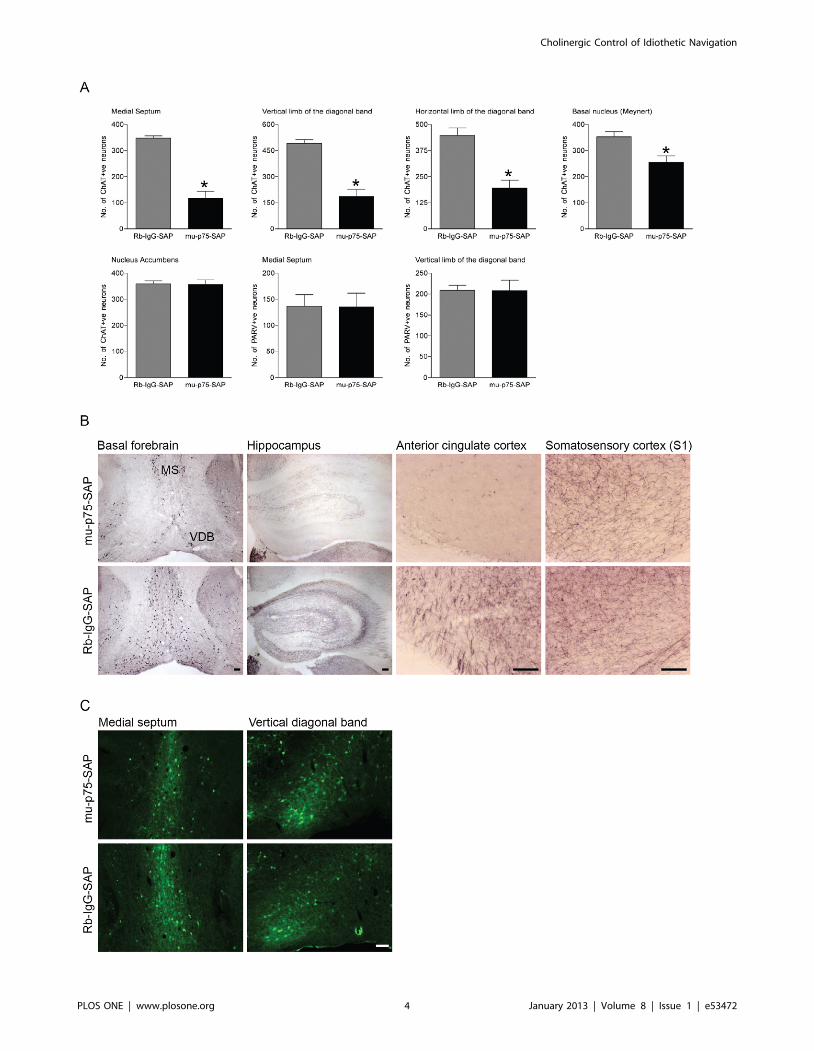
p75NTR-saporin Induces Specific Lesions of the BasalForebrain Cholinergic Neurons
To selectively ablate cholinergic basal forebrain neurons we
used a saporin-conjugated p75NTR antibody (mu-p75-SAP; [28–
31]. Intercerebroventricular injection of the murine-specific
ribosomal inactivating toxin mu-p75-SAP (0.4 mg) induced a
specific loss of cholinergic basal forebrain neurons (Figure 1). As
compared to a control injection of Rb-IgG-SAP, the number of
ChAT-positive neurons was significantly reduced in the medial
septum (F(1,65) = 40.427, p,0.001), vertical diagonal band of
Broca (F(1,65) = 38.892, p,0.001), and horizontal diagonal band
of Broca (F(1,65) = 19.148, p,0.001). A smaller but significant loss
of ChAT-positive neurons was also detected in the basal nucleus
(Meynerts) (F(1,65) = 29.88, p,0.001). In contrast, there was no loss
of parvalbumin-positive GABAergic neurons in the medial septum
or vertical diagonal band of Broca (F(1,16) = 0.065, p = 0.865;
F(1,16) = 0.089, p = 0.832, respectively; Figure 1), showing that p75-
SAP leads to a selective loss of cholinergic neurons. As expected
with the selective loss of ChAT-positive basal forebrain projection
neurons, there was an obvious loss of ChAT-positive terminals in
both the hippocampus and anterior cingulate cortex but not in the
somatosensory cortex (Figure 1 B). There was no change in the
number of ChAT-positive neurons in the nucleus accumbens
(F(1,65) = 0.004, p = 0.950), demonstrating that the toxin was
targeted to p75NTR-expressing neurons in the basal forebrain.
Mice with Cholinergic Basal Forebrain Lesions haveAltered Idiothetic Navigation
To assess idiothetic navigation, four groups of mice (mu-p75-
SAP+shock, Rb-IgG-SAP +shock, no surgery+shock, and no
surgery+no shock) were tested using the place avoidance task.
There was no difference in either the distance travelled
(F = (3,21) = 0.802, p = 0.507) or speed (F(3,21) = 0.804, p = 0.506)
between any of the groups during the 5 minute habituation period,
indicating that the lesioned mice did not have any deficits
associated with exploring a novel open field. During the training
session, conducted 1 hour after habituation, all mice entered a 60ushock zone that was located directly opposite where the animals
Cholinergic Control of Idiothetic Navigation
PLOS ONE | www.plosone.org 3 January 2013 | Volume 8 | Issue 1 | e53472
Cholinergic Control of Idiothetic Navigation
PLOS ONE | www.plosone.org 4 January 2013 | Volume 8 | Issue 1 | e53472

were introduced to the arena. Under these conditions, all mice
that received foot-shocks (0.4 mV) successfully avoided the shock
zone for the remainder of the 5 minute training session (data not
shown), showing that lesioned animals detected and avoided the
shock area during training.
However, clear differences were apparent between groups at
test. Both control groups that received foot-shocks (no surgery+-shock and Rb-IgG-SAP+shock) accurately avoided the shock zone
during the 5 minute test as compared to the animals that did not
receive a foot-shock during training (Figure 2A). Both groups had
significantly fewer entrances to the shock zone [Figure 2A;
ANOVA (F(3,21) = 8.079, p,0.01), no shock vs no surgery:
p,0.05; not shock vs Rb-IgG-SAP: p,0.05]. There was no
significant difference in the number of entries on test for lesioned
mice as compared to no shock mice (Figure 2A; p = 0.215).
However, the behaviour of the lesioned mice on test was clearly
different from that of the control mice that did not receive a foot-
shock during training, as revealed by the reduced distance
travelled on test [Fig. 2A; ANOVA (F(3,21) = 6.389, p,0.01), no
shock vs mu-p75-SAP, p,0.05], indicating that their memory for
the context was intact. This reduction in exploratory behaviour
displayed by the lesioned mice on test was similar to that of control
mice that received a foot-shock (mu-p75-SAP vs no surgery:
p = 0.805; mu-p75-SAP vs Rb-IgG-SAP: p,0.596; Figure 2A).
Even though lesioned mice travelled the same distance while
exploring the arena on test as control mice that received a foot-
shock during training, their exploratory behaviour was consider-
ably more random. As the mice were always introduced to the
arena directly opposite the shock zone, they learnt that this area of
the arena was a safe zone. To quantify the exploratory behaviour
of the animals, we measured the range of movement from this
home zone. Control animals that received no shocks randomly
explored the environment on test (Figure 2A, control). In contrast,
animals that had received a foot-shock explored the arena in an
organized manner, spending more time in the area closer to the
home zone (Figure 2A, no surgery+shock; Rb-Ig-SAP), whereas
lesioned animals explored the environment in a more random
manner, similar to control animals, and moved much further from
the safe zone (Figure 2B; ANOVA F(3,21) = 3.743, p,0.05; mu-
p75-SAP vs no surgery+shock: p,0.05; mu-p75-SAP vs Rb-IgG-
SAP: p,0.05). Moreover, some lesioned mice randomly entered
and stayed within the shock zone as indicated by the large
variability in the number of potential foot-shocks received by
lesioned mice (Figure 2). This change in exploratory behaviour on
test shows that lesioned animals had lost the ability to locate
themselves in space.
Lesioned Animals have Normal Spatial and ContextualFear Memory
To determine if the observed behaviour was due to a lack of
learning, the spatial working memory of mice was assessed in the
novelty seeking Y-maze test. Mice with a basal forebrain lesion
spent the same amount of time exploring the novel environment as
control mice during the 3 minute test (mu-p75-SAP 45.25 sec
68.6, Rb-IgG-SAP 51.25 sec 65.0) (F(1,7) = 0.363, p = 0.569),
indicating that in this test of spatial working memory lesioned mice
performed as well as control animals (Figure 3B).
Learning was further assessed using contextual fear condition-
ing. All mice that received the 5 random (0.8 mV) foot-shocks
during the training session showed significantly more freezing
(F(3,14) = 4.407, p,0.05) and less locomotor activity (F(3,14) = 4.807,
p,0.05) than control mice that received no shocks. No differences
in the percentage of freezing or locomotor activity were detected in
lesioned mice compared to the two control groups that received
shocks during training (Freezing: mu-p75-SAP vs Rb-IgG-SAP,
F(3,14) = 0.437, p = 0.671 and mu-p75-SAP vs no surgery+shock
F(3,14) = 0.951, p = 0.362; Locomotor: mu-p75-SAP vs Rb-IgG-
SAP, F(3,14) = 0.065, p = 0.949 and mu-p75-SAP vs no surgery+-shock F(3,14) = 0.859, p = 0.409). Similarly, all mice that received
foot-shocks during training showed significantly higher levels of
freezing and less locomotor activity when they were placed back
into the training context (F(3,14) = 7.569, p,0.05 and
F(3,14) = 3.518. p,0.05, respectively). Furthermore, no differences
in the percentage of freezing or locomotor activity during test were
detected in lesioned mice compared to the two control groups that
received foot-shocks on training (Freezing: mu-p75-SAP vs Rb-
IgG-SAP, F(3,14) = 0.151, p = 0.883 and mu-p75-SAP vs no
surgery+shock F(14,3) = 0.529, p = 0.607; Locomotor: mu-p75-
SAP vs Rb-IgG-SAP, F(3,14) = 0.177, p = 0.863 and mu-p75-SAP
vs no surgery+shock F(3,14) = 0.544, p = 0.597 ) (Figure 3A),
indicating that all mice learnt and recalled the contextual
information presented to them during training.
Anxiety, Sleep-wake Cycles and Motor Performance ofLesioned Mice are Normal
To determine whether lesions of the basal forebrain affected
levels of anxiety we employed the light/dark test and measured the
time taken to enter the light area, the number of entries, and the
total time spent in the light. ANOVA showed that the time taken
to enter the light, and the total time spent in the light, were
comparable between lesioned mice and controls (F(1,15) = 0.311
p = 0.587; F(1,15) = 0.383, p = 0.547, respectively). Interestingly,
lesioned mice made more entries into the light side of the
apparatus than controls during the 5 minute test (F(1,15) = 6.133,
p,0.05) (Figure 3D).
As altered motor performance could be a potential confounding
variable, mice that underwent contextual fear conditioning were
also assessed for motor performance using the accelerating Rota-
rod 24 hours after the conclusion of testing. All mice, regardless of
lesion status, reached the 3 minute threshold without any falls from
a Rota-rod that accelerated from 2 rpm to 20 rpm at a rate of
0.4 rpm/sec (Figure 3C).
Similarly, a subset of experimental mice were monitored for
locomotor activity for 36 hours during two wake and one sleep
cycles, as alterations in the sleep/wake cycle can have profound
effects on learning and memory performance. No statistical
difference in locomotor activity (distance travelled or velocity)
was detected by ANOVA (2-way repeated measures with between
Figure 1. p75NTR-saporin induces specific lesions of the basal forebrain cholinergic neurons. (A) Mean (6 SEM) numbers of ChAT-positiveneurons in basal forebrain nuclei and adjacent nucleus accumbens following intercerebroventricular application of mu-p75-SAP and the control toxinRb-IgG-SAP. (B,C) Photomicrographs showing (B) ChAT-positive neurons in the medial septum (MS) and vertical limb of the diagonal band of Broca(VDB) and ChAT-positive terminal labelling in the hippocampus, anterior cingulate cortex and primary somatosensory cortex and (C) parvalbumin-positive neurons in the medial septum and vertical limb of the diagonal band of Broca following mu-p75-SAP or Rb-IgG-SAP treatment. (D) Mean (6SEM) numbers of parvalbumin-positive neurons in basal forebrain nuclei following intercerebroventricular application of mu-p75-SAP and the controltoxin Rb-IgG-SAP. A specific loss of ChAT-positive neurons was detected in basal forebrain nuclei following mu-p75-SAP treatment (mu-p75-SAP vs.Rb-IgG-SAP, *p,0.001). Scale bars = 100 mm.doi:10.1371/journal.pone.0053472.g001
Cholinergic Control of Idiothetic Navigation
PLOS ONE | www.plosone.org 5 January 2013 | Volume 8 | Issue 1 | e53472

group factors [mu-p75-SAP vs Rb-IgG-Sap] and repeated
measure factor [time]) during the sleep/wake cycle
(F(1,7) = 0.005, p = 0.955; F(1,7) = 0.008, p = 0.931, respectively)
(Figure 3E).
Discussion
In this study we used intercerebroventricular application of mu-
p75-SAP to induce a discrete and specific loss of basal forebrain
cholinergic neurons in the mouse [28–31] in order to model the
behavioural consequences of degeneration. Although this lesion
resulted in a ,50% loss of medial septum cholinergic neurons that
project to the hippocampus, there was no overt histological
atrophy of this structure. This lesion therefore closely mimics the
cholinergic dysfunction observed in prodromal Alzheimer’s disease
[12].
To test the animals’ ability to navigate they were subjected to an
idiothetic version of the passive place avoidance test in which all
extramaze cues were removed. The design of this task forces the
mice to use kinaesthetic self-motion information to locate
themselves in space, relative to their home area and the aversive
shock zone. Ablation of the basal forebrain cholinergic neurons led
to highly disorganized exploratory behaviour. Control mice that
received foot-shocks during training successfully avoided the shock
zone, and displayed organized exploratory behaviour that centred
on the home area of the arena. In contrast, lesioned mice did not
stay within the safe area but randomly entered the shock zone,
even though they travelled a similar distance to control mice
during their exploration.
No deficits in contextual or working spatial memory were
apparent following loss of the basal forebrain cholinergic neurons.
Lesioned mice showed similar levels of freezing to control mice in
contextual fear conditioning, and explored the novel arm of the Y-
maze with the same intensity. Furthermore, although lesioned
mice were unable to locate the shock zone or home region of the
place avoidance arena on test, their behaviour was dramatically
different from their performance during habituation, as well as in
comparison with that of control mice that were never trained,
indicating that their memory for the context was intact. Moreover,
we found no deficits in motor performance, anxiety, or the sleep/
wake cycle. These findings indicate that the basal forebrain
cholinergic system is not critically involved in the cognitive abilities
required for context recognition or working spatial memory.
In general, animals use two different strategies to navigate,
allothetic and idiothetic. In allothetic navigation the animal uses
distal environmental cues to locate itself in space, and continually
reorients its motion, whereas in idiothetic navigation the animal
uses self-movement cues generated by internal sensory information
such as vestibular and proprioceptive input coupled with memory
[32]. Previous studies in which the entire medial septal area,
encompassing both the cholinergic and GABAergic neuronal
populations, was electrolytically ablated in rats reported that these
animals had specific deficits in idiothetic navigation [16].
Furthermore, both rats [33] and mice [15] with lesions of the
fimbria-fornix, the major pathway carrying cholinergic and
GABAergic efferents to the hippocampus, perform poorly in
idiothetic navigational tasks. By creating a specific lesion of the
basal forebrain cholinergic neurons, our results demonstrate that
this system is required for idiothetic navigation.
In a previous study, Moreau et al. [30], using the Morris
water maze, reported that, although mice with cholinergic basal
forebrain lesions display an early deficit in acquisition in cued
navigation, by day three of training they are performing at the
same level as control mice and show no deficit during the probe
Figure 2. Mice with cholinergic basal forebrain lesions havealtered idiothetic navigation. (A) Mean (6 SEM) entrances to theshock zone, distance travelled and range of exploratory behaviour fromthe home zone during the 5 minute passive avoidance test, andscatterplot of the number of potential foot-shock events during thistest. Control mice that received a foot-shock during training showedsignificantly fewer entrances to the shock zone on test than controlmice that received no shock during training (* p,0.05). All mice thatreceived a foot-shock during training travelled a significantly shorterdistance during test than control mice (* p,0.05). No difference in thedistance travelled during test was detected between mice that receiveda foot-shock during training. Lesioned mice explored further from thehome (safe) zone than control mice that received a foot-shock duringtraining (# p,0.05), and showed greater variability in the number ofpotential foot-shocks during test. (B) Representative tracking ofexploratory behaviour during the 5 minute passive avoidance test foreach of the four groups.doi:10.1371/journal.pone.0053472.g002
Cholinergic Control of Idiothetic Navigation
PLOS ONE | www.plosone.org 6 January 2013 | Volume 8 | Issue 1 | e53472

trial. This was interpreted as a spatial memory deficit. However
in the Morris water maze, animals have the opportunity to
utilize both allothetic and idiothetic navigation strategies. Our
results suggest that a more parsimonious interpretation is that
mice rely on idiothetic navigation in early trials, whereas
allothetic strategies dominate once extramaze cues become
learnt. Furthermore, Moreau et al. [30] reported that lesioned
mice had an increased incidence of thygmotaxic search
strategies and were slower at adopting a goal-directed strategy,
consistent with the animals exhibiting impaired idiothetic
navigation.
A major function of basal forebrain hippocampal innervation is
the generation of the 4–10 Hz theta rhythm. Lesion studies
targeting GABAergic or cholinergic inputs to the hippocampus
Figure 3. Spatial and contextual fear memory, anxiety, sleep-wake cycles and motor performance of lesioned mice are normal. (A)Mean (6 SEM) percent context-elicited freezing 24 hours after fear conditioning training (5 random 0.8 mV foot-shocks). No difference in percentfreezing during test was detected in lesioned mice compared to control mice that received foot-shocks,. (B) Mean (6 SEM) time spent in the novelarm in the Y-maze. Lesioned mice spent an equivalent amount of time exploring the novel arm of the Y-maze to control mice. (C) Mean (6 SEM) timeachieved in the accelerating Rota-rod test. All mice successfully completed the 3 minute test with no falls recorded. (D) Mean (6 SEM) initial time toenter, total time, and number of entries to the light area in the light/dark test of anxiety. No difference was detected between lesioned and controlmice in the initial time taken to enter the light area or in the total time spent in the this area. However, lesioned mice made more entries into the lightarea during the 5 minute test than control mice (mu-p75-SAP vs Rb-IgG-SAP, *p,0.05). (E) Mean (6 SEM) locomotor activity in 1 hour time bins over36 hours incorporating 2 wake and 1 sleep cycles. No difference in locomotor activity was found between lesioned and control mice in either thewake or sleep cycle.doi:10.1371/journal.pone.0053472.g003
Cholinergic Control of Idiothetic Navigation
PLOS ONE | www.plosone.org 7 January 2013 | Volume 8 | Issue 1 | e53472

have revealed that the GABAergic system is responsible for the
pacing of theta rhythm synchronization, whereas the cholinergic
system contributes to the amplitude or the power of the rhythm
[34]. Although the hippocampal theta rhythm is active during
voluntary movement and has been implicated in attention and
acquisition of sensory information [35,36], only recently has
vestibular stimulation, sensory information critical for idiothetic
navigation, been shown to stimulate this rhythm [37]. Moreover,
the locomotion-induced increase in theta amplitude can be
blocked by the muscarinic acetylcholine receptor antagonist
atropine, consistent with a role for the basal forebrain cholinergic
neurons in transmitting this sensorimotor information to the
hippocampus.
It is well established, based on single-cell recordings, that
hippocampal neurons respond to movement, place, and head
orientation [38,39]. Therefore, the hippocampus plays a signifi-
cant role in the generation of spatial awareness and the coding of
self-movement through efferent copies of movement-related
commands. In light of our evidence that basal forebrain
cholinergic neuron lesions produce a specific deficit in idiothetic
navigation, it is likely that the amplitude of the theta rhythm
modulated by acetylcholine [34,40] plays a critical role in the
processes required to be aware of where you are in space relative
to where you have been. Failure of appropriate hippocampal
integration of this self-movement information due to atrophy of
the basal forebrain cholinergic neurons or dysregulation of
acetylcholine receptor function in the hippocampus, as occurs in
Alzheimer’s disease [41,42], could underpin deficits in this form of
navigation.
Both basal forebrain atrophy and hippocampal volume loss, as
measured by magnetic resonance imaging, are emerging as
accurate markers for diagnosing at risk Alzheimer’s disease
patients, with basal forebrain atrophy and hippocampal volume
loss being highly correlated [11,14]. Furthermore, poor navigation
in patients with MCI is correlated with hippocampal atrophy [7].
The contribution of the basal forebrain cholinergic system to these
spatial memory deficits is yet to be established in humans.
However, our finding that an idiothetic navigational deficit is
caused by a loss of basal forebrain cholinergic neurons in mice
provides strong evidence-based support for the examination of
basal forebrain cholinergic neuron dysfunction, measured with
either magnetic resonance imaging or behavioural navigation
tasks, as an early diagnostic marker for the cognitive decline that is
associated with Alzheimer’s disease.
Acknowledgments
We would like to thank Nicola Marks and Sophie Hill for technical
assistance and Rowan Tweedale for editing the manuscript.
Author Contributions
Discussion and editing of manuscript: ZB PS. Conceived and designed the
experiments: ASH FW PS EJC. Performed the experiments: ASH FW ZB.
Analyzed the data: ASH FW ZB PS EJC. Wrote the paper: ASH FW EJC.
References
1. Monacelli AM, Cushman LA, Kavcic V, Duffy CJ (2003) Spatial disorientation
in Alzheimer’s disease: the remembrance of things passed. Neurology 61: 1491–
1497.
2. Mapstone M, Steffenella TM, Duffy CJ (2003) A visuospatial variant of mild
cognitive impairment: getting lost between aging and AD. Neurology 60: 802–
808.
3. O’Keefe J, Dostrovsky J (1971) The hippocampus as a spatial map. Preliminary
evidence from unit activity in the freely-moving rat. Brain Res 34: 171–175.
4. Morris RG, Garrud P, Rawlins JN, O’Keefe J (1982) Place navigation impaired
in rats with hippocampal lesions. Nature 297: 681–683.
5. Laczo J, Andel R, Vlcek K, Macoska V, Vyhnalek M, et al. (2011) Spatial
navigation and APOE in amnestic mild cognitive impairment. Neurodegener
Dis 8: 169–177.
6. Laczo J, Andel R, Vyhnalek M, Vlcek K, Magerova H, et al. (2010) Human
analogue of the morris water maze for testing subjects at risk of Alzheimer’s
disease. Neurodegener Dis 7: 148–152.
7. Hort J, Laczo J, Vyhnalek M, Bojar M, Bures J, et al. (2007) Spatial navigation
deficit in amnestic mild cognitive impairment. Proc Natl Acad Sci USA 104:
4042–4047.
8. Laczo J, Vlcek K, Vyhnalek M, Vajnerova O, Ort M, et al. (2009) Spatial
navigation testing discriminates two types of amnestic mild cognitive
impairment. Behav Brain Res 202: 252–259.
9. Czeh B, Stuchlik A, Wesierska M, Cimadevilla JM, Pokorny J, et al. (2001) Effect
of neonatal dentate gyrus lesion on allothetic and idiothetic navigation in rats.
Neurobiol Learn Mem 75: 190–213.
10. Feigenbaum JD, Morris RG (2004) Allocentric versus egocentric spatial memory
after unilateral temporal lobectomy in humans. Neuropsychology 18: 462–472.
11. Grothe M, Heinsen H, Teipel SJ (2012) Atrophy of the cholinergic Basal
forebrain over the adult age range and in early stages of Alzheimer’s disease. Biol
Psychiatry 71: 805–813.
12. Grothe M, Zaborszky L, Atienza M, Gil-Neciga E, Rodriguez-Romero R, et al.
(2010) Reduction of basal forebrain cholinergic system parallels cognitive
impairment in patients at high risk of developing Alzheimer’s disease. Cereb
Cortex 20: 1685–1695.
13. Muth K, Schonmeyer R, Matura S, Haenschel C, Schroder J, et al. (2010) Mild
cognitive impairment in the elderly is associated with volume loss of the
cholinergic basal forebrain region. Biol Psychiatry 67: 588–591.
14. Hall JR, Harvey MB (2008) Behavioral regulation: factor analysis and
application of the Behavioral Dyscontrol Scale in dementia and mild cognitive
impairment. Int J Geriatr Psychiatry 23: 314–318.
15. Gorny JH, Gorny B, Wallace DG, Whishaw IQ (2002) Fimbria-fornix lesions
disrupt the dead reckoning (homing) component of exploratory behavior in
mice. Learn Mem 9: 387–394.
16. Martin MM, Horn KL, Kusman KJ, Wallace DG (2007) Medial septum lesions
disrupt exploratory trip organization: evidence for septohippocampal involve-
ment in dead reckoning. Physiol Behav 90: 412–424.
17. Henny P, Jones BE (2008) Projections from basal forebrain to prefrontal cortex
comprise cholinergic, GABAergic and glutamatergic inputs to pyramidal cells or
interneurons. Eur J Neurosci 27: 654–670.
18. Blokland A (1995) Acetylcholine: a neurotransmitter for learning and memory?
Brain Res Brain Res Rev 21: 285–300.
19. Hasselmo ME (2006) The role of acetylcholine in learning and memory. Curr
Opin Neurobiol 16: 710–715.
20. Perry E, Walker M, Grace J, Perry R (1999) Acetylcholine in mind: a
neurotransmitter correlate of consciousness? Trends Neurosci 22: 273–280.
21. Schliebs R, Arendt T (2006) The significance of the cholinergic system in the
brain during aging and in Alzheimer’s disease. J Neural Transm 113: 1625–
1644.
22. Mesulam M (2004) The cholinergic lesion of Alzheimer’s disease: pivotal factor
or side show? Learn Mem 11: 43–49.
23. Perry EK, Tomlinson BE, Blessed G, Perry RH, Cross AJ, et al. (1981)
Noradrenergic and cholinergic systems in senile dementia of Alzheimer type.
Lancet 2: 149.
24. Craig LA, Hong NS, McDonald RJ (2011) Revisiting the cholinergic hypothesis
in the development of Alzheimer’s disease. Neurosci Biobehav Rev 35: 1397–
1409.
25. Pinto T, Lanctot KL, Herrmann N (2011) Revisiting the cholinergic hypothesis
of behavioral and psychological symptoms in dementia of the Alzheimer’s type.
Ageing Res Rev 10: 404–412.
26. Franklin KJB, Paxinos G (2007) The Mouse Brain in Stereotaxic Coordinates
Third Edition. San Diego: Academic Press.
27. Cimadevilla JM, Kaminsky Y, Fenton A, Bures J (2000) Passive and active place
avoidance as a tool of spatial memory research in rats. J Neurosci Methods 102:
155–164.
28. Berger-Sweeney J, Stearns NA, Murg SL, Floerke-Nashner LR, Lappi DA, et al.
(2001) Selective immunolesions of cholinergic neurons in mice: effects on
neuroanatomy, neurochemistry, and behavior. J Neurosci 21: 8164–8173.
29. Hunter CL, Quintero EM, Gilstrap L, Bhat NR, Granholm AC (2004)
Minocycline protects basal forebrain cholinergic neurons from mu p75-saporin
immunotoxic lesioning. Eur J Neurosci 19: 3305–3316.
30. Moreau PH, Cosquer B, Jeltsch H, Cassel JC, Mathis C (2008) Neuroanatomical
and behavioral effects of a novel version of the cholinergic immunotoxin mu
p75-saporin in mice. Hippocampus 18: 610–622.
31. Nag N, Baxter MG, Berger-Sweeney JE (2009) Efficacy of a murine-p75-saporin
immunotoxin for selective lesions of basal forebrain cholinergic neurons in mice.
Neurosci Lett 452: 247–251.
32. Gallistel CR (1990) The Organisation of Learning. Cambridge MA: MIT Press.
Cholinergic Control of Idiothetic Navigation
PLOS ONE | www.plosone.org 8 January 2013 | Volume 8 | Issue 1 | e53472

33. Whishaw IQ, Hines DJ, Wallace DG (2001) Dead reckoning (path integration)
requires the hippocampal formation: evidence from spontaneous explorationand spatial learning tasks in light (allothetic) and dark (idiothetic) tests. Behav
Brain Res 127: 49–69.
34. Lee MG, Chrobak JJ, Sik A, Wiley RG, Buzsaki G (1994) Hippocampal thetaactivity following selective lesion of the septal cholinergic system. Neuroscience
62: 1033–1047.35. Vanderwolf CH (1969) Hippocampal electrical activity and voluntary movement
in the rat. Electroencephalogr Clin Neurophysiol 26: 407–418.
36. Vanderwolf CH, Kramis R, Robinson TE (1977) Hippocampal electricalactivity during waking behaviour and sleep: analyses using centrally acting drugs.
CIBA Found Symp: 199–226.37. Tai SK, Ma J, Ossenkopp KP, Leung LS (2012) Activation of immobility-related
hippocampal theta by cholinergic septohippocampal neurons during vestibularstimulation. Hippocampus. 22: 914–925.
38. Foster TC, Castro CA, McNaughton BL (1989) Spatial selectivity of rat
hippocampal neurons: dependence on preparedness for movement. Science 244:
1580–1582.
39. Taube JS (1998) Head direction cells and the neurophysiological basis for a sense
of direction. Prog Neurobiol 55: 225–256.
40. Yoder RM, Pang KC (2005) Involvement of GABAergic and cholinergic medial
septal neurons in hippocampal theta rhythm. Hippocampus 15: 381–392.
41. Nordberg A (2001) Nicotinic receptor abnormalities of Alzheimer’s disease:
therapeutic implications. Biol Psychiatry 49: 200–210.
42. Levey AI (1996) Muscarinic acetylcholine receptor expression in memory
circuits: implications for treatment of Alzheimer disease. Proc Natl Acad Sci
USA 93: 13541–13546.
Cholinergic Control of Idiothetic Navigation
PLOS ONE | www.plosone.org 9 January 2013 | Volume 8 | Issue 1 | e53472
Copyright © 2022 FDOKUMEN