Laser micro-engineering of functionalised cyclic olefin polymers for microfluidic applications
6
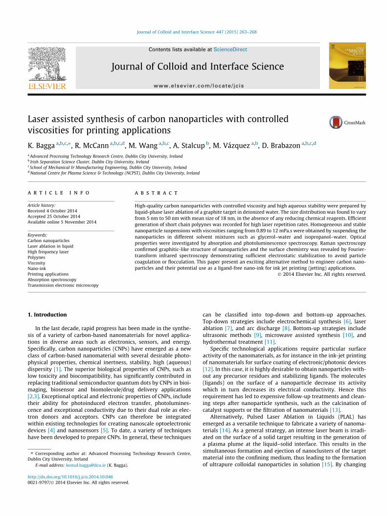
Laser assisted synthesis of carbon nanoparticles with controlled viscosities for printing applications K. Bagga a,b,c,⇑ , R. McCann a,b,c,d , M. Wang a,b,c , A. Stalcup b , M. Vázquez a,b , D. Brabazon a,b,c,d a Advanced Processing Technology Research Centre, Dublin City University, Ireland b Irish Separation Science Cluster, Dublin City University, Ireland c School of Mechanical & Manufacturing Engineering, Dublin City University, Ireland d National Centre for Plasma Science & Technology (NCPST), Dublin City University, Ireland article info Article history: Received 4 October 2014 Accepted 25 October 2014 Available online 5 November 2014 Keywords: Carbon nanoparticles Laser ablation in liquid High frequency laser Polyynes Viscosity Nano-ink Printing applications Absorption spectroscopy Transmission electronic microscopy abstract High-quality carbon nanoparticles with controlled viscosity and high aqueous stability were prepared by liquid-phase laser ablation of a graphite target in deionized water. The size distribution was found to vary from 5 nm to 50 nm with mean size of 18 nm, in the absence of any reducing chemical reagents. Efficient generation of short chain polyynes was recorded for high laser repetition rates. Homogeneous and stable nanoparticle suspensions with viscosities ranging from 0.89 to 12 mPa.s were obtained by suspending the nanoparticles in different solvent mixtures such as glycerol–water and isopropanol–water. Optical properties were investigated by absorption and photoluminescence spectroscopy. Raman spectroscopy confirmed graphitic-like structure of nanoparticles and the surface chemistry was revealed by Fourier- transform infrared spectroscopy demonstrating sufficient electrostatic stabilization to avoid particle coagulation or flocculation. This paper present an exciting alternative method to engineer carbon nano- particles and their potential use as a ligand-free nano-ink for ink jet printing (jetting) applications. Ó 2014 Elsevier Inc. All rights reserved. 1. Introduction In the last decade, rapid progress has been made in the synthe- sis of a variety of carbon-based nanomaterials for novel applica- tions in diverse areas such as electronics, sensors, and energy. Specifically, carbon nanoparticles (CNPs) have emerged as a new class of carbon-based nanomaterial with several desirable photo- physical properties, chemical inertness, stability, high (aqueous) dispersity [1]. The superior biological properties of CNPs, such as low toxicity and biocompatibility, has significantly contributed in replacing traditional semiconductor quantum dots by CNPs in bioi- maging, biosensor and biomolecule/drug delivery applications [2,3]. Exceptional optical and electronic properties of CNPs, include their ability for photoinduced electron transfer, photolumines- cence and exceptional conductivity due to their dual role as elec- tron donors and acceptors. CNPs can therefore be integrated within existing technologies for creating nanoscale optoelectronic devices [4] and nanosensors [5]. To date, a variety of techniques have been developed to prepare CNPs. In general, these techniques can be classified into top-down and bottom-up approaches. Top-down strategies include electrochemical synthesis [6], laser ablation [7], and arc discharge [8]. Bottom-up strategies include ultrasonic methods [9], microwave assisted synthesis [10], and hydrothermal treatment [11]. Specific technological applications require particular surface activity of the nanomaterials, as for instance in the ink-jet printing of nanomaterials for surface coating of electronic/photonic devices [12]. In this case, it is highly desirable to obtain nanoparticles with- out any precursor residues and stabilizing ligands. The molecules (ligands) on the surface of a nanoparticle decrease its activity which in turn decreases its electrical conductivity. Hence this requirement has led to expensive follow-up treatments and clean- ing steps after nanoparticle synthesis, such as the calcination of catalyst supports or the filtration of nanomaterials [13]. Alternatively, Pulsed Laser Ablation in Liquids (PLAL) has emerged as a versatile technique to fabricate a variety of nanoma- terials [14]. As a general strategy, an intense laser beam is irradi- ated on the surface of a solid target resulting in the generation of a plasma plume at the liquid–solid interface. This results in the simultaneous formation and ejection of nanoclusters of the target material into the confining medium, thus leading to the formation of ultrapure colloidal nanoparticles in solution [15]. By changing http://dx.doi.org/10.1016/j.jcis.2014.10.046 0021-9797/Ó 2014 Elsevier Inc. All rights reserved. ⇑ Corresponding author at: Advanced Processing Technology Research Centre, Dublin City University, Ireland E-mail address: [email protected] (K. Bagga). Journal of Colloid and Interface Science 447 (2015) 263–268 Contents lists available at ScienceDirect Journal of Colloid and Interface Science www.elsevier.com/locate/jcis
Transcript of Laser micro-engineering of functionalised cyclic olefin polymers for microfluidic applications
Journal of Colloid and Interface Science 447 (2015) 263–268
Contents lists available at ScienceDirect
Journal of Colloid and Interface Science
www.elsevier .com/locate / jc is
Laser assisted synthesis of carbon nanoparticles with controlledviscosities for printing applications
http://dx.doi.org/10.1016/j.jcis.2014.10.0460021-9797/� 2014 Elsevier Inc. All rights reserved.
⇑ Corresponding author at: Advanced Processing Technology Research Centre,Dublin City University, Ireland
E-mail address: [email protected] (K. Bagga).
K. Bagga a,b,c,⇑, R. McCann a,b,c,d, M. Wang a,b,c, A. Stalcup b, M. Vázquez a,b, D. Brabazon a,b,c,d
a Advanced Processing Technology Research Centre, Dublin City University, Irelandb Irish Separation Science Cluster, Dublin City University, Irelandc School of Mechanical & Manufacturing Engineering, Dublin City University, Irelandd National Centre for Plasma Science & Technology (NCPST), Dublin City University, Ireland
a r t i c l e i n f o a b s t r a c t
Article history:Received 4 October 2014Accepted 25 October 2014Available online 5 November 2014
Keywords:Carbon nanoparticlesLaser ablation in liquidHigh frequency laserPolyynesViscosityNano-inkPrinting applicationsAbsorption spectroscopyTransmission electronic microscopy
High-quality carbon nanoparticles with controlled viscosity and high aqueous stability were prepared byliquid-phase laser ablation of a graphite target in deionized water. The size distribution was found to varyfrom 5 nm to 50 nm with mean size of 18 nm, in the absence of any reducing chemical reagents. Efficientgeneration of short chain polyynes was recorded for high laser repetition rates. Homogeneous and stablenanoparticle suspensions with viscosities ranging from 0.89 to 12 mPa.s were obtained by suspending thenanoparticles in different solvent mixtures such as glycerol–water and isopropanol–water. Opticalproperties were investigated by absorption and photoluminescence spectroscopy. Raman spectroscopyconfirmed graphitic-like structure of nanoparticles and the surface chemistry was revealed by Fourier-transform infrared spectroscopy demonstrating sufficient electrostatic stabilization to avoid particlecoagulation or flocculation. This paper present an exciting alternative method to engineer carbon nano-particles and their potential use as a ligand-free nano-ink for ink jet printing (jetting) applications.
� 2014 Elsevier Inc. All rights reserved.
1. Introduction
In the last decade, rapid progress has been made in the synthe-sis of a variety of carbon-based nanomaterials for novel applica-tions in diverse areas such as electronics, sensors, and energy.Specifically, carbon nanoparticles (CNPs) have emerged as a newclass of carbon-based nanomaterial with several desirable photo-physical properties, chemical inertness, stability, high (aqueous)dispersity [1]. The superior biological properties of CNPs, such aslow toxicity and biocompatibility, has significantly contributed inreplacing traditional semiconductor quantum dots by CNPs in bioi-maging, biosensor and biomolecule/drug delivery applications[2,3]. Exceptional optical and electronic properties of CNPs, includetheir ability for photoinduced electron transfer, photolumines-cence and exceptional conductivity due to their dual role as elec-tron donors and acceptors. CNPs can therefore be integratedwithin existing technologies for creating nanoscale optoelectronicdevices [4] and nanosensors [5]. To date, a variety of techniqueshave been developed to prepare CNPs. In general, these techniques
can be classified into top-down and bottom-up approaches.Top-down strategies include electrochemical synthesis [6], laserablation [7], and arc discharge [8]. Bottom-up strategies includeultrasonic methods [9], microwave assisted synthesis [10], andhydrothermal treatment [11].
Specific technological applications require particular surfaceactivity of the nanomaterials, as for instance in the ink-jet printingof nanomaterials for surface coating of electronic/photonic devices[12]. In this case, it is highly desirable to obtain nanoparticles with-out any precursor residues and stabilizing ligands. The molecules(ligands) on the surface of a nanoparticle decrease its activitywhich in turn decreases its electrical conductivity. Hence thisrequirement has led to expensive follow-up treatments and clean-ing steps after nanoparticle synthesis, such as the calcination ofcatalyst supports or the filtration of nanomaterials [13].
Alternatively, Pulsed Laser Ablation in Liquids (PLAL) hasemerged as a versatile technique to fabricate a variety of nanoma-terials [14]. As a general strategy, an intense laser beam is irradi-ated on the surface of a solid target resulting in the generation ofa plasma plume at the liquid–solid interface. This results in thesimultaneous formation and ejection of nanoclusters of the targetmaterial into the confining medium, thus leading to the formationof ultrapure colloidal nanoparticles in solution [15]. By changing

264 K. Bagga et al. / Journal of Colloid and Interface Science 447 (2015) 263–268
the laser parameters, target material and liquid media, the size,morphology and surface chemistry of the nanoparticles can be pre-cisely controlled [16,17]. Several studies have been publishedreporting PLAL as an effective method for the generation of inter-esting carbon-based nanomaterials including nanoparticles [18]and polyynes [19]. Polyynes are of significance in materials sciencedue to their one-dimensional electronic structures, size-dependentband gaps [20] and nonlinear optical properties [21].
In this work, we exploit this unique technique (i.e. PLAL) togenerate nanoparticles with novel functionalities to be used asnano-inks in ink-jet printing (jetting) technology. Over the years,ink-jet printing techniques have become attractive alternatives toconventional photolithography or screen-printing for patterningvarious functional materials such as organic light emitting diodes(OLED), printed scaffolds for growth of living tissues, and building3D objects [22]. An important advantage of nanoparticle based inksis the large surface/volume ratio that these particles offer. This canallow low annealing temperature, short process time, highconductivity [23] and high density functionalisation to obtain awide variety of surface chemistries for different ink-jet printingapplications [24].
The present work focuses on the use of laser assisted method,PLAL, for preparation of high-quality carbon nanoparticles basedcolloids with controlled viscosity. To the best of our knowledge,this is the first time demonstration of laser generated carbon nano-particles, towards their application as nano-inks in jetting technol-ogy. Optical properties were investigated by absorption andphotoluminescence spectroscopy. Raman and Fourier-transforminfrared (FTIR) spectroscopy were performed to reveal the chemi-cal structure of the nanoparticles. Viscosity measurements indifferent solvent mixtures were performed in order to optimizethe viscosity of the CNP based colloids.
2. Materials and methods
2.1. Synthesis
Laser ablation experiments were carried out using a Nd:YAGlaser system (WEDGE HF 1064, Bright Solutions) providing pulsescentred at 1064 nm and with a pulse width of 700 ps at therepetition rate set at 7 kHz for production of CNPs and 10 kHz forgeneration of polyynes. The graphite target (99.999% pure fromSigma Aldrich) in cylindrical form with a diameter of 6 mm andheight of 8 mm was placed on the bottom of a glass cuvette(dimension 10 � 10 � 50 mm). The cuvette was filled with 2 mLdeionised water (DI water) which corresponded to 1 cm of liquidabove the surface of the target. The target was mechanically pol-ished, and then washed with deionised water several times toremove any impurities from the surface. The laser beam wasfocused onto the target material using a lens with a focal lengthof 30 cm. During all the experiments, the graphite target wasplaced in the focal plane of laser beam. A 2-dimensional scanninggalvanometer (SS-12, Raylase) was used to scan the laser beamacross the top of the graphite target in a circular pattern, at a scanrate of 1.2 mm/s. The laser fluence was measured experimentallyusing a 30A-P-17 OPHIR� power meter. The laser ablation param-eters are defined as follows, unless indicated otherwise. For theproduction of CNPs, the ablation was carried out at an energy den-sity ranging from 0.02 J.cm�2 to 0.71 J.cm�2 for a 30 min irradiationtime, and for the generation of polyynes the laser fluence was fixedat 0.40 J.cm�2 for a 10 min irradiation time. Additionally, the effectof ablation time on production of nanoparticles was also investi-gated at a fixed laser fluence. The formation of carbon nanostruc-tures could be estimated by the slight change of the colour (fromtransparent to varying tones of grey) of the liquid during ablation.
However it should be noted that changes in the colour of thecolloidal solution were only recorded for high levels of fluence.The sample preparation for each characterization measurementwas performed one day after preparation of the colloids.
2.2. Characterization
Optical absorption spectra were recorded in a quartz cuvette(10 mm pathlength, Helma) with Varian Cary� 50 UV–Vis spectro-photometer. The scan range was 200–1200 nm with a 600 nm/minscan rate. Photoluminescence of the colloidal solution was mea-sured using a Jasco FP-8500 fluorescence spectrometer. All theoptical spectra were corrected for water absorption, by subtractingthe contribution of water from the recorded spectrum.
Transmission Electron Microscopy (TEM) was performed with aFEI titan instrument, operating at 300 kV, equipped with aField Emission Gun (FEG), spherical aberration corrector system(Cs-corrector) of the objective lens. The samples were preparedby drop casting the colloidal solution onto carbon coated 300 meshcopper grid and left to evaporate at room temperature.
The nanoparticles were sonicated in distilled water for 20 minand then their stability in water was studied in a dynamic lightscattering (DLS) experiment using a Zetasizer Nano ZS (MalvernInstruments Ltd). Micro-probe Raman measurements were per-formed with Jobin-Yvon Horiba LabRam� HR800 system at20 mW and 1 lm2 spot size (with Ar+ 488 nm air cooled laser,accumulation time = 20 s) in a backscattering configuration withresolution of about 1.1 cm�1. Sample preparation for Raman mea-surements was done by depositing colloidal CNP solution over thesubstrate by drop casting method. The excess of liquid was thenleft to evaporate at room temperature leading to the formation ofa ‘‘coffee ring’’. This technique is commonly accepted as the prop-erty of the material in the coffee ring remains the same as in theoriginal sample [25]. Various measurements were performed atdifferent position on the coffee ring. Fourier Transform Infraredspectroscopy (FTIR) was performed on a Perkin–Elemer Spectrum100 FTIR spectrometer. The data was acquired in transmissionand ATR imaging modes over the spectral range the 600–4000 cm�1. Viscosities of the CNP suspensions were measured withan Anton Paar MCR 301 Rheometer system with maximum torquecapability of 200 mNm, resolution of 0.1 nNm, and a maximumangular velocity of 628 rad/s.
3. Results and discussion
3.1. TEM characterization
TEM characterization was carried out to obtain informationabout the morphology and size distributions in the colloidal solu-tions produced by laser ablation of the graphite target in deionizedwater. Fig. 1 shows TEM images of NPs obtained at 0.40 J.cm�2
laser irradiance for 5 min. Isolated and uniformly distributed CNPscould be observed at low (Fig 1a) and high magnification (Fig 1b).
3.2. Optical measurements
CNPs solutions were produced by laser ablation of a graphitetarget in deionized water, using picosecond laser pulses emittingat 1064 nm at 7 kHz, by varying the laser fluence 0.02, 0.40, 0.50,0.60 and 0.70 J.cm�2 for a fixed irradiation time of 30 min. Fig. 2shows the UV–visible absorption measurements of the obtainedcolloid solutions. The spectra of each colloidal solution consistedof a broad continuous band between 200 and 500 nm and a distinc-tive shoulder at around 260 nm, except for the solution obtained atlowest energy density. A strong increase in the UV absorption band

Fig. 1. TEM analyses of the CNPs obtained by picosecond laser ablation of graphite target in DI water at energy density 0.40 J.cm�2 at different magnifications. The scale barfor the TEM images is (a) 100 nm (b) 20 nm and inset of (b) is 5 nm. The mean and size distribution of the CNPs are reported in the inset of (a). A particle size distribution withmean NP diameter of 18 nm was obtained within almost all the CNPs colloidal solutions with size dispersion ranging from 5 nm to 50 nm.
Fig. 2. (a) Absorption spectra of the CNPs produced via picosecond laser ablation ofa graphite target in deionised water at different laser fluence (indicated) for a fixedirradiation time of 30 min and (b) photoluminescence spectra of the colloidobtained at 0.40 J.cm�2 (excitation wavelength: 360 nm).
K. Bagga et al. / Journal of Colloid and Interface Science 447 (2015) 263–268 265
was recorded from increased energy density indicating that theproduction of NP population is in the size range of around5–10 nm which is agreement with the TEM analysis.
In addition, a strong background absorption at wavelength upto about 500 nm could be observed. The occurrence of this typicalabsorbance band and background extinction has been reportedpreviously for CNPs [26]. The exact assignment of the peaks inthe absorption spectra is still not known, however the UV absorp-tion band at around 230 nm is attributed to n ? p* of C@C [27],and the shoulder at �260�300 nm corresponds to p ? p* transi-tion of the C@O bonds [28,29]. Carboxyl groups may form on thesurface of CNPs as they are produced during the plasma nucleationphase of the laser ablation process in DI water, while C@C could beproduced directly from the graphitic core. As previously reported[7], when excited at 360 nm, the CNPs showed strong blue–green photoluminescence (PL) centred at approximately 430 nm
(inset Fig. 2a). The luminescence from CNPs demonstrated thatthere was radiative recombination of excitons at the particlesurface during plasma assisted generation [30]. Additionally, thefunctional groups present on the surface of the CNPs mighthave also been responsible for the PL emission [7].
Apart from parameters such as pulse energy, repetition rate andlevel of the liquid above the target [31], another important param-eter in the PLAL technique which directly influences the productiv-ity/yield of NP material in the colloids is the irradiation time [32].Fig. 3(a) shows UV–Vis spectra of the colloidal solutions obtainedafter 1, 2, 5 and 10 min of ablation of graphite in DI water at0.90 J.cm�2. A significant increase in the intensity of the character-istic absorption peak is observed with increased laser irradiationtime which is attributed to an increase in the amount of nanopar-ticles in the colloids. The colour of the colloidal solution changedfrom light to dark grey with increasing ablation time. However, itshould be noted that there was no shift in the absorption maxi-mum (kmax) with increasing ablation time, indicating the particlesize remained constant with increasing irradiation time. This couldbe explained, considering that the produced CNPs with mean sizearound 18 nm do not absorb or interact with 1064 nm laser pulsesand therefore no photo fragmentation takes place over time [19].
Fig. 3(b) shows the carbon ablated mass in the colloidal solutionas the function of ablation time at high laser fluence (0.90 J.cm�2).A linear increase of the ablated mass as function of ablation timewas recorded. This value varied from 1.3 mg to 4.6 mg as theablation time was increased from 60 to 300 min, indicating aproductivity rate of 0.02 mg/min, which is highly competitive withchemical synthesis methods. It should be noted that the massablated was estimated by evaluation of the target mass lost afterlaser processing. In case of a highly monodisperse suspension (sizedispersion <5%, which is not our case) this value could be utilizedto estimate the number of NPs in the colloid [17] .
Laser ablation in liquid has been demonstrated as a highlysimple method to produce high concentration of polyynes [33],particularly with the 1064 nm laser wavelength. However, notmuch work has been reported of polyynes production at high pulserepetition frequency. Fig. 4 shows the UV–Vis spectra of the polyy-nes solutions prepared by laser ablation of graphite in deionized

Fig. 3. (a) Absorption spectra of CNPs synthesized by PLAL at high fluence 0.90 J.cm�2 for different irradiation times and (b) ablated mass as function of the ablation time.
Fig. 4. Typical UV absorption spectrum of a polyyne solution formed by laser ablation of a graphite target in deionized water at 10 kHz with varying fluence from 0.4 to0.95 J.cm�2.
266 K. Bagga et al. / Journal of Colloid and Interface Science 447 (2015) 263–268
water at 10 kHz (0.4–0.95 J.cm�2). Characteristic absorption peaksof polyynes were evident in the wavelength region of 190–250 nm,assigned according to the number of carbon atoms in the polyynes.For example 199 nm reflects the presence of C6H2, while those at215 nm and 226 nm belong to C8H2 [9]. As seen in Fig 4, the peakintensities of C6H2 were more pronounced than those of C8H2 forall our samples, indicating more favourable generation of shorterchain polyynes (C6H2) at higher laser repetition rate. During liquidphase laser ablation the high energy photochemical ablationprocess can overcome the high energy polyynes moiety activationbarriers in a short period of time and thereby results in mainlyshort-length polyynes (n < 10) [34].
Fig. 5. Raman analysis of the CNPs obtained at an ablation fluence of 0.40 J.cm�2.
3.3. Surface analysis: Raman and FTIR spectroscopy
Raman spectroscopy has been widely used to examine crystal-linity and reveal the defects in carbon structures by identifyingeven slight changes in orientation of CAC bonds [35]. Fig. 5 repre-sents Raman spectrum of laser generated CNPs, dominated by thetwo features typically observed for graphite-based materials:1365 cm�1 (D band – disordered carbon band related to thepresence of sp3 defects) and 1570 cm�1 (G band – graphitic carbonband related to in-plane vibration of sp2 carbon). The D-bandcorresponds to the defects and disordered in the carbon nanostruc-tures, and the G-band is attributed to the well-graphitized carbon.Hence the relative intensity ratio of the disordered D band and
the crystalline G band (ID/IG) is a measure of the degree of disorderand allows comparing the structural order between crystalline andamorphous graphitic systems. It is worth mentioning that ID/IG
values vary significantly depending on the synthesis method. TheID/IG for the CNPs obtained via PLAL was around 0.90, indicatingthat they have a similar graphitic-like structure [36].

Fig. 6. Fourier-transform infrared microscopy analysis of the CNPs obtained at anenergy density of 0.40 J.cm�2.
Fig. 8. Dynamic viscosity at 20 �C for CNP/water/glycerol solutions with differentglycerol concentrations.
K. Bagga et al. / Journal of Colloid and Interface Science 447 (2015) 263–268 267
FTIR analysis (Fig. 6) shows strong absorption intensitiesrecorded for the OAH stretching at around 3200–3500 cm�1 whichcontributed to the hydrophilicity and stability of CNPs in the aque-ous systems. The stability of NPs solution is known to be directlylinked to chemical nature of the NPs surface. This is also confirmedby complementary zeta potential values of CNPs, ranging from 21to 25 mV, revealing a sufficient electrostatic stabilization of parti-cles. Additionally, other functional groups associated with thesurface of CNPs were present, e.g. CAH at 1380 cm�1 and C@O at1700 cm�1. An overtone at 1648 cm�1 is due to AC@CA stretching.Presence of such functional groups could be utilized for the surfacefunctionalization of CNPs containing ACH3 and AOH as linkers formolecules.
3.4. Viscosity measurements
A primary obstacle for the use of nanoparticles in ink-jetprinting, is the presence of ligands, which hinder efficient chargetransport between the particles in electronic devices [37]. Theabove described optical and zeta potential measurements resultsrevealed the generation of stable ligand-free CNP suspension.
Fig. 7. Dynamic viscosity at 25 �C for carbon nanoparticle suspended in IPA/watersolutions as function of volumetric concentration of IPA. Each sample was producedfrom ablation lasting one hour with a laser fluence of 0.52 J.cm�2.
These bare NPs could be used for the production of nanofluid inkfor ink jet printing applications. Fluid properties of the ink, suchas the viscosity, density, and surface tension of the liquid, playan important role during the printing process. The value of theink viscosity is typically within the range of 1–20 mPa.s in orderto avoid the formation of undesirable flow (excessive fluidity orprinter head blockage).
In this work, the viscosity of the as-produced CNPs colloids wastuned to meet commercially used inkjet fluids by mixing CNPs col-loids with different solvents. Fig. 7 shows the dynamic viscosity ofCNPs in an Isopropyl Alcohol (IPA)–water mixture. The results arepresented as a function of volumetric concentration of IPA in finalsolution. A viscosity range of 0.94–2.98 mPa.s was achieved bycontrolling the IPA volume concentration, with the maximum vis-cosity (2.98 mPa.s) achieved at 0.7 volume fraction of IPA in water.Our results were consistent with previous results on the viscosityof IPA–water mixtures which also reported a non-linear response[38].
A number of factors, including the interaction between NP andthe solvent, would lead to modification of the liquid viscosity.Additional experiments were performed to investigate the effectof varying concentrations of glycerol addition on the viscositiesof the CNPs suspensions. Glycerol is a non-toxic, high viscoussolvent with which wide range of viscosity values can be easilyachieved by mixing water and glycerol in various proportions.
Fig. 8 reports the dynamic viscosity at 20 �C for CNPs/water/glycerol solutions with different glycerol concentrations (by vol-ume). The increase in viscosity from 2 mPa.s to 12 mPa.s wasachieved by regulating the volume concentration of glycerol inthe CNP suspensions. Maximum viscosity of 12 mPa.s wasachieved at 0.8 volume fraction of glycerol in water. Lin et al. hasdemonstrated the feasibility of inkjet printing using a carbon nano-tube suspension in water/glycerol mixture with a weight ratio of3:1 [39]. The FTIR spectra results of the CNPs revealed hydrophilicoxygen-containing groups on the NP surface, which wouldpromote the dispersion of the CNPs in the solvent.
4. Conclusions
In conclusion, this paper introduces the application of the‘green’ method of Pulsed Laser Ablation in Liquids to obtainhigh-quality colloids of carbon nanoparticles for ink jet printingapplications. With this laser assisted synthesis, not requiring added

268 K. Bagga et al. / Journal of Colloid and Interface Science 447 (2015) 263–268
any chemical precursors or reducing agents, ligand free carbonnanoparticles of mean size around 18 nm and size distributionfrom 10 nm to 50 nm were obtained. Optical spectroscopyconfirmed efficient generation of short chain polyynes at high laserrepetition rates. Raman and FTIR spectroscopy revealed that thechemical structure of the nanoparticles contained similar gra-phitic-like structures with sufficient electrostatic stabilization toavoid particle coagulation or flocculation. Viscosity measurementsin different solvent mixtures were performed in order to optimizethe viscosity of the CNP based colloids. Homogeneous and stablenanoparticle suspensions with viscosity ranging from 0.89 to12 mPa.s were obtained by suspending the nanoparticles in the dif-ferent base fluids of glycol–water and isopropyl alcohol–watermixtures. This presented approach addresses the previouslypublished report [40] highlighting the need to develop new strate-gies to obtain nanoparticles free from surface ligands for efficientcarrier transport and their effective use in printing technologies.To the best of the authors knowledge, this is the first time demon-stration of the ‘single’ step laser method to obtain ligand freecarbon nanoparticles, towards their application as nano-inkswithin jetting systems. This technique is more environmentallyfriendly and less time consuming in comparison to the conven-tional multistep ligand removal process. The proposed combina-tion of material and synthesis method to obtain nanoparticlebased colloids has wide potential applications in electronics (e.g.printing thin film devices), biomedical devices (e.g. nanoparticleprinted immunoassay for detection of biological species) andmanufacturing technologies (e.g. printed sensors on textiles). Highpurity laser generated nanoparticle colloids with controlled viscos-ities may emerge as a core and affordable technique for futureprinting applications.
Acknowledgments
This publication has emanated from research conducted withthe financial support of Science Foundation Ireland (SFI) underGrant Number 12/IA/1576. Authors would like to thank SimonGallagher for the help in FTIR measurements.
References
[1] Y.P. Sun, B. Zhou, Y. Lin, W. Wang, K.A.S. Fernando, P. Pathak, M.J. Meziani, B.A.Harruff, X. Wang, H. Wang, P.G. Luo, H. Yang, M.E. Kose, B. Chen, L.M. Veca, S.Y.Xie, J. Am. Chem. Soc. 128 (2006) 7756, http://dx.doi.org/10.1021/ja062677d.
[2] S.N. Baker, G.A. Baker, Angew. Chem. Int. Ed. 49 (2010) 6726, http://dx.doi.org/10.1002/anie.200906623.
[3] Y.P. Sun, X. Wang, F. Lu, L. Cao, M.J. Meziani, P.G. Luo, L. Gu, L.M. Veca, J. Phys.Chem. C 112 (2008) 18295, http://dx.doi.org/10.1021/jp8076485.
[4] F. Wang, Y. Chen, C. Liu, D. Ma, Chem. Commun. 47 (2011) 3502, http://dx.doi.org/10.1039/C0CC05391K.
[5] S.A. Hevia, P. Homm, F. Guzmán, H.M. Ruiz, G. Muñoz, L.S. Caballero, M. Favre,M. Flores, Surf. Coat. Technol. 253 (2014) 161, http://dx.doi.org/10.1016/j.surfcoat.2014.05.031.
[6] H.T. Li, X.D. He, Z.H. Kang, H. Huang, Y. Liu, J.L. Liu, S.Y. Lian, C.H.A. Tsang, X.B.Yang, S.T. Lee, Angew. Chem. Int. Ed. 49 (2010) 4430, http://dx.doi.org/10.1002/anie.200906154.
[7] S.L. Hu, K.Y. Niu, J. Sun, J. Yang, N.Q. Zhao, X.W. Du, J. Mater. Chem. 19 (2009)484, http://dx.doi.org/10.1039/B812943F.
[8] X.Y. Xu, R. Ray, Y.L. Gu, H.J. Ploehn, L. Gearheart, K. Raker, W.A. Scrivens, J. Am.Chem. Soc. 126 (2004) 12736, http://dx.doi.org/10.1021/ja040082h.
[9] H.T. Li, X.D. He, Y. Liu, H. Huang, S.Y. Lian, S.T. Lee, Z.H. Kang, Carbon 49 (2011)605, http://dx.doi.org/10.1016/j.carbon.2010.10.004.
[10] N. Gong, H. Wang, S. Li, Y. Deng, X. Chen, Ling. Ye, W. Gu, Langmuir 30 (2014)10933, http://dx.doi.org/10.1021/la502705g.
[11] A.B. Bourlinos, A. Stassinopoulos, D. Anglos, R. Zboril, M. Karakassides, E.P.Giannelis, Small 4 (2008) 455, http://dx.doi.org/10.1002/smll.200700578.
[12] S. Do, W. Kwon, S. Rhee, J. Mater. Chem. C. 2 (2014) 4221, http://dx.doi.org/10.1039/c4tc00090k.
[13] J.A. Lopez-Sanchez, N. Dimitratos, C. Hammond, et al., Nat. Chem. 3 (2011) 551,http://dx.doi.org/10.1038/nchem.1066.
[14] V. Amendola, M. Meneghetti, Phys. Chem. Chem. Phys. 11 (2009) 3805, http://dx.doi.org/10.1039/B900654K.
[15] R. Intartaglia, G. Das, K. Bagga, A. Gopalakrishanan, A. Genovese, M. Piova, E.Fabrizio, R. Cingolani, A. Diaspro, F. Brandi, Phys. Chem. Chem. Phys. 15 (2013)3075, http://dx.doi.org/10.1039/C2CP42656K.
[16] R. Intartaglia, K. Bagga, F. Brandi, G. Das, A. Genovese, E. Fabrizio, A. Diaspro, J.Phys. Chem. C 115 (2011) 5102, http://dx.doi.org/10.1021/jp109351t.
[17] S. Petersen, S. Barcikowski, Adv. Funct. Mater. 19 (2009) 1167, http://dx.doi.org/10.1002/adfm.200801526.
[18] A. Al-Hamaoy et al., Appl. Surf. Sci. 302 (2014) 141, http://dx.doi.org/10.1016/j.apsusc.2013.09.102.
[19] R. Matsutani, K. Inoue, N. Wadab, K. Kojima, Chem. Commun. 47 (2011) 5840,http://dx.doi.org/10.1039/C1CC00102G.
[20] S. Yang, M. Kertesz, J. Phys. Chem. A 110 (2006) 9771, http://dx.doi.org/10.1021/jp062701.
[21] R. Matsutani, F. Ozaki, R. Yamamoto, T. Sanada, Y. Okada, K. Kojima, Carbon 47(2009) 1659, http://dx.doi.org/10.1016/j.carbon.2009.02.026.
[22] A. Kamyshny, J. Steinke, S. Magdassi, Open Appl. Phys. J. 4 (2011) 19, http://dx.doi.org/10.2174/1874183501104010019.
[23] A. Kamyshny, M. Ben-Moshe, S. Aviezer, S. Magdassi, Macromol. RapidCommun. 26 (2005) 281, http://dx.doi.org/10.1002/marc.200400522.
[24] B.J. Park, B.O. Park, B.H. Ryu, Y.M. Choi, K.S. Kwon, H.J. Choi, J. Appl. Phys. 108(2010) 102803, http://dx.doi.org/10.1063/1.3511682.
[25] M.J. Pelletier, R. Altkorn, Anal. Chem. 73 (2001) 1393, http://dx.doi.org/10.1021/ac001220y.
[26] J. Wang, C. Wang, S. Chen, Angew. Chem. Int. Ed. 51 (2012) 9297, http://dx.doi.org/10.1002/anie.201204381.
[27] D. Pan, J. Zhang, Z. Li, M. Wu, Adv. Mater. 22 (2010) 734, http://dx.doi.org/10.1002/adma.200902825.
[28] B.J. Clark, T. Frost, M.A. Russell, vol. 4, Chapman & Hall, London, New York,1993.
[29] X. Li, S. Zhang, S.A. Kulinich, Y. Li, H. Zeng, Sci. Rep. 4 (2014) 1, http://dx.doi.org/10.1038/srep04976.
[30] J. Zheng, R. Yang, L. Xie, J. Qu, Y. Liu, X. Li, Adv. Mater. 22 (2010) 1451, http://dx.doi.org/10.1002/adma.200903147.
[31] C.L. Sajti, R. Sattari, B.N. Chichkov, S. Barcikowski, J. Phys. Chem. C 114 (2010)2421, http://dx.doi.org/10.1021/jp906960g.
[32] R. Intartaglia, K. Bagga, A. Genovese, A. Athanassiou, R. Cingolani, A. Diaspro, F.Brandi, Phys. Chem. Chem. Phys. 14 (2012) 15406, http://dx.doi.org/10.1039/C2CP42195J.
[33] M. Tsuji, S. Kuboyama, T. Matsuzaki, T. Tsuji, Carbon 41 (2003) 2141, http://dx.doi.org/10.1016/S0008-6223(03)00241-0.
[34] S. Shin, S. Park, Bull. Korean Chem. Soc. 33 (2012) 597, http://dx.doi.org/10.5012/bkcs.2012.33.2.597.
[35] R.M. Yadav, P.S. Dobal, T. Shripathi, R.S. Katiyar, O.N. Srivastava, Nanoscale Res.Lett. 4 (2009) 197, http://dx.doi.org/10.1007/s11671-008-9225-2.
[36] A.C. Ferrari, J. Robertson, Phys. Rev. B. 63 (2001) 121405(R), http://dx.doi.org/10.1103/PhysRevB.63.121405.
[37] T.V. Richter, F. Stelzl, J. Schulz-Gericke, B. Kerscher, U. Würfel, M. Niggemann,S. Ludwigs, J. Mater. Chem. 20 (2010) 874, http://dx.doi.org/10.1039/B916778C.
[38] J. Park, S. Lee, J. Ryu, Y. Hong, T. Kim, A.A. Busnaina, J. Electrochem. Soc. 153(2006) G811, http://dx.doi.org/10.1149/1.2214532.
[39] Z. Lin, T. Le, X. Song, Y. Yao, Z. Li, K.S. Moon, M.M. Tentzeris, C.P. Wong, J.Electron. Packag. 135 (2013) 011007, http://dx.doi.org/10.1115/1.4023671.
[40] J. Tang, K.W. Kemp, S. Hoogland, K.S. Jeong, H. Liu, L. Levina, M. Furukawa, X.Wang, R. Debnath, D. Cha, K.W. Chou, A. Fischer, A. Amassian, J.B. Asbury, E.H.Sargentet, Nat. Mater. 10 (2011) 765, http://dx.doi.org/10.1038/nmat3118.