Large-scale identification of tubulin-binding proteins provides insight on subcellular trafficking,...
14
Large-scale Identification of Tubulin-binding Proteins Provides Insight on Subcellular Trafficking, Metabolic Channeling, and Signaling in Plant Cells* □ S Simon D. X. Chuong‡, Allen G. Good§, Gregory J. Taylor§, Michelle C. Freeman‡, Greg B. G. Moorhead‡, and Douglas G. Muench‡¶ Microtubules play an essential role in the growth and development of plants and are known to be involved in regulating many cellular processes ranging from transla- tion to signaling. In this article, we describe the proteomic characterization of Arabidopsis tubulin-binding proteins that were purified using tubulin affinity chromatography. Microtubule co-sedimentation assays indicated that most, if not all, of the proteins in the tubulin-binding pro- tein fraction possessed microtubule-binding activity. Two-dimensional gel electrophoresis of the tubulin-bind- ing protein fraction was performed, and 86 protein spots were excised and analyzed for protein identification. A total of 122 proteins were identified with high confidence using LC-MS/MS. These proteins were grouped into six categories based on their predicted functions: microtu- bule-associated proteins, translation factors, RNA-bind- ing proteins, signaling proteins, metabolic enzymes, and proteins with other functions. Almost one-half of the pro- teins identified in this fraction were related to proteins that have previously been reported to interact with micro- tubules. This study represents the first large-scale pro- teomic identification of eukaryotic cytoskeleton-binding proteins, and provides insight on subcellular traffick- ing, metabolic channeling, and signaling in plant cells. Molecular & Cellular Proteomics 3:970 –983, 2004. The cytoskeleton is the single most important structure that contributes to the highly ordered organization of the eukary- otic cell. It provides a framework for cell division and the trafficking of organelles and macromolecules, and also serves to regulate important cellular processes such as signaling, translation, and metabolism. The cytoskeleton plays a key role in a number of plant-specific processes, such as assisting in the formation of the cell plate, regulating cell-to-cell move- ment, and influencing the direction of cell elongation (1). A role for the microtubule (MT) 1 component of the cytoskeleton in many of these processes has been demonstrated, and a number of MT-binding proteins that are responsible for reg- ulating these events have been identified. Plant MTs are assembled into four distinct arrays during the cell cycle (2). Three of these arrays—the interphase cortical array, the pre-prophase band, and the phragmoplast— have no counterpart in animal cells. The cortical MT array has been linked to the regulation of cellulose microfibril deposition and, hence, a role in cell expansion, while the pre-prophase band and the phragmoplast have important roles in the positioning and synthesis of the new cell plate in dividing cells. The fourth array, the spindle, has an evolutionarily conserved role in the segregation of chromosomes during cell division. The orga- nization and dynamics of MTs in these arrays depend on the activity of various MT-associated proteins (MAPs). Several plant MAPs have been identified, including the 65-kDa MAPs, MAP 190, and MOR1 (3). These proteins are important in cross-bridging MTs, linking MTs with actin filaments, and stabilizing MTs. Many other proteins are known also to bind to MTs but do not function as MAPs. These proteins, often called MT-interacting proteins (MIPs) (2, 4), likely bind to MTs as a mechanism to regulate their own activity, to direct their subcellular localization, or as a concentrating mechanism at specific locations within the cell. The interaction of these proteins with MTs is often transient, making it difficult to visualize their interaction with MTs in situ (5). The large surface area provided by the MT network, and the cytoskeleton as a whole, likely serves as a matrix for the binding of hundreds of proteins to an extent that is dependent on cell type and environmental conditions (6). Several approaches have been used to identify plant MT- binding proteins. These have included biochemical purifica- tion methods, mutant screens, and the identification of plant homologs to animal MT-binding proteins using antibody From the ‡Department of Biological Sciences, University of Cal- gary, Calgary, Alberta T2N 1N4, Canada; and the §Department of Biological Sciences, University of Alberta, Edmonton, Alberta T6G 2E9, Canada Received, April 20, 2004, and in revised form, July 12, 2004 Published, MCP Papers in Press, July 12, 2004, DOI 10.1074/mcp.M400053-MCP200 1 The abbreviations used are: MT, microtubule; CLASP, cytoplas- mic linker protein-associating protein 1; EF-1, elongation factor 1-; MAP, microtubule-associated protein; MFP, multifunctional protein; MIP, microtubule-interacting protein; PABP, polyadenylation-binding protein; RNP, ribonucleoprotein; TCP-1, T-complex protein-1. Research © 2004 by The American Society for Biochemistry and Molecular Biology, Inc. 970 Molecular & Cellular Proteomics 3.10 This paper is available on line at http://www.mcponline.org
-
Upload
independent -
Category
Documents
-
view
0 -
download
0
Transcript of Large-scale identification of tubulin-binding proteins provides insight on subcellular trafficking,...

Large-scale Identification of Tubulin-bindingProteins Provides Insight on SubcellularTrafficking, Metabolic Channeling, andSignaling in Plant Cells*□S
Simon D. X. Chuong‡, Allen G. Good§, Gregory J. Taylor§, Michelle C. Freeman‡,Greg B. G. Moorhead‡, and Douglas G. Muench‡¶
Microtubules play an essential role in the growth anddevelopment of plants and are known to be involved inregulating many cellular processes ranging from transla-tion to signaling. In this article, we describe the proteomiccharacterization of Arabidopsis tubulin-binding proteinsthat were purified using tubulin affinity chromatography.Microtubule co-sedimentation assays indicated thatmost, if not all, of the proteins in the tubulin-binding pro-tein fraction possessed microtubule-binding activity.Two-dimensional gel electrophoresis of the tubulin-bind-ing protein fraction was performed, and 86 protein spotswere excised and analyzed for protein identification. Atotal of 122 proteins were identified with high confidenceusing LC-MS/MS. These proteins were grouped into sixcategories based on their predicted functions: microtu-bule-associated proteins, translation factors, RNA-bind-ing proteins, signaling proteins, metabolic enzymes, andproteins with other functions. Almost one-half of the pro-teins identified in this fraction were related to proteinsthat have previously been reported to interact with micro-tubules. This study represents the first large-scale pro-teomic identification of eukaryotic cytoskeleton-bindingproteins, and provides insight on subcellular traffick-ing, metabolic channeling, and signaling in plant cells.Molecular & Cellular Proteomics 3:970–983, 2004.
The cytoskeleton is the single most important structure thatcontributes to the highly ordered organization of the eukary-otic cell. It provides a framework for cell division and thetrafficking of organelles and macromolecules, and also servesto regulate important cellular processes such as signaling,translation, and metabolism. The cytoskeleton plays a key rolein a number of plant-specific processes, such as assisting inthe formation of the cell plate, regulating cell-to-cell move-ment, and influencing the direction of cell elongation (1). A role
for the microtubule (MT)1 component of the cytoskeleton inmany of these processes has been demonstrated, and anumber of MT-binding proteins that are responsible for reg-ulating these events have been identified.
Plant MTs are assembled into four distinct arrays during thecell cycle (2). Three of these arrays—the interphase corticalarray, the pre-prophase band, and the phragmoplast—haveno counterpart in animal cells. The cortical MT array has beenlinked to the regulation of cellulose microfibril deposition and,hence, a role in cell expansion, while the pre-prophase bandand the phragmoplast have important roles in the positioningand synthesis of the new cell plate in dividing cells. The fourtharray, the spindle, has an evolutionarily conserved role in thesegregation of chromosomes during cell division. The orga-nization and dynamics of MTs in these arrays depend on theactivity of various MT-associated proteins (MAPs). Severalplant MAPs have been identified, including the 65-kDa MAPs,MAP 190, and MOR1 (3). These proteins are important incross-bridging MTs, linking MTs with actin filaments, andstabilizing MTs. Many other proteins are known also to bind toMTs but do not function as MAPs. These proteins, oftencalled MT-interacting proteins (MIPs) (2, 4), likely bind to MTsas a mechanism to regulate their own activity, to direct theirsubcellular localization, or as a concentrating mechanism atspecific locations within the cell. The interaction of theseproteins with MTs is often transient, making it difficult tovisualize their interaction with MTs in situ (5). The large surfacearea provided by the MT network, and the cytoskeleton as awhole, likely serves as a matrix for the binding of hundreds ofproteins to an extent that is dependent on cell type andenvironmental conditions (6).
Several approaches have been used to identify plant MT-binding proteins. These have included biochemical purifica-tion methods, mutant screens, and the identification of planthomologs to animal MT-binding proteins using antibody
From the ‡Department of Biological Sciences, University of Cal-gary, Calgary, Alberta T2N 1N4, Canada; and the §Department ofBiological Sciences, University of Alberta, Edmonton, Alberta T6G2E9, Canada
Received, April 20, 2004, and in revised form, July 12, 2004Published, MCP Papers in Press, July 12, 2004, DOI
10.1074/mcp.M400053-MCP200
1 The abbreviations used are: MT, microtubule; CLASP, cytoplas-mic linker protein-associating protein 1; EF-1�, elongation factor 1-�;MAP, microtubule-associated protein; MFP, multifunctional protein;MIP, microtubule-interacting protein; PABP, polyadenylation-bindingprotein; RNP, ribonucleoprotein; TCP-1, T-complex protein-1.
Research
© 2004 by The American Society for Biochemistry and Molecular Biology, Inc.970 Molecular & Cellular Proteomics 3.10This paper is available on line at http://www.mcponline.org
cross-reactivity and sequence database mining (7–10). One ofthe first biochemical purification approaches involved the po-lymerization of endogenous plant tubulin in the presence oftaxol, followed by pelleting of the resulting MTs and theirassociated proteins. This procedure was often followed byrounds of MT assembly and disassembly to enrich the MT-binding proteins (9, 11). The addition of taxol to assist in thestabilization of plant MTs was necessary due to the low con-centration of endogenous tubulin in plant cells. The additionof high concentrations of neuronal MTs has been used as analternative to facilitate the recovery of MT-binding proteins (7).Modifications to these techniques have been aimed at elimi-nating problems associated with the tough cell wall and lyticvacuoles in plant cells, and the development of “gentle” extrac-tion buffers that maintain cytoskeleton integrity has made theplant cytoskeleton purification procedure more efficient (12).
MT-binding proteins have also been successfully purifiedfrom both animal and plant cells using tubulin affinity chro-matography techniques. Two studies demonstrated that theSDS-PAGE profiles of plant proteins that eluted from tubulinand MT affinity chromatography columns were qualitativelysimilar (5, 13), and essentially all proteins that bound to thetubulin affinity columns subsequently co-sedimented withMTs in pelleting assays (14). This indicates that tubulin affinitychromatography can be used to successfully purify MT-bind-ing proteins. Indeed, tubulin affinity chromatography has beenused to identify three authentic plant MT-binding proteins—elongation factor-1� (EF-1�) (15), the peroxisomal multifunc-tional protein (5), and phospholipase D (14)—as well as addi-tional authentic and putative MT-binding proteins from bothplant and animal cells (16–18).
Although numerous reports on the identification of cy-toskeleton-binding proteins have been published, only a fewof these reports have described the identification of multipleproteins of this type. For instance, in neutrophil cells thepurification of a detergent-resistant membrane cytoskeletonled to the identification of 19 cytoskeleton-associated pro-teins involved in signaling (19). Similarly, a study of the actincytoskeleton in thrombin-stimulated human platelet cellsidentified 27 actin-binding proteins from a detergent-resistantcell extract (20). These types of studies have also been per-formed in plants. Using a detergent-resistant cytoskeleton/protein body fraction from rice and maize endosperm cells, 15and 5 cytoskeleton-associated proteins were identified, re-spectively (21, 22). Another plant study identified 8 proteinsfrom a detergent-resistant protein fraction from pea stems(12). Here we report the identification of 122 proteins thatwere purified from an Arabidopsis cell suspension cultureextract using tubulin affinity chromatography. Most, if not all,of these proteins possessed MT-binding activity in vitro, andalmost one-half of these proteins were related to proteins thatwere previously shown to interact with MTs. The identificationof these tubulin-binding proteins provides insight on the func-tion of specific MT/protein interactions in plant cells.
EXPERIMENTAL PROCEDURES
Growth of Arabidopsis Suspension Cells—Arabidopsis suspensioncells were cultured in Murashige and Skoog medium (pH 5.8, M-6899;Sigma, St. Louis, MO) containing 3% (w/v) sucrose and supple-mented with 1 mg/liter 1-naphthaleneacetic acid and 1 mg/liter kinetin(23). The cells were incubated at 23 °C in the dark and subculturedevery 7 days by pipetting the suspension (one-tenth of the finalvolume) into fresh medium. Cells were harvested 3 days after sub-culturing, and the cell paste was frozen in liquid nitrogen and storedat �80 °C.
Preparation of the Protein Extract—Eighty grams of Arabidopsiscell culture were ground to a fine powder in liquid nitrogen, and 40 mlof cold extraction buffer (200 mM HEPES, pH 7.6, 10 mM MgSO4, 10mM EGTA, 20 mM DTT, 2 mM PMSF, and 5 �g/ml each of leupeptin,pepstatin, and aprotinin) was then added, followed by further grind-ing. After filtering through two layers of Miracloth (Calbiochem, LaJolla, CA), the extract was centrifuged for 30 min at 48,000 � g at 4 °Cin an ultracentrifuge (model L7; Beckman, Fullerton, CA), and thesupernatant was collected and centrifuged for 90 min at 100,000 � gat 4 °C to remove cellular debris and insoluble proteins. The super-natant from the 100,000 � g centrifugation step was then filteredthrough a 0.45-�m nylon filter.
Tubulin Affinity Chromatography and MT Co-sedimentation As-says—A bovine brain tubulin column with a 5-ml bed volume wasprepared as described previously (5). A BSA control column was alsoprepared with an equivalent amount of protein as the tubulin columnand using the same coupling procedure. Cell extract containing �150mg of protein was loaded onto the columns at a rate of 0.5 columnvolumes per hour at 4 °C. The columns were washed thoroughly with20 column volumes of washing buffer (50 mM HEPES, pH 7.5, 1 mM
MgCl2, 1 mM EGTA, 10% glycerol, 50 mM KCl, 0.5 mM DTT, and 2.5�g/ml each of leupeptin, pepstatin, and aprotinin), and proteins thatbound to the columns were eluted with washing buffer containing 500mM KCl. The protein was acetone precipitated in preparation for gelelectrophoresis, or desalted and concentrated using a microfiltrationapparatus (Centricon, Millipore, Bedford, MA) for co-sedimentationexperiments.
MT co-sedimentation assays were performed as described previ-ously (5). Briefly, 5 �g of MTs and 2 �g of tubulin-binding proteinswere incubated either alone or together in 20 mM HEPES, pH, 7.5, 50mM KCl, 1 mM EGTA, 0.1 mM GTP, 1 mM DTT, and 2.5 �g/ml each ofleupeptin, pepstatin, and aprotinin at 24 °C for 30 min and thencentrifuged at 100,000 � g for 15 min in a benchtop ultracentrifuge(model TL-100; Beckman). The supernatant was carefully removedand the pellet was gently washed and then solubilized in SDS-PAGEgel-loading buffer, and the supernatants and pellets were then ana-lyzed by SDS-PAGE.
Gel Electrophoresis—SDS-PAGE, two-dimensional NEPHGE, andimmunoblot analysis were performed as described previously (5, 21).Protein gels were stained with either Coomassie Brilliant Blue stain(R-250) or by silver staining (Silver Stain Plus; Bio-Rad, Hercules, CA).For immunoblot experiments, rice multifunctional protein (MFP) andEF-1� antisera were both used at a dilution of 1:1,000, and horse-radish peroxidase-conjugated anti-rabbit secondary antibodies(Sigma) were used at a dilution of 1:3,000.
Mass Spectrometry—Coomassie blue-stained protein spots wereexcised from the two-dimensional gel and analyzed individually byLC-MS/MS at the University of Victoria Genome BC ProteomicsCentre. Protein samples were “in-gel” digested with trypsin in 50 mM
ammonium bicarbonate overnight at 37 °C, followed by extraction ofpeptides with 10% formic acid. Samples were pumped onto a300-�m inner diameter � 1-mm PepMap C18 5-�m, 100-A nanoprecolumn (LCPackings/Dionex, Sunnyvale, CA) to concentrate anddesalt the peptide mixture before MS analysis. The peptides were
Tubulin-binding Proteins from Arabidopsis
Molecular & Cellular Proteomics 3.10 971

then separated on a 75-�m inner diameter � 15-cm PepMap C183-�m, 100-A column (LCPackings/Dionex), followed by MS/MS on aPE Sciex QStar Pulsar I Q-TOF mass spectrometer (Sciex, Thornhill,Ontario, Canada). Proteins were identified using the MASCOT searchprogram (version 1.9; Matrix Science, London, United Kingdom) witha confidence limit value set at �0.05, where P is the probability thatthe observed match is a random event. The comprehensive MassSpectrometry Protein Sequence Database (MSDB) containing1,165,316 protein entries was searched, and the oxidation of methi-onine was selected as a variable modification.
RESULTS
Purification of Tubulin-binding Proteins—Tubulin affinitychromatography was used to purify tubulin-binding proteinsfrom an Arabidopsis cell suspension culture. Previously, wedemonstrated that bovine brain tubulin affinity columns wereas effective in purifying rice tubulin-binding proteins as werecolumns generated with rice tubulin as the ligand (5). In ad-dition, tubulin and MT affinity chromatography of plant ex-tracts resulted in the purification of proteins that had SDS-PAGE profiles that were qualitatively similar (5, 13). The easeof purifying large amounts of bovine brain tubulin and theenhanced stability of tubulin columns over MT columnsprompted us to use bovine tubulin affinity columns in thisstudy, as in our previous study (5).
The Arabidopsis protein fraction that bound to the bovinetubulin column was analyzed by SDS-PAGE (Fig. 1A). Numer-ous proteins were identified in this fraction, and the number
and pattern of proteins observed was consistent with that ofa similar fraction purified from rice and carrot cells (5, 13). Incontrast to the tubulin-binding protein fraction, no proteinswere evident in the eluate from a BSA control column (Fig.1A). The BSA column served as an appropriate control col-umn to identify any proteins that interacted solely by nonspe-cific ionic interactions, because the isoelectric points of BSA(pI � �4.7) and tubulin (pI � �4.6) are very similar. When thetubulin-binding protein fraction was used in MT co-sedimen-tation assays, most, if not all, of the proteins in this fractionco-sedimented with MTs (Fig. 1B). This was a similar obser-vation to what has been reported previously (14) and is con-sistent with reports demonstrating that tubulin and MT affinitycolumn eluates had similar SDS-PAGE protein profiles (5, 13).Additional evidence for the presence of MT-binding proteinsin the tubulin-binding protein fraction came from the immu-nological detection of the known MT-binding proteins EF-1�
(15) and the peroxisomal MFP (Ref. 5 and S. D. X. Chuong,R. T. Mullen, and D. G. Muench, unpublished observations),as shown in Fig. 1C.
Two-dimensional NEPHGE analysis of the tubulin-bindingprotein fraction followed by Coomassie blue staining identi-fied �100 prominent proteins spots (Fig. 2). Close examina-tion of these gels indicated that there were many additional,less abundant proteins in this fraction. The two-dimensionalgel profiles were consistent between independently purified
FIG. 1. Affinity purification of Arabidopsis cell culture tubulin-binding proteins. A, silver-stained SDS-PAGE gel of Arabidopsis cellculture proteins that bound to the tubulin affinity column (Tub). A BSA column served as a negative control for identifying any nonspecificprotein binding (BSA). B, Coomassie blue-stained SDS-PAGE gel of the soluble (S) and pellet (P) fractions from MT co-sedimentation assaysof the Arabidopsis tubulin-binding protein fraction. The �/� tubulin dimer is marked by an asterisk. C, an SDS-PAGE gel blot of thetubulin-binding proteins probed with a rice polyclonal EF-1� and MFP antisera. Molecular mass standards (in kDa) are indicated to the left ofthe panels.
Tubulin-binding Proteins from Arabidopsis
972 Molecular & Cellular Proteomics 3.10

tubulin column eluate fractions, showing only differences inspot intensity rather than in spot number or position (data notshown). These quantitative differences were likely the result ofvariation in the amount of protein loaded onto the columns.When a high concentration of protein was used, proteins witha higher affinity for tubulin could effectively outcompete otherproteins for tubulin-binding sites. For instance, the relativeabundance of EF-1� was variable between column purifica-tions, as observed by comparing the relative intensities of theabundant protein in spot 38 in Fig. 2 with the less obviouscorresponding band at the 50-kDa molecular mass markerposition in the tubulin-binding protein fraction lane in Fig. 1A.We observed previously that the tubulin-binding protein frac-tion was composed mostly of basic proteins, as determinedby NEPHGE (5). This was also the case here; therefore, ourfirst dimension, nonequilibrium tube gels were run so as toobserve the proteins within the pH 6–10 range of the gel toallow for better separation of proteins (Fig. 2). Due to the basicproperties of these proteins, the NEPHGE system was optimalas it preserved the solubility characteristics of the proteinfraction, whereas these proteins formed a precipitate instandard, fixed pH IEF gels strips. One disadvantage of usingthe NEPHGE system was that some smearing occurred at theacidic end of the first-dimension gels, often causing proteinspots to overlap in that region of the gel (Fig. 2).
LC-MS/MS Protein Identification—Ninety-three proteinspots containing the highest abundance of protein were iden-tified on the two-dimensional gel (Fig. 2). Several of these
proteins appeared to migrate as probable isoform pairs (forexample, spots 2 & 3 and 12 & 13), and in these instances onlya single member of each pair was selected for LC-MS/MSanalysis. The large cluster of heavily stained proteins thatmigrated at the pH 9.3 position in the first-dimension gel andhad a molecular mass of 50 kDa was assigned a singlenumber (spot 38). A total of 86 of these spots were chosen forLC-MS/MS analysis. Sufficient protein was present in eachspot to allow for the use of a single gel for protein spotexcision and subsequent LC-MS/MS analysis. Protein spotswere carefully excised from the gel, and tryptic peptides thatwere liberated from each spot were analyzed by LC-MS/MS.High-confidence peptide identity data (p � 0.05) was ob-tained from 74 of the 86 spots analyzed. An average of 5.0peptides were identified for each protein, with a range of 1–22peptides identified per protein. The high confidence limit set-tings that were used in the analysis of the peptide data cou-pled with the identification of multiple peptides for most of theproteins allowed for the unambiguous identification of individ-ual proteins using this technique.
The MS analysis of single gel spots often identified peptidesthat corresponded to more than one protein, indicating thatproteins within this fraction often migrated to the same loca-tion in the gel, or there was occasionally some overlap ofspots due to protein streaking in the gel. In total, 122 proteinswere identified, and these were grouped according to theirpredicted functions (Tables I–VI, Fig. 3). The experimentallydetermined molecular mass of each protein was generally
FIG. 2. Two-dimensional NEPHGE of the tubulin-binding protein fraction. Two hundred fifty micrograms of the tubulin-binding proteinfraction was separated by two-dimensional NEPHGE and stained with Coomassie blue. Protein spots with relatively high staining intensity werecandidates for MS analysis and were given a reference number as shown. The approximate pH value at specific positions of the first-dimensiongel is indicated at the top of the gel. Molecular mass standards (in kDa) are indicated to the left of the gel.
Tubulin-binding Proteins from Arabidopsis
Molecular & Cellular Proteomics 3.10 973

consistent with the predicted value of the corresponding pro-tein from the database (Tables I–VI). The discrepancy betweenthe experimental and theoretical values observed for someproteins could be explained by proteolytic degradation ofpolypeptides during sample preparation, or variability arisingfrom alternate splicing of mRNAs. Individual peptide se-quences and their probability scores are listed in Supplemen-tary Tables I–VI. Each of these peptides was confirmed as atryptic digest product based on the presence of flankingarginine or lysine residues.
Microtubule-associated Proteins—Table I lists the tubulin-binding proteins that were identified as MAP-like proteins.One hypothetical protein (spot 40) possessed 42 and 44%amino acid similarity to the human cytoplasmic linker protein-associating protein 1 (CLASP1) and its Drosophila homologORBIT, respectively. This protein functions in spindle devel-opment as well as other roles in MT organization in animalcells (24, 25). HSP70, a well-characterized MAP (26–28), wasalso identified in this study (spot 82), as was its co-factor,HSP40 (spot 57). This chaperone complex is believed toregulate MT dynamics (28). In addition, the � subunit of theT-complex protein-1 (TCP-1) chaperone was identified (spot23) and may assist the TCP-1 complex in the folding of �- and�-tubulin and in regulating MT nucleation (28, 29).
The remaining proteins listed in Table I contained domainsthat showed moderate to high amino acid similarity to motorproteins. Spot 47 contained a protein with 76% amino acidsimilarity to a myosin heavy chain-like protein from Arabidop-sis, as well as 59% similarity to an 80-aa region from a humancentriole-binding protein (centriolin), and 45% similarity in a
200-aa region with a chicken kinesin-binding protein (kinec-tin). A protein that was present in spots 58 and 60 containedan amino-terminal domain of �100 aa that has 52–57% sim-ilarity to mouse myosin heavy chain, human kinesin, andfungal dynein.
RNA-binding Proteins—A large number of proteins identi-fied in the tubulin-binding protein fraction are well-character-ized RNA-binding proteins or are hypothetical proteins con-taining RNA-binding domains (Table II). Among these were aSNc/Tudor domain protein (spot 3), a polyadenylation-bindingprotein (PABP; spot 10), and several RNA helicases (spots 10,15, 22, 23, 30, 32, 37). Homologs of these proteins have beenshown to associate with MTs, either directly or indirectly. Forexample, PABP co-purified with MTs from sea urchin em-bryos and HeLa cells (30, 31), and a human RNA helicase(Vasa) interacts with a MT-binding protein that regulates MTnucleation (32). A protein similar to the SNc/Tudor domain-containing protein has also been identified in cytoskeletonfractions from rice and pea (33, 34). The SNc domain isinvolved in single-stranded nucleic acid binding, while theTudor domain has been suggested to function in RNA traf-ficking/processing (34, 35). This protein is involved in seedstorage protein mRNA localization to subdomains of the en-doplasmic reticulum in rice endosperm cells (T. W. Okita,personal communication).
A number of the RNA-binding proteins identified here arepredicted to have a nuclear localization. For instance, nucleo-lin (spots 8, 14) functions in the synthesis, assembly, andmaturation of ribosome components and appears to shuttlebetween the nucleus and cytosol (36). Also identified here wasa protein similar to the La protein (spot 20) that functions in thenucleus to stabilize small RNAs, such as tRNA, and alsoshuttles between the nucleus and the cytosol (37, 38). Spots54, 64, 79, and 80 contained proteins identified as compo-nents of small nuclear ribonucleoproteins (snRNPs), a proteinclass that functions in splicing. Although there are no reportsof snRNPs having MT binding activity, two of these proteins(spots 54, 64) have sequence conservation (50% similarity)with fungal �-transducins, a WD-40-containing protein thatbinds to MTs in sea urchin cells (39). A transcriptional co-
FIG. 3. Distribution of tubulin-binding proteins according totheir proposed function.
TABLE IProteins similar to known MT-associated proteins that were identified in the Arabidopsis cell culture tubulin-binding protein fraction
Spotno.
Mr theoretical(experimental)
Accessionno.
Identity (no. of peptides matched)References supporting
a MT interaction
23 59.8 (62) Q9SF16 Putative TCP-1 � subunit (4) (28, 102)40 159 (48) Q8RWY6 Hypothetical protein with homology to CLASP1/ORBIT (3) (24)47 62.9 (50) G96703 Similar to myosin heavy chain related protein/kinectin/
centriolin (4)(103–105)
57 40.0 (39) T48161 Heat shock protein 40 (2) (28)58 36.4 (38) Q9SA50 Hypothetical protein with domain similar to myosin heavy
chain/dynein/kinesin (4)(105)
60 36.4 (37) Q9SA50 Hypothetical protein with domain similar to myosin heavychain/dynein/kinesin (4)
(105)
82 70.0 (29) Q9LHA8 70-kDa heat shock protein (5) (28)
Tubulin-binding Proteins from Arabidopsis
974 Molecular & Cellular Proteomics 3.10

activator protein with an RNA-recognition motif was also iden-tified (spot 91) and has high sequence similarity to the mam-malian ALY protein that is involved in RNA export from thenucleus (40, 41).
Proteins Involved in Translation—It is now well establishedthat the cytoskeleton has an important role in regulating trans-lational efficiency (1, 42). Several proteins that are known tofunction in this process were identified in the tubulin-bindingprotein fraction (Table III). As demonstrated previously usingrice and carrot cell extracts (5, 13) and here by protein gel blotanalysis (Fig. 1C) and LC-MS/MS (Table III), EF-1� (spot 38)was an abundant protein in the tubulin-binding protein frac-tion. Several other elongation and initiation factors were alsoidentified, some of which have been shown to bind to MTsand other cytoskeletal structures in previous studies (Table III)(12, 43–46). These included EF-2 (spot 4) and subunits of theeukaryotic translation initiation factors eIF3 and eIF4 (spots 7,18, 24, 27, 50, 54, 70). Spot 30 contained a hypotheticalprotein with 56% amino acid similarity throughout its se-quence to the PINHEAD/ZWILLE AND ARGONAUTE1 pro-teins that are involved in vegetative development and aremembers of a family of proteins that includes eIF2C (47).Additional evidence for the role of cytoskeletal elements intranslation comes from reports that aminoacyl-tRNA syn-thetases and ribosomes can associate with the cytoskeleton(21, 31, 48–51). Several aminoacyl-tRNA synthetases were
identified here (spots 5, 8, 9, 23, 85), as were various isoformsof large and small ribosomal subunits (spots 75, 86 to 91, 93).These ribosomal proteins may function to anchor ribosomesto MTs (51) or may have functions that extend beyondtranslation (52).
Proteins Involved in Signaling—Table IV lists a number ofpotential cell-signaling proteins that were identified in thetubulin-binding protein fraction. These included several pro-tein kinases, including casein kinase I and II (CK; spots 68,73), a cyclin-dependent protein kinase (CDK; spot 39), andseveral other serine/threonine kinases (spots 5, 15, 70, 84). Inaddition to the protein kinases, a hypothetical protein thatcontains a protein phosphatase 2A (PP2A) catalytic domain(spot 32) and a PP2A regulatory subunit (spot 33) were alsoidentified. Other proteins in the tubulin-binding protein frac-tion with probable signaling roles included a leucine-rich re-peat (LRR) containing protein (spot 22) involved in disease-resistance signaling (53). This protein shows 55% amino acidsimilarity to a Drosophila protein, FLIGHTLESS, which isthought to link the cytoskeleton to signal transduction path-ways, and which binds to MTs and actin filaments (54). Ahypothetical protein was identified (spot 44) that contains aSWI/SNF domain first identified in yeast (55). This domain isimportant in regulating transcriptional activity by remodelingchromatin, and it also localizes to the kinetechores of mitoticchromosomes where the SWI/SNF complex may interact with
TABLE IIProteins with RNA binding activity that were identified in the Arabidopsis cell culture tubulin-binding protein fraction
Spotno.
Mr theoretical(experimental)
Accessionno.
Identity (no. of peptides matched)References supporting
a MT interaction
3 108 (107) AAL57629 SNc/Tudor domain protein (19) (33, 34)8 58.8 (78) A96527 Probable nucleolin protein (4) (106)
10 67.6 (71) Q8LA13 ATP-dependent RNA helicase (7) (32)10 72.7 (71) C96534 Poly(A) binding protein (8) (30, 31)14 58.8 (68) A96527 Probable nucleolin protein (4) (106)15 65.8 (66) Q9LU46 DEAD-box RNA helicase (4) (32)20 55.8 (65) Q94K80 Hypothetical RNA binding protein with homology to La
protein (6)22 53.1 (63) Q8W4R3 DEAD box RNA helicase (6) (32)23 66 (62) Q8LA13 ATP-dependent RNA helicase (3) (32)30 65.4 (56) AAM13255 DEAD box RNA helicase (16) (32)32 55.6 (54) B96593 Ethylene response RNA helicase (5) (32)37 55.6 (52) Q9C718 Ethylene-response RNA helicase (22) (32)41 38.9 (47) AAL09735 Nuclear RNA-binding protein A-like protein (16)42 38.9 (49) Q8LB56 Nuclear RNA-binding protein A-like protein (6)43 38.9 (46) G714444 Nuclear RNA-binding protein (3)44 41.7 (48) Q94AC3 Hypothetical protein with RRM domain (1)51 38 (44) Q8LDQ7 Nuclear RNA-binding protein A-like protein (10)53 45 (43) B96600 Protein with PPR domain involved in RNA stability (3)54 37.9 (42) C84870 Probable small nuclear RNP (2)64 28.1 (32) S30580 U2 small nuclear RNP A (3)72 33.5 (33) Q8RWN5 RNA-binding protein-like (1)79 26.2 (29) Q8LB63 Putative small nuclear RNP U2B (2)80 26.2 (28) C84706 Small nuclear RNP U2B (3)90 25.9 (27) Q9C7C3 Hypothetical protein with KH domain (4) (107)91 25.7 (26) Q8L773 Transcription coactivator-like protein with RRM domain (2) (108)93 25.3 (21) Q8LDY1 Hypothetical protein with RRM domain (3) (107)
Tubulin-binding Proteins from Arabidopsis
Molecular & Cellular Proteomics 3.10 975
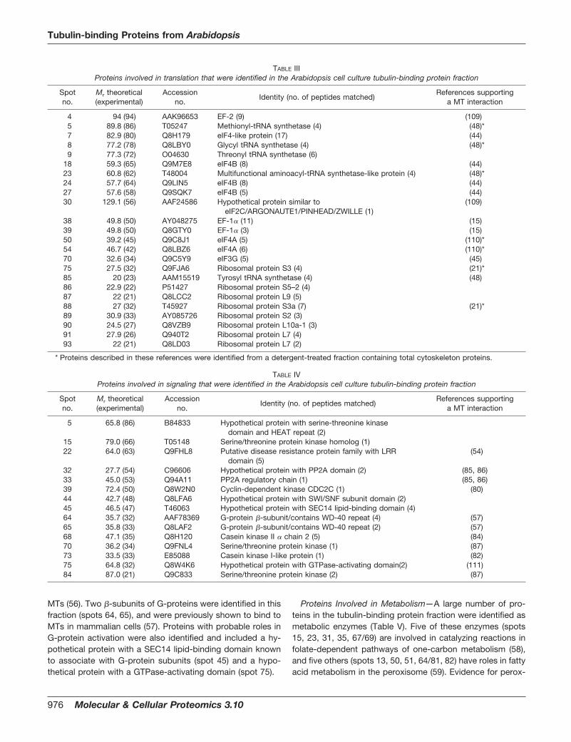
MTs (56). Two �-subunits of G-proteins were identified in thisfraction (spots 64, 65), and were previously shown to bind toMTs in mammalian cells (57). Proteins with probable roles inG-protein activation were also identified and included a hy-pothetical protein with a SEC14 lipid-binding domain knownto associate with G-protein subunits (spot 45) and a hypo-thetical protein with a GTPase-activating domain (spot 75).
Proteins Involved in Metabolism—A large number of pro-teins in the tubulin-binding protein fraction were identified asmetabolic enzymes (Table V). Five of these enzymes (spots15, 23, 31, 35, 67/69) are involved in catalyzing reactions infolate-dependent pathways of one-carbon metabolism (58),and five others (spots 13, 50, 51, 64/81, 82) have roles in fattyacid metabolism in the peroxisome (59). Evidence for perox-
TABLE IIIProteins involved in translation that were identified in the Arabidopsis cell culture tubulin-binding protein fraction
Spotno.
Mr theoretical(experimental)
Accessionno.
Identity (no. of peptides matched)References supporting
a MT interaction
4 94 (94) AAK96653 EF-2 (9) (109)5 89.8 (86) T05247 Methionyl-tRNA synthetase (4) (48)*7 82.9 (80) Q8H179 eIF4-like protein (17) (44)8 77.2 (78) Q8LBY0 Glycyl tRNA synthetase (4) (48)*9 77.3 (72) O04630 Threonyl tRNA synthetase (6)
18 59.3 (65) Q9M7E8 eIF4B (8) (44)23 60.8 (62) T48004 Multifunctional aminoacyl-tRNA synthetase-like protein (4) (48)*24 57.7 (64) Q9LIN5 eIF4B (8) (44)27 57.6 (58) Q9SQK7 eIF4B (5) (44)30 129.1 (56) AAF24586 Hypothetical protein similar to
eIF2C/ARGONAUTE1/PINHEAD/ZWILLE (1)(109)
38 49.8 (50) AY048275 EF-1� (11) (15)39 49.8 (50) Q8GTY0 EF-1� (3) (15)50 39.2 (45) Q9C8J1 eIF4A (5) (110)*54 46.7 (42) Q8LBZ6 eIF4A (6) (110)*70 32.6 (34) Q9C5Y9 eIF3G (5) (45)75 27.5 (32) Q9FJA6 Ribosomal protein S3 (4) (21)*85 20 (23) AAM15519 Tyrosyl tRNA synthetase (4) (48)86 22.9 (22) P51427 Ribosomal protein S5–2 (4)87 22 (21) Q8LCC2 Ribosomal protein L9 (5)88 27 (32) T45927 Ribosomal protein S3a (7) (21)*89 30.9 (33) AY085726 Ribosomal protein S2 (3)90 24.5 (27) Q8VZB9 Ribosomal protein L10a-1 (3)91 27.9 (26) Q940T2 Ribosomal protein L7 (4)93 22 (21) Q8LD03 Ribosomal protein L7 (2)
* Proteins described in these references were identified from a detergent-treated fraction containing total cytoskeleton proteins.
TABLE IVProteins involved in signaling that were identified in the Arabidopsis cell culture tubulin-binding protein fraction
Spotno.
Mr theoretical(experimental)
Accessionno.
Identity (no. of peptides matched)References supporting
a MT interaction
5 65.8 (86) B84833 Hypothetical protein with serine-threonine kinasedomain and HEAT repeat (2)
15 79.0 (66) T05148 Serine/threonine protein kinase homolog (1)22 64.0 (63) Q9FHL8 Putative disease resistance protein family with LRR
domain (5)(54)
32 27.7 (54) C96606 Hypothetical protein with PP2A domain (2) (85, 86)33 45.0 (53) Q94A11 PP2A regulatory chain (1) (85, 86)39 72.4 (50) Q8W2N0 Cyclin-dependent kinase CDC2C (1) (80)44 42.7 (48) Q8LFA6 Hypothetical protein with SWI/SNF subunit domain (2)45 46.5 (47) T46063 Hypothetical protein with SEC14 lipid-binding domain (4)64 35.7 (32) AAF78369 G-protein �-subunit/contains WD-40 repeat (4) (57)65 35.8 (33) Q8LAF2 G-protein �-subunit/contains WD-40 repeat (2) (57)68 47.1 (35) Q8H120 Casein kinase II � chain 2 (5) (84)70 36.2 (34) Q9FNL4 Serine/threonine protein kinase (1) (87)73 33.5 (33) E85088 Casein kinase I-like protein (1) (82)75 64.8 (32) Q8W4K6 Hypothetical protein with GTPase-activating domain(2) (111)84 87.0 (21) Q9C833 Serine/threonine protein kinase (2) (87)
Tubulin-binding Proteins from Arabidopsis
976 Molecular & Cellular Proteomics 3.10

isomal enzyme interactions with MTs has been reported pre-viously (5, 16, 60, 61). The glycolytic enzyme glyceraldehyde-3-phosphate dehydrogenase (GAPDH) was also identified inthis fraction (spot 62) and has been previously shown to haveMT-binding activity (22, 62, 63). Additional proteins were iden-tified that function in other metabolic pathways, including thepentose phosphate pathway (spots 31, 47), phosphate me-tabolism (spot 33), amino acid biosynthesis (spots 53, 81), thetricarboxylic acid cycle (spot 53), anaerobic glycolysis (spot57), and pantothenate biosynthesis (spots 59–61).
Other Proteins with Known or Unknown Functions—Thetubulin-binding proteins listed in Table VI likely participate incellular activities other than those described above. Theseproteins have a wide range of functions, and at least one ofthese proteins has previously been reported to bind to MTs.Spot 81 contained a regulatory subunit of the 26S proteo-some, a complex that was shown previously to bind to mul-tiple MT arrays in plants (64). Other interesting proteins wereidentified that have not been shown previously to bind to MTs;however, their predicted functions implicate a possible MTassociation. For instance, the sorting nexin-like protein (spots45, 46) is involved in vesicle trafficking in both plants andanimals and has a role in axonal guidance, a form of cellmovement requiring cytoskeletal rearrangements (65). Ac-tin-11 (spot 4), a unique and ancient actin subclass in theArabidopsis actin family that has a divergent sequence from
other actins, was also identified in this study. This proteinlikely participates in specific developmental roles (66) andmay function to link actin filaments and MTs together. Severalproteins were identified that possessed DNA-binding do-mains (spots 8, 46/47, 56, 84), including the transcriptionfactor Scarecrow. Transcription factor activity is known to beregulated in some instances by its shuttling into and out of thenucleus in a MT-dependent manner, as was reported for themammalian tumor suppressor protein p53 (67). A group ofproteins that were unexpectedly represented in the tubulin-binding protein fraction were those involved cell wall modifi-cation and included �-galactosidase (spot 6), xylosidase (spot19), pectate lyase (spot 58), xyloglucan endo-1,4-beta-D-glu-canase (spot 71), and endoxyloglucan glycosyl-transferase(spot 78).
DISCUSSION
Here we report the identification of 122 tubulin-bindingproteins that were purified from Arabidopsis suspension cellsusing tubulin affinity chromatography, a procedure that hasbeen used previously to purify bona fide MT-binding proteins(5, 13–15, 17). The following observations indicated that tu-bulin affinity chromatography also served as an effectivemethod for the purification of MT-binding proteins in thepresent study. First, antibodies raised against two knownMT-binding proteins, EF-1� (15) and the peroxisomal MFP (5),
TABLE VProteins involved in metabolism that were identified in the Arabidopsis cell culture tubulin-binding protein fraction
Spotno.
Mr theoretical(experimental)
Accessionno.
Identity (no. of peptides matched)References supporting
a MT interaction
8 73.4 (78) T06034 Phosphoenolpyruvate carboxykinase (8)13 77.8 (72) T08956 AIM protein (peroxisomal MFP) (8) (5)15 67.8 (66) C96541 10-Formyltetrahydrofolate synthetase (7)23 64.9 (62) Q8RWT5 Phosphoribosylaminoimidazole carboxamide
formyltransferase (2)28 59.2 (59) T52611 Glucose-6-phosphate 1- dehydrogenase (7)31 51.7 (55) O23254 Serine hydroxymethyl- transferase (14)33 58.0 (53) T06104 2-Dehydro-3-deoxyphospho- heptonate aldolase (2)35 50.7 (52) T05362 Glycine hydroxymethyl- transferase (3)47 52.9 (50) Q9FWA3 6-Phosphogluconate dehydrogenase (5)50 47.6 (45) T46895 Acyl-CoA dehydrogenase (2)51 48.6 (44) Q9S7M3 3-Ketoacyl-CoA thiolase (11)53 47.3 (43) T04668 Phosphoserine transaminase (7)53 43.3 (43) Q8H1Y0 Pyruvate dehydrogenase E1a-like subunit IAR4 (3)57 34.4 (39) T10203 Protein from the alcohol dehydrogenase family (6)59 36.7 (38) H84898 3-Methyl-2-oxobutanoate hydroxy-methyl-transferase (4)60 36.7 (37) O82357 3-Methyl-2-oxobutanoate hydroxy-methyl-transferase (9)61 37.4 (39) T47945 3-Methyl-2-oxobutanoate hydroxy-methyl-transferase (2)62 36.9 (38) Q8LAS0 Glyceraldehyde-3-phosphate dehydrogenase C subunit (8) (62)64 31.7 (32) Q9LDF5 3-Hydroxyacyl-CoA dehydrogenase-like protein (5)66 37.5 (34) G84616 Glyoxosomal malate dehydrogenase (6)67 31.6 (33) Q8LEA3 5,10-Methylenetetrahydrofolate dehydrogenase (4)69 31.6 (33) Q8LEA3 5,10-Methylenetetrahydrofolate dehydrogenase (13)81 31.7 (32) Q9LDF5 3-Hydroxyacyl-CoA dehydrogenase-like protein (3)81 36.5 (32) T46192 Acetylglutamate kinase-like protein (2)82 29.9 (29) Q9FHR8 Enoyl CoA hydratase (3)
Tubulin-binding Proteins from Arabidopsis
Molecular & Cellular Proteomics 3.10 977

cross-reacted with proteins of the expected size on proteingel blots containing the tubulin-binding proteins (Fig. 1C). Thepresence of these two proteins was confirmed by LC-MS/MSanalysis (results from spots 38 and 13 in Tables III and V,respectively). Second, it appears that most, if not all, of theproteins that bound to the tubulin affinity column, pelletedwith MTs in co-sedimentation assays (Fig. 1B). A similar ob-servation was made previously (14) and is consistent withobservations that SDS-PAGE profiles of proteins that boundto tubulin and MT affinity columns were qualitatively similar (5,13). Third, 43% of the proteins identified in this study (52/122)are related to proteins previously reported to have an asso-ciation with MTs (see references in Tables I–VI). These obser-vations gave us confidence that the proteins identified hereare authentic MT-binding proteins.
The tubulin-binding proteins that were identified in thisstudy were grouped according to their predicted primaryfunction as either MAPs, RNA-binding proteins, translationfactors, metabolic enzymes, signaling proteins, or proteinswith other functions (Tables I–VI, Fig. 3). Although we ex-pected to identify a broad range of protein types based on theidentities of previously characterized plant MT-binding pro-teins (1, 2, 10), certain groups of these proteins would not beexpected to bind to MTs because of their known subcellularlocation. In these cases, the association of these proteins with
MTs may play a role in their trafficking within the cell. Alter-natively, these proteins may possess multiple functions andcarry out the additional function(s) in the cytosol in associationwith MTs. A good example of a group of tubulin-bindingproteins identified here that is known to be localized to aparticular membrane-bound organelle were the peroxisomalmatrix enzymes. Five of these enzymes catalyze reactions infatty acid �-oxidation (Table V, spots 13, 50, 51, 64/81, 82),and one functions in the glyoxylate cycle (malate dehydrogen-ase; spot 66). Each of these enzymes possesses a putativeamino- or carboxyl-terminal signal sufficient to target the pro-tein to the peroxisome matrix (68). Peroxisomal matrix pro-teins are imported into the peroxisome in their fully foldedconformation and, therefore, have the potential to bind to MTsprior to their import. We have performed extensive studies onthe MT binding activity of the rice peroxisomal MFP (5) andhave demonstrated that it does indeed bind to MTs in vivo(S. D. X. Chuong, R. T. Mullen, and D. G. Muench, unpub-lished observations). We proposed previously that the bindingof MFP to MTs may be important in facilitating its import intothe peroxisome (69). The fact that at least six peroxisomalmatrix enzymes were present in the tubulin-binding proteinfraction in this study (Table V), and one reported in anotherstudy (16), supports a general role for MTs in peroxisomalmatrix protein trafficking in plant cells.
TABLE VIProteins with other known or unknown functions that were identified in the Arabidopsis cell culture tubulin-binding protein fraction
Spotno.
Mr theoretical(experimental)
Accessionno.
Identity (no. of peptides matched)References supporting
a MT interaction
4 41.9 (92) P54396 Actin 11 (3)6 93.6 (86) CAB64737 �-Galactosidase (11)8 79.0 (78) Q9LDX1 Hypothetical protein with XS zinc finger nucleic acid binding
domain (4)19 83.5 (68) Q9FGY1 Xylosidase (4)20 63.1 (65) Q9FK40 Histone acetyltransferase (5)23 56.9 (62) G86349 Hypothetical protein with homology to auxilin, trichohalin, and
has a DnaJ domain (3)45 46.5 (47) Q9FG38 Sorting nexin-like protein (7)46 46.5 (48) Q9FG38 Sorting nexin-like protein (9)46 44.0 (48) T51151 Nuclear DNA-binding protein G2p (7)47 44.0 (50) T51151 Nuclear DNA-binding protein G2p (7)50 42.5 (45) T05870 Cell division protein pelota (3)56 169.4 (42) Q9LNX6 Scarecrow-like transcription factor (2)56 49.0 (42) Q9FF81 Vacuolar protein sorting 36 (2)58 46.0 (38) Q9LTZ0 Putative pectate lyase 11 precursor (4)59 35.7 (38) Q8W457 Hypothetical protein with cysteine65 36.5 (33) T52133 Potassium channel protein, b subunit (5)67 34.3 (33) B96602 Hypothetical protein with homology to myosin heavy chain (3)70 51.8 (34) Q93Y09 Serine carboxypeptidase II (4)71 32.8 (31) C49539 Xyloglucan endo-1,4-�-D-glucanase (4)76 33.6 (31) Q8LB34 Phosphate-induced protein 1 (2)77 172 (30) T06677 Protein with Bromo adjacent homology domain (1)78 32.8 (30) Q39099 Endoxyloglucan glycosyl- transferase (12)81 33.5 (32) Q8LD11 26S proteasome non-ATPase regulatory subunit (6) (64)84 22.3 (21) BT004166 Protein with single-stranded DNA-binding domain (2)84 20.6 (21) Q8RXZ8 Hypothetical protein (2)
Tubulin-binding Proteins from Arabidopsis
978 Molecular & Cellular Proteomics 3.10

The identification of numerous cytosolically localized met-abolic enzymes in the tubulin-binding protein fraction (TableV) is consistent with reports suggesting that sequential en-zymes from individual metabolic pathways can cluster on thecytoskeleton to assist in the efficient and regulated control offlux through those pathways in a process called metabolicchanneling (70). For instance, five enzymes that catalyze cou-pled reactions in the folate-dependent pathways of one-car-bon metabolism (58) were identified as components of thetubulin-binding protein fraction (Table V, spots 15, 23, 31, 35,67/69). This suggests that these cytosolic enzymes bind toMTs individually or as a complex to facilitate channeling ofintermediates through these pathways. An interaction be-tween homologs of some of the one-carbon metabolism en-zymes that were identified here has been implicated in facil-itating metabolic channeling in mammalian cells (58, 71, 72).Our data suggests that any metabolic channeling that resultsfrom such an interaction would also require the binding of thiscomplex to the MT matrix in order to stabilize these inter-actions. The identification of other classes of metabolicenzymes in this study (Table V) indicates that MTs may havean important role in regulating the activity of many metabolicprocesses.
Interestingly, several enzymes that were identified in thetubulin-binding protein fraction have cell wall modificationactivities (spots 6, 19, 58, 71, 78), including endo-1,4-�-glu-canase and endoxyloglucan glycosyltransferase (Table VI).These enzymes are known to have a cooperative role in themodification of cell wall architecture that is required for cellelongation and fruit ripening (73). The complex organization ofthe cell wall requires a highly coordinated activation of wall-loosening proteins to achieve precise control of wall disas-sembly. Perhaps an interaction occurs between some ofthese proteins to form a multicomponent system of enzymesthat functions to channel substrates in the modification anddegradation of xyloglucans. This would be analogous to theplant cell wall-degrading “cellulosome” of certain bacteria(74). An endo-1,4-�-glucanase that contains a transmem-brane domain is localized to the plasma membrane and Golgiin expanding cells of tomato (75) and could potentially interactwith MTs through its cytosolic domain. An interaction be-tween endo-1,4-�-glucanase and MTs across the plasmamembrane, perhaps as a component of an enzyme complexin the cell wall, could function to precisely regulate the activityof these wall-loosening enzymes. Several examples of linkerproteins that are components of the cytoskeleton-plasmamembrane-cell wall continuum in plants have been identifiedand appear to mediate communication across the plasmamembrane (76).
In plant cells, the cortical MT network lines the plasmamembrane and responds to a diverse array of extracellularstimuli (77–79). Several putative signaling proteins that wereidentified in the tubulin-binding protein fraction (Table IV) havebeen shown previously to interact with MTs directly or indi-
rectly through their association with MAPs and play an impor-tant role in regulating the cell cycle, cytoskeletal dynamics,and stress responses. For instance, a cyclin-dependent ki-nase binds to MTs in the spindle and phragmoplast in plantcells (80), casein kinases regulate MT organization by inter-acting with MAPs (81–84), and PP2A assists in regulating MTorganization (85). MAPs are a target of kinases and phos-phatases, and their modification by phosphorylation/dephos-phorylation can result in changes in MT organization anddynamics, as well as the activation of signaling cascades(86–90). In plants, rearrangement of cortical MTs is known tooccur in response to various signals, including cold and alu-minum stress, wounding, and pathogen attack (91–93). Theputative signaling proteins identified in the tubulin bindingprotein fraction may be involved in the transduction of signalsgenerated from abiotic and biotic stresses such as these, or inresponse to light or developmentally regulated signals (95). Asvery little is known about cytoskeleton-associated proteinsand their role in plant-specific signaling pathways, these willserve as useful target proteins in future signaling studies.
Many proteins identified in this study have predicted roles inRNA binding and translation (Tables II and III, Fig. 3). Proteinsthat function in translation have been reported to bind to thecytoskeleton (21, 42), and several of these proteins wereidentified here, including translation initiation and elongationfactors, aminoacyl-tRNA synthetases, and ribosomal pro-teins. This concentration of the translational machinery on thecytoskeleton may function to increase the efficiency of trans-lation, or serve as a mechanism to regulate translation rate(42). Some of the RNA-binding proteins identified in this frac-tion may function to anchor RNAs to MTs in a specific orgeneral manner in order to facilitate translation, as has beendescribed previously (95, 96). Alternatively, these proteinsmay be involved in trafficking mRNAs from the nucleus toregions of the cell where they are to be translated. RNAlocalization is well characterized in many species and func-tions to contribute to the localized synthesis of protein atdistinct subcellular regions (97). The initial step in RNA traf-ficking involves nuclear export with the assistance of nuclear/cytosolic shuttling proteins. These shuttling proteins can re-main as a component of RNP particles that move specificRNAs throughout the cell along MTs (98). The transcriptionalco-activator protein identified here (spot 91) is homologous tothe animal nuclear RNA export factor ALY, which has a gen-eral role in RNA export from the nucleus and in the initial stageof localization of specific RNAs (41, 99). The identification ofseveral types of RNA-binding proteins and translation factorsin the tubulin-binding protein fraction is consistent with re-ports describing the existence of highly organized and effi-cient RNA localization and translation machineries that oper-ate in association with the cytoskeleton (42).
A group of proteins that represented a relatively small per-centage of the total proteins in the tubulin-binding proteinfraction were the MAPs (Table I). Proteins such as EF-1� and
Tubulin-binding Proteins from Arabidopsis
Molecular & Cellular Proteomics 3.10 979

GAPDH also have MAP activities (13, 100) and could havebeen included in this group; however, these were groupedaccording to their originally documented functions (Tables IIIand V). The absence in this fraction of well-characterized plantMAPs, such as the 65-kDa MAPs, MAP190, or MOR1 (3), mayhave been due to the fact that the tubulin column does notbind these proteins with high affinity. The 65-kDa MAPs andMAP 190 were originally purified by pelleting endogenoustaxol-stabilized plant MTs after rounds of polymerization anddepolymerization (11, 102). Perhaps these proteins were inlow abundance in our tubulin-binding protein fraction andwere not visible on the Coomassie blue-stained gel, andtherefore were not selected for MS analysis. The presence inthis fraction of MAPs that are common in eukaryotes, such asmolecular chaperones, CLASP, and putative motor proteins,indicates that at least some MAPs are enriched using thisprocedure.
The identity of the proteins reported in this study supportsprevious evidence suggesting that MTs serve as a matrixwhere proteins can bind and carry out numerous functionswithin the cell, and provides insight on the mechanisms re-sponsible for macromolecule trafficking, metabolic channel-ing, and signaling in plant cells. Additional, more direct meth-ods will be required to confirm that a bona fide interactionoccurs between these proteins and MTs, as it is possible forsome proteins that their interaction with tubulin in vitro occursas a result of nonspecific binding during the chromatographyprocedure. Also, some of the proteins identified here mayassociate indirectly with MTs by interacting with other MT-binding proteins as a component of a protein complex.
There were many additional, less abundant proteins in thetubulin-binding protein fraction that were not identified in thisstudy. Future cytoskeleton studies from plants and other eu-karyotic species using similar and different methods of puri-fication will be important in identifying additional MT-bindingproteins, as well as proteins that bind to the actin componentof the cytoskeleton. Technological advances that eliminatethe need for gel electrophoresis and allow for the identificationof low-abundance proteins will also accelerate the character-ization of the cytoskeleton-binding proteome.
Acknowledgments—We would like to thank the personnel at theUniversity of Victoria Protein Chemistry Centre staff for their expertiseand assistance with mass spectrometry.
* This work was funded by a Genome Canada/Genome PrairieGrant (to D. G. M., A. G. G., and G. J. T.). The costs of publication ofthis article were defrayed in part by the payment of page charges.This article must therefore be hereby marked “advertisement” inaccordance with 18 U.S.C. Section 1734 solely to indicate this fact.
□S The on-line version of this manuscript (available at http://www.mcponline.org) contains supplemental material.
¶ To whom correspondence should be addressed: Department ofBiological Sciences, University of Calgary, Calgary, Alberta T2N 1N4,Canada. Tel.: 403-220-7935; Fax: 403-289-9311; E-mail: [email protected].
REFERENCES
1. Davies, E., Fillingham, B. D., and Abe, S. (1996) The plant cytoskeleton, inThe Cytoskeleton, Vol. 3 (Hesketh, J. E., and Pryme, I. F., eds) pp.405–449, JAI Press, Gremwich, CT
2. Lloyd, C., and Hussey, P. (2001) Microtubule-associated proteins inplants—Why we need a MAP. Nat. Rev. Mol. Cell. Biol. 2, 40–47
3. Hussey, P. J., Hawkins, T. J., Igarashi, H., Kaloriti, D., and Smertenko, A.(2002) The plant cytoskeleton: Recent advances in the study of the plantmicrotubule-associated proteins MAP-65, MAP-190 and the XenopusMAP215-like protein, MOR1. Plant Mol. Biol. 50, 915–924
4. Maccioni, R. B., and Cambiazo, V. (1995) Role of microtubule-associatedproteins in the control of microtubule assembly. Physiol. Rev. 75,835–864
5. Chuong, S. D. X., Mullen, R., and Muench, D. G. (2002) Identification of arice RNA- and MT-binding protein as the multifunctional protein (MFP),a peroxisomal enzyme involved in the �-oxidation of fatty acids. J. Biol.Chem. 277, 2419–2429
6. Janmey, P. A. (1998) The cytoskeleton and cell signaling: Componentlocalization and mechanical coupling. Physiol. Rev. 78, 763–781
7. Cyr, R. J., and Palevitz, B. A. (1989) Microtubule-binding proteins fromcarrot 1. Initial characterization and microtubule bundling. Planta 177,245–260
8. Whittington, A. T., Vugrek, O., Wei, K. J., Hasenbein, N. G., Sugimoto, K.,Rashbrooke, M. C., and Wasteneys, G. O. (2001) MOR1 is essential fororganizing cortical microtubules in plants. Nature 411, 610–613
9. Vantard, M., Schellenbaum, P., Fellous, A., and Lambert, A. M. (1991)Characterization of maize microtubule-associated proteins, one ofwhich is immunologically related to tau. Biochemistry 30, 9334–9340
10. Gardiner, J., and Marc, J. (2003) Putative microtubule-associated proteinsfrom the Arabidopsis genome. Protoplasma 222, 61–74
11. Jiang, C.-J., and Sonobe, S. (1993) Identification and preliminary charac-terization of a 65 kDa higher-plant microtubule-associated protein.J. Cell Sci. 105, 891–901
12. Davies, E., Stankovic, B., Azama, K., Shibata, K., and Abe, S. (2001) Novelcomponents of the plant cytoskeleton: A beginning to plant ‘cytomics’.Plant Sci 160, 185–196
13. Durso, N. A., and Cyr, R. J. (1994) A calmodulin-sensitive interactionbetween microtubules and a higher plant homolog of elongation factor-1�. Plant Cell 6, 893–905
14. Marc, J., Sharkey, D. E., Durso, N. A., Zhang, M., and Cyr, R. J. (1996)Isolation of a 90-kD microtubule-associated protein from tobacco mem-branes. Plant Cell 8, 2127–2138
15. Durso, N. A., Leslie, J. D., and Cyr, R. J. (1996) In situ immunocytochem-ical evidence that a homolog of protein translation elongation factorEF-1-� is associated with microtubules in carrot cells. Protoplasma 190,141–150
16. Harper, J. D. I., Weerakoon, N. D., Gardiner, J. C., Blackman, L. M., andMarc, J. (2002) A 75-kDa plant protein isolated by tubulin-affinity chro-matography is a peroxisomal matrix enzyme. Can. J. Bot. 80,1018–1027
17. Balaban, N., and Goldman, R. (1992) Isolation and characterization of aunique 15 kilodalton trypanosome subpellicular microtubule-associatedprotein. Cell Motil. Cytoskeleton 21, 138–146
18. Jablonsky, P., Elliott, J., and Williamson, R. (1993) Purification of a mungbean protein binding to microtubules through two defined sites in thecarboxy-terminal domain of �-tubulin. Plant Sci. 94, 35–45
19. Nebl, T., Pestonjamasp, K. N., Leszyk, J. D., Crowley, J. L., Oh, S. W., andLuna, E. J. (2002) Proteomic analysis of a detergent-resistant mem-brane skeleton from neutrophil plasma membranes. J. Biol. Chem. 277,43399–43409
20. Gevaert, K., Eggermont, L., Demol, H., and Vandekerckhove, J. (2000) Afast and convenient MALDI-MS based proteomic approach: Identifica-tion of components scaffolded by the actin cytoskeleton of activatedhuman thrombocytes. J. Biotechnol. 78, 259–269
21. Wu, Y., Muench, D. G., Kim, Y.-T., Hwang, Y.-S., and Okita, T. W. (1998)Identification of polypeptides associated with an enriched cytoskeleton-protein body fraction from developing rice endosperm. Plant CellPhysiol. 39, 1251–1257
22. Azama, K., Abe, S., Sugimoto, H., and Davies, E. (2003) Lysine-containingproteins in maize endosperm: A major contribution from cytoskeleton-
Tubulin-binding Proteins from Arabidopsis
980 Molecular & Cellular Proteomics 3.10

associated carbohydrate-metabolizing enzymes. Planta 217, 628–63823. Mathur, J., and Koncz, C. (1998) Callus culture and regeneration. Methods
Mol. Biol. 82, 31–3424. Mathe, E., Inoue, Y. H., Palframan, W., Brown, G., and Glover, D. M. (2003)
Orbit/Mast, the CLASP orthologue of Drosophila, is required for asym-metric stem cell and cystocyte divisions and development of the po-larised microtubule network that interconnects oocyte and nurse cellsduring oogenesis. Development 130, 901–915
25. Inoue, Y. H., do Carmo Avides, M., Shiraki, M., Deak, P., Yamaguchi, M.,Nishimoto, Y., Matsukage, A., and Glover, D. M. (2000) Orbit, a novelmicrotubule-associated protein essential for mitosis in Drosophila mela-nogaster. J. Cell Biol. 149, 153–166
26. Garnier, C., Barbier, P., Gilli, R., Lopez, C., Peyrot, V., and Briand, C.(1998) Heat-shock protein 90 (hsp90) binds in vitro to tubulin dimer andinhibits microtubule formation. Biochem. Biophys. Res. Commun. 250,414–419
27. Czar, M. J., Welsh, M. J., and Pratt, W. B. (1996) Immunofluorescencelocalization of the 90-kDa heat-shock protein to cytoskeleton. EurJ. Cell Biol. 70, 322–330
28. Liang, P., and MacRae, T. H. (1997) Molecular chaperones and the cy-toskeleton. J. Cell Sci. 110, 1431–1440
29. Brown, C. R., Doxsey, S. J., Hong-Brown, L. Q., Martin, R. L., and Welch,W. J. (1996) Molecular chaperones and the centrosome. A role forTCP-1 in microtubule nucleation. J. Biol. Chem. 271, 824–832
30. Kraemer, D., and Blobel, G. (1997) mRNA binding protein mrnp41 local-izes to both nucleus and cytoplasm. Proc. Natl. Acad. Sci. U. S. A. 94,9119–9124
31. Hamill, D., Davis, J., Drawbridge, J., and Suprenant, K. A. (1994) Polyri-bosome targeting to microtubules: Enrichment of specific mRNAs in areconstituted microtubule preparation from sea urchin embryos. J. CellBiol. 127, 973–984
32. Shibata, N., Tsunekawa, N., Okamoto-Ito, S., Akasu, R., Tokumasu, A.,and Noce, T. (2004) Mouse RanBPM is a partner gene to a germlinespecific RNA helicase, mouse vasa homolog protein. Mol. Reprod. Dev.67, 1–7
33. Abe, S., Sakai, M., Yagi, K., Hagino, T., Ochi, K., Shibata, K., and Davies,E. (2003) A Tudor protein with multiple SNc domains from pea seed-lings: Cellular localization, partial characterization, sequence analysis,and phylogenetic relationships. J. Exp. Bot. 54, 971–983
34. Sami-Subbu, R., Choi, S. B., Wu, Y., Wang, C., and Okita, T. W. (2001)Identification of a cytoskeleton-associated 120 kDa RNA-binding pro-tein in developing rice seeds. Plant Mol. Biol. 46, 79–88
35. Amikura, R., Hanyu, K., Kashikawa, M., and Kobayashi, S. (2001) Tudorprotein is essential for the localization of mitochondrial RNAs in polargranules of Drosophila embryos. Mech. Dev. 107, 97–104
36. Borer, R. A., Lehner, C. F., Eppenberger, H. M., and Nigg, E. A. (1989)Major nucleolar proteins shuttle between nucleus and cytoplasm. Cell56, 379–390
37. Wolin, S. L., and Cedervall, T. (2002) The La protein. Annu. Rev. Biochem.71, 375–403
38. Bachmann, M., Pfeifer, K., Schroder, H. C., and Muller, W. E. (1989) TheLa antigen shuttles between the nucleus and the cytoplasm in CV-1cells. Mol. Cell. Biochem. 85, 103–114
39. Li, Q., and Suprenant, K. A. (1994) Molecular characterization of the77-kDa echinoderm microtubule-associated protein. Homology to the�-transducin family. J. Biol. Chem. 269, 31777–31784
40. Zhou, Z., Luo, M. J., Straesser, K., Katahira, J., Hurt, E., and Reed, R.(2000) The protein Aly links pre-messenger-RNA splicing to nuclearexport in metazoans. Nature 407, 401–405
41. Palacios, I. M. (2002) RNA processing: Splicing and the cytoplasmiclocalisation of mRNA. Curr. Biol. 12, R50–52
42. Jansen, R.-P. (1999) RNA-cytoskeletal associations. FASEB J. 13,455–466
43. Gavrilova, L. P., Rutkevitch, N. M., Gelfand, L. P., Stahl, U., Bommer, A.,and Bielka, H. (1987) Immunofluorescent localization of protein synthe-sis components in mouse embryo. Cell Biol. Int. Reports 11, 745–752
44. Bokros, C. L., Hugdahl, J. D., Kim, H. H., Hanesworth, V. R., van Heerden,A., Browning, K. S., and Morejohn, L. C. (1995) Function of the p86subunit of eukaryotic initiation factor (iso)4F as a microtubule-associ-ated protein in plant cells. Proc. Natl. Acad. Sci. U. S. A. 92, 7120–7124
45. Hasek, J., Kovarik, P., Valasek, L., Malinska, K., Schneider, J., Kohlwein,
S. D., and Ruis, H. (2000) Rpg1p, the subunit of the Saccharomycescerevisiae eIF3 core complex, is a microtubule-interacting protein. CellMotil. Cytoskeleton 45, 235–246
46. Palecek, J., Hasek, J., and Ruis, H. (2001) Rpg1p/Tif32p, a subunit oftranslation initiation factor 3, interacts with actin-associated proteinSla2p. Biochem. Biophys. Res. Commun. 282, 1244–1250
47. Lynn, K., Fernandez, A., Aida, M., Sedbrook, J., Tasaka, M., Masson, P.,and Barton, M. K. (1999) The PINHEAD/ZWILLE gene acts pleiotropi-cally in Arabidopsis development and has overlapping functions withthe ARGONAUTE1 gene. Development 126, 469–481
48. Mirande, M., Le Corre, D., Louvard, D., Reggio, H., Pailliez, J. P., andWaller, J. P. (1985) Association of an aminoacyl-tRNA synthetase com-plex and of phenylalanyl-tRNA synthetase with the cytoskeletal frame-work fraction from mammalian cells. Exp. Cell Res. 156, 91–102
49. Dang, C. V., Yang, D. C. H., and Pollard, T. D. (1983) Association ofmethionyl-tRNA synthetase with detergent-insoluble components of therough endoplasmic reticulum. J. Cell Biol. 96, 1138–1147
50. Melki, R., Kerjan, P., Waller, J. P., Carlier, M. F., and Pantaloni, D. (1991)Interaction of microtubule-associated proteins with microtubules: Yeastlysyl- and valyl-tRNA synthetases and tau 218–235 synthetic peptide asmodel systems. Biochemistry 30, 11536–11545
51. Davies, E., Comer, E. C., Lionberger, J. M., Stankovic, B., and Abe, S.(1993) Cytoskeleton-bound polysomes in plants. III. Polysome-cy-toskeleton-membrane interactions in corn endosperm. Cell Biol. Int. 17,331–340
52. Starkey, C. R., and Levy, L. S. (1995) Identification of differentially ex-pressed genes in T-lymphoid malignancies in an animal model system.Int. J. Cancer 62, 325–331
53. Dixon, M. S., Hatzixanthis, K., Jones, D. A., Harrison, K., and Jones, J. D.(1998) The tomato Cf-5 disease resistance gene and six homologs showpronounced allelic variation in leucine-rich repeat copy number. PlantCell 10, 1915–1925
54. Davy, D. A., Campbell, H. D., Fountain, S., de Jong, D., and Crouch, M. F.(2001) The flightless I protein colocalizes with actin- and microtubule-based structures in motile Swiss 3T3 fibroblasts: Evidence for theinvolvement of PI 3-kinase and Ras-related small GTPases. J. Cell Sci.114, 549–562
55. Cote, J., Quinn, J., Workman, J. L., and Peterson, C. L. (1994) Stimulationof GAL4 derivative binding to nucleosomal DNA by the yeast SWI/SNFcomplex. Science 265, 53–60
56. Xue, Y., Canman, J. C., Lee, C. S., Nie, Z., Yang, D., Moreno, G. T., Young,M. K., Salmon, E. D., and Wang, W. (2000) The human SWI/SNF-Bchromatin-remodeling complex is related to yeast rsc and localizes atkinetochores of mitotic chromosomes. Proc. Natl. Acad. Sci. U. S. A.97, 13015–13020
57. Wu, H. C., Huang, P. H., and Lin, C. T. (1998) G protein � subunit is closelyassociated with microtubules. J. Cell. Biochem. 70, 553–562
58. Chen, L., Chan, S. Y., and Cossins, E. A. (1997) Distribution of folatederivatives and enzymes for synthesis of 10-formyltetrahydrofolate incytosolic and mitochondrial fractions of pea leaves. Plant Physiol. 115,299–309
59. Hooks, M. A. (2002) Molecular biology, enzymology, and physiology of�-oxidation, in Plant Peroxisomes: Biochemistry, Cell Biology and Bio-technological Applications (Baker, A., and Graham, I. A., eds) pp.19–55, Kluwer Academic Publishers, Norwell, MA
60. Balaban, N., and Goldman, R. (1990) The association of glycosomalenzymes and microtubules: A physiological phenomenon or an exper-imental artifact? Exp. Cell Res. 191, 219–226
61. Kueng, V., Schleappi, K., Schneider, A., and Seebeck, T. (1989) A glyco-somal protein (p60) which is predominantly expressed in procyclicTrypanosoma brucei: Characterization and DNA sequence. J. Biol.Chem. 264, 5203–5209
62. Somers, M., Engelborghs, Y., and Baert, J. (1990) Analysis of the bindingof glyceraldehyde-3-phosphate dehydrogenase to microtubules, themechanism of bundle formation and the linkage effect. Eur. J. Biochem.193, 437–444
63. Walsh, J. L., Keith, T. J., and Knull, H. R. (1989) Glycolytic enzymeinteractions with tubulin and microtubules. Biochim. Biophys. Acta 999,64–70
64. Yanagawa, Y., Hasezawa, S., Kumagai, F., Oka, M., Fujimuro, M., Naito,T., Makino, T., Yokosawa, H., Tanaka, K., Komamine, A., Hashimoto, J.,
Tubulin-binding Proteins from Arabidopsis
Molecular & Cellular Proteomics 3.10 981

Sato, T., and Nakagawa, H. (2002) Cell-cycle dependent dynamicchange of 26S proteasome distribution in tobacco BY-2 cells. Plant CellPhysiol. 43, 604–613
65. Worby, C. A., Simonson-Leff, N., Clemens, J. C., Kruger, R. P., Muda, M.,and Dixon, J. E. (2001) The sorting nexin, DSH3PX1, connects theaxonal guidance receptor, Dscam, to the actin cytoskeleton. J. Biol.Chem. 276, 41782–41789
66. Huang, S., An, Y. Q., McDowell, J. M., McKinney, E. C., and Meagher,R. B. (1997) The Arabidopsis ACT11 actin gene is strongly expressed intissues of the emerging inflorescence, pollen, and developing ovules.Plant Mol. Biol. 33, 125–139
67. Giannakakou, P., Sackett, D. L., Ward, Y., Webster, K. R., Blagosklonny,M. V., and Fojo, T. (2000) p53 is associated with cellular microtubulesand is transported to the nucleus by dynein. Nat. Cell Biol. 2, 709–717
68. Mullen, R. T. (2002) Targeting and import of matrix proteins into peroxi-somes, in Plant Peroxisomes (Baker, A., and Graham, I. A., eds) pp.339–383, Kluwer Academic Publishers, Norwell, MA
69. Muench, D. G., and Mullen, R. T. (2003) Peroxisome dynamics in plantcells: A role for the cytoskeleton. Plant Sci. 164, 307–315
70. al-Habori, M. (1995) Microcompartmentation, metabolic channelling andcarbohydrate metabolism. Int. J. Biochem. Cell Biol. 27, 123–132
71. Schirch, V., and Strong, W. B. (1989) Interaction of folylpolyglutamateswith enzymes in one-carbon metabolism. Arch. Biochem. Biophys. 269,371–380
72. Strong, W. B., and Schirch, V. (1989) In vitro conversion of formate toserine: Effect of tetrahydropteroylpolyglutamates and serine hydroxym-ethyltransferase on the rate of 10-formyltatrahydrofolate synthetase.Biochemisrty 28, 9430–9439
73. Rose, J. K., and Bennett, A. B. (1999) Cooperative disassembly of thecellulose-xyloglucan network of plant cell walls: parallels between cellexpansion and fruit ripening. Trends Plant Sci. 4, 176–183
74. Doi, R. H., and Tamaru, Y. (2001) The Clostridium cellulovorans cellulo-some: an enzyme complex with plant cell wall degrading activity. Chem.Rec. 1, 24–32
75. Brummell, D. A., Catala, C., Lashbrook, C. C., and Bennett, A. B. (1997) Amembrane-anchored E-type endo-1,4-�-glucanase is localized onGolgi and plasma membranes of higher plants. Proc. Natl. Acad. Sci.U. S. A. 94, 4794–4799
76. Baluska, F., Samaj, J., Wojtaszek, P., Volkmann, D., and Menzel, D. (2003)Cytoskeleton-plasma membrane-cell wall continuum in plants. Emerg-ing links revisited. Plant Physiol. 133, 482–491
77. Gardiner, J. C., Harper, J. D. I., Weerakoon, N. D., Collings, D. A., Ritchie,S., Gilroy, S., Cyr, R. J., and Marc, J. (2001) A 90-kD phospholipase Dfrom tobacco binds to microtubules and the plasma membrane. PlantCell 13, 2143–2158
78. Nick, P., Bergfeld, R., Schafer, E., and Schopfer, P. (1990) Unilateralreorientation of microtubules at the outer epidermal wall during photo-and gravitropic curvature of maize coleoptiles and sunflower hypocot-yls. Planta 181, 162–168
79. Hush, J. M., Hawes, C., and Overall, R. L. (1990) Interphase microtubulere-orientation predicts a new cell polarity in wounded pea roots. J. CellSci. 96, 47–61
80. Weingartner, M., Binarova, P., Drykova, D., Schweighofer, A., David, J. P.,Heberle-Bors, E., Doonan, J., and Bogre, L. (2001) Dynamic recruitmentof Cdc2 to specific microtubule structures during mitosis. Plant Cell 13,1929–1943
81. Sapir, T., Cahana, A., Seger, R., Nekhai, S., and Reiner, O. (1999) LIS1 isa microtubule-associated phosphoprotein. Eur. J. Biochem. 265,181–188
82. Yang, P., and Sale, W. S. (2000) Casein kinase I is anchored on axonemaldoublet microtubules and regulates flagellar dynein phosphorylationand activity. J. Biol. Chem. 275, 18905–18912
83. Behrend, L., Stoter, M., Kurth, M., Rutter, G., Heukeshoven, J., Deppert,W., and Knippschild, U. (2000) Interaction of casein kinase 1 delta(CK1�) with post-Golgi structures, microtubules and the spindle appa-ratus. Eur. J. Cell Biol. 79, 240–251
84. Schaerer-Brodbeck, C., and Riezman, H. (2003) Genetic and biochemicalinteractions between the Arp2/3 complex, Cmd1p, casein kinase II, andTub4p in yeast. FEMS Yeast Res. 4, 37–49
85. Hiraga, A., and Tamura, S. (2000) Protein phosphatase 2A is associated inan inactive state with microtubules through 2A1-specific interaction
with tubulin. Biochem. J. 346, 433–43986. Camilleri, C., Azimzadeh, J., Pastuglia, M., Bellini, C., Grandjean, O., and
Bouchez, D. (2002) The Arabidopsis TONNEAU2 gene encodes a pu-tative novel protein phosphatase 2A regulatory subunit essential for thecontrol of the cortical cytoskeleton. Plant Cell 14, 833–845
87. Gundersen, G. G., and Cook, T. A. (1999) Microtubules and signal trans-duction. Curr. Opin. Cell Biol. 11, 81–94
88. Pedrotti, B., Francolini, M., Cotelli, F., and Islam, K. (1996) Modulation ofmicrotubule shape in vitro by high molecular weight microtubule asso-ciated proteins MAP1A, MAP1B, and MAP2. FEBS Lett. 384, 147–150
89. Hoshi, M., Ohta, K., Gotoh, Y., Mori, A., Murofushi, H., Sakai, H., andNishida, E. (1992) Mitogen-activated-protein-kinase-catalyzed phos-phorylation of microtubule-associated proteins, microtubule-associatedprotein 2 and microtubule-associated protein 4, induces an alteration intheir function. Eur. J. Biochem. 203, 43–52
90. Lindwall, G., and Cole, R. D. (1984) Phosphorylation affects the ability oftau protein to promote microtubule assembly. J. Biol. Chem. 259,5301–5305
91. Sivaguru, M., Pike, S., Gassmann, W., and Baskin, T. I. (2003) Aluminumrapidly depolymerizes cortical microtubules and depolarizes the plasmamembrane: Evidence that these responses are mediated by a glutamatereceptor. Plant Cell Physiol. 44, 667–675
92. Abdrakhamanova, A., Wang, Q. Y., Khokhlova, L., and Nick, P. (2003) Ismicrotubule disassembly a trigger for cold acclimation? Plant CellPhysiol. 44, 676–686
93. Kobayashi, Y., Kobayashi, I., Funaki, Y., Fujimoto, S., Takemoto, T., andKunoh, H. (1997) Dynamic reorganization of microfilaments and micro-tubules is necessary for the expression of non-host resistance in barleycoleoptile cells. Plant J. 11, 525–537
94. Nick, P. (1999) Signals, motors, morphogenesis—The cytoskeleton inplant development. Plant Biol. 1, 169–179
95. Wickham, L., Duchaı̂ne, T., Luo, M., Nabi, I. R., and DesGroseillers, L.(1999) Mammalian staufen is a double-stranded-RNA and tubulin-bind-ing protein which localizes to the rough endoplasmic reticulum. Mol.Cell. Biol. 19, 2220–2230
96. DeFranco, C., Chicurel, M. E., and Potter, H. (1998) A general RNA-binding protein copmplex that includes the cytoskeleton-associatedprotein MAP1A. Mol. Biol. Cell 9, 1695–1708
97. Okita, T. W., and Choi, S. B. (2002) mRNA localization in plants: Targetingto the cell’s cortical region and beyond. Curr. Opin. Plant Biol. 5,553–559
98. Brumwell, C., Antolik, C., Carson, J. H., and Barbarese, E. (2002) Intra-cellular trafficking of hnRNP A2 in oligodendrocytes. Exp. Cell Res. 279,310–320
99. Kataoka, N., Diem, M. D., Kim, V. N., Yong, J., and Dreyfuss, G. (2001)Magoh, a human homolog of Drosophila mago nashi protein, is acomponent of the splicing-dependent exon-exon junction complex.EMBO J. 20, 6424–6433
100. Sirover, M. A. (1999) New insights into an old protein: the functionaldiversity of mammalian glyceraldehyde-3-phosphate dehydrogenase.Biochim. Biophys. Acta 1432, 159–184
101. Igarashi, H., Orii, H., Mori, H., Shimmen, T., and Sonobe, S. (2000) Isola-tion of a novel 190 kDa protein from tobacco BY-2 cells: Possibleinvolvement in the interaction between actin filaments and microtu-bules. Plant Cell Physiol. 41, 920–931
102. Himmelspach, R., Nick, P., Schafer, E., and Ehmann, B. (1997) Develop-mental and light-dependent changes of the cytosolic chaperonin con-taining TCP-1 (CCT) subunits in maize seedlings, and the localization incoleoptiles. Plant J. 12, 1299–1310
103. Kumar, J., Yu, H., and Sheetz, M. P. (1995) Kinectin, an essential anchorfor kinesin-driven vesicle motility. Science 267, 1834–1837
104. Gromley, A., Jurczyk, A., Sillibourne, J., Halilovic, E., Mogensen, M.,Groisman, I., Blomberg, M., and Doxsey, S. (2003) A novel humanprotein of the maternal centriole is required for the final stages ofcytokinesis and entry into S phase. J. Cell Biol. 161, 535–545
105. Schliwa, M., and Woehlke, G. (2003) Molecular motors. Nature 422,759–765
106. Hollander, B. A., Liang, M. Y., and Besharse, J. C. (1999) Linkage of anucleolin-related protein and casein kinase II with the detergent-stablephotoreceptor cytoskeleton. Cell Motil. Cytoskeleton 43, 114–127
107. Havin, L., Git, A., Elisha, Z., Oberman, F., Yaniv, K., Schwartz, S. P.,
Tubulin-binding Proteins from Arabidopsis
982 Molecular & Cellular Proteomics 3.10
Standart, N., and Yisraeli, J. K. (1998) RNA-binding protein conserved inboth microtubule- and microfilament-based RNA localization. GenesDev. 12, 1593–1598
108. Qutob, M. S., Bhattacharjee, R. N., Pollari, E., Yee, S. P., and Torchia, J.(2002) Microtubule-dependent subcellular redistribution of the tran-scriptional coactivator p/CIP. Mol. Cell. Biol. 22, 6611–6626
109. Heuijerjans, J., Pieper, F., Ramaekers, F., Timmermans, L., Kuijpers, H.,Bloemendal, H., and van Venrooij, W. J. (1989) Association of mRNA
and eIF-2� with the cytoskeleton in cells lacking vimentin. Exp. Cell Res.181, 317–330
110. Howe, J. G., and Hershey, J. W. B. (1984) Translational initiation factor andribosome association with the cytoskeletal framework fraction fromHeLa cells. Cell 37, 85–93
111. Pay, A., Resch, K., Frohnmeyer, H., Fejes, E., Nagy, F., and Nick, P. (2002)Plant RanGAPs are localized at the nuclear envelope in interphase andassociated with microtubules in mitotic cells. Plant J. 30, 699–709
Tubulin-binding Proteins from Arabidopsis
Molecular & Cellular Proteomics 3.10 983