Profiling the autoantibody repertoire by serological antigen selection
Upload
independentCategory
view
0download
0
Large Functional Repertoire of Regulatory T Cell SuppressibleAutoimmune T Cells in Scurfy Mice1
Rahul Sharma*,2, Wael N. Jarjour*,2, Lingjie Zheng*, Felicia Gaskin†, Shu Man Fu*,3, andShyr-Te Ju*,3
*Department of Medicine, Division of Rheumatology and Immunology, University of Virginia Health System,Charlottesville, Virginia, 22908 USA
†Department of Psychiatry and Neurobehavioral Sciences, University of Virginia, Charlottesville, Virginia,22908 USA
AbstractScurfy mice, a strain lacking functional Foxp3 transcription factor and CD4+CD25+Foxp3+
regulatory T (Treg) cells, spontaneously develop autoimmune responses against skin, lung, liver andtail. However, many organs/tissues are spared from autoimmune attack. Here, we demonstrate thatscurfy mice contain dormant autoimmune T cells that induced new diseases such as sialoadenitis,dacryoadenitis, pancreatitis, gastritis, intestinal inflammation, colitis, and myositis in RAG-1 KOmice. Inflammation in as many as 12 organs/tissues was consistently induced in individual recipientswith scurfy lymph node cells containing as few as 1.25×10⁶ CD4+ T cells. Moreover, transfer of themultiple organ autoimmune diseases could be suppressed by as little as 0.5×10⁶ CD4+CD25+ Tregcells, mediated by inhibiting autoimmune T cell expansion. Our study provides evidence for thepresence of a large repertoire of autoimmune lymphocytes against various organs/tissues in scurfymice as well as Treg cells in B6 mice capable of suppressing the expansion of these autoimmunelymphocytes. Various conditions that control the expression of autoimmune T cells are discussed.
KeywordsAutoimmunity; Inflammation; Tolerance/Suppression
1. IntroductionThymocyte differentiation and selection impart distinct functions to different T cellpopulations. In the CD4+ T cell compartment, two subsets are generated: a CD25- set thatresponds to antigens in the host and a CD25+Foxp3+ subset that suppresses these antigen-specific responses (reviewed in 1-3). Autoimmune T cells with moderate affinities against self-antigens could escape negative selection and exit to the periphery (4-6). Also, some of theorgan-specific antigens are not presented in the thymus during negative selection and are not
1Supported by NIH grants AI-036938, DE-017579 and AR-051203 and Lightner grant (to STJ), AR-045222, AR-047988 and AR-049449(to SMF), and DK-059850 (to WNJ).2Co-first authors contributed equally to this study.3SMF and STJ contributed equally to this work.Disclosures The authors have no conflict of interest.Publisher's Disclaimer: This is a PDF file of an unedited manuscript that has been accepted for publication. As a service to our customerswe are providing this early version of the manuscript. The manuscript will undergo copyediting, typesetting, and review of the resultingproof before it is published in its final citable form. Please note that during the production process errors may be discovered which couldaffect the content, and all legal disclaimers that apply to the journal pertain.
NIH Public AccessAuthor ManuscriptJ Autoimmun. Author manuscript; available in PMC 2008 August 1.
Published in final edited form as:J Autoimmun. 2007 August ; 29(1): 10–19.
NIH
-PA Author Manuscript
NIH
-PA Author Manuscript
NIH
-PA Author Manuscript

negatively selected (7). These autoimmune T cells in the periphery are suppressed by thenaturally occurring CD4+CD25+Foxp3+ regulatory T (Treg) cells (1-3).
Based on our current understanding of Treg cell function, a general defect in Treg cellexpression should result in autoimmune responses against various organs in a systemic fashion.However, earlier pioneer studies either with the day-3 thymectomy mouse model in which Tregcell generation was interrupted or with the adoptive transfer model in which CD25-
lymphocytes were transferred into nude mice did not meet this expectation (8-11). In the day-3thymectomy model, leukocyte infiltration into various organs was limited. Even with the mostsensitive targets of salivary and lacrimal glands and reproductive organs, leukocyte infiltrationwas observed only in a few strains and often not in every individual (9). In the adoptive transfermodel, only gastritis and oophoritis were observed in 100% of the nude mouse recipients(10). The prevalence and frequency of organ-specific autoimmune diseases were higher inNOD-SCID recipients transferred with T cells depleted of glucocorticoid-induced TNFreceptor family-related protein (11). However, these experimental systems used a large numberof T cells for transfer and a long incubation time for disease development. In addition, therewas a significant autoimmune antibody contribution to the systemic autoimmune manifestation(10, 11).
It is possible that both day-3 thymectomy and antibody treatments failed to completelyeliminate Treg cells. However, even in hosts with a congenital defect in Treg cell expression,extensive autoimmune inflammation in multiple organs was rarely reported (12-17). In patientswith the Immune Dysregulation, Polyendocrinopathy, Enteropathy, X-linked syndrome(IPEX), a lethal autoimmune disorder resulted from Foxp3 mutation, organ involvement wasoften limited to a few (18). Even in Foxp3 mutant scurfy mice, which have noCD4+CD25+Foxp3+ Treg cells and die within 3-5 weeks after birth as a result of multi-organautoimmune syndrome (19, 20), major organs that are inflamed are limited to skin, tails andlungs (21). Inflammation was moderate in the liver and at best borderline in the pancreas. Nodiscernible inflammation was observed in salivary and lacrimal glands, gastrointestinal tract,central nervous system, joints, and other endocrine glands (21).
The limited involvement of multiple organs/tissues in scurfy mice raises several fundamentalquestions about Treg cell control of autoimmunity. Is there a lacunar defect in the thymicselection for autoimmune lymphocytes? Is the absence of autoimmune response in certainorgans/tissues attributable to the presence of non-naturally occurring Treg cells? Or are thereother mechanisms that prevent the development and expression of certain organ-specificautoimmune lymphocytes? Here, we provide evidence against the former two possibilities andfavoring the third. We developed models that rapidly transfer multi-organ autoimmuneresponses in RAG-1 knockout (KO) mice. Surprisingly, lymph node cells of scurfy mice eventransferred autoimmune diseases against organs that were not inflamed in the donorthemselves, most notably in the salivary glands and organs associated with the gastrointestinaltract but other organs were also severely inflamed under appropriate condition. As many as 12organs/tissues were inflamed in individual mice. These are Treg cell-regulated autoimmuneresponses because they are readily suppressed by the co-transfer of the CD4+CD25+ Treg cellsfrom normal mice. The suppression is mediated by a mechanism that inhibits the expansion ofautoimmune lymphocytes in the secondary lymphoid organs. By demonstrating the presenceof Treg cell-suppressible autoimmune lymphocytes against 12 organs in individual mice, ourstudy strongly suggests that there is no defect in the generation of a large repertoire ofautoimmune lymphocytes in scurfy mice nor do scurfy mice use non-naturally occurring Tregcells to protect those organs spared from autoimmune attack. In addition, it suggests thepresence of a large Treg cell repertoire capable of suppressing these autoimmune lymphocytesand maintaining peripheral tolerance in normal B6 mice.
Sharma et al. Page 2
J Autoimmun. Author manuscript; available in PMC 2008 August 1.
NIH
-PA Author Manuscript
NIH
-PA Author Manuscript
NIH
-PA Author Manuscript

2. Materials and MethodsMice
C57BL/6 (B6) mice, B6.SJL-PLprcaPcpcb/BoyJ (B6.CD45.1) and B6.129S7-Rag1tm1Mom/J(RAG-1 KO) mice were obtained from the Jackson Laboratories, Bar Harbor, ME.Heterozygous female B6.Cg-Foxp3sf/x/J mice (the Jackson Laboratories) were bred with maleB6 mice to produce scurfy mice (Foxp3sf/Y). The presence of the scurfy mutation was confirmedby PCR as detailed in the Jackson Laboratory’s website. Mice were examined twice weeklyfor clinical signs of the autoimmune disease including manifestation of skin inflammation,body weight loss, and wasting etc.
HistologyTissues/Organs were fixed with 10% neutral buffered formalin (Fisher Scientific) and sectionsof paraffin-embedded tissue were stained with H & E. Tissues/Organs examined includedsalivary glands, lacrimal glands, stomachs, small intestines, colons, lungs, hearts, thyroidglands, livers, kidneys, pancreas, skins, tails and skeletal muscles. We also scored inflammationlevels by the extent of leukocyte infiltration in 10 randomly selected fields and categorizedthem as severe (4+), strong (3+), moderate (2+), mild (1+), and no inflammation (0+).
Adoptive transferAxillary, brachial, inguinal, cervical lymph nodes from 3 to 4 weeks old scurfy or B6 micewere isolated, pooled, and single cell suspensions were prepared in normal saline. Variousnumbers of cells were injected intravenously (IV), intraperitoneally (IP), or intramuscularly(IM) into the recipient mice. Both adult and 6 days old RAG-1 KO mice were used as recipients.In other experiments, CD4+ T cells were purified by positive selection using magnetic beadsconjugated with anti-CD4 mAb (Miltenyi Biotec). The unbound fraction was used as CD4+ Tcell-depleted population. The purity of both populations was determined with a Flow cytometerbefore transfer.
The CD4+CD25+ Treg cells and CD4+CD25- T cells were isolated from lymph nodes ofB6.CD45.1 mice using CD4+CD25+ Treg cell isolation kit (Miltenyi Biotec). The purity of thecells as determined by Flow cytometry was more than 85% in all cases. Enriched Treg cells(0.5×10⁶ or 1.5×10⁶) or CD4+CD25- T cell control were co-transferred with 5×10⁶ scurfylymph node cells into RAG-1 KO mice. Mice were observed every other day for clinical signsof autoimmune syndrome. Histological determination of autoimmune responses againstvarious organs/tissues was conducted at 3 to 5 weeks after transfer.
Flow cytometryThe samples analyzed include lymph node cells, purified CD4+ T cells, cells depleted ofCD4+ T cells, and splenic cells of RAG-1 KO recipients. Cells (106) were suspended in 100μl of PBS solution (containing 4 mg with of bovine serum albumin and 1 μg of anti-FcR mAb2.4G2) and incubated with 0.2 μg of various fluorescent antibodies for 30 minutes at 4°C.FITC, PE, or PE-Cy5 conjugated anti-CD4 (GK1.5), anti-CD8 (53-6.7), anti-B220 (RA3-6B2),anti-CD44 (IM7), anti-CD62L (MEL-14) and anti-CD69 (H1.2F3) mAb were obtained fromBD Biosciences. At least 104 stained cells were analyzed using a FACScan equipped withCellQuest (BD Biosciences). Post acquisition analyses were carried out using FlowJo™software (Tree Star, Inc, OR).
Sharma et al. Page 3
J Autoimmun. Author manuscript; available in PMC 2008 August 1.
NIH
-PA Author Manuscript
NIH
-PA Author Manuscript
NIH
-PA Author Manuscript

3. ResultsScurfy mice developed autoimmune response against a few target organs
We examined the H & E stains of many different organs to determine the extent of the multi-organ autoimmune response in scurfy mice. Scurfy mice of 3 to 4 weeks old were used becausethey developed severe clinical signs of autoimmune syndrome. The organs examined were theear, skin, tail, lung, liver, brain, joint, salivary gland, lacrimal gland, stomach, small intestine,colon, muscle, kidney, pancreas, thyroid and adrenal glands. Autoimmune response is definedby leukocyte infiltration into target organ using age- and sex-matched B6 mice as control. Noinflammation was observed for these tissues/organs of B6 mice. A strong and extensiveinflammatory response was observed in the ears, the tail, the skin and the lung (A-D in Figure1). A moderate infiltration of leukocytes was also observed in the liver (E in Figure 1).Surprisingly, little or no inflammation was observed for the rest of the organs examined (notshown).
Transfer of autoimmune responses against 12 organs/tissues: ear, skin, lung, liver,pancreas, salivary gland, lacrimal gland, stomach, small intestine, colon, kidney, and muscle
To determine if there were autoimmune lymphocytes against organs that were spared fromautoimmune attack in scurfy mice, we transferred (IV) 25×106 or 5×10⁶ lymph node cells (25%CD4+ T cells) from scurfy mice into RAG-1 KO mice and determined inflammation in variousorgans 3 weeks and 5 weeks later, respectively. The results are shown in Figure 2. With theexception of tail, organs that were inflamed in the donor scurfy mice were also inflamed in theRAG-1 KO recipients. Their extent of inflammation was similar or only slightly less severethan that observed in the donor mice. Organs in this category include ear, skin, lung, and liver(A-D in Figure 2). By contrast, inflammation with varied intensities was seen in muscle, kidney,pancreas, salivary gland, lacrimal gland, stomach, small intestine, and colon of the recipientmice, although inflammation in these organs was not observed in the donor scurfy mice. Thehistology results showed mild inflammation in the pancreas (peri-islets) and the muscle (L andE in Figure 2), moderate inflammation in the kidney interstitium, moderate to severeinflammation in the salivary and the lacrimal glands (F-I in Figure 2), and severe inflammationin the stomach, small intestine, and colon (I-K in Figure 2). Organs that remained free ofinflammation in RAG-1 KO recipients were thyroid gland, adrenal gland, and joint (data notshown).
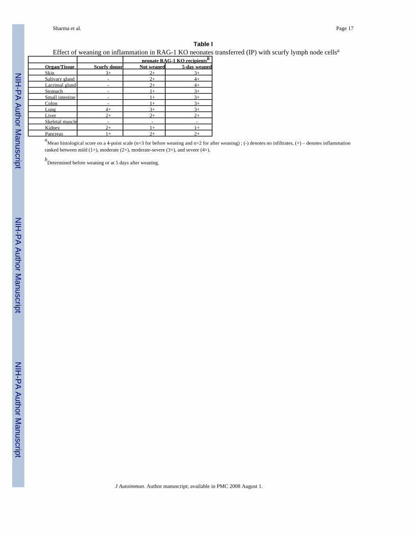
To determine the effect of neonatal condition on the expression of autoimmune lymphocytes,we transferred 5×10⁶ (IP) lymph node cells of 3.5 weeks old scurfy mice into 6 days old RAG-1KO mice and determined organ inflammation when they were 21 days old (last day beforeweaning) and 5 days after weaning. At 15 days after transfer and before weaning, the recipientsdeveloped severe inflammation in the lung and moderate inflammation in the skin, pancreas,liver, salivary glands and lacrimal glands. Moreover, a mild inflammation was observed in thekidney and gastrointestinal organs (Table 1). Interestingly, organs with direct exposure toexternal environment such as skin, salivary gland, lacrimal gland and gastrointestinal organs,but not pancreas and kidney, rapidly became severely inflamed at just 5 days after weaning(Table 1). Skeletal muscle remained free of inflammation in this transfer model.
We observed a moderate inflammation often extended into the adjacent muscle region layerin various organs that were strongly inflamed. However, such inflammation in skeletal muscletissue was seen neither in scurfy mice nor in the recipients of adoptive transfer of scurfy lymphnode cells by IV or IP routes. To determine if scurfy mice contain autoimmune lymphocytesagainst skeletal muscle, we transferred various numbers (1×10⁶, 5×10⁶, or 25×106) of theirlymph node cells into RAG-1 KO mice intramuscularly. Surprisingly, this rarely used route ofcell transfer is as efficient as IV for the induction of multi-organ autoimmune syndrome.
Sharma et al. Page 4
J Autoimmun. Author manuscript; available in PMC 2008 August 1.
NIH
-PA Author Manuscript
NIH
-PA Author Manuscript
NIH
-PA Author Manuscript

Multiple inflammations in 12 organs were observed even in the recipients of 106 transferredcells. Unlike IV transfer, severe inflammation was observed in the skeletal muscle tissues inthe area of injection (Figure 3A), in the thigh opposite to the transfer (Figure 3B), and in theupper limbs (Figure 3C). Both peri-vascular leukocyte infiltration and leukocytes surroundingthe muscle cells were observed. In addition, disintegrated and damaged muscle cells wereevident. This pattern of muscle pathology resembles the histology seen in dermatomyositis.This finding is significant as animal models for polymyositis and dermatomyositis are rare.The reason for the successful induction of inflammation in skeletal muscle is underinvestigation.
To determine whether CD4+ T cells are sufficient to induce autoimmune responses, wetransferred (IV) purified lymph node CD4+ T cells (2×10⁶, >97% CD4+ staining) of scurfymice to RAG-1 KO recipients. Lymph node cells depleted of CD4+ T cells (5×10⁶, <1%CD4+ staining) were transferred for comparison. With the CD4- cells, no organ inflammationwas observed at 5 weeks after transfer (not shown). Under identical conditions, CD4+ T cellsalone were sufficient to transfer the autoimmune diseases against various organs as describedabove in the IV transfer model. This transfer of autoimmune disease is significant becauseunlike earlier systems in which autoreactive antibodies may have played a significant role inthe multi-organ inflammation, the present study establishes that CD4+ T cells alone aresufficient to induce the multi-organ autoimmune inflammatory responses in RAG-1 KOrecipients.
Treg cells suppress the transfer of multiple autoimmune responsesTransfer of the CD4+CD25+ Treg cells into scurfy mice has generated variable results inprevious studies (22, 23). One group transferred 106 CD4+CD25+ Treg cells into scurfy miceof 3 days or 7 days old and this resulted in partial prolongation of lifespan (22). The other grouptransferred 4×10⁵ CD4+CD25+ Treg cells to 1-2 days old scurfy mice and these mice becamedisease-free as determined by disease phenotype (wasting and colitis) and lymph nodecellularity (23). In both reports, the protection from multi-organ inflammation was not studied,thus, leaving the issue open as to whether B6 CD4+CD25+ Treg cell repertoire is large enoughto protect mice from autoimmune inflammation in multiple organs. The adoptive transfermodel provides a more efficient and better controlled system to address this issue. Wetransferred 5×10⁶ lymph node cells of scurfy mice either alone or with 0.5×10⁶ CD4+CD25+
Treg cells from B6.CD45.1 mice into RAG-1 KO recipients. The mice were examined forinflammation in various organs at 5 weeks after transfer. Without co-transfer ofCD4+CD25+ Treg cells, the ear, the lung, the liver, the stomach, the small intestine, and thecolon were strongly inflamed (Figure 4; A-F and M). The histology of these organs wasselectively presented to show the extent of Treg cell-mediated suppression (Figure 4; G-L andM). Co-transfer CD4+CD25+ Treg cells and scurfy lymph node cells into RAG-1 KO micecompletely protected these organs from autoimmune attack in two of the three recipients (Fig.4). This suppression is specific because co-transfer of the CD4+CD25- T cells did not suppressthese autoimmune responses (data not shown). Interestingly, the remaining one recipient wasnot protected at all, showing multi-organ inflammation as severe as the RAG-1 KO mice thathad received scurfy lymph node cells. We detected CD4+CD25+Foxp3+ T cells in thesuppressed but not the unsuppressed recipients (data not shown). In subsequent experiments,1.5×106 CD4+CD25+ Treg cells were found to strongly suppress the multi-organ autoimmuneresponses in all four recipients. A mild leukocyte infiltration was only observed in the gut andlung and only in two of the four recipients. The compiled histological data are presented inFig. 4M. Thus, the repertoire of Treg cells in normal B6 mice is large enough to suppress theautoimmune lymphocytes present in the scurfy mice against 12 organs/tissues in individualmice4.
Sharma et al. Page 5
J Autoimmun. Author manuscript; available in PMC 2008 August 1.
NIH
-PA Author Manuscript
NIH
-PA Author Manuscript
NIH
-PA Author Manuscript

Mechanism of suppressionThe mechanism of the suppression of multi-organ inflammation was addressed. At 5 weeksafter transfer, there is a great expansion of transferred cells not only in the inflamed organs butalso in the spleen and lymph nodes. Spleens of RAG-1 KO mice that received 5×10⁶ scurfylymph node cells (in which 106 are CD4+ T cells) 5 weeks earlier expanded to 52±17×106 cells(in which 4×10⁶ are CD4+ T cells) (Figure 5A). In sharp contrast, RAG-1 KO mice that hadreceived both scurfy donor cells and B6.CD45.1 CD4+CD25+ Treg cells had few and nearlyundetectable leukocytes in various target organs. The spleens of the suppressed mice containedon the average of 11±4×10⁶ leukocytes in which 0.2×10⁶ were CD4+ T cells of the donor origin(Figure 5A). The scurfy donor cells contain a significant portion of memory CD4+ T cells (63%CD44high), recently activated CD4+ T cells (46% CD69high), and effector memory cells (54%CD62LlowCD44high) (Figure 5B and 5C). In contrast, the great majority of B6 lymph node Tcells express naïve phenotype of CD44lowCD62LhighCD69low. In the RAG-1 KO recipientsof scurfy donor cells, 95% of the splenic CD4+ T cells were CD44high and 43% of the CD4+
T cells were CD69high. The co-transfer of CD4+CD25+ Treg cells from B6.CD45.1 micereduced the percentage of CD69high expression to 10%, which is close to that of B6 control(Figure 5C). The suppressed CD4+ T cells remained CD44high. These results indicate that theCD4+CD25+ Treg cells suppress the multi-organ autoimmune inflammation mainly byinhibiting the activation and expansion of the transferred autoimmune memory CD4+ T cellsin the secondary lymphoid organs.
4. DiscussionConsidering the potentially extremely large lymphocyte repertoire and self antigens relativeto the small number of target organs in a host, it is reasonable to expect that in hosts with agenetic defect in the generation of Treg cells, autoimmune lymphocytes against various organsshould be relieved from autoimmune suppression, expand and infiltrate many organs/tissues.Nevertheless, such a wide and systemic autoimmune response has been reported neither inIPEX patients nor in mice with congenital Treg cell deficiency (12-20). The major point of thepresent study is that we have developed adoptive transfer models to demonstrate thatautoimmune responses against as many as 12 organs/tissues could be induced by lymphocytesof individual scurfy mice, thereby providing evidence that there is no defect in the generationof a large autoimmune repertoire in the scurfy mice, that there are no non-naturally-occurringTreg cells in scurfy mice responsible for the sparing of certain organs from autoimmune attack,and that congenital Treg cell-deficient mice contain autoimmune lymphocytes against the greatmajority if not all of the organs of the host. The second major point is the demonstration of anequally effective Treg cell population in normal mice capable of suppressing these autoimmuneresponses in individual mice.
There are several possibilities that can explain why scurfy mice display autoimmune responsesagainst only a few target organs but transfer of scurfy lymphocytes to RAG-1 KO mice inducessevere inflammation in additional organs/tissues. Scurfy mice live a short life so thatautoreactive lymphocytes against certain organs may be of low frequencies and do not havesufficient time to expand, resulting in lack of autoimmune manifestation. Scurfy mice remainfree of inflammation before 9 days old (21), a period in which lymphopenic expansion is critical(24). After this period, there are approximately 19 days for autoimmune diseases to developbefore death. Other factors such as organ development (salivary gland, prostate gland, etc) andenvironmental factors (immune status differences between scurfy and RAG-1 KO mice, foodintake before and after weaning, etc) are likely possibilities as well. The adoptive transfer of
4Using the IM transfer approach, co-transfer of B6 CD4+CD25+ Treg cells with scurfy lymph node cells also suppressed myositis(unpublished observation).
Sharma et al. Page 6
J Autoimmun. Author manuscript; available in PMC 2008 August 1.
NIH
-PA Author Manuscript
NIH
-PA Author Manuscript
NIH
-PA Author Manuscript
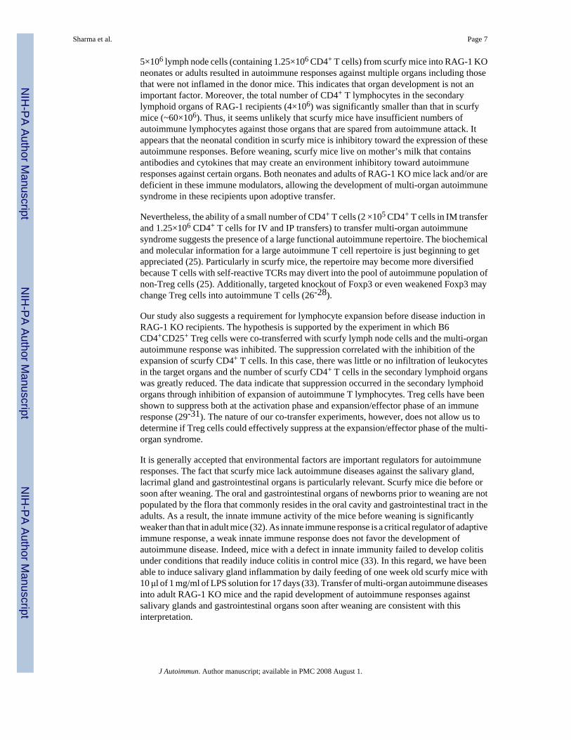
5×10⁶ lymph node cells (containing 1.25×10⁶ CD4+ T cells) from scurfy mice into RAG-1 KOneonates or adults resulted in autoimmune responses against multiple organs including thosethat were not inflamed in the donor mice. This indicates that organ development is not animportant factor. Moreover, the total number of CD4+ T lymphocytes in the secondarylymphoid organs of RAG-1 recipients (4×10⁶) was significantly smaller than that in scurfymice (~60×106). Thus, it seems unlikely that scurfy mice have insufficient numbers ofautoimmune lymphocytes against those organs that are spared from autoimmune attack. Itappears that the neonatal condition in scurfy mice is inhibitory toward the expression of theseautoimmune responses. Before weaning, scurfy mice live on mother’s milk that containsantibodies and cytokines that may create an environment inhibitory toward autoimmuneresponses against certain organs. Both neonates and adults of RAG-1 KO mice lack and/or aredeficient in these immune modulators, allowing the development of multi-organ autoimmunesyndrome in these recipients upon adoptive transfer.
Nevertheless, the ability of a small number of CD4+ T cells (2 ×10⁵ CD4+ T cells in IM transferand 1.25×10⁶ CD4+ T cells for IV and IP transfers) to transfer multi-organ autoimmunesyndrome suggests the presence of a large functional autoimmune repertoire. The biochemicaland molecular information for a large autoimmune T cell repertoire is just beginning to getappreciated (25). Particularly in scurfy mice, the repertoire may become more diversifiedbecause T cells with self-reactive TCRs may divert into the pool of autoimmune population ofnon-Treg cells (25). Additionally, targeted knockout of Foxp3 or even weakened Foxp3 maychange Treg cells into autoimmune T cells (26-28).
Our study also suggests a requirement for lymphocyte expansion before disease induction inRAG-1 KO recipients. The hypothesis is supported by the experiment in which B6CD4+CD25+ Treg cells were co-transferred with scurfy lymph node cells and the multi-organautoimmune response was inhibited. The suppression correlated with the inhibition of theexpansion of scurfy CD4+ T cells. In this case, there was little or no infiltration of leukocytesin the target organs and the number of scurfy CD4+ T cells in the secondary lymphoid organswas greatly reduced. The data indicate that suppression occurred in the secondary lymphoidorgans through inhibition of expansion of autoimmune T lymphocytes. Treg cells have beenshown to suppress both at the activation phase and expansion/effector phase of an immuneresponse (29-31). The nature of our co-transfer experiments, however, does not allow us todetermine if Treg cells could effectively suppress at the expansion/effector phase of the multi-organ syndrome.
It is generally accepted that environmental factors are important regulators for autoimmuneresponses. The fact that scurfy mice lack autoimmune diseases against the salivary gland,lacrimal gland and gastrointestinal organs is particularly relevant. Scurfy mice die before orsoon after weaning. The oral and gastrointestinal organs of newborns prior to weaning are notpopulated by the flora that commonly resides in the oral cavity and gastrointestinal tract in theadults. As a result, the innate immune activity of the mice before weaning is significantlyweaker than that in adult mice (32). As innate immune response is a critical regulator of adaptiveimmune response, a weak innate immune response does not favor the development ofautoimmune disease. Indeed, mice with a defect in innate immunity failed to develop colitisunder conditions that readily induce colitis in control mice (33). In this regard, we have beenable to induce salivary gland inflammation by daily feeding of one week old scurfy mice with10 μl of 1 mg/ml of LPS solution for 17 days (33). Transfer of multi-organ autoimmune diseasesinto adult RAG-1 KO mice and the rapid development of autoimmune responses againstsalivary glands and gastrointestinal organs soon after weaning are consistent with thisinterpretation.
Sharma et al. Page 7
J Autoimmun. Author manuscript; available in PMC 2008 August 1.
NIH
-PA Author Manuscript
NIH
-PA Author Manuscript
NIH
-PA Author Manuscript

The tails of donor scurfy mice were severely inflamed. Unlike other organs, transfer of tailinflammation was not observed in IV and IM transfer systems. A weak inflammation wasobserved in the IP transfer of scurfy cells into RAG-1 KO neonates (data not shown). Thereason remains unclear. Similarly, myositis was observed by the IM transfer approach but notIV and IP transfers. These results suggest that additional dormant autoimmune lymphocytesmay be present and may not be detected by these adoptive transfer approaches. In this respect,it should be noted that a number of organs/tissues were not inflamed despite the induction ofautoimmune inflammation in multiple organs/tissues in the recipients of adoptive cell transfer.Transfer of inflammation response may be difficult to organs that are not easily accessible tolymphocytes such as brain, spinal cord, and joints. Absence of B cells in RAG-1 KO mice mayalso affect manifestation of autoimmune responses that are dependent wholly or partly onautoantibody production.
Finally, the present study also demonstrated for the first time that a single transfer of lymphnode cells of individual scurfy mice induces multiple autoimmune responses against at least12 organs of the RAG-1 KO recipients, an observation that reflects the large autoimmunerepertoire and the true meaning of multi-organ autoimmune syndrome. Interestingly, some ofthem may be considered animal models for human diseases. For example, the salivary glandinflammation observed herein is associated with acinar cell destruction, ductal cell hypertrophyand reduced salivation function (34). Salivary gland malfunction is a typical manifestation ofSjögren’s syndrome. The data suggest that Treg cell deficiency may be one underlying factorfor Sjögren’s syndrome. Similar arguments could be made for other organ-specificautoimmune diseases such as myositis, gastritis, colitis, etc. Whether these diseases could becaused by a defect in organ/tissue-specific Treg cells is interesting and remains to be addressed.Organ-specific Treg cells that suppress organ-specific autoimmune diseases have beendescribed (35-38). Treg cells that recognize self antigens commonly expressed by variousorgans/tissues could also explain the results of the present study (7, 25). Recent studies havedemonstrated that Treg cell repertoire is very large (25, 39) and a sizable part of it recognizesubiquitously presented self antigens (25). In this respect, it is remarkable that a co-transfer ofas little as 0.5×10⁶ CD4+CD25+ Treg cells suppressed the autoimmune responses against allof these target organs, demonstrating the power of Treg cells and suggesting the presence ofTreg cells specific to many if not all organs/tissues in B6 mice for the maintenance of peripherytolerance. The simplicity, rapidity and effectiveness of the system suggest that it will be anextremely useful animal model to study the specificity, regulation and mechanisms of the multi-organ autoimmune syndrome at the levels of organ-specific autoimmune diseases, autoimmuneT cells and Treg cells that control these autoimmune T cells.
Acknowledgements
We thank Mrs. Chiao-Ying Angela Ju for her assistance.
References1. Wing K, Fehervari Z, Sakaguchi S. Emerging possibilities in the development and function of
regulatory T cells. Int Immunol 2006;18:991–1000. [PubMed: 16720616]2. Bluestone JA, Tang Q. How do CD4+CD25+ regulatory T cells control autoimmunity? Curr Opin
Immunol 2005;17:638–642. [PubMed: 16209918]3. Piccirillo CA, Shevach EM. Naturally-occurring CD4+CD25+ immunoregulatory T cells: central
players in the arena of peripheral tolerance. Semin Immunol 2004;16:81–88. [PubMed: 15036231]4. Danke NA, Koelle DM, Yee C, Beheray S, Kwok WW. Autoreactive T cells in healthy individuals. J
Immunol 2004;172:5967–5972. [PubMed: 15128778]5. Bouneaud C, Kourilsky P, Bousso P. Impact of negative selection on the T cell repertoire reactive to
a self-peptide: A large fraction of T cell clones escapes clonal deletion. Immunity 2000;13:829–840.[PubMed: 11163198]
Sharma et al. Page 8
J Autoimmun. Author manuscript; available in PMC 2008 August 1.
NIH
-PA Author Manuscript
NIH
-PA Author Manuscript
NIH
-PA Author Manuscript

6. Yan J, Mamula MJ. Autoreactive T cells revealed in the normal repertoire: escape from negativeselection and peripheral tolerance. J Immunol 2002;168:3188–3194. [PubMed: 11907071]
7. Anderson AC, Kuchroo VK. Expression of self-antigen in the thymus: A little goes a long way. J ExpMed 2003;198:1627–1629. [PubMed: 14657216]
8. Nishizuka Y, Sakakura T. Thymus and reproduction: sex-linked dysgenesia of the gonad after neonatalthymectomy in mice. Science 1969;166:753–775. [PubMed: 5823314]
9. Tung KS, Smith S, Teuscher C, Cook C, Anderson RE. Murine autoimmune oophoritis,epididymoorchitis, and gastritis induced by day 3 thymectomy. Immunopathology Am J Pathol1987;126:293–302.
10. Sakaguchi S, Sakaguchi N, Asano M, Itoh M, Toda M. Immunologic selftolerance maintained byactivated T cells expressing IL-2 receptor alphachains (CD25). Breakdown of a single mechanismof self-tolerance causes various autoimmune diseases. J Immunol 1995;155:1151–1164. [PubMed:7636184]
11. Ono M, Shimizu J, Miyachi Y, Sakaguchi S. Control of autoimmune myocarditis and multiorganinflammation by glucocorticoid-induced TNF receptor family-related protein (high), Foxp3-expressing CD25+ and CD25- regulatory T cells. J Immunol 2006;176:4748–4756. [PubMed:16585568]
12. Clark LB, Appleby MW, Brunkow ME, Wilkinson JE, Ziegler SF, Ramsdell F. Cellular and molecularcharacterization of the scurfy mouse mutant. J Immunol 1999;162:2546–2554. [PubMed: 10072494]
13. Schorle H, Holtschke T, Hunig T, Schimpl A, Horak I. Development and function of T cells in micerendered interleukin-2 deficient by gene targeting. Nature 1991;352:621–624. [PubMed: 1830926]
14. Suzuki H, Kündig TM, Furlonger C, Wakeham A, Timms E, Matsuyama T, Schmits R, Simard JL,Ohashi PS, Griesserl H. Deregulated T cell activation and autoimmunity in mice lacking interleukin-2receptor. Science 1995;268:1472–1476. [PubMed: 7770771]
15. Willerford DM, Chen J, Ferry JA, Davidson L, Ma A, Alt FW. Interleukin-2 receptor alpha chainregulates the size and content of the peripheral lymphoid compartment. Immunity 1995;3:521–530.[PubMed: 7584142]
16. Waterhouse P, Penninger JM, Timms E, Wakeham A, Shahinian A, Lee KP, Thompson CB, GriesserH, Mak TW. Lymphoproliferative disorders with early lethality in mice deficient in Ctla-4. Science1995;270:985–988. [PubMed: 7481803]
17. Shull MM, Ormsby I, Kier AB, Pawlowski S, Diebold RJ, Yin M, Allen R, Sidman C, Proetzel G,Calvin D, Annunziata N, Doetschman T. Targeted disruption of the mouse transforming growthfactor-beta 1 gene results in multifocal inflammatory disease. Nature 1992;359:693–699. [PubMed:1436033]
18. Wildin RS, Smyk-Pearson S, Filipovich AH. Clinical and molecular features of theimmunodysregulation, polyendocrinopathy, enteropathy, X-linked (IPEX) syndrome. J Med Genet2002;39:537–545. [PubMed: 12161590]
19. Brunkow ME, Jeffery EW, Hjerrild KA, Paeper B, Clark LB, Yasayko S-A, Wilkinson JE, Galas D,Ziegler SF, Ramsdell F. Disruption of a new forkhead/winged-helix protein, scurfin, results in thefatal lymphoproliferative disorder of the scurfy mouse. Nat Genet 2001;27:68–73. [PubMed:11138001]
20. Wildin RS, Ramsdell F, Peake J, Faravelli F, Casanova J-L, Buist N, Levy-Lahad E, Mazzella M,Goulet O, Perroni L, Bricarelli FD, Byrne G, McEuen M, Proll S, Appleby M, Brunkow ME. X-linked neonatal diabetes mellitus, enteropathy and endocrinopathy syndrome is the human equivalentof mouse scurfy. Nat Genet 2001;27:18–20. [PubMed: 11137992]
21. Chen Z, Benoist C, Mathis D. How defects in central tolerance impinge on a deficiency in regulatoryT cells. Proc Natl Acad Sci USA 2005;102:14735–14740. [PubMed: 16203996]
22. Smyk-Pearson SK, Bakke AC, Held PK, Wildin RS. Rescue of the autoimmune scurfy mouse bypartial bone marrow transplantation or by injection with T-enriched splenocytes. Clin Exp Immunol2003;133:193–199. [PubMed: 12869024]
23. Fontenot JD, Gavin MA, Rudensky AY. Foxp3 programs the development and function of CD4+CD25+ regulatory T cells. Nat Immunol 2003;4:330–336. [PubMed: 12612578]
24. Min B, McHugh R, Sempowski GD, Mackall C, Foucras G, Paul WE. Neonates support lymphopenia-induced proliferation. Immunity 2003;18:131–140. [PubMed: 12530982]
Sharma et al. Page 9
J Autoimmun. Author manuscript; available in PMC 2008 August 1.
NIH
-PA Author Manuscript
NIH
-PA Author Manuscript
NIH
-PA Author Manuscript

25. Hsieh CS, Zheng Y, Liang Y, Fontenot JD, Rudensky AY. An intersection between the self-reactiveregulatory and nonregulatory T cell receptor repertoires. Nat Immunol 2006;7:401–410. [PubMed:16532000]
26. Liston A, Farr AG, Chen Z, Benoist C, Mathis D, Manley NR, Rudensky AY. Lack of Foxp3 functionand expression in the thymic epithelium. J Exp Med 2007;204:475–480. [PubMed: 17353370]
27. Wan YY, Flavell RA. Regulatory T-cell functions are subverted and converted owing to attenuatedFoxp3 expression. Nature 2007;445:766–770. [PubMed: 17220876]
28. Kim JM, Rasmussen JP, Rudensky AY. Regulatory T cells prevent catastrophic autoimmunitythroughout the lifespan of mice. Nat Immunol 2007;8:191–197. [PubMed: 17136045]
29. Thornton AM, Shevach EM. Suppressor effector function of CD4+CD25+ immunoregulatory T cellsis antigen nonspecific. J Immunol 2000;164:183–190. [PubMed: 10605010]
30. Mottet C, Uhlig HH, Powrie F. Cutting Edge: Cure of colitis by CD4+CD25+ regulatory T cells. JImmunol 2003;170:3939–3943. [PubMed: 12682220]
31. Nguyen LT, Jacobs J, Mathis D, Benoist C. Where FoxP3-dependent regulatory T cells impinge onthe development of inflammatory arthritis. Arthritis Rheum 2007;56:509–520. [PubMed: 17265486]
32. Marodi L. Innate cellular immune responses in newborns. Clin Immunol 2006;118:137–144.[PubMed: 16377252]
33. Rakoff-Nahoum S, Paglino J, Eslami-Varzaneh F, Edberg S, Medzhitov R. Recognition of commensalmicroflora by toll-like receptors is required for intestinal homeostasis. Cell 2004;118:229–241.[PubMed: 15260992]
34. Sharma R, Zheng L, Guo X, Fu SM, Ju S-T, Jarjour WN. Novel animal models for Sjögren’ssyndrome: Transfer of salivary gland dysfunction from regulatory T cell-deficient mice. JAutoimmunity 2006;27:289–296. [PubMed: 17207605]
35. Green EA, Choi Y, Flavell RA. Pancreatic lymph node derived CD4+CD25+ T reg cells: highly potentregulators of diabetes that require TRANCE-RANK signals. Immunity 2002;16:183–191. [PubMed:11869680]
36. Verginis P, Li HS, Carayanniotis G. Tolerogenic semimature dendritic cells suppress experimentalautoimmune thyroiditis by activation of thyroglobulin specific CD4+CD25+ T cells. J Immunol2005;174:7433–7439. [PubMed: 15905592]
37. Yu P, Gregg RK, Bell JJ, Ellis JS, Divekar R, Lee HH, Jain R, Waldner H, Hardaway JC, Collins M,Kuchroo VK, Zaghouani H. Specific T regulatory cells display broad suppressive functions againstexperimental allergic encephalomyelitis upon activation with cognate antigen. J Immunol2005;174:6772–6780. [PubMed: 15905518]
38. Samy ET, Parker LA, Sharp CP, Tung KS. Continuous control of autoimmune disease by antigen-dependent polyclonal CD4+CD25+ regulatory T cells in the regional lymph node. J Exp Med2005;202:771–781. [PubMed: 16172257]
39. Pacholczyk R, Ignatowicz H, Kraj P, Ignatowicz L. Origin and T cell receptor diversity of Foxp3+CD4+CD25+ T cells. Immunity 2006;25:249–259. [PubMed: 16879995]
Sharma et al. Page 10
J Autoimmun. Author manuscript; available in PMC 2008 August 1.
NIH
-PA Author Manuscript
NIH
-PA Author Manuscript
NIH
-PA Author Manuscript

Figure 1. Scurfy mice developed autoimmune response against a few target organsVarious organs/tissues of scurfy mice (left column) and sex-, age-matched B6 mice (rightcolumn) were stained (H&E) and examined under microscope. Strong inflammation wasobserved in ear (A), neck skin (B), tail (C) and lung (D). A moderate inflammation wasobserved in liver (E). The corresponding organs of B6 mice did not display any inflammation(F-J). Magnification-100X.
Sharma et al. Page 11
J Autoimmun. Author manuscript; available in PMC 2008 August 1.
NIH
-PA Author Manuscript
NIH
-PA Author Manuscript
NIH
-PA Author Manuscript

Figure 2. Transfer of autoimmune responses against multiple organs/tissuesAdult RAG-1 KO male were transferred (IV) with 25×106 scurfy lymph node cells. Threeweeks later, the histologic slides of ear (A), skin (B), lung (C), liver (D), muscle (E), kidney(F), salivary gland (G), lacrimal gland (H), stomach (I), small intestine (J), colon (K), andpancreas (L) were examined for inflammation. Corresponding organs/tissues of age-matchedRAG-1 KO male mice that received B6 lymph node cells did not show detectable inflammationand were not presented for simplicity. Similar results were obtained in RAG-1 KO recipientstransferred with 5×10⁶ scurfy lymph node cells and examined 4 weeks later (not shown). Finalmagnification is 100x, except L which is 200x.
Sharma et al. Page 12
J Autoimmun. Author manuscript; available in PMC 2008 August 1.
NIH
-PA Author Manuscript
NIH
-PA Author Manuscript
NIH
-PA Author Manuscript

Figure 3. Transfer of myositisScurfy lymph node cells (25×106) were transferred (IM) into the right thigh of RAG-1 KOmice and skeletal muscle of site of injection (A), left thigh (B) and upper limb (C) wereexamined for inflammation at 3 weeks after transfer. Similar results were obtained in RAG-1KO recipients of 106 or 5×10⁶ scurfy lymph node cells (data not shown). Magnification-100x.
Sharma et al. Page 13
J Autoimmun. Author manuscript; available in PMC 2008 August 1.
NIH
-PA Author Manuscript
NIH
-PA Author Manuscript
NIH
-PA Author Manuscript

Figure 4. Treg cells suppress the transfer of autoimmune responses against multiple organs/tissuesAdult RAG-1 KO mice were transferred (IV) with 5×10⁶ scurfy lymph node (LN) cells alone(A-F) or with 0.5×10⁶ CD4+CD25+ T cells (G-L) purified from B6.CD45.1 mice (Treg). Fiveweeks later, ear (A & G), lung (B & H), liver (C & I), stomach (D & J), small intestine (E &K), and colon (F & L) were examined for inflammation by histology (Magnification-100x).Adult RAG-1 KO mice that had received 5×10⁶ scurfy lymph node cells showed comparabledegree of inflammation. Histological scores (Mean ± standard deviation) of suppressed miceand control (n=6) were presented in the bar diagram (M). The H & E stained sections of variousorgans/tissues were scored on a five-point scale from 0 to 4 depicting leukocyte infiltration asnone, mild, moderate, strong, and severe, respectively. Complete suppression was observed in
Sharma et al. Page 14
J Autoimmun. Author manuscript; available in PMC 2008 August 1.
NIH
-PA Author Manuscript
NIH
-PA Author Manuscript
NIH
-PA Author Manuscript

most organs/tissues and more than 80% suppression was observed in lungs, liver, stomach,small intestine, and colon.
Sharma et al. Page 15
J Autoimmun. Author manuscript; available in PMC 2008 August 1.
NIH
-PA Author Manuscript
NIH
-PA Author Manuscript
NIH
-PA Author Manuscript

Figure 5. Treg cells inhibit autoimmune memory T cell expansionThe donor cells and the spleens of both the suppressed and unsuppressed mice in Figure 4 wereexamined for the expression of CD44, CD62L and CD69. The total number of leukocytes inthe spleen was determined (A). The circles represent measurements of individual mice. Thebars represent the means. The expression of CD44 and CD62L on gated CD4+ T cells is shownin (B). The CD69 expression on gated CD4+ T cells is shown in (C).
Sharma et al. Page 16
J Autoimmun. Author manuscript; available in PMC 2008 August 1.
NIH
-PA Author Manuscript
NIH
-PA Author Manuscript
NIH
-PA Author Manuscript
NIH
-PA Author Manuscript
NIH
-PA Author Manuscript
NIH
-PA Author Manuscript
Sharma et al. Page 17
Table IEffect of weaning on inflammation in RAG-1 KO neonates transferred (IP) with scurfy lymph node cellsa
neonate RAG-1 KO recipientsbOrgan/Tissue Scurfy donor Not weaned 5-day weanedSkin 3+ 2+ 3+Salivary gland - 2+ 4+Lacrimal gland - 2+ 4+Stomach - 1+ 3+Small intestine - 1+ 3+Colon - 1+ 3+Lung 4+ 3+ 3+Liver 2+ 2+ 2+Skeletal muscle - - -Kidney 2+ 1+ 1+Pancreas 1+ 2+ 2+aMean histological score on a 4-point scale (n=3 for before weaning and n=2 for after weaning) ; (-) denotes no infiltrates, (+) – denotes inflammation
ranked between mild (1+), moderate (2+), moderate-severe (3+), and severe (4+).
bDetermined before weaning or at 5 days after weaning.
J Autoimmun. Author manuscript; available in PMC 2008 August 1.
Copyright © 2022 FDOKUMEN