
A Naturally Selected Dimorphism within the HLA-B44 Supertype Alters Class I Structure, Peptide...
13
The Journal of Experimental Medicine J. Exp. Med. The Rockefeller University Press • 0022-1007/2003/09/679/13 $8.00 Volume 198, Number 5, September 1, 2003 679–691 http://www.jem.org/cgi/doi/10.1084/jem.20030066 679 A Naturally Selected Dimorphism within the HLA-B44 Supertype Alters Class I Structure, Peptide Repertoire, and T Cell Recognition Whitney A. Macdonald, 1 Anthony W. Purcell, 1 Nicole A. Mifsud, 1 Lauren K. Ely, 2 David S. Williams, 1 Linus Chang, 1 Jeffrey J. Gorman, 3 Craig S. Clements, 2 Lars Kjer-Nielsen, 1 David M. Koelle, 4 Scott R. Burrows, 5 Brian D. Tait, 6 Rhonda Holdsworth, 6 Andrew G. Brooks, 1 George O. Lovrecz, 3 Louis Lu, 3 Jamie Rossjohn, 2 and James McCluskey 1 1 Department of Microbiology and Immunology, University of Melbourne, Parkville,Victoria 3010, Australia 2 The Protein Crystallography Unit, Department of Biochemistry and Molecular Biology, School of Biomedical Sciences, Monash University, Clayton,Victoria 3168, Australia 3 Commonwealth Scientific and Industrial Research Organization, Division of Health, Science and Nutrition, Parkville,Victoria 3052, Australia 4 Department of Medicine, University of Washington, Seattle,WA 98195 5 Queensland Institute of Medical Research,The Bancroft Centre, Herston 4029 Queensland, Australia 6 Victorian Transplantation and Immunogenetics Service, Australian Red Cross Blood Service, South Melbourne,Victoria 3205, Australia Abstract HLA-B*4402 and B*4403 are naturally occurring MHC class I alleles that are both found at a high frequency in all human populations, and yet they only differ by one residue on the 2 helix (B*4402 Asp156→B*4403 Leu156). CTLs discriminate between HLA-B*4402 and B*4403, and these allotypes stimulate strong mutual allogeneic responses reflecting their known barrier to hemopoeitic stem cell transplantation. Although HLA-B*4402 and B*4403 share 95% of their peptide repertoire, B*4403 presents more unique peptides than B*4402, consistent with the stronger T cell alloreactivity observed toward B*4403 compared with B*4402. Crystal structures of B*4402 and B*4403 show how the polymorphism at position 156 is completely buried and yet alters both the peptide and the heavy chain conformation, relaxing ligand selection by B*4403 compared with B*4402. Thus, the polymorphism between HLA-B*4402 and B*4403 modifies both peptide repertoire and T cell recognition, and is reflected in the para- doxically powerful alloreactivity that occurs across this “minimal” mismatch. The findings suggest that these closely related class I genes are maintained in diverse human populations through their differential impact on the selection of peptide ligands and the T cell repertoire. Key words: class I histocompatibility molecules • antigen presentation • crystallography • X-ray diffraction • graft rejection • polymorphism protective immunity against microbes (1–3). HLA alleles can differ from each other by only a single amino acid (“micropolymorphism”) or by 30 amino acids (4). It has been suggested that there are nine major HLA class I “super- types,” or clusters of alleles, that each possess a unique broad specificity for common anchor motifs in antigenic peptides (5). Alleles from each of these supertypic families are distributed in virtually all human populations and account W.A. Macdonald and A.W. Purcell contributed equally to this work. The online version of this article includes supplemental material. Address correspondence to James McCluskey, Dept. of Microbiology and Immunology, The University of Melbourne, Parkville, Victoria 3010, Australia. Phone: 613-8344-5709; Fax: 613-9347-3226; email: [email protected]; or Jamie Rossjohn, The Protein Crystallogra- phy Unit, Dept. of Biochemistry and Molecular Biology, School of Bio- medical Sciences, Monash University, Clayton, Victoria 3168, Australia. Phone: 613-9905-3736; Fax: 613-9905-4699; email: [email protected] Abbreviations used in this paper: H-bond, hydrogen bond; MALDI-TOF, matrix-assisted laser desorption/ionization time-of-flight; MS, mass spec- trometry; RP, reverse phase; v.d.w., van der Waals. Introduction HLA class I molecules are characterized by a high level of polymorphism presumed to reflect natural selection for on November 12, 2015 jem.rupress.org Downloaded from Published August 25, 2003 http://jem.rupress.org/content/suppl/2003/08/25/jem.20030066.DC1.html Supplemental Material can be found at:
-
Upload
independent -
Category
Documents
-
view
2 -
download
0
Transcript of A Naturally Selected Dimorphism within the HLA-B44 Supertype Alters Class I Structure, Peptide...
The
Journ
al o
f Exp
erim
enta
l M
edic
ine
J. Exp. Med.
The Rockefeller University Press • 0022-1007/2003/09/679/13 $8.00Volume 198, Number 5, September 1, 2003 679–691http://www.jem.org/cgi/doi/10.1084/jem.20030066
679
A Naturally Selected Dimorphism within the HLA-B44 Supertype Alters Class I Structure, Peptide Repertoire, and T Cell Recognition
Whitney A. Macdonald,
1
Anthony W. Purcell,
1
Nicole A. Mifsud,
1
Lauren K. Ely,
2
David S. Williams,
1
Linus Chang,
1
Jeffrey J. Gorman,
3
Craig S. Clements,
2
Lars Kjer-Nielsen,
1
David M. Koelle,
4
Scott R. Burrows,
5
Brian D. Tait,
6
Rhonda Holdsworth,
6
Andrew G. Brooks,
1
George O. Lovrecz,
3
Louis Lu,
3
Jamie Rossjohn,
2
and James McCluskey
1
1
Department of Microbiology and Immunology, University of Melbourne, Parkville, Victoria 3010, Australia
2
The Protein Crystallography Unit, Department of Biochemistry and Molecular Biology, School of Biomedical Sciences, Monash University, Clayton, Victoria 3168, Australia
3
Commonwealth Scientific and Industrial Research Organization, Division of Health, Science and Nutrition, Parkville, Victoria 3052, Australia
4
Department of Medicine, University of Washington, Seattle, WA 98195
5
Queensland Institute of Medical Research, The Bancroft Centre, Herston 4029 Queensland, Australia
6
Victorian Transplantation and Immunogenetics Service, Australian Red Cross Blood Service, South Melbourne,Victoria 3205, Australia
Abstract
HLA-B
*
4402 and B
*
4403 are naturally occurring MHC class I alleles that are both found at ahigh frequency in all human populations, and yet they only differ by one residue on the
�
2 helix
(B
*
4402 Asp156
→
B
*
4403 Leu156). CTLs discriminate between HLA-B
*
4402 and B
*
4403,and these allotypes stimulate strong mutual allogeneic responses reflecting their known barrierto hemopoeitic stem cell transplantation. Although HLA-B
*
4402 and B
*
4403 share
�
95% oftheir peptide repertoire, B
*
4403 presents more unique peptides than B
*
4402, consistent withthe stronger T cell alloreactivity observed toward B
*
4403 compared with B
*
4402. Crystalstructures of B
*
4402 and B
*
4403 show how the polymorphism at position 156 is completelyburied and yet alters both the peptide and the heavy chain conformation, relaxing ligand selectionby B
*
4403 compared with B
*
4402. Thus, the polymorphism between HLA-B
*
4402 andB
*
4403 modifies both peptide repertoire and T cell recognition, and is reflected in the para-doxically powerful alloreactivity that occurs across this “minimal” mismatch. The findings suggestthat these closely related class I genes are maintained in diverse human populations throughtheir differential impact on the selection of peptide ligands and the T cell repertoire.
Key words: class I histocompatibility molecules • antigen presentation • crystallography • X-ray diffraction • graft rejection • polymorphism
protective immunity against microbes (1–3). HLA allelescan differ from each other by only a single amino acid(“micropolymorphism”) or by
�
30 amino acids (4). It hasbeen suggested that there are nine major HLA class I “super-types,” or clusters of alleles, that each possess a uniquebroad specificity for common anchor motifs in antigenicpeptides (5). Alleles from each of these supertypic familiesare distributed in virtually all human populations and account
W.A. Macdonald and A.W. Purcell contributed equally to this work.The online version of this article includes supplemental material.
Address correspondence to James McCluskey, Dept. of Microbiologyand Immunology, The University of Melbourne, Parkville, Victoria3010, Australia. Phone: 613-8344-5709; Fax: 613-9347-3226; email:[email protected]; or Jamie Rossjohn, The Protein Crystallogra-phy Unit, Dept. of Biochemistry and Molecular Biology, School of Bio-medical Sciences, Monash University, Clayton, Victoria 3168, Australia.Phone: 613-9905-3736; Fax: 613-9905-4699; email: [email protected]
Abbreviations used in this paper:
H-bond, hydrogen bond; MALDI-TOF,matrix-assisted laser desorption/ionization time-of-flight; MS, mass spec-trometry; RP, reverse phase; v.d.w., van der Waals.
Introduction
HLA class I molecules are characterized by a high level ofpolymorphism presumed to reflect natural selection for
on Novem
ber 12, 2015jem
.rupress.orgD
ownloaded from
Published August 25, 2003
http://jem.rupress.org/content/suppl/2003/08/25/jem.20030066.DC1.html Supplemental Material can be found at:
The
Journ
al o
f Exp
erim
enta
l M
edic
ine
680
Impact of a Naturally Selected Polymorphism on HLA Class I
for the majority HLA-A and -B polymorphism, suggestingthat their maintenance reflects an adaptable strategy for in-duction of immunity toward diverse microbial ligands.
The widespread distribution of many low frequencyHLA polymorphisms is probably an artifact of human mi-gration and admixture of population groups that originallypossessed a limited degree of HLA polymorphism (2). Thismight explain why most human populations often haveonly one or two alleles from each of the different HLA classI supertypes (5), such that a complete picture of HLA diver-sity is only appreciated when multiple racial groups are ex-amined. An exception to this notion is the HLA-B44 familyof alleles, in which both B
*
4402 and B
*
4403 are present inmost human populations at combined phenotypic frequen-cies of up to 40% (6). HLA-B
*
4402 and B
*
4403 are the twomost common members of the HLA-B44 supertype andparticipate in antiviral (7–10), antitumor (11–13), and minorAg-specific responses (14, 15). HLA-B
*
4402 (Asp156) andB
*
4403 (Leu156) differ by only a single amino acid locatedon the
�
2 helix, essentially constituting a dimorphismwithin the HLA-B44 family because the other HLA-B44alleles are much less common (generally, gene frequency
�
0.01; reference 6). These two B44 allotypes are also asso-ciated with different ancestral MHC haplotypes, suggestingan ancient origin for this di-allelism (16, 17).
Members of the B44 supertype share a preference forpeptides with negatively charged residues at P2 (Glu) andhydrophobic residues at the COOH terminus (18, 19).Previous comparisons of ligands eluted from B
*
4402 andB
*
4403 molecules have not revealed any difference in theirpeptide repertoire, raising the question of what functionaldifferences between these alleles support their high preva-lence in all human populations.
Despite the similarity of their peptide repertoires, mis-matching of B
*
4402 and B
*
4403 in hemopoeitic stem celltransplantation has been associated with transplant rejection(19, 20) and acute graft versus host disease (21), indicatingthat they represent a significant barrier to clinical transplan-tation. Moreover, CTL can discriminate between B
*
4402and B
*
4403 allotypes, indicating structural differences thatare highly relevant to T cells (22–24). The similarity inpeptide repertoires of B
*
4402 and B
*
4403 suggests that thestrong alloresponses between these alleles do not reflectpresentation of different peptides (18, 19), but may reflectthe accessibility of the
�
2 domain residue 156 for direct in-teraction with the TcR (24). Moreover, analyses of HLA-A2 mutants have confirmed the strong influence of posi-tion 156 on T cell recognition (25) and suggest a limitedinfluence for this residue on peptide binding (26). On theother hand, residue 156 has also been predicted to influ-
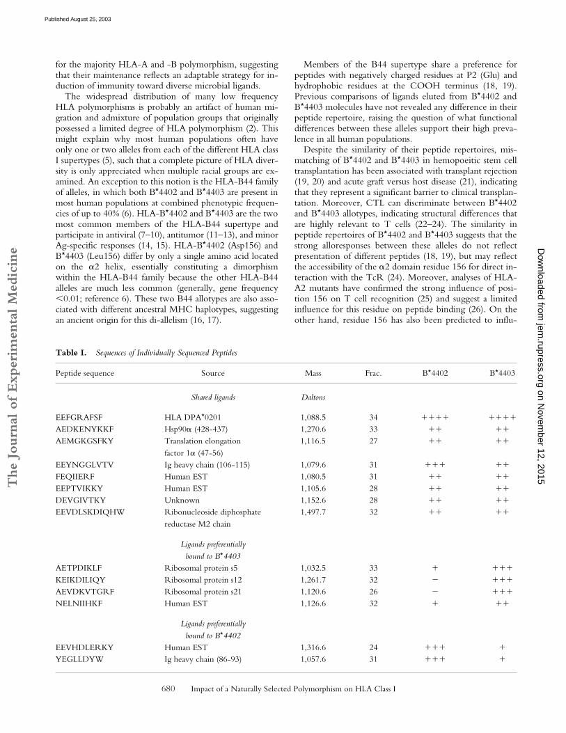
Table I.
Sequences of Individually Sequenced Peptides
Peptide sequence Source Mass Frac. B
*
4402 B
*
4403
Shared ligands Daltons
EEFGRAFSF HLA DPA
*
0201 1,088.5 34
���� ����
AEDKENYKKF Hsp90
�
(428-437) 1,270.6 33
�� ��
AEMGKGSFKY Translation elongation factor 1
�
(47-56)1,116.5 27
�� ��
EEYNGGLVTV Ig heavy chain (106-115) 1,079.6 31
��� ��
FEQIIERF Human EST 1,080.5 31
�� ��
EEPTVIKKY Human EST 1,105.6 28
�� ��
DEVGIVTKY Unknown 1,152.6 28
�� ��
EEVDLSKDIQHW Ribonucleoside diphosphatereductase M2 chain
1,497.7 32
�� ��
Ligands preferentiallybound to B
*
4403
AETPDIKLF Ribosomal protein s5 1,032.5 33
� ���
KEIKDILIQY Ribosomal protein s12 1,261.7 32
� ���
AEVDKVTGRF Ribosomal protein s21 1,120.6 26
� ���
NELNIIHKF Human EST 1,126.6 32
� ��
Ligands preferentiallybound to B
*
4402
EEVHDLERKY Human EST 1,316.6 24
��� �
YEGLLDYW Ig heavy chain (86-93) 1,057.6 31
��� �
on Novem
ber 12, 2015jem
.rupress.orgD
ownloaded from
Published August 25, 2003
The
Journ
al o
f Exp
erim
enta
l M
edic
ine
681
Macdonald et al.
ence the D/E pockets (24) that interact with the centralcore of the bound peptide (P4-P7; reference 27).
To help resolve these issues, we have evaluated thestructural and functional properties of HLA-B
*
4402 andB
*
4403.
Materials and Methods
Cell Lines and Culture.
The B-lymphoblastoid cell lineHmy2.C1R (28) is class I–deficient, having no detectable HLA-A, very low levels of HLA-B35, normal HLA-Cw4, and intactantigen processing and presentation pathways (29). Transfectionof HLA-B
*
4402 and B
*
4403 into C1R (C1R.B
*
4402 andC1R.B
*
4403, respectively) and cell culture conditions have beendescribed previously (29). LC-13 and DD1 CTL clones weremaintained in RPMI 1640 with supplements and restimulated ev-ery 10–14 d in the presence of recombinant IL-2 and HLA-matched lymphoblastoid cell lines or PBMCs pulsed with the ap-propriate peptide antigen. Derivation of CD8
�
CTL clone5101.1999.23 has been described previously (30).
Allogeneic MLR and Intracellular Cytokine Staining.
MLRswere performed using PBMCs from healthy blood donors (
n
�
13) matched for HLA-A, -B, and sometimes -C, but mismatchedfor HLA-B
*
4402 and B
*
4403 (and HLA class II). The HLA classI typing was as follows: B
*
4402 group (
n
�
10: A
*
02011/9,03011; B
*
44021, 5701; and C
*
0501/02/03, 0602/03/07) andB
*4403 group (n � 3: A*02011/9, 3011; B*44031, 5701; andC*0602/03/07, 1601).
In brief, 107 responder and 5 � 106 irradiated (3,000 rad) stim-ulator cells were cultured in RPMI 1640 plus 10% fetal calf se-rum, supplemented with 10 U/ml of recombinant IL-2 (CetusCorporation) for 13 d at 37C. On day 13, 2 � 105 responder Tcells were harvested and restimulated with a panel of APCs(C1R, C1R.B*4402, and C1R.B*4403) at a cell concentration of105 for 2 h at 37C, 5% CO2. 10 g/ml brefeldin was added foran additional 4 h, and responder T cells were stained with anti-CD4 PE (clone SK3; Becton Dickinson) and anti-CD8 CyChrome(BD Biosciences). Cells were fixed with 1% paraformadehyde(ProSciTech), permeabilized with 0.3% saponin (Sigma-Aldrich),and intracellular IFN-� was detected with an anti–IFN-� mAb(clone 25723.11; Becton Dickinson). The percentage of CD8� Tcells producing IFN-� was determined by flow cytometry usingFlowJo software (Tree Star Inc.).
Purification of Cell Surface–Associated HLA–B44 Complexes andPeptide Analysis. Purification of HLA-B*4402 and B*4403 wasperformed from 5 � 109 C1R.B*4402 and C1R.B*4403 cellsgrown in roller bottles as described previously (31). Peptides wererecovered as described previously (31). Peptides were separatedby reverse phase (RP)-HPLC using a SMART system HPLC(Amersham Biosciences) with a RPC C2 /C18 column (2.1mm [inside diameter] � 10 cm). Eluted peptides were resolvedfrom contaminating detergent polymers by using a rapid gradientfrom 0 to 60% acetonitrile in 0.1% aqueous TFA (12% increasein buffer B (organic)/min, 200 l/min). This material was sub-jected to pool Edman sequencing and matrix-assisted laser de-sorption/ionization time-of-flight mass spectrometry (MALDI-TOF MS).
MS. MALDI-TOF MS was performed using a Reflex massspectrometer (Bruker) as described previously (31). Care wastaken to ensure reproducibility of MS results on HPLC fractions(Figs. S1–S3 available at http://www.jem.org/cgi/content/full/jem.20030066/DC1). Peptide sequencing by Q-TOF electro-
spray ionization MS was performed as described previously (31,32) on a Q-STAR pulsar-i Q-TOF MS (Applied Biosystems).Putative peptide sequences were obtained by database compari-
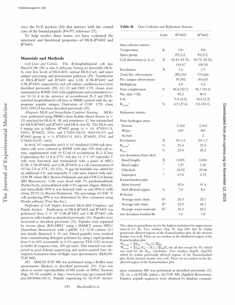
Table II. Data Collection and Refinement Statistics
Units B*4402 B*4403
Data collection statisticsTemperature K 100 100Space group P212121 P212121
Cell dimensions (a, b, c) Å 50.81, 81.78, 110.07
50.72, 82.30,109.55
Resolution Å 1.6 1.7Total No. observations 282,153 176,666No. unique observations 59,302 50,615Multiplicity 4.8 3.5Data completeness % 96.8 (92.7) 96.7 (93.5)No. data �2�1 85.2 80.9I/�1 9.4 (2.0) 18.8 (3.2)Rmerge
a % 4.9 (27.6) 5.6 (54.1)
Refinement statistics
Non–hydrogen atomProtein 3,162 3,162Water 659 481Acetate 2
Resolution Å 50-1.6 50-1.7Rfactor
b % 21.4 22.9Rfree % 23.4 26.2rms deviations from idealBond lengths Å 0.005 0.006Bond angles 1.27 1.26Dihedrals 24.92 25.08Impropers 0.74 0.72
Ramachandran plotMost favored 92.0 91.4And allowed region % 7.4 8.0
B-factorsAverage main chain Å2 20.3 25.1Average side chain Å2 22.9 28.1Average water molecule Å2 36.3 40.5
rms deviation bonded Bs 1.6 1.8
The values in parentheses are for the highest resolution bin (approximateinterval 0.1 Å). Two residues (Asp 29, Arg 239) that lie withingenerously allowed regions of the Ramachandran plot, fit the electrondensity very well. There are no residues in the disallowed region of theRamachandran plot.aRmerge � �Ihkl � <Ihkl>� � IhklbRfactor � Ihkl� �Fo� � �Fc� � � Ihkl�Fo� for all data except for 3%, whichwas used for the Rfree calculation. Two residues (Asp29, Arg239),which lie within generously allowed regions of the Ramachandranplot, fit the electron density very well. There are no residues in the dis-allowed region of the Ramachandran plot.
on Novem
ber 12, 2015jem
.rupress.orgD
ownloaded from
Published August 25, 2003
The
Journ
al o
f Exp
erim
enta
l M
edic
ine
682 Impact of a Naturally Selected Polymorphism on HLA Class I
son of the fragmentation spectra using the MASCOT algorithm(33) followed by manual assignment of expected fragments fromthe highest score sequences (Table I).
Crystallization, Data Collection, and Structure Determination.Recombinant B44 complexes were prepared from Escherichia coli,refolded, and crystallized as described previously (34). The datawere processed and scaled using the HKL package (35) andD*TREK (36), Table II. The HLA-B*4402 structure was solvedfirst using the molecular replacement method, using programsfrom CCP4 suite (37). The progress of refinement was monitoredby the Rfree value (3% of the data) with neither a sigma, nor a lowresolution cut off being applied to the data. The structure was re-fined using rigid-body fitting followed by the simulated-anneal-ing protocol implemented in CNS (version 1.0; reference 38) andinterspersed with rounds of model building using the program“O” (39). Water molecules were included in the model usingstandard criteria. The subsequent HLA-B*4403 structure wassolved using the fully refined HLA-B*4402 structure, minus thebound peptide, water molecules, and site of polymorphism mu-tated to alanine. The refinement protocol implemented was iden-tical to that of the HLA-B*4402 structure. For the HLA-B*4403structure, two acetate ions were clearly visible in the electrondensity map. The quality of the electron density and the stereo-chemistry for the two fully refined, high resolution HLA-B44 al-leles is excellent. The electron density for the bound peptide, aswell as the interacting residues, is unambiguous. A summary ofthe refinement statistics is given in Table II. The coordinates havebeen deposited in the Protein Data Bank (http://www.rcsb.org/pdb), accession codes 1M60 (B*4402) and 1N2R (B*4403).
Online Supplemental Material. The general issue of reproduc-ibility and quantitation of the mass spectra are addressed in Figs.S1–S3. Fig. S1 shows the results of an experiment analyzing frac-
tions from two independent elution experiments; Fig. S2 showshow the different peak heights in the mass spectra were comparedby using natural internal reference standards; and Fig. S3 showsthe results obtained when comparing the same samples but usingdifferent types of mass spectroscopy technology (MALDI-TOFversus ESI-QqTOF). Table S1 provides a detailed list of MHC-peptide contacts for the crystal structures of B*4402 and B*4403.Online supplemental material is available at http://www.jem.org/cgi/content/full/jem.20030066/DC1.
ResultsT Cell Discrimination between HLA-B*4402 and B*4403.
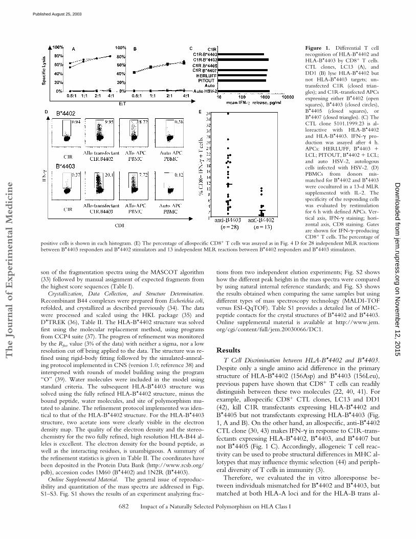
Despite only a single amino acid difference in the primarystructure of HLA-B*4402 (156Asp) and B*4403 (156Leu),previous papers have shown that CD8� T cells can readilydistinguish between these two molecules (22, 40, 41). Forexample, allospecific CD8� CTL clones, LC13 and DD1(42), kill C1R transfectants expressing HLA-B*4402 andB*4405 but not transfectants expressing HLA-B*4403 (Fig.1, A and B). On the other hand, an allospecific, anti-B*4402CTL clone (30, 43) makes IFN-� in response to C1R-trans-fectants expressing HLA-B*4402, B*4403, and B*4407 butnot B*4405 (Fig. 1 C). Accordingly, allogeneic T cell reac-tivity can be used to probe structural differences in MHC al-lotypes that may influence thymic selection (44) and periph-eral diversity of T cells in immunity (3).
Therefore, we evaluated the in vitro alloresponse be-tween individuals mismatched for B*4402 and B*4403, butmatched at both HLA-A loci and for the HLA-B trans al-
Figure 1. Differential T cellrecognition of HLA-B*4402 andHLA-B*4403 by CD8� T cells.CTL clones, LC13 (A), andDD1 (B) lyse HLA-B*4402 butnot HLA-B*4403 targets; un-transfected C1R (closed trian-gles); and C1R-transfected APCsexpressing either B*4402 (opensquares), B*4403 (closed circles),B*4405 (closed squares), orB*4407 (closed triangles). (C) TheCTL clone 5101.1999.23 is al-loreactive with HLA-B*4402and HLA-B*4403. IFN-� pro-duction was assayed after 4 h.APCs: HERLUFF, B*4403 �LCL; PITOUT, B*4402 � LCL;and auto HSV-2, autologouscells infected with HSV-2. (D)PBMCs from donors mis-matched for B*4402 and B*4403were cocultured in a 13-d MLRsupplemented with IL-2. Thespecificity of the responding cellswas evaluated by restimulationfor 6 h with defined APCs. Ver-tical axis, IFN-� staining; hori-zontal axis, CD8 staining. Gatesare shown for IFN-�–producingCD8� T cells. The percentage of
positive cells is shown in each histogram. (E) The percentage of allospecific CD8� T cells was assayed as in Fig. 4 D for 28 independent MLR reactionsbetween B*4403 responders and B*4402 stimulators and 13 independent MLR reactions between B*4402 responders and B*4403 stimulators.
on Novem
ber 12, 2015jem
.rupress.orgD
ownloaded from
Published August 25, 2003
The
Journ
al o
f Exp
erim
enta
l M
edic
ine
683 Macdonald et al.
lele. After reciprocal allogeneic MLRs, CD8� T lympho-cytes were restimulated with defined APCs to determinetheir specificity, and T cells making IFN-� were enumer-ated. Less than 1% of CD8� T cells responded after the al-logeneic MLR when either syngeneic PBMCs or untrans-fected C1R cells were used as APCs (Fig. 1 D, left).However, �20% of CD8� T cells from B*4402� respond-ers were activated by C1R.B*4403 APCs (Fig. 1 D, bot-tom). Similarly, restimulation of B*4403� responders withC1R.B*4402 APCs provoked IFN-� production in �10%of CD8� T cells (Fig. 1 D, top).
The median percentage of alloreactive CD8� T cells inthe B*4402 anti-B*4403 vector was 11.3% (n � 28; inde-pendent donor pairs) compared with a median of 2%B*4403 anti-B*4402 CD8� T cells (n � 13; independentdonor pairs). Hence, there were nearly sixfold more respond-ing T cells identified in MLRs from B*4402 anti-B*4403mismatches than vice versa, indicating an asymmetry in themagnitude of the alloresponse stimulated between thesetwo HLA allotypes in vitro (Fig. 1 E). These findings indi-cate that the single residue that distinguishes B*4402 fromB*4403 has a profound effect on T cell recognition of thesealloantigens, which is likely to result in differential T cellrepertoire selection by these allotypes.
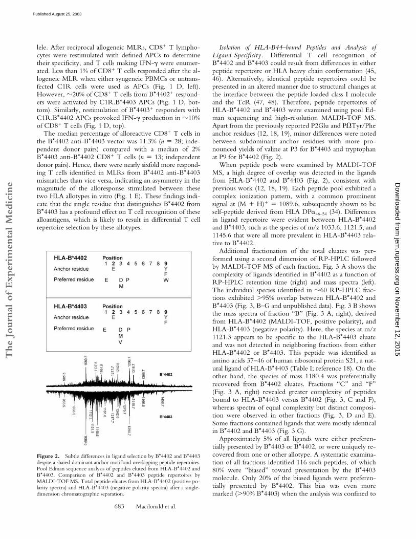
Isolation of HLA-B44–bound Peptides and Analysis ofLigand Specificity. Differential T cell recognition ofB*4402 and B*4403 could result from differences in eitherpeptide repertoire or HLA heavy chain conformation (45,46). Alternatively, identical peptide repertoires could bepresented in an altered manner due to structural changes atthe interface between the peptide loaded class I moleculeand the TcR (47, 48). Therefore, peptide repertoires ofHLA-B*4402 and B*4403 were examined using pool Ed-man sequencing and high-resolution MALDI-TOF MS.Apart from the previously reported P2Glu and P�Tyr/Pheanchor residues (12, 18, 19), minor differences were notedbetween subdominant anchor residues with more pro-nounced yields of valine at P3 for B*4403 and tryptophanat P9 for B*4402 (Fig. 2).
When peptide pools were examined by MALDI-TOFMS, a high degree of overlap was detected in the ligandsfrom HLA-B*4402 and B*4403 (Fig. 2), consistent withprevious work (12, 18, 19). Each peptide pool exhibited acomplex ionization pattern, with a common prominentsignal at (M � H)� � 1089.6, subsequently shown to beself-peptide derived from HLA DP�46–54 (34). Differencesin ligand repertoire were evident between HLA-B*4402and B*4403, such as the species of m/z 1033.6, 1121.5, and1145.6 that were all more prevalent in HLA-B*4403 rela-tive to B*4402.
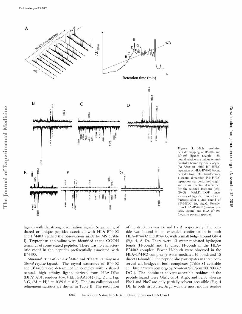
Additional fractionation of the total eluates was per-formed using a second dimension of RP-HPLC followedby MALDI-TOF MS of each fraction. Fig. 3 A shows thecomplexity of ligands identified in B*4402 as a function ofRP-HPLC retention time (right) and mass spectra (left).The individual species identified in �60 RP-HPLC frac-tions exhibited �95% overlap between HLA-B*4402 andB*4403 (Fig. 3, B–G and unpublished data). Fig. 3 B showsthe mass spectra of fraction “B” (Fig. 3 A, right), derivedfrom HLA-B*4402 (MALDI-TOF, positive polarity), andHLA-B*4403 (negative polarity). Here, the species at m/z1121.3 appears to be specific to the HLA-B*4403 eluateand was not detected in neighboring fractions from eitherHLA-B*4402 or B*4403. This peptide was identified asamino acids 37–46 of human ribosomal protein S21, a nat-ural ligand of HLA-B*4403 (Table I; reference 18). On theother hand, the species of mass 1180.4 was preferentiallyrecovered from B*4402 eluates. Fractions “C” and “F”(Fig. 3 A, right) revealed greater complexity of peptidesbound to HLA-B*4403 versus B*4402 (Fig. 3, C and F),whereas spectra of equal complexity but distinct composi-tion were observed in other fractions (Fig. 3, D and E).Some fractions contained ligands that were mostly identicalin B*4402 and B*4403 (Fig. 3 G).
Approximately 5% of all ligands were either preferen-tially presented by B*4403 or B*4402, or were uniquely re-covered from one or other allotype. A systematic examina-tion of all fractions identified 116 such peptides, of which80% were “biased” toward presentation by the B*4403molecule. Only 20% of the biased ligands were preferen-tially presented by B*4402. This bias was even moremarked (�90% B*4403) when the analysis was confined to
Figure 2. Subtle differences in ligand selection by B*4402 and B*4403despite a shared dominant anchor motif and overlapping peptide repertoires.Pool Edman sequence analysis of peptides eluted from HLA-B*4402 andB*4403. Comparison of B*4402 and B*4403 peptide repertoires byMALDI-TOF MS. Total peptide eluates from HLA-B*4402 (positive po-larity spectra) and HLA-B*4403 (negative polarity spectra) after a single-dimension chromatographic separation.
on Novem
ber 12, 2015jem
.rupress.orgD
ownloaded from
Published August 25, 2003
The
Journ
al o
f Exp
erim
enta
l M
edic
ine
684 Impact of a Naturally Selected Polymorphism on HLA Class I
ligands with the strongest ionization signals. Sequencing ofshared or unique peptides associated with HLA-B*4402and B*4403 verified the observations made by MS (TableI). Tryptophan and valine were identified at the COOHterminus of some eluted peptides. There was no character-istic motif in the peptides preferentially associated withB*4403.
Structural Basis of HLA-B*4402 and B*4403 Binding to aShared Peptide Ligand. The crystal structures of B*4402and B*4403 were determined in complex with a sharednatural, high affinity ligand derived from HLA-DP�(DPA*0201, residues 46–54 EEFGRAFSF) (Fig. 2 and Fig.3 G, (M � H)� � 1089.6 � 0.2). The data collection andrefinement statistics are shown in Table II. The resolution
of the structures was 1.6 and 1.7 Å, respectively. The pep-tide was bound in an extended conformation in bothHLA-B*4402 and B*4403, with a small bulge around Gly 4(Fig. 4, A–D). There were 13 water-mediated hydrogenbonds (H-bonds) and 15 direct H-bonds in the HLA–B*4402 complex. Fewer H-bonds were observed in theHLA–B*4403 complex (9 water mediated H-bonds and 15direct H-bonds). The peptide also participates in three con-served salt bridges in both complexes (Table S1 availableat http://www.jem.org/cgi/content/full/jem.20030066/DC1). The dominant solvent-accessible residues of thepeptide ligand were Glu1, Gly4, Arg5, and Ser8, whereasPhe3 and Phe7 are only partially solvent accessible (Fig. 4D). In both structures, Arg5 was the most mobile residue
Figure 3. High resolutionpeptide mapping of B*4402 andB*4403 ligands reveals �5%bound peptides are unique or pref-erentially bound by one allotype.(A) After an initial RP-HPLCseparation of HLA-B*4402 boundpeptides from C1R transfectants,a second dimension RP-HPLCseparation was performed (right)and mass spectra determinedfor the selected fractions (left).(B–G) MALDI-TOF massspectra of ligands from selectedfractions after a 2nd round ofRP-HPLC (A, right). Peptidesfrom HLA-B*4402 (positive po-larity spectra) and HLA-B*4403(negative polarity spectra).
on Novem
ber 12, 2015jem
.rupress.orgD
ownloaded from
Published August 25, 2003
The
Journ
al o
f Exp
erim
enta
l M
edic
ine
685 Macdonald et al.
(B factors 29–35, Å2), reflecting the greater solvent accessi-bility of the polar head group of this residue.
Structural Basis for Selection of Shared Anchor Residues by theB and F pockets. The antigen-binding cleft of class I HLAmolecules consists of a series of pockets designated A–F(27, 49). In different alleles, these pockets vary in theirdepth, electrostatic potential, and hydrophobicity, thus de-termining their ligand specificity. Specificity for P2-Gluoccurs in HLA-B44, HLA-A29, and H-2Kk. However, be-cause the structural basis for this specificity has not yet been
described, these interactions are described in some detailhere and compared with the B pockets of B*2705 (50) andB*3501 (51, 52) that possess alternative specificities (Fig. 5).The B pocket is formed by residues at positions 9, 24, 45,66, 67, and 99 of the class I heavy chain; the P2-Glu inter-acts with all these residues except for Ile66 in our structuresand makes an additional interaction (position 70). InB*4402 and B*4403, the peptide anchor Glu2 forms a saltbridge with Lys45, which is situated at the bottom of the Bpocket and presumably provides substantial energetic con-
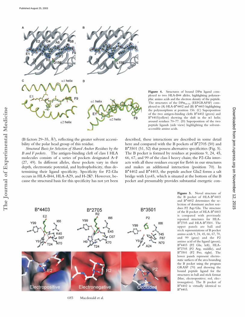
Figure 4. Structures of bound DP� ligand com-plexed to two HLA-B44 alleles, highlighting polymor-phic amino acids and the electron density of the peptide.The structures of the DP�46–54 (EEFGRAFSF) com-plexed to (A) HLA-B*4402 and (B) B*4403 highlightingthe polymorphism at position 156. (C) Superpositionof the two antigen-binding clefts B*4402 (green) andB*4403(yellow) showing the shift in the �1 helixaround residues 70–77. (D) Superposition of the twopeptide ligands (side view) highlighting the solvent-accessible amino acids.
Figure 5. Novel structure ofthe B pocket of HLA-B*4403and B*4402 determines the se-lection of dominant anchor resi-dues P2 Asp/Glu. The structureof the B pocket of HLA-B*4403is compared with previouslyreported structures for HLA-B*2705 and HLA-B*3501. Theupper panels are ball andstick representations of B pocketamino acids 9, 24, 45, 66, 67, 70,and 99 (gray) and the P2amino acid of the ligand (green);B*4403 (P2 Glu, left), HLA-B*2705 (P2 Arg, middle), andB*3501 (P2 Pro, right). Thelower panels represent electro-static surfaces of the area boundingthe B pocket using the programGRASP (70) and showing thebound peptide ligand for thestructures in ball and stick format(blue, electropositive; red, elec-tronegative). The B pocket ofB*4402 is virtually identical toB*4403.
on Novem
ber 12, 2015jem
.rupress.orgD
ownloaded from
Published August 25, 2003
The
Journ
al o
f Exp
erim
enta
l M
edic
ine
686 Impact of a Naturally Selected Polymorphism on HLA Class I
tribution to the specificity of the B pocket (Fig. 5). Fur-thermore, the charged P2-Glu carboxylate forms directH-bonds to Tyr9 and Tyr99. Additionally, there is a bur-ied, well-ordered water molecule at the base of the pocketthat mediates H-bonds to Ser67 and Thr24 in the HLA-B*4402/03 structures. In B*4403, this conserved watermolecule forms a H-bond to Asn70, a residue within the�1-helix that flexes as a result of the polymorphism at posi-tion 156. The long aliphatic moiety of Glu2 packs predom-inantly against the aromatic ring of Tyr7 and is flanked byIle66, although it doesn’t interact with the latter residue.
Some of the structural framework of the B pocket isconserved between B*4403, B*2705, and B*3501 (Fig. 5).Thus, the B pockets of B*2705 (50) and B*4402/B*4403are deep to accommodate the long aliphatic side chains ofarginine or glutamate. However, in contrast to B*4402 andB*4403, the deep electronegative B pocket of B*2705 (50)accommodates a positively charged canonical P2-Arg resi-due involved in charge complementation with the glutamicacid residue at position 45 of the heavy chain. In the Bpocket of HLA-B*3501, the projecting Phe67 creates a hy-drophobic, sterically occluded pocket that preferentially ac-commodates proline residues at P2 of bound peptideligands (51, 52).
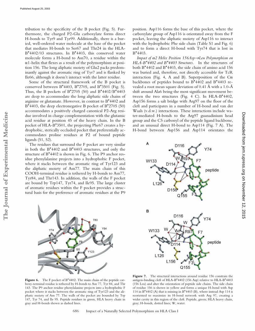
The residues that surround the F pocket are very similarin both the B*4402 and B*4403 structures, and only thestructure of B*4402 is shown in Fig. 6. The P9 anchor res-idue phenylalanine projects into a hydrophobic F pocket,where it stacks between the aromatic ring of Tyr123 andthe aliphatic moiety of Asn77. The main chain of thisCOOH-terminal residue is tethered by H-bonds to Asn77,Tyr84, and Thr143. In addition, the walls of the F pocketare bound by Trp147, Tyr74, and Ile95. The large clusterof aromatic residues within the F pocket provides a struc-tural basis for the preference of aromatic residues at the P9
position. Asp116 forms the base of this pocket, where thecarboxylate group of Asp116 is orientated away from the Fpocket, leaving the aliphatic moiety of Asp116 to interactwith the hydrophobic Phe side chain (Table S1 and Fig. 6)and to form a direct H-bond with Tyr74 that is lost inB*4403.
Impact of �2 Helix Position 156Asp→Leu Polymorphism onHLA-B*4402 and B*4403 Structures. In the structures ofboth B*4402 and B*4403, the side chain of amino acid 156was buried and, therefore, not directly accessible for TcRinteraction (Fig. 4, A and B). Superposition of the C�backbones of peptides bound to B*4402 and B*4403 re-vealed a root mean square deviation of 0.41 Å with a 1.0-Åshift around Ala6 being the most significant movement be-tween the two structures (Fig. 4 C). In HLA-B*4402,Asp156 forms a salt bridge with Arg97 on the floor of thecleft and participates in a number of H-bond and van derWaals (v.d.w.) interactions. These interactions include wa-ter-mediated H-bonds to the Arg97 guanadinium headgroup and the C5 carbonyl of the peptide ligand backbone,and an unusual direct H-bond to Asp114 (Fig. 7 A). TheH-bond between Asp156 and Asp114 orientates the
Figure 6. The F pocket of B*4402. The main chain of the peptide car-boxy-terminal residue is tethered by H-bonds to Asn 77, Tyr 84, and Thr143. The P9 anchor residue phenylalanine projects into a hydrophobic Fpocket where it stacks between the aromatic ring of Tyr123 and the ali-phatic moiety of Asn 77. The walls of the pocket are bounded by Trp147, Tyr 74, and Ile 95. Peptide residues in green, HLA heavy chain ingray and H-bonds shown as dashed lines.
Figure 7. The structural interactions around residue 156 constrain theantigen-binding cleft of HLA-B*4402 (156 Asp) relative to HLA-B*4403(156 Leu) and alter the orientation of peptide side chains. The side chainof residue 156 is shown in yellow and forms a unique H-bond with Asp114 in B*4402 (A) that is missing in B*4403 (B), where instead Asp 114 isreoriented to maximize its H-bond network with Arg 97, creating awider cavity in this region of the cleft. Peptide, green; HLA heavy chain,gray; H-bonds, dotted lines; W, water.
on Novem
ber 12, 2015jem
.rupress.orgD
ownloaded from
Published August 25, 2003
The
Journ
al o
f Exp
erim
enta
l M
edic
ine
687 Macdonald et al.
Asp114 side chain, allowing a salt bridge to the guanadin-ium head group of Arg97 as well as a water-mediatedH-bond to Asp116. The pairing of Asp156 and Asp114 hasnot been seen in any other crystal structures of HLA class Imolecules, and is presumably stabilized by salt bridgingbetween both these residues and Arg97, allowing anO–H·····H interaction between carboxylates. However,the potential for H-bond formation between Asp156 andAsp114 exists in several other HLA-B molecules, includingother members of the HLA-B44 family. In additionAsp156 forms a v.d.w. interaction with Trp133 andLeu126, as well as with Phe3 of the bound peptide ligand.Here, the peptide Phe3–Asp156 interaction is facilitated bythe side-on orientation adopted by the Phe3 side chain.
In contrast to B*4402, the nonpolar nature of Leu156 inB*4403 results in loss of a water-mediated H-bond to thepeptide backbone, giving rise to a significant repositioningof the peptide between the B*4403 �-helices. On the otherhand, the side chain of Leu156 in HLA-B*4403 is rotatedby �90 (Fig. 7 B), enhancing v.d.w. contacts within thehydrophobic pocket formed by Leu126, Trp133, andVal152. In addition, to avoid unfavorable interactions withLeu156, the orientation of the Asp114 side chain inB*4403 changes by �90, simultaneously maximizing in-teractions with Arg97 and maintaining a water-mediatedH-bond to the C5 carbonyl of the peptide backbone.Hence, despite the loss of some H-bonds in B*4403, in-cluding the Asp116–Tyr74 interaction, the stability of theEEFGRAFSF peptide does not seem to be significantly im-paired in the B*4403 complex consistent with experimentsthat show little difference in the thermostability of B*4402versus B*4403 in complex with the EEFGRAFSF peptide(unpublished data).
However, the altered interactions between residue 156and neighboring heavy chain residues, and the peptideligand, significantly change the dimensions of the Ag-bind-ing cleft in B*4402 and B*4403 (Fig. 4, C and D). A “rigidbody shift” in the �1 helix, particularly around positions70–77, results in a 0.6-Å displacement of the �-helices be-tween the antigen-binding cleft of HLA-B*4402 (green)versus B*4403 (Fig. 4 C, yellow). Amino acid residues 70–77 lie adjacent to the central region of the peptide that hasundergone a shift of 1.0 Å in the C� backbone aroundAla6. This conformational adjustment is directly associatedwith the loss of a direct H-bond between Asp116 andTyr74 of the B*4403 heavy chain (Fig. 7, A and B). Thus,in the HLA-B*4402 structure the direct H-bond betweenAsp116 and Tyr74 results in a narrower antigen-bindingcleft (Figs. 6 and 7 A), whereas in the B*4403 structure lossof this interaction is associated with distortion and relative“breathing” of the binding cleft (Fig. 7 B). Instead of mak-ing a direct H-bond with Asp116, Tyr74 participates in awater-mediated H-bond with this residue in HLA-B*4403;this is attributable to changes in the peptide orientation, re-sulting from the substitution at amino acid 156. Conse-quently, the slightly larger cavity of the HLA-B*4403 cleftis occupied by an acetate ion in this structure, reflectingpotentially greater accommodation of peptide side chains
by the B*4403 allotype compared with B*4402 (Fig. 7 B).Any thermodynamic penalty for “breathing” of the B*4403cleft may not be apparent with some ligands because ofcompensatory peptide interactions arising from the alteredcleft structure, as appears to be the case for B*4403 bindingof the EEFGRAFSF peptide.
DiscussionThe impact of point mutations and other micropolymor-
phisms on the structure and function of class I molecules hasbeen studied extensively by in vitro mutagenesis (53, 54)and through exploitation of the experimentally derived Kb
mutant system (3, 44, 47, 48). These systems have beenenormously important for unraveling key details of struc-ture–function relationships in MHC molecules. However,it is equally important to study the structural and functionalconsequences of naturally selected microvariation in HLAallotypes found in outbred human populations. To this end,we have determined the structural and functional propertiesof two closely related, naturally occurring human class I al-leles, HLA-B*4402 and B*4403. These were studied be-cause of their evolutionary success as common alleles in di-verse human populations, notwithstanding their markedstructural similarity and shared peptide binding motif.
Structural differences in B*4402 and B*4403 are distin-guishable by many T cells and these allotypes provokestrong, mutual allogeneic T cell responses in vitro. Indeed,up to 25% of CD8� T cells are specifically alloreactive inextended MLRs between B*4402/B*4403 mismatched in-dividuals. These observations indicate that B*4402 andB*4403 molecules select both shared and unique T cellsduring thymic development.
The structures of B*4402 and B*4403 show that aminoacid 156 is neither solvent accessible, nor directly involvedin forming major specificity pockets. Nonetheless, the156Asp→Leu polymorphism is a nonconservative substitu-tion and markedly alters the characteristics of the P3 speci-ficity (E pocket) as well as the secondary effects of the anti-gen-binding cleft. Hence, the 156Asp→Leu substitutionalters the repertoire of peptide ligands bound by both mol-ecules and directly alters the conformation of the antigen-binding cleft. Subtle differences were observed in preferredresidues at minor anchor sites in B*4403 versus HLA-B*4402, however, these differences may be more apparentthan real. For instance, a Trp can be modeled at P� inB*4402 and B*4403 (unpublished data) and a tryptophan isfound at P� in the EEVDLSKDIQHW peptide elutedfrom both B*4402 and 4403 (Table I). Thus, any prefer-ence for Trp at P� in B*4402 is probably quantitative (sta-tistical) or context-dependent.
The peptide repertoire of HLA-B*4402 and B*4403showed �95% overlap but most of the unique peptideswere associated with B*4403. These differences in peptideselection can be partly understood by an examination of thetwo structures. Comparison of the antigen-binding cleft ofHLA-B*4402 and B*4403 revealed a relative narrowing ofthe B*4402 cleft between the �-helices, particularly around
on Novem
ber 12, 2015jem
.rupress.orgD
ownloaded from
Published August 25, 2003
The
Journ
al o
f Exp
erim
enta
l M
edic
ine
688 Impact of a Naturally Selected Polymorphism on HLA Class I
amino acids 70–77. This distortion of the cleft shape appearsto be the result of both indirect and ligand-mediated con-formational changes associated with the 156Asp→Leu poly-morphism. The Asp114–Asp156 interaction in B*4402 isunusual, and being constrained, appears to limit the type ofresidue that can sit in the P3, and/or the P7 pocket. A largenetwork of polar interactions at position 156 dissipates thisunfavorable interaction and draws the helix 1 closer to thepeptide, thereby sterically constraining the makeup of po-tential bound peptides in B*4402. Thus, the relatively re-stricted range of ligands recovered from B*4402 comparedwith B*4403 probably results from this structural constraintthat limits the size of side chains that can be accommodatedin the narrowed region. In contrast, the 156Asp→Leu sub-stitution in B*4403 results in a loss of this bonding network,allowing the helices to breathe in this region. This breathingof the cleft is further reflected by the presence of an acetatemoiety in the extra space around amino acids 70–77 in theB*4403/EEFGRAFSF complex. Breathing, or opening ofthe binding cleft, is also observed in the structures of HLA-DQ8 (55) and I-Ag7 (56) when these are compared withother class II molecules.
The orientation of side chains in the EEFGRAFSF pep-tide bound by HLA-B*4402 and B*4403 was also alteredby the 156Asp→Leu polymorphism. Structural adjustmentswithin identical ligands bound by related MHC allotypescan be sufficient to induce T cell alloreactivity (24). How-ever, altered heavy chain conformation (57) and binding ofunique ligands (58) can also contribute to T cell alloreactiv-ity. Thus, T cell discrimination of B*4402 and B*4403, ei-ther during positive selection of the T cell repertoire, orduring B*4402 anti-B*4403 alloreactions, could involveany one, or all of the modes of recognition discussed here.
Ironically, the structural similarity between B*4402 andB*4403 is likely to engender strong reciprocal T cell al-loreactivity due to thymic selection of a large repertoire ofT cells with intrinsic “B44”-reactivity biased toward theseallotypes. Hence, the number of T cells that can distin-guish subtle structural differences in B*4402 and B*4403 islikely to be very significant. Thus, the magnitude of the al-loresponse is paradoxically as great, or even greater, thanobserved across more disparate HLA allelic differences(19–21)
The subtle differences in peptide repertoire betweenB*4402 and B*4403 contrast with the differences observedbetween many HLA class I allotypes, especially where sub-stitutions occur in the dominant anchor residues locatedwithin the B and F pockets (59–61). For instance, HLA-B*2705 and B*2709 differ at position 116 (Asp→His) at thebase of the F pocket (62, 63), resulting in peptide reper-toires that overlap by �79 and �88%, respectively (64),and producing mutual alloreactivity (62, 65).
However, natural class I polymorphism outside the P2and P� peptide anchor sites appears to modulate peptiderepertoire in a much more subtle, often imperceptiblemanner, as reported for subtypes of HLA-B7, B17 and B15(66), B5 and B22 (67), and B27 (65–69). Thus, our find-ings in relation to B*4402 and B*4403 are likely to be of
general relevance in understanding the impact of microva-riation in HLA class I genes. For instance, among allknown HLA class I molecules, there are five differentamino acids observed at position 156 (Asp, Leu, Arg, Gln,and Trp). Dimorphism at this residue is found in virtuallyall the common class I subtypes, suggesting that this posi-tion exerts a strong influence on the structure and functionof other class I allotypes as well as B44 (25).
The functional basis underlying the retention of theB*4402 and B*4403 alleles in diverse human populationspresumably rests with their independent selective advantagein immunity. Selection could be related to differentialligand selection by B*4402 and B*4403 (1); or, alterna-tively, may reflect differences in either thymic positive se-lection of T cells (44) or clonal diversification of the pe-ripheral immune response (3). In addition, selection couldbe influenced by other elements of conserved HLA-B44haplotypes, although these are not well-preserved in allpopulations, making this explanation less likely.
We conclude that the relatively small changes in peptiderepertoire and MHC–peptide conformation between nat-urally occurring HLA polymorphisms can be sufficient tomaintain selection of these alleles in diverse human popu-lations. These micropolymorphisms are potentially capableof enhancing protective immunity by expanding ligand se-lection but are also likely to diversify T cell repertoire dur-ing thymic positive selection and at the time of the im-mune response.
We would like to thank F. Carbone for critical reading of themanuscript and W. Heath for helpful suggestions. We thank thestaff at BioCARS and the Australian Synchrotron Research Pro-gram for assistance.
J. Rossjohn is supported by a Wellcome Trust Senior ResearchFellowship in Biomedical Science in Australia; A.W. Purcell is aC.R. Roper Fellow of the Faculty of Medicine, Dentistry, andHealth Science at The University of Melbourne; A.G. Brooks issupported by a R.D. Wright Fellowship. This work was supportedby the National Health Council and the Medical Research Coun-cil, the Australian Kidney Foundation, the Roche Organ Trans-plantation Research Foundation, and the Juvenile Diabetes Re-search Foundation.
Submitted: 15 January 2003Revised: 10 June 2003Accepted: 10 June 2003
References1. Hill, A.V., C.E. Allsopp, D. Kwiatkowski, N.M. Anstey, P.
Twumasi, P.A. Rowe, S. Bennett, D. Brewster, A.J. Mc-Michael, and B.M. Greenwood. 1991. Common west Afri-can HLA antigens are associated with protection from severemalaria. Nature. 352:595–600.
2. Parham, P., and T. Ohta. 1996. Population biology of anti-gen presentation by MHC class I molecules. Science. 272:67–74.
3. Messaoudi, I., J.A. Patino, R. Dyall, J. LeMaoult, and J. Ni-kolich-Zugich. 2002. Direct link between MHC polymor-phism, T cell avidity, and diversity in immune defense. Sci-ence. 298:1797–1800.
on Novem
ber 12, 2015jem
.rupress.orgD
ownloaded from
Published August 25, 2003
The
Journ
al o
f Exp
erim
enta
l M
edic
ine
689 Macdonald et al.
4. Marsh, S.G., P. Parham, and L.D. Barber. 2000. The HLAFacts Book. Academic Press, London. 398 pp.
5. Sette, A., and J. Sidney. 1999. Nine major HLA class I super-types account for the vast preponderance of HLA-A and -Bpolymorphism. Immunogenetics. 50:201–212.
6. Raffoux, C., R.C. Williams, C. Gorodezky, E.D. Albert, E.Gyodi, M.G. Hammond, Z. Layrisse, M. Mariani, G. de laRosa, J.M. Tiercy, et al. 1997. AHS#7: HLA-B12, B13,B21, B37, B40, B41, B81, B4005. In Genetic Diversity ofHLA Functional and Medical Implication. D. Charron, edi-tor. EDK Medical and Scientific International Publisher,Paris. 69–72.
7. Hiroishi, K., H. Kita, M. Kojima, H. Okamoto, T.Moriyama, T. Kaneko, T. Ishikawa, S. Ohnishi, T. Aikawa,N. Tanaka, et al. 1997. Cytotoxic T lymphocyte responseand viral load in hepatitis C virus infection. Hepatology. 25:705–712.
8. Khanna, R., S.R. Burrows, A. Neisig, J. Neefjes, D.J. Moss,and S.L. Silins. 1997. Hierarchy of Epstein-Barr virus-specificcytotoxic T-cell responses in individuals carrying differentsubtypes of an HLA allele: implications for epitope-based an-tiviral vaccines. J. Virol. 71:7429–7435.
9. Kita, H., T. Moriyama, T. Kaneko, I. Harase, M. Nomura,H. Miura, I. Nakamura, Y. Yazaki, and M. Imawari. 1993.HLA B44-restricted cytotoxic T lymphocytes recognizing anepitope on hepatitis C virus nucleocapsid protein. Hepatology.18:1039–1044.
10. Kita, H., T. Moriyama, T. Kaneko, H. Okamoto, K. Hi-roishi, S. Ohnishi, and M. Imawari. 1995. HLA B44-restricted cytotoxic T lymphocyte responses to the peptidesof HCV nucleoprotein residues 81-100 in patients withchronic hepatitis C. J. Gastroenterol. 30:809–812.
11. Brichard, V.G., J. Herman, A. Vanpel, C. Wildmann, B.Gaugler, T. Wolfel, T. Boon, and B. Lethe. 1996. A tyrosi-nase nonapeptide presented by HLA-B44 is recognized on ahuman melanoma by autologous cytolytic T lymphocytes.Eur. J. Immunol. 26:224–230.
12. Fleischhauer, K., D. Fruci, P. Van Endert, J. Herman, S.Tanzarella, H.J. Wallny, P. Coulie, C. Bordignon, and C.Traversari. 1996. Characterization of antigenic peptides pre-sented by HLA-B44 molecules on tumor cells expressing thegene MAGE-3. Int. J. Cancer. 68:622–628.
13. Herman, J., P. van der Bruggen, I.F. Luescher, S. Mandruz-zato, P. Romero, J. Thonnard, K. Fleischhauer, T. Boon,and P.G. Coulie. 1996. A peptide encoded by the humanMAGE3 gene and presented by HLA-B44 induces cytolyticT lymphocytes that recognize tumor cells expressing MAGE3.Immunogenetics. 43:377–383.
14. Dolstra, H., H. Fredrix, F. Preijers, E. Goulmy, C.G. Figdor,T.M. de Witte, and E. van de Wiel-van Kemenade. 1997.Recognition of a B cell leukemia-associated minor histocom-patibility antigen by CTL. J. Immunol. 158:560–565.
15. Tiercy, J.M., M. Jeannet, and E. Roosnek. 1995. HLA-B44-restricted minor antigen. Transplantation. 60:113–114.
16. Kruskall, M.S., E.E. Eynon, Z. Awdeh, C.A. Alper, and E.J.Yunis. 1987. Identification of HLA-B44 subtypes associatedwith extended MHC haplotypes. Immunogenetics. 26:216–219.
17. Mattiuz, P.L., E. Di Paolo, V. Fossombroni, A. Menicucci, F.Pradella, B. Porfirio, and G. Rombola. 1997. HLA-B44 sub-types and the chance of finding HLA compatible donor/re-cipient pairs for bone marrow transplantation: a haplotypestudy of 303 Italian families. Tissue Antigens. 50:602–609.
18. DiBrino, M., K.C. Parker, D.H. Margulies, J. Shiloach, R.V.Turner, W.E. Biddison, and J.E. Coligan. 1995. Identifica-tion of the peptide binding motif for HLA-B44, one of themost common HLA-B alleles in the Caucasian population.Biochemistry. 34:10130–10138.
19. Fleischhauer, K., D. Avila, F. Vilbois, C. Traversari, C. Bor-dignon, and H.J. Wallny. 1994. Characterization of naturalpeptide ligands for HLA-B*4402 and -B*4403: implicationsfor peptide involvement in allorecognition of a single aminoacid change in the HLA-B44 heavy chain. Tissue Antigens.44:311–317.
20. Fleischhauer, K., N.A. Kernan, R.J. O’Reilly, B. Dupont,and S.Y. Yang. 1990. Bone marrow-allograft rejection by Tlymphocytes recognizing a single amino acid difference inHLA-B44. N. Engl. J. Med. 323:1818–1822.
21. Keever, C.A., N. Leong, I. Cunningham, E.A. Copelan,B.R. Avalos, J. Klein, N. Kapoor, P.W. Adams, C.G. Orosz,P.J. Tutschka, et al. 1994. HLA-B44-directed cytotoxic Tcells associated with acute graft-versus-host disease followingunrelated bone marrow transplantation. Bone Marrow Trans-plant. 14:137–145.
22. Burrows, S.R., R. Khanna, J.M. Burrows, and D.J. Moss.1994. An alloresponse in humans is dominated by cytotoxicT lymphocytes (CTL) cross-reactive with a single Epstein-Barr virus CTL epitope: implications for graft-versus-hostdisease. J. Exp. Med. 179:1155–1161.
23. Burrows, S.R., S.L. Silins, S.M. Cross, C.A. Peh, M.Rischmueller, J.M. Burrows, S.L. Elliott, and J. McCluskey.1997. Human leukocyte antigen phenotype imposes complexconstraints on the antigen-specific cytotoxic T lymphocyterepertoire. Eur. J. Immunol. 27:178–182.
24. Herman, J., V. Jongeneel, D. Kuznetsov, and P.G. Coulie.1999. Differences in the recognition by CTL of peptides pre-sented by the HLA-B*4402 and the HLA-B*4403 moleculeswhich differ by a single amino acid. Tissue Antigens. 53:111–121.
25. Hogan, K.T., C. Clayberger, E.J. Bernhard, S.F. Walk, J.P.Ridge, P. Parham, A.M. Krensky, and V.H. Engelhard.1988. Identification by site-directed mutagenesis of aminoacid residues contributing to serologic and CTL-definedepitope differences between HLA-A2.1 and HLA-A2.3. J.Immunol. 141:2519–2525.
26. Tussey, L.G., M. Matsui, S. Rowland-Jones, R. Warburton,J.A. Frelinger, and A. McMichael. 1994. Analysis of mutantHLA-A2 molecules. Differential effects on peptide bindingand CTL recognition. J. Immunol. 152:1213–1221.
27. Saper, M.A., P.J. Bjorkman, and D.C. Wiley. 1991. Refinedstructure of the human histocompatibility antigen HLA-A2 at2.6 A resolution. J. Mol. Biol. 219:277–319.
28. Zemmour, J., A.M. Little, D.J. Schendel, and P. Parham.1992. The HLA-A,B “negative” mutant cell line C1R ex-presses a novel HLA-B35 allele, which also has a point muta-tion in the translation initiation codon. J. Immunol. 148:1941–1948.
29. Alexander, J., J.A. Payne, R. Murray, J.A. Frelinger, and P.Cresswell. 1989. Differential transport requirements of HLAand H-2 class I glycoproteins. Immunogenetics. 29:380–388.
30. Koelle, D.M., H.B. Chen, M.A. Gavin, A. Wald, W.W.Kwok, and L. Corey. 2001. CD8 CTL from genital herpessimplex lesions: recognition of viral tegument and immediateearly proteins and lysis of infected cutaneous cells. J. Immunol.166:4049–4058.
31. Purcell, A.W., J.J. Gorman, M. Garcia-Peydro, A. Paradela,
on Novem
ber 12, 2015jem
.rupress.orgD
ownloaded from
Published August 25, 2003
The
Journ
al o
f Exp
erim
enta
l M
edic
ine
690 Impact of a Naturally Selected Polymorphism on HLA Class I
S.R. Burrows, G.H. Talbo, N. Laham, C.A. Peh, E.C. Rey-nolds, J.A. Lopez De Castro, and J. McCluskey. 2001. Quan-titative and qualitative influences of tapasin on the class I pep-tide repertoire. J. Immunol. 166:1016–1027.
32. Purcell, A.W., and J.J. Gorman. 2001. The use of post sourcedecay in matrix assisted laser desorption-ionisation mass spec-trometry to delineate T cell determinants. J. Immunol. Meth-ods. 249:17–31.
33. Perkins, D.N., D.J. Pappin, D.M. Creasy, and J.S. Cottrell.1999. Probability-based protein identification by searchingsequence databases using mass spectrometry data. Electrophore-sis. 20:3551–3567.
34. Macdonald, W., D.S. Williams, C.S. Clements, J.J. Gorman,L. Kjer-Nielsen, A.G. Brooks, J. McCluskey, J. Rossjohn,and A.W. Purcell. 2002. Identification of a dominant self-ligand bound to three HLA B44 alleles and the preliminarycrystallographic analysis of recombinant forms of each com-plex. FEBS Letters. 527:27–32.
35. Ottwinowski, Z. 1993. Oscillation data reduction program.In Data Collection and Processing, Proceedings of the CCP4Study Weekend, 29–30, January, 1993. L. Sawyer, N. Issacs,and S.W. Bailey, editors. SERC Daresbury Laboratory, U.K.,Warrington, U.K. 56–62.
36. Pflugrath, J.W. 1999. The finer things in X-ray diffractiondata collection. Acta Crystallogr. D Biol. Crystallogr. 55:1718–1725.
37. CCP4. 1994. CCP4. The CCP4 suite: programs for proteincrystallography. Acta Crystallogr. D50:750–763.
38. Brunger, A.T., P.D. Adams, G.M. Clore, W.L. DeLano, P.Gros, R.W. Grosse-Kunstleve, J.S. Jiang, J. Kuszewski, M.Nilges, N.S. Pannu, et al. 1998. Crystallography & NMRsystem: A new software suite for macromolecular structuredetermination. Acta Crystallogr. D Biol. Crystallogr. 54:905–921.
39. Jones, T.A., J.-Y. Zou, S.W. Cowan, and M. Kjeldgaard.1991. Improved methods for building models in electrondensity maps and the location of errors in these models. ActaCrystallogr. 47:110–119.
40. Burrows, S.R., S.L. Silins, D.J. Moss, R. Khanna, I.S. Misko,and V.P. Argaet. 1995. T cell receptor repertoire for a viralepitope in humans is diversified by tolerance to a backgroundmajor histocompatibility complex antigen. J. Exp. Med. 182:1703–1715.
41. Rufer, N., B.S. Breur-Vriesendorp, J.M. Tiercy, A.S.Slavcev, N.M. Lardy, P. Francis, R. Kressig, D.E. Speiser, C.Helg, B. Chapuis, et al. 1993. High-resolution histocompati-bility testing of a group of sixteen B44-positive, ABDR sero-logically matched unrelated donor-recipient pairs. Analysis ofserologically undisclosed incompatibilities by cellular tech-niques, isoelectrofocusing, and HLA oligotyping. Hum. Im-munol. 38:235–239.
42. Argaet, V.P., C.W. Schmidt, S.R. Burrows, S.L. Silins, M.G.Kurilla, D.L. Doolan, A. Suhrbier, D.J. Moss, E. Kieff, T.B.Suclley, et al. 1994. Dominant selection of an invariant T cellantigen receptor in response to persistent infection by Ep-stein-Barr virus. J. Exp. Med. 180:2335–2340.
43. Koelle, D.M., H.B. Chen, C.M. McClurkan, and E.W. Pe-tersdorf. 2002. Herpes simplex virus type 2-specific CD8 cy-totoxic T lymphocyte cross-reactivity against prevalent HLAclass I alleles. Blood. 99:3844–3847.
44. Dyall, R., I. Messaoudi, S. Janetzki, and J. Nikolic-Zugic.2000. MHC polymorphism can enrich the T cell repertoireof the species by shifts in intrathymic selection. J. Immunol.
164:1695–1698.45. Eckels, D.D. 1990. Alloreactivity: allogeneic presentation of
endogenous peptide or direct recognition of MHC polymor-phism? A review. Tissue Antigens. 35:49–55.
46. Rotzschke, O., K. Falk, S. Faath, and H.G. Rammensee.1991. On the nature of peptides involved in T cell alloreac-tivity. J. Exp. Med. 174:1059–1071.
47. Luz, J.G., M. Huang, K.C. Garcia, M.G. Rudolph, V. Apos-tolopoulos, L. Teyton, and I.A. Wilson. 2002. Structuralcomparison of allogeneic and syngeneic T cell receptor-pep-tide-major histocompatibility complex complexes: a buriedalloreactive mutation subtly alters peptide presentation sub-stantially increasing V� interactions. J. Exp. Med. 195:1175–1186.
48. Rudolph, M.G., J.A. Speir, A. Brunmark, N. Mattsson,M.R. Jackson, P.A. Peterson, L. Teyton, and I.A. Wilson.2001. The crystal structures of K(bm1) and K(bm8) revealthat subtle changes in the peptide environment impact ther-mostability and alloreactivity. Immunity. 14:231–242.
49. Garrett, T.P., M.A. Saper, P.J. Bjorkman, J.L. Strominger,and D.C. Wiley. 1989. Specificity pockets for the side chainsof peptide antigens in HLA-Aw68. Nature. 342:692–696.
50. Madden, D.R., J.C. Gorga, J.L. Strominger, and D.C. Wiley.1991. The structure of HLA-B27 reveals nonamer self-pep-tides bound in an extended conformation. Nature. 353:321–325.
51. Menssen, R., P. Orth, A. Ziegler, and W. Saenger. 1999.Decamer-like conformation of a nona-peptide bound toHLA-B*3501 due to non-standard positioning of the C ter-minus. J. Mol. Biol. 285:645–653.
52. Smith, K.J., S.W. Reid, K. Harlos, A.J. McMichael, D.I. Stu-art, J.I. Bell, and E.Y. Jones. 1996. Bound water structureand polymorphic amino acids act together to allow the bind-ing of different peptides to MHC class I HLA-B53. Immunity.4:215–228.
53. Murray, R., C.A. Hutchison III, and J.A. Frelinger. 1988.Saturation mutagenesis of a major histocompatibility complexprotein domain: identification of a single conserved aminoacid important for allorecognition. Proc. Natl. Acad. Sci. USA.85:3535–3539.
54. Nathenson, S.G., J. Geliebter, G.M. Pfaffenbach, and R.A.Zeff. 1986. Murine major histocompatibility complex class-Imutants: molecular analysis and structure-function implica-tions. Annu. Rev. Immunol. 4:471–502.
55. Latek, R.R., A. Suri, S.J. Petzold, C.A. Nelson, O. Kana-gawa, E.R. Unanue, and D.H. Fremont. 2000. Structural ba-sis of peptide binding and presentation by the type I diabetes-associated MHC class II molecule of NOD mice. Immunity.12:699–710.
56. Lee, K.H., K.W. Wucherpfennig, and D.C. Wiley. 2001.Structure of a human insulin peptide-HLA-DQ8 complexand susceptibility to type 1 diabetes. Nat. Immunol. 2:501–507.
57. Ajitkumar, P., S.S. Geier, K.V. Kesari, F. Borriello, M. Naka-gawa, J.A. Bluestone, M.A. Saper, D.C. Wiley, and S.G. Na-thenson. 1988. Evidence that multiple residues on both thealpha-helices of the class I MHC molecule are simultaneouslyrecognized by the T cell receptor. Cell. 54:47–56.
58. Heath, W.R., K.P. Kane, M.F. Mescher, and L.A. Sherman.1991. Alloreactive T cells discriminate among a diverse set ofendogenous peptides. Proc. Natl. Acad. Sci. USA. 88:5101–5105.
59. Rojo, S., F. Garcia, J.A. Villadangos, and J.A. Lopez de Cas-
on Novem
ber 12, 2015jem
.rupress.orgD
ownloaded from
Published August 25, 2003
The
Journ
al o
f Exp
erim
enta
l M
edic
ine
691 Macdonald et al.
tro. 1993. Changes in the repertoire of peptides bound toHLA-B27 subtypes and to site-specific mutants inside andoutside pocket B. J. Exp. Med. 177:613–620.
60. Barouch, D., T. Friede, S. Stevanovic, L. Tussey, K. Smith,S. Rowland-Jones, V. Braud, A. McMichael, and H.G.Rammensee. 1995. HLA-A2 subtypes are functionally dis-tinct in peptide binding and presentation. J. Exp. Med. 182:1847–1856.
61. Prilliman, K.R., K.W. Jackson, M. Lindsey, J. Wang, D.Crawford, and W.H. Hildebrand. 1999. HLA-B15 peptideligands are preferentially anchored at their C termini. J. Im-munol. 162:7277–7284.
62. Ramos, M., and J.A. Lopez de Castro. 2002. HLA-B27 andthe pathogenesis of spondyloarthritis. Tissue Antigens. 60:191–205.
63. Hulsmeyer, M., R.C. Hillig, A. Volz, M. Ruhl, W. Schro-der, W. Saenger, A. Ziegler, and B. Uchanska-Ziegler. 2002.HLA-B27 subtypes differentially associated with disease ex-hibit subtle structural alterations. J. Biol. Chem. 277:47844–47853.
64. Ramos, M., A. Paradela, M. Vazquez, A. Marina, J. Vazquez,and J.A. Lopez de Castro. 2002. Differential association ofHLA-B*2705 and B*2709 to ankylosing spondylitis correlateswith limited peptide subsets but not with altered cell surfacestability. J. Biol. Chem. 277:28749–28756.
65. Garcia-Peydro, M., M. Marti, and J. Lopez de Castro. 1999.High T cell epitope sharing between two HLA-B27 subtypes(B*2705 and B*2709) differentially associated to ankylosingspondylitis. J. Immunol. 163:2299–2305.
66. Barber, L.D., L. Percival, K.L. Arnett, J.E. Gumperz, L.Chen, and P. Parham. 1997. Polymorphism in the alpha 1helix of the HLA-B heavy chain can have an overriding in-fluence on peptide-binding specificity. J. Immunol. 158:1660–1669.
67. Barber, L.D., B. Gillece-Castro, L. Percival, X. Li, C. Clay-berger, and P. Parham. 1995. Overlap in the repertoires ofpeptides bound in vivo by a group of related class I HLA-Ballotypes. Curr. Biol. 5:179–190.
68. Lopez, D., S. Rojo, V. Calvo, and J.A. Lopez de Castro.1992. Peptide-presenting similarities among functionally dis-tant HLA-B27 subtypes revealed by alloreactive T lympho-cytes of unusual specificity. J. Immunol. 148:996–1002.
69. Tanigaki, N., D. Fruci, E. Vigneti, G. Starace, P. Rovero, M.Londei, R.H. Butler, and R. Tosi. 1994. The peptide bind-ing specificity of HLA-B27 subtypes. Immunogenetics. 40:192–198.
70. Taylor, N.R., and R. Smith. 1996. The World Wide Web asa graphical user interface to program macros for moleculargraphics, molecular modeling, and structure-based drug de-sign. J. Mol. Graph. 14:291–296, 280–282.
on Novem
ber 12, 2015jem
.rupress.orgD
ownloaded from
Published August 25, 2003