Elevated ATG5 expression in autoimmune demyelination and multiple sclerosis
Upload
independentCategory
view
0download
0
Defective positive selection results in T cell lymphopenia andincreased autoimmune diabetes in ADAP deficient BDC2.5-Bl/6mice
Liangxing Zou*, Felipe Mendez*,=, Natalia Martin-Orozco@, and Erik J. Peterson*
*Department of Internal Medicine and Center for Immunology, University of Minnesota,Minneapolis, MN 55455 USA@Department of Immunology, MD Anderson Cancer Center. Houston, TX 77019 USA
AbstractAdhesion and Degranulation Promoting Adapter protein (ADAP), a positive regulator of T cellreceptor (TCR) signaling, is required for thymocyte development and T cell homeostasis. Toinvestigate the role of ADAP in a T cell-driven autoimmune response, we generated ADAP-deficient, BDC2.5 TCR transgenic, diabetes-prone (C57Bl/6) mice (BDC/B6). We observed astriking enhancement of diabetes incidence in ADAP-deficient mice, both in animals homozygousfor I-Ag7, and in mice carrying one I-Ab allele (BDC/B6g7/b). Increased disease correlates withsignificantly reduced numbers of pathologic CD4+ T cells in the mice. Consistent with a state offunctional lymphopenia in ADAP-deficient BDC/B6g7/b mice, T cells display increasedhomeostatic proliferation. Transfer of syngeneic lymphocytes or T cells both blocks ADAP-dependent diabetes and relieves exaggerated homeostatic T cell proliferation observed in ADAP-deficient mice. Marked attenuation in cellularity of the CD4+ single-positive (SP) thymocytecompartment in ADAP-deficient BDC/B6g7/b animals suggests a mechanism for induction of thelymphopenia. We conclude that inefficient positive selection in ADAP deficiency results inlymphopenia that leads to enhanced autoimmune diabetes in the BDC/B6g7/b model. Our findingssupport the notion that ineffective thymic T cell output can be a powerful causative factor inlymphopenia-driven autoimmune diabetes.
KeywordsADAP; Autoimmune Diabetes; BDC2.5; Lymphopenia; Thymocyte development
INTRODUCTIONType 1 diabetes (T1D) develops due to an organ-specific autoimmune reaction mediated byT lymphocytes responsive to β-islet cell self-antigen(s)[1,2]. In part, failure to maintain self-tolerance in T1D may be ascribed to dysregulation of peripheral T cell homeostasis.Alterations in both the quality (reduced repertoire diversity) and the quantity (T celllymphopenia) of the T cell compartment have been causally linked with increased diabetesincidence in animals [3-5]. In non-obese diabetic (NOD) mice, T cell lymphopenia has beenstrongly implicated in diabetes development [6]. A prevailing model of T lymphocytehomeostasis holds that in a lymphopenic animal, decreased competition for growth-
Correspondence: Erik J. Peterson, M.D. 6-122 Hasselmo Hall 312 Church Street University of Minnesota Minneapolis, MN [email protected] Telephone: 612-625-5634 Fax: 612-625-2199.=Current address: Department of Medicine/Section of Endocrinology, University of Chicago, Chicago, IL 60637 USA
NIH Public AccessAuthor ManuscriptEur J Immunol. Author manuscript; available in PMC 2010 February 26.
Published in final edited form as:Eur J Immunol. 2008 April ; 38(4): 986–994. doi:10.1002/eji.200737881.
NIH
-PA Author Manuscript
NIH
-PA Author Manuscript
NIH
-PA Author Manuscript

regulating resources and for antigen/Major Histocompatibility Complex (MHC)[7] results inenhanced proliferation of the remaining cells. Selective expansion and activation ofautoreactive clones induced by T cell lymphopenia may result in augmented destructive Tcell potential at the site of autoimmune attack and exacerbation of disease [8].
Causes of lymphopenia may include the actions of environmental toxins (e.g. drugs,irradiation) and impaired regulation of T cell homeostasis determined by either decreased Tcell survival or T cell production [8]. A causal link between increased turnover andsusceptibility of autoreactive NOD T cells to apoptosis and lymphopenia-induced diabeteswas recently established [6]. Ablation of thymic output by thymectomy results inlymphopenia associated with exaggerated diabetes incidence [9,10]. However, a causalrelationship between abnormalities in T cell receptor-dependent positive selection ofautoreactive T cells and lymphopenia leading to disease has not been demonstrated.
ADAP (previously designated SLAP-130 or Fyb) is a recently-characterized positiveregulator of both thymocyte development and of TCR signaling[11,12]. ADAP is acytoplasmic adapter protein with expression limited to hematopoietic cells [13,14]. Loss ofADAP results in impaired positive and negative thymocyte selection, as well as inefficientpopulation of the peripheral lymphoid organs[15-17]. However, the role of ADAP inregulating autoimmune responses in vivo has not been characterized. We set out todetermine whether defects in thymic selection and/or T cell homeostasis engendered byADAP deficiency could modulate the course of T1D.
We bred ADAP-deficient mice to BDC2.5 (on the C57Bl/6 background and congenic forMHC H-2g7) TCR transgenic mice (hereafter designated “BDC/B6”) [18], a well-characterized model for T1D[18]. Disease features in BDC/B6 mice closely mimic thoseobserved in humans and NOD mice. TCR transgenic, CD4+ T cells bearing a rearrangedislet-antigen specific TCR (Vβ4+) are both required and sufficient to cause highly-penetrantand synchronized disease in BDC2.5 mice. However, development of frank hyperglycemiain BDC2.5 mice occurs within weeks—as opposed to the delay of many months observed inthe NOD strain--allowing relatively rapid evaluation of genetic influences on theautoimmune process [19]. BDC/B6 heterozygous for MHC II molecule H-2 g7/b areprotected from the disease because of the influence of non-diabetogenic clones selected byH-2b molecules (3).
We observed that ADAP deficiency results in a dramatically higher incidence of diabetes inboth diabetes prone (BDC/B6 g7/g7) and resistant (BDC/B6 g7/b) mice. Further, we foundthat the enhanced disease correlates with both decreased numbers of diabetogenic T cells inthe periphery and with augmented T cell homeostatic proliferation in ADAP-deficient BDC/B6 mice. Upon intravenous transfer of syngeneic leukocytes, ADAP−/− mice are protectedfrom disease, strongly suggesting a causal influence of lymphopenia on the enhanced rate ofdiabetes. Pre-diabetic ADAP-deficient BDC/B6 mice display markedly reduced numbers ofmature CD4+ SP thymocytes. Our data reveal that loss of ADAP results in a defect inperipheral T cell homeostasis that is causally linked to enhanced autoimmune diabetes in theBDC2.5 model. Further, our findings strongly suggest that ADAP promotes efficientthymocyte positive selection and thymic output, processes important for support ofperipheral T cell numbers in BDC2.5 mice.
RESULTSADAP-deficient BDC2.5 mice display increased incidence of autoimmune diabetes
To evaluate the function of ADAP in T-cell dependent autoimmune disease, we crossedADAP-deficient mice with diabetes-prone BDC2.5 (C57Bl/6 background) TCR Tg mice
Zou et al. Page 2
Eur J Immunol. Author manuscript; available in PMC 2010 February 26.
NIH
-PA Author Manuscript
NIH
-PA Author Manuscript
NIH
-PA Author Manuscript

[18]. Animals homozygous for I-Ag7 (BDC/B6g7/g7), the Class II MHC selecting elementfor the diabetogenic TCR, were monitored for diabetes from age 4 to 20 weeks. Aspreviously reported [3], we observed that wild type BDC/B6g7/g7 mice develop diabetesbetween 4 and 10 weeks of age with an incidence of 60% (Fig. 1). Greater numbers ofADAP-deficient BDC/B6g7/g7 mice (94%) become diabetic over the same age range. Weobserved an even greater difference between diabetes incidence rates for control (26%) andADAP-deficient mice (78%) when we examined BDC2.5 transgenic mice heterozygous forI-Ab and I-Ag7 (BDC/B6g7/b). Expression of I-Ab exerts a protective effect against disease inwild-type BDC2.5 mice [3], but this effect was ablated by ADAP deficiency. Takentogether, the diabetes incidence data indicate that ADAP-deficient BDC/B6 mice exhibitenhanced incidence of autoimmune diabetes.
To search for a mechanism of enhanced diabetes resulting from ADAP deficiency, weelected to focus further investigation upon the BDC/B6g7/b mice, given the wide diseaseincidence gap between ADAP-deficient and control animals of this strain. Prior todevelopment of overt hyperglycemia, BDC2.5 mice display progressive pancreatic isletlymphocyte infiltration[19]. To determine whether ADAP deficiency regulates islet immunecell infiltration, we evaluated hematoxylin and eosin-stained pancreata [4,20] from age-matched, 5.5 week-old control and ADAP-deficient, prediabetic BDC/B6g7/b mice (Fig. 1C).We found no significant difference in the fraction of ADAP-deficient islets exhibiting eithernon-aggressive (“level 1”; p = 0.15) or aggressive (“level 2”; p = 0.27) insulitis. Thesefindings suggested that the difference in diabetes incidence between control and ADAP-deficient BDC/B6g7/b mice could not be explained by altered kinetics of islet infiltration byinflammatory cells.
ADAP deficient BDC/B6g7/b mice show reduced diabetogenic T cell numbersDisordered T lymphocyte homeostasis resulting in lymphopenia has been causally linked toautoimmunity [8]. ADAP-deficient lymph node and spleen display modestly decreased Tcell numbers in non-transgenic mice [16,17]. To test whether ADAP deficiency results inaltered T cell homeostasis in BDC/B6g7/b mice, we enumerated peripheral T cells expressingTCRVβ4 and CD4, markers expressed by diabetogenic, transgenic cells [3]. In both draining(pancreatic, P-LN) and non-draining (inguinal/axillary, N-LN) lymph nodes, as well as inspleen, we observed a 50-66% reduction in the percentages (Fig. 2A) and numbers (Fig. 2B)of Vβ4+CD4+ T cells. Total cellularity of spleen and lymph nodes was not significantlyaffected by ADAP deficiency. However, total T cell (combined CD4+ and CD8+ cells; wild-type =12 × 106 ± 2 versus ADAP-deficient= 5.8 × 10⁶ ± 1.2; p < 0.01) and total CD4+ cellnumbers (wild-type =11 × 106 ± 1.6 versus ADAP-deficient= 4.4 × 10⁶ ± 0.9; p < 0.01) inthe lymph nodes are also significantly decreased in the ADAP-deficient animals. Weconcluded from these data that enhanced diabetes incidence is associated with reduction inthe numbers of CD4+ diabetogenic T cells in ADAP-deficient, BDC/B6g7/b peripherallymphoid tissues.
ADAP is dispensable for development and in vivo regulatory function of TregsThymus-derived regulatory T cells (CD4+foxP3+; Tregs) play an essential disease-suppressive role in the BDC2.5 model[21]. As ADAP is required for efficient selection ofCD4+ T cells[15], we asked whether loss of ADAP regulates Treg number or function inBDC/B6g7/b mice. We observed increased (approximately 2-fold) fractions of the CD4+ Tcell subset expressing Foxp3 (Fig. 3A) and CD25 (data not shown) in spleen and lymphnodes from pre-diabetic, 5.5 week-old, ADAP-deficient BDC/B6g7/b mice. However,absolute numbers of Tregs are not significantly altered by ADAP deficiency (data notshown). These observations strongly suggest that the enhanced diabetes incidence in ADAP-deficient mice cannot be explained by altered development of Tregs.
Zou et al. Page 3
Eur J Immunol. Author manuscript; available in PMC 2010 February 26.
NIH
-PA Author Manuscript
NIH
-PA Author Manuscript
NIH
-PA Author Manuscript
To evaluate cell-intrinsic Treg function in vivo, we co-transferred purified CD25+ BDC/B6g7/b splenocytes (Tregs) in combination with CD25lo BDC/B6g7/b (effector) T cells intoNOD/SCID mice and monitored recipients for diabetes [22]. We observed that co-transfer ofTregs derived from control mice results in cell dose-dependent protection of recipients fromeffector-induced diabetes for up to 30 days (Fig. 3B). ADAP-null Tregs show a non-significant trend toward enhancement of disease-suppressive capacity when co-transferred inlow numbers. ADAP-deficient Tregs also mediate suppression of TCR-stimulated effectorproliferation in vitro at wild type levels (data not shown). These data strongly suggest thatthe increased diabetes in ADAP-deficient mice is not caused by defects in BDC/B6g7/b Tregdevelopment or function.
Lymphopenia associated with loss of ADAP promotes homeostatic proliferationCurrent models of lymphoid homeostasis hold that T cells in a lymphopenic environmentdisplay increased propensity to proliferate due to decreased interclonal competition for finiteantigen/MHC and cytokine resources [23]. Given the CD4+ T cell lymphopenia in ADAP-deficient BDC/B6g7/b mice, we hypothesized that ADAP-deficient animals would supportenhanced homeostatic T cell expansion. To test this idea, we injected CFSE-labeled,congenic (CD45.1+), non-transgenic ”reporter” lymphocytes into wild type or ADAP-null(CD45.2+) BDC/B6g7/b mice. Transferred lymphocytes, distinguished by congenic markerexpression, were recovered from inguinal lymph nodes and spleen 4 days after injection. Weobserved that significantly higher percentages of reporter CD4+ and CD8+ T cells recoveredfrom ADAP-deficient recipients show evidence of CFSE dilution (indicating cell division)compared to cells from control BDC/B6g7/b recipients (Fig. 4A). These data indicate thatdiminished T cell numbers correlate with the presence of an ADAP-deficient BDC/B6g7/b
tissue environment that supports enhanced T cell proliferation.
To examine basal proliferation of endogenous, autoreactive T cells, we fed Brdu to pre-diabetic BDC/B6g7/b mice. FACS analysis revealed Brdu uptake in a greater percentage ofsplenic and lymph node CD4+Vβ4+ cells from ADAP-deficient animals than in cells fromwild type mice (Fig. 4B). Increased T cell “turnover” was also suggested by the finding, inpre-diabetic mice, of higher proportions of ADAP-deficient, CD4+Vβ4+ lymph node T cellsthat express the “memory” and activation markers CD44 [6] and CD69 (data not shown).Together, these data strongly suggest that ADAP-deficient BDC/B6g7/b mice supportenhanced homeostatic T cell proliferation.
Syngeneic lymphocytes rescue ADAP-deficient BDC/B6g7/b mice from diabetes and blockendogenous T cell proliferation
T cell lymphopenia can be relieved, and diabetes prevented, by transfer of syngeneicsplenocytes and T cells into NOD mice [6]. We used a similar lymphocyte replacementapproach to investigate whether lymphopenia plays a causal role in ADAP-dependentdiabetes. We transferred ADAP-deficient BDC/B6g7/b leukocytes into 4-5 week-old, pre-diabetic syngeneic recipients. We studied the effect of a “replacement” cell dose (240 × 106
splenocytes) calculated to restore total numbers of CD4+Vβ4+ T cells in prediabetic, ADAPdeficient mice to levels observed in age-matched controls (not shown). A dosage of 80 × 106
cells was chosen for an alternative “low dose” cell infusion.
We observed that transfer of a low dose of syngeneic leukocytes into ADAP-deficient,transgenic BDC/B6g7/b mice results in delayed onset and partial prevention of disease (Fig.5A). Strikingly, replacement dose leukocyte transfer reduces diabetes incidence in ADAP-deficient mice to the rate observed in wild-type animals. Transfer of purified splenic T cellsresults in a similar dramatic reduction in diabetes incidence (Fig. 5A), strongly suggestingthat the protective effects of cell transfer are determined by the T cell compartment. Taken
Zou et al. Page 4
Eur J Immunol. Author manuscript; available in PMC 2010 February 26.
NIH
-PA Author Manuscript
NIH
-PA Author Manuscript
NIH
-PA Author Manuscript
together, these data suggest that reconstitution of T cell lymphopenic BDC/B6g7/b mice witheither unfractionated leukocytes or with purified T cells is sufficient to protect animals fromADAP-dependent enhanced diabetes.
To investigate the mechanism of protection from disease conferred by leukocyte transfers,we examined basal T cell proliferation in transfer recipients. 7 days after replacement dosetransfer as in Fig. 5A, Brdu was fed to transfer recipients. Spleen and lymph node cells werethen analyzed for Brdu uptake. We observed that after transfer of syngeneic cells, thepercentage of Brdu+ cells in ADAP-deficient recipients is reduced to levels observed inunmanipulated BDC/B6 wild-type mice (Fig. 5B). We concluded that disease-preventingtransfer of syngeneic leukocytes also reduces CD4+ T cell homeostatic expansion in ADAP-deficient BDC/B6g7/b mice.
ADAP is required for efficient positive selection of diabetogenic T cells, but is dispensablefor their survival
The T cell lymphopenia observed in ADAP-deficient BDC/B6g7/b mice might be caused bydecreased thymic output or by reduced T cell survival in the periphery. To address the firstpossibility, we counted thymocytes. While overall thymic cellularity of young, pre-diabeticBDC/B6g7/b mice is unaffected by ADAP deficiency, we found a marked reduction in boththe percentage and absolute numbers of ADAP-deficient CD4+ SP thymocytes (6A). Similarimpaired production of CD4 SP thymocytes has also been noted in other ADAP-deficientTCR transgenic mice [15]. The deficit in ADAP-deficient CD4+ SP correlates withincreased numbers of CD4+CD8+ double positive (DP) thymocytes (not shown). These datasuggest a developmental block in ADAP-deficient BDC/B6g7/b mice at the DP to SPtransition, a stage that correlates with positive selection of CD4+ thymocytes prior tomigration into peripheral tissues.
We next addressed the possibility that altered survival capacity of ADAP-deficient BDC2.5/B6 T cells contributes to lymphopenia (Fig. 6). Mixtures of congenically-marked control orADAP-deficient CD4+Vββ+ T cells (harvested from pre-diabetic BDC/B6g7/b donors) weretransferred into non-transgenic hosts (see “Input” percentages, left hand panel, Fig. 6B). Wethen determined the contributions of either control or ADAP-deficient T cells to the donor Tcell population in peripheral lymphoid organs after 9 days. We found no difference betweencontrol and ADAP-deficient T cells in their abilities to survive (Fig. 6B) or to proliferate(Fig. 6C) in either non-draining or draining lymph nodes. Together, these data stronglysuggest that a thymic production deficit, and not reduced T cell survival, is the underlyingcause of T cell lymphopenia in ADAP-deficient BDC/B6g7/b mice.
DISCUSSIONResults presented here corroborate recent findings that lymphopenia is a causative influencein T-cell dependent diabetes [6]. In ADAP-deficient BDC/B6g7/b mice, reduced numbers ofdiabetogenic T cells and increased homeostatic proliferation of both transferred andendogenous T cells suggest the presence of functional lymphopenia. That such lymphopeniais causally linked to disease is strongly suggested by the finding that simple enhancement ofperipheral T cell numbers normalizes both increased diabetes incidence and T cell turnoverin ADAP-deficient BDC/B6g7/b mice. Our data implicate a marked defect in thymic outputof CD4+ T cells as the source of lymphopenia in ADAP-deficient mice.
T lymphocyte homeostatic proliferation is critical to restoration of host immune competence[23]. However, in the genetically-predisposed host, the lymphopenia-driven lymphocyteexpansion may translate into destructive autoimmune responses [8]. For example, recentwork showed that transferred, islet antigen-specific T cells are normally tolerized in NOD
Zou et al. Page 5
Eur J Immunol. Author manuscript; available in PMC 2010 February 26.
NIH
-PA Author Manuscript
NIH
-PA Author Manuscript
NIH
-PA Author Manuscript
recipients. However, the same cells proliferate and cause severe disease when transferredinto NOD/SCID lymphopenic recipients [24]. Furthermore, transfer of diabetogenicleukocytes into modestly-lymphopenic NOD recipients protects mice from disease,suggesting that relief of lymphopenia can restrain the diabetogenic potential of T cells [6].
A key novel finding arising from our studies is that defects in thymic auto-reactive cellproductive capacity can result in T1D-inducing lymphopenia. Others have shown that totalablation of thymic output through thymectomy accelerates murine diabetes [9,10]. However,for these studies, numerous thymus-dependent populations known to regulate the course ofdiabetes were removed by experimental manipulation. For example, numbers ofCD4+CD25+ Treg cells, recently shown capable of suppressing diabetes in the BDC2.5model [21], are reduced in thymectomized animals [25]. Others have shown diseaseregulatory properties for thymus-derived NK-T cells and CD8αα intraepithelial lymphocytesin T1D [9,26]. However, we found no differences between control and ADAP-deficientBDC/B6 lymphoid tissues in Tregs, in numbers of NK-T cells (as detected by CD1dtetramers) or of CD8αα–expressing IEL (data not shown). Finally, enhanced usage ofalternative (non-transgenic) TCRα chains by transgenic Vβ4+ expressing cells has beenassociated with reduced diabetes in BDC2.5g7/b/Bl/6 mice [3]. Surprisingly, we observed amodest increase in the percentage of peripheral CD4+ T cells expressing alternative TCRαin ADAP-deficient BDC/B6g7/b mice (not shown). There is no literature precedent tosupport the notion that such alternative TCRα-bearing cells could account for enhanceddiabetes, however. Thus, the T cell lymphopenia driving enhanced diabetes in ADAP-deficient BDC/B6 mice is likely not attributable to loss of important regulatory populations.
Although Treg defects do not account for enhanced diabetes in ADAP-deficient BDC/B6mice (Fig. 3), Tregs may be a contributing factor in the observed amelioration of ADAP-dependent diabetes by syngeneic leukocyte transfer (Fig. 5). Recent studies have suggestedthat Tregs can control both CD4+ homeostatic proliferation and disease expression intransfer models of autoimmunity [27,28]. Further, Herman et al. showed that co-transfer ofCD25+ Treg could block BDC2.5 T cell-transfer induced diabetes in NOD-Scid recipients ina dose-dependent manner [21]. However, unlike Herman et al., we have transferred Treg (inthe context of leukocytes or T cells) into ADAP-deficient mice harboring an already-intact,homeostatically-proliferating, autoreactive repertoire and displaying pancreatic infiltration.Because we transferred ADAP-deficient BDC/B6 leukocytes cells, the ratio of Treg toeffector T cells in the recipients was not changed. Thus, if Tregs are playing a role in diseasesuppression by transferred ADAP-deficient cells, enhanced Treg absolute number inrecipient mice is likely the major determinant of their effect. Quantification of the role ofTregs in suppression of disease and/or suppression of homeostatic expansion in our modelmust await completion of experiments achieving Treg depletion by physical or geneticmeans.
Severity of the T cell lymphopenia in ADAP-deficient BDC/B6g7/b mice is dependent uponexpression of the BDC2.5 TCR transgene. Indeed, thymi from ADAP-deficient H-Y,DO11.10 and AND TCR transgenic mice all display reduced numbers of SP thymocytes,similar to observations in the BDC2.5 mice [15]. However, non-transgenic, ADAP-deficientmice show only mild deficits in peripheral CD3+ and CD4+ lymphocyte numbers [16,17],and do not support increased spontaneous proliferation of transferred reporter T cells (notshown). The possibility that ADAP regulates development of T cell-dependent, but TCRtransgene-independent, autoimmune responses such as diabetes (in the NOD model) orsystemic lupus (exemplified by MRL/lpr mice) remains under study.
The molecular mechanism whereby ADAP regulates positive selection of CD4 SPthymocytes remains incompletely understood. ADAP is a known modulator of TCR
Zou et al. Page 6
Eur J Immunol. Author manuscript; available in PMC 2010 February 26.
NIH
-PA Author Manuscript
NIH
-PA Author Manuscript
NIH
-PA Author Manuscript
signaling, which is critical for efficient thymocyte selection [29]. ADAP is dispensable forTCR-mediated ERK activation and calcium flux in thymocytes [15]. However, throughassociation with the adapter SKAP55, ADAP regulates Rap1-dependent TCR-stimulatedintegrin activation and thymocyte conjugate formation [30,31]. Recent work has alsoestablished a requirement for ADAP in TCR-dependent formation of a CARMA-1-containing complex and subsequent activation of the NF-kB pathway [32]. Experiments todetermine whether immature thymocytes also require ADAP to activate NF-kB upon receiptof a selecting TCR stimulus are underway.
MATERIALS AND METHODSMice and diabetes diagnosis
BDC2.5 TCR transgenic (C57Bl/6) mice were generous gifts from D. Mathis and C.Benoist, Harvard University. ADAP-deficient mice (x7 cross to C57Bl/6) [16] wereinterbred with BDC2.5 mice and housed in conventional facilities. MHC haplotype wasdetermined by blood cell expression of I-Ab and I-Ag7. NOD/SCID mice were purchasedfrom the Jackson laboratory. Detection of glycosuria with Diastix strips (Bayer Corporation,Elkhart, IN) during weekly screening was confirmed by blood glucose measurement(glucometer from Ascensia Elite XL, Bayer). Diabetes was defined by the occurrence of twoconsecutive weekly blood glucose measurements greater than 200mg/dl. Animal work wasdone in compliance with regulations of the IACUC at the University of Minnesota.
HistologyPancreata isolated from 5.5 week-old, prediabetic mice were fixed in 10% formaldehyde,paraffin-embedded, and sectioned for hematoxylin and eosin staining per establishedprotocol [4]. At least 100 β-islets from each pancreas were categorized as “aggressive”,“non-aggressive”, “peri-insulitis”, or “no infiltration” in a blinded fashion by a single trainedobserver (N. O-M.) using light microscopy [20].
Antibodies, Flow cytometryCD44-FITC (IM7), CD45.2-FITC (104), Foxp3-PE (FJK-16s), Vα2-FITC (B20.1) CD4-APC (GK1.5), CD45.1-APC (A20), and CD25-biotin (PC61) were purchased fromeBioscience (San Diego, CA). CD8b.2-FITC (Ly-3.2), I-Ak-FITC (Aβk 10-3.6), Vβ4-PE(KT4), and I-Ab-PE (AF60120.1) were purchased from BD PharMingen (San Diego, CA).Cells were stained in FACS buffer (2% FBS in PBS with FcBlock (2.4G2)) and subjected toflow cytometric analysis using a Becton-Dickinson FACScaliber.
T cell purification~ 100 × 106 unfractionated splenocytes and lymph node cells were negatively selected afterincubation with FITC-conjugated mAb to B220, CD11b, and NK1.1 (1.5ug/10 × 106 cells).After washing twice, cells were resuspended in 500 ul FACS buffer containing BioMag anti-FITC conjugated magnetic beads (Polyscience Inc. Warrington, PA) and rocked at RT for 30minutes. Magnetic depletion then yielded >90% pure CD3+ T cells.
T cell proliferation and survival assaysTo measure endogenous DNA synthesis, mice were fed Brdu (Sigma, St. Louis, MO)dissolved in drinking water (0.8 mg/mL). Intracellular staining for Brdu in harvestedlymphocytes was performed according to kit manufacturer protocol (BD PharMingen). Tomeasure proliferation of adoptively transferred cells, splenocytes were labeled with CFSE(5uM in PBS) for 10 minutes at 37 °C. 3 × 10⁶ CFSE-labeled cells were transferred via tailvein. Proliferation was determined by FACS-detected CFSE dilution amongst transferred
Zou et al. Page 7
Eur J Immunol. Author manuscript; available in PMC 2010 February 26.
NIH
-PA Author Manuscript
NIH
-PA Author Manuscript
NIH
-PA Author Manuscript
cells. To examine T cell survival, pooled, RBC-depleted “reporter” splenocytes and lymphnode cells from wild-type CD45.1+ BDC/B6g7/b mice were labeled with CFSE. 10 × 106
labeled cells were injected into CD45.2+, non-transgenic recipients. After 4 days, lymphnode cells were recovered for FACS analysis.
Diabetes and lymphopenia suppression by leukocyte transferPooled, ADAP−/− BDC/B6g7/b splenocytes and lymph node T cells were transferred into4-5 week-old ADAP−/− BDC/B6g7/b recipients and mice were monitored up to age 11weeks. To detect suppression of lymphopenia, Brdu was fed to recipients for 4 days starting7 days after injection of 240 million cells into ADAP−/− BDC/B6g7/b mice; Brdu-positivesplenocytes and lymph node cells were detected by flow cytometric analysis.
Treg purification and functional assessmentBDC/B6g7/b Splenocytes were stained with biotin-anti-CD25 mAb and then positivelyselected using streptavidin-conjugated magnetic beads and LS midiMACS columns(Miltenyi Biotec, Auburn, CA). Diabetogenic effectors (column-non-adherent CD25− cells)were mixed with CD25+ cells (“Tregs”) at indicated ratios and were injected by tail vein intoNOD/SCID recipients. Recipients were followed for diabetes 7-28 days after injection.
AcknowledgmentsAuthors gratefully acknowledge Drs. Diane Mathis and Christophe Benoist for the gift of the BDC2.5 mice, Dr.Osami Kanagawa for the gift of anti-BDC2.5 clonotype antibody, and Drs. Koho Iizuka, Yoji Shimizu, and MichaelFarrar for critical reading of the manuscript and for helpful discussions.
Work is supported by the American Diabetes Association (Award 7-05-RA-111 to EJP), and the NIH-NIAID(AI1056016-01 to EJP).
Abbreviations used in this paper
ADAP Adhesion and Degranulation-promoting Adapter Protein
APC antigen presenting cell
BDC/B6g7/b BDC2.5 TCR transgenic mice, C57Bl/6 background, bearing I-Ag7/b
Ctl control
DP Double Positive
IL Interleukin
NOD Non-Obese Diabetic
NK Natural Killer
NKT Natural Killer T cells
N-LN Non-draining Lymph Node
MHC Major Histocompatibility Complex
P-LN Pancreatic Lymph Node
SP Single Positive
TCR T cell receptor
T1D Type 1 Diabetes
Zou et al. Page 8
Eur J Immunol. Author manuscript; available in PMC 2010 February 26.
NIH
-PA Author Manuscript
NIH
-PA Author Manuscript
NIH
-PA Author Manuscript
LITERATURE CITED1. Hoglund P, Mintern J, Waltzinger C, Heath W, Benoist C, Mathis D. Initiation of autoimmune
diabetes by developmentally regulated presentation of islet cell antigens in the pancreatic lymphnodes. J Exp Med. 1999; 189:331–339. [PubMed: 9892615]
2. Tisch R, McDevitt H. Insulin-dependent diabetes mellitus. Cell. 1996; 85:291–297. [PubMed:8616883]
3. Luhder F, Katz J, Benoist C, Mathis D. Major histocompatibility complex class II molecules canprotect from diabetes by positively selecting T cells with additional specificities. J Exp Med. 1998;187:379–387. [PubMed: 9449718]
4. Gonzalez A, Andre-Schmutz I, Carnaud C, Mathis D, Benoist C. Damage control, rather thanunresponsiveness, effected by protective DX5+ T cells in autoimmune diabetes. Nat Immunol.2001; 2:1117–1125. [PubMed: 11713466]
5. Kishimoto H, Sprent J. A defect in central tolerance in NOD mice. Nat Immunol. 2001; 2:1025–1031. [PubMed: 11668341]
6. King C, Ilic A, Koelsch K, Sarvetnick N. Homeostatic expansion of T cells during immuneinsufficiency generates autoimmunity. Cell. 2004; 117:265–277. [PubMed: 15084263]
7. Boyman O, Purton JF, Surh CD, Sprent J. Cytokines and T-cell homeostasis. Curr Opin Immunol.2007; 19:320–326. [PubMed: 17433869]
8. Marleau AM, Sarvetnick N. T cell homeostasis in tolerance and immunity. J Leukoc Biol. 2005;78:575–584. [PubMed: 15894586]
9. Locke NR, Stankovic S, Funda DP, Harrison LC. TCR gamma delta intraepithelial lymphocytes arerequired for self-tolerance. J Immunol. 2006; 176:6553–6559. [PubMed: 16709812]
10. Dardenne M, Lepault F, Bendelac A, Bach JF. Acceleration of the onset of diabetes in NOD miceby thymectomy at weaning. Eur J Immunol. 1989; 19:889–895. [PubMed: 2661244]
11. Griffiths EK, Penninger JM. Communication between the TCR and integrins: role of the molecularadapter ADAP/Fyb/Slap. Curr Opin Immunol. 2002; 14:317–322. [PubMed: 11973129]
12. Peterson EJ. The TCR ADAPts to integrin-mediated cell adhesion. Immunol Rev. 2003; 192:113–121. [PubMed: 12670399]
13. Musci MA, Hendricks-Taylor LR, Motto DG, Paskind M, Kamens J, Turck CW, Koretzky GA.Molecular cloning of SLAP-130, an SLP-76-associated substrate of the T cell antigen receptor-stimulated protein tyrosine kinases. J Biol Chem. 1997; 272:11674–11677. [PubMed: 9115214]
14. da Silva AJ, Li Z, de Vera C, Canto E, Findell P, Rudd CE. Cloning of a novel T-cell protein FYBthat binds FYN and SH2-domain-containing leukocyte protein 76 and modulates interleukin 2production. Proc Natl Acad Sci U S A. 1997; 94:7493–7498. [PubMed: 9207119]
15. Wu JN, Gheith S, Bezman NA, Liu QH, Fostel LV, Swanson AM, Freedman BD, Koretzky GA,Peterson EJ. Adhesion- and degranulation-promoting adapter protein is required for efficientthymocyte development and selection. J Immunol. 2006; 176:6681–6689. [PubMed: 16709827]
16. Peterson EJ, Woods ML, Dmowski SA, Derimanov G, Jordan MS, Wu JN, Myung PS, Liu QH,Pribila JT, Freedman BD, Shimizu Y, Koretzky GA. Coupling of the TCR to integrin activation bySlap-130/Fyb. Science. 2001; 293:2263–2265. [PubMed: 11567141]
17. Griffiths EK, Krawczyk C, Kong YY, Raab M, Hyduk SJ, Bouchard D, Chan VS, Kozieradzki I,Oliveira-Dos-Santos AJ, Wakeham A, Ohashi PS, Cybulsky MI, Rudd CE, Penninger JM. Positiveregulation of T cell activation and integrin adhesion by the adapter Fyb/Slap. Science. 2001;293:2260–2263. [PubMed: 11567140]
18. Katz JD, Wang B, Haskins K, Benoist C, Mathis D. Following a diabetogenic T cell from genesisthrough pathogenesis. Cell. 1993; 74:1089–1100. [PubMed: 8402882]
19. Gonzalez A, Katz JD, Mattei MG, Kikutani H, Benoist C, Mathis D. Genetic control of diabetesprogression. Immunity. 1997; 7:873–883. [PubMed: 9430232]
20. Martin-Orozco N, Chen Z, Poirot L, Hyatt E, Chen A, Kanagawa O, Sharpe A, Mathis D, BenoistC. Paradoxical dampening of anti-islet self-reactivity but promotion of diabetes by OX40 ligand. JImmunol. 2003; 171:6954–6960. [PubMed: 14662903]
Zou et al. Page 9
Eur J Immunol. Author manuscript; available in PMC 2010 February 26.
NIH
-PA Author Manuscript
NIH
-PA Author Manuscript
NIH
-PA Author Manuscript
21. Herman AE, Freeman GJ, Mathis D, Benoist C. CD4+CD25+ T regulatory cells dependent onICOS promote regulation of effector cells in the prediabetic lesion. J Exp Med. 2004; 199:1479–1489. [PubMed: 15184501]
22. You S, Slehoffer G, Barriot S, Bach JF, Chatenoud L. Unique role of CD4+CD62L+ regulatory Tcells in the control of autoimmune diabetes in T cell receptor transgenic mice. Proc Natl Acad SciU S A. 2004; 101(Suppl 2):14580–14585. [PubMed: 15340148]
23. Baccala R, Theofilopoulos AN. The new paradigm of T-cell homeostatic proliferation-inducedautoimmunity. Trends Immunol. 2005; 26:5–8. [PubMed: 15629402]
24. Long B, Wong CP, Wang Y, Tisch R. Lymphopenia-driven CD8(+) T cells are resistant to antigen-induced tolerance in NOD.scid mice. Eur J Immunol. 2006; 36:2003–2012. [PubMed: 16821236]
25. Asano M, Toda M, Sakaguchi N, Sakaguchi S. Autoimmune disease as a consequence ofdevelopmental abnormality of a T cell subpopulation. J Exp Med. 1996; 184:387–396. [PubMed:8760792]
26. Beaudoin L, Laloux V, Novak J, Lucas B, Lehuen A. NKT cells inhibit the onset of diabetes byimpairing the development of pathogenic T cells specific for pancreatic beta cells. Immunity.2002; 17:725–736. [PubMed: 12479819]
27. Annacker O, Pimenta-Araujo R, Burlen-Defranoux O, Barbosa TC, Cumano A, Bandeira A.CD25+ CD4+ T cells regulate the expansion of peripheral CD4 T cells through the production ofIL-10. J Immunol. 2001; 166:3008–3018. [PubMed: 11207250]
28. Shen S, Ding Y, Tadokoro CE, Olivares-Villagomez D, Camps-Ramirez M, de Lafaille M. A.Curotto, Lafaille JJ. Control of homeostatic proliferation by regulatory T cells. J Clin Invest. 2005;115:3517–3526. [PubMed: 16294223]
29. Starr TK, Jameson SC, Hogquist KA. Positive and negative selection of T cells. Annu RevImmunol. 2003; 21:139–176. [PubMed: 12414722]
30. Kliche S, Breitling D, Togni M, Pusch R, Heuer K, Wang X, Freund C, Kasirer-Friede A,Menasche G, Koretzky GA, Schraven B. The ADAP/SKAP55 signaling module regulates T-cellreceptor-mediated integrin activation through plasma membrane targeting of Rap1. Mol Cell Biol.2006; 26:7130–7144. [PubMed: 16980616]
31. Menasche G, Kliche S, Chen EJ, Stradal TE, Schraven B, Koretzky G. RIAM Links the ADAP/SKAP-55 Signaling Module to Rap1 Facilitating TCR-Mediated Integrin Activation. Mol CellBiol. 2007
32. Medeiros RB, Burbach BJ, Mueller KL, Srivastava R, Moon JJ, Highfill S, Peterson EJ, ShimizuY. Regulation of NF-kappaB activation in T cells via association of the adapter proteins ADAPand CARMA1. Science. 2007; 316:754–758. [PubMed: 17478723]
Zou et al. Page 10
Eur J Immunol. Author manuscript; available in PMC 2010 February 26.
NIH
-PA Author Manuscript
NIH
-PA Author Manuscript
NIH
-PA Author Manuscript
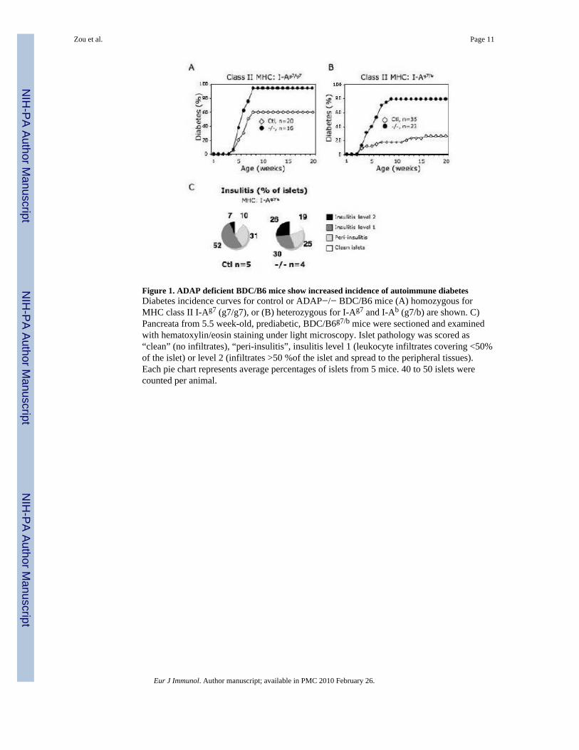
Figure 1. ADAP deficient BDC/B6 mice show increased incidence of autoimmune diabetesDiabetes incidence curves for control or ADAP−/− BDC/B6 mice (A) homozygous forMHC class II I-Ag7 (g7/g7), or (B) heterozygous for I-Ag7 and I-Ab (g7/b) are shown. C)Pancreata from 5.5 week-old, prediabetic, BDC/B6g7/b mice were sectioned and examinedwith hematoxylin/eosin staining under light microscopy. Islet pathology was scored as“clean” (no infiltrates), “peri-insulitis”, insulitis level 1 (leukocyte infiltrates covering <50%of the islet) or level 2 (infiltrates >50 %of the islet and spread to the peripheral tissues).Each pie chart represents average percentages of islets from 5 mice. 40 to 50 islets werecounted per animal.
Zou et al. Page 11
Eur J Immunol. Author manuscript; available in PMC 2010 February 26.
NIH
-PA Author Manuscript
NIH
-PA Author Manuscript
NIH
-PA Author Manuscript

Figure 2. ADAP−/− BDC/B6g7/b peripheral lymphoid tissues contain reduced numbers of CD4+
T cellsA) Percentages of live cells in indicated tissues (P-LN = pancreatic lymph nodes) fromcontrol or ADAP−/−, 5 week-old BDC/B6g7/b mice staining positive for CD4 and Vβ4 areshown. B) Comparison of control and ADAP−/− BDC/B6g7/b absolute CD4+Vβ4+ T cellcounts in spleen and inguinal, non-draining (N-LN) lymph nodes.
Zou et al. Page 12
Eur J Immunol. Author manuscript; available in PMC 2010 February 26.
NIH
-PA Author Manuscript
NIH
-PA Author Manuscript
NIH
-PA Author Manuscript

Figure 3. Treg development and function are normal in ADAP−/− BDC/B6g7/b miceA) Mean (+/− s.d.) percentage of FoxP3+ staining among CD4+ cells from prediabetic BDC/B6g7/b non-draining inguinal (N-LN) or pancreatic (P-LN) lymph nodes is shown. B)Indicated numbers of CD25+ BDC/B6g7/b splenocytes (Tregs) from ADAP+/− (Ctl) orADAP-deficient (−/−) were co-transferred with 0.6 × 10⁶ wild type, CD25lo BDC/B6g7/b
“effector” spleen cells into NOD/SCID mice (n=5 mice per group). Recipients weremonitored for 4 weeks.
Zou et al. Page 13
Eur J Immunol. Author manuscript; available in PMC 2010 February 26.
NIH
-PA Author Manuscript
NIH
-PA Author Manuscript
NIH
-PA Author Manuscript
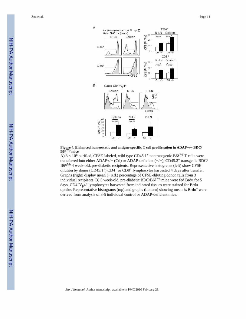
Figure 4. Enhanced homeostatic and antigen-specific T cell proliferation in ADAP−/− BDC/B6g7/b miceA) 3 × 10⁶ purified, CFSE-labeled, wild type CD45.1+ nontransgenic B6g7/b T cells weretransferred into either ADAP+/− (Ctl) or ADAP-deficient (−/−), CD45.2+ transgenic BDC/B6g7/b 4 week-old, pre-diabetic recipients. Representative histograms (left) show CFSEdilution by donor (CD45.1+) CD4+ or CD8+ lymphocytes harvested 4 days after transfer.Graphs (right) display mean (+ s.d.) percentage of CFSE-diluting donor cells from 3individual recipients. B) 5 week-old, pre-diabetic BDC/B6g7/b mice were fed Brdu for 5days. CD4+Vβ4+ lymphocytes harvested from indicated tissues were stained for Brduuptake. Representative histograms (top) and graphs (bottom) showing mean % Brdu+ werederived from analysis of 3-5 individual control or ADAP-deficient mice.
Zou et al. Page 14
Eur J Immunol. Author manuscript; available in PMC 2010 February 26.
NIH
-PA Author Manuscript
NIH
-PA Author Manuscript
NIH
-PA Author Manuscript

Figure 5. Transfer of syngeneic leukocytes inhibits ADAP-dependent diabetes and reducesspontaneous T cell proliferationA) 4-5 week-old ADAP-deficient mice were injected (age range at time of injectionindicated by black bar) with indicated numbers of pooled ADAP-deficient BDC/B6g7/b
spleen and lymph node cells or purified (> 90% pure CD3+) T cells harvested from age-matched mice. Graph (left) shows diabetes incidence curves for transfer recipients and forunmanipulated (no transfer) control or ADAP−/− mice. B) 4 week-old BDC/B6g7/b micewere injected with pooled ADAP−/− BDC/B6g7/b spleen and lymph nodes cells or were leftunmanipulated. One week after injection, mice were fed Brdu for 4 days. The fraction ofBrdu+ CD4+Vβ4+ T lymphocytes among live cells from indicated tissues is shown inrepresentative histograms (top). Mean (+ s.d.) Brdu+ cell fraction (bottom graph) wascalculated from analysis of 3-5 individual animals in each group. Legends indicate subjectgenotypes and the number of injected cells.
Zou et al. Page 15
Eur J Immunol. Author manuscript; available in PMC 2010 February 26.
NIH
-PA Author Manuscript
NIH
-PA Author Manuscript
NIH
-PA Author Manuscript
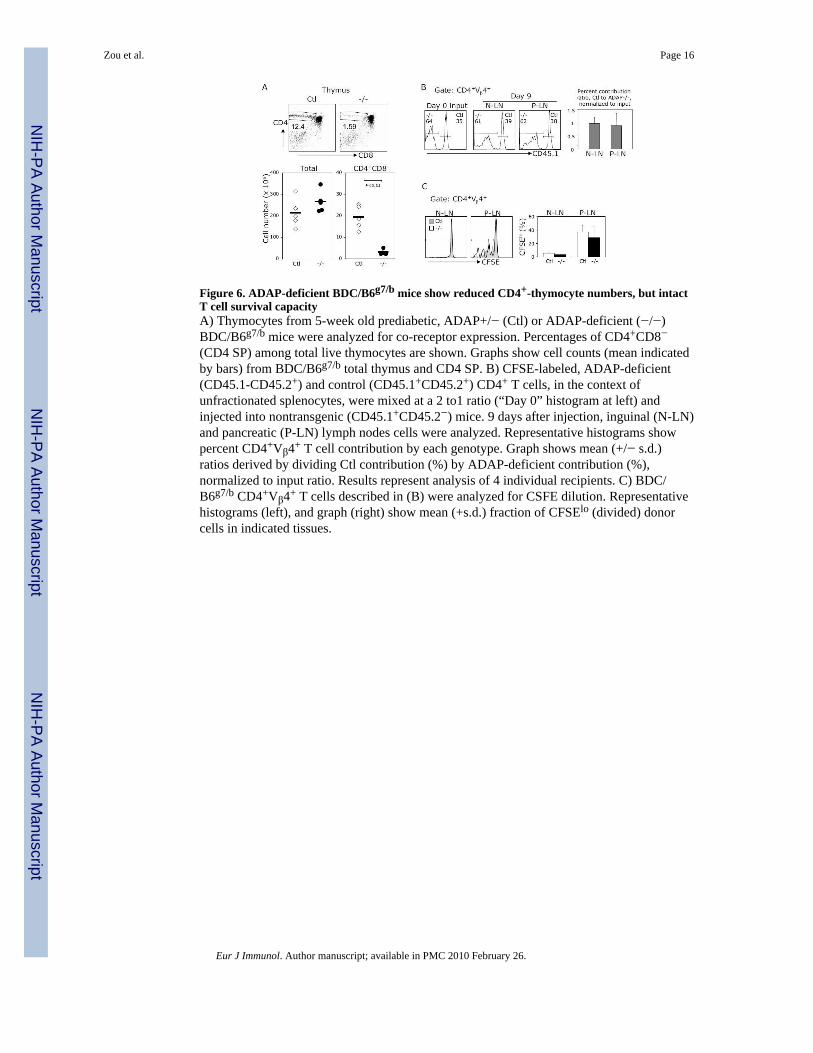
Figure 6. ADAP-deficient BDC/B6g7/b mice show reduced CD4+-thymocyte numbers, but intactT cell survival capacityA) Thymocytes from 5-week old prediabetic, ADAP+/− (Ctl) or ADAP-deficient (−/−)BDC/B6g7/b mice were analyzed for co-receptor expression. Percentages of CD4+CD8−(CD4 SP) among total live thymocytes are shown. Graphs show cell counts (mean indicatedby bars) from BDC/B6g7/b total thymus and CD4 SP. B) CFSE-labeled, ADAP-deficient(CD45.1-CD45.2+) and control (CD45.1+CD45.2+) CD4+ T cells, in the context ofunfractionated splenocytes, were mixed at a 2 to1 ratio (“Day 0” histogram at left) andinjected into nontransgenic (CD45.1+CD45.2−) mice. 9 days after injection, inguinal (N-LN)and pancreatic (P-LN) lymph nodes cells were analyzed. Representative histograms showpercent CD4+Vβ4+ T cell contribution by each genotype. Graph shows mean (+/− s.d.)ratios derived by dividing Ctl contribution (%) by ADAP-deficient contribution (%),normalized to input ratio. Results represent analysis of 4 individual recipients. C) BDC/B6g7/b CD4+Vβ4+ T cells described in (B) were analyzed for CSFE dilution. Representativehistograms (left), and graph (right) show mean (+s.d.) fraction of CFSElo (divided) donorcells in indicated tissues.
Zou et al. Page 16
Eur J Immunol. Author manuscript; available in PMC 2010 February 26.
NIH
-PA Author Manuscript
NIH
-PA Author Manuscript
NIH
-PA Author Manuscript
Copyright © 2022 FDOKUMEN