Behaviour of four different B16 murine melanoma cell sublines: C57BL/6J skin
8
ORIGINAL ARTICLE Behaviour of four different B16 murine melanoma cell sublines: C57BL/6J skin Corina Danciu*, Camelia Oprean † , Dorina E. Coricovac ‡ , Cioca Andreea § , Anca Cimpean ¶ , Heinfried Radeke**, Codruta Soica † and Cristina Dehelean ‡ *Department of Pharmacognosy, University of Medicine and Pharmacy ‘Victor Babes’, Timisoara, Romania, † Department of Pharmaceutical Chemistry, University of Medicine and Pharmacy ‘Victor Babes’, Timisoara, Romania, ‡ Department of Toxicology, University of Medicine and Pharmacy ‘Victor Babes’, Timisoara, Romania, § Department of Pathology, ‘Iuliu Hatieganu’ University of Medicine and Pharmacy, Cluj-Napoca, Romania, ¶ Department of Microscopic Morphology/Histology, Angiogenesis Research Center, University of Medicine and Pharmacy ‘Victor Babes’, Timisoara, Romania and **Pharmazentrum Frankfurt/Center for Drug Research, Development, and Safety, Clinic of J. W. Goethe University, Frankfurt, Germany INTERNATIONAL JOURNAL OF EXPERIMENTAL PATHOLOGY doi: 10.1111/iep.12114 Received for publication: 17 July 2014 Accepted for publication: 1 December 2014 Correspondence: Dorina E. Coricovac Department of Toxicology University of Medicine and Pharmacy ‘Victor Babes’ Eftimie Murgu Square No. 2, 300041 Timisoara Romania Tel.: +0040744648844 Fax: +0256/490626 E-mail: [email protected] SUMMARY Transplantable murine melanomas are well-established models for the study of experimental cancer therapies. The aim of this study was to analyse the behaviour of four different B16 murine melanoma cell sublines after inoculation in C57BL/6J host, more specifically skin-targeted analysis, with respect to two parameters: clinical (tumour volume, melanin amount, erythema) and histological (H & E, S100, VEGF expression). Both non-invasive and invasive determinations showed that B164A5 is the most aggressive melanoma cell line for C57BL/6J’s skin, succeeded by B16F10 and followed in a similar diminished manner of aggressiveness by B16GMCSF and B16FLT3 cell lines. Keywords B164A5, B16F10, B16FLT3, B16GMCSF, C57BL/6J Skin cancers include basal cell carcinoma, squamous cell car- cinoma and malignant melanoma. The first two types of skin cancer are the most frequent malignant neoplasms among fair-skinned population (Andrade et al. 2012). Recent studies showed that the incidence of melanoma was also increasing especially in case of women young adults (Reed et al. 2012). Ultraviolet (UV) radiation can promote the limited prolifera- tive capacity of melanocytes (Gupta et al. 2013). Although the incidence of melanoma among other types of skin cancer is a parameter with increased variability, the severity of this disease is undisputed. Due to its highly metastatic potential and resistance to chemotherapy, it is responsible for most deaths (Rigel 2005; Svobodova et al. 2006). Over the past years, Food and Drug Administration approved three agents for the treatment of melanoma, namely pegylated interferon alpha-2b, vemurafenib and ipilimumab (Lee et al. 2012). More research is still required to find a highly effective drug, with low side effects, against this challenging type of cancer. For this purpose, preclinical studies in animal models provide valuable clues for clinical trials. Transplantable murine melanomas are well-established models for the study of experimental cancer therapies. Many immunotherapeutic protocols have been tested using the mur- ine B16 melanoma cell line (and its sublines) that originates in the syngeneic C57BL/6 (H-2b) mouse strain. Although B164A5 is one of the most widely used cell line for the mur- ine melanoma model, as evidenced by the latest papers in the field (Danciu et al. 2013a,b; Lee et al. 2013; Villareal et al. 2013; Ookubo et al. 2014), several subline derivates have been obtained to study different therapeutic strategies. © 2015 The Authors. International Journal of Experimental Pathology © 2015 International Journal of Experimental Pathology 1 Int. J. Exp. Path. (2015)
Transcript of Behaviour of four different B16 murine melanoma cell sublines: C57BL/6J skin

ORIG INAL ART ICLE
Behaviour of four different B16 murine melanoma cell sublines:C57BL/6J skinCorina Danciu*, Camelia Oprean†, Dorina E. Coricovac‡, Cioca Andreea§, Anca Cimpean¶,Heinfried Radeke**, Codruta Soica† and Cristina Dehelean‡
*Department of Pharmacognosy, University of Medicine and Pharmacy ‘Victor Babes’, Timisoara, Romania, †Department ofPharmaceutical Chemistry, University of Medicine and Pharmacy ‘Victor Babes’, Timisoara, Romania, ‡Department of Toxicology,University of Medicine and Pharmacy ‘Victor Babes’, Timisoara, Romania, §Department of Pathology, ‘Iuliu Hatieganu’ Universityof Medicine and Pharmacy, Cluj-Napoca, Romania, ¶Department of Microscopic Morphology/Histology, Angiogenesis ResearchCenter, University of Medicine and Pharmacy ‘Victor Babes’, Timisoara, Romania and **Pharmazentrum Frankfurt/Center for DrugResearch, Development, and Safety, Clinic of J. W. Goethe University, Frankfurt, Germany
INTERNATIONAL
JOURNAL OF
EXPERIMENTAL
PATHOLOGY
doi: 10.1111/iep.12114
Received for publication: 17 July 2014Accepted for publication: 1 December2014
Correspondence:Dorina E. CoricovacDepartment of ToxicologyUniversity of Medicine and Pharmacy‘Victor Babes’Eftimie Murgu SquareNo. 2, 300041 TimisoaraRomaniaTel.: +0040744648844Fax: +0256/490626E-mail: [email protected]
SUMMARY
Transplantable murine melanomas are well-established models for the study of
experimental cancer therapies. The aim of this study was to analyse the behaviour of
four different B16 murine melanoma cell sublines after inoculation in C57BL/6J
host, more specifically skin-targeted analysis, with respect to two parameters: clinical
(tumour volume, melanin amount, erythema) and histological (H & E, S100, VEGF
expression). Both non-invasive and invasive determinations showed that B164A5 is
the most aggressive melanoma cell line for C57BL/6J’s skin, succeeded by B16F10
and followed in a similar diminished manner of aggressiveness by B16GMCSF and
B16FLT3 cell lines.
Keywords
B164A5, B16F10, B16FLT3, B16GMCSF, C57BL/6J
Skin cancers include basal cell carcinoma, squamous cell car-
cinoma and malignant melanoma. The first two types of skin
cancer are the most frequent malignant neoplasms among
fair-skinned population (Andrade et al. 2012). Recent studies
showed that the incidence of melanoma was also increasing
especially in case of women young adults (Reed et al. 2012).
Ultraviolet (UV) radiation can promote the limited prolifera-
tive capacity of melanocytes (Gupta et al. 2013). Although
the incidence of melanoma among other types of skin cancer
is a parameter with increased variability, the severity of this
disease is undisputed. Due to its highly metastatic potential
and resistance to chemotherapy, it is responsible for most
deaths (Rigel 2005; Svobodova et al. 2006). Over the past
years, Food and Drug Administration approved three agents
for the treatment of melanoma, namely pegylated interferon
alpha-2b, vemurafenib and ipilimumab (Lee et al. 2012).
More research is still required to find a highly effective drug,
with low side effects, against this challenging type of cancer.
For this purpose, preclinical studies in animal models provide
valuable clues for clinical trials.
Transplantable murine melanomas are well-established
models for the study of experimental cancer therapies. Many
immunotherapeutic protocols have been tested using the mur-
ine B16 melanoma cell line (and its sublines) that originates
in the syngeneic C57BL/6 (H-2b) mouse strain. Although
B164A5 is one of the most widely used cell line for the mur-
ine melanoma model, as evidenced by the latest papers in the
field (Danciu et al. 2013a,b; Lee et al. 2013; Villareal et al.
2013; Ookubo et al. 2014), several subline derivates have
been obtained to study different therapeutic strategies.
© 2015 The Authors.
International Journal of Experimental Pathology © 2015 International Journal of Experimental Pathology 1
Int. J. Exp. Path. (2015)

The sublines B16F1 and B16F10 were derived from the
mother B16 line by selection for their ability to form lung col-
onies in vivo after intravenous injection and subsequently
established in vitro after one (B16F1) or 10 (B16F10) cycles
of lung colony formation. B16F10 is a subline that possesses
high lung metastasis ability, whereas B16F1 is a subline with
low metastatic potential (Fidler 1973; Teicher 2010).
B16GMCSF is a subline derived from B16F10 by transduc-
tion, employing an MFG retroviral vector-encoding murine
Granulocyte - Macrophage Colony Stimulating Factor (GM-
CSF) (Kumar et al. 1999). It has been shown that GM-CSF
surface-modified B16F10 melanoma cell vaccine may induce
protection against the wild-type tumour challenge (Gao et al.
2006; Danciu et al. 2013a,b). The fourth cell subline analy-
sed in this study is B16FLT3. It was also obtained from the
murine B16 cell line, which was transfected with the gene for
the Fms-like tyrosine kinase 3 (Flt3)-L cytokine (Vargas et al.
2006; Danciu et al. 2013a,b). Although both GM-CSF and
Flt3 ligand induce marked expansion of dendritic cells, it has
been shown that GM-CSF-secreting tumour cells promoted
higher levels of protective immunity than vaccination with
FLT3-L-secreting tumour cells (Zarei et al. 2009).
The aim of this study was to analyse the behaviour of this
four different B16 murine melanoma cell sublines in the
C57BL/6J host, more specifically skin-targeted analysis, with
respect to two parameters: clinical (tumour volume, melanin
amount, erythema) and histological features [Haematoxylin
and Eosin (H&E), S100, Vascular Endothelial Growth Fac-
tor (VEGF) expression].
Materials and methods
Cells
Mouse adherent melanoma cell line B164A5 was purchased
from ECACC (European Collection of Cell Cultures,
Salisbury, UK). B16F10, B16GMCSF and B16FLT3 cells
were provided by Prof. Radeke, Pharmazentrum Frankfurt/
Center for Drug Research, Development, and Safety, Clinic
of JW Goethe University, Frankfurt, Germany. Cells were
grown in Dulbecco’s modified Eagle’s medium (DMEM)
supplemented with 10% heat-inactivated foetal calf serum
(FCS), 1% non-essential amino acids and 1% penicillin–streptomycin in a humidified atmosphere containing 5%
CO2 at 37°C. All cell culture media and supplements were
obtained from Life Technologies (Paisley, UK). To prepare
the cells for inoculation, cells were trypsinized, counted
using trypan blue, washed with PBS, resuspended at a con-
centration of 106 cells/0.1 ml in saline solution and injected
immediately as described below.
Animal studies
Animal studies were conducted on 7- to 8-week-old C57BL/
6J female mice with an average weight of 20–25 g. Mice were
purchased from Charles River (Sulzfeld, Germany). The work
protocol followed all National Institute of Animal Health
(NIAH) rules: animals were maintained during the experiment
in standard conditions: 12 h light–dark cycle, food and water
ad libitum, temperature 24°C and humidity above 55%. The
experiment was conducted according to the rules of the Ethical
Committee of the ‘Victor Babes�’ University of Medicine and
Pharmacy of Timis�oara, Romania. The number of mice
included in the study was thirty, and the mice were divided
equally in five groups as follows: group A – blank group, group
B – mice inoculated with B164A5 cells, group C – mice inocu-
lated with B16F10 cells, group D – mice inoculated with
B16GMCSF cells and group E – mice inoculated with B16FLT3
cells. In day 0 of the experiment, mice in groups B, C, D and E
were subcutaneous (s.c.) inoculated into the hair-depilated lat-
eral abdomen with 0.1 ml of 106 cells/mouse. Group A, the
blank group was injected with saline solution, the vehicle used
for the injection of cells. Mice were inspected daily for the
development of tumours or other changes. Tumour growth was
measured in millimetres, daily, using callipers, and tumour
volume was estimated by the formula: length 9 width2/2
(Giavazzi et al. 1986). On day 21 post-inoculation, after the
last measurements had been taken, mice were sacrificed by
cervical dislocation. Tumours were collected, measured,
weighed, and afterwards, histological analysis was performed.
Non-invasive skin measurements
All the measurements on mice skin were carried out with a
Multiprobe Adapter System (MPA5) from Courage-Khazaka,
K€oln, Germany. For measurement of melanin and erythema
Mexameter� MX 18 was used to obtain quantitative infor-
mation regarding melanin and erythema (haemoglobin)
amounts and to monitor modifications of these features
during tumour evolution The device emits light over 3 wave-
lengths, namely 568, 660 and 880 nm, and measures remit-
ted light over a 5 mm diameter. The erythema and melanin
indices are determined as follows:
Mx ¼ 500
log 5: log
Infrared
Redþ log 5 Ex
¼ 500
log 5: log
Red
Greenþ log 5
Mx = melanin index, Ex = erythema index and infrared/red/
green = infrared/red/green remittance.
These indices are relative values, and the maximum ratio
between each colour is 1:5. The range of values is 0–1000, ahigher value representing more melanin or erythema, and a
value of 500 representing a remittance ratio of 1:1. The mea-
surement was carried out for four parts of the skin located
near the tumour, and mean and standard deviations were
calculated (Hoshino et al. 2010). Melanin and erythema
values were measured at baseline (day 0) and every two days
until day 17 of the experiment. The measurement area was
5 mm in diameter. Afterwards, the mice were sacrificed and
the skin and main visceral organs were collected. Histologi-
cal and immunohistochemical analyses were performed.
International Journal of Experimental Pathology
2 D. Corina et al.

Histological and immunohistochemical analyses
For the histological analysis, skin samples were fixed in
10% formalin solution and were embedded in paraffin
and cut at 4 lm. Finally, after dewaxing, the samples
were stained with the conventional H & E method and
examined.
Additional slides containing 5-lm thick sections were per-
formed from each case and were stained with anti-VEGF
antibody (monoclonal mouse anti-human antibody, Clone
VG1, code no. M7273; Dako Co, Denmark) and with S100
protein antibody (polyclonal anti-human antibody, Dako
Co, Denmark code 1573, ready-to-use). The dewaxing and
rehydration of the sections was followed by heat-induced
epitope retrieval in citrate buffer pH 9 for 20 min, respec-
tively, pH 7.2 for 30 min (with PT link module;
DakoCytomation, Denmark). The immunohistochemical
technique continued with blocking of the endogenous perox-
idases, using hydrogen peroxide 3%. Incubation with the
VEGF primary antibody (dilution 1:30) and S100 primary
antibody (dilution 1:100) was for 30 min. After incubation
with the primary antibody, the slides were exposed to
labelled streptavidin–biotin system and then 3,3-diamino-
benzidine dihydrochloride was applied as chromogen.
Nuclei were stained with Lillie’s modified haematoxylin.
The entire immunohistochemical procedure was performed
with Dako Autostainer Plus (DakoCytomation). Image
acquisition and analysis were performed using Nikon Eclipse
E 600 microscope and Lucia G software for microscopic
image analysis (NIKON, Germany).
For both antibodies, the intensity of reaction was assessed
as 1 – low staining, 2 – moderate staining and 3 – intense
staining. The immunostaining was considered 0 – negative
when <10% of tumour cells showed positivity. Examination
was performed with the microscope Eclipse E80i Nikon,
and images were acquired with Lucia G soft for microscopic
image analysis.
Statistical analysis
One-way ANOVA followed by Bonferroni post-test was used
to determine the statistical difference between various exper-
imental and control groups.*, ** and *** indicate P < 0.05,
P < 0.01 and P < 0.001 compared to control group.
Results
Clinical results
Our results (Figure 1) show a linear development of the
tumour, directly proportional with the number of days post-
inoculation in all four groups of mice. The results started to
be statistically significant in all groups compared to the blank
group from day 12 postinoculation (P = 0.021) until the end
of the experiment (P = 0.009). In group B and C, tumours
arose on day 6 postinoculation while in groups D and E,
tumours arose on day 8 postinoculation. As a general obser-
vation, tumours were larger throughout the experiment in
groups B and C, compared to groups D and E. At the end of
the experiment, the average tumour volume was as follows:
1033.67 � 400 mm3 in group B, 895.33 � 445 mm3 in
group C, 638.647 � 426 mm3 in groups D and
732 � 465 mm3 in group E. One-way ANOVA followed by
Bonferroni post-test showed no significant difference between
the volume of the tumour in the four groups (B, C, D and E)
at the end of the experiment (P = 0.702).
Non-invasive measurements for melanin amount and
degree of erythema were conducted every two days starting
from day 0 until the end of the experiment. Normal values
for melanin in the blank group (C57BL/6J host) varied
between 650 and 665 arbitrary units (as explained in the
Materials and Methods section). After inoculation the
amount of melanin increases linearly in all four groups, as
shown in Figure 2. Until day 7 postinoculation, values were
similar in all inoculated groups. Differences started to
increase on day 9 postinoculation and continued until the
end of the experiment. As a ‘general rule’ observed during
days 9–21, mice in group B presented the highest values,
Figure 1 Tumour volume (mm3) evolution throughout theexperiment.
Figure 2 Melanin amount (arbitrary units) evolution in thedifferent experimental groups among the 21 days ofexperiment.
International Journal of Experimental Pathology
B16 melanoma cell sublines and C57BL/6J skin 3

corresponding to amounts of melanin. For groups C, D and
E, the amount was quite similar during days 9–21. One-way
ANOVA followed by Bonferroni post-test showed no signifi-
cant difference between the melanin amount in the four
groups (B, C, D and E) at the end of the experiment
(P = 0.570).
The difference was not significant when compared to the
blank group (P = 0.269). At the end of the experiment, the
average amount of melanin was as follows: 650 � 13 arbi-
trary units in group A, 1147 � 287 arbitrary units in group
B, 993 � 322 arbitrary units in group C, 851 � 128 arbi-
trary units in group D and 879 � 108 arbitrary units in
group E.
Together with the melanin amount determination,
another non-invasive measurement that was conducted
was the degree of erythema. Normal values for the blank
group (C57BL/6J host) vary between 50 and 60 arbitrary
units (as explained in the Materials and Methods section).
After inoculation, the degree of erythema increased line-
arly in all four groups, as shown in Figure 3. The values
in all four inoculated groups (corresponding to a certain
day of measurement) are quite similar. One-way ANOVA
followed by Bonferroni post-test showed no significant dif-
ference between the erythema degree in the four groups
(B, C, D and E) at the end of the experiment (P = 0.704).
The changes were significant when compared to the blank
group (P = 0.008). At the end of the experiment, the aver-
age value corresponding to the degree of erythema was as
follows: 59 � 3 arbitrary units in group A, 191 � 47
arbitrary units in group B, 193 � 322 arbitrary units in
group C, 163 � 14 arbitrary units in group D and
185 � 11 arbitrary units in group E.
Histological and immunohistochemical results
In group B, the conventional H&E analysis showed intense
pigmentation in almost all tumour cells and with predomi-
nance at the periphery of the tumour (Figure 4a). The
tumour borders were infiltrative, and there were large areas
of tumoral necrosis. There was no ulceration or lymph-
vascular invasion. The mitotic index was 15 mitoses/1 mm2.
In this group, S100 and VEGF were strongly positive, con-
sistent in almost all tumour cells (Figure 5a and 6a).
The evaluation of H&E in group C revealed moderate
pigmentation with intense pigmentation in nests of tumour
melanocytes (Figure 4b). There were large areas of tumoral
necrosis with haemorrhage and infiltrative borders. There
was no ulceration in this group and no lymph-vascular
invasion. The mitotic index was 10 mitoses/1 mm2. S100
Figure 3 Erythema (arbitrary units) evolution in the differentexperimental groups among the 21 days of experiment.
(a) (b)
(d)(c) Figure 4 HE staining in the differentgroups with skin melanoma: (a) groupB – intense pigmentation in almost alltumour cells with a predominance atthe periphery of the tumour; (b) groupC – moderate pigmentation withintense pigmentation in nests of tumourmelanocytes; (c) group D – skinmelanoma with local presence ofmelanin in isolated cells; (d) group E –presence of melanin with an aspect ofdiffusion and loosing of pigmentation.
International Journal of Experimental Pathology
4 D. Corina et al.
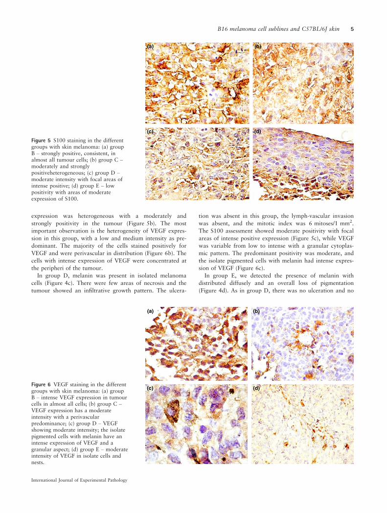
expression was heterogeneous with a moderately and
strongly positivity in the tumour (Figure 5b). The most
important observation is the heterogeneity of VEGF expres-
sion in this group, with a low and medium intensity as pre-
dominant. The majority of the cells stained positively for
VEGF and were perivascular in distribution (Figure 6b). The
cells with intense expression of VEGF were concentrated at
the peripheri of the tumour.
In group D, melanin was present in isolated melanoma
cells (Figure 4c). There were few areas of necrosis and the
tumour showed an infiltrative growth pattern. The ulcera-
tion was absent in this group, the lymph-vascular invasion
was absent, and the mitotic index was 6 mitoses/1 mm2.
The S100 assessment showed moderate positivity with focal
areas of intense positive expression (Figure 5c), while VEGF
was variable from low to intense with a granular cytoplas-
mic pattern. The predominant positivity was moderate, and
the isolate pigmented cells with melanin had intense expres-
sion of VEGF (Figure 6c).
In group E, we detected the presence of melanin with
distributed diffusely and an overall loss of pigmentation
(Figure 4d). As in group D, there was no ulceration and no
(a) (b)
(d)(c)
Figure 5 S100 staining in the differentgroups with skin melanoma: (a) groupB – strongly positive, consistent, inalmost all tumour cells; (b) group C –moderately and stronglypositiveheterogeneous; (c) group D –moderate intensity with focal areas ofintense positive; (d) group E – lowpositivity with areas of moderateexpression of S100.
(a) (b)
(d)(c)Figure 6 VEGF staining in the differentgroups with skin melanoma: (a) groupB – intense VEGF expression in tumourcells in almost all cells; (b) group C –VEGF expression has a moderateintensity with a perivascularpredominance; (c) group D – VEGFshowing moderate intensity; the isolatepigmented cells with melanin have anintense expression of VEGF and agranular aspect; (d) group E – moderateintensity of VEGF in isolate cells andnests.
International Journal of Experimental Pathology
B16 melanoma cell sublines and C57BL/6J skin 5

lymph–vascular invasion, the tumoral necrosis was isolated,
the borders were infiltrative, and the mitotic index was
8 mitoses/1 mm2. In this group, S100 showed low positivity
with areas of moderate expression (Figure 5d). VEGF had a
low to moderate intensity in melanoma cells. The cells were
small with a spindle-like aspect (Figure 6d).
Discussion
The present study shows that among the tested cell lines,
B164A5 is the most aggressive for C57BL/6J’s skin. Melanin
is the most important pigment of skin, synthesized by mela-
nocytes, as a normal defence to diverse stimuli (Pinon et al.
2011; Sass et al. 2013). The major cutaneous melanin role
is protection against epidermal carcinogenesis and malignant
melanomagenesis (Brozyna et al. 2008). Pinon et al. (2011).
This is an index of the fact that melanocytes respond to
UVB exposure and synthesise melanin and proliferate,
instead of apoptosis. As melanoma is a malignant tumour of
melanocytes, the role of this pigment-producing cells (Tho-
mas et al. 2007), and moreover, the implication of melanin
in melanoma, can give important clues about melanoma dis-
ease and its response to different therapeutic approaches. It
is well known that melanin content is variable in different
types of melanomas, melanomas being evaluated as ‘deeply’,
‘heavily’, ‘lightly’ pigmented or amelanotic, as Watts et al.
(1981) reported in his work many years ago. Brozyna et al.
(2008) demonstrated that amelanotic melanomas have a
better response to radiotherapy than those with higher
melanin content. Liu et al. (2006) showed that in human
subjects, fast-growing melanoma (usually associated with a
worse prognosis) have a higher percentage of amelanotic
cells. Furthermore, recently the relationship between mitotic
rate and the lack of pigment in melanoma has been
described (Shen et al. 2014).
Our data show that in all B16 sublines, after inoculation,
the level of melanin increased as compared to the control
group. Among the tested groups, mice injected with B164A5
cells (group B) had the highest content of melanin. The values
started to increase from day 9 after inoculation, as described
in the Results section. The second high melanin level was reg-
istered in the group injected with B16F10 cells. Fedele et al.’s
experiment with B16F10 melanoma cells implanted into
C57BL/6J mouse strain, revealed that melanin was released in
excess by the tumour cells, resulting in tumours with blackish
colour extending into the subcutaneous tissue. Also, a large
number of metastases in lung and liver parenchyma were
noticed in their experiment (Fedele et al. 2013).
As reported previously, in most cases, melanin values can
also be correlated with erythema values (Kaur & Saraf
2011; Danciu et al. 2013a,b; Hexse et al. 2013). Brenner
et al. first described the erythematous rash that accompanies
melanoma (Brenner & Wolf 1992; Mashiah et al. 2009).
The erythematous eruption, termed ‘Brenner sign’, is corre-
lated with angiogenesis (Russo et al. 2011). It is known that
tumour growth in vivo is dependent on vascularization
on the first day after inoculation. When tumours are not vas-
cularized then, the tumour growth is slow and linear. After
angiogenesis has occurred, the tumours begin to grow rap-
idly (Rose et al. 1999; Zhao et al. 2010; S�oica et al. 2012).
The erythema and tumour growth measurements show that
both parameters increased linearly after cell inoculation.
Although the ‘behaviour’ of erythema values was similar for
all four cell lines subtypes, as described in the Results sec-
tion, tumour volume was higher in the group of B164A5,
followed by B16F10. Ulceration is a very important prog-
nostic factor in case of human melanoma (Balch et al.
2009). In case of this study, during the 21 days of observa-
tion, ulceration of the tumour was not detected in any of the
experimental groups. Overwijk and Restifo (2001) reported
that in the case of implanted B164A5 cells, tumours become
necrotic in the centre and start to ulcerate or bleed when
allowed to grow larger, for a period exceeding 21 days.
For all three non-invasive measurements, tumour volume,
melanin index and erythema, inoculation with B16GMCSF
and B16FLT3 cell lines leads to the lowest values. These
results are consistent with other studies concerning vaccines
expressing GM-CSF and Flt3 (Curran & Allison 2009). It is
well known that GM-CSF is one of the most potent
cytokine capable of inducing tumour-specific systemic
immunity (Dranoff et al. 1993; Stagg et al. 2004; Curran &
Allison 2009). Being associated with the growth and differ-
entiation of hematopoietic progenitors, GM-CSF cytokine
exerts its effects on antigen-presenting cells and recruitment
of dendritic cells (Dranoff et al. 1993; Li et al. 2006). It also
chemo-attracts macrophages, lymphocytes and granulocytes
to the vaccine site (Curran & Allison 2009). Dranoff et al.
reported that GM-CSF generates protection against a distant
tumour, while other cytokines, such as IL-2, can induce lo-
coregional tumour rejection (Dranoff et al. 1993; Stagg
et al. 2004). The same protective role is played by the cyto-
kine Flt3L When used prophylactically, it promotes tumour
regression in experimental models (Curran & Allison 2009).
Although it is known that B16-GMCSF can give a better
immune protection than B16-Flt3L, Curran and Allison
(2009) reported that the Flt3L efficacy increases with higher
doses, whereas GM-CSF activity decreases at higher expres-
sion levels.
Histological and immunohistochemical analyses support
the above findings. As H&E staining shows, mice skin inoc-
ulated with B164A5 cell expressed the highest amount of
pigment. Mouse skin inoculated with B16F10 and
B16GMCSF cells showed moderate pigmentation with
isolated areas of intense pigmentation. In mouse skin inocu-
lated with the B16FLT3 cell line, the presence of diffuse
melanin and loss of pigmentation is probably due to the
dedifferentiation of malignant melanocytes.
S100 protein is widely accepted as a marker of choice in
the immunohistochemical detection of malignant melanoma
(Henze et al. 1997). S100 showed positivity in all groups
with the most intense expression in mouse skin inoculated
with the B164A5 cell line. In B16F10 and B16GMCSF inoc-
ulated groups, S100 was heterogeneous with moderate to
intense positivity, while in B16FLT3 group, the expression
International Journal of Experimental Pathology
6 D. Corina et al.

was low to moderate. A strong expression of S100 protein
is associated with tumour progression (Harpio & Einarsoon
2004). B164A5 is a cell line derived from the skin of a mur-
ine melanoma of a C57BL/6J mouse strain (Danciu et al.
2013a,b). S100 expression shows that this cell line is the
most ‘well accepted’, in the C57BL/6J host, which is a perti-
nent behaviour, bearing in mind its origin.
VEGF is a signal protein produced by cells that stimulates
the growth of new blood vessels, (Hoeben et al. 2004). An
increased expression of VEGF in the tumour microenviron-
ment is associated with progression of malignant melanomas
(Brychtova et al. 2008). In this study, VEGF was positive in
all groups. The most intense positivity was noticed in mouse
skin inoculated with the B164A5A cell line while the other
groups showed approximately equal moderate expression. In
mouse skin inoculated with B16F10, perivascular predomi-
nance of VEGF expression was seen on many vessels.
Conclusions
Both clinical (tumour volume, melanin amount, erythema)
and histological (H&E, S100, VEGF expression) approaches
show that B164A5 is the most aggressive melanoma cell line
for C57BL/6J’s skin, followed by B16F10 and followed, in a
similar, diminished manner of aggression, by the B16-
GMCSF and B16FLT3 cell lines.
Acknowledgements
The work of the first author, Danciu Corina was supported
by the UMFT grant Parteneriate in cercetarea fundamentala
inovativa PIII-C2-PCFI-2015-2016.
Conflicts of interest
The authors declare no conflict of interest.
References
Andrade P., Brites M.M., Vieira R. et al. (2012) Epidemiology of
basal cell carcinomas and squamous cell carcinomas in a
Department of Dermatology: a 5 year review. An. Bras. Derma-
tol. 87, 212–219.Balch C.M., Gershenwald J.E., Soong S.J. et al. (2009) Final version
of 2009 AJCC melanoma staging and classification. J. Clin.
Oncol. 27, 6199–6206.Brenner S. & Wolf R. (1992) The “red face” – a warning sign of
malignant melanoma? Acta Derm. Venereol. 72, 464.
Brozyna A., VanMiddlesworth L., Slominski A.T. (2008) Inhibition
of melanogenesis as a radiation sensitizer for melanoma therapy.
Int. J. Cancer 123, 448–456.Brychtova S., Bezdekova M., Brychta T., Tichy M. (2008) The role
of vascular endothelial growth factors and their receptors in
malignant melanomas. Neoplasma 55, 273–279.Curran M.A. & Allison J.P. (2009) Tumor vaccines expressing Flt3
ligand synergize with CTLA-4 blockade to reject preimplanted
tumors. Cancer Res. 69, 7747–7755.
Danciu C., Borcan F., Bojin F., Zupko I., Dehelean C. (2013a)
Effect of the isoflavone genistein on tumor size, metastasis poten-
tial and melanization in a B16 mouse model of murine melanoma.
Nat. Prod. Commun. 8, 343–346.Danciu C., Falamas A., Dehelean C. et al. (2013b) A characteriza-
tion of four B16 murine melanoma cell sublines molecular finger-
print and proliferation behavior. Cancer Cell Int. 13, 75.
Dranoff G., Jaffee E., Lazenby A. et al. (1993) Vaccination with
irradiated tumor cells engineered to secrete murine granulocyte-
macrophage colony-stimulating factor stimulates potent, specific,
and long-lasting anti-tumor immunity. Proc. Natl Acad. Sci. USA
90, 3539–3543.Fedele T.A., Galdos-Riveros A.C., Jose de Farias e Melo H.,
Megalhaes A., Maria D.A. (2013) Prognostic relationship of met-
abolic profile obtained of melanoma B16F10. Biomed. Pharmac-
other. 67, 146–156.Fidler I.J. (1973) The relationship of embolic homogeneity, number,
size and viability to the incidence of experimental metastasis. Eur.
J. Cancer 9, 223–227.Gao J., Huang S., Li M., Luo R., Wang X., Takashima A. (2006)
GM-CSF-surface-modified B16. F10 melanoma cell vaccine. Vac-
cine 24, 5265–5268.Giavazzi R., Campbell D.E., Jessup J.M., Cleary K., Fidler I.J.
(1986) Metastatic behavior of tumor cells isolated from
primary and metastatic human colorectal carcinomas implanted
into different sites in nude mice. Cancer Res. 46(4 Pt 2),
1928–1933.Gupta A., Avci P., Dai T., Huang Y.Y., Hamblin M.R. (2013)
Ultraviolet radiation in wound care: sterilization and stimulation.
Adv. Wound Care (New Rochelle) 2, 422–437.Harpio R. & Einarsoon R. (2004) S100 proteins as cancer biomar-
kers with focus on S100B in malignant melanoma. Clin. Biochem.
37, 512–518.Henze G., Dummer R., Joller-Jemelka H.I., B€oni R., Burg G. (1997)
Serum S100-a marker for disease monitoring in metastatic mela-
noma. Dermatology 194, 208–212.Hexse D., Caspary P., Dal Forno Dini T., Schilling-Souza J., Siega
C. (2013) Variation of melanin levels in the skin areas exposed
and not exposed to the sun following winter and summer. Surg.
Cosmet. Dermatol. 5, 298–301.Hoeben A., Landuvt B., Highley M.S., Wildiers H., Van Oosterom
A.T., De Brujin E.A. (2004) Vascular endothelial growth factor
and angiogenesis. Pharmacol. Rev. 56, 549–580.Hoshino T., Matsuda M., Yamashita Y. et al. (2010) Suppression
of melanin production by expression of HSP70. J. Biol. Chem.
285, 13254–13263.Kaur C.D. & Saraf S. (2011) Skin care assessment on the basis of
skin hydration, melanin, erythema and sebum at various body
sites. Int. J. Pharm. Pharm. Sci. 3, 209–213.Kumar R., Yoneda J., Fidler I.J., Dong Z. (1999) GM-CSF-
transduced B16 melanoma cells are highly susceptible to lysis by
normal murine macrophages and poorly tumorigenic in immune-
compromised mice. J. Leukoc. Biol. 65, 102–108.Lee B., Mukhi N., Liu D. (2012) Current management and novel
agents for malignant melanoma. J. Hematol. Oncol. 5, 3.
Lee M.H., Huang Z., Kim D.J. et al. (2013) Direct targeting of
MEK1/2 and RSK2 by silybin induces cell-cycle arrest and inhibits
melanoma cell growth. Cancer Prev. Res. 6, 455–465.Li B., Lalani A.S., Harding T.C. et al. (2006) Vascular endothelial
growth factor blockade reduces intratumoral regulatory T cells
and enhances the efficacy of a GM-CSF-secreting cancer immuno-
therapy. Clin. Cancer Res. 12, 6808–6816.
International Journal of Experimental Pathology
B16 melanoma cell sublines and C57BL/6J skin 7

Liu W., Dowling J.P., Murray W.K., McArthur W.A. et al. (2006)
Rate of growth in melanomas. Characteristics and associations of
rapidly growing melanomas. Arch. Dermatol. 142, 1551–1558.Mashiah J., Brenner S., Pessach Y., Barak V., Schachter J. (2009)
Differences in cytokine levels in melanoma patients with and
without redness (brenner sign). Anticancer Res. 29, 1793–1796.Ookubo N., Michiue H., Kitamatsu M. et al. (2014) The transder-
mal inhibition of melanogenesis by a cell-membrane-permeable
peptide delivery system based on poly-arginine. Biomaterials 35,
4508–4516.Overwijk W.W. & Restifo N.P. (2001) B16 as a mouse model for
human melanoma. Curr. Protoc. Immunol. Chapter 20:Unit 20.1,
doi:10.1002/0471142735.im2001s39.
Pinon A., Limami Y., Micallef L. et al. (2011) A novel form of mel-
anoma apoptosis resistance: melanogenesis up-regulation in apop-
totic B16-F0 cells delays ursolic acid-triggered cell death. Exp.
Cell Res. 317, 1669–1676.Reed K.B., Brewer J.D., Lohse C.M., Bringe K.E., Pruitt C.N., Gib-
son L.E. (2012) Increasing incidence of melanoma among young
adults: an epidemiological study in Olmsted County, Minnesota.
Mayo Clin. Proc. 87, 328–334.Rigel D.S. (2005) Cancer of the Skin. China: Elsevier Saunders.
Rose M.L., Madren J., Bunzendahl H., Thurman R.G. (1999) Die-
tary glycine inhibits the growth of B16 melanoma tumors in mice.
Carcinogenesis 20, 793–798.Russo J., Barr K., Scanlan L., Vincek V. (2011) Signet ring cell mel-
anoma, Brenner sign, and elevated VEGF. J. Am. Acad. Derma-
tol. 65, 444–446.Sass C., Bojin F., Dehelean C., Soica C., P�aunescu V. (2013)
Insights into melanoma. Histological aspects, progression and
prognostic. Fiziologia 23, 14–20.Shen S., Wolfe R., McLean C.A., Haskett M., Kelly J.W. (2014)
Characteristics and associations of high-mitotic-rate melanoma.
JAMA Dermatol. 150(10), 1048–55; doi: 10.1001/jamadermatol.
2014.635.
S�oica C., Dehelean C., Danciu C. et al. (2012) Betulin complex
in c-cyclodextrin derivatives: properties and antineoplasic activi-
ties in in vitro and in vivo tumor models. Int. J. Mol. Sci. 13,
14992–15011.Stagg J., Wu J.H., Bouganim N. et al. (2004) Granulocyte-macro-
phage colony-stimulating factor and interleukin-2 fusion cDNA
for cancer gene immunotherapy. Cancer Res. 64, 8795–8799.Svobodova A., Walterova D., Vostalova J. (2006) Ultraviolet light
induced alteration to the skin. Biomed. Pap. Med. Fac. Univ.
Palacky Olomouc Czech Repub. 150, 25–38.Teicher B.A. (2010) Tumor Models in Cancer Research, pp. 682.
Boston, USA: Springer.
Thomas J., Liu T., Cotter M.A. et al. (2007) Melanocyte expression
of survivin promotes development and metastasis of UV-induced
melanoma in HGF-transgenic mice. Cancer Res. 67, 5172–5178.Vargas P., Cort�es C., Vargas L., Rosemblatt M., Bono M.R. (2006)
Immunization with antigen-pulsed dendritic cells significantly
improves the immune response to weak self-antigens. Immunobi-
ology 211, 29–36.Villareal M., Han J., Matsuyama K. et al. (2013) Lupenone from
erica multiflora leaf extract stimulates melanogenesis in B16 mur-
ine melanoma cells through the inhibition of ERK1/2 activation.
Planta Med. 79, 236–243.Watts K.P., Fairchild R.G., Slatkin D.N. et al. (1981) Melanin con-
tent of hamster tissues, human tissues, and various melanomas.
Cancer Res. 41, 467–472.Zarei S., Schwenter F., Luy P. et al. (2009) Role of GM-CSF signal-
ing in cell-based tumor immunization. Blood 113, 6658–6668.Zhao Q.H., Zhang Y., Liu Y. et al. (2010) Anticancer effect of real-
gar nanoparticles on mouse melanoma skin cancer in vivo via
transdermal drug delivery. Med. Oncol. 27, 203–212.
International Journal of Experimental Pathology
8 D. Corina et al.