Migration defects by DISC1 knockdown in C57BL/6, 129X1/SvJ, and ICR strains via in utero gene...
14
Migration defects by DISC1 knockdown in C57BL/6, 129X1/SvJ, and ICR strains via in utero gene transfer and virus-mediated RNAi Ken-ichiro Kubo a,# , Kenji Tomita a,# , Asuka Uto a , Keisuke Kuroda b , Saurav Seshadri c , Jared S. Cohen c , Kozo Kaibuchi b , Atsushi Kamiya c,* , and Kazunori Nakajima a,** a Department of Anatomy, Keio University School of Medicine, 35 Shinanomachi, Shinjuku-ku, Tokyo, Japan b Department of Cell Pharmacology, Graduate School of Medicine, Nagoya University, Showa, Nagoya, Japan c Department of Psychiatry and Behavioral Sciences, Johns Hopkins University School of Medicine, Baltimore, MD, USA Abstract Disrupted-in-Schizophrenia 1 (DISC1) is a promising genetic risk factor for major mental disorders. Many groups repeatedly reported a role for DISC1 in brain development in various strains of mice and rats by using RNA interference (RNAi) approach. Nonetheless, due to the complexity of its molecular disposition, such as many splice variants and a spontaneous deletion in a coding exon of the DISC1 gene in some mouse strains, there have been debates on the interpretation on these published data. Thus, in this study, we address this question by DISC1 knockdown via short-hairpin RNAs (shRNAs) against several distinct target sequences with more than one delivery methodologies into several mouse strains, including C57BL/6, ICR, and 129X1/SvJ. Here, we show that DISC1 knockdown by in utero electroporation of shRNA against exons 2, 6, and 10 consistently results in neuronal migration defects in the developing cerebral cortex, which are successfully rescued by co- expression of full-length DISC1. Furthermore, lentivirus-mediated shRNA also led to migration defects, which is consistent with two other methodologies already published, such as plasmid- mediated and retrovirus-mediated ones. The previous study by Song’s group also reported that, in the adult hippocampus, the phenotype elicited by DISC1 knockdown with shRNA targeting exon 2 was consistently seen in both C57BL/6 and 129S6 mice. Taken together, we propose that some of DISC1 isoforms that are feasible to be knocked down by shRNAs to exon 2, 6, and 10 of the DISC1 gene play a key role for neuronal migration commonly in various mouse strains and rats. Keywords DISC1; In utero electroporation; Neuronal migration; Brain development © 2010 Elsevier Inc. All rights reserved. ** Coresponding author Department of Anatomy, Keio University School of Medicine, 35 Shinanomachi, Shinjuku-ku, Tokyo, Japan Tel: +81-3-5363-3743, Fax: +81-3-5379-1977 [email protected] . * Coresponding author Department of Psychiatry and Behavioral sciences, Johns Hopkins University School of Medicine, 600N Wolfe St, Baltimore, MD 21208 USA Tel: +1-410-502-0060, Fax: +1-410-614-1792 [email protected] . # These authors equally contributed to this work Publisher's Disclaimer: This is a PDF file of an unedited manuscript that has been accepted for publication. As a service to our customers we are providing this early version of the manuscript. The manuscript will undergo copyediting, typesetting, and review of the resulting proof before it is published in its final citable form. Please note that during the production process errors may be discovered which could affect the content, and all legal disclaimers that apply to the journal pertain. NIH Public Access Author Manuscript Biochem Biophys Res Commun. Author manuscript; available in PMC 2011 October 1. Published in final edited form as: Biochem Biophys Res Commun. 2010 October 1; 400(4): 631–637. doi:10.1016/j.bbrc.2010.08.117. NIH-PA Author Manuscript NIH-PA Author Manuscript NIH-PA Author Manuscript
-
Upload
independent -
Category
Documents
-
view
4 -
download
0
Transcript of Migration defects by DISC1 knockdown in C57BL/6, 129X1/SvJ, and ICR strains via in utero gene...

Migration defects by DISC1 knockdown in C57BL/6, 129X1/SvJ,and ICR strains via in utero gene transfer and virus-mediated RNAi
Ken-ichiro Kuboa,#, Kenji Tomitaa,#, Asuka Utoa, Keisuke Kurodab, Saurav Seshadric, JaredS. Cohenc, Kozo Kaibuchib, Atsushi Kamiyac,*, and Kazunori Nakajimaa,**aDepartment of Anatomy, Keio University School of Medicine, 35 Shinanomachi, Shinjuku-ku,Tokyo, JapanbDepartment of Cell Pharmacology, Graduate School of Medicine, Nagoya University, Showa,Nagoya, JapancDepartment of Psychiatry and Behavioral Sciences, Johns Hopkins University School of Medicine,Baltimore, MD, USA
AbstractDisrupted-in-Schizophrenia 1 (DISC1) is a promising genetic risk factor for major mental disorders.Many groups repeatedly reported a role for DISC1 in brain development in various strains of miceand rats by using RNA interference (RNAi) approach. Nonetheless, due to the complexity of itsmolecular disposition, such as many splice variants and a spontaneous deletion in a coding exon ofthe DISC1 gene in some mouse strains, there have been debates on the interpretation on thesepublished data. Thus, in this study, we address this question by DISC1 knockdown via short-hairpinRNAs (shRNAs) against several distinct target sequences with more than one delivery methodologiesinto several mouse strains, including C57BL/6, ICR, and 129X1/SvJ. Here, we show that DISC1knockdown by in utero electroporation of shRNA against exons 2, 6, and 10 consistently results inneuronal migration defects in the developing cerebral cortex, which are successfully rescued by co-expression of full-length DISC1. Furthermore, lentivirus-mediated shRNA also led to migrationdefects, which is consistent with two other methodologies already published, such as plasmid-mediated and retrovirus-mediated ones. The previous study by Song’s group also reported that, inthe adult hippocampus, the phenotype elicited by DISC1 knockdown with shRNA targeting exon 2was consistently seen in both C57BL/6 and 129S6 mice. Taken together, we propose that some ofDISC1 isoforms that are feasible to be knocked down by shRNAs to exon 2, 6, and 10 of theDISC1 gene play a key role for neuronal migration commonly in various mouse strains and rats.
KeywordsDISC1; In utero electroporation; Neuronal migration; Brain development
© 2010 Elsevier Inc. All rights reserved.**Coresponding author Department of Anatomy, Keio University School of Medicine, 35 Shinanomachi, Shinjuku-ku, Tokyo, Japan Tel:+81-3-5363-3743, Fax: +81-3-5379-1977 [email protected] . *Coresponding author Department of Psychiatry and Behavioralsciences, Johns Hopkins University School of Medicine, 600N Wolfe St, Baltimore, MD 21208 USA Tel: +1-410-502-0060, Fax:+1-410-614-1792 [email protected] .#These authors equally contributed to this workPublisher's Disclaimer: This is a PDF file of an unedited manuscript that has been accepted for publication. As a service to our customerswe are providing this early version of the manuscript. The manuscript will undergo copyediting, typesetting, and review of the resultingproof before it is published in its final citable form. Please note that during the production process errors may be discovered which couldaffect the content, and all legal disclaimers that apply to the journal pertain.
NIH Public AccessAuthor ManuscriptBiochem Biophys Res Commun. Author manuscript; available in PMC 2011 October 1.
Published in final edited form as:Biochem Biophys Res Commun. 2010 October 1; 400(4): 631–637. doi:10.1016/j.bbrc.2010.08.117.
NIH
-PA Author Manuscript
NIH
-PA Author Manuscript
NIH
-PA Author Manuscript

1. IntroductionDisrupted-in-Schizophrenia-1 (DISC1), a promising genetic risk factor for major psychiatricdisorders, has multiple roles in brain development [1-12]. Several independent groups haveconsistently demonstrated that DISC1 is important in regulating migration or coordinating thetempo of migration in a context-dependent manner, by using RNA interference (RNAi) [1,4-6,8,10,12] (Table. 1). Nonetheless, because of the complexity of its molecular disposition,including many splice variants and a spontaneous deletion in a coding exon of the DISC1 genein some mouse strains [13-19], there have been many debates on the interpretation of thesepublished data. This study is designed to address these questions systematically by focusingon radial neuronal migration in the developing cerebral cortex. Thus far, four independentgroups have reported migration defects by knockdown of DISC1 in developing cerebral cortex(Table. 1). DISC1 short hairpin RNA (shRNA) targeted to sequences in exon 10 consistentlyleads to migration defects. Tsai and colleagues [10] reported this effect in Swiss Webster mice,an outbred strain, by plasmid-mediated shRNA via in utero electroporation. By using the sametarget sequences commonly conserved between rats and mice, Selkoe and Young-Pearseconsistently found migration defects in Sprague Dawley rats [12]. The same interventionagainst exon 10 of DISC1 also led to the similar defects in ICR mice, another outbred strain[4,5]. Song and colleagues used retrovirus-mediated shRNA targeting to exon 2 of DISC1 andalso reported the migration defect in the developing cerebral cortex in C57BL/6 [1]. Althoughthese results from independent studies appear consistent, each study used different strains andspecies of animals and target sequences of RNAi, and different methods to deliver shRNA.
In this study, we compared the effects of three independent shRNAs to DISC1, including twoalready characterized, in plasmid-based in utero gene transfer. Importantly, the migrationdefects elicited by all these shRNAs were significantly rescued by co-expression with RNAi-resistant wild-type mouse DISC1, known as the full-length DISC1. The migration defectspreviously commonly reported in more than one outbred strain via DISC1 RNAi werereproduced in C57BL/6N. We also assessed how consistently we could elicit migration defectsby DISC1 RNAi by a different delivering methodology, a lentivirus-mediated knockdownapproach. Finally, we further characterized the time course of migration defects from prenatalto neonatal stages.
2. Materials and methods2. 1. Plasmid constructs
Plasmids expressing shRNA were used for the suppression of DISC1 expression [20]. Theeffects of two shRNAs to DISC1 (RNAi-1 and -3) were well characterized in cell culture andin utero gene transfer by more than one group [4,5,7,9,12,21]. Another target sequence forRNAi to DISC1, previously characterized by retrovirus-mediated shRNA, was also used(RNAi-2) [1,6]. A scrambled target sequence without homology to any known messenger RNAwas used to produce control RNAi (Con RNAi). Target sequences of these shRNAs to DISC1were designed to distinct exons of DISC1 as follows.
RNAi-1 with mild suppression, 5′-CGGCTGAGCCAAGAGTTGG-3′ in exon 6.
RNAi-2 with moderate suppression, 5′-CGGCTTCCAAGACACCTTC-3′ in exon 2.
RNAi-3 with strong suppression, 5′-GGCAAACACTGTGAAGTGC-3′ in exon 10.
Expression constructs of RNAi-resistant forms of mouse DISC1 with silent mutations in thetarget sequence of shRNAs (DISC1-R−1, -R−2, and -R−3 are resistant to DISC1 RNAi-1, -2,and -3, respectively) were tested for functional complementation in migration assays. LentiviralshRNA vectors for knockdown of DISC1 (FUGW-D1) and control (FUGW-C1) were providedby Dr. Hongjun Song. Viral shRNA was produced by our published methods [21].
Kubo et al. Page 2
Biochem Biophys Res Commun. Author manuscript; available in PMC 2011 October 1.
NIH
-PA Author Manuscript
NIH
-PA Author Manuscript
NIH
-PA Author Manuscript

2. 2. In utero electroporationC57BL/6NCr, ICR, and 129X1/SvJ pregnant mice (SLC) were used in this study. The day ofvaginal plug occurrence was considered to be embryonic day 0 (E0). In utero electroporationwas performed as described previously [4,5,22]. Briefly, DISC1 shRNAs (0.1-7.5 mg/ml)together with green fluorescent protein (GFP) expression vector (1 mg/ml) were injected intothe lateral ventricle and electroporated into the ventricular zone at E15.0. RNAi-resistant formsof mouse DISC1 expression constructs on the pCAGGS1 vector [23] or pCAGGS1 vector (2.5mg/ml) were co-electroporated to test functional complementation. All experiments wereperformed according to the institutional guidelines for animal experiments.
2. 3. Brain slice preparation and immunohistochemistryPreparation of coronal slices of cerebral cortex and immunohistochemistry were performed asdescribed previously [4,5,9,22,24,25]. Images from brain sections with self-inactivatinglentiviral shRNA (pFUGW) [1,21] were captured after immunofluorescent staining with arabbit polyclonal antibody against GFP (1:400; Molecular Probes). Slice images were acquiredwith confocal microscopes (FV300 or FV1000; Olympus, and LSM510; Carl Zeiss).
2. 4. In vivo bromodeoxyuridine (BrdU) labellingBrdU (50 mg/kg body weight; Sigma) was injected intraperitoneally into pregnant mice 48 hafter electroporation. Brain sections were stained with a rat monoclonal antibody against BrdU(1:1000, Abcam).
2. 5. Quantitative bin analysis of brain slicesTo analyze the RNAi effect on cell positions, the cell positions of GFP-positive cells werequantified by bin analysis as previously described [5]. Statistical analyses were conducted withone-way analysis of variance (ANOVA) followed by the Tukey-Kramer multiple comparisontest. Student’s t-test was used in comparing two sets of data. A value of p <0.05 is consideredstatistically significant. Values were given as mean ± standard error of the mean.
2. 6. Cell culture and transfectionCell culture and transfection were performed by our published protocols [4,5].
2. 7. Cell extraction and immunoblottingCell extraction and immunoblotting were performed by our published protocols [5,9]. Thefollowing primary antibodies were used for immunoblotting; mouse monoclonal antibodiesagainst HA-tag (1:1000, Covance) and against β-tubulin (1:1000, Sigma-Aldrich). Quantitativedensitometric measurement of Western blotting was performed by Image J.
3. Results3. 1. Effect of RNAi targeting distinct exons of DISC1 on radial migration in the developingcerebral cortex
To validate the effects of RNAi to DISC1, we used three independent DISC1 shRNAconstructs, DISC1 RNAi-1, -2, and -3, for targeting exon 6, 2, or 10, respectively (Fig. 1A).All DISC1 shRNAs were confirmed to suppress DISC1 protein expression, consistent withprevious studies [1,4,5,7]. RNAi-1, -2, and -3 suppressed 64, 81, and 92% of mouse DISC1protein expression, respectively, in HEK293T cells (Fig. 1B). RNAi-resistant forms of mouseDISC1 (mDISC1-R−1, −2, and −3 are resistant to RNAi-1, -2, and -3, respectively) were alsoconfirmed to be resistant to each shRNA by co-expression with the shRNA (data not shown).
Kubo et al. Page 3
Biochem Biophys Res Commun. Author manuscript; available in PMC 2011 October 1.
NIH
-PA Author Manuscript
NIH
-PA Author Manuscript
NIH
-PA Author Manuscript

We then tested the effects of DISC1 RNAi in the developing cerebral cortex in C57BL/6NCrmice by in utero gene transfer. Embryos were electroporated with DISC1 shRNAs expressionconstruct together with GFP expression plasmid at embryonic day 15 (E15.0) and the effect ofDISC1 knockdown on neuronal migration was evaluated by bin analysis of GFP-positiveneurons at postnatal day 1 (P1.0). In brain sections with control shRNA, 59% of GFP-positivecells left the ventricular zone (VZ), subventricular zone (SVZ) and intermediate zone (IZ), andmigrated into the upper cortical plate (CP) beneath the marginal zone (MZ) of the cortex (Fig.1C). In contrast, in brain sections with DISC1 RNAi, a large number of GFP-positive neuronsstill remained in the midst of their migration in the CP, IZ, and VZ/SVZ (Fig. 1C). Of note,the effect of DISC1 RNAi on neuronal migration correlated with the level of DISC1suppression, and was significantly restored by overexpression of RNAi-resistant forms ofmouse DISC1 (DISC1-R−1, −2, and −3), suggesting that disturbed migration by DISC1 RNAidid not originate from off-target effects (Fig. 1C and D). Migration defects by DISC1 RNAi-1and -3 in ICR mice as well as migration defects by DISC1 RNAi-2 and-3 in 129X1/SvJ micewere also confirmed (Fig. S1). These phenotypes are unlikely to be non-specific effects ofmolecular disturbance by in utero electroporation, since overexpression of p27Kip1 (a cyclin-dependent kinase inhibitor [26]) at E15.0, which is known to mediate acceleration of migration[27], advanced migration by the same experimental paradigms (Fig. S2).
3. 2. Effect of DISC1 RNAi on neuronal migration via in utero lentivirus-mediated shRNAdelivery
The effect of DISC1 knockdown was also been evaluated by an alternative approach,retrovirus-mediated shRNA delivery by in utero injection, which showed migration defect inthe developing cerebral cortex [1,6]. To further confirm that migration defect is not a non-specific consequence of in utero electroporation, we tested another virus-mediated method,lentivirus-mediated RNAi. These RNAi constructs have the same target sequences as thoseused in the retrovirus-mediated RNAi, whose efficacies were well characterized [1,6]. Embryoswere injected with lentivirus expressing DISC1 shRNA at E15.0 and the effect of DISC1knockdown was evaluated at P1.0. As expected, knockdown of DISC1 by lentivirus expressingDISC1 shRNA also led to neuronal migration failure (Fig. 2A and B), suggesting that themigration defect was not caused non-specifically by the in utero electroporation. Of note, incontrast to the focal distribution of the transfected cells by in utero electroporation, lentivirus-infected cells showed wide distribution in developing brain, including the cerebral cortex andganglionic eminences (GEs), main sources of GABAergic interneurons [28] (Fig. 2C).
3. 3. Altered cell positions caused by delayed migration during brain developmentWe next examined how migration defects by DISC1 knockdown would affect cell positions atdifferent developmental stages. Brain slices with DISC1 RNAi-2 were used for time seriesanalysis at E17.5, P1.0, P3.5, and P6.0 (Fig. 3A). The majority of GFP-positive cells withcontrol RNAi were located at the top of the CP at P1.0 and were distributed in the middle oflayers II-IV at P6.0, because the later-born GFP-negative cells had passed and piled up abovethe GFP-positive cells (Fig. 3B). In the brain sections with DISC1 RNAi-2, the majority ofGFP-positive cells were still migrating throughout the CP and only a few had arrived beneaththe MZ at P1.0 as shown in Fig. 1C. In contrast, a number of GFP-positive cells reached nearthe top of the CP at P3.5, suggesting that knockdown of DISC1 led to delayed migration,although a minor defect at the final step of radial migration near the MZ could also exist. ByP6.0, most of the cells arrived near the MZ (Fig. 3).
3. 4. Effect of DISC1 RNAi on cell position at the postnatal stage via migration defectWe next compared the effect of DISC1 RNAi-1, -2, and -3 on cell positions at P6.0. Since themajority of the GFP-positive cells with shRNAs were distributed in the upper cortical layers
Kubo et al. Page 4
Biochem Biophys Res Commun. Author manuscript; available in PMC 2011 October 1.
NIH
-PA Author Manuscript
NIH
-PA Author Manuscript
NIH
-PA Author Manuscript

(II/III and IV) at P6.0 (Fig. S3A), the cell position within these layers was further quantified.We observed that the peak of distribution of GFP-positive cells with DISC1 shRNAs wasshifted slightly to more superficial positions in layer II-IV, compared to those with ControlRNAi (Fig. S3B and C). This phenotype was robust in cells with DISC1 RNAi-2, and cellswith DISC1 RNAi-3, displayed a broad distribution in layer II-IV (Fig. S3). To confirm thatthe effect of DISC1 knockdown on cell position at P6.0 depended on the level of DISC1suppression, we tested the effect of DISC1 RNAi-2 with varying doses: at concentrations of1.0 mg/ml (low dose), 2.5 mg/ml (middle dose), and 7.5 mg/ml (high dose), and analyzedmigration phenotypes at P6.0. In these experiments, we injected BrdU at E17.0, 2 days afterelectroporation, to label neurons born later than the electroporated neurons. As expected, thecells with control RNAi were mainly distributed deeper in the cortex than BrdU-labeled cells,reflecting the “inside-out” fashion of cortical layer formation (Fig. 4). Many cells with the lowand middle dose of DISC1 RNAi-2 were found more superficially in the cortex, overlappingwith the distribution of the BrdU-labeled cells more than control cells. On the other hand, amajority of cells with the high dose of DISC1 RNAi-2 was distributed in middle and deeppositions in the cortex, similar to the result of DISC1 RNAi-3 (Fig. S3B), reflecting a defectin the radial migration toward the MZ. Taken together, these results suggest that corticalmigrating neurons can eventually reach near the MZ with a delay when DISC1 is weakly ormoderately suppressed, while strong suppression of DISC1 causes severe delay or arrest inradial migration. Consequently, at P6.0, weakly or moderately suppressed neurons are locatedmore superficially than control cells, while strongly suppressed neurons are distributed in deeppositions.
DiscussionThere are three main finding in this study. First, we showed that suppression of DISC1 leadsto radial neuronal migration defect in the developing cerebral cortex in C57BL/6NCr mice, acommon phenotype elicited by three independent shRNAs targeting distinct exons of DISC1by in utero electroporation. Our present data together with previous studies, including othergroups, indicate that such migration defect elicited by DISC1 shRNA also occurs in ICR, SwissWebster, and 129X1/SvJ mice as well as Sprague Dawley rats [4,5,8,10,12]. All RNAi effectswere significantly normalized by overexpression of RNAi-resistant forms of mouse full-lengthDISC1, indicating that migration defects are truly associated with DISC1, but unlikely withoff-target effects unrelated to DISC1 functions.
These conclusions in this study, however, may raise new important questions. In outbredstrains, such as ICR and Swiss Webster mice, some but not all of them, carry a spontaneous25-base pair deletion in a coding exon of DISC1 (unpublished data), that was originally reportedin 129 strains [13,14]. This mutation leads to generation of a stop codon by the frameshift ofthe non-triplet deletion and can theoretically deplete the known full-length DISC1 isoformresulting in about a 100 kDa protein [14]. Nonetheless, the patterns of DISC1 immunoreactivitybetween the 129 mice and C57BL/6 mice without this mutation are very similar [29]. Sincethis observation was confirmed by many investigators working on DISC1 with most of theantibodies available against DISC1, it is unlikely that this conclusion is just simply due tocross-reactivity of antibodies. How can we reconcile these data? One of the most reasonableinterpretations at present is that there are unidentified functional isoforms of DISC1 even inthe presence of the 25-base pair deletion. This idea is supported by the observation that a mutantDISC1 model originally utilized this 129-drived genetic deletion led only to impaired workingmemory [14], whereas many other DISC1 animal models, even those with single amino acidpoint mutations, display a greater range of abnormal behavior [30]. These data indicate thatthe 25-base pair deletion may not robustly affect overall DISC1 functions, although it is likelyto delete the known full-length isoform. A recent paper indicating a similar migration defectof the adult dentate gyrus in both C57BL/6 and 129 mice by the knockdown of DISC1 via
Kubo et al. Page 5
Biochem Biophys Res Commun. Author manuscript; available in PMC 2011 October 1.
NIH
-PA Author Manuscript
NIH
-PA Author Manuscript
NIH
-PA Author Manuscript

retrovirus-mediated RNAi approach further supports this idea [6]. We believe that via takingsystematic experiments and careful interpretation, the molecular disposition of DISC1 is,although still complicated, a key to elucidate its diverse and important functions in mentalillnesses.
Second, we observed defective migration of the cells infected with lentivirus expressing DISC1shRNA, which is consistent with the previous study with oncoretrovirus-mediated RNAi [1].Since many cells are ubiquitously infected in several brain regions, including the GE, a majorsource of cortical interneurons [28], cells that did not originate from the pallial VZ/SVZ mightbe mixed with neurons from pallial VZ/SVZ in the CP. Thus, electroporation of shRNA-expression plasmids is a better approach, at least for addressing the developmental processesof specific cell populations in a distinct brain region.
Third, we also demonstrated that many cells with delayed migration by DISC1 knockdownseemed eventually to reach near the MZ at later time points. Thus, the timing of observationof cell positions is very important in examining the effect of DISC1 RNAi on neuronalmigration. Nonetheless, conventional shRNA may not be temporally well controlled for themodulation of gene expression. DISC1 has multiple roles in various cellular events in braindevelopment [1,2,4,5,7,8-12]. Thus, to address the molecular mechanisms precisely, it isimportant to segregate their functions in specific cellular processes. In this respect, the use ofin utero electroporation with inducible and cell type specific gene targeting systems [31,32]would be expected for further exploration of DISC1-mediated molecular pathways and cellbehaviors in brain development.
Supplementary MaterialRefer to Web version on PubMed Central for supplementary material.
AcknowledgmentsWe thank Drs. Hongjun Song, Takeshi Kawauchi, and Jun-ichi Miyazaki for lentiviral RNAi constructs, p27Kip1expression construct, and the CAG promoter, respectively. We thank members of Nakajima laboratory for valuablediscussions and Ms. Yukiko Lema for organizing the manuscript. This work was supported by grants from HealthLabour Sciences Research Grant (K.K.), Japan Society for the Promotion of Science (K.N.), Ministry of Education,Culture, Sports, and Science and Technology of Japan (K.K. and K.N.), the Takeda Science Foundation (K.N.), theNaito Foundation (K.N.), the Japan Brain Foundation (K.N.), the Promotion and Mutual Aid Corporation for PrivateSchools of Japan (K.N.), MH-091230 (A.K.), NARSAD (A.K.), and S-R (A.K.).
References[1]. Duan X, Chang JH, Ge S, et al. Disrupted-In-Schizophrenia 1 regulates integration of newly generated
neurons in the adult brain. Cell 2007;130:1146–1158. [PubMed: 17825401][2]. Enomoto A, Asai N, Namba T, et al. Roles of disrupted-in-schizophrenia 1-interacting protein girdin
in postnatal development of the dentate gyrus. Neuron 2009;63:774–787. [PubMed: 19778507][3]. Jaaro-Peled H, Hayashi-Takagi A, Seshadri S, et al. Neurodevelopmental mechanisms of
schizophrenia: understanding disturbed postnatal brain maturation through neuregulin-1-ErbB4 andDISC1. Trends Neurosci 2009;32:485–495. [PubMed: 19712980]
[4]. Kamiya A, Kubo K, Tomoda T, et al. A schizophrenia-associated mutation of DISC1 perturbs cerebralcortex development. Nat Cell Biol 2005;7:1167–1178. [PubMed: 16299498]
[5]. Kamiya A, Tan PL, Kubo K, et al. Recruitment of PCM1 to the centrosome by the cooperative actionof DISC1 and BBS4: a candidate for psychiatric illnesses. Arch Gen Psychiatry 2008;65:996–1006.[PubMed: 18762586]
[6]. Kim JY, Duan X, Liu CY, et al. DISC1 regulates new neuron development in the adult brain viamodulation of AKT-mTOR signaling through KIAA1212. Neuron 2009;63:761–773. [PubMed:19778506]
Kubo et al. Page 6
Biochem Biophys Res Commun. Author manuscript; available in PMC 2011 October 1.
NIH
-PA Author Manuscript
NIH
-PA Author Manuscript
NIH
-PA Author Manuscript

[7]. Mao Y, Ge X, Frank CL, et al. Disrupted in schizophrenia 1 regulates neuronal progenitorproliferation via modulation of GSK3beta/beta-catenin signaling. Cell 2009;136:1017–1031.[PubMed: 19303846]
[8]. Meyer KD, Morris JA. Disc1 regulates granule cell migration in the developing hippocampus. HumMol Genet 2009;18:3286–3297. [PubMed: 19502360]
[9]. Niwa M, Kamiya A, Murai R, et al. Knockdown of DISC1 by in utero gene transfer disturbs postnataldopaminergic maturation in the frontal cortex and leads to adult behavioral deficits. Neuron2010;65:480–489. [PubMed: 20188653]
[10]. Singh KK, Ge X, Mao Y, et al. Dixdc1 is a critical regulator of DISC1 and embryonic corticaldevelopment. Neuron 2010;67:33–48. [PubMed: 20624590]
[11]. Taya S, Shinoda T, Tsuboi D, et al. DISC1 regulates the transport of the NUDEL/LIS1/14-3-3epsiloncomplex through kinesin-1. J Neurosci 2007;27:15–26. [PubMed: 17202468]
[12]. Young-Pearse TL, Suth S, Luth ES, et al. Biocemical and Functional Interaction of Disrupted-in-Schizophrenia 1 and Amyloid Precursor Protein Regulates Neuronal Migration during MammalianCortical Development. J Neurosci 2010;30:10431–10440. [PubMed: 20685985]
[13]. Clapcote SJ, Roder JC. Deletion polymorphism of Disc1 is common to all 129 mouse substrains:implications for gene-targeting studies of brain function. Genetics 2006;173:2407–2410. [PubMed:16751659]
[14]. Koike H, Arguello PA, Kvajo M, et al. Disc1 is mutated in the 129S6/SvEv strain and modulatesworking memory in mice. Proc Natl Acad Sci U S A 2006;103:3693–3697. [PubMed: 16484369]
[15]. Kvajo M, McKellar H, Arguello PA, et al. A mutation in mouse Disc1 that models a schizophreniarisk allele leads to specific alterations in neuronal architecture and cognition. Proc Natl Acad SciU S A 2008;105:7076–7081. [PubMed: 18458327]
[16]. Ishizuka K, Paek M, Kamiya A, et al. A review of Disrupted-In-Schizophrenia-1 (DISC1):neurodevelopment, cognition, and mental conditions. Biol Psychiatry 2006;59:1189–1197.[PubMed: 16797264]
[17]. Nakata K, Lipska BK, Hyde TM, et al. DISC1 splice variants are upregulated in schizophrenia andassociated with risk polymorphisms. Proc Natl Acad Sci U S A 2009;106:15873–15878. [PubMed:19805229]
[18]. Hennah W, Thomson P, Peltonen L, et al. Genes and schizophrenia: beyond schizophrenia: the roleof DISC1 in major mental illness. Schizophr Bull 2006;32:409–416. [PubMed: 16699061]
[19]. Brandon NJ, Millar JK, Korth C, et al. Understanding the role of DISC1 in psychiatric disease andduring normal development. J Neurosci 2009;29:12768–12775. [PubMed: 19828788]
[20]. Brummelkamp TR, Bernards R, Agami R. A system for stable expression of short interfering RNAsin mammalian cells. Science 2002;296:550–553. [PubMed: 11910072]
[21]. Hayashi-Takagi A, Takaki M, Graziane N, et al. Disrupted-in-Schizophrenia 1 (DISC1) regulatesspines of the glutamate synapse via Rac1. Nat Neurosci 2010;13:327–332. [PubMed: 20139976]
[22]. Tabata H, Nakajima K. Efficient in utero gene transfer system to the developing mouse brain usingelectroporation: visualization of neuronal migration in the developing cortex. Neuroscience2001;103:865–872. [PubMed: 11301197]
[23]. Niwa H, Yamamura K, Miyazaki J. Efficient selection for high-expression transfectants with a noveleukaryotic vector. Gene 1991;108:193–199. [PubMed: 1660837]
[24]. Nakajima K, Mikoshiba K, Miyata T, et al. Disruption of hippocampal development in vivo byCR-50 mAb against reelin. Proc Natl Acad Sci U S A 1997;94:8196–8201. [PubMed: 9223338]
[25]. Tabata H, Nakajima K. Multipolar migration: the third mode of radial neuronal migration in thedeveloping cerebral cortex. J Neurosci 2003;23:9996–10001. [PubMed: 14602813]
[26]. Kawauchi T, Chihama K, Nabeshima Y, et al. Cdk5 phosphorylates and stabilizes p27kip1contributing to actin organization and cortical neuronal migration. Nat Cell Biol 2006;8:17–26.[PubMed: 16341208]
[27]. Nguyen L, Besson A, Heng JI, et al. p27kip1 independently promotes neuronal differentiation andmigration in the cerebral cortex. Genes Dev 2006;20:1511–1524. [PubMed: 16705040]
[28]. Marin O, Rubenstein JL. Cell migration in the forebrain. Annu Rev Neurosci 2003;26:441–483.[PubMed: 12626695]
Kubo et al. Page 7
Biochem Biophys Res Commun. Author manuscript; available in PMC 2011 October 1.
NIH
-PA Author Manuscript
NIH
-PA Author Manuscript
NIH
-PA Author Manuscript

[29]. Ishizuka K, Chen J, Taya S, et al. Evidence that many of the DISC1 isoforms in C57BL/6J miceare Ralso also expressed in 129S6/SvEv mice. Mol Psychiatry 2007;12:897–899. [PubMed:17895924]
[30]. Clapcote SJ, Lipina TV, Millar JK, et al. Behavioral phenotypes of Disc1 missense mutations inmice. Neuron 2007;54:387–402. [PubMed: 17481393]
[31]. Kamiya A. Animal models for schizophrenia via in utero gene transfer: understanding roles forgenetic susceptibility factors in brain development. Prog. Brain Res 2009;179:9–15. [PubMed:20302813]
[32]. LoTurco J, Manent JB, Sidiqi F. New and improved tools for in utero electroporation studies ofdeveloping cerebral cortex. Cereb Cortex 2009;1(19 Suppl):i120–125. [PubMed: 19395528]
Kubo et al. Page 8
Biochem Biophys Res Commun. Author manuscript; available in PMC 2011 October 1.
NIH
-PA Author Manuscript
NIH
-PA Author Manuscript
NIH
-PA Author Manuscript

Fig. 1. Knockdown of DISC1 by RNAi targeting distinct exons leads to migration defects in thedeveloping cerebral cortex(A) A schematic representation of the mouse DISC1 exons targeted by shRNA constructs.DISC1 RNAi-1, -2, and -3 target exon 6, 2, and 10 of DISC1, respectively. (B) The graphrepresents densitometry analysis of western blotting after normalization by the expression levelof β-tubulin. Con RNAi, control RNAi. (C) RNAi constructs and GFP expression vectors wereelectroporated into the ventricular zone (VZ) at E15.0 and analyzed at P1.0. 59% of GFP-positive cells terminated migration into the upper cortical plate (CP). By contrast, only 23, 12,and 4% of GFP-labeled cells reached in the most superficial region (Bins 9 and 10) with DISC1RNAi-1, -2, and -3, respectively, of which migration defect was normalized by co-electroporation of RNAi-resistant wild-type mouse DISC1 (DISC1-R−1, −2, or −3). SVZ,subventricular zone. IZ, intermediate zone. Green, GFP. Magenta, propidium iodide (PI). Scalebar, 100 μm. Error bars indicate SEM. (D) The effect of DISC1 shRNAs on migration correlates
Kubo et al. Page 9
Biochem Biophys Res Commun. Author manuscript; available in PMC 2011 October 1.
NIH
-PA Author Manuscript
NIH
-PA Author Manuscript
NIH
-PA Author Manuscript

with the suppression level of DISC1 expression. Overexpression of RNAi-resistant form ofDISC1 rescued migration defect. Error bars indicate SEM. *p < 0.01.
Kubo et al. Page 10
Biochem Biophys Res Commun. Author manuscript; available in PMC 2011 October 1.
NIH
-PA Author Manuscript
NIH
-PA Author Manuscript
NIH
-PA Author Manuscript

Fig. 2. Effect of DISC1 RNAi on neuronal migration via in utero lentivirus-mediated RNAi(A) Lentivus expressing shRNA and GFP were injected into the lateral ventricule at E15.0 andanalyzed at P1.0. In the brains with Control RNAi (FUGW-C1), 21% of GFP-positive cellslocated into the superficial region of CP. By contrast, only 6 % of GFP-labeled cells reachedthe superficial region of CP in brain slices with DISC1 RNAi-D1 (FUGW-D1). Green, GFP.Blue, DAPI. Scale bar, 100 μm. Error bars indicate SEM. (B) Silencing of DISC1 by lentiviralRNAi induces migration defect. Error bars indicate SEM. *p < 0.05. (C) The distribution oflentivirus-infected cells in forebrain at P1.0. GFP-positive cells were diffusely detected inforebrain, including cerebral cortex (CC) and ganglionic eminence (GE). Scale bar, 300 μm.
Kubo et al. Page 11
Biochem Biophys Res Commun. Author manuscript; available in PMC 2011 October 1.
NIH
-PA Author Manuscript
NIH
-PA Author Manuscript
NIH
-PA Author Manuscript
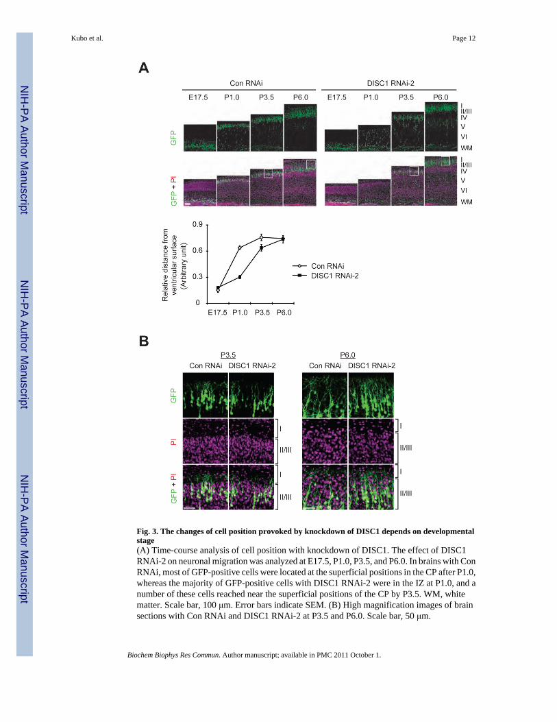
Fig. 3. The changes of cell position provoked by knockdown of DISC1 depends on developmentalstage(A) Time-course analysis of cell position with knockdown of DISC1. The effect of DISC1RNAi-2 on neuronal migration was analyzed at E17.5, P1.0, P3.5, and P6.0. In brains with ConRNAi, most of GFP-positive cells were located at the superficial positions in the CP after P1.0,whereas the majority of GFP-positive cells with DISC1 RNAi-2 were in the IZ at P1.0, and anumber of these cells reached near the superficial positions of the CP by P3.5. WM, whitematter. Scale bar, 100 μm. Error bars indicate SEM. (B) High magnification images of brainsections with Con RNAi and DISC1 RNAi-2 at P3.5 and P6.0. Scale bar, 50 μm.
Kubo et al. Page 12
Biochem Biophys Res Commun. Author manuscript; available in PMC 2011 October 1.
NIH
-PA Author Manuscript
NIH
-PA Author Manuscript
NIH
-PA Author Manuscript
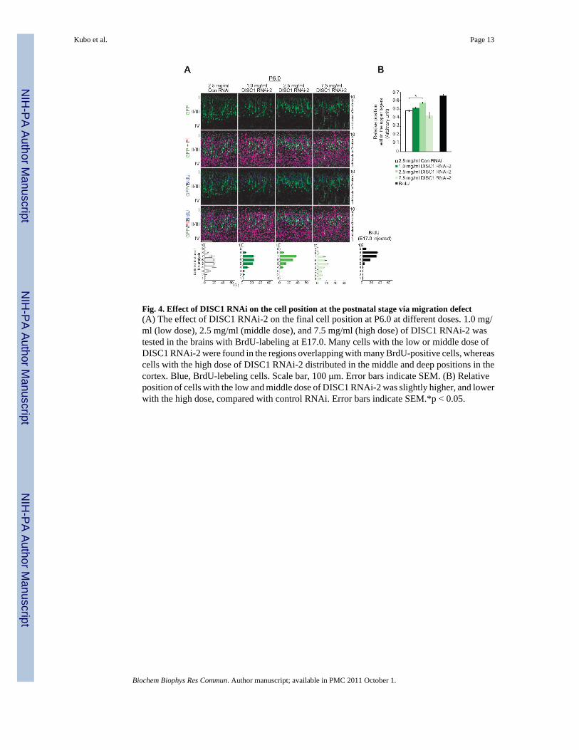
Fig. 4. Effect of DISC1 RNAi on the cell position at the postnatal stage via migration defect(A) The effect of DISC1 RNAi-2 on the final cell position at P6.0 at different doses. 1.0 mg/ml (low dose), 2.5 mg/ml (middle dose), and 7.5 mg/ml (high dose) of DISC1 RNAi-2 wastested in the brains with BrdU-labeling at E17.0. Many cells with the low or middle dose ofDISC1 RNAi-2 were found in the regions overlapping with many BrdU-positive cells, whereascells with the high dose of DISC1 RNAi-2 distributed in the middle and deep positions in thecortex. Blue, BrdU-lebeling cells. Scale bar, 100 μm. Error bars indicate SEM. (B) Relativeposition of cells with the low and middle dose of DISC1 RNAi-2 was slightly higher, and lowerwith the high dose, compared with control RNAi. Error bars indicate SEM.*p < 0.05.
Kubo et al. Page 13
Biochem Biophys Res Commun. Author manuscript; available in PMC 2011 October 1.
NIH
-PA Author Manuscript
NIH
-PA Author Manuscript
NIH
-PA Author Manuscript

NIH
-PA Author Manuscript
NIH
-PA Author Manuscript
NIH
-PA Author Manuscript
Kubo et al. Page 14
Tabl
e. 1
The
effe
ct o
f DIS
C1
Kno
ckdo
wn
on n
euro
nal m
igra
tion
The
role
for D
ISC
1 in
neu
rona
l mig
ratio
n ha
ve b
een
exam
ined
in se
vera
l mou
se st
rain
s and
rat,
usin
g R
NA
inte
rfer
ence
(RN
Ai),
via
in u
tero
gen
e tra
nsfe
ran
d vi
rus-
med
iate
d kn
ockd
own
appr
oach
by
in v
ivo
inje
ctio
n. C
C, c
ereb
ral c
orte
x; D
G, d
enta
te g
yrus
.
Stag
eR
egio
nR
NA
iT
arge
t exo
nSp
ecie
s/St
rain
Phen
otyp
eR
efer
ence
s
Dev
elop
men
tC
C
Plas
mid
10M
ouse
/Sw
iss
Web
ster
Dis
turb
ed m
igra
tion
[10]
Plas
mid
10 a
nd 6
Mou
se/IC
RD
istu
rbed
mig
ratio
n[4
,5]
Plas
mid
10R
at/S
prag
ueD
awle
yD
istu
rbed
mig
ratio
n[1
2]
Ret
rovi
rus
2M
ouse
/C57
BL/
6D
istu
rbed
mig
ratio
n[1
]
Plas
mid
2, 6
, and
10
Mou
se/C
57B
L/6,
ICR
, 129
X1/
SvJ
Dis
turb
ed m
igra
tion
Cur
rent
Stud
y
Lent
iviru
s2
Mou
se/C
57B
L/6
Dis
turb
ed m
igra
tion
Cur
rent
Stud
y
DG
Plas
mid
2M
ouse
/Sw
iss
Web
ster
Dis
turb
ed m
igra
tion
[8]
Adu
ltR
etro
viru
s2
Mou
se/C
57B
L/6
Ove
rext
ende
dm
igra
tion
[1]
Ret
rovi
rus
2M
ouse
/C57
BL/
6an
d 12
9S6
Ove
rext
ende
dm
igra
tion
[6]
Biochem Biophys Res Commun. Author manuscript; available in PMC 2011 October 1.