Knockdown of miR21 in human breast cancer cell lines inhibits proliferation, in vitro migration and...
14
RESEARCH ARTICLE Open Access Knockdown of miR-21 in human breast cancer cell lines inhibits proliferation, in vitro migration and in vivo tumor growth Li Xu Yan 1,2† , Qi Nian Wu 1,2† , Yan Zhang 1,2† , Yang Yang Li 1,2 , Ding Zhun Liao 1,2 , Jing Hui Hou 1,2 , Jia Fu 1,2 , Mu Sheng Zeng 1,3 , Jing Ping Yun 1,2 , Qiu Liang Wu 1,2 , Yi Xin Zeng 1,3 , Jian Yong Shao 1,2,3* Abstract Introduction: MicroRNAs (miRNAs) are a class of small non-coding RNAs (20 to 24 nucleotides) that post- transcriptionally modulate gene expression. A key oncomir in carcinogenesis is miR-21, which is consistently up-regulated in a wide range of cancers. However, few functional studies are available for miR-21, and few targets have been identified. In this study, we explored the role of miR-21 in human breast cancer cells and tissues, and searched for miR-21 targets. Methods: We used in vitro and in vivo assays to explore the role of miR-21 in the malignant progression of human breast cancer, using miR-21 knockdown. Using LNA silencing combined to microarray technology and target prediction, we screened for potential targets of miR-21 and validated direct targets by using luciferase reporter assay and Western blot. Two candidate target genes (EIF4A2 and ANKRD46) were selected for analysis of correlation with clinicopathological characteristics and prognosis using immunohistochemistry on cancer tissue microrrays. Results: Anti-miR-21 inhibited growth and migration of MCF-7 and MDA-MB-231 cells in vitro, and tumor growth in nude mice. Knockdown of miR-21 significantly increased the expression of ANKRD46 at both mRNA and protein levels. Luciferase assays using a reporter carrying a putative target site in the 3’ untranslated region of ANKRD46 revealed that miR-21 directly targeted ANKRD46. miR-21 and EIF4A2 protein were inversely expressed in breast cancers (r s = -0.283, P = 0.005, Spearman’s correlation analysis). Conclusions: Knockdown of miR-21 in MCF-7 and MDA-MB-231 cells inhibits in vitro and in vivo growth as well as in vitro migration. ANKRD46 is newly identified as a direct target of miR-21 in BC. These results suggest that inhibitory strategies against miR-21 using peptide nucleic acids (PNAs)-antimiR-21 may provide potential therapeutic applications in breast cancer treatment. Introduction Breast cancer (BC) is by far the most frequent cancer of women (23% of all cancers), with an estimated 1.15 mil- lion new cases worldwide in 2002 [1]. It is still the lead- ing cause of cancer mortality in women [1]. Despite research and resources dedicated to elucidating the molecular mechanisms of BC, the precise mechanisms of its initiation and progression remain unclear. MicroRNAs (miRNAs) are small non-coding RNAs (20 to 24 nucleotides) that post-transcriptionally modu- late gene expression by negatively regulating the stability or translational efficiency of their target mRNAs [2]. After the discovery of miRNAs, and findings indicating that they play a role in cancer, the concept of “onco- mirs” was proposed [3]. In particular, miR-21 [miRBase: MIMAT0000076] has emerged as a key oncomir, since it is the most consistently up-regulated miRNA in a wide range of cancers [4-7]. Functional studies showed that knockdown of miR-21 in MCF7 cells led to reduced proliferation and tumor * Correspondence: [email protected] † Contributed equally 1 State Key Laboratory of Oncology in Southern China, Sun Yat-Sen University Cancer Center, 651 Dong Feng Road East, Guangzhou, 510060, PR China Full list of author information is available at the end of the article Yan et al. Breast Cancer Research 2011, 13:R2 http://breast-cancer-research.com/content/13/1/R2 © 2011 Yan et al.; licensee BioMed Central Ltd. This is an open access article distributed under the terms of the Creative Commons Attribution License (http://creativecommons.org/licenses/by/2.0), which permits unrestricted use, distribution, and reproduction in any medium, provided the original work is properly cited.
-
Upload
independent -
Category
Documents
-
view
0 -
download
0
Transcript of Knockdown of miR21 in human breast cancer cell lines inhibits proliferation, in vitro migration and...

RESEARCH ARTICLE Open Access
Knockdown of miR-21 in human breast cancercell lines inhibits proliferation, in vitro migrationand in vivo tumor growthLi Xu Yan1,2†, Qi Nian Wu1,2†, Yan Zhang1,2†, Yang Yang Li1,2, Ding Zhun Liao1,2, Jing Hui Hou1,2, Jia Fu1,2,Mu Sheng Zeng1,3, Jing Ping Yun1,2, Qiu Liang Wu1,2, Yi Xin Zeng1,3, Jian Yong Shao1,2,3*
Abstract
Introduction: MicroRNAs (miRNAs) are a class of small non-coding RNAs (20 to 24 nucleotides) that post-transcriptionally modulate gene expression. A key oncomir in carcinogenesis is miR-21, which is consistentlyup-regulated in a wide range of cancers. However, few functional studies are available for miR-21, and few targetshave been identified. In this study, we explored the role of miR-21 in human breast cancer cells and tissues, andsearched for miR-21 targets.
Methods: We used in vitro and in vivo assays to explore the role of miR-21 in the malignant progression of humanbreast cancer, using miR-21 knockdown. Using LNA silencing combined to microarray technology and targetprediction, we screened for potential targets of miR-21 and validated direct targets by using luciferase reporterassay and Western blot. Two candidate target genes (EIF4A2 and ANKRD46) were selected for analysis ofcorrelation with clinicopathological characteristics and prognosis using immunohistochemistry on cancer tissuemicrorrays.
Results: Anti-miR-21 inhibited growth and migration of MCF-7 and MDA-MB-231 cells in vitro, and tumor growthin nude mice. Knockdown of miR-21 significantly increased the expression of ANKRD46 at both mRNA and proteinlevels. Luciferase assays using a reporter carrying a putative target site in the 3’ untranslated region of ANKRD46revealed that miR-21 directly targeted ANKRD46. miR-21 and EIF4A2 protein were inversely expressed in breastcancers (rs = -0.283, P = 0.005, Spearman’s correlation analysis).
Conclusions: Knockdown of miR-21 in MCF-7 and MDA-MB-231 cells inhibits in vitro and in vivo growth as well asin vitro migration. ANKRD46 is newly identified as a direct target of miR-21 in BC. These results suggest thatinhibitory strategies against miR-21 using peptide nucleic acids (PNAs)-antimiR-21 may provide potentialtherapeutic applications in breast cancer treatment.
IntroductionBreast cancer (BC) is by far the most frequent cancer ofwomen (23% of all cancers), with an estimated 1.15 mil-lion new cases worldwide in 2002 [1]. It is still the lead-ing cause of cancer mortality in women [1]. Despiteresearch and resources dedicated to elucidating themolecular mechanisms of BC, the precise mechanismsof its initiation and progression remain unclear.
MicroRNAs (miRNAs) are small non-coding RNAs(20 to 24 nucleotides) that post-transcriptionally modu-late gene expression by negatively regulating the stabilityor translational efficiency of their target mRNAs [2].After the discovery of miRNAs, and findings indicatingthat they play a role in cancer, the concept of “onco-mirs” was proposed [3]. In particular, miR-21 [miRBase:MIMAT0000076] has emerged as a key oncomir, sinceit is the most consistently up-regulated miRNA in awide range of cancers [4-7].Functional studies showed that knockdown of miR-21
in MCF7 cells led to reduced proliferation and tumor
* Correspondence: [email protected]† Contributed equally1State Key Laboratory of Oncology in Southern China, Sun Yat-Sen UniversityCancer Center, 651 Dong Feng Road East, Guangzhou, 510060, PR ChinaFull list of author information is available at the end of the article
Yan et al. Breast Cancer Research 2011, 13:R2http://breast-cancer-research.com/content/13/1/R2
© 2011 Yan et al.; licensee BioMed Central Ltd. This is an open access article distributed under the terms of the Creative CommonsAttribution License (http://creativecommons.org/licenses/by/2.0), which permits unrestricted use, distribution, and reproduction inany medium, provided the original work is properly cited.

growth [8,9]. Knockdown of miR-21 in MDA-MB-231cells significantly reduced invasion and lung metastasis[10]. These data clearly implicate miR-21 as a key mole-cule in carcinogenesis, but functional studies thatdemonstrate cause and effect relationships betweenmiR-21 and target genes are lacking. Given that miRNAsusually target multiple genes post-transcriptionally, miR-21 is likely to exert its effects by regulating many genesinvolved in BC.The inhibition of miRNAs using antisense oligonu-
cleotides (ASOs) is a unique and effective technique forinvestigating miRNA functions and targets. Peptidenucleic acids (PNAs) are artificial oligonucleotides con-structed on a peptide-like backbone. PNAs have a stron-ger affinity and greater specificity for DNA or RNA thannatural nucleic acids, and are resistant to nucleases [11].PNA-based ASOs can be used without transfectionreagents, and are highly effective and sequence-specific.They provide long-lasting inhibition of miRNAs, andshow no cytotoxicity up to 1 μM [11]. Therefore, weused a PNA miR-21 inhibitor for in vivo investigation.In this study, we explored the role of miR-21 in the
malignant progression of human BC by assaying in vitroand in vivo function after miR-21 knockdown. We alsosearched for miR-21 targets using gene prediction-basedand systematic screening approaches. Two potential tar-get genes eukaryotic translation initiation factor 4A2(EIF4A2) [NCBI: NM001967] and ankyrin repeatdomain 46 (ANKRD46) [NCBI: NM198401] wereselected for correlation analysis between protein levelsand clinicopathological characteristics as well as prog-nosis using immunohistochemistry (IHC) on cancer tis-sue microrrays (TMAs).
Materials and methodsTissue specimens and TMAs constructionIn situ hybridization analysis was performed on freshsamples from BC or fibroadenoma (FA) tissues withpaired normal adjacent tissues (NATs, > 2 cm fromtumor tissues) obtained from Sun Yat-sen UniversityCancer Center (SYSUCC) (Guangzhou, China) betweenJanuary and March 2009. For IHC staining of miR-21predicted target genes, formalin-fixed paraffin-embeddedtissues were obtained from 99 randomly selected BCpatients without neoadjuvant therapy at SYSUCC fromJanuary 2000 to November 2004, from whom informedconsent and agreement, and clinicopathological informa-tion was available. A pathologist reviewed slides from allblocks, selecting representative areas of invasive tumortissue to be cored. Selected cores were analyzed induplicate using a MiniCore Tissue Arrayer (Alphelys,Passage Paul Langevin, Plaisir, France) with a 1-mmneedle. The diagnosis and histological grade of eachcase were independently confirmed by two pathologists
based on World Health Organization classification [12].The clinical stage was classified according to the Ameri-can Joint Committee on Cancer (AJCC) tumor-lymphnode-metastasis (TNM) classification system [13]. Thestudy was approved by the Research Ethics Committeeof SYSUCC (Reference number: YP-2009168). The clini-copathological characteristics and follow-up data of thepatients are summarized in Table 1.
Locked nucleic acid (LNA)-based in situ hybridization formiRNATo study the spatial and temporal expression of miR-NAs with high sensitivity and resolution, the miRNAchromogenic in situ hybridization (CISH) and fluores-cein in situ hybridization (FISH) protocol [14] wereoptimized (Additional file 1).
Transfection of LNA-antimiR-21 into BC cellsMCF-7 and MDA-MB-231 cells were maintained inDulbecco’s modified Eagle’s medium, supplemented with100 U/ml penicillin, 100 μg/ml streptomycin and 10%fetal bovine serum (GIBCO-Invitrogen, Carlsbad, CA,USA). For transfection, the LNA-antimiR-21 or LNA-control (Exiqon A/S, Skelstedet, Vedbaek, Denmark)
Table 1 Clinicopathological characteristics and follow-updata for 99 patients with BC
Characteristics Number of patients/Numberanalyzed (%)
Median age (range) 48 (30 to 74) (years)
Histological type*
Ductal 93/99 (94%)
Lobular 1/99 (1%)
Other 5/99 (5%)
Histological grade*
I 22/99 (22%)
II 58/99 (58%)
III 19/99 (20%)
Lymph node status at time of primary diagnosis
Metastasis 57/99 (58%)
No metastasis 42/99 (42%)
AJCC clinical stage**
I 8/99 (8%)
II 68/99 (69%)
III 23/99 (23%)
Overall survival (median, range) 74 (6 to 112) (months)
Alive without evidence ofcancer
45/99 (68%)
Alive with cancer 14/99 (14%)
Died of cancer 40/99 (40%)
Died of other disease 0/99 (0%)
* According to the WHO classification of BC. ** According to the AJCC stagingsystem; AJCC, American Joint Committee on Cancer; BC, breast cancer; WHO,World Health Organization.
Yan et al. Breast Cancer Research 2011, 13:R2http://breast-cancer-research.com/content/13/1/R2
Page 2 of 14
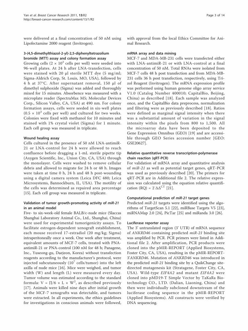
were delivered at a final concentration of 50 nM usingLipofectamine 2000 reagent (Invitrogen).
3-(4,5-dimethylthiazol-2-yl)-2,5-diphenyltetrazoliumbromide (MTT) assay and colony formation assayGrowing cells (2 × 10³ cells per well) were seeded into96-well plates. At 24 h after LNA-transfection, cellswere stained with 20 μl sterile MTT dye (5 mg/ml;Sigma-Aldrich Corp, St. Louis, MO, USA), followed by4 h at 37°C. After supernatant removal, 150 μl ofdimethyl sulphoxide (Sigma) was added and thoroughlymixed for 15 minutes. Absorbence was measured with amicroplate reader (SpectraMax M5; Molecular DevicesCorp., Silicon Valley, CA, USA) at 490 nm. For colonyformation assays, cells were seeded in six-well plates(0.5 × 10³ cells per well) and cultured for two weeks.Colonies were fixed with methanol for 10 minutes andstained with 1% crystal violet (Sigma) for 1 minute.Each cell group was measured in triplicate.
Wound healing assayCells cultured in the presence of 50 nM LNA-antimiR-21 or LNA-control for 24 h were allowed to reachconfluence before dragging a 1-mL sterile pipette tip(Axygen Scientific, Inc., Union City, CA, USA) throughthe monolayer. Cells were washed to remove cellulardebris and allowed to migrate for 24 h or 48 h. Imageswere taken at time 0 h, 24 h and 48 h post-woundingusing a digital camera system (Leica DFC 480; LeicaMicrosystems, Bannockburn, IL, USA). The motility ofthe cells was determined as repaired area percentage[15]. Each cell group was measured in triplicate.
Validation of tumor growth-promoting activity of miR-21in an animal modelFive- to six-week-old female BALB/c-nude mice (SlaccasShanghai Laboratory Animal Co., Ltd., Shanghai, China)were used for experimental tumorigenicity assays. Tofacilitate estrogen-dependent xenograft establishment,each mouse received 17-estradiol (20 mg/kg; Sigma)intraperitoneally once a week. One week after treatment,equivalent amounts of MCF-7 cells, treated with PNA-antimiR-21 or PNA-control (100 nM for 48 h; Panagene,Inc., Yuseong-gu, Daejeon, Korea) without transfectionreagents according to the manufacturer’s protocol, wereinjected subcutaneously (107 cells/tumor) into the leftaxilla of nude mice [16]. Mice were weighed, and tumorwidth (W) and length (L) were measured every day.Tumor volume was estimated according to the standardformula: V = ∏/6 × L × W2, as described previously[17]. Animals were killed nine days after initial growthof the MCF-7 xenografts was detectable, and tumorswere extracted. In all experiments, the ethics guidelinesfor investigations in conscious animals were followed,
with approval from the local Ethics Committee for Ani-mal Research.
mRNA array and data miningMCF-7 and MDA-MB-231 cells were transfected eitherwith LNA-antimiR-21 or with LNA-control at a finalconcentration of 50 nM. Total RNAs were isolated fromMCF-7 cells 48 h post transfection and from MDA-MB-231 cells 36 h post transfection, respectively, using Tri-zol Reagent (Invitrogen). The mRNA expression profilewas performed using human genome oligo array serviceV1.0 (Catalog Number 400010; CapitalBio, Beijing,China) as described [18]. Each sample was analyzedonce, and the CapitalBio data preprocess, normalizationand filtering were as previously described [18]. Ratioswere defined as marginal signal intensity when therewas a substantial amount of variation in the signalintensity within the pixels from 800 to 1,500. Allthe microarray data have been deposited to theGene Expression Omnibus (GEO) [19] and are accessi-ble through GEO Series accession number [GEO:GSE20627].
Relative quantitative reverse transcription-polymerasechain reaction (qRT-PCR)For validation of mRNA array and quantitative analysisof miR-21 as well as potential target genes, qRT-PCRwas used as previously described [20]. The primers forqRT-PCR are in Additional file 2. The relative expres-sion was calculated using the equation relative quantifi-cation (RQ) = 2-ΔΔCT [21].
Computational prediction of miR-21 target genesPredicted miR-21 targets were identified using the algo-rithms of TargetScan 5.1 [22], miRBase Targets V5 [23],miRNAMap 2.0 [24], PicTar [25] and miRanda 3.0 [26].
Luciferase reporter assayThe 3’ untranslated region (3’ UTR) of mRNA sequenceof ANKRD46 containing predicted miR-21 binding sitewas amplified by PCR. PCR primers were listed in Addi-tional file 2. After amplification, PCR products werecloned into the pMIR-REPORT (Applied Biosystems,Foster City, CA, USA), resulting in the pMIR-REPORT-3’ANKRD46. Mutation of ANKRD46 was introduced inthe predicted miR-21 binding site by a QuikChange site-directed mutagenesis kit (Stratagene, Foster City, CA,USA). Wild-type EIF4A2 and mutant EIF4A2 werecloned into pMD19-T Simple Vector by TaKaRa Bio-technology CO., LTD. (Dalian, Liaoning, China) andthen were individually subcloned downstream of theluciferase coding sequence in the pMIR-REPORT(Applied Biosystems). All constructs were verified byDNA sequencing.
Yan et al. Breast Cancer Research 2011, 13:R2http://breast-cancer-research.com/content/13/1/R2
Page 3 of 14

For reporter assays, wide-type or mutant reporter con-structs (15 ng) were cotransfected into 293T cells intwelve-well plates with miR-21 or miR-control (50 nM;GenePharma, Shanghai, China) and Renilla plasmid(5 ng) using lipofectamine 2000 (Invitrogen). Firefly andRenilla luciferase activities were measured by using aDual Luciferase Assay (Promega, Madison, WI, USA)24 h after transfection. Firefly luciferase values werenormalized to Renilla, and the ratio of firefly/renilla waspresented.
Immunoblot analysisCells were harvested and lysed in radioimmune precipi-tation buffer (Upstate, Lake Placid, NY, USA) at theindicted time post-transfection. Antibodies used forimmunoblot analysis were against ANKRD46 (1:500dilution; sc-87548, Santa Cruz Biotechnology, Inc., SantaCruz, CA, USA), EIF4A2 (1:1000 dilution; ab31218,Abcam, Cambridge, UK) and GAPDH (1:3,000 dilution;sc-32233, Santa Cruz Biotechnology), as a loading con-trol. All bands were detected using a SuperSignal WestPico Chemiluminescent Substrate (Pierce, Rockford,IL, USA).
Immunohistochemical stainingIHC and scoring of the estrogen receptor (ER), proges-terone receptor (PR) and CerbB2 were performed aspreviously described [20]. Slides were incubated withprimary antibodies against ANKRD46 (1:150 dilution;sc-87548, Santa Cruz Biotechnology); or EIF4A2 (1:700dilution; ab31218, Abcam). All slides were processedsimultaneously in identical conditions per the manufac-turer’s instructions. Three observers independentlydetermined consensus scoring of EIF4A2 and ANKRD46immunostaining using a semi-quantitative estimationaccording to the percentage of positive cells and theintensity of staining as described previously [27]. Withthese data, the composite score was obtained by addingthe values of the staining intensity and relative abun-dance [28]. Samples with scores lower than the medianscore were grouped as low protein expression [29].
Statistical analysisSpearman’s rank correlation test was used for correla-tion analysis between predicted target gene proteinlevels and endogenous miR-21 levels measured pre-viously by qRT-PCR [20]. Pearson’s Chi-Square testswere used to compare target gene expression levels toclinicopathological characteristics. Survival curves wereestimated by the Kaplan-Meier method and log-ranktest. All analysis used SPSS 16.0 for Windows (SPSS Inc,Chicago, IL, USA). All tests were two-tailed, and the sig-nificance level was set at P < 0.05.
ResultsmiR-21 is overexpressed in BC tissues and cell linesExpression of miR-21 was detected in the cytoplasm incancerous and luminal epithelial cells, and occasionallyin fibroblasts. In BC patients, an increase in miR-21staining intensity was observed in BC and FA tissuescompared with corresponding NATs (Figure 1a). Paralleldetection by FISH is shown in Additional file 3. Consis-tent with the CISH results, quantitative analysis indi-cated that miR-21 expression was significantly increasedby 4.44- to 2.02-fold in BC tissues compared with NATs(P = 0.019, n = 4), and increased in FA tissues by 3.03-to 1.89-fold (P = 0.008, n = 4, Figure 1b). FISH andqRT-PCR were used to measure miR-21 levels in fiveBC cell lines (MCF-7, MDA-MB-231, MDA-MB-453,MDA-MB-435, and SK-BR-3) and one non-tumorigenicepithelial cell line (MCF-10A). Consistent with previousfindings [8,10,30], miR-21 overexpressed in MCF-7,MDA-MB-231 and MDA-MB-453 cell lines from 6.48-to 4.04-fold compared with MCF-10A cell line (P <0.01, Figure 1d).
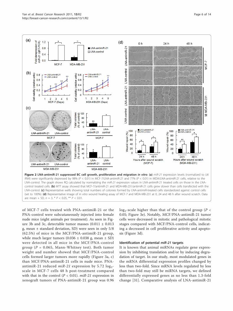
LNA-antimiR-21 inhibits BC cell growth, proliferation andmigration in vitroMCF-7 and MDA-MB-231 cell lines were selected toinvestigate miR-21 functions and targets by usingsequence-specific functional inhibition of miR-21,because both cell lines express higher levels of miR-21compared with MCF-10A cells. Optimal doses and timepoints for transfection of LNA reagents were deter-mined by evaluating miR-21 levels using qRT-PCR(Additional file 4). Knockdown of miR-21 reduced miR-21 levels by 98% in MCF-7 cells, and 77% in MDA-MB-231 cells (P < 0.01) (Figure 2a). LNA-antimiR-21 led toa decrease in MCF-7 cell growth (Figure 2b) and prolif-eration (29%, P = 0.003, Figure 2c). Similar inhibition ofcell growth and proliferation effects (51%, P = 0.011,Figure 2c) was also observed in MDA/LNA-antimiR-21cells (data not shown). In vitro wound healing assaysshowed that wound repair in MCF/LNA-antimiR-21 andMDA/LNA-antimiR-21 was delayed compared withMCF/LNA-control and MDA/LNA-control cells (datanot shown). Knockdown of miR-21 suppressed MCF-7cell migration by up to 69% (P = 0.013), and MDA-MB-231 migration by 51% (P = 0.001), compared with theLNA-control at 24 h after wound scratch (Figure 2d).These data demonstrate the tumorigenic properties ofmiR-21 in regulating cell growth, proliferation andmigration.
PNA-antimiR-21 inhibits tumor growth in vivoTo address the potential effects of PNA-antimiR-21 invivo on the growth of BC cells, equal numbers (3 × 10⁷)
Yan et al. Breast Cancer Research 2011, 13:R2http://breast-cancer-research.com/content/13/1/R2
Page 4 of 14

Figure 1 Altered expression of miR-21 in different breast tumor types and breast cell lines. (a) CISH detection using miR-21 LNAdetection probe (blue) or scramble-miR as negative control were performed on consecutive 8 μM cryo-sections obtained from BC/FA tissuesand corresponding NATs. DNA was counterstained with methyl green (green, × 400 magnification). (b) Total RNA was used to quantify miR-21expression by relative qRT-PCR, normalizing on U6 RNA levels. The graph shows a log2-scale RQ calculated by normalizing the miR-21 expressionvalues in the tumors on those in the NATs. Data indicate the mean (+SD) of four independent samples. * P < 0.05. (c) FISH detection of miR-21in five BC cell lines (MCF-7, MDA-MB-231, MDA-MB-453, MDA-MB-435, SK-BR-3) and one non-tumorigenic epithelial cell line (MCF-10A). Positive insitu hybridization signals are visualized in green, while blue depicts DAPI nuclear stain (× 1,000 magnification). (d) qRT-PCR analysis show thatMCF-7, MDA-MB-231 and MDA-MB-453 cells express higher levels of miR-21 compared with MCF-10A cells. Data are mean (+SD) of threereplicates. ** P < 0.01; BC, breast cancer; FA, fibroadenoma; NATs, normal adjacent tissues.
Yan et al. Breast Cancer Research 2011, 13:R2http://breast-cancer-research.com/content/13/1/R2
Page 5 of 14
of MCF-7 cells treated with PNA-antimiR-21 or thePNA-control were subcutaneously injected into femalenude mice (eight animals per treatment). As seen in Fig-ure 3b and 3c, detectable tumor masses (0.011 ± 0.013g, mean ± standard deviation, SD) were seen in only 5/8(62.5%) of mice in the MCF/PNA-antimiR-21 group,while much larger tumors (0.036 ± 0.038 g, mean ± SD)were detected in all mice in the MCF/PNA-controlgroup (P = 0.065, Mann-Whitney test). Both tumorweight and number showed that MCF/PNA-controlcells formed larger tumors more rapidly (Figure 3a, c)than MCF/PNA-antimiR-21 cells in nude mice. PNA-antimiR-21 reduced miR-21 expression by 5.72 log2-scale in MCF-7 cells 48 h post-treatment comparedwith that in the control (P < 0.01). miR-21 expression inxenograft tumors of PNA-antimiR-21 group was 0.96
log2-scale higher than that of the control group (P <0.05; Figure 3e). Notably, MCF/PNA-antimiR-21 tumorcells were decreased in mitotic and pathological mitoticstages compared with MCF/PNA-control cells, indicat-ing a decreased in cell proliferative activity and apopto-sis (Figure 3d).
Identification of potential miR-21 targetsIt is known that animal miRNAs regulate gene expres-sion by inhibiting translation and/or by inducing degra-dation of target. In our study, most modulated genes inthe mRNA differential expression profiles changed byless than two-fold. Since mRNA levels regulated by lessthan two-fold may still be miRNA targets, we defineddifferentially expressed genes as no less than 1.3-foldchange [31]. Comparative analysis of LNA-antimiR-21
Figure 2 LNA-antimiR-21 suppressed BC cell growth, proliferation and migration in vitro. (a) miR-21 expression levels (normalized to U6RNA) were significantly depressed by 98% (P < 0.01) in MCF-7/LNA-antimiR-21 and 77% (P < 0.01) in MDA/LNA-antimiR-21 cells, relative to theLNA-control. The graph shows RQ calculated by normalizing the miR-21 expression values in LNA-antimiR-21 treated cells on those in the LNA-control treated cells. (b) MTT assay showed that MCF-7/antimiR-21 and MDA-MB-231/antimiR-21 cells grew slower than cells transfected with theLNA-control. (c) Representative wells showing total numbers of colonies formed by LNA-antimiR-treated cells standardized against control cells(set to 100%). (d) Representative image of in vitro wound healing assay of MCF-7 and MDA-MB-231 at 0, 24 and 48 h after wound scratch. Dataare mean + SD, n = 3. * P < 0.05, ** P < 0.01.
Yan et al. Breast Cancer Research 2011, 13:R2http://breast-cancer-research.com/content/13/1/R2
Page 6 of 14

and LNA-control mRNA profiles showed differentialregulation of 394 genes in MCF/LNA-antimiR-21, ofwhich 228 (58%) were up-regulated and 166 (42%) weredown-regulated. 321 genes were differentially expressedin MDA/LNA-antimiR-21 cells, of which 190 (59%)were up-regulated, and 131 (41%) down-regulated
(Figure 4a). The intersection of MCF/LNA-antimiR-21and MDA/LNA-antimiR-21 consisted of 27 genes (18up- and 9 down-regulated; Figure 4b and Table 2).To further screen potential direct targets, we com-
pared the 27 candidate mRNAs with miR-21 targetspredicted by TargetScan 5.1, miRBase Targets V.5,
Figure 3 Effect of miR-21-knockdown on MCF-7 cells growth in nude mice: in vivo functional studies. MCF-7 cells (107 cells/tumor),treated with PNA-antimiR-21 or PNA-control (100 nM for 48 h) without transfection reagents, were injected subcutaneously into the left axilla ofnude mice. (a) Growth curves for MCF/PNA-antimiR-21 (n = 5) vs. MCF/PNA-control (n = 8) cells in an in vivo proliferation assay. All P-values >0.05. (b) Tumors were weighed after animals were killed at 17 days post-tumor-cell injection. Decreasing trend for both number and size oftumors from MCF/PNA-antimiR-21 compared with MCF/PNA-control group, although differences were not significant (P = 0.065, Mann-Whitneytest). (c) Mice and tumors extracted from MCF/PNA-antimiR-21 and MCF/PNA-control groups. (d) Representative photomicrographs of CISH formiR-21 on xenograft tumor sections obtained from mice bearing MCF/PNA-antimiR-21 or MCF/PNA-control groups (× 400). (e) qRT-PCR analysisof miR-21 expression in MCF-7 cells 48 h post PNA-treatment and in xenograft tumors after mice sacrifice. Data in (a), (b) and (e) indicate themean + SD. * P < 0.05, ** P < 0.01. PNA, peptide nucleic acid.
Yan et al. Breast Cancer Research 2011, 13:R2http://breast-cancer-research.com/content/13/1/R2
Page 7 of 14
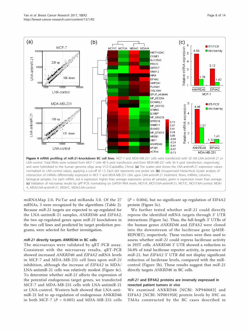
miRNAMap 2.0, PicTar and miRanda 3.0. Of the 27mRNAs, 3 were recognized by the algorithms (Table 2).Because miR-21 targets are expected to up-regulated forthe LNA-antimiR-21 samples, ANKRD46 and EIF4A2,the two up-regulated genes upon miR-21 knockdown inthe two cell lines and predicted by target prediction pro-grams, were selected for further investigation.
miR-21 directly targets ANKRD46 in BC cellsThe microarrays were validated by qRT-PCR assay.Consistent with the microarray results, qRT-PCRshowed increased ANKRD46 and EIF4A2 mRNA levelsin MCF-7 and MDA-MB-231 cell lines upon miR-21inhibition, although the increase of EIF4A2 in MDA/LNA-antimiR-21 cells was relatively modest (Figure 4c).To determine whether miR-21 affects the expression ofthe potential endogenous target genes, we transfectedMCF-7 and MDA-MB-231 cells with LNA-antimiR-21or LNA-control. Western bolt showed that LNA-anti-miR-21 led to up-regulation of endogenous ANKRD46in both MCF-7 (P = 0.005) and MDA-MB-231 cells
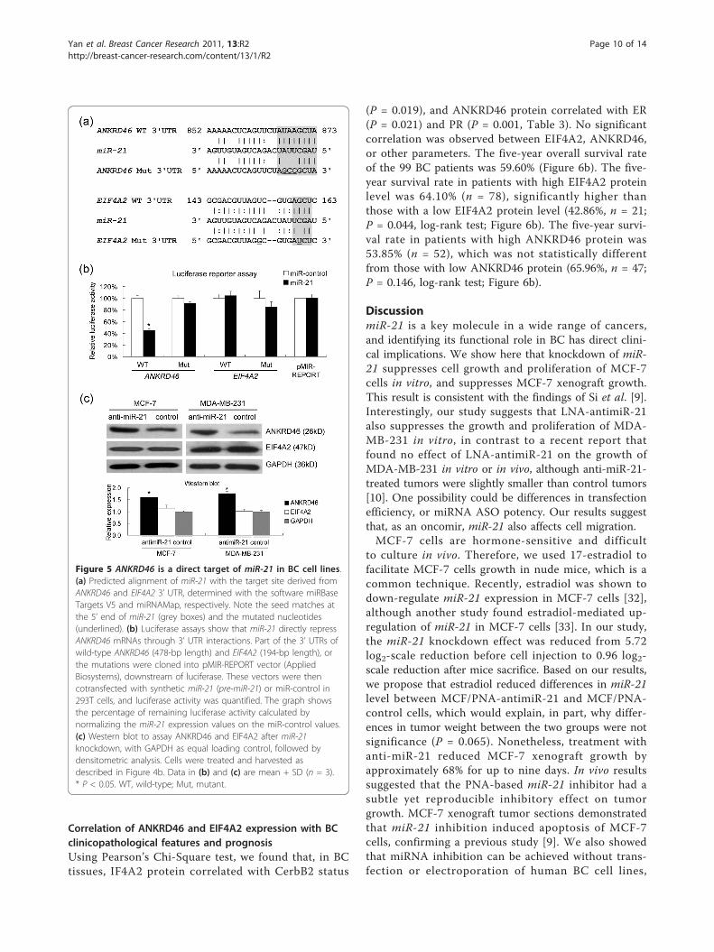
(P = 0.004), but no significant up-regulation of EIF4A2protein (Figure 5c).We further tested whether miR-21 could directly
repress the identified mRNA targets through 3’ UTRinteractions (Figure 5a). Thus, the full-length 3’ UTRs ofthe human genes ANKRD46 and EIF4A2 were clonedinto the downstream of the luciferase gene (pMIR-REPORT), respectively. These vectors were then used toassess whether miR-21 could repress luciferase activityin 293T cells. ANKRD46 3’ UTR showed a reduction to54.8% of total luciferase reporter activity, in presence ofmiR-21, but EIF4A2 3’ UTR did not display significantreduction of luciferase levels, compared with the miR-control (Figure 5b). These results suggest that miR-21directly targets ANKRD46 in BC cells.
miR-21 and EIF4A2 proteins are inversely expressed inresected patient tumors in vivoWe examined ANKRD46 [NCBI: NP940683] andEIF4A2 [NCBI: NP001958] protein levels by IHC onTMAs constructed by the BC cases described in
Figure 4 mRNA profiling of miR-21-knockdown BC cell lines. MCF-7 and MDA-MB-231 cells were transfected with 50 nM LNA-antimiR-21 orLNA-control. Total RNAs were isolated from MCF-7 cells 48 h post transfection and from MDA-MB-231 cells 36 h post transfection, respectively,and were hybridized to the human genome oligo array V1.0 (CapitalBio, China). (a) The scatter plot shows the LNA-antimiR-21 expression valuesnormalized to LNA-control values, applying a cut-off of 1.3. Each dot represents one probe set. (b) Unsupervised hierarchical cluster analysis ofintersection of mRNAs differentially expressed in MCF-7 and MDA-MB-231 cells upon LNA-antimiR-21 treatment. Rows, mRNAs; columns,biological samples. For each mRNA, red is expression higher than average expression across all samples, green is expression lower than average.(c) Validation of microarray results by qRT-PCR, normalizing on GAPDH RNA levels. MCF/A, MCF/LNA-antimiR-21; MCF/C, MCF/LNA-control; MDA/A, MDA/LNA-antimiR-21, MDA/C, MDA/LNA-control.
Yan et al. Breast Cancer Research 2011, 13:R2http://breast-cancer-research.com/content/13/1/R2
Page 8 of 14

Materials and Methods. EIF4A2 was found in the cyto-plasm, and ANKRD46 was seen in both the cytoplasmand nucleus (Figure 6a). ANKRD46 low expression wasfound in 47.5% (staining score < 4; median = 4); EIF4A2low expression was found in 21.2% (staining score < 7;median = 7) of the 99 BC cases. Next, we investigatedthe negative regulation of endogenous EIF4A2 and
ANKRD46 protein by endogenous miR-21 in vivo. In 99successfully tested cases out of 113, endogenous miR-21and EIF4A2 protein levels were inversely expressed inresected patient tumors (rs = -0.283, n = 99, P = 0.005,Spearman’s correlation analysis). However, no significantassociation between miR-21 and ANKRD46 (P = 0.181,Spearman’s correlation analysis) was observed.
Table 2 Regulated mRNAs in BC cells after miR-21 knockdown determined by oligo array
Fold change
Gene Name NCBI accession number Genbankaccessionnumber
Description MCF-7 MDA-MB-231
Predictionprogram
ANKRD46 NM_198401 U79297, BC035087 ankyrin repeat domain 46 1.78 2.57# miRBaseTargets V5
ACTN4 NM_004924 BC005033 Alpha-actin 4 (F-actin cross linking protein) 1.75 1.55
NR0B1 NM_000475 S74720 Nuclear receptor 0B1 (Nuclear receptor DAX-1)
1.60 1.32
HIRA NM_003325 BC039835, X89887 HIRA protein 1.48 1.32
COL18A1 NM_030582, NM_130444,NM_130445
AF018081 Collagen alpha 1(XVIII) chain precursor 1.43 1.35
SAMD11 NM_152486 BC024295,AK054643
sterile alpha motif domain containing 11 1.42 1.34
EIF4A2 NM_001967 AL117412 Eukaryotic initiation factor 4A-II 1.42 1.41 miRNAMap
SFRS16 NM_007056 AF042800,AK094681
Splicing factor, arginine/serine-rich 16 1.40 1.34
NP_079316 - BC004930 zinc finger protein 614 1.37 1.36
IL18BP NM_173043, NM_005699,NM_173042
AF110801 Interleukin-18 binding protein precursor 1.36 1.34
GBF1 NM_004193 AK025330,AF068755
Golgi-specific brefeldin A-resistance guaninenucleotide exchange factor 1
1.35 1.51
GNPDA1 NM_005471 AJ002231 Glucosamine-6-phosphate isomerase 1.34 1.32
SPUF_HUMAN NM_013349 - SPUF protein precursor 1.34 1.31
PLXNA1 NM_032242 AK127254 plexin A1; plexin 1 1.32 1.57
STMN3 NM_015894 AK094112 Stathmin 3 (SCG10-like protein) 1.32 1.47
NP_056236 NM_015421 AK093383,AL080088
DKFZP564K2062 protein 1.31 1.33
- - AK056473 - 1.31 1.30
AP2B1 NM_001282 M34175 adaptor-related protein complex 2, beta 1subunit
1.31 1.45
HIST1H2AC - CR608156 Histone H2A.l (H2A/l) -1.47 -1.67
PRKCI NM_002740 L33881, BC042405,BC022016
Protein kinase C, iota type -1.43 -1.32
SLC25A3 NM_002635, NM_213611,NM_213612, NM_005888
BX647062,AK057575
Phosphate carrier protein, mitochondrialprecursor (PTP)
-1.39 -1.39
CCNE1 NM_057182, NM_001238 M74093,BC035498
G1/S-specific cyclin E1 -1.37 -1.39
DCAMKL1 NM_004734 AB002367 Serine/threonine-protein kinase DCAMKL1 -1.34 -1.46
GNG12 NM_018841 AL832431 Guanine nucleotide-binding protein G(I)/G(S)/G(O) gamma-12 subunit
-1.32 -1.40 Miranda
Q8TAY7 NM_024869 BC025658 hypothetical protein FLJ14050 -1.32 -1.31
NP_653284 NM_144683 BC015582 hypothetical protein MGC23280 -1.31 -1.35
NP_055182 NM_014367 - growth and transformation-dependentprotein
-1.30 -1.34
Fold-regulation of mRNAs affected by miR-21 knockdown compared with control-transfected cells (MCF-7 and MDA-MB-231) at indicated time points. Fold-changes for all mRNAs are derived from a single microarray experiment, with two relevant genes confirmed by qRT-PCR (Figure 4B). Shown are mRNAs changingby at least 1.3-fold in both cell lines. Genes in bold were selected for candidate gene analyses by qRT-PCR.; # marginal signal intensity; NCBI, National Center forBiotechnology Information; GB, GenBank.
Yan et al. Breast Cancer Research 2011, 13:R2http://breast-cancer-research.com/content/13/1/R2
Page 9 of 14
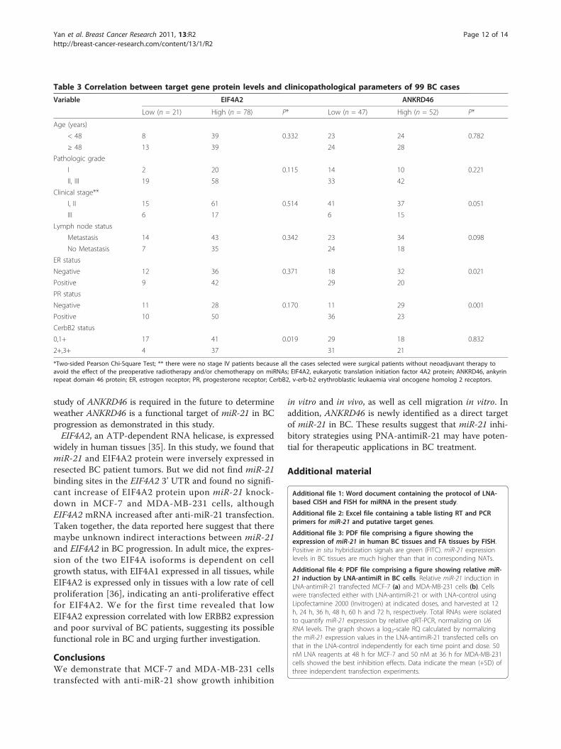
Correlation of ANKRD46 and EIF4A2 expression with BCclinicopathological features and prognosisUsing Pearson’s Chi-Square test, we found that, in BCtissues, IF4A2 protein correlated with CerbB2 status
(P = 0.019), and ANKRD46 protein correlated with ER(P = 0.021) and PR (P = 0.001, Table 3). No significantcorrelation was observed between EIF4A2, ANKRD46,or other parameters. The five-year overall survival rateof the 99 BC patients was 59.60% (Figure 6b). The five-year survival rate in patients with high EIF4A2 proteinlevel was 64.10% (n = 78), significantly higher thanthose with a low EIF4A2 protein level (42.86%, n = 21;P = 0.044, log-rank test; Figure 6b). The five-year survi-val rate in patients with high ANKRD46 protein was53.85% (n = 52), which was not statistically differentfrom those with low ANKRD46 protein (65.96%, n = 47;P = 0.146, log-rank test; Figure 6b).
DiscussionmiR-21 is a key molecule in a wide range of cancers,and identifying its functional role in BC has direct clini-cal implications. We show here that knockdown of miR-21 suppresses cell growth and proliferation of MCF-7cells in vitro, and suppresses MCF-7 xenograft growth.This result is consistent with the findings of Si et al. [9].Interestingly, our study suggests that LNA-antimiR-21also suppresses the growth and proliferation of MDA-MB-231 in vitro, in contrast to a recent report thatfound no effect of LNA-antimiR-21 on the growth ofMDA-MB-231 in vitro or in vivo, although anti-miR-21-treated tumors were slightly smaller than control tumors[10]. One possibility could be differences in transfectionefficiency, or miRNA ASO potency. Our results suggestthat, as an oncomir, miR-21 also affects cell migration.MCF-7 cells are hormone-sensitive and difficult
to culture in vivo. Therefore, we used 17-estradiol tofacilitate MCF-7 cells growth in nude mice, which is acommon technique. Recently, estradiol was shown todown-regulate miR-21 expression in MCF-7 cells [32],although another study found estradiol-mediated up-regulation of miR-21 in MCF-7 cells [33]. In our study,the miR-21 knockdown effect was reduced from 5.72log2-scale reduction before cell injection to 0.96 log2-scale reduction after mice sacrifice. Based on our results,we propose that estradiol reduced differences in miR-21level between MCF/PNA-antimiR-21 and MCF/PNA-control cells, which would explain, in part, why differ-ences in tumor weight between the two groups were notsignificance (P = 0.065). Nonetheless, treatment withanti-miR-21 reduced MCF-7 xenograft growth byapproximately 68% for up to nine days. In vivo resultssuggested that the PNA-based miR-21 inhibitor had asubtle yet reproducible inhibitory effect on tumorgrowth. MCF-7 xenograft tumor sections demonstratedthat miR-21 inhibition induced apoptosis of MCF-7cells, confirming a previous study [9]. We also showedthat miRNA inhibition can be achieved without trans-fection or electroporation of human BC cell lines,
Figure 5 ANKRD46 is a direct target of miR-21 in BC cell lines.(a) Predicted alignment of miR-21 with the target site derived fromANKRD46 and EIF4A2 3’ UTR, determined with the software miRBaseTargets V5 and miRNAMap, respectively. Note the seed matches atthe 5’ end of miR-21 (grey boxes) and the mutated nucleotides(underlined). (b) Luciferase assays show that miR-21 directly repressANKRD46 mRNAs through 3’ UTR interactions. Part of the 3’ UTRs ofwild-type ANKRD46 (478-bp length) and EIF4A2 (194-bp length), orthe mutations were cloned into pMIR-REPORT vector (AppliedBiosystems), downstream of luciferase. These vectors were thencotransfected with synthetic miR-21 (pre-miR-21) or miR-control in293T cells, and luciferase activity was quantified. The graph showsthe percentage of remaining luciferase activity calculated bynormalizing the miR-21 expression values on the miR-control values.(c) Western blot to assay ANKRD46 and EIF4A2 after miR-21knockdown, with GAPDH as equal loading control, followed bydensitometric analysis. Cells were treated and harvested asdescribed in Figure 4b. Data in (b) and (c) are mean + SD (n = 3).* P < 0.05. WT, wild-type; Mut, mutant.
Yan et al. Breast Cancer Research 2011, 13:R2http://breast-cancer-research.com/content/13/1/R2
Page 10 of 14

highlighting the potential of PNA for future therapeuticapplications.ANKRD46, also known as ankyrin repeat small protein
(ANK-S), is a 228-amino acid single-pass membraneprotein, of unclear function. For the first time, we iden-tify miR-21 as an important regulator of ANKRD46mRNA and protein levels in BC cells. Our data showedthat miR-21 directly interacted with the ANKRD46 3’UTR and inhibited ANKRD46 expression, though therewas no significant association between miR-21 andANKRD46 in resected patient tumors. This discrepancy
may be due to three reasons. First, the artificial lucifer-ase reporter assays do not fully recapitulate miRNAregulation in vivo [34]; second, the expression ofANKRD46 protein in patient tumors reflected specifictime-point feature, which maybe different to the in vitrosubsequent increase of ANKRD46 protein at the timepoint of observation (the indicated hours after transfec-tion); third, immunohistochemistry (IHC) is conven-tional a semi-quantitative method with relatively limitedsensitivity. IHC may not be sensitive enough to observethe down-regulation of ANKRD46 by miR-21. Functional
Figure 6 EIF4A2 or ANKRD46 expression and survival in BC patients. (a) Representative TMAs sections stained for ANKRD46 and EIF4A2from different BC. Examples for staining score 0 (negative), 3, 5 and 7 of marker expression are shown. All images were taken at × 200.(b) Kaplan-Meier survival curve and log-rank test for 99 BC patients showing five-year overall survival. ANKRD46 expression had no significantrelationship to patient survival (P = 0.146). High EIF4A2 expression was associated with significantly improved OS (P = 0.044) in women with BC.TMAs, tissue microarrays; OS, overall survival.
Yan et al. Breast Cancer Research 2011, 13:R2http://breast-cancer-research.com/content/13/1/R2
Page 11 of 14
study of ANKRD46 is required in the future to determineweather ANKRD46 is a functional target of miR-21 in BCprogression as demonstrated in this study.EIF4A2, an ATP-dependent RNA helicase, is expressed
widely in human tissues [35]. In this study, we found thatmiR-21 and EIF4A2 protein were inversely expressed inresected BC patient tumors. But we did not find miR-21binding sites in the EIF4A2 3’ UTR and found no signifi-cant increase of EIF4A2 protein upon miR-21 knock-down in MCF-7 and MDA-MB-231 cells, althoughEIF4A2 mRNA increased after anti-miR-21 transfection.Taken together, the data reported here suggest that theremaybe unknown indirect interactions between miR-21and EIF4A2 in BC progression. In adult mice, the expres-sion of the two EIF4A isoforms is dependent on cellgrowth status, with EIF4A1 expressed in all tissues, whileEIF4A2 is expressed only in tissues with a low rate of cellproliferation [36], indicating an anti-proliferative effectfor EIF4A2. We for the first time revealed that lowEIF4A2 expression correlated with low ERBB2 expressionand poor survival of BC patients, suggesting its possiblefunctional role in BC and urging further investigation.
ConclusionsWe demonstrate that MCF-7 and MDA-MB-231 cellstransfected with anti-miR-21 show growth inhibition
in vitro and in vivo, as well as cell migration in vitro. Inaddition, ANKRD46 is newly identified as a direct targetof miR-21 in BC. These results suggest that miR-21 inhi-bitory strategies using PNA-antimiR-21 may have poten-tial for therapeutic applications in BC treatment.
Additional material
Additional file 1: Word document containing the protocol of LNA-based CISH and FISH for miRNA in the present study.
Additional file 2: Excel file containing a table listing RT and PCRprimers for miR-21 and putative target genes.
Additional file 3: PDF file comprising a figure showing theexpression of miR-21 in human BC tissues and FA tissues by FISH.Positive in situ hybridization signals are green (FITC). miR-21 expressionlevels in BC tissues are much higher than that in corresponding NATs.
Additional file 4: PDF file comprising a figure showing relative miR-21 induction by LNA-antimiR in BC cells. Relative miR-21 induction inLNA-antimiR-21 transfected MCF-7 (a) and MDA-MB-231 cells (b). Cellswere transfected either with LNA-antimiR-21 or with LNA-control usingLipofectamine 2000 (Invitrogen) at indicated doses, and harvested at 12h, 24 h, 36 h, 48 h, 60 h and 72 h, respectively. Total RNAs were isolatedto quantify miR-21 expression by relative qRT-PCR, normalizing on U6RNA levels. The graph shows a log2-scale RQ calculated by normalizingthe miR-21 expression values in the LNA-antimiR-21 transfected cells onthat in the LNA-control independently for each time point and dose. 50nM LNA reagents at 48 h for MCF-7 and 50 nM at 36 h for MDA-MB-231cells showed the best inhibition effects. Data indicate the mean (+SD) ofthree independent transfection experiments.
Table 3 Correlation between target gene protein levels and clinicopathological parameters of 99 BC cases
Variable EIF4A2 ANKRD46
Low (n = 21) High (n = 78) P* Low (n = 47) High (n = 52) P*
Age (years)
< 48 8 39 0.332 23 24 0.782
≥ 48 13 39 24 28
Pathologic grade
I 2 20 0.115 14 10 0.221
II, III 19 58 33 42
Clinical stage**
I, II 15 61 0.514 41 37 0.051
III 6 17 6 15
Lymph node status
Metastasis 14 43 0.342 23 34 0.098
No Metastasis 7 35 24 18
ER status
Negative 12 36 0.371 18 32 0.021
Positive 9 42 29 20
PR status
Negative 11 28 0.170 11 29 0.001
Positive 10 50 36 23
CerbB2 status
0,1+ 17 41 0.019 29 18 0.832
2+,3+ 4 37 31 21
*Two-sided Pearson Chi-Square Test; ** there were no stage IV patients because all the cases selected were surgical patients without neoadjuvant therapy toavoid the effect of the preoperative radiotherapy and/or chemotherapy on miRNAs; EIF4A2, eukaryotic translation initiation factor 4A2 protein; ANKRD46, ankyrinrepeat domain 46 protein; ER, estrogen receptor; PR, progesterone receptor; CerbB2, v-erb-b2 erythroblastic leukaemia viral oncogene homolog 2 receptors.
Yan et al. Breast Cancer Research 2011, 13:R2http://breast-cancer-research.com/content/13/1/R2
Page 12 of 14

Abbreviations3’ UTR: 3’ untranslated region; AJCC: American Joint Committee on Cancer;ANK-S: ankyrin repeat small protein; ASOs: antisense oligonucleotides;BC: breast cancer; CerbB2: v-erb-b2 erythroblastic leukaemia viral oncogenehomolog 2 receptors; CISH: chromogenic in situ hybridization; DAPI: 4’,6-diamidino-2-phenylindole; EIF4A2: eukaryotic initiation factor 4A: isoform 2;ER: estrogen receptor; F: forward primer; FA: fibroadenoma; FISH: fluoresceinin situ hybridization; FITC: fluorescein isothiocyanate; GEO: Gene ExpressionOmnibus; IHC: immunohistochemistry; L: length; LNA: Locked nucleic acid;miRNAs: microRNAs; MTT: 3-(4,5-dimethylthiazol-2-yl)-2,5-diphenyltetrazoliumbromide; Mut: mutation; NATs: normal adjacent tissues; NBT/BCIP: nitro bluetetrazolium/5-bromo-4-chloro-3-indolyl phosphate; NCBI: National Center forBiotechnology Information; OS: overall survival; PNAs: peptide nucleic acids;PR: progesterone receptor; qRT-PCR: quantitative reverse transcription-polymerase chain reaction; R: reverse primer; RQ: relative quantification;RT: reverse transcription primer; SABC-AP: alkaline phosphatase-conjugatedstrept-avidin-biotin complex; SD: standard deviation: SYSUCC: Sun Yat-senUniversity Cancer Center; TMAs: tissue microrrays; TNM: tumor-lymph node-metastasis; W: width; WT: wild-type.
AcknowledgementsThis work was supported in part by the National High Technology Researchand Development Program of China (863 Program) (No. 20060102A4002), aresearch grant from State Key Laboratory of Oncology in Southern China,985-II Project.
Author details1State Key Laboratory of Oncology in Southern China, Sun Yat-Sen UniversityCancer Center, 651 Dong Feng Road East, Guangzhou, 510060, PR China.2Department of Pathology, Sun Yat-Sen University Cancer Center, 651 DongFeng Road East, Guangzhou, 510060, PR China. 3Department of ExperimentResearch, Sun Yat-Sen University Cancer Center, 651 Dong Feng Road East,Guangzhou, 510060, PR China.
Authors’ contributionsLXY, QNW and YZ carried out the substantial experiment work and draftedthe manuscript. JYS designed and financially supported the study. QNW andDZL were responsible for patient samples and tissue array construction. JHHand JF supported the immunohistochemistry. YYL carried out the luciferasereporter assay. JPY, MSZ, QLW and YXZ helped carry out the research designand critically reviewed the final version of the manuscript for submission. Allauthors read and approved the final manuscript.
Competing interestsMiss Li Xu Yan and Mrs Yan Zhang are doctoral degree candidates; Miss QiNian Wu and Mrs Yang Yang Li are master’s degree candidates at SYSUCC.Mr Ding Zhun Liao, Mr Jing Hui Hou and Mrs Jia Fu are technicians atSYSUCC. Dr Mu-Sheng Zeng, Jing Ping Yun, Qiu Liang Wu and Yi Xin Zengare Professors at SYSUCC. Dr Shao is a Professor and Vice Director atDepartment of Pathology of SYSUCC. The authors declare that they havenot received any reimbursements, fees, funding, or salary, nor hold anystocks or shares in an organization that may in any way gain or losefinancially from the publication of this manuscript, either now or in thefuture. The authors do not hold or are not currently applying for anypatents relating to the content of the manuscript. The authors declare thatthey do not have any other financial or non-financial competing interests.
Received: 7 March 2010 Revised: 15 October 2010Accepted: 10 January 2011 Published: 10 January 2011
References1. Parkin DM, Bray F, Ferlay J, Pisani P: Global cancer statistics, 2002. CA
Cancer J Clin 2005, 55:74-108.2. Ambros V: MicroRNA pathways in flies and worms: growth, death, fat,
stress, and timing. Cell 2003, 113:673-676.3. Esquela-Kerscher A, Slack FJ: Oncomirs - microRNAs with a role in cancer.
Nat Rev Cancer 2006, 6:259-269.4. Chan JA, Krichevsky AM, Kosik KS: MicroRNA-21 is an antiapoptotic factor
in human glioblastoma cells. Cancer Res 2005, 65:6029-6033.5. Fulci V, Chiaretti S, Goldoni M, Azzalin G, Carucci N, Tavolaro S, Castellano L,
Magrelli A, Citarella F, Messina M, Maggio R, Peragine N, Santangelo S,
Mauro FR, Landgraf P, Tuschl T, Weir DB, Chien M, Russo JJ, Ju J,Sheridan R, Sander C, Zavolan M, Guarini A, Foa R, Macino G: Quantitativetechnologies establish a novel microRNA profile of chronic lymphocyticleukemia. Blood 2007, 109:4944-4951.
6. Yanaihara N, Caplen N, Bowman E, Seike M, Kumamoto K, Yi M,Stephens RM, Okamoto A, Yokota J, Tanaka T, Calin GA, Liu CG, Croce CM,Harris CC: Unique microRNA molecular profiles in lung cancer diagnosisand prognosis. Cancer Cell 2006, 9:189-198.
7. Iorio MV, Ferracin M, Liu CG, Veronese A, Spizzo R, Sabbioni S, Magri E,Pedriali M, Fabbri M, Campiglio M, Menard S, Palazzo JP, Rosenberg A,Musiani P, Volinia S, Nenci I, Calin GA, Querzoli P, Negrini M, Croce CM:MicroRNA gene expression deregulation in human breast cancer. CancerRes 2005, 65:7065-7070.
8. Frankel LB, Christoffersen NR, Jacobsen A, Lindow M, Krogh A, Lund AH:Programmed cell death 4 (PDCD4) is an important functional target ofthe microRNA miR-21 in breast cancer cells. J Biol Chem 2008,283:1026-1033.
9. Si ML, Zhu S, Wu H, Lu Z, Wu F, Mo YY: miR-21-mediated tumor growth.Oncogene 2007, 26:2799-2803.
10. Zhu S, Wu H, Wu F, Nie D, Sheng S, Mo YY: MicroRNA-21 targets tumorsuppressor genes in invasion and metastasis. Cell Res 2008, 18:350-359.
11. Oh SY, Ju Y, Park H: A highly effective and long-lasting inhibition ofmiRNAs with PNA-based antisense oligonucleotides. Mol Cells 2009,28:341-345.
12. In Pathology and Genetics of Tumours of the Breast and Female GenitalOrgans. Edited by: Tavassoli F, Devilee P. Lyon: IARC Press; 2003.
13. Singletary SE, Allred C, Ashley P, Bassett LW, Berry D, Bland KI, Borgen PI,Clark GM, Edge SB, Hayes DF, Hughes LL, Hutter RV, Morrow M, Page DL,Recht A, Theriault RL, Thor A, Weaver DL, Wieand HS, Greene FL: Stagingsystem for breast cancer: revisions for the 6th edition of the AJCCCancer Staging Manual. The Surgical Clinics of North America 2003,83:803-819.
14. Obernosterer G, Martinez J, Alenius M: Locked nucleic acid-based in situdetection of microRNAs in mouse tissue sections. Nature Protocols 2007,2:1508-1514.
15. Lee SH, Kunz J, Lin SH, Yu-Lee LY: 16-kDa prolactin inhibits endothelialcell migration by down-regulating the Ras-Tiam1-Rac1-Pak1 signalingpathway. Cancer Res 2007, 67:11045-11053.
16. Wu L, Li Z, Zhang Y, Zhang P, Zhu X, Huang J, Ma T, Lu T, Song Q, Li Q,Guo Y, Tang J, Ma D, Chen KH, Qiu X: Adenovirus-expressed humanhyperplasia suppressor gene induces apoptosis in cancer cells. MolCancer Ther 2008, 7:222-232.
17. Melo SA, Ropero S, Moutinho C, Aaltonen LA, Yamamoto H, Calin GA,Rossi S, Fernandez AF, Carneiro F, Oliveira C, Ferreira B, Liu CG, Villanueva A,Capella G, Schwartz SJr, Shiekhattar R, Esteller M: A TARBP2 mutation inhuman cancer impairs microRNA processing and DICER1 function. NatGenet 2009, 41:365-370.
18. Patterson TA, Lobenhofer EK, Fulmer-Smentek SB, Collins PJ, Chu TM,Bao W, Fang H, Kawasaki ES, Hager J, Tikhonova IR, Walker SJ, Zhang L,Hurban P, de Longueville F, Fuscoe JC, Tong W, Shi L, Wolfinger RD:Performance comparison of one-color and two-color platforms withinthe MicroArray Quality Control (MAQC) project. Nat Biotechnol 2006,24:1140-1150.
19. Gene Expression Omnibus, GEO. [http://www.ncbi.nlm.nih.gov/geo/].20. Yan LX, Huang XF, Shao Q, Huang MY, Deng L, Wu QL, Zeng YX, Shao JY:
MicroRNA miR-21 overexpression in human breast cancer is associatedwith advanced clinical stage, lymph node metastasis and patient poorprognosis. RNA 2008, 14:2348-2360.
21. Gramantieri L, Ferracin M, Fornari F, Veronese A, Sabbioni S, Liu CG,Calin GA, Giovannini C, Ferrazzi E, Grazi GL, Croce CM, Bolondi L, Negrini M:Cyclin G1 is a target of miR-122a, a microRNA frequently down-regulated in human hepatocellular carcinoma. Cancer Res 2007,67:6092-6099.
22. Grimson A, Farh KK, Johnston WK, Garrett-Engele P, Lim LP, Bartel DP:MicroRNA targeting specificity in mammals: determinants beyond seedpairing. Mol Cell 2007, 27:91-105.
23. Griffiths-Jones S, Saini HK, van Dongen S, Enright AJ: miRBase: tools formicroRNA genomics. Nucleic Acids Res 2008, 36:D154-158.
24. Hsu SD, Chu CH, Tsou AP, Chen SJ, Chen HC, Hsu PW, Wong YH, Chen YH,Chen GH, Huang HD: miRNAMap 2.0: genomic maps of microRNAs inmetazoan genomes. Nucleic Acids Res 2008, 36:D165-169.
Yan et al. Breast Cancer Research 2011, 13:R2http://breast-cancer-research.com/content/13/1/R2
Page 13 of 14

25. Krek A, Grun D, Poy MN, Wolf R, Rosenberg L, Epstein EJ, MacMenamin P,da Piedade I, Gunsalus KC, Stoffel M, Rajewsky N: Combinatorial microRNAtarget predictions. Nat Genet 2005, 37:495-500.
26. John B, Enright AJ, Aravin A, Tuschl T, Sander C, Marks DS: HumanMicroRNA targets. PLoS Biol 2004, 2:e363.
27. Song CH, Park SY, Eom KY, Kim JH, Kim SW, Kim JS, Kim IA: Potentialprognostic value of heat-shock protein 90 in the presence ofphosphatidylinositol-3-kinase overexpression or loss of PTEN, in invasivebreast cancers. Breast Cancer Res 2010, 12:R20.
28. Gamallo C, Palacios J, Suarez A, Pizarro A, Navarro P, Quintanilla M, Cano A:Correlation of E-cadherin expression with differentiation grade andhistological type in breast carcinoma. The American Journal of Pathology1993, 142:987-993.
29. Tuominen VJ, Ruotoistenmaki S, Viitanen A, Jumppanen M, Isola J:ImmunoRatio: a publicly available web application for quantitativeimage analysis of estrogen receptor (ER), progesterone receptor (PR),and Ki-67. Breast Cancer Res 2010, 12:R56.
30. Sempere LF, Christensen M, Silahtaroglu A, Bak M, Heath CV, Schwartz G,Wells W, Kauppinen S, Cole CN: Altered MicroRNA expression confined tospecific epithelial cell subpopulations in breast cancer. Cancer Res 2007,67:11612-11620.
31. Matkovich SJ, Van Booven DJ, Youker KA, Torre-Amione G, Diwan A,Eschenbacher WH, Dorn LE, Watson MA, Margulies KB, Dorn GW: Reciprocalregulation of myocardial microRNAs and messenger RNA in humancardiomyopathy and reversal of the microRNA signature bybiomechanical support. Circulation 2009, 119:1263-1271.
32. Wickramasinghe NS, Manavalan TT, Dougherty SM, Riggs KA, Li Y,Klinge CM: Estradiol downregulates miR-21 expression and increasesmiR-21 target gene expression in MCF-7 breast cancer cells. Nucleic AcidsRes 2009, 37:2584-2595.
33. Bhat-Nakshatri P, Wang G, Collins NR, Thomson MJ, Geistlinger TR,Carroll JS, Brown M, Hammond S, Srour EF, Liu Y, Nakshatri H: Estradiol-regulated microRNAs control estradiol response in breast cancer cells.Nucleic Acids Res 2009, 37:4850-4861.
34. Eulalio A, Huntzinger E, Izaurralde E: Getting to the root of miRNA-mediated gene silencing. Cell 2008, 132:9-14.
35. Sudo K, Takahashi E, Nakamura Y: Isolation and mapping of the humanEIF4A2 gene homologous to the murine protein synthesis initiationfactor 4A-II gene Eif4a2. Cytogenet Cell Genet 1995, 71:385-388.
36. Williams-Hill DM, Duncan RF, Nielsen PJ, Tahara SM: Differential expressionof the murine eukaryotic translation initiation factor isogenes eIF4A(I)and eIF4A(II) is dependent upon cellular growth status. Arch BiochemBiophys 1997, 338:111-120.
doi:10.1186/bcr2803Cite this article as: Yan et al.: Knockdown of miR-21 in human breastcancer cell lines inhibits proliferation, in vitro migration and in vivotumor growth. Breast Cancer Research 2011 13:R2.
Submit your next manuscript to BioMed Centraland take full advantage of:
• Convenient online submission
• Thorough peer review
• No space constraints or color figure charges
• Immediate publication on acceptance
• Inclusion in PubMed, CAS, Scopus and Google Scholar
• Research which is freely available for redistribution
Submit your manuscript at www.biomedcentral.com/submit
Yan et al. Breast Cancer Research 2011, 13:R2http://breast-cancer-research.com/content/13/1/R2
Page 14 of 14