Widespread Bronchogenic Dissemination Makes DBA/2 Mice More Susceptible than C57BL/6 Mice to...
10
INFECTION AND IMMUNITY, Oct. 2003, p. 5845–5854 Vol. 71, No. 10 0019-9567/03/$08.000 DOI: 10.1128/IAI.71.10.5845–5854.2003 Copyright © 2003, American Society for Microbiology. All Rights Reserved. Widespread Bronchogenic Dissemination Makes DBA/2 Mice More Susceptible than C57BL/6 Mice to Experimental Aerosol Infection with Mycobacterium tuberculosis Pere-Joan Cardona, 1 * Sergi Gordillo, 1 Jorge Díaz, 1 Gustavo Tapia, 2 Isabel Amat, 1 A ´ ngeles Pallare ´s, 1 Cristina Vilaplana, 1 Aurelio Ariza, 2 and Vicenc ¸ Ausina 1 Unitat de Tuberculosi Experimental, Department of Microbiology, 1 and Department of Pathology, 2 Hospital Universitari “Germans Trias i Pujol” and Universitat Auto `noma de Barcelona, Badalona, Catalonia, Spain Received 4 March 2003/Returned for modification 8 April 2003/Accepted 1 July 2003 We have used the murine model of aerosol-induced experimental tuberculosis to assess the effects of four clinical isolates and a reference strain of Mycobacterium tuberculosis on resistant C57BL/6 mice and susceptible DBA/2 mice. Histological studies and detection of 25 cytokines potentially involved in the infection were carried out. DBA/2 mice showed higher concentrations of bacilli in bronchoalveolar lavage fluid and lung tissue. Furthermore, these mice evidenced a larger granulomatous infiltration in the parenchyma due to an increased rate of emigration of infected foamy macrophages from the granulomas to the neighboring pulmonary alveolar spaces. The better control of bacillary concentrations and pulmonary infiltration observed in C57BL/6 mice from week 3 postinfection could result from their higher RANTES, ICAM-1, and gamma interferon (IFN-) mRNA levels. On the other hand, the higher MIP-2 and MCP-3 mRNA levels seen in DBA/2 mice would result in stronger lung recruitment of macrophages and neutrophils. Additionally, DBA/2 mice showed increased inducible nitric oxide synthase expression, induced by the larger number of foamy macrophages, at weeks 18 and 22. This increment was a consequence of phagocytosed bacillary debris, was independent of IFN- expression, and could exert only a bacteriostatic effect. The results of the study suggest that DBA/2 mice are more susceptible than C57BL/6 mice to M. tuberculosis infection due to a higher bronchial dissemination of bacilli inside poorly activated foamy macrophages. At the beginning of the 21st century, tuberculosis continues to be an important cause of death and a major public health concern. According to World Health Organization estimates, one-third of humankind (2 billion people) is infected with Mycobacterium tuberculosis. This extraordinarily high preva- lence of the latent infection is one of the main factors contrib- uting to the high incidence of active tuberculosis (more than 8 million new cases per year). Although most infected people never develop active disease, in approximately 10% of cases reactivation of latent infection results in active tuberculosis (49). Experimental models of murine tuberculosis are of para- mount importance for gaining a deeper knowledge of latent infection and the genetic host factors favoring reactivation. Different strains of mice have long been known to have differ- ent innate resistances to tuberculous challenge (18, 36). Me- dina and North first identified susceptibility variations in sev- eral inbred mouse strains, showing different survival times after aerosol and intravenous inoculation. These authors related such differences to bcg gene (Nramp1) and histocompatibility complex haplotype effects (27, 28) and classified six inbred mouse strains as either highly susceptible (CBA, DBA/2, C3H, and 129/SvJ) or highly resistant (BALB/c and C57BL/6). Many subsequent studies have contributed to a better un- derstanding of the basis for these differences, but the subject still awaits full clarification. Thus, the fact that mycobacteria seem to disseminate earlier from the lungs of resistant mice than from those of susceptible mice has been associated with the stronger response shown by the former (6). It has also been demonstrated that the inability of reactivation-prone mice to recruit lymphocytes to the infection site may underlie their predisposition to chronic-infection control failure and associ- ated bacterial regrowth (46). In the same way, the F 1 progeny of susceptible (I/St) and resistant (A/Sn) parental strains have shown an infection-hyperresistant intermediate phenotype providing a better defense against tuberculosis (25). On the other hand, identification of the genetic basis of susceptibility to tuberculosis has provided deeper insights into immuno- pathological aspects (20, 24, 30, 33), and the link to the tbs1 and tbs2 loci has suggested the existence of a basic genetic network for the control of intracellular parasites (42). A complex chemokine and cytokine profile is produced dur- ing M. tuberculosis infection (38). Members of the beta sub- family of chemokines (C-C chemokines), such as macrophage chemotactic protein 1 (MCP-1), macrophage inflammatory protein 1 alpha/beta (MIP-1/), and RANTES (regulated upon activation normal T-cell expression sequence), seem to be strongly involved not only in attracting monocytes and lym- phocytes to the sites of infection but also in favoring a T helper type 1 (Th1) response (9). Members of the alpha subfamily of chemokines (C-X-C chemokines), such as interferon-inducible protein (IP-10), are usually related to the attraction of neutro- phils but also act as potent attractors of monocytes and acti- vated T lymphocytes (22). * Corresponding author. Mailing address: Unitat de Tuberculosi Experimental, Department of Microbiology, Hospital Universitari “Germans Trias i Pujol,” Crta del Canyet s/n, 08916 Badalona, Cata- lonia, Spain. Phone: 34 93 497 88 94. Fax: 34 93 497 88 95. E-mail: [email protected]. 5845
-
Upload
independent -
Category
Documents
-
view
3 -
download
0
Transcript of Widespread Bronchogenic Dissemination Makes DBA/2 Mice More Susceptible than C57BL/6 Mice to...
INFECTION AND IMMUNITY, Oct. 2003, p. 5845–5854 Vol. 71, No. 100019-9567/03/$08.00�0 DOI: 10.1128/IAI.71.10.5845–5854.2003Copyright © 2003, American Society for Microbiology. All Rights Reserved.
Widespread Bronchogenic Dissemination Makes DBA/2 Mice MoreSusceptible than C57BL/6 Mice to Experimental Aerosol
Infection with Mycobacterium tuberculosisPere-Joan Cardona,1* Sergi Gordillo,1 Jorge Díaz,1 Gustavo Tapia,2 Isabel Amat,1
Angeles Pallares,1 Cristina Vilaplana,1 Aurelio Ariza,2 and Vicenc Ausina1
Unitat de Tuberculosi Experimental, Department of Microbiology,1 and Department of Pathology,2 Hospital Universitari“Germans Trias i Pujol” and Universitat Autonoma de Barcelona, Badalona, Catalonia, Spain
Received 4 March 2003/Returned for modification 8 April 2003/Accepted 1 July 2003
We have used the murine model of aerosol-induced experimental tuberculosis to assess the effects of fourclinical isolates and a reference strain of Mycobacterium tuberculosis on resistant C57BL/6 mice and susceptibleDBA/2 mice. Histological studies and detection of 25 cytokines potentially involved in the infection were carriedout. DBA/2 mice showed higher concentrations of bacilli in bronchoalveolar lavage fluid and lung tissue.Furthermore, these mice evidenced a larger granulomatous infiltration in the parenchyma due to an increasedrate of emigration of infected foamy macrophages from the granulomas to the neighboring pulmonary alveolarspaces. The better control of bacillary concentrations and pulmonary infiltration observed in C57BL/6 micefrom week 3 postinfection could result from their higher RANTES, ICAM-1, and gamma interferon (IFN-�)mRNA levels. On the other hand, the higher MIP-2 and MCP-3 mRNA levels seen in DBA/2 mice would resultin stronger lung recruitment of macrophages and neutrophils. Additionally, DBA/2 mice showed increasedinducible nitric oxide synthase expression, induced by the larger number of foamy macrophages, at weeks 18and 22. This increment was a consequence of phagocytosed bacillary debris, was independent of IFN-�expression, and could exert only a bacteriostatic effect. The results of the study suggest that DBA/2 mice aremore susceptible than C57BL/6 mice to M. tuberculosis infection due to a higher bronchial dissemination ofbacilli inside poorly activated foamy macrophages.
At the beginning of the 21st century, tuberculosis continuesto be an important cause of death and a major public healthconcern. According to World Health Organization estimates,one-third of humankind (2 billion people) is infected withMycobacterium tuberculosis. This extraordinarily high preva-lence of the latent infection is one of the main factors contrib-uting to the high incidence of active tuberculosis (more than 8million new cases per year). Although most infected peoplenever develop active disease, in approximately 10% of casesreactivation of latent infection results in active tuberculosis(49).
Experimental models of murine tuberculosis are of para-mount importance for gaining a deeper knowledge of latentinfection and the genetic host factors favoring reactivation.Different strains of mice have long been known to have differ-ent innate resistances to tuberculous challenge (18, 36). Me-dina and North first identified susceptibility variations in sev-eral inbred mouse strains, showing different survival times afteraerosol and intravenous inoculation. These authors relatedsuch differences to bcg gene (Nramp1) and histocompatibilitycomplex haplotype effects (27, 28) and classified six inbredmouse strains as either highly susceptible (CBA, DBA/2, C3H,and 129/SvJ) or highly resistant (BALB/c and C57BL/6).
Many subsequent studies have contributed to a better un-
derstanding of the basis for these differences, but the subjectstill awaits full clarification. Thus, the fact that mycobacteriaseem to disseminate earlier from the lungs of resistant micethan from those of susceptible mice has been associated withthe stronger response shown by the former (6). It has also beendemonstrated that the inability of reactivation-prone mice torecruit lymphocytes to the infection site may underlie theirpredisposition to chronic-infection control failure and associ-ated bacterial regrowth (46). In the same way, the F1 progenyof susceptible (I/St) and resistant (A/Sn) parental strains haveshown an infection-hyperresistant intermediate phenotypeproviding a better defense against tuberculosis (25). On theother hand, identification of the genetic basis of susceptibilityto tuberculosis has provided deeper insights into immuno-pathological aspects (20, 24, 30, 33), and the link to the tbs1and tbs2 loci has suggested the existence of a basic geneticnetwork for the control of intracellular parasites (42).
A complex chemokine and cytokine profile is produced dur-ing M. tuberculosis infection (38). Members of the beta sub-family of chemokines (C-C chemokines), such as macrophagechemotactic protein 1 (MCP-1), macrophage inflammatoryprotein 1 alpha/beta (MIP-1�/�), and RANTES (regulatedupon activation normal T-cell expression sequence), seem tobe strongly involved not only in attracting monocytes and lym-phocytes to the sites of infection but also in favoring a T helpertype 1 (Th1) response (9). Members of the alpha subfamily ofchemokines (C-X-C chemokines), such as interferon-inducibleprotein (IP-10), are usually related to the attraction of neutro-phils but also act as potent attractors of monocytes and acti-vated T lymphocytes (22).
* Corresponding author. Mailing address: Unitat de TuberculosiExperimental, Department of Microbiology, Hospital Universitari“Germans Trias i Pujol,” Crta del Canyet s/n, 08916 Badalona, Cata-lonia, Spain. Phone: 34 93 497 88 94. Fax: 34 93 497 88 95. E-mail:[email protected].
5845
Generation of specific CD4� T cells is initiated by the rec-ognition of peptides in the context of major histocompatibilitycomplex class II (MHC II) molecules (12, 23). These CD4� Tcells differentiate into effector cells that are able to produceappropriate levels of cytokines. The early immune response ischaracterized by the predominance of Th1 cells, which activateinfected macrophages through gamma interferon (IFN-�) pro-duction to thwart M. tuberculosis growth (11, 17, 34).
It is well known that IFN-� induces macrophage activationand promotes inducible nitric oxide synthase (iNOS) expres-sion, leading to the production of nitric oxide (NO) and reac-tive nitrogen intermediates (RNI). It is unclear, however,whether IFN-� exerts a mycobactericidal (8, 14, 26) or a my-cobacteriostatic (13, 16, 39) effect in murine tuberculosis. Bethat as it may, IFN-� augments the acidity of infected phago-somes (45, 48), which seems to be crucial for the enhancementof RNI activity (as is the M. tuberculosis strain involved) (39).In contrast, the larger NO amounts produced by murine lungmacrophages limit the magnitude of local mucosal T-cell re-sponses by suppressing T-cell activity (44). In this regard, itmust be stated that IFN-� is not solely responsible for theproduction of NO by macrophages, which can also be elicitedby several M. tuberculosis cell wall components, such as li-poarabinomannan or the 19-kDa antigen (7).
In the present work, two strains of mice have been infectedvia aerosol inhalation with four virulent M. tuberculosis clinicalisolates and strain H37Rv. We provide evidence of suscepti-bility differences between DBA/2 and C57BL/6 mice, whichshow dissimilar bacillary concentrations, histopathologicalfindings, and chemokine and cytokine expression profiles (asdetermined by real-time PCR).
Specifically, susceptibility differences manifested themselvesby the early presence in DBA/2 mice of large granulomas withmultiple layers of infected foamy macrophages, high CFUcounts in bronchoalveolar lavage (BAL) fluid and lung tissue,and low expression of IFN-� and RANTES in the lung. Incontrast, C57BL/6 mice showed smaller and scantier granulo-mas, moderate CFU counts, and significantly increased IFN-�and RANTES expression. We have also shown that the higheriNOS expression triggered in DBA/2 mice during the finalstages of infection failed to prevent fatal progression. Thepresent study, therefore, suggests that DBA/2 mice are moresusceptible to M. tuberculosis infection than C57BL/6 mice.Probably this is the consequence of a more widespread bron-chogenic dissemination of bacilli transported by insufficientlyactivated foamy macrophages in DBA/2 mice.
(Part of this research has been presented previously [P. J.Cardona, S. Gordillo, J. Dıaz, I. Amat, J. Lonca, and V.Ausina, Abstr. 102nd Gen. Meet. Am. Soc. Microbiol. 2002,abstr. U-61, p. 486, 2002; P. J. Cardona S. Gordillo, J. Dıaz, R.Llatjos, A. Ariza, and V. Ausina, Abstr. 5th Int. Conf. Pathog.Mycobacterial Infect. 2002, abstr. P17]).
MATERIALS AND METHODS
Mice. Our study was performed using specific-pathogen-free (spf) DBA/2(H-2d) and C57BL/6 (H-2b) female mice, 6 to 8 weeks old, which had beenobtained from Charles River (Bagneux Cedex, France). Mice were shippedunder conditions appropriate for travel, with the corresponding certificate ofhealth and origin. All the animals were kept under controlled conditions in a P3high-security facility with sterile food and water ad libitum.
Bacteria and infection. The M. tuberculosis standard strain NC007416(H37Rv) and four virulent human clinical isolates were grown in Proskauer Beckmedium containing 0.01% Tween 80 to mid-log phase and stored at �70°C in2-ml aliquots. Mice were placed in the exposure chamber of an airborne infectionapparatus (Glas-col Inc., Terre Haute, Ind.). The nebulizer compartment wasfilled with 7 ml of an M. tuberculosis suspension at a concentration previouslycalculated to provide an uptake of approximately 20 viable bacilli within thelungs. Four mice were used for every time point in every experimental group.The numbers of viable bacteria in homogenates of the left lung and spleen andin BAL fluid at weeks 3, 9, 18, and 22 were monitored by plating serial dilutionson nutrient Middlebrook 7H11 agar (Biomedics s.l., Madrid, Spain) and count-ing bacterial colony formation after 21 days of incubation at 37°C. Special carewas taken not to include hilar lymph nodes at the time of removal of the left lungso as not to artificially increase the CFU count. BAL fluid was obtained by gentleintratracheal injection of 1 ml of phosphate-buffered saline, and lungs wereimmediately extracted after euthanasia by means of a halothane overdose (Zen-eca Farma, Pontevedra, Spain).
Animal health. Mice were weighed once a week. They were supervised everyday under a protocol that called for paying attention to weight loss, apparent illhealth (bristled hair and wounded skin), and behavior (signs of aggressiveness orisolation). Animals were euthanized by a halothane overdose so as to avoid anysuffering. Sentinel animals were used to check spf conditions in the facility. Testsfor 25 known mouse pathogens were all negative. All experimental proceedingswere approved and supervised by the Animal Care Committee of “GermansTrias i Pujol” University Hospital in agreement with the European Union lawsfor protection of experimental animals.
Histology and morphometry. Histology and morphometry procedures havebeen described in previous works (32). Briefly, two right lung lobes from eachmouse were fixed in buffered formalin and subsequently embedded in paraffin.Every sample was stained with Masson’s trichrome stain and hematoxylin-eosin(37). For histometry, 5-�m-thick sections from each specimen were stained withhematoxylin-eosin and photographed at �50 by using an Eclipse E400 micro-scope and a Coolpix 990 digital camera (both from Nikon, Tokyo, Japan).Sections of eight lung lobes were studied in each case. By using appropriatesoftware (SigmaScan; SPSS Software, San Rafael, Calif.), the area of each singlelesion and the total tissue area were determined on photomicrographs at eachtime point. Sections were blindly evaluated in order to obtain a more objectivemeasurement.
Immunohistochemistry. Detection of iNOS in lung sections involved proce-dures described previously (31). Briefly, deparaffinized lung sections were incu-bated with 0.1 �g of affinity-purified monospecific rabbit immunoglobulin (Ig)against mouse iNOS and NOS type II (BD Transduction Laboratories, FranklinLakes, N.J.) as the primary antibody. A rabbit polyclonal antibody against the38-kDa protein of the M. tuberculosis cell wall (Novocastra Laboratories, New-castle upon Tyne, United Kingdom) was also used. In both cases, sections werereacted with biotinylated goat Ig against rabbit Ig as the second reagent, andafter a wash, they were incubated with avidin-coupled biotinylated horseradishperoxidase (Vectastain ABC kit; Vector Laboratories, Burlingame, Calif.). Dia-minobenzidine was used as the chromogen, according to the supplier’s instruc-tions (Vector Laboratories). Photomicrographs were taken with an Eclipse E400microscope and a Coolpix 990 digital camera. As a negative control, 1% bovineserum albumin–phosphate-buffered saline and an irrelevant antibody from thesame species with the same Ig type as the primary antibody were used instead ofthe primary antibody. As a control of specificity, slides from noninfected micewere included blindly in the process.
RNA extraction and cDNA synthesis. Right middle lobes were homogenized inRNAzol (Cinna/Biotecx, Friendswood, Tex.) by using a glass homogenizer andwere snap-frozen to �70°C. Total RNA was extracted by a phenol-chloroformmethod (10). Total RNA concentrations were determined by spectrophotometry.In addition, a denaturing agarose gel was used to check RNA stability. TotalRNA was subjected to a DNase treatment with a DNA-free kit (Ambion, Wood-ward, Austin, Tex.). Subsequently, 5 �g of RNA was reverse transcribed by usinga Superscript RT kit (Gibco BRL, Grand Island, N.Y.) according to the manu-facturer’s recommendations in order to obtain cDNA.
Quantitative analysis using real time PCR. Quantitative analysis for IFN-�,tumor necrosis factor alpha (TNF-�), interleukin-1� (IL-1�), IL-2, IL-4, IL-5,IL-6, IL-8r, IL-10, IL-12p40, transforming growth factor beta (TGF-�), iNOS,MIP-1�, MIP-1�, MIP-2, MIP-3�, MIP-3�r, TNF-� (LT-�), MCP-1, MCP-3,intercellular cell adhesion molecule 1 (ICAM-1), RANTES, IP-10, vascular celladhesion molecule 1 (VCAM-1), and granulocyte-macrophage colony-stimulat-ing factor (GM-CSF) was performed at weeks 0, 3, 9, 18, and 22 postinfection byusing a LightCycler system (Roche Biochemicals, Idaho Falls, Idaho). Real-timePCR was carried out in glass capillaries to a final volume of 10 �l in the presence
5846 CARDONA ET AL. INFECT. IMMUN.

of 1 �l of 10� reaction buffer (Taq polymerase, deoxynucleoside triphosphates,MgCl2, SYBR Green; Roche Biochemicals) and 1 �l of cDNA (or water as anegative control, which was always included), with MgCl2 added to a finalconcentration of 2 to 5 mM and primers added to a final concentration of 0.5�M. For each PCR product, a single peak was obtained by melting curve analysisand only one band of the estimated size was observed on the agarose gel.
Standards for cytokines. PCR was performed in a conventional thermocycler(GeneAmp PCR System 2700; Applied Biosystems, Foster City, Calif.) in orderto obtain an unique product for every cytokine with the same primers to be usedin the LightCycler. These PCR products were purified in columns of membranesilica (Qiagen purification kit; Qiagen, Valencia, Calif.) and quantified by den-sitometric comparison to X-174 (Roche Biochemicals) using SigmaScan software(SPSS) on an agarose gel. If only one single band of the predicted size appearedfor every cytokine, dilutions were made in order to generate a standard curveplotting Ct values (crossing cycle number) against the known input copy numberto enable quantification with the LightCycler software (version 3.5; Roche Bio-chemicals).
Primer design and normalization to a housekeeping gene. Primers for thecytokines and the housekeeping gene hypoxanthine phosphoribosyltransferase(HPRT) were designed using the LightCycler Probe Design software (RocheBiochemicals). All the primers used are listed on Table 1. HPRT mRNA ex-pression was analyzed for every target sample in order to normalize for efficien-cies in cDNA synthesis and RNA input amounts. The ratio to HPRT mRNAexpression was obtained for every sample.
Statistical analysis. Sigma Stat (SPSS Software) was used to compare differ-ences among groups by the Student t test. Differences were considered significantwhen P was �0.05.
RESULTS
Evolution of CFU. Evolution of CFU showed the expectedtendency (4), although the amounts of BAL fluid and lungtissue bacilli were larger in DBA/2 than in C57BL/6 mice. Thishigher bacillary concentration was reproducible for all fourisolates, but it was not always significant (Fig. 1). In the ma-jority of cases, the increase in bacterial load was not the resultof a reactivated chronic infection but the outcome of inferiorcontainment of the early infection, related to higher CFUcounts from week 3 postinfection. Concentrations of bacilli inthe spleen showed almost no differences between the twomouse strains. The reason why CFU counts in the lungs ofanimal did not rise any further, including in animals infectedwith H37Rv, may be that we use a low-dose model of aerosolinfection. The fact that we provide an uptake of approximately20 viable bacilli within the lungs of every animal must be takeninto account.
Evolution of granulomatous inflammation. Figures 2 and 3depict the evolution of pulmonary infiltration by granuloma-tous lesions. The arithmetical progression shown by this vari-able over time is the most pronounced genetic difference, es-
FIG. 1. Evolution of BAL fluid, lung, and spleen bacillary concentrations for each isolate. Data are log10 CFU obtained for each isolate,expressed as means standard deviations for four mice. Open and filled symbols, DBA/2 and C57BL/6 mice, respectively. Differences betweenmeans were compared using the Student’s t test. Asterisks indicate significant differences (P � 0.05).
VOL. 71, 2003 TB SUSCEPTIBILITY IN C57BL/6 VERSUS DBA/2 MICE 5847
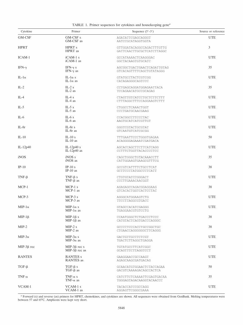
TABLE 1. Primer sequences for cytokines and housekeeping genea
Cytokine Primer Sequence (5–3) Source or reference
GM-CSF GM-CSF s AGATATTCGAGCAGGGT UTEGM-CSF as AATCCGCATAGGTGGTA
HPRT HPRT s GTTGGATACAGGCCAGACTTTGTTG 3HPRT as GATTCAACTTGCGCTCATCTTAGGC
ICAM-1 iCAM-1 s GCCATAAAACTCAAGGGAC UTEiCAM-1 as GGCTACAAGTGTGCATC
IFN-� IFN-� s AGCGGCTGACTGAACTCAGATTGTAG 35IFN-� as GTCACAGTTTTCAGCTGTATAGGG
IL-1� IL-1� s GTATGCCTACTCGTCGG UTEIL-1� as CATAGAGGGCAGTCCC
IL-2 IL-2 s CCTGAGCAGGATGGAGAATTACA 35IL-2 as TCCAGAACATGCCGCAGAG
IL-4 IL-4 s CTAGTTGTCATCCTGCTCTTCTTT UTEIL-4 as CTTTAGGCTTTCCAGGAAGTCTTT
IL-5 IL-5 s CTGGCCTCAAACTGGT UTEIL-5 as CCCTGATGCAACGAAG
IL-6 IL-6 s CCACGGCCTTCCCTAC UTEIL-6 as AAGTGCATCATCGTTGT
IL-8r IL-8r s GGGTCGTACTGCGTAT UTEIL-8r as GTCAATGTCATCGCGG
IL-10 IL-10 s TTTGAATTCCCTGGGTGAGAA 50IL-10 as ACAGGGGAGAAATCGATGACA
IL-12p40 IL-12p40 s AGCACCAGCTTCTTCATCAGG UTEIL-12p40 as CCTTTCTGGTTACACCCCTCC
iNOS iNOS s CAGCTGGGCTGTACAAACCTT 35iNOS as CATTGGAAGTGAAGCGTTTCG
IP-10 IP-10 s GCCGTCATTTTCTGCCTCAT 38IP-10 as GCTTCCCTATGGCCCTCATT
TNF-� TNF-� s CTGTGTATCCGGGACT UTETNF-� as CCCTTGAAACAACGGT
MCP-1 MCP-1 s AGAGAGCCAGACGGAGGAAG 38MCP-1 as GTCACACTGGTCACTCCTAC
MCP-3 MCP-3 s AGGGCATGGAAGTCTG UTEMCP-3 as TTCCTTAGGCGTGACC
MIP-1� MIP-1� s GTAGCCACATCGAGGG UTEMIP-1� as TGAGGAACGTGTCCTG
MIP-1� MIP-1� s CCAATGGGCTCTGACCCTCCC 38MIP-1� as CATGTACTCAGTGACCCAGGGC
MIP-2 MIP-2 s GCCCCTCCCACCTGCCGGCTGC 38MIP-2 as CTGAACCAGGGGGGCTTCAGGG
MIP-3� MIP-3� s GACTGTTGCCTCTCGT UTEMIP-3� as TGACTCTTAGGCTGAGGA
MIP-3� rec MIP-3� rec s TGTATGCCTTCATCGGC UTEMIP-3� rec as GCAGTTTCTTAGGTCCT
RANTES RANTES s GAAGGAACCGCCAAGT UTERANTES as AGAGCAAGCGATGACAG
TGF-� TGF-� s GCAACATGTGGAACTCTACCAGAA 50TGF-� as GACGTCAAAAGACAGCCACTCA
TNF-� TNF-� s CATCTTCTCAAAATTCGAGTGACAA 35TNF-� as TGGGAGTAGACAAGGTACAACCC
VCAM-1 VCAM-1 s TACACCATCCGCCAGG UTEVCAM-1 as AGGAGTTCGGGCGAAA
a Forward (s) and reverse (as) primers for HPRT, chemokines, and cytokines are shown. All sequences were obtained from GenBank. Melting temperatures werebetween 57 and 65°C. Amplicons were kept very short.
5848
pecially with regard to DBA/2 mice and isolates UTE 0423Rand UTE 0705R, and supports the notion of a progressiveprocess determined by the virulence of the M. tuberculosisstrain tested from the beginning of the infection. The histolog-ical appearance of granulomatous lesions in both mousestrains was essentially as described previously (4). Clustering ofneutrophils, macrophages, and lymphocytes, giving rise to pri-mary pregranulomas, was seen during the first weeks postin-fection (data not shown). Subsequently, granulomas becamebetter structured, and by week 9, neutrophils, lymphocytes, andmacrophages of primary granulomas were surrounded by athick mantle of lymphocytes (Fig. 4A and E). At the same time,secondary granulomas became visible as compact lesions con-sisting of abundant lymphocytes that surrounded sparse mac-rophages (Fig. 4C). Outside this lymphocytic mantle, foamymacrophages were seen to initiate the occupation of alveolarspaces (Fig. 4C). By weeks 18 and 22 postinfection, this alve-
olar occupation became very noticeable and led to a pro-nounced size increase and eventual coalescence of granulomas(Fig. 3D and H and 4D, G, and H). In these enlarged granu-lomas it was possible to distinguish between older layers, richin disrupted macrophages and cholesterol crystals (Fig. 5B),and newer layers, in which macrophages were well preserved.Additionally, disrupted infected macrophages were accompa-nied by a progressively denser neutrophilic infiltrate in DBA/2mice.
Figure 5 depicts the evolution of granuloma-related fibrosisas evidenced by collagen staining with Masson’s trichrometechnique. Effacement of pulmonary architecture and subtlesigns of interstitial fibrosis in association with pregranulomaswere already apparent at the beginning of the infection (datanot shown). Starting at week 9 postinfection, as granulomasbecame significantly larger (Fig. 2 and 3), septa surroundingmacrophage-filled alveolar spaces showed marked thickening
FIG. 2. Evolution of granulomatous infiltration in the lungs. Data are percentages calculated by dividing the granuloma-involved area by theglobal tissue area and multiplying by 100. Two lung lobes were analyzed for each mouse. Open and filled symbols, DBA/2 and C57BL/6 mice,respectively. Linear regressions were determined by the Pearson product moment correlation coefficient (P � 0.05 in all cases).
FIG. 3. Evolution of infiltration in C57BL/6 (A through D) and DBA/2 (E through H) mice infected with isolate UTE 0423R at weeks 3 (Aand E), 9 (B and F), 18 (C and G), and 22 (D and H) postinfection. Microphotographs were taken with a Nikon stereoscopic microscope SMZ800(Nikon) at a magnification of �10. Bar, 500 �m.
VOL. 71, 2003 TB SUSCEPTIBILITY IN C57BL/6 VERSUS DBA/2 MICE 5849

secondary to heavy collagen deposition and infiltration by lym-phocytes, especially in DBA/2 mice (Fig. 5C and D). Intra-granulomatous necrosis is characterized by the accumulationof karyorrhectic debris and fibrillar eosinophilic material ac-companied by a strong fibrous reaction within the granuloma.From week 9 postinfection onward, isolates UTE 0335R andUTE 0423R (Fig. 4B) were able to induce intragranulomatousnecrosis in C57BL/6 mice but not in DBA/2 mice (Fig. 4F).
Presence of acid-fast bacilli in the lung. At week 3 postin-fection, bacilli could be easily visualized within macrophages ofpregranulomas. In contrast, at week 9 postinfection, bacilliwere hardly discernible within macrophages of primary gran-ulomas (data not shown). The thick lymphocytic mantle sur-rounding these primary granulomas failed to prevent bacillarydissemination, as evidenced by the fact that single or groupedbacilli were commonly seen within alveolar macrophages out-side the lymphocytic mantle (data not shown). Groups of ba-cilli were more easily identified in DBA/2 mice than inC57BL/6 mice. Bacillus-containing macrophages admixed withabundant neutrophils were also seen in BAL specimens fromDBA/2 mice, mostly in the late stages of infection (data notshown).
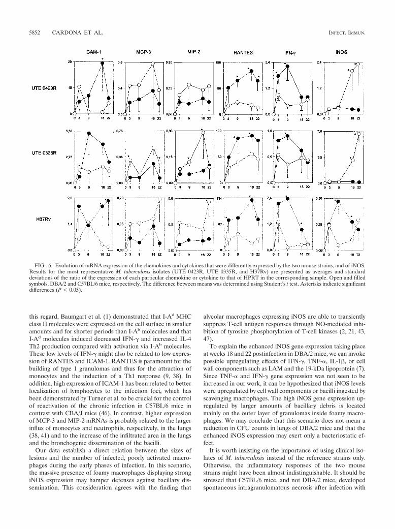
Production of chemokines and cytokines in the lung. Che-mokine and cytokine gene expression in the lung was deter-mined at weeks 0, 3, 9, 18, and 22 postinfection (Fig. 6). Noexpression of IL-1�, IL-2, IL-4, IL-5, IL-6, MIP-1�, MIP-1�,MCP-1, or VCAM-1 was detected (data not shown). Levels ofTNF-�, IL-8r, IL-12p40, MIP-3�, MIP-3�r, TNF-� (LT-�),TGF-�, IP-10, GM-CSF, and IL-10 mRNAs were similar inC57BL/6 and DBA/2 mice (data not shown).
It is noteworthy that IL-1�, IL-6, MIP-1�, MIP-1�, andMCP-1 were not detected, although expression of these che-mokines has been reported by other authors (38). We suggest
that the absence of expression in the study may be the conse-quence of a higher specificity due to the technique used toremove DNA and the real-time PCR protocol itself.
C57BL/6 and DBA/2 mice exhibited different inductionkinetics for ICAM-1, MCP-3, MIP-2, RANTES, IFN-�, andiNOS. Expression of the adhesion molecule ICAM-1 wasincreased in C57BL/6 mice, which clearly facilitates the abil-ity of T lymphocytes to enter the lung. Also meaningful werethe higher MCP-3 and MIP-2 mRNA levels detected inDBA/2 mice, since MIP-2 is a potent neutrophil-recruitingchemokine and MCP-3 exerts selective effects on circulatingmonocytes. As for RANTES, which is associated with aTh1-related immune response in mice, its expression levelswere higher in C57BL/6 mice than in DBA/2 mice. Interest-ingly, in both mouse strains, RANTES expression levelswere higher than those of all other chemokines and cyto-kines. IFN-� expression levels, also seen to be higher inC57BL/6 mice, peaked at week 3 or 9 postinfection (depend-ing on the M. tuberculosis strain) and went down afterwards(3). iNOS mRNA levels increased dramatically in DBA/2mice at weeks 18 and 22 postinfection, but surprisingly, thissurge was neither preceded by increased IFN-� levels (ex-cept for isolate UTE 0824R [data not shown]) nor related todecreased lung bacillary concentrations.
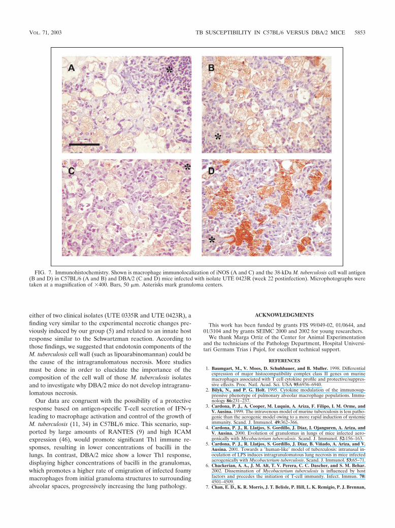
In view of these results, we tried to locate the site of iNOSproduction by use of immunohistochemistry. Our results didnot differ from data obtained by others (31). Thus, iNOS wasdetected mostly in foamy macrophages (Fig. 7A and C), espe-cially in DBA/2 mice at weeks 18 and 22 postinfection. Simi-larly, M. tuberculosis fragments that were able to induce tissueproduction of iNOS were mainly located within foamy macro-phages (Fig. 7B and D), as described by others (32).
FIG. 4. Evolution of granulomas in C57BL/6 (A through D) and DBA/2 (E through H) mice infected with isolate UTE 0423R at weeks 9 (Athrough C and E through G) and 22 (D and H) postinfection. (A, B, E, and F) Primary granulomas, with a center of infected macrophages anda mantle of lymphocytes. Panels B and F show intragranulomatous necrosis at a higher power (�400). The asterisk in panel A marks the zoneamplified in panel B. (C and G) Secondary and tertiary granulomas, respectively, with a compact center of lymphocytes admixed with scantyinfected macrophages. In panel G two secondary granulomas are surrounded by a layer of foamy macrophages that connects them both. (D andH) Tertiary granulomas, with septal thickening of alveoli filled with foamy macrophages. All microphotographs except those in panels B and E weretaken at a magnification of �100. Bars, 100 �m.
5850 CARDONA ET AL. INFECT. IMMUN.

DISCUSSION
As shown by other authors and now confirmed by our study,DBA/2 mice are more susceptible to tuberculous infectionthan C57BL/6 mice (27, 46). The present work explores thebasis of these susceptibility differences. The data obtained sup-port the hypothesis that DBA/2 mice are more susceptible toM. tuberculosis aerosol infection because bronchogenic dissem-ination of bacilli transported by foamy macrophages is morewidespread in this strain.
This assertion is well supported by the increased concentra-tions of bacilli in BAL fluid and lungs, and the accumulation ofintra-alveolar macrophages, shown by DBA/2 mice. Thesehigher concentrations were the outcome of less effective con-tainment of the early infection, as CFU counts are higher fromweek 3 postinfection. In fact, the arithmetic progression of thegranulomatous infiltration of the lungs (Fig. 2) lends supportto this notion.
Our data expand on previous work that has assigned a par-amount role to foamy macrophages in the chronicity of infec-tion in the murine model of tuberculosis. As in other pulmo-nary infections, foamy macrophages exit granulomas afteringesting debris generated by the inflammatory response (19).Afterwards, macrophages invade alveolar spaces and are pro-pelled by the mucociliary elevator along the bronchial tree tobe finally expectorated or swallowed. Previous reports (4, 31,32) have emphasized that at least some of these foamy mac-
rophages were infected with bacilli. It may be assumed that thesurvival of bacilli within macrophages is the result of eithertheir insufficient activation by the IFN-�-induced immune re-sponse or new macrophage ingestion of bacilli released fromdisrupted macrophages. Furthermore, the host immune de-fenses seem to relax following bacillary destruction associatedwith the initial response, and afterwards, surviving bacilli adaptto stress conditions by acquiring the so-called latent status. Inagreement with this, it has been observed that the mouseimmune response is induced by antigens secreted by growingM. tuberculosis (12, 34) and that bacilli adopt a different cellwall antigenic composition and a decreased metabolic rateunder stress conditions (29).
After the initial steps of chronic infection, far from beingenclosed by a fibrous capsule as happens in humans (40),murine granulomas undergo progressive enlargement by occu-pation of alveolar spaces. Probably the immune responseagainst M. tuberculosis in this new scenario is not as efficient asin earlier phases, since disrupted macrophages cannot achievephysical containment of bacilli. Even though the host attractslymphocytes and deposits collagen in the septa of alveoli filledwith foamy macrophages (15), this response is unable to pre-vent infected cells from disseminating through alveolar spaces.
This disseminating process seems to be more dramatic inDBA/2 mice, in which the weakness of initial IFN-� inductionseems to result in poor activation of infected macrophages. In
FIG. 5. Fibrosis and inflammation at weeks 9 (A and C) and 18 (B and D) postinfection in C57BL/6 (A and B) and DBA/2 (C and D) miceinfected with isolate UTE 0423R. Microphotographs were taken at a magnification of �200. Bars, 100 �m. Asterisks mark granuloma centers.Black arrow indicates cholesterol crystals; white arrows point to foamy macrophages.
VOL. 71, 2003 TB SUSCEPTIBILITY IN C57BL/6 VERSUS DBA/2 MICE 5851
this regard, Baumgart et al. (1) demonstrated that I-Ad MHCclass II molecules were expressed on the cell surface in smalleramounts and for shorter periods than I-Ab molecules and thatI-Ad molecules induced decreased IFN-� and increased IL-4Th2 production compared with activation via I-Ab molecules.These low levels of IFN-� might also be related to low expres-sion of RANTES and ICAM-1. RANTES is paramount for thebuilding of type 1 granulomas and thus for the attraction ofmonocytes and the induction of a Th1 response (9, 38). Inaddition, high expression of ICAM-1 has been related to betterlocalization of lymphocytes to the infection foci, which hasbeen demonstrated by Turner et al. to be crucial for the controlof reactivation of the chronic infection in C57BL/6 mice incontrast with CBA/J mice (46). In contrast, higher expressionof MCP-3 and MIP-2 mRNAs is probably related to the largerinflux of monocytes and neutrophils, respectively, in the lungs(38, 41) and to the increase of the infiltrated area in the lungsand the bronchogenic dissemination of the bacilli.
Our data establish a direct relation between the sizes oflesions and the number of infected, poorly activated macro-phages during the early phases of infection. In this scenario,the massive presence of foamy macrophages displaying strongiNOS expression may hamper defenses against bacillary dis-semination. This consideration agrees with the finding that
alveolar macrophages expressing iNOS are able to transientlysuppress T-cell antigen responses through NO-mediated inhi-bition of tyrosine phosphorylation of T-cell kinases (2, 21, 43,47).
To explain the enhanced iNOS gene expression taking placeat weeks 18 and 22 postinfection in DBA/2 mice, we can invokepossible upregulating effects of IFN-�, TNF-�, IL-1�, or cellwall components such as LAM and the 19-kDa lipoprotein (7).Since TNF-� and IFN-� gene expression was not seen to beincreased in our work, it can be hypothesized that iNOS levelswere upregulated by cell wall components or bacilli ingested byscavenging macrophages. The high iNOS gene expression up-regulated by larger amounts of bacillary debris is locatedmainly on the outer layer of granulomas inside foamy macro-phages. We may conclude that this scenario does not mean areduction in CFU counts in lungs of DBA/2 mice and that theenhanced iNOS expression may exert only a bacteriostatic ef-fect.
It is worth insisting on the importance of using clinical iso-lates of M. tuberculosis instead of the reference strains only.Otherwise, the inflammatory responses of the two mousestrains might have been almost indistinguishable. It should bestressed that C57BL/6 mice, and not DBA/2 mice, developedspontaneous intragranulomatous necrosis after infection with
FIG. 6. Evolution of mRNA expression of the chemokines and cytokines that were differently expressed by the two mouse strains, and of iNOS.Results for the most representative M. tuberculosis isolates (UTE 0423R, UTE 0335R, and H37Rv) are presented as averages and standarddeviations of the ratio of the expression of each particular chemokine or cytokine to that of HPRT in the corresponding sample. Open and filledsymbols, DBA/2 and C57BL/6 mice, respectively. The difference between means was determined using Student’s t test. Asterisks indicate significantdifferences (P � 0.05).
5852 CARDONA ET AL. INFECT. IMMUN.
either of two clinical isolates (UTE 0335R and UTE 0423R), afinding very similar to the experimental necrotic changes pre-viously induced by our group (5) and related to an innate hostresponse similar to the Schwartzman reaction. According tothose findings, we suggested that endotoxin components of theM. tuberculosis cell wall (such as lipoarabinomannan) could bethe cause of the intragranulomatous necrosis. More studiesmust be done in order to elucidate the importance of thecomposition of the cell wall of those M. tuberculosis isolatesand to investigate why DBA/2 mice do not develop intragranu-lomatous necrosis.
Our data are congruent with the possibility of a protectiveresponse based on antigen-specific T-cell secretion of IFN-�leading to macrophage activation and control of the growth ofM. tuberculosis (11, 34) in C57BL/6 mice. This scenario, sup-ported by large amounts of RANTES (9) and high ICAMexpression (46), would promote significant Th1 immune re-sponses, resulting in lower concentrations of bacilli in thelungs. In contrast, DBA/2 mice show a lower Th1 response,displaying higher concentrations of bacilli in the granulomas,which promotes a higher rate of emigration of infected foamymacrophages from initial granuloma structures to surroundingalveolar spaces, progressively increasing the lung pathology.
ACKNOWLEDGMENTS
This work has been funded by grants FIS 99/049-02, 01/0644, and01/3104 and by grants SEIMC 2000 and 2002 for young researchers.
We thank Marga Ortiz of the Center for Animal Experimentationand the technicians of the Pathology Department, Hospital Universi-tari Germans Trias i Pujol, for excellent technical support.
REFERENCES
1. Baumgart, M., V. Moos, D. Schuhbauer, and B. Muller. 1998. Differentialexpression of major histocompatibility complex class II genes on murinemacrophages associated with T cell cytokine profile and protective/suppres-sive effects. Proc. Natl. Acad. Sci. USA 95:6936–6940.
2. Bilyk, N., and P. G. Holt. 1995. Cytokine modulation of the immunosup-pressive phenotype of pulmonary alveolar macrophage populations. Immu-nology 86:231–237.
3. Cardona, P. J., A. Cooper, M. Luquin, A. Ariza, F. Filipo, I. M. Orme, andV. Ausina. 1999. The intravenous model of murine tuberculosis is less patho-genic than the aerogenic model owing to a more rapid induction of systemicimmunity. Scand. J. Immunol. 49:362–366.
4. Cardona, P. J., R. Llatjos, S. Gordillo, J. Díaz, I. Ojanguren, A. Ariza, andV. Ausina. 2000. Evolution of granulomas in lungs of mice infected aero-genically with Mycobacterium tuberculosis. Scand. J. Immunol. 52:156–163.
5. Cardona, P. J., R. Llatjos, S. Gordillo, J. Díaz, B. Vinado, A. Ariza, and V.Ausina. 2001. Towards a human-like’ model of tuberculosis: intranasal in-oculation of LPS induces intragranulomatous lung necrosis in mice infectedaerogenically with Mycobacterium tuberculosis. Scand. J. Immunol. 53:65–71.
6. Chackerian, A. A., J. M. Alt, T. V. Perera, C. C. Dascher, and S. M. Behar.2002. Dissemination of Mycobacterium tuberculosis is influenced by hostfactors and precedes the initiation of T-cell immunity. Infect. Immun. 70:4501–4509.
7. Chan, E. D., K. R. Morris, J. T. Belisle, P. Hill, L. K. Remigio, P. J. Brennan,
FIG. 7. Immunohistochemistry. Shown is macrophage immunolocalization of iNOS (A and C) and the 38-kDa M. tuberculosis cell wall antigen(B and D) in C57BL/6 (A and B) and DBA/2 (C and D) mice infected with isolate UTE 0423R (week 22 postinfection). Microphotographs weretaken at a magnification of �400. Bars, 50 �m. Asterisks mark granuloma centers.
VOL. 71, 2003 TB SUSCEPTIBILITY IN C57BL/6 VERSUS DBA/2 MICE 5853
and D. W. Riches. 2001. Induction of inducible nitric oxide synthase-NO* bylipoarabinomannan of Mycobacterium tuberculosis is mediated by MEK1-ERK, MKK7-JNK, and NF-�B signaling pathways. Infect. Immun. 69:2001–2010.
8. Chan, J., Y. Xing, R. S. Magliozzo, and B. R. Bloom. 1992. Killing of virulentMycobacterium tuberculosis by reactive nitrogen intermediates produced byactivated murine macrophages. J. Exp. Med. 175:1111–1122.
9. Chensue, S. W., K. S. Warmington, E. J. Allenspach, B. Lu, C. Gerard, S. L.Kunkel, and N. W. Lukacs. 1999. Differential expression and cross-regula-tory function of RANTES during mycobacterial (type 1) and schistosomal(type 2) antigen-elicited granulomatous inflammation. J. Immunol. 163:165–173.
10. Chomczynski, P., and N. Sacchi. 1987. Single-step method of RNA isolationby acid guanidinium thiocyanate-phenol-chloroform extraction. Anal. Bio-chem. 162:156–159.
11. Cooper, A. M., D. K. Dalton, T. A. Stewart, J. P. Griffin, D. G. Russell, andI. M. Orme. 1993. Disseminated tuberculosis in interferon gamma gene-disrupted mice. J. Exp. Med. 178:2243–2247.
12. Cooper, A. M., and J. L. Flynn. 1995. The protective immune response toMycobacterium tuberculosis. Curr. Opin. Immunol. 7:512–516.
13. Denis, M. 1991. Interferon-gamma-treated murine macrophages inhibitgrowth of tubercle bacilli via the generation of reactive nitrogen intermedi-ates. Cell. Immunol. 132:150–157.
14. Denis, M. 1991. Tumor necrosis factor and granulocyte macrophage-colonystimulating factor stimulate human macrophages to restrict growth of viru-lent Mycobacterium avium and kill avirulent M. avium: killing effector mech-anism depends on the generation of reactive nitrogen intermediates. J. Leu-koc. Biol. 49:380–387.
15. Dunn, P. L., and R. J. North. 1995. Virulence ranking of some Mycobacte-rium tuberculosis and Mycobacterium bovis strains according to their ability tomultiply in the lungs, induce lung pathology, and cause mortality in mice.Infect. Immun. 63:3428–3437.
16. Flesch, I. E., and S. H. Kaufmann. 1991. Mechanisms involved in mycobac-terial growth inhibition by gamma interferon-activated bone marrow mac-rophages: role of reactive nitrogen intermediates. Infect. Immun. 59:3213–3218.
17. Flynn, J. L., J. Chan, K. J. Triebold, D. K. Dalton, T. A. Stewart, and B. R.Bloom. 1993. An essential role for interferon gamma in resistance to Myco-bacterium tuberculosis infection. J. Exp. Med. 178:2249–2254.
18. Forget, A., E. Skamene, P. Gros, A. C. Miailhe, and R. Turcotte. 1981.Differences in response among inbred mouse strains to infection with smalldoses of Mycobacterium bovis BCG. Infect. Immun. 32:42–47.
19. Green, G. M. 1973. Alveolobronchiolar transport mechanisms. Arch. Intern.Med. 131:109–114.
20. Gros, P., E. Skamene, and A. Forget. 1983. Cellular mechanisms of geneti-cally controlled host resistance to Mycobacterium bovis (BCG). J. Immunol.131:1966–1972.
21. Holt, P. G. 1993. Regulation of antigen-presenting cell function(s) in lungand airway tissues. Eur. Respir. J. 6:120–129.
22. Kaplan, G., A. D. Luster, G. Hancock, and Z. A. Cohn. 1987. The expressionof a gamma interferon-induced protein (IP-10) in delayed immune responsesin human skin. J. Exp. Med. 166:1098–1108.
23. Kaufmann, S. H. 1995. Immunity to intracellular microbial pathogens. Im-munol. Today 16:338–342.
24. Lengeling, A., K. Pfeffer, and R. Balling. 2001. The battle of two genomes:genetics of bacterial host/pathogen interactions in mice. Mamm. Genome12:261–271.
25. Lyadova, I. V., E. B. Eruslanov, S. V. Khaidukov, V. V. Yeremeev, K. B.Majorov, A. V. Pichugin, B. V. Nikonenko, T. K. Kondratieva, and A. S. Apt.2000. Comparative analysis of T lymphocytes recovered from the lungs ofmice genetically susceptible, resistant, and hyperresistant to Mycobacteriumtuberculosis-triggered disease. J. Immunol. 165:5921–5931.
26. MacMicking, J. D., R. J. North, R. LaCourse, J. S. Mudgett, S. K. Shah, andC. F. Nathan. 1997. Identification of nitric oxide synthase as a protectivelocus against tuberculosis. Proc. Natl. Acad. Sci. USA 94:5243–5248.
27. Medina, E., and R. J. North. 1998. Resistance ranking of some commoninbred mouse strains to Mycobacterium tuberculosis and relationship to majorhistocompatibility complex haplotype and Nramp1 genotype. Immunology93:270–274.
28. Medina, E., B. J. Rogerson, and R. J. North. 1996. The Nramp1 antimicro-bial resistance gene segregates independently of resistance to virulent My-cobacterium tuberculosis. Immunology 88:479–481.
29. Michele, T. M., C. Ko, and W. R. Bishai. 1999. Exposure to antibioticsinduces expression of the Mycobacterium tuberculosis sigF gene: implicationsfor chemotherapy against mycobacterial persistors. Antimicrob. Agents Che-mother. 43:218–225.
30. Mitsos, L. M., L. R. Cardon, A. Fortin, L. Ryan, R. LaCourse, R. J. North,and P. Gros. 2000. Genetic control of susceptibility to infection with Myco-bacterium tuberculosis in mice. Genes Immun. 1:467–477.
31. Mogues, T., M. E. Goodrich, L. Ryan, R. LaCourse, and R. J. North. 2001.The relative importance of T cell subsets in immunity and immunopathologyof airborne Mycobacterium tuberculosis infection in mice. J. Exp. Med. 193:271–280.
32. Mustafa, T., S. Phyu, R. Nilsen, R. Jonsson, and G. Bjune. 1999. A mousemodel for slowly progressive primary tuberculosis. Scand. J. Immunol. 50:127–136.
33. Nikonenko, B. V., M. M. Averbakh, Jr., C. Lavebratt, E. Schurr, and A. S.Apt. 2000. Comparative analysis of mycobacterial infections in susceptibleI/St and resistant A/Sn inbred mice. Tuber. Lung Dis. 80:15–25.
34. Orme, I. M., P. Andersen, and W. H. Boom. 1993. T cell response to Myco-bacterium tuberculosis. J. Infect. Dis. 167:1481–1497.
35. Overbergh, L., D. Valckx, M. Waer, and C. Mathieu. 1999. Quantification ofmurine cytokine mRNAs using real time quantitative reverse transcriptasePCR. Cytokine 11:305–312.
36. Pierce, C., R. Dubos, and G. Middlebrook. 1947. Infection of mice withmammalian tubercle bacilli grown in Tween-albumin liquid medium. J. Exp.Med. 86:159–174.
37. Prophet, E. B., B. Mills, J. B. Arrington, and L. H. Sobin (ed.). 1992. AFIPlaboratory methods in histotechnology. American Registry of Pathology,Washington, D.C.
38. Rhoades, E. R., A. M. Cooper, and I. M. Orme. 1995. Chemokine responsein mice infected with Mycobacterium tuberculosis. Infect. Immun. 63:3871–3877.
39. Rhoades, E. R., and I. M. Orme. 1997. Susceptibility of a panel of virulentstrains of Mycobacterium tuberculosis to reactive nitrogen intermediates.Infect. Immun. 65:1189–1195.
40. Ridley, D. S., and M. J. Ridley. 1987. Rationale for the histological spectrumof tuberculosis. A basis for classification. Pathology 19:186–192.
41. Rollins, B. J. 2001. MCP-1, MCP-2, MCP-3, MCP-4 and MCP-5, p. 1145–1160. In J. J. Oppenheim and M. Feldmann (ed.), Cytokine reference, vol 1.Academic Press, San Diego, Calif.
42. Sanchez, F., T. V. Radaeva, B. V. Nikonenko, A. S. Persson, S. Sengul, M.Schalling, E. Schurr, A. S. Apt, and C. Lavebratt. 2003. Multigenic controlof disease severity after virulent Mycobacterium tuberculosis infection inmice. Infect. Immun. 71:126–131.
43. Strickland, D., U. R. Kees, and P. G. Holt. 1996. Regulation of T-cellactivation in the lung: alveolar macrophages induce reversible T-cell anergyin vitro associated with inhibition of interleukin-2 receptor signal transduc-tion. Immunology 87:250–258.
44. Stumbles, P. A., A. S. McWilliam, and P. G. Holt. 1999. Dendritic cells andmucosal macrophages, p. 397–412. In P. L. Ogra, J. Mestecky, M. E. Lamm,W. Strober, J. Bienenstock, and J. R. McGhee (ed.), Mucosal immunology.Academic Press, San Diego, Calif.
45. Tsang, A. W., K. Oestergaard, J. T. Myers, and J. A. Swanson. 2000. Alteredmembrane trafficking in activated bone marrow-derived macrophages.J. Leukoc. Biol. 68:487–494.
46. Turner, J., M. Gonzalez-Juarrero, B. M. Saunders, J. V. Brooks, P. Marietta,D. L. Ellis, A. A. Frank, A. M. Cooper, and I. M. Orme. 2001. Immunologicalbasis for reactivation of tuberculosis in mice. Infect. Immun. 69:3264–3270.
47. Upham, J. W., D. H. Strickland, N. Bilyk, B. W. Robinson, and P. G. Holt.1995. Alveolar macrophages from humans and rodents selectively inhibitT-cell proliferation but permit T-cell activation and cytokine secretion. Im-munology 84:142–147.
48. Via, L. E., R. A. Fratti, M. McFalone, E. Pagan-Ramos, D. Deretic, and V.Deretic. 1998. Effects of cytokines on mycobacterial phagosome maturation.J. Cell Sci. 111:897–905.
49. World Health Organization. 2001. Global tuberculosis control. WHO Re-port 2001. WHO/CDS/TB/2001.287. World Health Organization, Geneva,Switzerland.
50. Xia, D., A. Sanders, M. Shah, A. Bickerstaff, and C. Orosz. 2001. Real-timepolymerase chain reaction analysis reveals an evolution of cytokine mRNAproduction in allograft acceptor mice. Transplantation 72:907–914.
Editor: S. H. E. Kaufmann
5854 CARDONA ET AL. INFECT. IMMUN.