LANDMARK RELATING TO NATIONAL RESILIENCE WITH IMPACT ON RISK OF GEOPOLITICAL PERSPECTIVE
Upload
independentCategory
view
0download
0
Landmark Control and Updating of Self-Movement Cues Are LargelyMaintained in Head Direction Cells After Lesions of the Posterior
Parietal Cortex
Jeffrey L. CaltonCalifornia State University–Sacramento
and Dartmouth College
Carol S. Turner, De-Laine M. Cyrenne,and Brian R. Lee
California State University–Sacramento
Jeffrey S. TaubeDartmouth College
Head direction (HD) cells discharge as a function of the rat’s directional orientation with respect to itsenvironment. Because animals with posterior parietal cortex (PPC) lesions exhibit spatial and naviga-tional deficits, and the PPC is indirectly connected to areas containing HD cells, we determined theeffects of bilateral PPC lesions on HD cells recorded in the anterodorsal thalamus. HD cells from lesionedanimals had similar firing properties compared to controls and their preferred firing directions shifted acorresponding amount following rotation of the major visual landmark. Because animals were notexposed to the visual landmark until after surgical recovery, these results provide evidence that the PPCis not necessary for visual landmark control or the establishment of landmark stability. Further, cells fromlesioned animals maintained a stable preferred firing direction when they foraged in the dark and wereonly slightly less stable than controls when they self-locomoted into a novel enclosure. These findingssuggest that PPC does not play a major role in the use of landmark and self-movement cues in updatingthe HD cell signal, or in its generation.
Keywords: path-integration, vestibular, navigation, anterodorsal thalamic nucleus, spatial orientation
The posterior parietal cortex (PPC) is postulated to be involvedin the orientation of the body in space and the representation ofspace around the body (Andersen, Snyder, Bradley, & Xing, 1997;Arbib, 1997; Colby & Goldberg, 1999; Corwin & Reep, 1998;McNaughton, Leonard, & Chen, 1989; Save & Poucet, 2000a). Inaccordance with findings in primates, rats with PPC lesions exhibitan array of spatial deficits including sensory neglect (King &Corwin, 1993), an inability to notice a change in the spatialrelationship between objects in the environment (DeCoteau &Kesner, 1998), and deficits in tasks requiring the integration of
visual cues and motor responses (Davis & McDaniel, 1993; DiMattia& Kesner, 1988; Kolb & Walkey, 1987). Electrophysiological studiesof rat PPC have supported the view that this area has a role indirecting spatial behavior, with cells showing modulation based on themovement state of the animal relative to a spatial task (Chen, Lin,Barnes, & McNaughton, 1994; Chen, Lin, Green, Barnes, & Mc-Naughton, 1994; McNaughton, Mizumori, Barnes, Marquis, &Green, 1994; Nitz, 2006), and modulation based on the location ofauditory targets relative to the animal (Nakamura, 1999).
Using the rodent model, investigators have identified threeimportant classes of cells that are believed to be involved innavigation. Place cells, recorded primarily in the hippocampalformation, are preferentially active when the animal is within aspecific area in an environment - referred to as the “place field” ofthe recorded cell (O’Keefe & Dostrovsky, 1971). Grid cells, foundprimarily in the dorsal caudal medial entorhinal cortex, dischargeat multiple locations in an environment and these locations form aregular, repeating grid-like pattern (Hafting, Fyhn, Molden, Moser,& Moser, 2005). Head direction (HD) cells, found throughout theclassic Papez circuit, become active when the animal orients itshead in the “preferred direction” of the recorded cell, independentof its location and ongoing behavior (Taube, 1995; Taube, Muller,& Ranck, 1990a). All three cell types respond to visual landmarkand self-movement (idiothetic) information, signals that are con-sidered important for animal navigation (Gallistel, 1990; Muller &Kubie, 1987; Quirk, Muller, & Kubie, 1990; Taube & Burton,1995; Taube, Muller, & Ranck, 1990b).
Jeffrey L. Calton, Department of Psychology, California StateUniversity–Sacramento and Department of Psychological and Brain Sci-ences, Center for Cognitive Neuroscience, Dartmouth College; Carol S.Turner, De-Laine M. Cyrenne, and Brian R. Lee, Department of Psychol-ogy, California State University–Sacramento; Jeffrey S. Taube, Depart-ment of Psychological and Brain Sciences, Center for Cognitive Neuro-science, Dartmouth College.
This work was supported through the NASA Cooperative AgreementNCC9-58 with the National Space Biomedical Research Institute andNational Institute of Mental Health Grants MH48924 and MH01286 to J.S.Taube and MH06258 to J.L. Calton. A preliminary report of this researchwas presented at the 31st annual meeting of the Society for Neuroscienceheld in San Diego, CA in 2001.
Correspondence concerning this article should be addressed to Jeffrey S.Taube, Department of Psychological and Brain Sciences, Dartmouth Col-lege, 6207 Moore Hall, Hanover, NH 03755. E-mail: [email protected]
Behavioral Neuroscience Copyright 2008 by the American Psychological Association2008, Vol. 122, No. 4, 827–840 0735-7044/08/$12.00 DOI: 10.1037/0735-7044.122.4.827
827

Connectivity studies have led many to suggest that PPC isideally suited to provide convergence of motor and sensory infor-mation to navigational circuitry (Corwin & Reep, 1998; Save &Poucet, 2000a). Figure 1 presents a schematic diagram showingthe major connections by which information could flow from PPCto limbic regions presumably involved in navigation and, in par-ticular, the HD cell circuitry. Rat PPC shares connections withprimary and secondary visual cortex (Kolb & Walkey, 1987;Miller & Vogt, 1984; Reep, Chandler, King, & Corwin, 1994),auditory cortex (Reep et al., 1994), somatosensory cortex (Kolb &Walkey, 1987; Reep et al., 1994) and prefrontal areas known tocontain motor signals (Kolb & Walkey, 1987; Reep et al., 1994).PPC also shares reciprocal connections with retrosplenial cortex(Kolb & Walkey, 1987; Reep et al., 1994), which in turn projectsto a number of limbic structures involved in navigation such as theentorhinal cortex (Wyss & Van Groen, 1992), postsubiculum(Vogt & Miller, 1983), and anterodorsal thalamus (ADN; Sripand-kulchai & Wyss, 1986). Entorhinal cortex is the primary corticalinput region of the hippocampal complex, and HD cells can be
found in the retrosplenial cortex, anterodorsal thalamus, and post-subiculum (reviewed in Taube, 2007). In short, the pathways existfor PPC to serve as a multimodal association area by whichcortically generated signals utilized during navigation reach limbiccircuitry.
Given the close anatomical connections between PPC and nav-igational structures and electrophysiological findings that PPCcells are modulated by visual, idiothetic, and even HD information(Chen et al., 1994a, b; McNaughton et al., 1994), it is not surpris-ing that investigators have proposed a major role for PPC in thenetworks mediating navigational behavior. Conceptualizations ofnavigational circuitry have assigned PPC the role of processingvisual landmark information and the integration of self-movementand visuospatial cues (Arbib, 1997; McNaughton et al., 1996; Save& Poucet, 2000a; Taube, 1998). To test this hypothesis, we pro-duced lesions of PPC using two commonly used coordinates forPPC. We then monitored HD cell activity in the ADN during threetasks, one assessing visual landmark control and the other twoassessing idiothetic control. To determine whether the PPC con-
Figure 1. Schematic diagram of connections between PPC, other cortical areas, and subcortical areas relatedto the HD circuit. Arrows represent direction of information flow between connected areas.
828 CALTON ET AL.

tributed to the acquisition or selection of visual cues as landmarks,animals were not exposed to the testing environment and its salientvisual cues until after the lesions were conducted. We found thatPPC lesions did not disrupt generation of direction-specific firingin ADN HD cells or landmark control of HD cell preferred firingdirections. In addition, PPC lesions led only to mild impairmentsin idiothetic control of HD cell activity.
Method
Subjects
There were 28 female Long-Evans rats that served as subjects.Weights ranged between 220 and 300 g at the start of the study.The animals were singly housed in clear plastic cages in a roommaintained on a 12-hr light/12-hr dark cycle. A food-restricted dietwas used to maintain the rats at approximately 85% of free feedingweight. Water was freely available in the home cage.
Surgical Procedures
Each animal received implantation of a recording electrodearray to monitor neural activity during the behavioral task. Elec-trodes were constructed using methods described by Kubie (1984),and consisted of a bundle of 10 25 �m diameter nichrome wiresthat were insulated except at the tip. The wires were threadedthrough a stainless steel cannula that was moveable in the dorsal/ventral direction after being fixed to the skull using dental acrylic.Standard surgical procedures were followed for implantation of therecording electrodes (Taube, 1995). Briefly, animals were anes-thetized with a ketamine-acepromazine-xylazine cocktail. Afterexposure of the skull, a hole was drilled and the electrode im-planted just dorsal to the right ADN based on coordinates (1.5 mmposterior to bregma, 1.3 mm lateral to bregma and 3.7 mm belowthe cortical surface) provided by Paxinos and Watson (1998).
During this same surgery some animals (n � 15) receivedbilateral aspiration lesions of PPC. The rat parietal cortex isgenerally divided into three distinct areas—anterior, ventral, andposterior, and the posterior area can be further subdivided intothree zones designated as dorsal, rostral, and caudal (Palomero-Gallagher & Zilles, 2004). Because definitive functions have notbeen ascribed to each of the three zones in PPC, we designed ourexperiments to lesion the entire PPC. For these animals, the skullwas opened over the lesion sites, the dura-mater was cut, and aglass pipette attached to a suction source was used to aspirate braintissue. Because there is disagreement in the literature as to thelocation of PPC, we utilized two different sized lesions. Theprimary disagreement has concerned the anterior-posterior (AP)placement of the area, with a few investigators delineating the areaas a 4 mm long strip beginning 0.5 mm anterior to bregma(DiMattia & Kesner, 1988), while others consider the boundariesas 2.0 to 6.0 mm posterior to Bregma (Save, Paz-Villagran, Alex-insky, & Poucet, 2005). Still other studies have placed the PPCsomewhere in-between these two areas (Goodrich-Hunsaker, Hun-saker, & Kessner, 2005; Kolb & Walkey, 1987; Ward & Brown,1997). Accordingly, animals within group PPC-Large (n � 5)were given large bilateral lesions intended to overlie all these areasutilized previously (AP: � 0.5 to �6.0 relative to Bregma, andmedial-lateral (ML): � 1.5 to 5.5 relative to midline). Group
PPC-Small (n � 10) received smaller lesions intended to overliethe more posterior lesion coordinates that are commonly used (AP:�2.0 to �6.0 relative to bregma and ML: � 1.5 to 5.5 relative tomidline). For both lesion sizes, tissue was aspirated to a depth ofapproximately 1 mm. After aspirations, lesion sites were packedwith sterile gel foam and the previously overlying skull pieceswere cemented back in place using orthopedic cement. Animalswere allowed at least 1 week of recovery before behavioral train-ing. All procedures were conducted under an institutionally ap-proved animal care and use protocol.
Apparatus
Two different enclosures were utilized during recordings. Bothstructures were painted gray and had floors of paper that werechanged between sessions. A wooden cylinder (76 cm diameter, 51cm high) was used when searching for cells and to assess landmarkcontrol of HD activity (Figure 2A). The wall of the cylinder wasfeatureless except for a white card attached to the inside wall,which encompassed approximately 100° of arc, centered at the 0°position. A dual-chamber apparatus similar to that first used byTaube and Burton (1995) was used to assess idiothetic control ofHD activity (Figure 2B). This enclosure included a cylinder similarto that described above with the exception of a section of wall thatcould be removed revealing a U-shaped passageway (15 cm wide,147 cm total length) that ended in a rectangular enclosure 71 cmwide, 89 cm long). The walls of the rectangular compartment werefeatureless except for a cue card identical to that found in thecylinder attached to the wall opposite the entryway; thus this cardwas rotated 90° counterclockwise (CCW) with respect to the cuecard in the cylindrical compartment (Figure 2B).
The room that housed the enclosures was dimly illuminated byfour or eight lights symmetrically attached to the ceiling. Alsomounted to the ceiling was a food pellet dispenser for motivatingforaging behavior and a white noise generator to mask potentialauditory cues. The enclosures were surrounded from floor toceiling by a black curtain that eliminated exterior visual cues. Theposition and directional orientation of the rat was determined by anautomated video tracking system (Ebtronics; Elmont, NY). Thisvideo tracking hardware provided x and y coordinates (256 � 256)
Figure 2. (A) Diagram of the Cylinder Task procedure. (B) Diagram ofthe Dual-Chamber Task procedure.
829INTACT HD CELL RESPONSES AFTER PARIETAL LESIONS

of red and green light-emitting diodes secured 6 cm apart abovethe head and back of the animal, respectively.
Electrical signals recorded in the brain were passed through afield-effect transistor in a source-follower configuration. Signalswere amplified by a factor of 10K to 50K, bandpass filtered(300–10,000 Hz, �3 dB/octave) and sent through a dual windowdiscriminator (BAK Electronics) for spike discrimination. Thefiring rates of recorded cells and LED positions were saved onto apersonal computer at a rate of 60 Hz. Data analysis was accom-plished off-line using custom software (LabView, National Instru-ments, Austin, TX). The HD of the animal was determined by therelative position of the red and green LEDs.
Behavioral Tasks
Screening for HD cells began after surgical recovery. Thisinvolved examining the electrical signal on each of the 10 im-planted wires while the animal foraged in the recording cylinderfor food pellets. If no HD cells were detected, the electrode wasadvanced 25 to 50 �m and the animal was returned to its homecage. If a HD cell was identified, the animal was removed and theappropriate enclosure was prepared for data collection. Threedifferent behavioral tasks were used in this study.
The first three cylinder sessions (the Cue Rotation series) as-sessed whether PPC lesions affected the stability of the HD signaland visual landmark control of HD activity. These sessions in-volved examining the directional activity of the HD cell while theanimal foraged in the recording cylinder for 8 minutes. Figure 2Apresents the procedure for these sessions. In the Cylinder-Standardsession the cue card was placed at the 0° position. After thissession, the visual landmark was rotated 90° in the CCW direction;this session was referred to as the Cylinder-Rotation session.Finally, in the Cylinder-Return session the cue card was returnedto the 0° position. For each of these sessions the animal was givendisorientation treatment before the recording session by placing itin a cardboard box and rotating it at approximately 0.5 Hz forapproximately 60 seconds. Analysis of these sessions involvedcomparing the preferred directions of the recorded cells across thethree sessions. To accomplish this analysis, we constructed a firingrate versus HD plot for each session by dividing the 360° direc-tional range into 60 6° bins and then calculating the average firingrate for each bin. We then compared the plots from each session todetermine how the position of the cue card affected the directionalsignal of recorded cells (see below).
We used two different tasks to assess whether PPC lesionsaffected idiothetic control of HD cell activity. Cylinder-Dark/NoCue sessions (Figure 2A) given to Control cells (n � 8) andPPC-Large cells (n � 11) after Cylinder-Return sessions, exam-ined the stability of HD cells in the absence of visual landmarks.In these sessions the cue card was removed, the lights wereextinguished, and the animal was allowed to forage for 24 minutesin the dark. The animal was not disoriented at the start of thesession and the floor paper was not changed. The Dual-Chambertask involved the animal self-locomoting into a novel enclosure(Figure 2B). After disorientation treatment the animal was placedin the cylindrical compartment of the apparatus with the hiddendoor closed. After allowing the animal to forage for 16 minutes thedoor was opened and the animal was free to walk into the pas-sageway and rectangular compartment. After the animal loco-
moted into the rectangular compartment the door was usuallyclosed, trapping the animal inside, and the animal was allowed toforage for 16 minutes while the activity of the HD cell wasmonitored. In a few cases (n � 2 for Group PPC-Large, and n �1 for Group Control) the door was left open and the animal wasfree to move between the enclosures of the apparatus. Unlike in thecylinder task, the Dual-Chamber sessions could be conducted onlyonce per animal, since the task requires the rectangular compart-ment to be novel. Hence, only one HD cell recording session wascollected per animal in this task.
Analysis of Cellular Data
Basic directional characteristics of the recorded cells were de-termined by examining cellular activity during the Cylinder-Standard session. Cells from lesioned and control animals werecompared on measures of peak firing rate, background firing rate,signal-to-noise ratio, directional firing range, directional informa-tion content, and anticipatory time interval. The preferred directionwas defined as the directional bin with the highest average firingrate. The peak firing rate was the average firing rate correspondingto the preferred direction. The background firing rate was theaverage firing rate of the three directional bins centered at 180°opposite the preferred direction. Signal-to-noise ratio was calcu-lated as the peak firing rate divided by the background firing rate.The directional firing range was defined as the width at the base ofthe firing rate versus HD tuning curve (Taube et al., 1990a).Directional information content is a measure of how many bits ofHD information is conveyed by each spike (Skaggs, McNaughton,Gothard, & Markus., 1993) and was calculated by the followingformula: Directional Information Content � � pi (�i/�) log2 (�i/�),where pi is the probability that the head pointed in the ith direc-tional bin, �i is the mean firing rate for bin i, and � is the meanfiring rate across all directional bins.
The anticipatory time interval (ATI) is a measure of the amountof time that cell firing best predicts where the animal will bepointing its head in the future. Previous work has estimated that theactivity of ADN HD cells reflect HDs 25 ms into the futurebetter than current HDs (Blair & Sharp, 1995; Taube & Muller,1998). Both sensory and motor inputs into the HD network havebeen postulated as a mechanism for this anticipatory activity(Bassett et al., 2005; Blair & Sharp, 1995; Taube & Muller, 1998).Because both types of signals may be found in PPC (reviewedabove), it is possible that PPC may play a role in this phenomenon.Accordingly, we compared the ATI of Standard-Cylinder sessionsin cells from control and PPC-lesioned animals using the methodsof Blair and Sharp (1995). For this method, HD cell tuningfunctions were calculated for CW and CCW directions and thedifference between the preferred firing directions for the twofunctions (separation angle) determined. Then the spike record wasshifted forward and backward in time in steps of 16.67 ms (themaximum temporal resolution of the recording hardware) and theseparation angle between the CW and CCW functions for headmovements �90° computed for each shift. The spike series wasshifted incrementally �6 times ( �100 ms) relative to the HDseries, providing 13 values of CW-CCW separation angles. Ascattergram was then constructed from the 13 CW-CCW separa-tion angles and their corresponding time shift. The x-intercept ofthe best-fit line of this plot is referred to as the ATI and is
830 CALTON ET AL.

equivalent to the amount of time that the spike series has to beshifted to achieve overlapping CW and CCW functions.
Circular statistics (Batschelet, 1981) were used to determine thestability of the directional signal in the cue rotation series, the cueremoval sessions, and between the cylindrical and rectangularenclosures of the Dual-Chamber apparatus. Directional deviationscores across these conditions were calculated using a cross-correlation method (Taube & Burton, 1995). This approach in-volves shifting the firing rate/HD function of the first session in 6°increments while correlating this shifted function with the non-shifted function from the other session. The amount of shift re-quired to produce the maximal Pearson r correlation between thetwo sessions is defined as the directional deviation score betweenthe sessions. These scores were then subjected to Rayleigh tests(Batschelet, 1981) to determine if the scores were clustered ran-domly (as would be the case if the preferred directions were notcontrolled by landmark or idiothetic signals) or if the preferreddirections tended to shift in the same direction and amount. Thecritical statistic of the Rayleigh test is the mean vector length, r,which varies between 0 and 1, with higher values indicating thatthe distribution of directions is more concentrated, (i.e., clusterednonrandomly; Batschelet, 1981). Also, calculated using these di-rectional deviations was the mean vector angle, m, which estimatesthe mean angle of the sample (Batschelet, 1981).
Two inferential tests were utilized for between-groups compar-isons of directional deviation scores produced during the cylinderand dual-chamber sessions. ANOVAs were used to determine ifthe absolute directional deviations between conditions were sig-nificantly different. The absolute values of the directional devia-tions were used because they could be either positive or negativedepending on whether the deviations were CW or CCW from theexpected value. We also utilized an F test of the concentrationparameter of the directional deviation scores (Batschelet, 1981).This latter test determines if there is a difference among the groupsin the concentration of directional scores around the mean value;an expected finding if lesioned animals show less coupling ofpreferred directions between multiple sessions of the cylinder taskor between the two enclosures of the dual-chamber task.
Histology
At the completion of the experiments, animals were anesthetizeddeeply and a small anodal current (20 �A, 10 seconds) was passedthrough one electrode wire to later conduct a Prussian blue reac-tion. The animals were then perfused transcardially with salinefollowed by 10% formalin in saline. The brains were removed andplaced in 10% formalin for at least 48 hours. The brains were thenplaced in a 10% formalin solution containing 2% potassium fer-rocyanide for 24 hours and then reimmersed in 10% formalin (24hours) before being placed in 20% sucrose for 24 hours. They werethen frozen, sectioned (40 �m) in the coronal plane, stained withcresyl violet, and examined microscopically for localization of therecording and lesion sites. All recording electrodes were localizedto the ADN.
Results
A total of 50 HD cells were recorded from the 28 animals, with17, 14, and 19 cells recorded from PPC-Large, PPC-Small, andControl animals, respectively.
Lesion Data
Figure 3 shows the extent of lesions of PPC-Large and PPC-Small animals at select plates from Paxinos and Watson (1998). Asis typical with aspiration lesions, deeper cortical tissue tended to bespared at the intended lesion borders. All animals showed at leastpartial damage to the external capsule below the targeted cortex.Some cingulum damage was found in all PPC-Large animals andfive PPC-Small animals. Hippocampal damage down to the pyra-midal layer of dorsal CA1 was observed in two PPC-Small ani-mals. No other hippocampal damage was noted, and no damagewas detected for deeper structures.
Basic Directional Characteristics
HD cell activity during the Cylinder-Standard sessions was usedto determine the basic directionally dependent firing characteris-tics of cells from control and lesioned animals. Figure 4 presentsaverage values for peak firing rate, background firing rate, signal-to-noise ratio, directional firing range, directional information con-tent, and ATI for recorded cells. As the figure indicates, there wereno obvious differences between lesioned animals and controls forthese measures. Single factor ANOVAs found no effect of condi-tion for any of these measures [Fs(2,33) � 1.81, 1.12, 1.06, 0.008,2.81, and 0.06 for the six measures, ps 0.05]. In summary,lesions of PPC failed to produce any significant effects on thebasic directional-specific firing characteristics of HD cells in theADN.
Figure 3. Lesions for PPC-Large and PPC-Small animals at selected APcoordinates relative to Bregma. The lesion extent of each animal is repre-sented by a different line color within each group. Because of damagecaused during brain removal, histology could not be performed for onePPC-Large animal for the left side of plates �0.48 through 3.8, and for onePPC-Small animal for both sides of plate �2.12. Adapted from The RatBrain in Stereotaxic Coordinates (4th ed.), G. Paxinos and C. Watson,1998. Copyright 1998, with permission from Elsevier.
831INTACT HD CELL RESPONSES AFTER PARIETAL LESIONS

Cylinder Sessions: Cue Rotation Series
All animals received their first exposure to the cylinder’s cuecard during cell screening sessions, which began after the surgicallesions. Thus, the HD cells that we recorded had to assimilate thespatial information about the cue card without the PPC. Conse-quently, the amount of exposure each animal had to the cue cardat the time of the recording session varied according to when theHD cells were encountered in our recordings. Because we oftenrecorded multiple HD cells at different times from each animal inthe cylinder task (average number of cells recorded per animal was2.0; range � 1–5 cells), some cells were recorded after the animalhad experienced many sessions in the cylinder. On average, theanimals were exposed to the cue card for 15.6 � 1.7 recordingsessions (14.5 � 1.5 days; ranges of 2–48 sessions and 2–41 days)before conducting the first cue rotation series for each cell. With atypical cell screening session lasting 20 minutes, this means thatthe animals had been exposed to the cue card for 312 minutes, onaverage, at the time the cue rotation series were conducted.
Figure 5 presents firing rate versus HD functions of represen-tative cells from each of the three conditions recorded during thecue rotation series. As expected, cells from control animalsshowed reliable cue control of HD cell activity, with the preferreddirection of recorded cells typically shifting in the same directionand amount as the cue rotation in the Cue Rotation sessions.Importantly, cells from both lesion groups also exhibited reliable
cue control in these sessions. This result is also illustrated in thescatter diagrams of directional deviation scores between the dif-ferent sessions shown in Figure 6. Specifically, during the transi-tion between Standard and Rotation sessions, cells in all threegroups showed reliable cue card control, with the preferreddirection of most cells rotating in a similar direction andamount as the cue card. Rayleigh tests of deviation scores forthese transitions were significantly distributed nonrandomly inall three conditions indicating that directional tuning was main-tained across sessions [mean vector lengths (r) � 0.995, 0.987,and 0.984; mean vectors (m) � 86.0°, 84.0°, and 81.8° forControl, PPC-Large, and PPC-Small conditions, respectively;ps � 0.05]. A single factor ANOVA found no effect of lesiontype on the absolute deviations from the expected preferreddirections [F(2, 33) � 1.70, p .05], with absolute directionaldeviations of 4.8 � 1.5°, 9.7 � 1.6°, and 8.3 � 3.9° for Control,PPC-Large, and PPC-Small conditions, respectively. In addi-tion, an F test of these data found no significant differences in
Figure 5. Representative firing rate/HD tuning curves for HD cells fromControl, PPC-Large, and PPC-Small animals during the three sessions ofthe Cylinder Task. The solid line indicates the tuning curve during theCylinder-Standard Task, the dotted line indicates the tuning curve duringthe Cylinder-Rotation Task, and the dashed line indicates the tuning curveduring the Cylinder-Return Task.
Figure 4. Basic directional firing characteristics of HD cells in Control,PPC-Large, and PPC-Small animals. (A) Peak firing rate, (B) Backgroundfiring rate, (C) Signal-to-noise ratio, (D) Directional firing range (DFR),(E) Directional information content (DIC), (F) Anticipatory time interval(ATI). No significant differences existed between the groups for any ofthese measures.
832 CALTON ET AL.

the concentration parameter of these scores [F(12, 14) � 2.42,p .05 for PPC-Large versus Control, and F(7, 14) � 3.09, p .05, for PPC-Small versus Control].
Similar findings occurred during transitions between the Ro-tation and Return sessions. Rayleigh tests indicated intact di-rectional specificity between the Rotation and Return sessionsfor all three conditions (rs � 0.992, 0.987, and 0.971; ms ��90.3°, �79.9°, and �79.5°, for Control, PPC-Large, andPPC-Small conditions, respectively; p � .05], and there were nodifferences in absolute shift deviation across the three lesiontypes [F(2, 31) � 2.08, p .05; deviations of 5.5 � 1.4°,10.2 � 2.8°, and 13.5 � 4.2° respectively]. An F test of thesedata found no significant differences in the concentration pa-rameter of the directional deviations between PPC-Large andcontrol animals [F(12, 12) � 1.66, p .05]. Interestingly, therewas a significant increase in angular variance for cells fromPC-Small animals compared with control [F(7, 12) � 3.75, p �
.05]. Lastly, when the preferred directions of Standard sessionswere compared to Return sessions, all three groups maintaineddirectional specificity across the sessions (rs � 0.990, 0.967, and0.991; ms � �6.4°, 4.1°, and 1.5° for Control, PPC-Large, andPPC-Small, respectively; p � .05), there were no group differencesin absolute preferred directions across these sessions [F � 1;deviations of 7.4 � 2.2°, 7.9 � 2.0°, and 6.0 � 2.0°, respectively],nor were there any significant differences in the concentrationparameter between cells from lesioned animals and controls [F(12,12) � 1.32, p .05 for PPC-Large vs. control; F(12, 7) � 1.07,p .05 for PPC-Small vs. control].
In summary, landmark control of HD activity in the cue rotationseries appeared largely unaffected by PPC lesions. Although theobserved absolute directional deviations between the first twotransitions appeared somewhat larger for lesioned groups, thisincrease failed to reach significance by nearly every test and moreimportantly, these increases did not approach what would be
Figure 6. Scatter diagrams showing the amount of angular shift between Cylinder Task sessions for eachexperimental group. Each plot shows the angular shifts of preferred directions between pairs of sessions. The leftcolumn shows the amount of angular shift observed in the Cylinder-Rotation session relative to the Cylinder-Standard session. The middle column shows the amount of shift observed in the Cylinder-Return session relativeto the Cylinder-Rotation session. The right column shows the amount of shift observed in the Cylinder-Returnsession relative to the Cylinder-Standard session. Each circle represents the amount of angular shift of thepreferred direction of a single cell relative to the position of the cue card for the second session. The solid arrowdenotes the expected mean vector angle if the angular shift is perfectly predicted by the cue card and the dottedarrow denotes the observed mean vector angle. The length of the dotted arrow denotes the mean vector length,with a length of 1.0 (no variability in shift scores) represented by a vector spanning the radius of the circle. Eachplot uses Cartesian coordinates with 0° at the 3 o’clock position and increasing values proceeding in a CCWdirection.
833INTACT HD CELL RESPONSES AFTER PARIETAL LESIONS

expected if the visual landmark failed to control HD cell activity.These results indicate that despite lesions of the PPC, HD cellscould acquire the necessary information about visual cues toenable them to become landmarks and control the preferred direc-tions of HD cells.
Cue Removal Sessions in the Dark
HD cells from both control and lesioned animals maintainedstable directional correlates during the transition between Cylin-der/Return sessions and Cylinder Dark/No Cue sessions, and thisstable directional specificity typically remained during the entire24 Minute Dark/No Cue sessions. Figure 7A demonstrates thisstability in example cells from Control and PPC-Large conditions.In this figure, HD is indicated on the ordinate, while time isindicated on the abscissa with minutes �8 to 0 encompassing theCylinder/Return session and Minutes 0 to 24 encompassing theDark/No Cue session. The filled circles represent occasions inwhich the recorded cell fired at a rate equal or greater than 2 SDsabove the mean firing rate, and for purposes of the figure areoperationalized as instances of the animal facing the preferreddirection of the cell. Using this type of analysis, cells that maintainstable directional correlates over time are readily detectable byshowing instances of elevated cell activity clustered within anarrow range of HDs. To quantify this effect, we calculated theamount of shift between the 8-min Cylinder/Return session and thelast 8 minutes of the Dark/No Cue session. Scatterplots of thesedirectional deviation scores for the two conditions are shown inFigure 7B. Shifts were significantly nonrandomly distributedaround the zero degree direction in both Control and PPC-Largeanimals [rs � 0.94 and 0.90, ms � �5.0° and �5.6°, for Controland PPC-Large conditions, respectively]. Mean absolute direc-
tional deviations between the light and dark sessions were 18 �4.4° for control animals and 19.1 � 6.4° for PPC-Large conditions,a difference that failed to reach significance [F � 1]. Lastly, therewas no difference in the concentration parameter of these scores[F(10, 7) � 1.5, p .05]. This analysis showed that HD cells fromlesioned animals were able to maintain a stable directional corre-late even in the absence of visual landmarks.
Dual-Chamber Sessions
Representative firing rate versus HD functions from the threeconditions during the Dual-Chamber sessions are shown in Figure8A. Cells from control animals typically exhibited fairly stablepreferred directions when the animal locomoted from the cylindri-cal enclosure to the rectangular enclosure. This finding is similarto that reported by Taube and Burton (1995), and indicates that HDcells in normal animals are able to utilize idiothetic cues tomaintain a stable preferred firing direction during navigation intoa novel environment. Compared to Controls, cells from both lesionconditions exhibited slightly less stable preferred firing directionswhen the animal made this transition. Although cells from Controlanimals showed an average absolute directional deviation of19.5 � 3.5° between the two enclosures, cells from PPC-Large andPPC-Small animals showed average absolute deviations of 36.0 �13.6° and 42.0 � 5.2°, respectively. An ANOVA of these absolutedeviations across the groups just missed significance [F(2, 17) �3.12, p � .08)].
In those PPC-lesioned animals that exhibited instability in thepreferred direction between the cylinder and rectangle, the insta-bility could be because of shifts occurring upon entry into the newenvironment or because of a slow drift occurring within thatenvironment during a session. The former seemed to be the case—the large shifts occurred as the animal entered the rectangle. Thisresult was also apparent in the two lesioned animals that wereallowed to move repeatedly between the two enclosures. Figure 8Bshows data from one of these cells in the same format as shown inFigure 7A. This cell clearly showed a 30° to 60° shift of preferreddirection as the animal moved between the cylindrical (denoted byC) and rectangular (denoted by R) enclosures, and this shift wasconsistent across several excursions between the enclosures.
Because of the nature of the dual-chamber task (i.e., the rect-angular enclosure is truly novel only once) we are only able to useone recording session from each animal, severely limiting our cellnumbers for statistical analyses. Therefore, because the PPC-Largeand PPC-Small groups failed to significantly differ from eachother in this and all other analyses, we combined the two groupsfor the purposes of further statistical analyses. Figure 8C showsscatter diagrams of the directional deviations of each cell inlesioned animals (top panel) and control animals (bottom panel). ARayleigh analysis found that cells from both groups showed non-random directional deviations (rs � 0.936 and 0.798, ms � 6.1°and 25.3° for Control and PPC animals, respectively). Although asingle factor ANOVA using the absolute values of the directionaldeviations found that cells from lesioned animals showed greaterdeviations than cells from control animals [F(1, 18) � 6.14, p �.05; absolute deviations of 19.5° � 3.5 and 39.5° � 6.1 for Controland PPC animals, respectively], scores from lesioned animalsfailed to significantly differ from controls on the concentrationparameter [F(11, 7) � 2.98, p .05]; an unexpected outcome if
Figure 7. (A) Real-time plots of preferred directions of an example HDcell from a Control animal (top panel) and a PPC-Large animal (bottompanel) during Cylinder-Return and Cylinder-Dark/No-Cue sessions. HD isindicated on the ordinate while time is indicated on the abscissa. The grayregion represents data from the Cylinder-Dark/No-Cue session. Filledcircles represent occasions in which the recorded cell fired at a rate equalor greater than 2 SDs above the mean firing rate, and are operationalizedas instances of the animal facing the preferred direction of the recordedcell. (B) Scatter diagrams showing the amount of angular shift between theCylinder-Return session and the last 8 minutes of the Cylinder-Dark/NoCue sessions for each experimental group; 0° is located at the 3 o’clockposition with increasing degree values proceeding in a CCW direction.
834 CALTON ET AL.
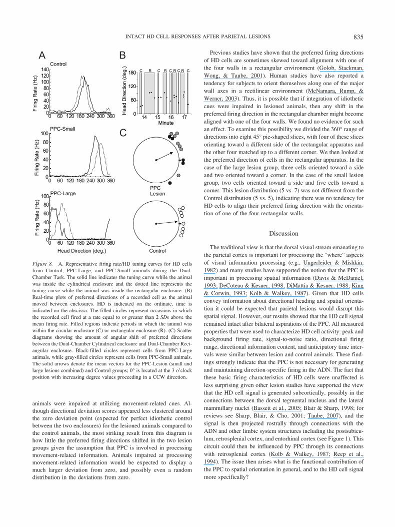
animals were impaired at utilizing movement-related cues. Al-though directional deviation scores appeared less clustered aroundthe zero deviation point (expected for perfect idiothetic controlbetween the two enclosures) for the lesioned animals compared tothe control animals, the most striking result from this diagram ishow little the preferred firing directions shifted in the two lesiongroups given the assumption that PPC is involved in processingmovement-related information. Animals impaired at processingmovement-related information would be expected to display amuch larger deviation from zero, and possibly even a randomdistribution in the deviations from zero.
Previous studies have shown that the preferred firing directionsof HD cells are sometimes skewed toward alignment with one ofthe four walls in a rectangular environment (Golob, Stackman,Wong, & Taube, 2001). Human studies have also reported atendency for subjects to orient themselves along one of the majorwall axes in a rectilinear environment (McNamara, Rump, &Werner, 2003). Thus, it is possible that if integration of idiotheticcues were impaired in lesioned animals, then any shift in thepreferred firing direction in the rectangular chamber might becomealigned with one of the four walls. We found no evidence for suchan effect. To examine this possibility we divided the 360° range ofdirections into eight 45° pie-shaped slices, with four of these slicesorienting toward a different side of the rectangular apparatus andthe other four matched up to a different corner. We then looked atthe preferred direction of cells in the rectangular apparatus. In thecase of the large lesion group, three cells oriented toward a sideand two oriented toward a corner. In the case of the small lesiongroup, two cells oriented toward a side and five cells toward acorner. This lesion distribution (5 vs. 7) was not different from theControl distribution (5 vs. 5), indicating there was no tendency forHD cells to align their preferred firing direction with the orienta-tion of one of the four rectangular walls.
Discussion
The traditional view is that the dorsal visual stream emanating tothe parietal cortex is important for processing the “where” aspectsof visual information processing (e.g., Ungerleider & Mishkin,1982) and many studies have supported the notion that the PPC isimportant in processing spatial information (Davis & McDaniel,1993; DeCoteau & Kesner, 1998; DiMattia & Kesner, 1988; King& Corwin, 1993; Kolb & Walkey, 1987). Given that HD cellsconvey information about directional heading and spatial orienta-tion it could be expected that parietal lesions would disrupt thisspatial signal. However, our results showed that the HD cell signalremained intact after bilateral aspirations of the PPC. All measuredproperties that were used to characterize HD cell activity: peak andbackground firing rate, signal-to-noise ratio, directional firingrange, directional information content, and anticipatory time inter-vals were similar between lesion and control animals. These find-ings strongly indicate that the PPC is not necessary for generatingand maintaining direction-specific firing in the ADN. The fact thatthese basic firing characteristics of HD cells were unaffected isless surprising given other lesion studies have supported the viewthat the HD cell signal is generated subcortically, possibly in theconnections between the dorsal tegmental nucleus and the lateralmammillary nuclei (Bassett et al., 2005; Blair & Sharp, 1998; forreviews see Sharp, Blair, & Cho, 2001; Taube, 2007), and thesignal is then projected rostrally through connections with theADN and other limbic system structures including the postsubicu-lum, retrosplenial cortex, and entorhinal cortex (see Figure 1). Thiscircuit could then be influenced by PPC through its connectionswith retrosplenial cortex (Kolb & Walkey, 1987; Reep et al.,1994). The issue then arises what is the functional contribution ofthe PPC to spatial orientation in general, and to the HD cell signalmore specifically?
Figure 8. A. Representative firing rate/HD tuning curves for HD cellsfrom Control, PPC-Large, and PPC-Small animals during the Dual-Chamber Task. The solid line indicates the tuning curve while the animalwas inside the cylindrical enclosure and the dotted line represents thetuning curve while the animal was inside the rectangular enclosure. (B)Real-time plots of preferred directions of a recorded cell as the animalmoved between enclosures. HD is indicated on the ordinate, time isindicated on the abscissa. The filled circles represent occasions in whichthe recorded cell fired at a rate equal to or greater than 2 SDs above themean firing rate. Filled regions indicate periods in which the animal waswithin the circular enclosure (C) or rectangular enclosure (R). (C) Scatterdiagrams showing the amount of angular shift of preferred directionsbetween the Dual-Chamber Cylindrical enclosure and Dual-Chamber Rect-angular enclosure. Black-filled circles represent cells from PPC-Largeanimals, while gray-filled circles represent cells from PPC-Small animals.The solid arrows denote the mean vectors for the PPC-Lesion (small andlarge lesions combined) and Control groups; 0° is located at the 3 o’clockposition with increasing degree values proceeding in a CCW direction.
835INTACT HD CELL RESPONSES AFTER PARIETAL LESIONS

PPC and Landmark Control of HD Cell PreferredDirections
Previous studies have found that animals are less able to navi-gate using visual landmarks after PPC lesions (Kolb & Walkey,1987; Save & Poucet, 2000b), making our intact landmark controlof HD cells after PPC lesions surprising. This lack of an effect issomewhat consistent with findings from hippocampal place cellsin PPC lesioned animals (Save et al., 2005). In that study, 28 outof 36 place fields rotated near equal amounts when three objectsplaced in the arena were rotated out of view of the lesioned animal.Their findings differ from ours, however, in that all of our HD cellsfrom lesioned animals rotated with the visual landmark. In addi-tion, in the Save et al. (2005) study most of the place cells that hadrotated with the landmarks reverted to their original (nonrotated)positions when the landmarks were removed in the presence of thelesioned animals. The authors concluded that the most likelyexplanation for this finding was that animals with PPC lesionsshowed a preference for the use of distal room cues for orientationrelative to proximal cues. Many theories postulate that place cellsand HD cells are interconnected elements of navigational circuitry(McNaughton et al., 1996; Sharp, 1999; Touretzky & Redish,1996), and these cell types often respond similarly to manipula-tions (Knierim et al., 1995, 1998), making the difference betweenthis place cell study and ours surprising. A likely explanation forthis difference lies in the nature of the visual landmarks utilized inthe present study and by Save et al. (2005). Our study used a whitecard attached to the wall of the enclosure as the visual landmark,whereas Save et al. (2005) used a trio of three-dimensional figuresplaced near the wall of the circular apparatus. Whereas there wasno great difference in the distance of the landmarks relative to theanimal, the three-dimensional characteristic of the landmarks uti-lized by Save et al. (2005) afforded the animal the chance tointeract with the landmarks in a much more proximal way. Givenprevious findings that PPC lesions are more likely to impairnavigation using proximal rather than distal landmarks (Save &Poucet, 2000b), and findings that when a conflict occurs betweenthe location of proximal and distal cues some place cells are morecontrolled by proximal landmarks, others are more controlled bydistal landmarks, while still others show remapping (Renaudineau,Poucet, & Save, 2007; Shapiro, Tanila, & Eichenbaum, 1997;Yoganarasimha, Yu, & Knierim, 2006), it is not surprising thatPPC lesions affected proximal landmark control of some, but notall, place cells in the Save et al. (2005) study.
Several studies, however, have found that visual landmark con-trol of HD cell functioning is controlled by the most distantlandmark in the enclosure. For instance, Zugaro, Berthoz, andWiener (2001) recorded HD cells while the animal foraged on acircular platform with three geometric figures spaced equallyalong the perimeter of the platform. When a wall was placedbetween the geometric figures and the edge of the platform (block-ing the animal’s view of the surrounding room), rotation of thefigures caused an equal rotation of HD cell preferred direction. Incontrast, when the wall was absent, making the most distant visuallandmarks the features of the surrounding room, the position of thegeometric figures was ineffective at controlling HD cell direction-ality. In addition, Yoganarasimha et al. (2006) showed that whilewithin the same session some place cells were controlled byproximal landmarks and other place cells were controlled by distal
landmarks, HD cells were nearly universally controlled by thedistal landmarks. The authors suggested that proximal and distalinformation impinges on the place cell system via different routes,with proximal information arriving through pathways independentof the HD system. In summary, it is likely that while the HDsystem is preferentially controlled by the most distal visual land-marks, the place cell system is controlled by both proximal anddistal landmarks. PPC, by virtue of its role as a convergence areaof both motor and sensory information could play an importantrole in orientating to proximal landmarks with which the animalcan intimately interact.
Our failure to observe decrements of cue card control in PPC-lesioned animals is evidence that substantial landmark informationenters the HD circuitry via a route other than through PPC. Anumber of alternative routes exist (see Figure 1). For instance,cortically processed visual information could bypass PPC by wayof inputs from primary and secondary visual cortex to retrosplenialcortex and postsubiculum (Vogt & Miller, 1983). In support of thispossibility, animals with retrosplenial lesions show a variety ofvisuospatial deficits (Vann & Aggleton, 2005; Whishaw,Maaswinkel, Gonzalez, & Kolb, 2001) and there is preliminaryevidence for the instability in the preferred direction of HD cellsafter retrosplenial lesions (Bassett & Taube, 1999). Additionally,previous studies have suggested that the postsubiculum is instru-mental for processing information concerning visual landmarks, aslesions of the postsubiculum led to unequal shifts in ADN HD cellpreferred directions (Goodridge & Taube, 1997) and in place fieldsof hippocampal place cells after rotation of the prominent visuallandmark in cue rotation experiments (Calton et al., 2003). An-other source of visual input is the tectocortical pathway, throughwhich most retinal ganglion cells project (Linden & Perry, 1983).In this pathway, visual inputs could arrive via connections betweensuperior colliculus, pretectal areas, and thalamic nuclei; mostnotably the lateral dorsal thalamus (Thompson & Robertson,1987), an area also containing HD cells (Mizumori & Williams,1993). Lesions of the superior colliculus result in deficits oforientation and navigation (Dean & Key, 1981; Dean & Redgrave,1984; Lines & Milner, 1985), and rat superior collicular neuronshave been shown to possess visually dependent movement relatedactivity during a navigational task (Cooper, Miya, & Mizumori,1998). However, the finding that lesions of the lateral dorsalthalamus do not disrupt cue control in HD cells (Golob, Wolk, &Taube, 1998) discounts the likelihood that the tectocortical path-way is important.
Selection of Visual Cues as Landmarks
Our failure to observe a deficit in cue card control after PPClesions is also evidence that intact PPC functioning is not neces-sary for a new landmark cue to gain control over the directionalspecificity of the HD network. McNaughton, Chen, and Markus(1991) suggested that for a landmark to provide a useful signal fornavigational circuitry, an association must be made between thestable landmark and a preexisting directional reference, likelyidiothetic in nature. Furthermore, a number of studies have shownthat the ability of a landmark to control HD cell and place cellactivity can be modified by experience (Goodridge, Dudchenko,Worboys, Golob, & Taube, 1998; Knierim et al., 1995, 1998). Theconvergence of sensory and motor signals in PPC, and the close
836 CALTON ET AL.

anatomical connections between PPC and navigational circuitry,provides a possible role for the PPC in this form of learning(McNaughton et al., 1989, 1996; Save & Poucet, 2000a). Ouranimals received their lesions before exposure to the recordingenclosure, and yet HD cells were still accurately controlled by theposition of the salient visual landmark in the apparatus, providingstrong evidence that PPC does not play a crucial role in the abilityof a visual cue to become selected as a stable directional referencefor the HD system. This finding suggests that the ventral visualpathways through the inferior temporal and perirhinal cortices(Miller & Vogt, 1984) may be instrumental in the selection andacquisition of appropriate visual landmarks. Consistent with thisview, studies in rats that have lesioned portions of the perirhinalcortex that are referred to as postrhinal cortex (Burwell & Amaral,1998a, b), have found deficits in visual spatial tasks (Aggleton,Keen, Warburton, & Bussey, 1997; Liu & Bilkey, 2002; Winters,Forwood, Cowell, Saksida, & Bussey, 2004). Alternatively, asmentioned above visual landmark acquisition may emerge throughdirect projections from visual cortex to the retrosplenial cortex andpostsubiculum (Vogt & Miller, 1983).
PPC and Path Integration
Two forms of navigation have been described based on the typesof guidance cues utilized. In the case of landmark navigation, theanimal is guided by the location of familiar landmarks in theenvironment and knowledge of the position of those landmarksrelative to the destination. Alternatively, path-integration involvesthe use of idiothetic signals generated during movement (e.g.,vestibular, proprioceptive, or efferent copy signals) to localizeposition and guide future movement (Mittelstaedt & Mittelstaedt,1980). HD cells respond to both types of navigational cues, pro-viding support for the view that the HD network is an importantcomponent to the navigational system. Our finding that visuallandmarks exerted normal control over the preferred firing direc-tion of HD cells from PPC lesioned animals suggests that the PPCis not necessary for accurate use of visual landmarks by the HDsystem.
The Cylinder-Dark/No Cue and Dual-Chamber tasks assessedwhether PPC-lesions affected the use of idiothetic cues by the HDcell network. Cells from lesioned animals were largely unaffectedby the removal of the visual landmarks in the Cylinder-Dark/NoCue task, demonstrating that these cells were able to use nonvisualinformation to maintain an accurate directional signal. On the otherhand, HD cells and place cells have been shown to respond partlyto nonvisual cues (presumably olfactory in nature) after the elim-ination of visual landmarks (Goodridge et al., 1998; Save, Nerad,& Poucet, 2000) so it is possible that these Dark-No Cue sessionsdid not force the animal to rely on path-integration. The dual-chamber task provided a more complete assessment of the abilityof HD cells to use idiothetic cues after PPC lesions.
Our results from the dual-chamber task show that HD cells inPPC-lesioned animals can maintain a relatively stable preferredfiring direction as the animal moves between a familiar and novelenvironment. One interpretation of this finding is that this taskrequires the use of idiothetic cues and that PPC lesioned animalsremain capable of processing these cues over time and integratingthem with their current orientation. This possibility may not besurprising since there are appropriate idiothetic signals available in
many of the subcortical areas involved in generating and process-ing the HD cell signal (reviewed by Taube, 2007). Alternatively,the maintenance of a stable preferred direction in the dual chambertask may arise because of mechanisms other than processingidiothetic cues. For example, it is possible that the preferreddirections are maintained through a series of associative processesthat, through the use of geometric cues across the different envi-ronments, can link the preferred direction between the cylinder andthe rectangle chambers. Further experiments that use more circu-itous and lengthy routes, which more rigorously test path integra-tion mechanisms, will be necessary before firmly concluding thatthe PPC is not involved in mechanisms of path integration.
Although the dual-chamber task is relatively simple, maintain-ing a stable preferred firing direction for a HD cell on the task hasbeen sufficiently sensitive to various manipulations. For example,lesions of the hippocampus have been shown to impair the abilityof HD cells to maintain a stable preferred direction in this task(Golob & Taube, 1999). Similarly, depriving the animal of normalvolitional motor cues during the task (i.e., proprioception andmotor efference copy) also impairs the ability of HD cells tomaintain a stable preferred direction in this task (Stackman, Golob,Bassett, & Taube, 2003). Thus, the absence of a major impairmentin this task after PPC lesions is consistent with the view that thePPC is not needed to process and update idiothetic cues for HDcell activity. Our results in the no cue card dark sessions are alsoconsistent with this view, because the preferred firing directions inthe lesioned animals were relatively stable throughout these ses-sions.
Our findings that PPC lesions only mildly impaired the ability ofHD cells to use idiothetic cues to maintain a stable preferreddirection are similar to results reported by Golob and Taube (1999)who, using the same task, lesioned cortical areas overlying thedorsal hippocampus as a control for collateral damage whichoccurred when lesioning the hippocampus. Our PPC lesions in-cluded most of the areas lesioned by Golob and Taube, includingportions of Oc1 and Oc2, but also included more lateral areas thatare considered PPC; thereby making these lesions more completePPC lesions. Shifts in the preferred direction were relatively mildfor both studies and suggests that it is unlikely that these corticalareas, including PPC, play a major role in the ability of HD cellsto use idiothetic cues in maintaining a stable preferred direction.One explanation for these results is that large areas of retrosplenialcortex were spared in both studies, and previous experiments haveshown that animals with retrosplenial cortex lesions are impairedin tasks that emphasize the use of idiothetic cues (Cooper, Manka,& Mizumori, 2001; Cooper & Mizumori, 1999; Whishaw et al.,2001).
A number of investigators, however, have found that PPClesions cause deficits in path integration (Commins, Gemmell,Anderson, Gigg, & O’Mara, 1999; Save & Moghaddam, 1996).For instance, using the table-top homing task Save et al. (2001)found that animals with PPC lesions were less likely to take adirect route when returning to the nest from a foraging excursion.Further, Rogers and Kesner (2006) recently reported that parietallesioned animals show deficits in both acquisition and retention ofa spatial task when required to use idiothetic cues in a modifiedHebb-Williams maze. Our findings, however, support the viewthat parietal cortex contributes some other aspect to path integra-tion processes. Keeping track of ones path through an environment
837INTACT HD CELL RESPONSES AFTER PARIETAL LESIONS

may involve a number of different processes and brain areas. It ispossible that the processing of idiothetic cues used to update theperceived directional heading occurs elsewhere in the brain, butthen is integrated with other spatial information in the parietalcortex about location, reference frames, or the order of naviga-tional epochs along a route (Nitz, 2006). Frohardt, Bassett andTaube (2006) found that lesions of the dorsal tegmental nucleus, anarea known to be important for generating the directional signal(Bassett, Tullman, & Taube, 2006), severely impaired perfor-mance in a spatial task requiring path integration. Our findingsalong with other findings demonstrating path-integration deficitsafter PPC lesions could be explained if other HD cell pathways tothe cortex (dorsal tegmental nucleus3 lateral mammillary nuclei3 ADN3 postsubiculum3 entorhinal cortex; Sharp et al., 2001;Taube & Bassett, 2003) enabled normal updating of the directionalsignal, but then this information was unable to be integrated withspatial information in the PPC.
Several other brain areas are thought to be important for pathintegration. The nature of the grid cell correlate in entorhinalcortex makes this signal an ideal candidate for processes involvedwith updating an animal’s path through the environment (Haftinget al., 2005; McNaughton, Battaglia, Jensen, Moser, & Moser,2006). It would be interesting to determine whether lesions of thePPC interfere with the grid cell correlate. However, because theentorhinal cortex receives a significant portion of its afferentinformation from the ADN 3 postsubiculum pathway, the func-tion of which appears relatively intact after PPC lesions, we wouldpostulate that HD and grid cells in the entorhinal cortex would besimilarly unaffected by PPC lesions in terms of landmark controland spatial updating. Finally, the hippocampus may also be in-volved in processing path integration information (Maaswinkel etal., 1999; McNaughton et al., 1996; cf., Alyan & McNaughton,1999). As with lesions of PPC, hippocampal lesions do not disruptHD cell responses or landmark control of their preferred firingdirections (Golob & Taube, 1997), but they do, however, impairthe ability to maintain a stable preferred firing direction whenanimals move to a novel environment in the dual-chamber forag-ing task (Golob & Taube, 1999).
In summary, the PPC does not appear to be important for eitherlandmark control or integrating idiothetic cues for “normal” HDcell responses. Because numerous studies have indicated an im-portant role for PPC in spatial information processing, the PPC islikely involved in processing other types or aspects of spatialinformation that are not critical for normal HD cell function.
References
Aggleton, J. P., Keen, S., Warburton, E. C., & Bussey, T. J. (1997).Extensive cytotoxic lesions involving both the rhinal cortices and areaTE impair recognition but spare spatial alternation in the rat. BrainResearch Bulletin, 43, 279–287.
Alyan, S., & McNaughton, B. L. (1999). Hippocampectomized rats arecapable of homing by path integration. Behavioral Neuroscience, 113,19–31.
Andersen, R. A., Snyder, L. H., Bradley, D. C., & Xing, J. (1997).Multimodal representation of space in the posterior parietal cortex andits use in planning movements. Annual Reviews of Neuroscience, 20,303–330.
Arbib, M. A. (1997). From visual affordances in monkey parietal cortex tohippocampo-parietal interactions underlying rat navigation. Philosophi-
cal Transactions of the Royal Society of London, Series B BiologicalSciences, 352, 1429–1436.
Bassett, J. P., & Taube, J. S. (1999). Retrosplenial cortex lesions disruptstability of head direction cell activity. Society of Neuroscience Ab-stracts, 25, 1383.
Bassett, J. P., Tullman, M. L., & Taube, J. S. (2006). Lesions of thetegmento-mammillary circuit in the head direction system disrupts thehead direction signal in the anterior thalamus. Journal of Neuroscience,27, 7564–7577.
Bassett, J. P., Zugaro, M. B., Muir, G. M., Golob, E. J., Muller, R. U., &Taube, J. S. (2005). Passive movements of the head do not abolishanticipatory firing properties of head direction cells. Journal of Neuro-physiology, 93, 1304–1316.
Batschelet, E. (1981). Circular statistics in biology. New York: AcademicPress.
Blair, H. T., & Sharp, P. E. (1995). Anticipatory head direction signals inanterior thalamus: Evidence for a thalamocortical circuit that integratesangular head motion to compute head direction. Journal of Neuro-science, 15, 6260–6270.
Burwell, R. D., & Amaral, D. A. (1998a). Perirhinal and postrhinal corticesof the rat: Interconnectivity and connections with the entorhinal cortex.Journal of Comparative Neurology, 391, 293–321.
Burwell, R. D., & Amaral, D. A. (1998b). Cortical afferents of the perirhi-nal, postrhinal, and entorhinal cortices of the rat. Journal of ComparativeNeurology, 398, 179–205.
Calton, J. L., Stackman, R. W., Goodridge, J. P., Archey, W. B., Dud-chenko, P. A., & Taube, J. S. (2003). Hippocampal place cell instabilityfollowing lesions of the head direction cell network. Journal of Neuro-science, 23, 9719–9731.
Chen, L. L., Lin, L. H., Barnes, C. A., & McNaughton, B. L. (1994a).Head-direction cells in the rat posterior cortex: I. Anatomical distribu-tion and behavioral modulation. Experimental Brain Research, 101,8–23.
Chen, L. L., Lin, L. H., Green, E. J., Barnes, C. A., & McNaughton, B. L.(1994b). Head-direction cells in the rat posterior cortex: II. Contribu-tions of visual and idiothetic information to the directional firing. Ex-perimental Brain Research, 101, 24–34.
Colby, C. L., & Goldberg, M. E. (1999). Space and attention in parietalcortex. Annual Reviews of Neuroscience, 22, 319–349.
Commins, S., Gemmell, C., Anderson, M., Gigg, J., & O’Mara, S. M.(1999). Disorientation combined with bilateral parietal cortex lesionscauses path integration deficits in the water maze. Behavioral BrainResearch, 104, 197–200.
Cooper, B. G., Manka, T. F., & Mizumori, S. J. Y. (2001). Finding yourway in the dark: The retrosplenial cortex contributes to spatial memoryand navigation without visual cues. Behavioral Neuroscience, 115,1012–1028.
Cooper, B. G., Miya, D. Y., & Mizumori, S. J. Y. (1998). Superiorcolliculus and active navigation: Role of visual and nonvisual cues incontrolling cellular representations of space. Hippocampus, 8, 340–372.
Cooper, B. G., & Mizumori, S. J. Y. (1999). Retrosplenial cortex inacti-vation selectively impairs navigation in darkness. Neuroreport, 10, 625–630.
Corwin, J. V., & Reep, R. L. (1998). Rodent posterior parietal cortex as acomponent of a cortical network mediating directed spatial attention.Psychobiology, 26, 87–102.
Davis, B. K., & McDaniel, W. F. (1993). Visual memory and visual spatialfunctions in the rat following parietal and temporal cortex injuries.Physiology and Behavior, 53, 145–151.
Dean, P., & Key, C. (1981). Spatial deficits on radial maze after large tectallesions in rats: Possible role of impaired scanning. Behavioral andNeural Biology, 32, 170–190.
Dean, P., & Redgrave, P. (1984). The superior colliculus and visual neglect
838 CALTON ET AL.
in rat and hamster. I. Behavioural evidence. Brain Research, 320,129–141.
DeCoteau, W. E., & Kesner, R. P. (1998). Effects of hippocampal andparietal cortex lesions on the processing of multiple-object scenes.Behavioral Neuroscience, 112, 68–82.
DiMattia, B. V., & Kesner, R. P. (1988). Role of the posterior parietalassociation cortex in the processing of spatial event information. Behav-ioral Neuroscience, 102, 397–403.
Frohardt, R. J., Bassett, J. P., & Taube, J. S. (2006). Path integration andlesions within the head direction cell circuit: Comparison between theroles of the anterodorsal thalamus and dorsal tegmental nucleus. Behav-ioral Neuroscience, 120, 135–149.
Gallistel, C. R. (1990). The organization of learning. Cambridge, MA:MIT Press.
Golob, E. J., Stackman, R. W., Wong, A. C., & Taube, J. S. (2001). On thebehavioral significance of head direction cells: Neural and behavioraldynamics during spatial memory tasks. Behavioral Neuroscience, 115,285–304.
Golob, E. J., & Taube, J. S. (1997). Head direction cells and episodicspatial information in rats without a hippocampus. Proceedings of theNational Academy of Science USA, 94, 7645–7650.
Golob, E. J., & Taube, J. S. (1999). Head direction cells in rats withhippocampal or overlying neocortical lesions: Evidence for impairedangular path integration. Journal of Neuroscience, 19, 7198–7211.
Golob, E. J., Wolk, D. A., & Taube, J. S. (1998). Recordings of postsub-icular head direction cells following lesions of the lateral dorsal thalamicnucleus. Brain Research, 780, 9–19.
Goodrich-Hunsaker, N. J., Hunsaker, M. R., & Kessner, R. P. (2005).Dissociating the role of the parietal cortex and dorsal hippocampus forspatial information processing. Behavioral Neuroscience, 119, 1307–1315.
Goodridge, J. P., Dudchenko, P. A., Worboys, K. A., Golob, E. J., &Taube, J. S. (1998). Cue control and head direction cells. BehavioralNeuroscience, 112, 749–761.
Goodridge, J. P., & Taube, J. S. (1997). Interaction between postsubiculumand anterior thalamus in the generation of head direction cell activity.Journal of Neuroscience, 17, 9315–9330.
Hafting, T., Fyhn, M., Molden, S., Moser, M. B., & Moser, E. I. (2005).Microstructure of a spatial map in the entorhinal cortex. Nature, 436,801–806.
King, V. R., & Corwin, J. V. (1993). Comparisons of hemi-inattentionproduced by unilateral lesions of the posterior parietal cortex or medialagranular prefrontal cortex in rats: Neglect, extinction, and the role ofstimulus distance. Behavioral Brain Research, 54, 117–131.
Knierim, J. J., Kudrimoti, H. S., & McNaughton, B. L. (1995). Place cells,head direction cells, and the learning of landmark stability. Journal ofNeuroscience, 15, 1648–1659.
Knierim, J. J., Kudrimoti, H. S., & McNaughton, B. L. (1998). Interactionsbetween idiothetic cues and external landmarks in the control of placecells and head direction cells. Journal of Neurophysiology, 80, 425–446.
Kolb, B., & Walkey, J. (1987). Behavioral and anatomical studies of theposterior parietal cortex in the rat. Behavioral Brain Research, 23,127–145.
Kubie, J. L. (1984). A driveable bundle of microwires for collectingsingle-unit data from freely-moving rats. Physiology and Behavior, 32,115–118.
Linden, R., & Perry, V. H. (1983). Massive retinotectal projection in rats.Brain Research, 272, 145–149.
Lines, C. R., & Milner, A. D. (1985). A deficit in ambient visual guidancefollowing superior colliculus lesions in rats. Behavioral Neuroscience,99, 707–716.
Liu, P., & Bilkey, D. K. (2002). The effects of NMDA lesions centered onthe postrhinal cortex on spatial memory tasks in the rat. BehavioralNeuroscience, 116, 860–873.
Maaswinkel, H., Jarrard, L. E., & Whishaw, I. Q. (1999). Hippocampec-tomized rats are impaired in homing by path integration. Hippocampus,9, 553–561.
McNamara, T. P., Rump, B., & Werner, S. (2003). Egocentric and geo-centric frames of reference in memory of large-scale space. Psy-chonomic Bulletin and Review, 10, 589–595.
McNaughton, B. L., Barnes, C. A., Gerrard, J. L., Gothard, K., Jung,M. W., Knierim, J. J., et al. (1996). Deciphering the hippocampalpolyglot: The hippocampus as a path integration system. Journal ofExperimental Biology, 199, 173–185.
McNaughton, B. L., Battaglia, F. P., Jensen, O., Moser, E. I., Moser, &M. B. (2006). Path integration and the neural basis of the ‘cognitivemap’. Nature Reviews Neuroscience, 7, 663–678.
McNaughton, B. L., Chen, L. L., & Markus, E. J. (1991). “Dead reckon-ing”, landmark learning, and the sense of direction: A neurophysiolog-ical and computational hypothesis. Journal of Cognitive Neuroscience,3, 190–202.
McNaughton, B. L., Leonard, B., & Chen, L. (1989). Cortical-hippocampalinteractions and cognitive mapping: A hypothesis based on reintegrationof the parietal and inferotemporal pathways for visual processing. Psy-chobiology, 17, 230–235.
McNaughton, B. L., Mizumori, S. J. Y., Barnes, C. A., Marquis, J. L. M.,& Green, E. J. (1994). Cortical representation of motion during unre-strained spatial navigation in the rat. Cerebral Cortex, 4, 7–39.
Miller, M. W., & Vogt, B. A. (1984). Direct connections of rat visualcortex with sensory, motor, and association cortices. Journal of Com-parative Neurology, 226, 184–202.
Mittelstaedt, M. L., & Mittelstaedt, H. (1980). Homing by path integrationin a mammal. Naturwissenschaften, 67, 566–567.
Mizumori, S. J., & Williams, J. D. (1993). Directionally selective mne-monic properties of neurons in the lateral dosal nucleus of the thalamusof rats. Journal of Neuroscience, 13, 4015–4028.
Muller, R. U., & Kubie, J. L. (1987). The effects of changes in theenvironment on the spatial firing properties of hippocampal complex-spike cells. Journal of Neuroscience, 7, 1951–1968.
Nakamura, K. (1999). Auditory spatial discriminatory and mnemonic neu-rons in rat posterior parietal cortex. Journal of Neurophysiology, 82,2503–2517.
Nitz, D. A. (2006). Tracking route progression in the posterior parietalcortex. Neuron, 49, 747–756.
O’Keefe, J., & Dostrovsky, J. (1971). The hippocampus as a spatial map:Preliminary evidence from unit activity in the freely-moving rat. BrainResearch, 34, 171–175.
Palomero-Gallagher, N., & Zilles, K. (2004). Isocortex. In G. Paxinos(Ed.), The rat nervous system (2nd ed., pp. 729–757). New York:Academic Press.
Paxinos, G., & Watson, C. (1998). The rat brain in stereotaxic coordinates(4th ed.). San Diego, CA: Academic Press.
Quirk, G. J., Muller, R. U., & Kubie, J. L. (1990). The firing of hippocam-pal place cells in the dark depends on the rat’s recent experience. Journalof Neuroscience, 10, 2008–2017.
Reep, R. L., Chandler, H. C., King, V., & Corwin, J. V. (1994). Ratposterior parietal cortex: Topography of corticocortical and thalamicconnections. Experimental Brain Research, 100, 67–84.
Renaudineau, S., Poucet, B., & Save, E. (2007). Flexible use of proximalobjects and distal cues by hippocampal place cells. Hippocampus, 17,381–385.
Rogers, J. L., & Kesner, R. P. (2006). Lesions of the dorsal hippocampusor parietal cortex differentially affect spatial information processing.Behavioral Neuroscience, 120, 852–860.
Save, E., Guazzelli, A., & Poucet, B. (2001). Dissociation of the effects ofbilateral lesions of the dorsal hippocampus and parietal cortex on pathintegration in the rat. Behavioral Neuroscience, 115, 1212–1223.
Save, E., & Moghaddam, M. (1996). Effects of lesions of the associative
839INTACT HD CELL RESPONSES AFTER PARIETAL LESIONS

parietal cortex on the acquisition and use of spatial memory in egocen-tric and allocentric navigation tasks in the rat. Behavioral Neuroscience,110, 74–85.
Save, E., Nerad, L., & Poucet, B. (2000). Contribution of multiple sensoryinformation to place field stability in hippocampal place cells. Hip-pocampus, 10, 64–76.
Save, E., Paz-Villagran, V., Alexinsky, T., & Poucet, B. (2005). Functionalinteraction between the associative parietal cortex and hippocampalplace cell firing in the rat. European Journal of Neuroscience, 21,522–530.
Save, E., & Poucet, B. (2000a). Hippocampal-parietal cortical interactionsin spatial cognition. Hippocampus, 10, 491–499.
Save, E., & Poucet, B. (2000b). Involvement of the hippocampus andassociative parietal cortex in the use of proximal and distal landmarksfor navigation. Behavioral Brain Research, 109, 195–206.
Shapiro, M. L., Tanila, H., & Eichenbaum, H. (1997). Cues that hippocam-pal place cells encode: Dynamic and hierarchical representation of localand distal stimuli. Hippocampus, 7, 624–642.
Sharp, P. E. (1999). Complimentary roles for hippocampal versus subicu-lar/entorhinal place cells in coding place, context, and events. Hip-pocampus, 9, 432–443.
Sharp, P. E., Blair, H. T., & Cho, J. (2001). The anatomical and compu-tational basis of the rat head-direction cell signal. Trends in Neuro-science, 24, 289–294.
Skaggs, W. E., McNaughton, B. L., Gothard, K. M., & Markus, E. J.(1993). An information-theoretic approach to deciphering the hippocam-pal code. In S. J. Hanson, J. D. Cowan, & C. L. Giles (Eds.), Advancesin neural information processing systems (Vol. 5, pp. 1030–1037). SanMateo, CA: Morgan Kaufmann.
Sripanidkulchai, K., & Wyss, J. M. (1986). Thalamic projections to retro-splenial cortex in the rat. Journal of Comparative Neurology, 254,143–165.
Stackman, R. W., Golob, E. J., Bassett, J. P., & Taube, J. S. (2003). Passivetransport disrupts directional path integration by rat head direction cells.Journal of Neurophysiology, 90, 2862–2874.
Taube, J. S. (1995). Head direction cells recorded in the anterior thalamicnuclei of freely moving rats. Journal of Neuroscience, 15, 70–86.
Taube, J. S. (1998). Head direction cells and the neurophysiological basisfor a sense of direction. Progress in Neurobiology, 55, 225–256.
Taube, J. S. (2007). The head direction signal: Origins and sensory-motorintegration. Annual Reviews of Neuroscience, 30, 181–207.
Taube, J. S., & Bassett, J. P. (2003). Persistent neural activity in headdirection cells. Cerebral Cortex, 13, 1162–1172.
Taube, J. S., & Burton, H. L. (1995). Head direction cell activity monitoredin a novel environment and during a cue conflict situation. Journal ofNeurophysiology, 74, 1953–1971.
Taube, J. S., & Muller, R. U. (1998). Comparisons of head direction cellactivity in the postsubiculum and anterior thalamus of freely movingrats. Hippocampus, 8, 87–108.
Taube, J. S., Muller, R. U., & Ranck, J. B. Jr. (1990a). Head-direction cellsrecorded from the postsubiculum in freely moving rats. I. Descriptionand quantitative analysis. Journal of Neuroscience, 10, 420–435.
Taube, J. S., Muller, R. U., & Ranck, J. B. Jr. (1990b). Head-direction cellsrecorded from the postsubiculum in freely moving rats. II. Effects ofenvironmental manipulations. Journal of Neuroscience, 10, 436–447.
Thompson, S. M., & Robertson, R. T. (1987). Organization of subcorticalpathways for sensory projections to the limbic cortex. II. Afferentprojections to the thalamic lateral dorsal nucleus in the rat. Journal ofComparative Neurology, 265, 189–202.
Touretzky, D. S., & Redish, A. D. (1996). Theory of rodent navigationbased on interacting representations of space. Hippocampus, 6, 247–270.
Ungerleider, L., & Mishkin, M. (1982). Two cortical visual streams. InD. J. Ingle, M. A. Goodale, & R. J. W. Mansfield (Eds.), Analysis ofvisual behavior (pp. 549–586). Cambridge, MA: MIT Press.
Vann, S. D., & Aggleton, J. P. (2005). Selective dysgranular retrosplenialcortex lesions in rats disrupt allocentric performance of the radial-armmaze task. Behavioral Neuroscience, 119, 1682–1686.
Vogt, B. A., & Miller, M. W. (1983). Cortical connections between ratcingulated cortex and visual, motor, and postsubicular cortices. Journalof Comparative Neurology, 216, 192–210.
Ward, N. M., & Brown, V. J. (1997). Deficits in response initiation, but notattention, following excitotoxic lesions of posterior parietal cortex in rat.Brain Research, 775, 81–90.
Whishaw, I. Q., Maaswinkel, H., Gonzalez, C. L., & Kolb, B. (2001).Deficits in allothetic and idiothetic spatial behavior in rats with posteriorcingulated cortex lesions. Behavioral Brain Research, 118, 67–76.
Winters, B. D., Forwood, S. E., Cowell, R. A., Saksida, L. M., & Bussey,T. J. (2004). Double dissociation between the effects of peri-postrhinalcortex and hippocampal lesions on tests of object recognition and spatialmemory: Heterogeneity of function within the temporal lobe. Journal ofNeuroscience, 24, 5901–5908.
Wyss, J. M., & Van Groen, T. (1992). Connections between the retrosple-nial cortex and the hippocampal formation in the rat: A review. Hip-pocampus, 2, 1–12.
Yoganarasimha, D., Yu, X., & Knierim, J. J. (2006). Head direction cellrepresentations maintain internal coherence during conflicting proximaland distal cue rotations: Comparison with hippocampal place cells.Journal of Neuroscience, 11, 622–631.
Zugaro, M. B., Berthoz, A., & Wiener, S. I. (2001). Background, but notforeground, spatial cues are taken as references for head directionresponses by rat anterodorsal thalamus neurons. Journal of Neuro-science, 21, RC154.
Received September 17, 2007Revision received December 18, 2007
Accepted January 24, 2008 �
840 CALTON ET AL.
Copyright © 2022 FDOKUMEN