Key role of alkaline phosphatase in the development of human-derived nanoparticles in vitro
17
Key role of alkaline phosphatase for development of human- derived nanoparticles in vitro Larry W. Hunter a , Farooq A. Shiekh b , George T. Pisimisis a , Sung-Hoon Kim b,e , Samuel N. Edeh b , Virginia M. Miller a,c , and John C. Lieske b,c,d a Department of Surgery, Mayo Clinic Rochester, MN, USA b Department of Internal Medicine, Mayo Clinic Rochester, MN, USA c Department of Physiology and Biomedical Engineering, Mayo Clinic Rochester, MN, USA d Department of Laboratory Medicine and Pathology, Mayo Clinic Rochester, MN, USA e College of Oriental Medicine, Kyunghee University, Republic of Korea Abstract Alkaline phosphatase (ALP) is an enzyme critical for physiological and pathological biomineralization. Experiments were designed to determine if ALP participates in formation of calcifying nanometer-sized particles (NPs) in vitro. Filtered homogenates of human calcified carotid artery, aorta and kidney stones were inoculated into cell culture medium containing 10% fetal bovine serum in the absence or presence of inhibitors of ALP or pyrophosphate. Calcific NP biofilm developed within one week after inoculation and their development was reduced by pyrophosphate and inhibitors of ALP. ALP protein and enzymatic activity were detected in washed NPs whether calcified or decalcified. Therefore, ALP activity is required for formation of calcifying NPs in vitro, as has previously been implicated during pathological calcification in vivo. Keywords Biologic nanoparticles; calcification; heterotopic mineralization; hydroxyapatite; nanobacteria 1. INTRODUCTION Spherical nano-sized structures comprised partly of calcium and phosphorous, historically called matrix vesicles, are present at sites of physiological and pathological mineral deposition such as normal bone and soft-tissues in mammals and birds [1–3]. Formation of these matrix vesicles was considered to be a passive process resulting from pathological increases in plasma calcium and phosphate levels. However, it is now accepted that their formation is a cell-mediated, regulated process which is similar to that of normal osteogenesis, and which is modulated by a group of proteins which either promote or inhibit calcification. Release of these membrane-vesicles by chondrocytes, osteoblasts, odontoblasts © 2010 Acta Materialia Inc. Published by Elsevier Ltd. All rights reserved. Correspondence: John C. Lieske, MD, Mayo Clinic Division of Nephrology and Hypertension, 200 1 st Street SW, Rochester, MN 55905, USA, Phone: (507) 266 - 7960, Fax: (507) 266 - 9315, [email protected]. Publisher's Disclaimer: This is a PDF file of an unedited manuscript that has been accepted for publication. As a service to our customers we are providing this early version of the manuscript. The manuscript will undergo copyediting, typesetting, and review of the resulting proof before it is published in its final citable form. Please note that during the production process errors may be discovered which could affect the content, and all legal disclaimers that apply to the journal pertain. NIH Public Access Author Manuscript Acta Biomater. Author manuscript; available in PMC 2012 March 1. Published in final edited form as: Acta Biomater. 2011 March ; 7(3): 1339–1345. doi:10.1016/j.actbio.2010.10.027. NIH-PA Author Manuscript NIH-PA Author Manuscript NIH-PA Author Manuscript
-
Upload
mayoclinic -
Category
Documents
-
view
5 -
download
0
Transcript of Key role of alkaline phosphatase in the development of human-derived nanoparticles in vitro
Key role of alkaline phosphatase for development of human-derived nanoparticles in vitro
Larry W. Huntera, Farooq A. Shiekhb, George T. Pisimisisa, Sung-Hoon Kimb,e, Samuel N.Edehb, Virginia M. Millera,c, and John C. Lieskeb,c,daDepartment of Surgery, Mayo Clinic Rochester, MN, USAbDepartment of Internal Medicine, Mayo Clinic Rochester, MN, USAcDepartment of Physiology and Biomedical Engineering, Mayo Clinic Rochester, MN, USAdDepartment of Laboratory Medicine and Pathology, Mayo Clinic Rochester, MN, USAeCollege of Oriental Medicine, Kyunghee University, Republic of Korea
AbstractAlkaline phosphatase (ALP) is an enzyme critical for physiological and pathologicalbiomineralization. Experiments were designed to determine if ALP participates in formation ofcalcifying nanometer-sized particles (NPs) in vitro. Filtered homogenates of human calcifiedcarotid artery, aorta and kidney stones were inoculated into cell culture medium containing 10%fetal bovine serum in the absence or presence of inhibitors of ALP or pyrophosphate. Calcific NPbiofilm developed within one week after inoculation and their development was reduced bypyrophosphate and inhibitors of ALP. ALP protein and enzymatic activity were detected inwashed NPs whether calcified or decalcified. Therefore, ALP activity is required for formation ofcalcifying NPs in vitro, as has previously been implicated during pathological calcification in vivo.
KeywordsBiologic nanoparticles; calcification; heterotopic mineralization; hydroxyapatite; nanobacteria
1. INTRODUCTIONSpherical nano-sized structures comprised partly of calcium and phosphorous, historicallycalled matrix vesicles, are present at sites of physiological and pathological mineraldeposition such as normal bone and soft-tissues in mammals and birds [1–3]. Formation ofthese matrix vesicles was considered to be a passive process resulting from pathologicalincreases in plasma calcium and phosphate levels. However, it is now accepted that theirformation is a cell-mediated, regulated process which is similar to that of normalosteogenesis, and which is modulated by a group of proteins which either promote or inhibitcalcification. Release of these membrane-vesicles by chondrocytes, osteoblasts, odontoblasts
© 2010 Acta Materialia Inc. Published by Elsevier Ltd. All rights reserved.Correspondence: John C. Lieske, MD, Mayo Clinic Division of Nephrology and Hypertension, 200 1st Street SW, Rochester, MN55905, USA, Phone: (507) 266 - 7960, Fax: (507) 266 - 9315, [email protected]'s Disclaimer: This is a PDF file of an unedited manuscript that has been accepted for publication. As a service to ourcustomers we are providing this early version of the manuscript. The manuscript will undergo copyediting, typesetting, and review ofthe resulting proof before it is published in its final citable form. Please note that during the production process errors may bediscovered which could affect the content, and all legal disclaimers that apply to the journal pertain.
NIH Public AccessAuthor ManuscriptActa Biomater. Author manuscript; available in PMC 2012 March 1.
Published in final edited form as:Acta Biomater. 2011 March ; 7(3): 1339–1345. doi:10.1016/j.actbio.2010.10.027.
NIH
-PA Author Manuscript
NIH
-PA Author Manuscript
NIH
-PA Author Manuscript
and activated or apoptotic cells provides an extracellular environment which promotes theinitial steps for generation of hydroxyapatite (HA) crystals, leading to the deposition ofextracellular mineral [4–7].
Structures similar to matrix vesicles have also been identified in histological sections ofhuman calcified vascular tissue, renal papillae and kidney stones, whereas they are absent incomparable uncalcified tissues [8–12]. When filtered homogenates of calcified humantissue, blood or saliva are placed into cell culture, a calcific biofilm develops which iscomposed of nanosized complexes of HA and protein similar in anatomical appearance tomatrix vesicles, but previously described as either nanobacteria [8,13,14] or calcifyingnanoparticles [9,11,12,15,16]. Due to the failure to consistently sequence unique nucleicacid from these vesicles/particles, and because development of nano-sized structures isdependent upon the chemical composition of the in vitro milieu, the term nanoparticles(NPs) is now preferred to describe these structures [17]. Because nano-sized structures areassociated with sites of ectopic mineralization, study of the capacity of NPs to propagate andinduce HA mineral deposition in vitro may provide insight into the biochemical processes ofpathological mineralization.
When incubated under tissue culture conditions, NPs develop both as an adherent calcifiedbiofilm and exist as free-floating "planktonic” forms [15]. Both structures contain an outercalcium phosphate shell and inner core composed of both mammalian [18,19] andprokaryotic proteins [12]. Some of the NP-associated proteins are structural or carriermolecules, for example fetuin-A, also known as α2-HS-Glycoprotein, a circulatinghepatocyte-derived calcification inhibitor [20–23]
Alkaline phosphatase (ALP) belongs to a group of enzymes which individually promotes orinhibits mineralization in a highly-regulated process [24–26]. ALP is a cell membrane-associated enzyme that hydrolyzes inorganic pyrophosphate (PPi), a potent suppressor ofHA crystal growth. Hydrolysis of PPi yields inorganic phosphate (Pi), a substrate for HAmineral [27–29], and thus, contributes to regulation of normal bone formation as well as inpathological extraosseous mineralization. The association of ALP with physiological andpathological mineralization is supported by observations that mice deficient in the gene fortissue non-specific ALP develop severe hypophosphatasia and exhibit abnormal bonedevelopment [30]. Furthermore, cultured aortic smooth muscle cells derived fromspontaneously hypertensive rats (SHR) develop extracellular calcification, whereas similarcells derived from normotensive animals do not. Gene expression of ALP protein is higherin the cells from the SHR animals than the normotensive controls [31]. Serum levels of Piand ALP also are correlated with vascular and renal calcification in humans [32,33]. Giventhe participation of ALP in calcification processes, experiments were conducted to test thehypothesis that in vitro formation of calcifying NPs derived from human diseased tissuesrequires ALP.
2. MATERIALS AND METHODS2.1. Reagents and drugs
Beryllium sulfate tetrahydrate, levamisole-hydrochloride, sodium pyrophosphate,phenylmethanesulfonyl fluoride and hydroxyapatite suspension (in 0.001 M phosphatebuffer, pH 6.8; approx. 25% solid) were obtained from Sigma Chemical, St. Louis, MO.Beta-Glycerophosphate was from Calbiochem, Gibbstown, NJ. Dulbecco’s Modified EagleMedium (DMEM) was purchased from Mediatech Inc., Manassas, VA. Gamma-irradiatedfetal bovine serum was from Atlanta Biologicals, Lawrenceville, GA. All other chemicalswere from ThermoFisher Scientific Inc., Waltham, MA. Beryllium sulfate and levamisolestocks were prepared in water and DMEM, respectively. Water used for these experiments
Hunter et al. Page 2
Acta Biomater. Author manuscript; available in PMC 2012 March 1.
NIH
-PA Author Manuscript
NIH
-PA Author Manuscript
NIH
-PA Author Manuscript
was double-distilled; all water and phosphate-buffered saline (PBS, pH 7.4) were filtered(0.2 µm) prior to use.
2.2. Preparation of NP from tissue isolatesCalcified and uncalcified human segments from sub-adventitial endarterectomy or fullthickness pieces of abdominal aorta and carotid arteries were collected under asepticconditions as waste during surgical procedures for vascular repair in accordance with theguidelines of the Institutional Review Board governing use of human materials at MayoClinic, Rochester, MN [9]. Each segment was placed into sterile phosphate-buffered saline(PBS) on ice, and sent to the laboratory for immediate processing within an hour ofexplantation. Segments with high, low and no calcification were collected from separateindividuals. A piece (200–300 mg) was cut from each vascular segment, rinsed, placed into2 ml PBS and finely minced with scissors. These pieces were disrupted using several strokesin a Potter-Elvehjem homogenizer. The sample was then centrifuged for 15 minutes at2,500Xg to pellet debris. The supernatant was filtered successively through 1.2, 0.45, and0.2 µm cellulose acetate filters (Whatman Inc., Piscataway, NJ). This filtered isolate wasused for the culture of NP (see below).
To characterize the material remaining after the 0.2 µm filtration, the filtrate from somepreparations was divided into two equal portions; one portion was placed into culture for NPpropagation as described below. The other was centrifuged at 125,000Xg, 4°C for 60 min.The supernatant was removed, diluted in medium and placed in culture. A sample of high-speed pellet was examined by transmission electron microscopy for structural elements (i.e.matrix vesicles); the remainder of the pellet was re-suspended in medium and cultured.
Kidney stones classified as calcium phosphate kidney stones (strain AP11) aftercompositional analysis in the Mayo Clinic Metals Laboratory, were washed with distilledwater, dried, pulverized using a mortar and pestle, and stored at 4°C until use. Pulverizedstones were demineralized in 1N HCl for 10 minutes with constant stirring, then neutralizedwith 1N NaOH. This suspension was centrifuged at 60,000Xg (Sorvall Evolution-RCcentrifuge, ThermoFisher) for 1 hr at 4°C, followed by re-suspension of the resulting pelletin DMEM. After vortexing, the sample was filtered, first through a No. 42 filter (WhatmanInc., Piscataway, NJ), then through a 0.2 µm filter. An aliquot of filtrated isolate was used toculture NP.
2.3. Propagation and collection of NPsThe filtered isolate from either the vascular or kidney stone homogenates was diluted 1:20 incell culture medium (DMEM) containing 10% γ–irradiated fetal bovine serum (FBS).Concentrations of Ca2+ and Pi were 1.8 mM and 0.9 mM, respectively, being derived fromboth the medium and FBS.
Filtered tissue homogenate (0.2 mL) was injected into culture medium (5 ml) and placedinto 25 cm2 vented culture flasks and maintained in a humid 13% CO2 incubator at 37°C.After 2–3 week, the adherent calcific biofilm containing NPs was scraped from the bottomof each flask into the medium. The turbidity of the medium was then measured inNephelometric Turbidity Units (NTU) using a Hach Model 2100N turbidimeter (Hach Co.,Loveland, CO) which was used as an index of NP propagation. This methods measures totalNPs, those from the biofilm and those which were free floating (planktonic) in the media.Random flasks were screened for bacterial contamination, including Mycoplasma, using asensitive rapid PCR test, by the Mayo Clinic Microbiology Laboratory; all results werenegative.
Hunter et al. Page 3
Acta Biomater. Author manuscript; available in PMC 2012 March 1.
NIH
-PA Author Manuscript
NIH
-PA Author Manuscript
NIH
-PA Author Manuscript

In some experiments, commercial synthetic HA crystals were added to the culture media(instead of tissue filtrate) and processed in parallel with flasks containing the tissue filtrateto serve as a non-biological control.
To determine whether ALP was required for NP propagation, the culture medium wasmodified by addition of either an ALP inhibitor (beryllium, 30 µM or levamisole 1mM), anALP substrate (β-glycerophosphate, 5 mM) [34] or an inhibitor of apatite mineral deposition(PPi, 10 µM).
2.4. Transmission electron microscopy (TEM)To examine structural elements, the 125,000Xg fraction of a freshly isolated artery wasfixed in 3% glutaraldehyde overnight at 4°C and then embedded in Epon/Araldite resin, thinsectioned and transferred to a carbon-coated grid. The grid was then negatively stained with2% uranyl acetate, dried, and examined using TEM (Tecnai T12, FEI Co., Hillsboro, OR).In other experiments, NPs propagated for 14 days were examined using whole mounts. TheNPs were harvested then fixed as described above. A 25 µl drop of suspended NPs wastransferred to a grid; each grid was then stained, dried and examined using TEM.
2.5. Enzyme activity, Protein isolation and Western blottingALP activity in medium and in harvested NPs before and after decalcification was measuredcolorimetrically as the hydrolysis of p-nitrophenyl phosphate at pH 9.8 according to themanufacturer’s instructions (Sigma, Inc.). To decalcify NPs, the harvested NPs weresuspended in 0.5M EDTA with continuous shaking overnight at 4°C. Decalcified NPs(dNPs) were then pelleted by centrifugation at 60,000Xg and washed twice in PBS.
To measure protein, pellet of decalcified NPs was suspended in PBS containing 1mMphenylmethanesulfonyl fluoride and the resulting suspension was sonicated for 20 minuteson ice (10% wave intensity), followed by centrifugation at 8,000Xg for 5 minutes at 4°C.Total protein in the supernatant was determined colorimetrically using a kit based onbicinchoninic acid (BCA; Pierce Biotechnology, Rockford, IL); individual proteins wereresolved via SDS-PAGE under reducing conditions and these resulting bands were identifiedvia Coomassie brilliant blue staining. For Western blotting, gels were destained, electro-transferred to a PVDF membrane, and probed using a polyclonal primary antibody raised ingoats which maps an epitope within an internal region of ALP of human origin (ALP I-12;Santa Cruz Biotechnology, Santa Cruz, CA), followed by donkey anti-goat IgG-HRP (SantaCruz).
2.6. StatisticsStatistical data are presented as mean ± SEM; n = the number of times the experiment wasrepeated using filtered tissue homogenates from different patients. The Student’s t-test wasused to determine differences from control conditions; statistical significance was acceptedat P< 0.05. One exception was the initial survey of various tissues to identify possiblesources of NPs (n=1 each).
3. RESULTS3.1. Absence of matrix vesicles in tissue homogenates which proprogate NPs
The filtered homogenate prepared from calcified vascular tissue contained structures similarin size and appearance to matrix vesicles previously isolated from human atheroscleroticaorta (Fig. 1) [35]. Supernatant resulting from the125,000Xg centrifugation of thehomogenate retained the capacity to propagate NPs in culture, whereas the pelleted fractionthat contained matrix vesicle-like structures from the homogenate did not (Fig. 1).
Hunter et al. Page 4
Acta Biomater. Author manuscript; available in PMC 2012 March 1.
NIH
-PA Author Manuscript
NIH
-PA Author Manuscript
NIH
-PA Author Manuscript
3.2. Evidence for ALP activity in the culture mediumALP activity in freshly-prepared DMEM containing 10% FBS, measured colorimetricallywith p-nitrophenol phosphate was 0.02 U/ml; this activity decreased by 91% with additionof 1 mM levamisole (not shown). Increases in turbidity of the media were used as an indirectmeasure of NP formation. The turbidity of DMEM plus 10% γ– irradiated FBS incubatedunder culture conditions for 14 days increased significantly with the addition of 5 mM β-glycerophosphate, a non-biological substrate for ALP. (Fig. 2). Concurrent addition oflevamisole to β-glycerophosphate-supplemented medium reduced the turbidity to controllevels (Fig. 2).
3.3. NPs propagated in vitro from isolates of calcified tissue require ALP activityTurbidity of media (NP formation) increased when seeded with filtered supernatant derivedfrom a 2,500Xg centrifugation of calcified tissues homogenates (calcified aortic aneurysm,carotid artery and kidney stone), but not from a non-calcified aneurysm (Fig. 3). Thecompetitive ALP inhibitor, beryllium (30 µM), or an inhibitor of apatite mineral deposition,PPi (10 µM), reduced turbidity (NP formation) to 6% – 28% of levels observed in flaskswithout inhibitors (controls, Fig. 3).
To confirm these observations using other calcified tissue isolates and other ALP inhibitors,kidney stone-derived homogenates were seeded into flask containing DMEM and FBS.Turbidity of media without tissue homogenates (NPs;controls) and of media seeded withkidney stone homogenates was low at baseline (day 1; 2.7 ± 0.1 and 4.7 ± 0.3 NTU,respectively; n = 5 each; Fig. 4). After 21 days, turbidity of media seeded with kidney stonehomogenates significantly increased (296.2 ± 33.4 NTU, n = 5) compared to the turbidity ofcontrol flasks (6.1 ± 0.2 NTU, n = 5). This increase was significantly attenuated in thepresence of levamisole (1 mM; 23.1 ± 0.4 NTU, n = 5). NPs that formed under controlconditions ranged in size from 150–300 nm, with crystals on the exterior structure (Fig. 4b).However, NPs cultured in the presence of levamisole appeared smaller with less associatedcrystal (Fig. 4b). Previous studies demonstrated that these crystals are composed of calciumphosphate hydroxyapatite [9].
3.4. ALP activity in NPsALP activity was detected in NPs harvested after 14 days in culture and washed three timeswith physiological saline to remove adherent proteins (Fig. 5). ALP activity also waspresent, although reduced, in NPs after decalcification with 0.5M EDTA. HA crystalsincubated in DMEM containing 10% FBS under similar conditions and processed in thesame way also contained measurable but low ALP activity. However, ALP activity was nearbackground levels in HA crystals incubated in DMEM without FBS (Fig. 5).
3.5. Immunological detection of ALP antigenIn a typical experiment approximately 250 µg of protein was recovered after decalcificationof one flask of NPs. Numerous protein bands that stained with Comassie-blue were detectedby SDS-PAGE of decalcified NP pellets (Fig. 6A).
A commercial antibody raised against human ALP recognized at least 5 bands in decalcifiedNP lysate by Western blotting, 2 of these bands migrated to the approximate molecularweight of human or bovine ALP, and 3 others of smaller molecular weight (Fig. 6B). Thebands recognized by the anti-ALP antibody were then cut from a duplicate gel for furtheranalysis in order to identify the proteins by trypsin digestion and tandem mass spectrometry.Neither eukaryotic nor prokaryotic ALP was detected via this analysis, even though thesebands were recognized by the ALP antibody.
Hunter et al. Page 5
Acta Biomater. Author manuscript; available in PMC 2012 March 1.
NIH
-PA Author Manuscript
NIH
-PA Author Manuscript
NIH
-PA Author Manuscript
4. DISCUSSIONThis study confirms that calcifying biologic NPs form in vitro from filtered homogenates ofcalcified vascular tissue and kidney stones, but not from non-calcified vascular tissue [9,12].It is unlikely that NPs studied in the present experiments were matrix vesicles since NPscould not be propagated from pelleted fractions resulting from high speed centrifugation oftissue homogenates whereas NPs did propagate from the corresponding supernatant.Furthermore, although ALP is expressed in matrix vesicles derived from various cells[7,38,39], it is absent or present at low levels in matrix vesicles isolated from freshatherosclerotic blood vessels. These vesicles are enriched in ATPase, AMPase and NTPpyrophosphohydrolase. These other hydrolases regulate calcification [35,40].
In contrast, results of this study provide evidence that ALP is required for propagation of NPderived from homogenates of calcified tissue. Found in most species from bacteria to man,ALP is a ubiquitous dimeric enzyme which catalyzes hydrolysis of phosphomonoesters,with release of Pi [24] and is implicated in the formation of renal stones and in arterialcalcification [27,28]. Development and propagation of NPs from these tissues were inhibitedby both beryllium and levamisole, inhibitors of ALP [26,29,41]. Beryllium, a competitiveALP inhibitor [42] reduces mineralization as well as corresponding ALP activity [41]. Incontrast, levamisole, an uncompetitive ALP inhibitor [36,37] binds to the ALP/substratecomplex. In the present study, levamisole reduced NP formation and inhibited ALP activityby similar degrees (91%) in addition to reducing the amount of apatite mineral in associationwith NPs as determined by TEM. To our knowledge, neither beryllium nor levamisole hasbeen shown to inhibit mineralization via a direct effect on crystal growth. ALP activity inculture medium containing 10% FBS, as used in this study, was measured at 0.02 U/ml,sufficient to hydrolyze 5 mM β-glycerophosphate and increase medium turbidity, mostlikely due to precipitation of calcium phosphate [34]. The lack of change in turbidity in β-glycerophosphate-treated medium in the presence of levamisole confirms the capacity ofantagonists of ALP to modulate NP propagation.
Exogenous PPi was used in this study as a tool to inhibit apatite mineral deposition. Theeffect of endogenous PPi on mineralization appears to be concentration-dependent andbimodal [43]. Lower concentrations (< 0.1mM) favor mineralization because PPi isefficiently hydrolyzed by ALP liberating Pi, making it available for formation of apatite.However, at supraphysiological concentrations, as used in the present study, a largeproportion of PPi remains unhydrolyzed and available to bind HA thereby inhibitingmineralization.
Electron microscopic examination of calcified vascular tissue sections by energy-dispersivex-ray analysis indicated that the mineral present in these segments was in the form ofcalcium phosphate HA [9]. NPs that develop in vitro also appear to be complexes of proteinstogether with calcium phosphate HA with no evidence of carbonated HA or of otherminerals [8,9,44]. In the present study, the mineral content of NPs developed under variouspH or calcium concentrations was not examined.
This study extends previous findings by demonstrating that addition of PPi at aconcentration which suppresses calcification in vitro [27,45] inhibits formation of thiscalcified shell, whereas ALP activity promotes it. Certain plasma proteins bind to HA [46],and several groups including ours have shown that two of these, fetuin-A and albumin, arelocalized within NPs [18,19,44]. The present results also suggest that ALP may bind to HAsince ALP activity was reduced by demineralization of the NPs. Indeed, ALP activity waspresent with commercial apatite crystals that were incubated in DMEM containing 10%FBS.
Hunter et al. Page 6
Acta Biomater. Author manuscript; available in PMC 2012 March 1.
NIH
-PA Author Manuscript
NIH
-PA Author Manuscript
NIH
-PA Author Manuscript
Numerous proteins were identified in NPs by SDS-PAGE; five of these proteins wererecognized by an antibody to human ALP in Western blots. However, attempts to identifythe source of the ALP antigen i.e., mammalian vs prokaryotic, by mass spectroscopy, wereunsuccessful. Due to the data-dependent nature of this type of analysis, it is apparent thatALP is not the major protein in the bands recognized by the antibody, and a more purifiedsample will need to be submitted for analysis.
Several bone-related proteins are potential candidates for involvement in NP mineralizationand propagation including fetuin-A [47], matrix Gla protein [48], osteopontin [49], andosteoprotegerin [50], all of which have been shown to be active participants in pathologicalcalcification, and importantly, are regulated at the gene level. Others have provided evidencethat NPs contain serum proteins, including albumin and fetuin-A, which assemble throughcommon bio and physical chemical interactions, but have dismissed their pathogenicpotential [16,18,19,51].
It is not yet known if ALP activity associated with calcifying NPs derived from humandiseased tissue is derived entirely from eukaryotic sources. The recent finding of theprokaryotic elongation factor-Tu (EF-Tu) in calcifying NPs suggests that bacterial proteinsmay participate in these events as well, perhaps entering the system as fragments orremnants of viable bacteria from tissue homogenates [15,44]. Interestingly, EF-Tu can belocalized at the cell surface and has a capacity to induce a pro-inflammatory response [52].If similar events occur in the blood, NPs could be pathogenic factors in humans as they werefound to be when injected into rabbits with vascular injury [53,54]. These results suggestthat propagation of calcific NP-biofilm from human calcified disease tissue is not simplynonspecific protein precipitation since both ALP activity and ongoing calcification appear tobe required. Therefore, development of calcifying NPs may involve a complex sequence ofevents in which ALP, one component protein, acts locally in a calcium-rich solution (blood,urine, DMEM) to create an environment conducive to mineralization, i.e., hydrolysis of PPiwith concomitant increase in Pi levels (Fig. 7). Once formed, calcified NPs can circulate inthe blood, bind to sites of injury and promote calcification. Formation and circulation of NPsassociated with hyrdoxyapatite in vivo could represent a novel mechanism that promotes softtissue calcification in humans [55].
5. CONCLUSIONSelf-propagating biologic NPs with the capacity to accumulate hydroxyapatite mineraldeveloped in vitro from 0.2 µm-filtered isolates of calcified but not from non-calcifiedhuman vascular tissues and calcium phosphate kidney stones. NPs appear to be comprised ofa mixture of proteins and hydroxyapatite. One such protein was ALP, an enzyme which iscritical to NP development. NPs propagated in vitro utilized ALP derived partly from theserum present in the culture medium. However, NPs themselves possessed ALP activity, notall of which was complexed with mineral. Biologic NPs are localized to calcific regions ofvascular tissue in situ, and ALP is up-regulated in diseases such as chronic kidney diseasewhich is characterized by vascular calcification. Thus, propagation of NPs in vitro is auseful tool to examine the possible role of NPs in heterotopic mineralization, and thefunction of ALP as an active component of the mineralization process.
AcknowledgmentsGrant support: National Institutes of Health HL 88988, the John E. Fetzer Memorial Trust, and the Ministry ofEducation and Science Technology (R13-2007-019-00000-0), Republic of South Korea.
Hunter et al. Page 7
Acta Biomater. Author manuscript; available in PMC 2012 March 1.
NIH
-PA Author Manuscript
NIH
-PA Author Manuscript
NIH
-PA Author Manuscript
References1. Greenhill NS, Presland MR, Rogers KM, Stehbens WE. X-ray microanalysis of mineralized matrix
vesicles of experimental saccular aneurysms. Exp Mol Pathol 1985;43:220–232. [PubMed:4043341]
2. Kim KM. Calcification of matrix vesicles in human aortic valve and aortic media. Federation Proc1976;35:156–162. [PubMed: 1248649]
3. Anderson HC, Reynolds JJ. Pyrophosphate stimulation of calcium uptake into cultured embryonicbones. Fine structure of matrix vesicles and their role in calcification. Dev Biol 1973;34:211–227.[PubMed: 4363671]
4. Wu LNY, Sauer GR, Genge BR, Valhmu WB, Wuthier RE. Effects of analogues of inorganicphosphate and sodium ion on mineralization of matrix vesicles isolated from growth plate cartilageof normal rapidly growing chickens. J Inorganic Biochem 2003;94:221–235.
5. Boyan BD, Schwartz ZVI, Carnes DL Jr, Ramirez V. The effects of vitamin D metabolites on theplasma and matrix vesicle membranes of growth and resting cartilage cells in vitro. Endocrinology1988;122:2851–2860. [PubMed: 2836176]
6. Kirsch T, Nah H-D, Shapiro IM, Pacifici M. Regulated production of mineralizationcompetentmatrix vesicles in hypertrophic chondrocytes. J Cell Biol 1997;137:1149–1160. [PubMed: 9166414]
7. Anderson HC. Molecular biology of matrix vesicles. Clin Orthop Relat Res 1995;314:266–280.[PubMed: 7634645]
8. Kajander EO, Ciftcioglu N. Nanobacteria: An alternative mechanism for pathogenic intra- andextracellular calcification and stone formation. Proc Natl Acad Sci USA 1998;95:8274–8279.[PubMed: 9653177]
9. Miller VM, Rodgers G, Charlesworth JA, Kirkland B, Severson SR, Rasmussen TE, Yagubyan M,et al. Evidence of nanobacterial-like structures in human calcified arteries and cardiac valves. Am JPhysiol: Heart Circ Physiol 2004;287:H1115–H1124. [PubMed: 15142839]
10. Shiekh FA, Khullar M, Singh SK. Lithogenesis: Induction of renal calcifications by nanobacteria.Urol Res 2006;34:53–57. [PubMed: 16425019]
11. Ciftcioglu N, Vejdani K, Lee O, Mathew G, Aho KM, Kajander EO, McKay DS, et al. Associationbetween Randall's plaque and calcifyng nanoparticles. International Journal of Nanomedicine2008;3:105–115. [PubMed: 18488421]
12. Kumar V, Farell G, Yu S, Harrington S, Fitzpatrick L, Rzewuska E, Miller VM, et al. Cell biologyof pathologic renal calcification: contribution of crystal transcytosis, cellmediated calcification,and nanoparticles. J Investig Med 2006;54:412–424.
13. Folk RL. Nannobacteria in the natural environment and in medicine. Alpe Adria Microbiol J1998;7:87–95.
14. Ciftcioglu N, Pelttari A, Kajander EO. Extraordinary growth phases of Nanobacteria isolated frommammalian blood. SPIE 1997;3111:429–435.
15. Schwartz MK, Hunter LW, Huebner M, Lieske JC, Miller VM. Characterization of biofilm formedby human-derived nanoparticles. Nanomedicine 2009;4:931–941. [PubMed: 19958229]
16. Cisar JO, Xu D-Q, Thompson J, Swaim W, Hu L, Kopecko DJ. An alternative interpretation ofnanobacteria-induced biomineralization. Proc Natl Acad Sci USA 2000;97:11511–11515.[PubMed: 11027350]
17. Young JD, Martel J. The Rise and Fall of Nanobacteria. Sci Am 2010;302:52–59. [PubMed:20063636]
18. Young JD, Martel J, Young L, Wu CY, Young A, Young D. Putative nanobacteria representphysiological remnants and culture by-products of normal calcium homeostasis. PLoS ONE2009;4:e4417. [PubMed: 19198665]
19. Raoult D, Drancourt M, Azza S, Nappex C, Guieu R, Rolain J, Fourquet P, et al. Nanobacteria AreMineralo Fetuin Complexes. PLoS Pathogens 2008;4:e41. [PubMed: 18282102]
20. Schinke T, Amendt C, Trindl A, Poschke O, Muller-Esterl W, Jahnen-Dechent W. The serumprotein alpha2-HS glycoprotein/fetuin inhibits apatite formation in vitro and in mineralizingcalvaria cells. A possible role in mineralization and calcium homeostasis. J Biol Chem1996;271:20789–20796. [PubMed: 8702833]
Hunter et al. Page 8
Acta Biomater. Author manuscript; available in PMC 2012 March 1.
NIH
-PA Author Manuscript
NIH
-PA Author Manuscript
NIH
-PA Author Manuscript
21. Price PA, Lim JE. The inhibition of calcium phosphate precipitation by fetuin is accompanied bythe formation of a fetuin-mineral complex. J Biol Chem 2003;278:22144–22152. [PubMed:12676929]
22. Heiss A, DuChesne A, Denecke B, Grotzinger J, Yamamoto K, Renne T, Jahnen-Dechent W.Structural basis of calcification inhibition by alpha 2-HS glycoprotein/fetuin-A. Formation ofcolloidal calciprotein particles. J Biol Chem 2003;278:13333–13341. [PubMed: 12556469]
23. Wu C-Y, Martel J, Young D, Young JD. Fetuin-A/Albumin-Mineral Complexes ResemblingSerum Calcium Granules and Putative Nanobacteria: Demonstration of a Dual Inhibition-SeedingConcept. PLoS ONE 2009;4:e8058. [PubMed: 19956594]
24. Coleman JE. Structure and mechanism of alkaline phosphatase. Annu Rev Biophys Biomol Struct1992;21:441–483. [PubMed: 1525473]
25. Schoppet M, Shanahan CM. Role for alkaline phosphatase as an inducer of vascular calcification inrenal failure? Kidney Int 2008;73:989–991. [PubMed: 18414436]
26. Lomashvili KA, Garg P, Narisawa S, Millan JL, O'Neill WC. Upregulation of alkaline phosphataseand pyrophosphate hydrolysis: potential mechanism for uremic vascular calcification. Kidney Int2008;73:1024–1030. [PubMed: 18288101]
27. Lomashvili K, Cobbs S, Hennigar R, Hardcastle K, O'Neill W. Phosphate-Induced VascularCalcification: Role of Pyrophosphate and Osteopontin. J Am Soc Nephrol 2004;15:1392–1401.[PubMed: 15153550]
28. Moochhala SH, Sayer JA, Carr G, Simmons NL. Renal calcium stones: insights from the control ofbone mineralization. Exp Physiol 2008;93:43–49. [PubMed: 17911353]
29. Huang MS, Sage AP, Lu J, Demer LL, Tintut Y. Phosphate and pyrophosphate mediate PKA-induced vascular cell calcification. Biochem Biophys Res Commun 2008;374:553–558. [PubMed:18655772]
30. Narisawa S, Frohlander N, Millan JL. Inactivation of two mouse alkaline phosphatase genes andestablishment of a model of infantile hypophosphatasia. Dev Dyn 1997;208:432–446. [PubMed:9056646]
31. Kanemaru K, Seya K, Miki I, Motomura S, Furukawa KI. Calcification of Aortic Smooth MuscleCells Isolated From Spontaneously Hypertensive Rats. Journal of Pharmacological Sciences2008;106:280–286. [PubMed: 18270471]
32. Block GA, Hulbert-Shearon TE, Levin NW, Port FK. Association of Serum Phosphorus andCalcium X Phosphate Product With Mortality Risk in Chronic Hemodialysis Patients: A NationalStudy. Am J Kidney Dis 1998;31:607–617. [PubMed: 9531176]
33. Shantouf R, Kovesdy CP, Kim Y, Ahmadi N, Luna A, Luna C, Rambod M, et al. Association ofSerum Alkaline Phosphatase with Coronary Artery Calcification in Maintenance HemodialysisPatients. Clinical Journal of the American Society of Nephrology 2009;4:1106–1114. [PubMed:19423565]
34. Khouja HI, Bevington A, Kemp GJ, Russell RG. Calcium and orthophosphate deposits in vitro donot imply osteoblast-mediated mineralization: mineralization by betaglycerophosphate in theabsence of osteoblasts. Bone 1990;11:385–391. [PubMed: 2078432]
35. Hsu HHT, Camacho NP. Isolation of calcifiable vesicles from human atherosclerotic aortas.Atherosclerosis 1999;143:353–362. [PubMed: 10217364]
36. Van Belle H, Alkaline phosphatase I. Kinetics and inhibition by levamisole of purified isoenzymesfrom humans. Clin Chem 1976;22:972–976. [PubMed: 6169]
37. Kozlenkov A, Le Du MH, Cuniasse P, Ny T, Hoylaerts MF, Millan JL. Residues determining thebinding specificity of uncompetitive inhibitors to tissue-nonspecific alkaline phosphatase. J BoneMiner Res 2004;19:1862–1872. [PubMed: 15476587]
38. Matsuzawa T, Anderson HC. Phosphatases of epiphyseal cartilage studied by electron microscopiccytochemical methods. J Histochem Cytochem 1971;19:801–808. [PubMed: 4335252]
39. Chen NX, O'Neill KD, Chen X, Moe SM. Annexin-Mediated Matrix Vesicle Calcification inVascular Smooth Muscle Cells. J Bone Miner Res 2008;23:1798–1805. [PubMed: 18597635]
40. Hsu HHT, Camacho NP, Sun F, Tawfik O, Aono H. Isolation of calcifiable vesicles from aortas ofrabbits fed with high cholesterol diets. Atherosclerosis 2000;153:337–348. [PubMed: 11164422]
Hunter et al. Page 9
Acta Biomater. Author manuscript; available in PMC 2012 March 1.
NIH
-PA Author Manuscript
NIH
-PA Author Manuscript
NIH
-PA Author Manuscript
41. Price PA, Toroian D, Chan WS. Tissue-nonspecific alkaline phosphatase is required for thecalcification of collagen in serum: a possible mechanism for biomineralization. J Biol Chem2009;284:4594–4604. [PubMed: 19098289]
42. Rej R, Bretaudiere J-P. Effects of Metal Ions on the Measurement of Alkaline PhosphataseActivity. Clin Chem 1980;26:423–428. [PubMed: 7363462]
43. Garimella R, Bi X, Anderson HC, Camacho NP. Nature of phosphate substrate as a majordeterminant of mineral type formed in matrix vesicle-mediated in vitro mineralization: An FTIRimaging study. Bone 2005;38:811–817. [PubMed: 16461032]
44. Shiekh FA, Charlesworth JA, Sung-Hoon K, Hunter LW, Jayachandran M, Miller VM, Lieske JC.Proteomic evaluation of biological nanoparticles isolated from human kidney stones and calcifiedarteries. Acta Biomaterialia 2010;6:4065–4072. [PubMed: 20466084]
45. Meyer JL. Can Biological Calcification Occur in the Presence of Pyrophosphate? Arch BiochemBiophys 1984;231:1–8. [PubMed: 6326671]
46. Terkeltaub RA, Santoro DA, Mandel G, Mandel N. Serum and plasma inhibit neutrophilstimulation by hydroxyapatite crystals. Evidence that serum alpha 2-HS glycoprotein is a potentand specific crystal-bound inhibitor. Arthritis Rheum 1988;31:1081–1089. [PubMed: 2844196]
47. Mori K, Emoto M, Araki T, Yokoyama H, Teramura M, Lee E, Motoyama K, et al. Association ofserum fetuin-A with carotid arterial stiffness. Clin Endocrinol (Oxf) 2007;66:246–250. [PubMed:17223995]
48. Shanahan CM, Cary NRB, Salisbury JR, Proudfoot D, Weissberg PL, Edmonds ME. Mediallocalization of mineralization-regulating proteins in association with Monckeberg's sclerosis:Evidence for smooth muscle cell-mediated vascular calcification. Circulation 1999;100:2168–2176. [PubMed: 10571976]
49. Nitschke Y, Hartmann S, Torsello G, Horstmann R, Seifarth H, Weissen-Plenz G, Rutsch F.Expression of NPP1 is regulated during atheromatous plaque calcification. J Cell Mol Med. 2009
50. Jono S, Ikari Y, Shioi A, Mori K, Miki T, Hara K, Nishizawa Y. Serum Osteoprotegerin LevelsAre Associated With the Presence and Severity of Coronary Artery Disease. Circulation2002;106:1192–1194. [PubMed: 12208791]
51. Martel J, Ding-E Young J. Purported nanobacteria in human blood as calcium carbonatenanoparticles. PNAS 2008;105:5549–5554. [PubMed: 18385376]
52. Granato D, Bergonzelli GE, Pridmore RD, Marvin L, Rouvet M, Corthesy-Theulaz IE. Cellsurface-associated elongation factor Tu mediates the attachment of Lactobacillus johnsoniiNCC533 (La1) to human intestinal cells and mucins. Infect Immun 2004;72:2160–2169. [PubMed:15039339]
53. Schwartz MA-K, Lieske JC, Kumar V, Farell-Baril G, Miller VM. Human-derived nanoparticlesand vascular responses to injury in rabbit carotid arteries: proof of principle. International Journalof Nanomedicine 2008;3:243–248. [PubMed: 18686783]
54. Schwartz MK, Lieske JC, Hunter LW, Miller VM. Systemic Injection of Planktonic Forms ofMammalian-derived Nanoparticles Alters Arterial Response to Injury in Rabbits. AJP Heart &Circ 2009;296:1434–1441.
55. Sommer AP. Cytotoxicity of Calcium Phosphate Crystals and Human-Derived Nanoparticles: AnOverlooked Link. Circ Res 2010;106:e10. [PubMed: 20508196]
Hunter et al. Page 10
Acta Biomater. Author manuscript; available in PMC 2012 March 1.
NIH
-PA Author Manuscript
NIH
-PA Author Manuscript
NIH
-PA Author Manuscript
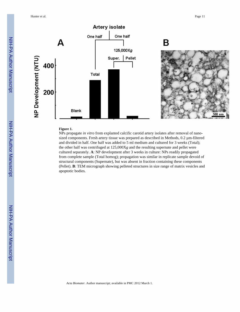
Figure 1.NPs propagate in vitro from explanted calcific carotid artery isolates after removal of nano-sized components. Fresh artery tissue was prepared as described in Methods, 0.2 µm-filteredand divided in half. One half was added to 5 ml medium and cultured for 3 weeks (Total);the other half was centrifuged at 125,000Xg and the resulting supernate and pellet werecultured separately. A: NP development after 3 weeks in culture: NPs readily propagatedfrom complete sample (Total homog); propagation was similar in replicate sample devoid ofstructural components (Supernate), but was absent in fraction containing these components(Pellet). B: TEM micrograph showing pelleted structures in size range of matrix vesicles andapoptotic bodies.
Hunter et al. Page 11
Acta Biomater. Author manuscript; available in PMC 2012 March 1.
NIH
-PA Author Manuscript
NIH
-PA Author Manuscript
NIH
-PA Author Manuscript
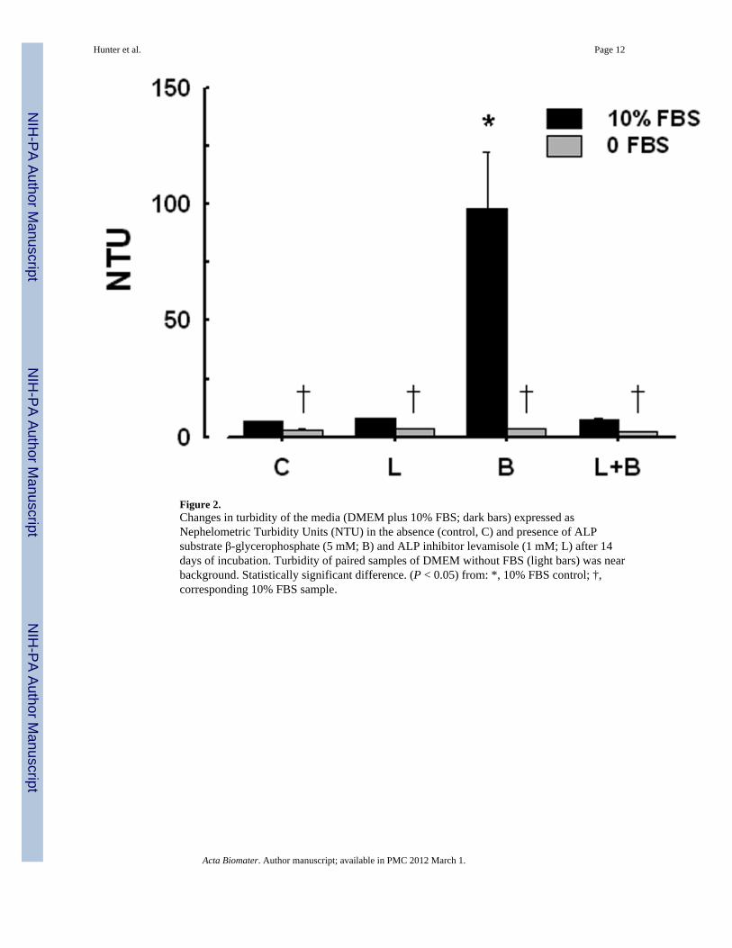
Figure 2.Changes in turbidity of the media (DMEM plus 10% FBS; dark bars) expressed asNephelometric Turbidity Units (NTU) in the absence (control, C) and presence of ALPsubstrate β-glycerophosphate (5 mM; B) and ALP inhibitor levamisole (1 mM; L) after 14days of incubation. Turbidity of paired samples of DMEM without FBS (light bars) was nearbackground. Statistically significant difference. (P < 0.05) from: *, 10% FBS control; †,corresponding 10% FBS sample.
Hunter et al. Page 12
Acta Biomater. Author manuscript; available in PMC 2012 March 1.
NIH
-PA Author Manuscript
NIH
-PA Author Manuscript
NIH
-PA Author Manuscript
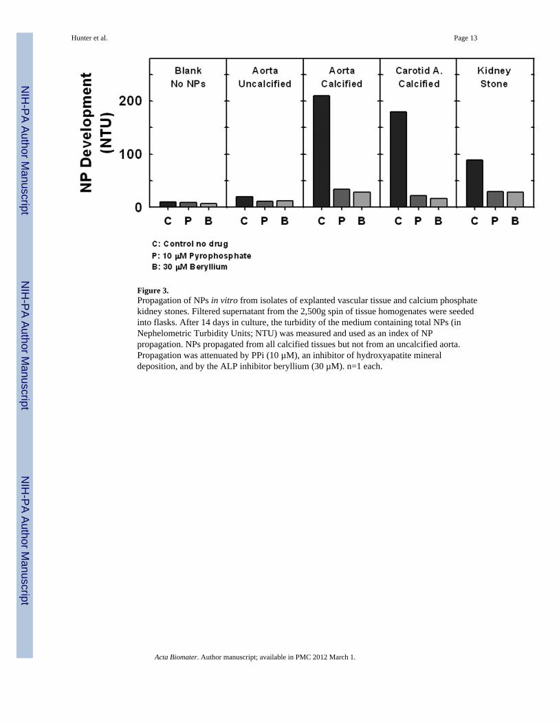
Figure 3.Propagation of NPs in vitro from isolates of explanted vascular tissue and calcium phosphatekidney stones. Filtered supernatant from the 2,500g spin of tissue homogenates were seededinto flasks. After 14 days in culture, the turbidity of the medium containing total NPs (inNephelometric Turbidity Units; NTU) was measured and used as an index of NPpropagation. NPs propagated from all calcified tissues but not from an uncalcified aorta.Propagation was attenuated by PPi (10 µM), an inhibitor of hydroxyapatite mineraldeposition, and by the ALP inhibitor beryllium (30 µM). n=1 each.
Hunter et al. Page 13
Acta Biomater. Author manuscript; available in PMC 2012 March 1.
NIH
-PA Author Manuscript
NIH
-PA Author Manuscript
NIH
-PA Author Manuscript
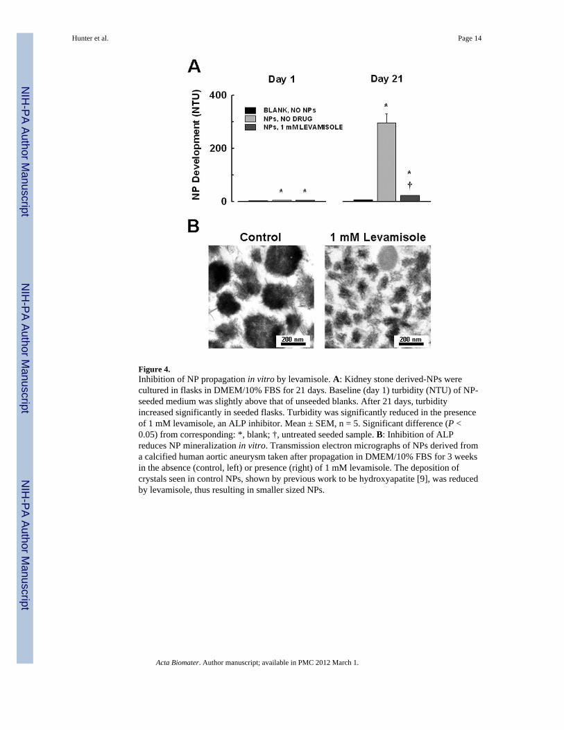
Figure 4.Inhibition of NP propagation in vitro by levamisole. A: Kidney stone derived-NPs werecultured in flasks in DMEM/10% FBS for 21 days. Baseline (day 1) turbidity (NTU) of NP-seeded medium was slightly above that of unseeded blanks. After 21 days, turbidityincreased significantly in seeded flasks. Turbidity was significantly reduced in the presenceof 1 mM levamisole, an ALP inhibitor. Mean ± SEM, n = 5. Significant difference (P <0.05) from corresponding: *, blank; †, untreated seeded sample. B: Inhibition of ALPreduces NP mineralization in vitro. Transmission electron micrographs of NPs derived froma calcified human aortic aneurysm taken after propagation in DMEM/10% FBS for 3 weeksin the absence (control, left) or presence (right) of 1 mM levamisole. The deposition ofcrystals seen in control NPs, shown by previous work to be hydroxyapatite [9], was reducedby levamisole, thus resulting in smaller sized NPs.
Hunter et al. Page 14
Acta Biomater. Author manuscript; available in PMC 2012 March 1.
NIH
-PA Author Manuscript
NIH
-PA Author Manuscript
NIH
-PA Author Manuscript
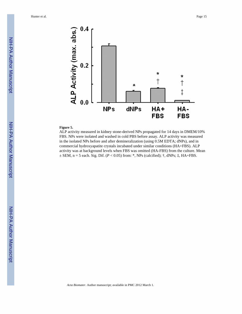
Figure 5.ALP activity measured in kidney stone-derived NPs propagated for 14 days in DMEM/10%FBS. NPs were isolated and washed in cold PBS before assay. ALP activity was measuredin the isolated NPs before and after demineralization (using 0.5M EDTA; dNPs), and incommercial hydroxyapatite crystals incubated under similar conditions (HA+FBS). ALPactivity was at background levels when FBS was omitted (HA-FBS) from the culture. Mean± SEM, n = 5 each. Sig. Dif. (P < 0.05) from: *, NPs (calcified); †, dNPs; ‡, HA+FBS.
Hunter et al. Page 15
Acta Biomater. Author manuscript; available in PMC 2012 March 1.
NIH
-PA Author Manuscript
NIH
-PA Author Manuscript
NIH
-PA Author Manuscript
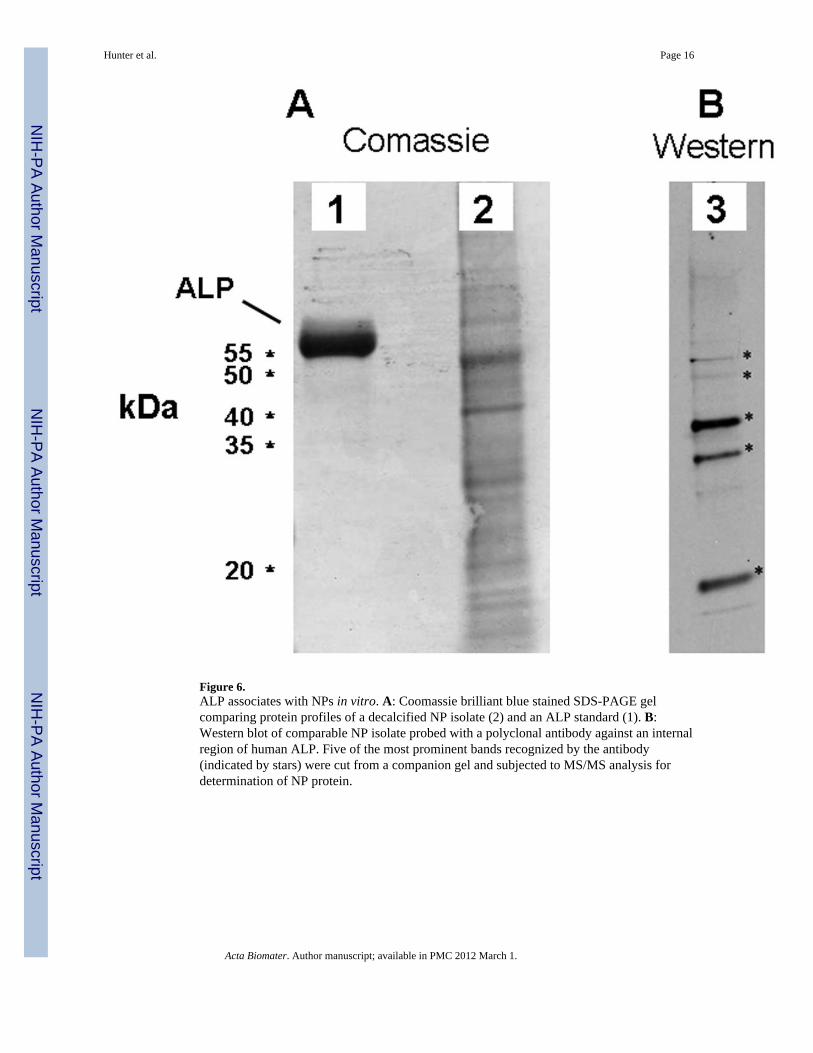
Figure 6.ALP associates with NPs in vitro. A: Coomassie brilliant blue stained SDS-PAGE gelcomparing protein profiles of a decalcified NP isolate (2) and an ALP standard (1). B:Western blot of comparable NP isolate probed with a polyclonal antibody against an internalregion of human ALP. Five of the most prominent bands recognized by the antibody(indicated by stars) were cut from a companion gel and subjected to MS/MS analysis fordetermination of NP protein.
Hunter et al. Page 16
Acta Biomater. Author manuscript; available in PMC 2012 March 1.
NIH
-PA Author Manuscript
NIH
-PA Author Manuscript
NIH
-PA Author Manuscript
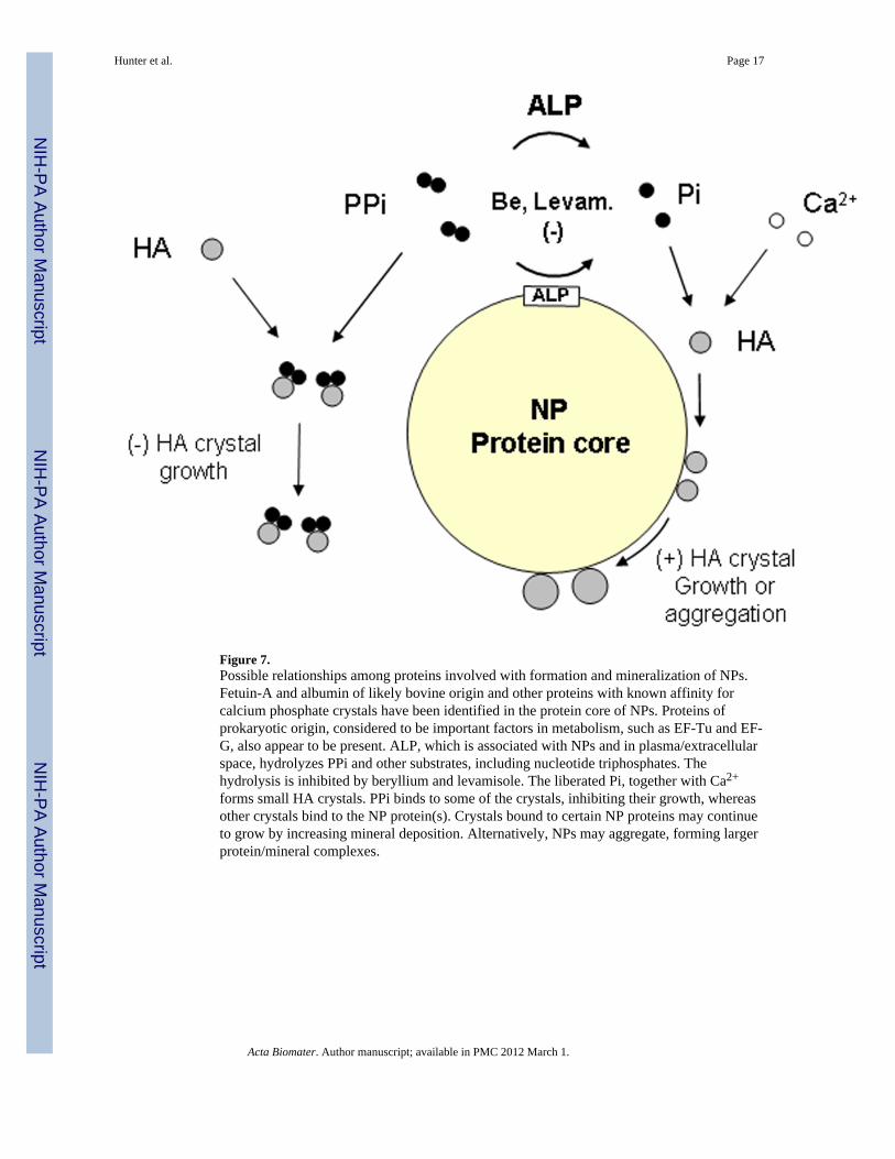
Figure 7.Possible relationships among proteins involved with formation and mineralization of NPs.Fetuin-A and albumin of likely bovine origin and other proteins with known affinity forcalcium phosphate crystals have been identified in the protein core of NPs. Proteins ofprokaryotic origin, considered to be important factors in metabolism, such as EF-Tu and EF-G, also appear to be present. ALP, which is associated with NPs and in plasma/extracellularspace, hydrolyzes PPi and other substrates, including nucleotide triphosphates. Thehydrolysis is inhibited by beryllium and levamisole. The liberated Pi, together with Ca2+
forms small HA crystals. PPi binds to some of the crystals, inhibiting their growth, whereasother crystals bind to the NP protein(s). Crystals bound to certain NP proteins may continueto grow by increasing mineral deposition. Alternatively, NPs may aggregate, forming largerprotein/mineral complexes.
Hunter et al. Page 17
Acta Biomater. Author manuscript; available in PMC 2012 March 1.
NIH
-PA Author Manuscript
NIH
-PA Author Manuscript
NIH
-PA Author Manuscript