Osteoblast-Like Cell Behavior on Porous Scaffolds Based on Poly(styrene) Fibers
Upload
independentCategory
view
0download
0
Journal of
J. Trace Elements Med. BioI. Vol. 11. pp. 110-115 (1997) Trace Elements In Medicine and Biology
© 1997 by Gustav Fischer Verlag
Maltol Complexes of Vanadium (IV) and (V) Regulate In Vitro Alkaline Phosphatase Activity and Osteoblast-like Cell Growth
D. A. BARRIO*, M. D. BRAZIUNAS*, S. B. ETCHEVERRY*** andA. M. CORTIZO*I
"C,ltedra de Bioqufmica Patol6gica. **CEQUINOR. Facultad de Ciencias Exactas.
Universidad de la Plata. 47 y 115 (1900) La Plata. Argentina
(Received January/May 1997)
Summary
Vanadium compounds have been found to possess insulin- and growth factor-mimetic etlects. In conse-
quence. these derivatives are potentially useful as effective oral therapeutic agents in diabetic patients. Howev-
er. their use has been limited by various toxic side-effects and by the low solubility of different derivatives. Re-
cently. vanadium complexes with maltol, a sugar used as a common food additive. have been synthesised and
investigated in animals. showing possible insulin-mimetic effects with low toxic side-effects. In the present
study we have investigated the effect ofbis(maltolato)oxovanadium (IV) (BMOV) and bis(maltolato )dioxova-
nadium (V) (B\!IV) on bone cells in culture as well as their direct effect on alkaline phosphatase in I'itro. A
comparison was also made with the action of vanadate and vanadyl cation. Vanadium compounds regulated cell proliferation in a biphasic manner with similar potencies. Osteoblast differentiation. assessed by alkaline phos-
phatase activity. was found to be dose-dependent. with the inhibitory effect being stronger for vanadate and
BMOV than for vanadyl and BMY. All vanadium compounds directly inhibited bovine intestinal ALP with a
similar potency. Thus. maltol vanadium derivatives behave in a similar way to vanadate and vanadyl in osteob-
last-like UMR 106 cells in culture.
Kcnmrds:Vanadium-maltol complexes. proliferation. differentiation. osteoblasts. alkaline phosphatase.
Introduction
Vanadium. a trace transition element. promotes a
number of cellular events. both ill ritro and ill rim (1-3).
It regulates several enzymatic activities crucial to inter-
mediate metabolism. nucleic acid synthesis. protein ex-pression. growth promotion and cell differentiation.
These observations have led to the suggestion that vana-
dium acts both as an insulin- and growth factor-mimetic
compound. Oral vanadium administration in rodent mod-
eb of diabetes was found to improve hyperglycacmia (4-
'To whom correspondence should be addressed.
6). Thus. vanadium has been proposed as a potential ther-
apeutic agent for diabetic patients. However. extended
treatment with vanadate induces toxic effects in animals as well as in cell culture systems (3.7-9). It is for this rea-
son that the bioactivity and toxicity of new vanadium de-
rivatives are being investigated. Previous studies in our laboratory have shown that
low doses of different vanadium compounds promote the proliferation of fibroblasts and osteoblast-like cells (10-
12). However. this effect was biphasic in our culture
models. Both an inhibition of cell growth and an induc-
tion of morphological transformation were observed at
concentrations over 50 for most of the vanadium (V)
compounds. On the other hand. the vanadium (IV) deriv-
atives appeared to be less toxic. which is in agreement
with previous results (13.14). Using two osteoblastic lines. U MR I 06 and MC3T3E I. we have also shown that
vanadium regulates the osteoblast phenotype as assessed
by alkaline phosphatase (ALP) specific activity (10.12).
ALP inhibition was observed after 24 h incubation with
vanadium (V) compounds. On the other hand. a weak
regulation of cell differentiation was observed with vana-
clium (IV) derivatives. In addition. using il1l'itro cell-free
systems. we previously demonstrated that both vanadium (IV) and (V) directly inhibited alkaline and acid phos-
phatases in a dose-dependent manner (15.16). Thus. the
ohserved vanadium inhibition of osteoblast differentia-
tion may be due to a direct effect on ALP activity. On the
other hand. vanadium compounds might specifically reg-
ulate ALP gene expression. Our studies suggest that dif-
ferent vanadium compounds might be able to exert selec-
tive/specific effects on different cell and tissue models. Further studies are required. therefore. to determine the
usefulness of vanadium compounds as therapeutic tools.
In the present study. we investigated the effects of bis (maltolato) oxovanadium (IV) and bis (maltolato) diox-
ovanadium (V) compounds on osteoblast-like UMR 106
cell growth. In addition. their direct effect on bovine in-testinal ALP was also studied. These results were com-
pared with the effects of vanadate and vanadyl on the
same parameters.
Material and Methods
Materials
Vanadium (IV) oxide sulfate (vanadyl sulfate) (VO)
was obtained from Merck. Vanadate (V). p-nitrophenyl-
phosphate (pNPP). ethylenediaminetetraacetic acid
(EOTA). maltol (3-hydroxy-2-methyl-4-pyrone). fast
blue RR salt. -naphthyl phosphate and bovine intestinal alkaline phosphatase were obtained from Sigma. Tissue
culture material was provided by Corning or Falcon: Oul-
becco's modified Eagle's medium (OMEM) and trypsin
were supplied by Gibco. and fetal bovine serum (FBS) by Gen. Argentina.
\(/J1adi[(m Compounds and Solutio/1 Preparation
Fresh stock solutions (100 mM) of vanadate and va-
Maltol vanadium complexes in ostcoblasts III
nadyl were prepared hy dissolving the crystalline materi-
al in distilled water. Bis (maltolato) oxovanadium (IV)
(BMOV) rC1,H",o7Vj was provided by Dr. E.1. Baran and prepared according to McNeill et al. (17). Aqueous
solutions of maltol and vanadyl sulfate were mixed in a
2: I ratio. The pH of the solution was raised to 8.5 and the
solution was retluxed overnight; the green compound
was collected upon cooling. The solid product was char-acterized by IR spectroscopy. Bis (maltolato) dioxovana-
dium (V) (BMV) was prepared according to Elvingson et
al. (18). Brictly. 10 mM vanadate and 20 mM maltol in
water were mixed (I :2 molar ratio) and the pH of the so-
lution was kept between 6.5 and 7.0. The solution was
protected from light and was used within 48 h.
Cell clIltllre
Rat osteosarcoma cell line UMR 1 06 was grown in OMEM supplemented with 10 o/c FBS and antibiotics
(100 U/ml penicillin - 100 streptomycin) in a hu-
midified atmosphere of 95o/c air / 5o/c CO, ( 10). Cells
were grown to near confluence (70-800/c) when they were subcultured using 0.19', trypsin - lmM EOTA in Ca'+-
Mg'+ - free phosphate huffered saline (PBS). For experi-
ments. about 2.5 • I cells / well were plated into 24-well
plates. After the culture reached 700/, contluence. the
cells were washed with OMEM without serum and incu-
bated in 0.5 ml OMEM plus different vanadium com-
pounds for 24 h. We chose this cell line since it has been
reported to respond to insulin and vanadium compounds
(3.10.12).
C el/lJroliferation assay
A mitogenic bioassay was carried out as described by
Okajima et al. (19) with some modifications. Brietly.
cells in 24-well plates were washed with PBS and fixed
with 5clc glutaraldehyde / PBS at room temperature for 10
min. Then they were stained with 0.5o/c crystal violet /
2SC,1r methanol for 10 min. after which the dye solution was discarded and the plate was washed with water and
dried. The dye taken up by the cells was extracted using
0.5 mi/well 0.1 M glycine/HCl butler. pH=3.0 /30O/C
methanol and transferred to test tubes. Absorbance was
read at 540 nm after a convenient sample dilution. We
previously showed that under these conditions this color-
imetric bioassay strongly correlates with cell count (10).
II:: D. A. Barrio. M. D. Braziunas. S. B. Etcheverry and A. M. Cortizo
130 140,----------------------,
120
'iij r U>
.. '" 110 ID
;;l:: I
100
90 __ __ __ L-__ L-__
o 20 40 60 80 100 12C
Vanadium b.tM]
Figure I. Effect of vanadium compounds on UMR 106 osteoblast-like cell proliferation. Symbols are: .V .• VO .• BMV ..... BMOY. Data are expressed as '/, of the basal value with mean±SEM (n=9) being given.
C ell differentiation assay
Cells were incubated at 37°C / 24h in serum-free DMEM containing different agents. The cell layer was then washed with PBS and solubilized in 0.5 ml 0.1°11' Tri-ton X-100. Aliquots of the total cell extract (lO-200/d were saved for protein determination using the Bradford technique (20) and for measurement of alkaline phos-phatase activity as has been described (10). This assay has been used as a marker of osteoblast phenotype ex-pression in cell cultures (3.10.12.21).
120
100
-' 80 « en « ID ;;l:: 60
40
20
o 20 40 60 80 100
Vanadium [liM]
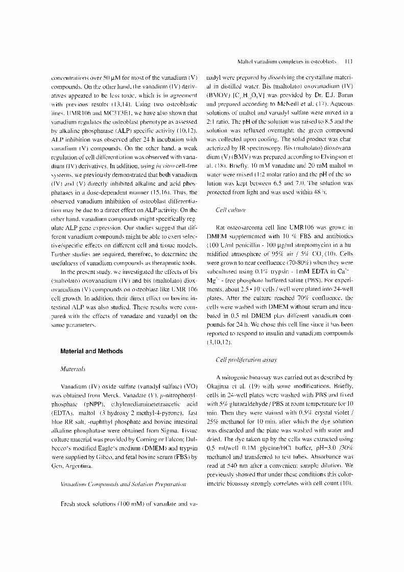
Figure I. Effect of vanadium compounds on UMR I 06 differencia-tion. Symbols arc: • V .• YO .• BMV ..... BMOV. Data are ex-pressed as '1c of the basal value with mean±SEM (n=9) being given.
Intestinal alkaline phosphatase acti"i!.'>'
The effect of vanadium compounds on ALP from bo-vine intestinal mucosa was investigated. The initial rate of ALP activity was determined using I Ilg/ml bovine in-testinal ALP - 5 mM pNPP in the incubation buffer (55 mM Glycine - 0.55 mM MgCI, buffer. pH=10.5) at 37°C for 7 min. Under these experimental conditions. the prod-uct formation was linear until 10 min. The effect of vana-dium complexes was assessed by adding different con-centrations of each compound in the ALP assay. The pro-duction of pNP was determined measuring absorbance at 405 nm.
We chose this assay as a model of an in 1'itro cell-free Alkaline phosphatase histochemistry system to test the direct effect of vanadium compounds
on alkaline phosphatase activity. as we have previously UMR 106 osteoblasts were cultured into 6 well plates described ( 15).
at a density of 2.4 • I cell/m!. For the experiments. cul-tures were washed with PBS and fixed with citrate-buff-ered acetone-formaldehyde. according to Gay et al (22). After rinsing with water. cells were incubated with fresh-ly made -naphthyl phosphate and fast blue RR (O.Olo/c and 0.06 o/e. respectively). in 0.1 M Tris-HCI pH=8.5 with or without different vanadium compounds for 15 min. After this incubation period. eells were extensively washed with water. The alkaline phosphatase staining distribution on the cells was observed and photographs of each dish were made at 40 x Obj magnification with a black and white Kodak 125 ASA film.
Statistical methods
At least three experiments were performed in trip-licate for each experimental condition. Data were ex-pressed as mean±SEM. Statistical differences were ana-lysed using Student's t-test or analysis of variance. when suitable. Linear correlation analysis was performed by Pearson's correlation coefficient.
Results
Figure I shows the effect of different vanadium com-pounds on the proliferation of UMRI06 osteoblast-like
A
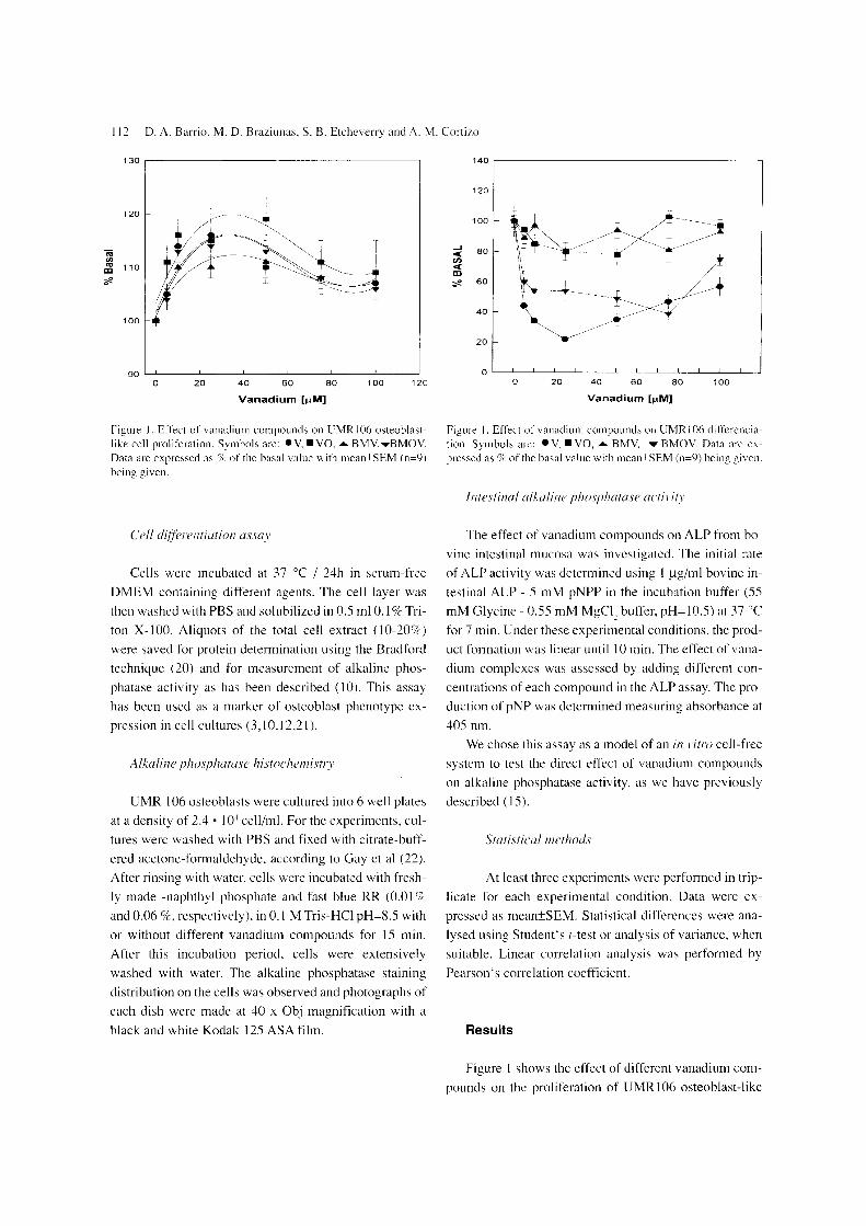
B
Figure 3. Alkaline phosphatase activity determined by histochemis-try. Cells were incubated at 37 for 15 min in absence (A) or pres-ence (B) of 100 11M BMOV. Obi 40x.
cells. All compounds significantly stimulated cell growth
in a biphasic manner. A maximal potency, similar for all
vanadium derivatives, was found at doses between 20 to
SO 11M (ranging from 110 - 120 'k over basal, p<0.02 - p<
0.001).
We also evaluated the effect on total protein content
after 24 h incubation with different concentrations of va-
nadium complexes. A dose-dependent effect was ob-
served for the BM-vanadium complexes and va (range
110- 124 'I, of basal. p<O.02 - p<O.OO I ) (data not shown).
In contrast. V showed a biphasic effect with no stimula-
tion of protein content at 100 11M concentration (data not
shown). Maximal induction of protein content for all va-
nadium complexes was in the same concentration range
as for the cell proliferation effect (20-50 11M).
The effect of malto vanadium complexes on osteob-
last-like cell differentiation was assessed by alkaline
phosphatase specific activity. Figure 2 shows that BMOV
and V strongly inhibited cell differentiation in a dose-de-
pendent manner. The maximal effect was at 10- SO or 75
11M, for BMOV or V respectively. This effect showed a
tendency to disappear above 80 11M for both compounds.
On the other hand, BMV complex and va show only a
slight effect on cell differentiation « 20'k inhibition) in
all the concentration ranges investigated. These results
Maltol vanadium complexes in osteoblasts 113
Table I. Intensity of ALP staining associated with the membrane of osteoblast-like cells
Concentration BMV BMOV V VO
25 11M ++++ ++++ +++ ++++
50 +++ +++ +++ +++
100 ++ ++ ++ ++
500 + + + +
1000
Pluses indicate relative amounts of reaction product. minuses signi-fy absence of reaction product. Basal value was determined in ab-sence of vanadium compounds (++++)
are in agreement with our previous studies of vanadyl ac-
tion on osteoblast-like cell differentiation (10,12).
We also analyzed the direct effect of BMOV and
BMV on alkaline phosphatase activity from osteoblast-
like cells and bovine intestinc. Figure 3 shows ALP activ-
ity in UMR I 06 osteoblast-like cells detected by histo-
chemistry. The ALP reaction product was mainly associ-
ated with the cell membrane. and to a lesser extent in the
cytoplasm. Control cells showed a strong brown staining
reaction (Fig 3A). ALP activity was inhibited by both
BMOV and BMV complexes to the same extent and in a
dose-response manner (Table I). Thus. there was a pro-
gressive decrease in the histochemically detectable mem-
brane-associated ALP activity (Fig 3B). Vanadate and
va also inhibited the ALP reaction depending on the
dose. Approximately 50 o/r. of the control staining intensi-
ty was observed after incubation with 100 11M of any of
the vanadium compounds (Table I). These results seem
to indicate that osteoblast ALP is equally sensitive to all
the vanadium compounds tested.
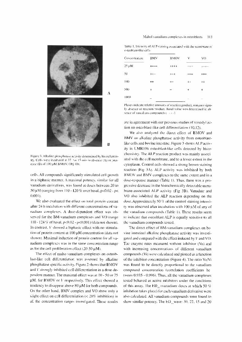
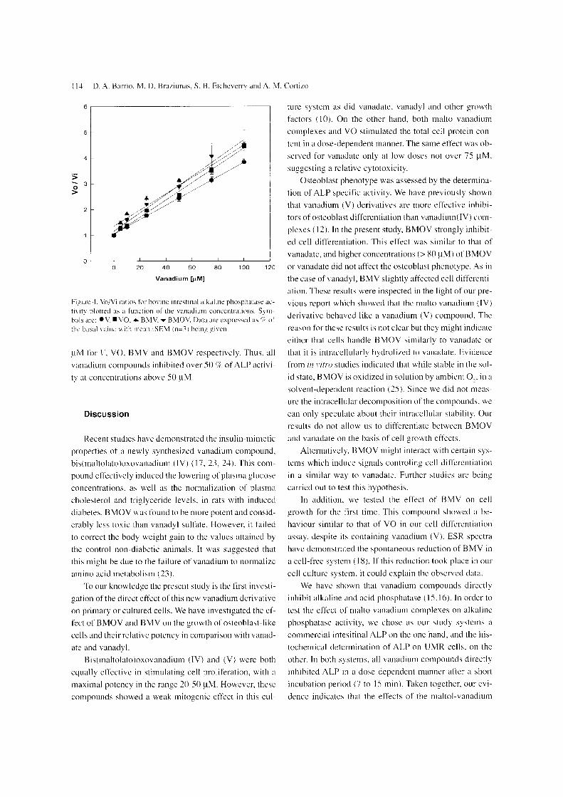
The direct effect of BM-vanadium complexes on bo-
vine intestinal alkaline phosphatase activity was investi-
gated and compared with the effect induced by Vand va. The enzyme rates measured without inhibitor (Vo) and
with increasing concentrations of different vanadium
compounds (Vi) were calculated and plotted as a function
of the inhibitor concentration (Figure 4). The ratio VoNi
was found to be directly proportional to the vanadium
compound concentration (correlation coetTicients be-
tween 0.935 - 0.996). Thus. all the vanadium complexes
tested behaved as active inhibitors under the conditions
of this assay. The EDeo (vanadium doses at which 50 o/c inhibition takes place) for each vanadium derivative were
also calculated. All vanadium compounds were found to
show similar potency. The ED)II were: 30, 27. IS and 20
11-1 D. A. Barrio. M. D. Br'lLiunas. S. B. Etcheverry and A. M. Corlizo
6 ,-------------------------------------,
5
4
:> o 3 >
2
o
ture system as did vanadate, vanadyl and othcr growth
factors (10). On the other hand. both malto vanadium
complexes and VO stimulated the total cell protein con-tent in a dose-dependent manner. The same effect was ob-
served for vanadate only at low doses not over 75 11M.
suggesting a relative cytotoxicity.
Osteoblast phenotype was assessed by the determina-
tion of ALP specific activity. We have previously shown that vanadium (V) derivatives are more effective inhibi-
tors of osteoblast differentiation than vanadium(lV) com-
plcxes (12). In the present study. BMOV strongly inhibit-
ed cell differentiation. This effect was similar to that of
vanadate. and higher concentrations (> 80 11M) of BMOV o 20 40 60 80 100 12C or vanadate did not affect the osteoblast phenotype. As in
Vanadium
Figure -I. Vo/Vi ratios for bovine intestinal alkaline phosphatase ac-tivity plotted as a function of the vanadium concentrations. Sym-bols arc: ev. .VO." BMV ..... BMOV. Data are expressed as 'I; of the basal value \v ith I11can±SEM (n=-,) being given.
11M for \. YO. BMV and BMOV respectivcly. Thus. all
vanadium compounds inhibited over SO '7r of ALP activi-
ty at concentrations above SO 11M.
Discussion
Recent studies have demonstrated the insulin-mimetic
properties of a newly synthesized vanadium compound. bis(maltolato)oxovanadium (IV) (17. 23. 24). This com-
pound effectively induccd the lowering of plasma glucose concentrations. as well as the normalization of plasma
cholesterol and triglyceride levels. in rats with induced
diabetes. BMOV was found to be more potent and consid-erably less toxic than vanadyl sulfate. However. it failed
to correct the body weight gain to the values attained by
thc control non-diabetic animals. It was suggested that
this might be due to the failure of vanadium to normalize amino acid metabolism (23).
To our knowledge the present study is the first investi-gation of the direct etfect of this new vanadium derivative
on primary or cultured cells. We have investigated the ef-
fect of BMOV and BMV on the growth of osteoblast-like
cells and their relative potency in comparison with vanad-ate and vanadyl.
Bis(maltolato)oxovanadium (IV) and (V) were both
equally effective in stimulating cell proliferation. with a
maximal potency in the range 20-50 11M. However. these
compounds showed a weak mitogenic effect in this cul-
the case ofvanadyL BMV slightly affected cell differenti-
ation. These results were inspected in the light of our pre-vious report which showed that the malto vanadium (IV)
derivative behaved like a vanadium (V) compound. The
reason for thcse results is not clear but they might indicatc
either that cells handle BMOV similarly to vanadate or
that it is intracellularly hydrolized to vanadate. Evidence
from ill l'itro studies indicated that while stable in the sol-
id state. BMOV is oxidized in solution by ambient 0 .. in a solvent-dependent reaction (25). Since we did not meas-
ure the intracellular decomposition of the compounds. we
can only speculate about their intracellular stability. Our
results do not allow us to differentiate between BMOV
and vanadate on the basis of cell growth effects.
Alternatively. BMOV might interact with certain sys-tems which induce signals controling cell ditfcrentiation
in a similar way to vanadate. Further studies are being
carried out to test this hypothesis.
In addition. we tested the effect of BMV on cell growth for the first time. This compound showed a be-
haviour similar to that of VO in our cell differentiation
assay. despite its containing vanadium (V). ESR spectra
have demonstrated the spontaneous reduction of BMV in a cell-free system (18), If this reduction took place in our
cell culture system. it could explain the observed data.
We have shown that vanadium compounds directly
inhibit alkaline and acid phosphatase (15.16). In order to
test the effect of malto vanadium complexes on alkaline
phosphatase activity, we chose as our study systems a commercial intesitinal ALP on the one hand, and the his-
tochemical detcrmination of ALP on UMR on the
other. In both systems, all vanadium compounds directly
inhibited ALP in a dose-dependent manner after a short
incubation period (7 to IS min). Taken together. our evi-
dence indicates that the effects of the maltol-vanadium

derivatives are similar to those of their parent compounds vanadate and vanadyl. However, they are not more potent than their parent compounds in our osteoblast-like cell system.
Acknowledgements
This work was supported by grants from Facultad de Ciencias Exactas, UNLP; Universidad Nacional de La Plata, Argentina. SBE is a member of the Carrera del In-vestigador, CONICET, Argentina; AMC is member of the Carrera del Investigador, CICPBA, Argentina.
References
I. SCHECHTER. Y. (1990). Perspectives in diabetes: insulin mi-metic effects of vanadate. possible implications for future treat-ment of diabetes. Diabetes 39. 1-5.
,.., A.: YIN. X .. TSANG. S-S .. DAVISON. A., MOON, 1. ( 1993). Vanadium as a modulator of cellular regulatory cas-cades and oncogene expression. Biochem. Cell BioI. 71. 103-112.
3. ETCHEVERRY. S.B .. CORTIZO. A.M. (1997). Bioactivity of vanadium compounds on cell in culture. In: Vanadium in the environment. Parts I and 11. Vol (Nriagu (Ed.) In press.
4. BRICHARD. S.M .. BAILEY. C.J. AND HENQCIN. J.e. ( 1990). 'vIarked improvement of glucose homeostasis in diabet-ic ob/ob mice given oral vanadate. Diabetes 39. 1326-1332.
5. MEYEROVITCH. J .. ROTHENBERG. P .. SHECHTER. Y. AND KAHN. e.R. (1991). Vanadate normaliLcd hyperglyc-emia in two mouse models of non-insulindependent diabetes mellitus. J. Clin. Invest. 87.1286-1294.
6. RAMANADHAM. S .. MONGOLD. J.J .. BROWNSEY. R.W.. CROS. G.H .. AND MCNEILL. 1.H. (1989). Oral vanadvl sul-fate in treatment of diabetes mellitus in rats. Am. 1. PhysiEJI. 257. H904-H911.
7. AL-BAYATL M. A .. GIRL S.H .. RAABE. O.G .. ROSEN-BLATT. L.S. and SHIFRINE. M. (1989). Time and dose-re-sponse study of the effects of vanadate on rats: morphological and biochemical changes in organs. JEPTO 9. 435-455.
R. SABBIONI. E.. POZZI. G .. DEVOS. S .. PINTAR. A .. CASEL-LA. L FISCHBACH. M. (1993). The intensity of vanadiumtV)-induced cytotoxicity and morphological matlon 111 BALB/3T3 cells is dependent on glutathione-mediat-ed bioreduction to vanadium(lV). Carcinogenesis 14. 2565-2568.
9. DAI. S .. AND McNEILL. 1.H. (1995). Lack of hematolo"cial effect of oral vanadium treatment in rats. Pharmacol. Toxicol. 76. 263-26X.
10. CORTIZO. A.M. AND ETCHEVERRY. S.B. ( 1995). Vanadium derivatives act a;. growth factor-mimetic compounds upon dif-ferentiation and proliferation of osteoblast-like UMRI06 cells. Mol. Cell Biochem 145.97-102.
Maltol vanadium complexes in osteoblasts 115
II. CORTIZO AM. SALlCE Ve. VESCIl\;A CM. ETCHEVERRY SB. t 1997) Proliferative and morphological changes induced by vanadium compounds in Swiss 3T3 fibroblast;. Biometals 10.127-133
12. ETCHEVERRY. S.B .. CRANS. D.c.. KERAMlDAS. A.D .. CORTIZO. A.M. (1997). Insulin-mimetic action of vanadium compounds on osteoblast-like cells in culture. Arch Biochem Biophys 33S. 7-14.
13. OAt S .. AND McNEILL 1.H. (1994). One-year treatment of non-diabetic and streplOzotocin-diabetic rats with vanadyl sul-fate did not alter blood presure or haematological indices. Phar-macol. J. Toxicol. 7-+.110-115.
14. WILLSKY. G.R .. WHITE. D.A .. McCABE. B.e. (19S4). Me-tabolism of added orthovanadate to vanadvl and hioh-molecu-lar 'height vanadate bv Sacc/iaromrc{'s 1. BioI. Chem. 259: 13273-132'S I. .
15. CORTIZO.A.M .. SA LICE. Vc.. ETCHEVERRY. S.B. (1994). Vanadium compounds: their action on alkaline phosphata;.e ac-tivity. BioI. Trace Element. Res. 41. 331-339.
16. VESCINA. C.M .. SALICE. Vc.. CORTlZO. A.M .. ETCH-EVERRY. S.B. (1996). Effect of vanadium compounds on acid phosphatase activity. BioI. Trace Elem. Re;. 53. 1 R5-191.
17. McNEILL 1.H .. YUEN. VG .. HOVEYDA. H.R .. ORVIG. e. t 1992). Bis(maltolato)oxovanadium (IV) is a potent insulin mimic. 1. Med. Chem. 35.1489-1491.
IS. ELVINGSON. K .. GONZALEZ BARO. A .. PETTERSON. L. (1996). Speciation in vanadium bioinorganic systems. 3.An NMR ESR and potentiometric study of H' -vanad-ate-maltol system. Inorg. Chem. (in press).
19.0KAJIMA. T.. NAKAMURA. K .. ZHANG. H .. LING. N .. TANABE. T.. YASUDA. T.. ROSENFELD. R.G. (1992). Sen-sitive colorimetric bioassays for insulin-like growth factor (lGF) stimulation of cell proliferation and glucose consump-tIon: use in studies of IGF analogs. Endocrinology 130. 2201-2212.
20. BRADFORD. M. (1976). Rapid and sensitive method for quantitation of microgram quantities of protein utilizing the principle of protein-dye binding. Anal. Biochem. n. 24X-254.
21. STEIN, G.S .. LlAN. J.B. (1993). Molecular mechanism medi-ating proliferation/differentiation interrelationships during pro-gressIve development of the osteoblast phenotype. Endocrine Rev. 14. 424-442.
22. GAY. e.V. LLOYD. Q.P .. GILMAN. YR. (1994). Characteris-tics and culture of osteoblast> derived from avian long bone. In Vitro Cell. Dev. BioI. 30A. 379-3S3.
23. YUEN. VG .. ORVIG. e.. AND McNEILL. 1.H. t 1993). Glu-cose-lowering effects of a new organic vanadium complex. bis(maltolato)oxovanadium (IV). Can. 1. Phvsiol. Pharmacol. 71: 263-269. .
24. YUEN. VG .. ORVIG. c.. AND McNEILL, 1.H. (1995). Com-parison of the glucose-lowering properties of vanadvl sulfate and bis(maltolato)oxovanadium (IV) following and chronic administration. Can. 1. Physiol. Pharmacol. 73. 55-64.
25. CARAVAN. P.. GELMINL L.. GLOVER. N .. HERRING. F.G .. LL H .. MCNEILL 1.H .. RETTIG. S.J .. SETYAWATI I A SHUTER. E .. SUN. Y.. TRACEY. A.S .. YUEN. VG .. ORVIG: e. (1995). Reaction chemistry of BMVO. Bis(maltolato) ox-ovanadium (IV). A potent insulin mimetic agent. J. Am. Chem. Soc. 117. 12759-12770. L
Copyright © 2022 FDOKUMEN