Isolation, characterization and antimicrobial activity characterization of endophitic bacterial...
9
Vol. 8(11), pp. 1178-1186, 12 March, 2014 DOI: 10.5897/AJMR2013.6444 ISSN 1996-0808 Copyright © 2014 Author(s) retain the copyright of this article http://www.academicjournals.org/AJMR African Journal of Microbiology Research Full Length Research Paper Isolation, characterization and antimicrobial activity of a Streptomyces strain isolated from deteriorated wood Hajar Maataoui, Mohammed Iraqui, Siham Jihani, Saad Ibnsouda and Abdellatif Haggoud* Microbial Biotechnology Laboratory, Faculty of Sciences and Techniques, University Sidi Mohamed Ben Abdellah, Fez, Morocco. Received 21 October, 2013; Accepted 21 February, 2014 Emergence of drug resistance among pathogenic bacteria to currently used antibiotics has made the search for novel bioactive compounds from natural and unexplored habitats a necessity. In this study, we reported the isolation, characterization and antimicrobial activity of an actinomycete strain isolated from deteriorated wood of an old house located in the Medina of Fez. The isolate, named H2, was identified by 16S rDNA sequencing and was shown to belong to the genus Streptomyces. The isolate was screened for antimicrobial activity against Gram positive bacteria, Gram negative bacteria, Mycobacteria, yeasts and fungi. Partial characterization of the active substance (resistance to proteinase K and heat) showed that it would be of non-protein nature. The kinetics of production of the active substance showed that the maximum production occurs between the 7th and 10th day of fermentation. In addition, organic extract of the isolate was able to release genomic DNA of Staphylococcus aureus suggesting that it acts probably on the bacterial cell wall. Thin layer chromatography (TLC) of the ethyl acetate extract followed by bioautography has allowed localizing the active substances. This will open the way to further investigations to demonstrate their potential importance in combating pathogenic bacteria. Key words: Actinomycetes, Streptomyces, antimicrobial activity, molecular identification. INTRODUCTION Since the discovery of antibiotics, bacterial resistance to these chemicals has continued to evolve. Thus, we are witnessing more and more multiresistant bacteria that pose a serious public health problem. Therefore, novel antibiotics against drug resistant bacteria are urgently needed. Microbial natural products have been one of the major sources of novel drugs. Among microorganisms, actinomycetes are the most economically and biotechnologically useful prokaryotes (Lam, 2006; Valli et al., 2012). They produce antibiotics and other industrially important secondary metabolites (Okami and Hotta, 1988; Koehn and Carter, 2005; Nermeen and Gehan, 2006; Kekuda et al., 2010; Naine et al., 2011). From all the genera of actinomycetes, the genus Streptomyces is represented in nature by the largest number of species and varieties. These species can produce a large number of antibiotic and active secondary metabolites (Devi et al., 2006). In fact, 80% of the recognized antibiotics are sourced from this genus *Corresponding author. E-mail: [email protected]. Tel: +212 (0) 535 60 80 14. Fax: +212 (0) 535 60 82 14. Author(s) agree that this article remain permanently open access under the terms of the Creative Commons Attribution License 4.0 International License
-
Upload
independent -
Category
Documents
-
view
1 -
download
0
Transcript of Isolation, characterization and antimicrobial activity characterization of endophitic bacterial...

Vol. 8(11), pp. 1178-1186, 12 March, 2014
DOI: 10.5897/AJMR2013.6444
ISSN 1996-0808
Copyright © 2014
Author(s) retain the copyright of this article
http://www.academicjournals.org/AJMR
African Journal of Microbiology Research
Full Length Research Paper
Isolation, characterization and antimicrobial activity of a Streptomyces strain isolated from deteriorated wood
Hajar Maataoui, Mohammed Iraqui, Siham Jihani, Saad Ibnsouda and Abdellatif Haggoud*
Microbial Biotechnology Laboratory, Faculty of Sciences and Techniques, University Sidi Mohamed Ben Abdellah, Fez, Morocco.
Received 21 October, 2013; Accepted 21 February, 2014
Emergence of drug resistance among pathogenic bacteria to currently used antibiotics has made the search for novel bioactive compounds from natural and unexplored habitats a necessity. In this study, we reported the isolation, characterization and antimicrobial activity of an actinomycete strain isolated from deteriorated wood of an old house located in the Medina of Fez. The isolate, named H2, was identified by 16S rDNA sequencing and was shown to belong to the genus Streptomyces. The isolate was screened for antimicrobial activity against Gram positive bacteria, Gram negative bacteria, Mycobacteria, yeasts and fungi. Partial characterization of the active substance (resistance to proteinase K and heat) showed that it would be of non-protein nature. The kinetics of production of the active substance showed that the maximum production occurs between the 7th and 10th day of fermentation. In addition, organic extract of the isolate was able to release genomic DNA of Staphylococcus aureus suggesting that it acts probably on the bacterial cell wall. Thin layer chromatography (TLC) of the ethyl acetate extract followed by bioautography has allowed localizing the active substances. This will open the way to further investigations to demonstrate their potential importance in combating pathogenic bacteria. Key words: Actinomycetes, Streptomyces, antimicrobial activity, molecular identification.
INTRODUCTION Since the discovery of antibiotics, bacterial resistance to these chemicals has continued to evolve. Thus, we are witnessing more and more multiresistant bacteria that pose a serious public health problem. Therefore, novel antibiotics against drug resistant bacteria are urgently needed. Microbial natural products have been one of the major sources of novel drugs.
Among microorganisms, actinomycetes are the most economically and biotechnologically useful prokaryotes (Lam, 2006; Valli et al., 2012). They produce antibiotics
and other industrially important secondary metabolites (Okami and Hotta, 1988; Koehn and Carter, 2005; Nermeen and Gehan, 2006; Kekuda et al., 2010; Naine et al., 2011).
From all the genera of actinomycetes, the genus Streptomyces is represented in nature by the largest number of species and varieties. These species can produce a large number of antibiotic and active secondary metabolites (Devi et al., 2006). In fact, 80% of the recognized antibiotics are sourced from this genus
*Corresponding author. E-mail: [email protected]. Tel: +212 (0) 535 60 80 14. Fax: +212 (0) 535 60 82 14. Author(s) agree that this article remain permanently open access under the terms of the Creative Commons Attribution License 4.0 International License
(Procópio et al., 2012), and many representatives of this group produce substances of high commercial value and are being extensively screened for novel bioactive compounds (Anderson and Wellington, 2001; Vijayakumar et al., 2007).
Exploring new habitats is one of the most promising ways to isolate new strains of actinomycetes endowed with antimicrobial activity (Zitouni et al., 2005; Khanna et al., 2011; Wadetwar and Patil, 2013).
Thus, we report here the isolation and molecular identification of a Streptomyces strain endowed with antibacterial activity. Partial characterization of the active substance as well as its mode of action is also reported. MATERIALS AND METHODS
Sample collection
Pieces of deteriorated wood were taken from an old house, built 450 years ago, located in the former Derb Lamté in the Medina of Fez and were transported aseptically in sterile containers to the laboratory. Isolation and culture conditions Ten grams of wood samples were crushed and suspended in
physiological water, then vigorously mixed for 2 h. The suspension was serially diluted up to 10
-7. An amount of 0.1 ml of each dilution
was spread on the surface of the Actinomycetes Isolation Agar medium (AIA) (Thakur et al., 2007) and incubated at 30°C. After 2-4 weeks of incubation, the plates were examined for the presence of actinomycetes colony. The colonies of actinomycetes were recognized according to their macroscopic and microscopic characteristics (optical microscopy and Gram stain) then purified
and conserved at 4°C for short periods and at -20°C in glycerol stock (20%, v/v) for a longer period. Twelve isolates showing different morphological characteristics were obtained. In this work, an isolate named H2 was chosen for the study of its antimicrobial activity. Characterization of the isolate H2
The isolate H2 was tested for its ability to grow at pH 5 to 10 and at a temperature range of 25 to 42°C. Thus, actinomycetes cultures were spot inoculated onto plates of ISP2 (Shirling and Gottlieb, 1966) media to pH 5, 6, 7, 8, 9 and 10. The plates were checked for growth after seven days of incubation at 30°C. The same procedure was followed for the temperatures test except the use of ISP2 medium at pH 7.2. The temperatures tested were 25, 30, 37 and 42°C.
Actinomycetes synthesize and excrete dark pigments, melanin or
melanoid, which are considered to be a useful criterion for taxonomical studies (Zonova, 1965; Arai and Mikami, 1972). Therefore the production of melanoid pigments was carried out on ISP6 and ISP7agar (Shirling and Gottlieb, 1966).
DNA extraction, PCR amplification and sequencing of 16S rDNA
The isolate H2 was grown for 4 days at 28°C with agitation in 10 ml of ISP2 medium. Biomass was harvested by centrifugation at 8000
Maataoui et al. 1179 rpm for 10 min and washed twice with sterile distilled water. The pellet was dispersed in 800 µl of the aqueous lysis solution (100 mM Tris-HCl, pH 7; 20 mM EDTA; 250 mM NaCl; 2% SDS; 1 mg/ml lysozyme). The suspension was incubated at 37°C for 60 min. About 10 µl of a proteinase K solution (20 mg/ml) was added, and the lysis solution was reincubated at 65°C for 60 min. The lysate was extracted with an equal volume of phenol and centrifuged at 7000 rpm for 10 min. The aqueous layer was reextracted with phenol (50-50%, v/v) and then by chloroform (50-50%, v/v). DNA was recovered from the aqueous phase by the addition of NaCl (150 mM final concentration) and two volumes of cool 95% (v/v) ethanol prior to centrifugation. The precipitated DNA was cleaned with 500 µl of 70% (v/v) ethanol, centrifuged at 7000 rpm for 10
min, resuspended in 20 µl of TE buffer (10 mM Tris-HCl, pH 7.4; 1 mM EDTA, pH 8), and stored at -20°C (Duraipandiyan et al., 2010).
The 16S rDNA gene of the isolate H2 was amplified by poly-merase chain reaction (PCR) using primers fD1 (5’ AGAGTTTGATCCTGGCTCAG 3’) and Rs16 (5’TACGGCTACCTTGTTACGACTT 3’) (Weisberg et al., 1991). The same primers were then used separately in two sequencing reactions from the two ends of the amplified fragment (about 1.5 kbp). The two sequences obtained were compared for similarity
with those contained in genomic database banks, using the NCBI BLAST (Altschul et al., 1997). Antimicrobial activity according to the culture medium The actinomycete isolate was sown in scratches tightened in different agar media GLM (Kitouni et al., 2005), GYEA (Athalye et al., 1985), ISP2 (Shirling and Gottlieb, 1966), Bennett (Badji et al.,
2005) and ISP1 (Shirling and Gottlieb, 1966) to show which medium stimulates maximum antimicrobial activity. After incubation for 7 days at 30°C, agar cylinders were then taken with hollow punch (Shomura et al., 1979; Saadoun and Al moumani, 1997; Petrosyan et al., 2003) and placed on Mueller Hinton agar plates, previously seeded with the test microorganism (10
5-10
6 CFU/ml).
Plates were kept at 4°C for 2 h, and then incubated at 30°C for yeasts and at 37°C for bacteria and observed for antibiosis after 24
to 48 h. The bacteria and yeasts used as target were Escherichia
coli DH5α, E. coli CIP 7624, Staphylococcus aureus CIP 53154, Mycobacterium smegmatis MC
2 155, Mycobacterium aurum A
+,
Bacillus subtilis CIP 5262, Bacillus cereus CIP 14579, Pseudomonas aeruginosa 27853, P. aeruginosa A22, Candida
albicans and Candida tropicalis. Screening of antifungal activity
Antifungal activity was evaluated on ISP2 medium by the double layer method against a strain of Aspergillus niger. The isolate H2 was sown by touch in the center of Petri dish and incubated at 30°C during 7 days. The culture was covered by 8 ml of ISP2 medium containing 10 g of agar already sowed by A. niger. The diameter of inhibition was determined after 48 h of incubation at 30°C (Boughachiche et al., 2005). Extraction of antimicrobial metabolites The isolate of actinomycete was cultivated in ISP2 broth medium in 500 ml Erlenmeyer flask containing 100 ml of medium. The culture was incubated at 30°C for 7 days under constant agitation of 250 rpm. The production medium was centrifuged for 20 min at 8000 rpm to remove the mycelium. Ethyl acetate was added to the
supernatant in the ratio 1:1 (v/v) and shaken vigorously for 2 h at room temperature. The organic extract was evaporated to dryness using a Rotavapor (Zitouni et al., 2005). The resulting dry extract

1180 Afr. J. Microbiol. Res. was recuperated in 1 ml of methanol and subjected to biological assay (disk of 6 mm in diameter) against S. aureus. The control corresponded to a disc containing an identical volume of methanol. The experiment was repeated twice. The dry extract was recovered in 1 ml of physiological water (NaCl 0.9%) instead of methanol to evaluate its effect on S. aureus cell wall (see below “extraction of staphylococcal DNA by the organic extract of the isolate H2”). Kinetics of antimicrobial activity of the isolate
Kinetics of antimicrobial activity was assessed on ISP2 broth. Pre-culture of the isolate H2 was carried out in 250 ml Erlenmeyer flask
containing 50 ml of the ISP2 medium. After incubation for 48 h at 30°C under constant agitation at 250 rpm, the flask was homo-genized and 5 ml of pre-culture was used to inoculate a 500 ml flask containing 100 ml of ISP2 medium. The flask was then incubi-ted under the same conditions as above (Boudjella et al., 2006). The activity against S. aureus was regularly recorded each day by the agar diffusion method (well technique). The experiment was repeated twice. Partial characterization of the antimicrobial products
The sensitivity to heat was examined by boiling the supernatant of the isolate H2 to 60, 80 and 100°C for 15 min then the treated supernatant was tested against S. aureus to determine its antibac-terial activity by the well method. After incubation for 24 h at 37°C, the inhibition zone was measured. In addition, the sensitivity of organic extract to proteinase K was tested as follow: 40 μl of a
Proteinase K solution (1 mg/ml) was mixed with 100 μl of the organic extract (using the protocol described previously) and incubated for 3 h at 37°C (Wu et al., 2005). Then, the effect of the Proteinase K treated extract was tested against S. aureus using the well method. The control was a solution of Proteinase K at the same concentration (the experiment was repeated twice). Extraction of staphylococcal DNA by the organic extract of the isolate H2 A volume of 1 ml of an overnight culture of S. aureus was centrifuged at 5000 rpm during 5 min. The bacterial pellet was re-suspended in 360 µl of the organic extract recuperated in 1 ml of physiological water. This bacterial suspension was incubated at 37°C during 5 min and then centrifuged at 5000 rpm for 5 min. DNA was precipitated with two volumes of cool 95% (v/v) ethanol and 40 µl of NaCl 5 M. After incubation at -20°C during 30 min and centri-fugation at 10000 rpm for 20 min, the pellet was dried and re-suspended in 20 µl of sterile distilled water. The control used corresponded to the same protocol as above, except for the use of 360 µl of physiological water instead of the organic extract (Hassi et al., 2007).
The genomic DNA obtained was detected by electrophoresis on 1% of agarose gel in a TAE buffer (Tris base 242 g; glacial acetic acid 57.1 ml; 0.5 M EDTA pH 8.0; H2O 1000 ml) and was visualized by ultraviolet (UV) fluorescence after ethidium bromide staining. The experiment was repeated three times. Thin layer chromatography
Ethyl acetate extract was used for primary analysis of the antibac-terial substances. It was performed by thin layered chromatography
(TLC) on silica gel slides by using chloroform-methanol (9:1, v/v) as solvent system. The chromatogram was observed under UV light. The experiment was repeated three times.
Bioautography TLC slide was dried and was put in empty sterile Petri plate, in which 15 ml of sterile, LB agar seeded with S. aureus was poured. LB agar plate was incubated at 37°C for 24 h. After incubation, zone of inhibition around the spot was observed. The sterile zone on the media proved the presence of active antibacterial compo-nents in the studied samples (Holt, 1994). The Rf values of antibacterial compounds were determined.
In order to confirm the results from this experiment, the silica gel around the inhibition zone was eluted using ethyl acetate. After evaporation, the product was recuperated in sterile distilled water and 20 μl was spotted onto a sterile 6 mm diameter paper disc
placed at the center of LB agar plate previously innoculated with a liquid culture of S. aureus. The experiment was repeated twice. RESULTS AND DISCUSSION Isolation of actinomycetes In this study, an actinomycete isolate named H2 was isolated from deteriorated wood in an old house built 450 years ago located in the former Derb Lamté in the Medina of Fez. The isolate was Gram positive and formed colored tough and filamentous colonies that were hard to pick from the culture media as a characteristic of actinomycetes. It also produced colored pigments (purple), which were secreted into the culture media. Characterization of the isolate The growth of the isolate H2 was tested on ISP2 media at different pH and temperatures. We found that the isolate grew at pH and temperatures ranged from 5 to 10 and from 25 to 37°C, respectively.
For most actinomycetes, the optimum growth tempe-rature is 23-37°C (Breidt et al., 1995; Stal and Moezelaar, 1997; Spyropoulou et al., 2001; Chen et al., 2003) whereas Goodfellow and Williams (1983) reported that most of the actinomycetes behave as mesophiles with an optimum growth at 30°C. There are also thermotolerant and thermophilic actinomycetes (Xu et al., 1998).
Streptomycetes are known to prefer neutral to alkaline environmental pH, the optimal growth pH range being 6.5 to 8.0 (Kutzner, 1986; Locci, 1989). However, acidophilic and alkalophilic streptomycetes have also been found (Kontro et al., 2005).
Melanin production was observed on the ISP6 medium but not on the ISP7 medium. This result is comparable with Antonova-Nikolova et al., (2006-2007) who found that Streptomyces sp. strain 34-1 synthesizes melanin on the ISP6 medium only and does not possess tyrosinase activity.
Some actinomycetes are capable of producing dark-brown substances in the culture media, generally referred to as melanin or melanoid pigments. Melanins are nega-tively charged composed of multi-functional polymers and polyphenolic compounds that are produced by various

Maataoui et al. 1181
Figure 1. Antimicrobial activity of the isolate H2 on different agar media.
microorganisms by fermentative oxidation. They have also the radioprotective and antioxidant properties that can effectively protect the living organisms from ultra-violet radiation (Dastager et al., 2006). In addition they are used in medicine, pharmacology and cosmetics preparations (Dastager et al., 2006). Vasanthabharathi et al. (2011) found that the melanin pigment obtained from marine Streptomyces had antibacterial activity whereas Ali et al. (2011) found that pigment (melanin) produced by Streptomyces virginiae had antimicrobial activity against A. niger.
Molecular identification of the isolate H2
In this study, universal primers were used for amplifica-tion and sequencing the gene 16S rDNA of the isolate H2. The lengths of the 16S rDNA sequences analyzed were 540 and 550 bp for the primers fD1 and Rs16, res-pectively. Their analysis in comparison with the sequen-ces available in Gen Bank, EMBL, DDBJ and PDB databases showed that the isolate H2 was closely related to Streptomyces sp. with 98 and 100% similarity for sequences obtained by fD1 and Rs16, respectively. This result is consistent with literature; in fact, the universal primers seem to be sufficient for identifying the genus but not the species (Boudemagh et al., 2005; Jeffrey, 2008; Arasu et al., 2008). Therefore, it is necessary to consider both the genetic and phenotypic aspects to identify the actinomycetes (Goodfellow et al., 2004). Other technics were used such as PCR-RFLP gene encoding 65-kilo-dalton heat shock protein (Steingrube et al., 1997), repetitive intergenic DNA sequences (Sadowsky et al., 1996) and sequencing of the rpoB gene encoding the β subunit of RNA polymerase (Kim et al., 2004).
Antimicrobial activity according to the culture medium Antibiotic production by actinomycetes is dependent on the composition of the medium (Singh et al., 2009) and especially on carbon and nitrogen sources (Iwai and Omura, 1982).
To determine the best production medium of antimicro-bial substances by the isolate H2, the following culture medium were used: GLM, GYEA, ISP2, ISP1 and Bennett (Figure 1). First, we noticed that the spectrum of activity was different depending on the culture medium used. ISP1 seems to be a poor production medium since no activity was observed on it. Badji et al. (2005) found that GYEA and ISP1 media enabled satisfactory produc-tion in comparison with Bennett, ISP2 and nutrient agar whereas Cheraiti and Gacemi kirane (2012) showed that the activity of strain Act sp5 was very important on GYEA medium as compared to ISP1, Bennett and ISP2.
No activity was observed on all media for strains E. coli CIP 7624 and P. aeruginosa A22 indicating that these strains might be resistant to the substances produced by the isolate H2. These substances were active on S. aureus CIP 53154 and B. subtilis CIP 5262 on GLM, Bennett, ISP2 and GYEA. It is interesting to note that P. aeruginosa 27853 was sensitive only on ISP2 medium. This medium is recognized in the literature as a good pro-duction medium (Badji et al., 2006, 2007). So, in the follo-wing, ISP2 medium were used as the production medium.
Screening of antimicrobial activity of the isolate H2 on ISP2 medium
The isolate H2 was active against Gram positive bacteria
1182 Afr. J. Microbiol. Res.
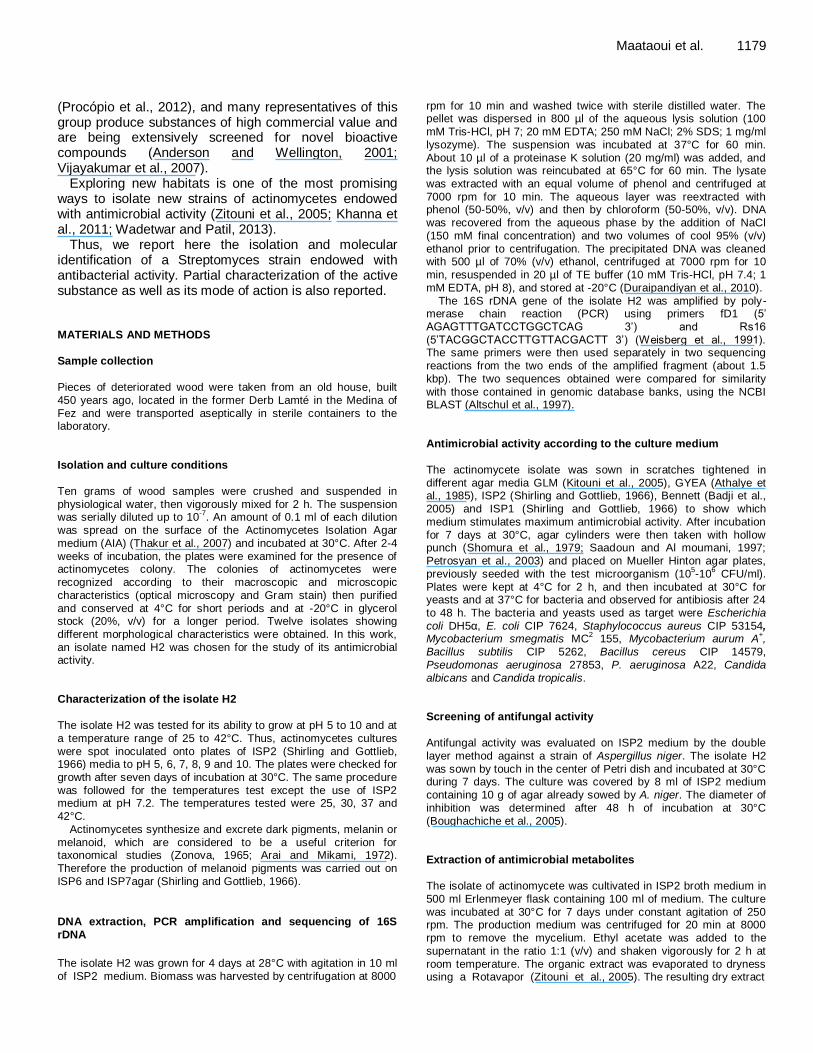
Table 1. Antibacterial activity of the isolate H2.
Test isolate*
EcDH5α Ec CIP Bs Bc Pa A22 Pa Sa Ms Ma
- - + + - + + + +
+ Inhibition; - no inhibition. *EcDH5α, E. coli DH5α; Ec CIP, E. coli CIP 7624; Bs, B. subtilis CIP 5262 ; Bc, B. cereus CIP 14579 ; Pa A22, P. aeruginosa A22; Pa, P. aeruginosa 27853; Sa, S.
aureus CIP 53154; Ms, M. smegmatis MC2 155 and Ma, M. aurum A
+
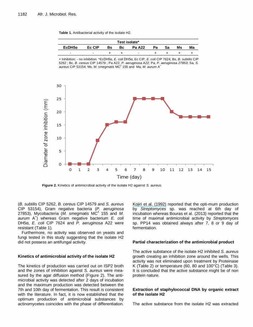
Figure 2. Kinetics of antimicrobial activity of the isolate H2 against S. aureus.
(B. subtilis CIP 5262, B. cereus CIP 14579 and S. aureus CIP 53154), Gram negative bacteria (P. aeruginosa 27853), Mycobacteria (M. smegmatis MC
2 155 and M.
aurum A+) whereas Gram negative bacterium E. coli
DH5α, E. coli CIP 7624 and P. aeruginosa A22 were resistant (Table 1).
Furthermore, no activity was observed on yeasts and fungi tested in this study suggesting that the isolate H2 did not possess an antifungal activity. Kinetics of antimicrobial activity of the isolate H2 The kinetics of production was carried out on ISP2 broth and the zones of inhibition against S. aureus were mea-sured by the agar diffusion method (Figure 2). The anti-microbial activity was detected after 2 days of incubation and the maximum production was detected between the 7th and 10th day of fermentation. This result is consistent with the literature. In fact, it is now established that the optimum production of antimicrobial substances by actinomycetes coincides with the phase of differentiation.
Kojiri et al. (1992) reported that the opti-mum production by Streptomyces sp. was reached at 6th day of incubation whereas Bouras et al. (2013) reported that the time of maximal antimicrobial activity by Streptomyces sp. PP14 was obtained always after 7, 8 or 9 day of fermentation. Partial characterization of the antimicrobial product The active substance of the isolate H2 inhibited S. aureus growth creating an inhibition zone around the wells. This activity was not eliminated upon treatment by Proteinase K (Table 2) or temperature (60, 80 and 100°C) (Table 3). It is concluded that the active substance might be of non protein nature. Extraction of staphylococcal DNA by organic extract of the isolate H2 The active substance from the isolate H2 was extracted

Maataoui et al. 1183
Table 2. Sensitivity of the active substance from the isolate H2 to Proteinase K.
Inhibition diameter against S. aureus (cm)
Without treatment with Proteinase K
After treatment with Proteinase K
Control
Organic extract of the isolate H2 3 ± 0.28 2.85 ± 0.07 0
The control used was a solution of Proteinase K. Values are means of two replicates ± standard deviation.
Table 3. Sensitivity of the active substance from the isolate H2 to temperature.
Inhibition diameter against S. aureus (cm)
Not subjected to heat treatment After heat treatment
60°C 80°C 100°C
Supernatant of the isolate H2 3 ± 0.14 3 ± 0.00 3 ± 0.14 2,6 ± 0.00
Values are means of two replicates ± standard deviation.
Figure 3. 1% Agarose gel electrophoresis analysis of
S. aureus genomic DNA extracted with the organic
extract of the isolate H2. 1: Control corresponding to DNA extraction of S. aureus with physiological water. 2: DNA extraction of S. aureus with the organic extract of the isolate H2.
by ethyl acetate, recuperated in physiological water and then tested for its ability to extract genomic DNA from S. aureus (previously explained in methods). Figure 3 shows that genomic DNA was obtained when S. aureus was treated by the organic extract of the isolate H2 in comparison with the control where organic extract was replaced by physiological water. This extraction was carried out in the absence of conventional agents of lysis, such as lysozyme, SDS or proteinase K, suggesting that the active ingredient acts at the S. aureus wall level.
Other active substances produced by actinomycetes
act on cell wall biosynthesis (Schneider et al., 2009; Hendlin et al., 1969; Krogstad et al.,1980; Singh et al., 2003).
More experiments are needed to characterize the structure of the active substance produced by the isolate H2 and to illustrate its effect on the bacterial cell wall of S. aureus. The effect might be also studied on other bacteria, mainly Gram negative ones. Thus, a protocol based on this substance could be developed and used in extraction of bacterial genomic DNA. Thin layer chromatography and bioautography
TLC and bioautography are often used for preliminary characterization of antimicrobial substances from produ-cing microorganisms’ organic extract, especially determi-nation of the position of the hot spot on TLC (Considinet and Mallette, 1965; Awais et al., 2007).
In this study, chloroform-methanol (9:1, v/v) was used as solvent system to study ethyl acetate extract of the isolate H2. TLC silica gel analysis showed three bands having respectively the following Rf values: B1=0.37; B2=0.6 and B3=0.7 (Figure 4a). Bioautography of the TLC slide against S. aureus resulted in the formation of inhibition zones around the components corresponding to B2 and B3 bands (Figure 4b). No inhibition zone was observed around the B1 band.
Silica gel around the three bands was separately elua-ted and evaporated. The resulting products were sepa-rately recuperated in 20 μl of sterile distilled water and tested using the disc method. The results obtained con-firms that the active bands were B2 and B3 (Figure 5). Further work is needed to determine the chemical struc-ture of the products contained in the bands 2 and 3. The spectrum of activity of each of the two products should be determined on different bacteria and fungi. It would also
Genomic DNA
supérieure
1 2

1184 Afr. J. Microbiol. Res.
Figure 4. a) TLC analysis of the crude extract of H2 strain after migration; b) Bioautography of the
H2 extract against S. aureus.
Figure 5. Effect of the three products (B1, B2 and B3) on
S. aureus growth.
be interesting to see if the two products have a synergic effect or not.
In summary, a Streptomyces strain producing at least two antimicrobial substances was isolated from deterio-rated wood. The activity of the isolate H2 against S. aureus, Mycobacteria and P. aeruginosa makes it more interesting to study because of the problems of resistance and multi-resistance encountered by these bacteria.
Conflict of Interests The author(s) have not declared any conflict of interests.
REFERENCES
Ali MA, Keera AA, Helmy MS, Abd El-Nasser HN, Ahmed KA, El-
Hennawi HM (2011). Selection of Pigment (Melanin) production in Streptomyces and their application in Printing and Dyeing of Wool Fabrics. Res. J. Chem. Sci. 1(5):22-28.
Altschul SF, Madden TL, Schäffer AA, Zhang J, Zhang Z, Miller W, Lipman DJ (1997). Gapped BLAST and PSI-BLAST: a new generation of protein database search programs. Nucleic Acids Res.
25(17):3389-3402. Anderson AS, Wellington MHE (2001). The taxonomy of Streptomyces
and related genera. Int. J. Syst. Evol. Microbiol. 51:797-814.
Antonova-Nikolova S, Stefanova V, Yocheva L (2006-2007). Taxonomic study of Streptomyces sp. strain 34-1. J. Cult. Collect. 5:10-15.
Arai T, Mikami Y (1972). Choromogenecity of Streptomyces. Appl.
Microbiol. 23:402-406. Arasu MV, Duraipandiyan V, Agastian P, Ignacimuthu S (2008).
Antimicrobial activity of Streptomyces spp. ERI-26 recovered from
Western Ghats of Tamil Nadu. J. Med. Mycol. 18:147-153. Athalye M, Goodfellow M, Lacey J, White RP (1985). Numerical
classification of Actinomadura and Nocardiopsis. Int. J. Syst.
Bacteriol. 35:86-98. Awais M, Shah A, Hammed A, Hasan F (2007). Isolation, identification
and optimization of bacitracin produced by Bacillus sp. Pak. J. Bot.
39:1303-1312.
a b
B2
B1
B3
Point of deposition
Actives bands
B1 B2
B3

Badji B, Mostefaoui A, Sabaou N, Lebrihi A, Mathieu F, Seguin E,
Tillequin F (2007). Isolation and partial characterization of antimicrobial compounds from a new strain Nonomuraea sp. NM94.
J. Ind. Microbiol. Biotechnol. 34:403-412. Badji B, Riba A, Mathieu F, Lebrihi A, Sabaou N (2005). Antifungal
activity of a saharan Actinomadura strain against various pathogenic
and toxinogenic fungi. J. Med. Mycol. 15(4):211-219. Badji B, Zitouni A, Mathieu F, Lebrihi A, Sabaou N (2006). Antimicrobial
compounds produced by Actinomadura sp. AC104 isolated from an
Algerian Saharan soil. Can. J. Microbiol. 52:373-382. Boudemagh A, Kitouni M, Boughachiche F, Hamdiken H, Oulmi L,
Reghioua S, Zerizer H, Couble A, Mouniee D, Boulahrouf A, Boiron P
(2005). Isolation and molecular identification of actinomycete microflora, of some saharian soils of south east Algeria (Biskra, EL-Oued and Ourgla) study of antifungal activity of isolated strains. J.
Med. Mycol. 15:39-44. Boudjella H, Bouti K, Zitouni A, Mathieu F, Lebrihi A, Sabaou N (2006).
Taxonomy and chemical characterization of antibiotics of Streptosporangium Sg 10 isolated from a Saharan soil. Microbiol.
Res. 161:288-298. Boughachiche F, Reghioua S, Oulmi L, Zerizer H, Kitouni M,
Boudemagh A (2005). Isolation of actinomycetes producing antimicrobial substances from Sebkha Ain Mlila. Sci. Technol. 23:5-10.
Bouras N, Meklat A, Toumatia O, Mokrane S, Holtz MD, Strelkov SE, Sabaou N (2013). Bioactive potential of a new strain of Streptomyces sp. PP14 isolated from Canadian soil. Afr. J.
Microbiol. Res. 7(25):3199-3208. Breidt F, Crowley KA, Fleming HP (1995). Controlling cabbage
fermentations with nisin and nisin-resistant Leuconostoc
mesenteroides. Food Microbiol. 12:109-116.
Chen J, Du GC, Li Y (2003). Fermentation experimental techniques. Chemical Industry Press, Beijing.
Cheraiti N, Gacemi Kirane D (2012). Isolation of actinomycetes strains producing new antifungal molecules. Rev. Microbiol. Ind. San. Environn. 6(1):18-34.
Considinet JM, Mallette MF (1965). Production and partial purification of antibiotic materials formed by Physarum gyrosum. Appl. Microbiol.
13:464-468.
Dastager S, Li WJ, Dayanand A, Tang SK, Tian XP, Zhi XY, Xu LH, Jiang CL (2006). Separation, identification and analysis of pigment (melanin) production in Streptomyces. Afr. J. Biotechnol. 5:1131-
1134. Devi A, Jeyarani M, Balakrishnan K (2006). Isolation and Identification
of Marine Actinomycetes and their Potential in Antimicrobial Activity.
Pak. J. Biol. Sci. 9(3):470-472. Duraipandiyan V, Sasi AH, Islam VIH , Valanarasu M, Ignacimuthu S
(2010). Antimicrobial properties of actinomycetes from the soil of Himalaya. J. Med. Mycol. 20:15-20.
Goodfellow M, Jones AL, Maldonado LA, Salanitro J (2004). Rhodococcus aetherivorans sp. nov., a new species that contains
methyl-t-butyl ether degrading actinomycetes. Syst. Appl. Microbiol.
27:61-65. Goodfellow M, Williams ST (1983). Ecology of actinomycetes. Ann.
Rev. Microbiol. 37:189-215.
Hassi M, Haggoud A, El Mzibri M, Ibnsouda S, Houari A, Iraqui M (2007). Isolation and identification of a staphylococcal strain with an anti-mycobacterial activity and study of it’s mode of action. Ann.
Microbiol. 57(4):651-656. Hendlin D, Stapley EO, Jackson M, Wallick H, Miller AK, Wolf FJ, Miller
TW, Chaiet L, Kahan FM, Foltz EL, Woodruff HB, Mata JM,
Hernandez S, Mochales S (1969). Phosphonomycin, a new antibiotic produced by strains of Streptomyces. Science 66(3901):122-123.
Holt JG (1994). Bergey’s Manual of Determinative Bacteriology 9th
edition (Williams and Wilkins, Baltimore). pp. 667-669. Iwai Y, Omura S (1982). Culture conditions for screening of new
antibiotics. J. Antibiot. 35:123-141.
Jeffrey LSH (2008). Isolation, characterization and identification of actinomycetes from agriculture soils at Semongok, Sarawak. Afr. J. Biotechnol. 7:3697-3702.
Kekuda TRP, Shobha KS, Onkarappa R (2010). Fascinating diversity and potent biological activities of actinomycetes metabolites. J.
Maataoui et al. 1185
Pharm. Res. 3:250-256. Khanna M, Solanki R, Lal R (2011). Selective isolation of rare
actinomycetes producing novel antimicrobial compounds. Int. J. Adv.
Biotechnol. Res. 2:357-375. Kim BJ, Kim CJ, Chun J, Koh YH, Lee SH, Hyun JW, Cha CY, Kook YH
(2004). Phylogenetic analysis of the genera Streptomyces and
Kitasatospora based on partial RNA polymerase β-subunit gene
(rpoB) sequences. Int. J. Syst. Evol. Microbiol. 54:593-598. Kitouni M, Boudemagh A, Oulmi L, Reghioua S, Boughachiche F,
Zerizer H, Hamdiken H, Couble A, Mouniee D, Boulahrouf A, Boiron P (2005). Isolation of actinomycetes producing bioactive substances from water, soil and tree bark samples of the north–east of Algeria. J.
Med. Mycol. 15:45-51. Koehn FE, Carter GT (2005). The evolving role of natural products in
drug discovery. Nat. Rev. Drug. Discov. 4:206-220.
Kojiri K, Nakajima S, Suzuki H, Kondo H, Suda H (1992). A new macrocyclic lactam antibiotic, BE-14106. I. Taxonomy, isolation, biological activity and structural elucidation. J. Antibiot. 45:868-874.
Kontro M, Lignell U, Hirvonen MR, Nevalainen A (2005). pH effects on 10 Streptomyces spp. growth and sporulation depend on nutrients. Lett. Appl. Microbiol. 41:32-38.
Krogstad DJ, Moellering RC, Greenblatt DJ (1980). Single-dose kinetics of intravenous vancomycin. J. Clin. Pharmacol. 20:197-201.
Kutzner HJ (1986). The family Streptomycetaceae. In M.P. Starr, H.
Stolp, H.G. Trüper, A. Balows, and H.G. Schlegel (ed.), The prokaryotes, a handbook on habitats, isolation, and identification of bacteria, Springer-Verlag, New York 2:2028-2090.
Lam KS (2006). Discovery of novel metabolites from marine actinomycetes. Curr. Opin. Microbiol. 9:245-251.
Locci R (1989). Streptomycetes and related genera. Bergey's Manual of
Systematic Bacteriology, Williams and Wilkins Company, Baltimore 4:2451-2508.
Naine J, Srinivasan MV, Devi SC (2011). Novel anticancer compounds
from marine actinomycetes: a review. J. Pharm. Res. 4:1285-1287. Nermeen AE, Gehan MA (2006). Antagonistic effect of marine Nocardia
brasiliensis against the fish pathogen Vibrio damsela: Application of
Plackett-Burman experimental design to evaluate factors affecting the production of the antibacterial agent. Int. J. Oceans Oceanogr. 1:141-150.
Okami Y, Hotta K (1988). Search and discovery of new antibiotics, in: Goodfellow M, Williams ST, Mordaski M (Eds). Actinomycetes in biotechnology. Academic Press, London, United Kingdom. pp 33-67.
Petrosyan P, Garcia-Varela M, Luz-Madrigal A, Huitron C, Flores ME (2003). Streptomyces mexicanus sp. Nov., a xylanolytic
microorganism isolated from soil. Inter. J. Syst. Evol. Microbiol.
53:269-273. Procópio RE, Silva IR, Martins MK, Azevedo JL, Araújo JM (2012).
Antibiotics produced by Streptomyces. Braz. J. Infect. Dis. 16(5):466-
471.
Saadoun I, Al moumani F (1997). Streptomycetes from jordan soils active against Agrobacterium tumefaciens. Actinomycetes 8:29-36.
Sadowsky MJ, Kinkel LL, Bowers JH, Schottel JL (1996). Use of
repetitive intergenic DNA sequences to classify pathogenic and disease-suppressive Streptomyces strains. Appl. Environ. Microbiol. 62:3489-3493.
Schneider T, Gries K, Josten M, Wiedemann I, Pelzer S, Labischinski H, Sahl HG (2009). The lipopeptide antibiotic Friulimicin B inhibits cell wall biosynthesis through complex formation with bactoprenol
phosphate. Antimicrob. Agents Chemother. 53(4):1610-1618. Shirling EB, Gottlieb D (1966). Methods for characterization of
Streptomyces species. Int. J. Syst. Bacteriol. 16:313-340.
Shomura T, Yoshida J, Amano S, Kojima M, Inouye S, Niida T (1979). Studies on Actinomycetales producing antibiotics only on agar culture. I. Screening, taxonomy and morphology-productivity relationship of Streptomyces halstedii, strain SF-1993. J. Antibiot.
32(5):427-435. Singh LS, Mazumder S, Bora TC (2009). Optimisation of process
parameters for growth and bioactive metabolite produced by a salt- tolerant and alkaliphilic actinomycete, Streptomyces tanashiensis
strain A2D. J. Med. Mycol. 19:225-233.
Singh MP, Petersen PJ, Weiss WJ, Janso JE, Luckman SW, Lenoy EB, Bradford PA, Testa RT, Greenstein M (2003).

1186 Afr. J. Microbiol. Res.
Mannopeptimycins, New Cyclic Glycopeptide Antibiotics Produced by Streptomyces hygroscopicus LL-AC98: Antibacterial and Mechanistic
Activities. Antimicrob. Agents Chemother. 47(1):62-69.
Spyropoulou KE, Chorianopoulous NG, Skandamis PN, Nychas GJN (2001). Survival of Escherichia coli O157: H7 during the fermentation
of Spanish-style green table olives (conservolea variety) supple-
mented with different carbon sources. Int. J. Food Microbiol. 66:3-11. Stal LJ, Moezelaar R (1997). Fermentiaon in cyanobacteria. FEMS
Microbiol. Rev. 21:179-211.
Steingrube VA, Wilson RW, Brown BA, Jost KC, Blacklock Z, Gibson JL, Wallace RJ (1997). Rapid identification of clinically significant species and taxa of aerobic actinomycetes, including Actinomadura,
Gordona, Nocardia, Rhodococcus, Streptomyces, and Tsukamurella
isolates, by DNA amplification and restriction endonuclease analysis. J. Clin. Microbiol. 35:817-822.
Thakur D, Yadav A, Gogoi BK, Bora TC (2007). Isolation and screening of Streptomyces in soil of protected forest areas from the states of Assam and Tripura, India, for antimicrobial metabolites. J. Mycol.
Med. 17:242-249. Valli S, Suvathi SS, Aysha OS, Nirmala P, Vinoth KP, Reena A (2012).
Antimicrobial potential of Actinomycetes species isolated from marine
environment. Asian Pac. J. Trop. Biomed. 2(6):469-473. Vasanthabharathi V, Lakshminarayanan R, Jayalakshmi S (2011)
.Melanin production from marine Streptomyces. Afr. J. Biotechnol.
10:11224-11234. Vijayakumar R, Muthukumar C, Thajuddin N, Panneerselvam A,
Saravanamuthu R (2007). Studies on the diversity of actinomycetes
in the Palk Strait region of Bay of Bengal, India. Actinomycetologica 21:59-65.
Wadetwar RN, Patil AT (2013). Isolation and characterization of
bioactive actinomycetes from soil in and around Nagpur. Int. J. Pharm. Sci. Res. 4(4):1428-1433.
Weisberg WG, Barns SM, Pelletier DA, Lane DJ (1991). 16S ribosomal DNA amplification for phylogenetic study. J. Bacteriol. 173:679-703.
Wu S, Jia S, Sun D, Chen M, Chen X, Zhong J, Huan L (2005).
Purification and characterization of two novel antimicrobial peptides subpeptin JM4-A and subpeptin JM4-B produced by Bacillus subtilis
JM4. Curr. Microbiol. 51:292-296. Xu LH, Tiang YQ, Zhang YF, Zhao LX, Jiang CL (1998). Streptomyces
thermogriseus a new species of the genus Streptomyces from soil,
lake and hot spring. Int. J. Syst. Bacteriol. 48:1087-1093.
Zitouni A, Boudjella H, Lamari L, Badji B, Mathieu F, Lebrihi A, Sabaou N (2005). Nocardiopsis and Saccharothrix genera in Saharan soils in
Algeria: Isolation, biological activities and partial characterization of
antibiotics. Res. Microbiol. 156:984-993. Zonova GM (1965). Melanoid pigments of Actinomycetes. Mikrobio-
logiya 34:278-283.