
Isolation and characterization of a Drosophila gene essential for early embryonic development and...
12
Isolation and Characterization of a Drosophila Gene Essential for Early Embryonic Development and Formation of Cortical Cleavage Furrows Claire X. Zhang, Maxwell P. Lee, Alice D. Chen, Sheryl D. Brown, and Tao-shih Hsieh Department of Biochemistry, Duke University Medical Center, Durham, North Carolina 27710 Abstract. We have isolated a new female sterile mu- tant from Drosophila melanogaster, which arrests the embryonic development during the transition from syn- cytial to cellular blastoderm. Cytological analysis of the mutant embryos indicates that pseudocleavage furrows in the syncytial blastoderm are abnormal but not com- pletely disrupted. However, cleavage furrows during cellularization are totally disorganized, and no embryos can develop beyond this stage. Consistent with this ob- servation, the expression of this gene peaks around the cellular blastoderm and not in any later developmental stages. Based on immunofluorescence experiments, the protein product of this gene is localized in both pseudocleavage furrows at the syncytial blastoderm and in the cleavage furrows during the cellularization stage. Sequence homology analysis demonstrates a modest, but statistically significant, similarity of this protein with the carboxyl-terminal domains of dystro- phin and a family of proteins collectively known as apo- dystrophins. It is possible that this protein may play an essential role in organizing and maintaining a special- ized cytoskeletal structure, a function also suggested for dystrophin and apodystrophins. T uz early development of the Drosophila embryo is completely under maternal control (Zalokar, 1976; Merrill et al., 1988; Wieschaus and Sweeton, 1988). It is characterized by rapid DNA replication and nuclear division without intervening cytokinesis (Rabinowitz, 1941; Foe and Alberts, 1983). The first eight nuclear divisions happen in the interior of the embryo. Starting from the te- lophase of cycle 8 and finishing at the interphase of cycle 10, the majority of nuclei migrate synchronously toward the cortex of embryo (Foe and Alberts, 1983; for review see Foe et al., 1993). At cycle 9, ~10 nuclei migrate ahead of the main body of nuclei and reach the posterior end to form pole cells, the precursors of germ line cells. The corti- cal nuclei then undergo four synchronous divisions as a regularly spaced monolayer underneath the plasma mem- brane. These four nuclear divisions, cycles 10 to 13, are termed the syncytial blastoderm stage. Cellularization commences at the beginning of interphase of cycle 14, when zygotic machinery starts to take over embryogenesis. During the ensuing period of ~45 rain, membrane biogen- C.X. Zhang and M.P. Lee share first authorship. Address all correspondence to Tao-shih Hsieh, Department of Bio- chemistry, Duke University Medical Center, Durham, NC 27710. Tel.: (919) 684-6501. Fax: (919) 684-8885. e-mail: [email protected]. edu. M.P. Lee's present address is Department of Medicine, Johns Hopkins University School of Medicine, 1064 Ross, 720 Rutland Ave., Baltimore, MD 212O5. esis coincides with the formation of cleavage furrows and allows ,--~6,000 cortical nuclei to be enclosed with plasma membrane. Gastrulation occurs immediately after the completion of the cellularization process. The precision and order of these early developmental processes are under the control of and dependent upon the cytoskeletal structure of embryos. During Drosophila early embryogenesis when all nuclear divisions occur in a syncytium, cytoskeletal organization is especially impor- tant in the formation of a highly regular structure of corti- cal nuclei (for review see Fyrberg and Goldstein, 1990; Schejter and Wieschaus, 1993). In preblastoderm stages, nuclei divide in the center of the embryo, and each nucleus is surrounded by a special domain enriched in cytoskeletal elements. The cytoskeleton is also localized in a cortical layer beneath the entire plasma membrane at that stage. Inhibitor and cytological studies demonstrate that mi- crofilaments and microtubules are responsible for early nuclear movement and migration toward the cortex (Foe and Alberts, 1983; Hatanaka and Okada, 1991; Baker et al., 1993). The cytoskeletal elements undergo rearrangements during the syncytial blastoderm stage (Karr and Alberts, 1986; Kellogg et al., 1988). During interphase, F-actin forms a cap above a pair of centrosomes that lie on the apical side of each nucleus. Starting from prophase, each of the pair of centrosomes separates and migrates to oppo- site sides of the nucleus to assemble a mitotic spindle, while the actin cap enlarges and spreads into pseudocleav- © The Rockefeller University Press, 0021-9525196/08/923/12 $2.00 The Journal of Cell Biology, Volume 134, Number 4, August 1996 923-934 923 on January 18, 2014 jcb.rupress.org Downloaded from Published August 15, 1996
-
Upload
independent -
Category
Documents
-
view
3 -
download
0
Transcript of Isolation and characterization of a Drosophila gene essential for early embryonic development and...
Isolation and Characterization of a Drosophila Gene Essential for Early Embryonic Development and Formation of Cortical Cleavage Furrows Claire X. Zhang, Maxwell P. Lee, Alice D. Chen, Sheryl D. Brown, and Tao-shih Hsieh Department of Biochemistry, Duke University Medical Center, Durham, North Carolina 27710
Abstract. We have isolated a new female sterile mu- tant from Drosophila melanogaster, which arrests the embryonic development during the transition from syn- cytial to cellular blastoderm. Cytological analysis of the mutant embryos indicates that pseudocleavage furrows in the syncytial blastoderm are abnormal but not com- pletely disrupted. However, cleavage furrows during cellularization are totally disorganized, and no embryos can develop beyond this stage. Consistent with this ob- servation, the expression of this gene peaks around the cellular blastoderm and not in any later developmental stages. Based on immunofluorescence experiments, the
protein product of this gene is localized in both pseudocleavage furrows at the syncytial blastoderm and in the cleavage furrows during the cellularization stage. Sequence homology analysis demonstrates a modest, but statistically significant, similarity of this protein with the carboxyl-terminal domains of dystro- phin and a family of proteins collectively known as apo- dystrophins. It is possible that this protein may play an essential role in organizing and maintaining a special- ized cytoskeletal structure, a function also suggested for dystrophin and apodystrophins.
T uz early development of the Drosophila embryo is completely under maternal control (Zalokar, 1976; Merrill et al., 1988; Wieschaus and Sweeton, 1988).
It is characterized by rapid DNA replication and nuclear division without intervening cytokinesis (Rabinowitz, 1941; Foe and Alberts, 1983). The first eight nuclear divisions happen in the interior of the embryo. Starting from the te- lophase of cycle 8 and finishing at the interphase of cycle 10, the majority of nuclei migrate synchronously toward the cortex of embryo (Foe and Alberts, 1983; for review see Foe et al., 1993). At cycle 9, ~10 nuclei migrate ahead of the main body of nuclei and reach the posterior end to form pole cells, the precursors of germ line cells. The corti- cal nuclei then undergo four synchronous divisions as a regularly spaced monolayer underneath the plasma mem- brane. These four nuclear divisions, cycles 10 to 13, are termed the syncytial blastoderm stage. Cellularization commences at the beginning of interphase of cycle 14, when zygotic machinery starts to take over embryogenesis. During the ensuing period of ~45 rain, membrane biogen-
C.X. Zhang and M.P. Lee share first authorship. Address all correspondence to Tao-shih Hsieh, Department of Bio-
chemistry, Duke University Medical Center, Durham, NC 27710. Tel.: (919) 684-6501. Fax: (919) 684-8885. e-mail: [email protected]. edu.
M.P. Lee's present address is Department of Medicine, Johns Hopkins University School of Medicine, 1064 Ross, 720 Rutland Ave., Baltimore, MD 212O5.
esis coincides with the formation of cleavage furrows and allows ,--~6,000 cortical nuclei to be enclosed with plasma membrane. Gastrulation occurs immediately after the completion of the cellularization process.
The precision and order of these early developmental processes are under the control of and dependent upon the cytoskeletal structure of embryos. During Drosophila early embryogenesis when all nuclear divisions occur in a syncytium, cytoskeletal organization is especially impor- tant in the formation of a highly regular structure of corti- cal nuclei (for review see Fyrberg and Goldstein, 1990; Schejter and Wieschaus, 1993). In preblastoderm stages, nuclei divide in the center of the embryo, and each nucleus is surrounded by a special domain enriched in cytoskeletal elements. The cytoskeleton is also localized in a cortical layer beneath the entire plasma membrane at that stage. Inhibitor and cytological studies demonstrate that mi- crofilaments and microtubules are responsible for early nuclear movement and migration toward the cortex (Foe and Alberts, 1983; Hatanaka and Okada, 1991; Baker et al., 1993). The cytoskeletal elements undergo rearrangements during the syncytial blastoderm stage (Karr and Alberts, 1986; Kellogg et al., 1988). During interphase, F-actin forms a cap above a pair of centrosomes that lie on the apical side of each nucleus. Starting from prophase, each of the pair of centrosomes separates and migrates to oppo- site sides of the nucleus to assemble a mitotic spindle, while the actin cap enlarges and spreads into pseudocleav-
© The Rockefeller University Press, 0021-9525196/08/923/12 $2.00 The Journal of Cell Biology, Volume 134, Number 4, August 1996 923-934 923
on January 18, 2014jcb.rupress.org
Dow
nloaded from
Published August 15, 1996
age furrows, the transient membrane invaginations sur- rounding each spindle. In anaphase, F-actin undergoes a recapping process above each tubulin aster and stays in the caps throughout telophase. The actin-based microfila- ment networks seem to be important for the separation of the adjacent mitotic apparati, since disruptions in actin or- ganization would lead to centrosome clumping and de- fective spindles (Callaini et al., 1992; Postner et al., 1992; Sullivan et al., 1990, 1993). Soon after cycle 14 begins, mi- crotubules grow from the centrosomes to surround each elongating nucleus, and actin filaments form a membrane- associated hexagonal array that invaginates, passing the bottom of each nucleus, and then closes off to form cells. The integrity of the cytoskeleton is shown to be required for mitosis and cellularization in the early embryo. Failure of proper cytoskeletal organization leads to abnormal nu- clear morphology, such as nondisjunction of chromosomes and nuclear fusion (Sullivan et al., 1993). Although cytoskel- etal behavior in early cycles has been extensively studied and well characterized, the molecular mechanism remains unknown. A key step toward understanding the molecular mechanism is to define the cytoskeletal components in biochemical detail. A group of actin-binding proteins from Drosophila embryos has been isolated by F-actin affinity chromatography (Miller et al., 1989). Most of them (>90%) colocalize with actin at some point during early develop- ment. Both myosin VI and annilin are identified from such analysis, and they seem to play important roles in organiz- ing the embryonic cytoskeleton (Field and Alberts, 1995; Mermall and Miller, 1995). Genetic approaches have also been applied in identifying components in embryonic cy- toskeleton, either by analyzing the maternal effect mutants (Postner et al., 1992; Sullivan et al., 1990, 1993) or by re- verse genetics (Karess et al., 1991).
We have recently generated a number of female sterile mutants, and one of them is a novel mutant that arrests the embryonic development during the transition from syncy- tial to cellular blastoderm. The cleavage furrows in the mutant embryos are grossly disrupted, suggesting this ma- ternal effect gene is involved in the formation of embry- onic cytoskeleton. According to the mutant phenotype, we name the gene affected in this mutant as _discontinuous ac- tin hexagon (dah). We have cloned and sequenced this gene, and its amino acid sequence shares limited homol- ogy with a family of cytoskeletal proteins, dystrophin, and apodystrophins. We have also generated specific antibody to monitor the expression and distribution of this protein. The maximal expression is at the syncytial/cellular blasto- derm, thus coinciding with the genetic function of this pro- tein. The immunofluorescence experiments have localized this protein in pseudocleavage and cleavage furrows, sug- gesting a functional role of this protein in the organization of early embryonic cytoskeleton.
Materials and Methods
Isolation of Female Sterile Mutants Drosophila strain 438 (Bloomington Stock Center, Bloomington, 1N) con- tains a P-element insertion at 13C1 in the X chromosome (de Cicco and Spradling, 1984). We have screened for mutants generated by imprecise excision events after remobilizing the P-element in 438 flies using a ge- nomic source of transposase (Lee ct al., 1993). Among the mutants char-
acterized after this genetic screen, a lethal complementation group includ- ing 77, 111, 112, and 113 was isolated. Their mutations were due to a deficiency in top1 gene, which encodes DNA topoisomerase I, and could be completely rescued by an ectopic copy of topl introduced through germ- line transformation (Lee et al., 1993 ; see also Fig. 1 A). We have also iso- lated a female sterile group including alleles of 30 and 36, which is in a dif- ferent genetic complementation group from topl lethals. All of these mutants were subjected to the rescue tests by various transgenes contain- ing genomic segments covering topl and neighboring sequences. Comple- mentation tests of topl lethals were carried out as described before (Lee et al., 1993). For the female sterile mutants, genetic crosses were carried out to obtain homozygous female steriles offs(l)30 or 36 with either a bal- ancer second chromosome CyO/+ or a second chromosome containing an ectopic copy of transgene, like p[DSR]/+ or p[topl]/+. These females were then mated with wild-type males. Only females with p[DSR] trans- genes could produce progeny. For a given transgene construct, we have obtained several independent transformant lines, and the genetic rescue data were all consistent among each other.
Molecular Genetics of dah The molecular cloning of the genomic and cDNA sequences, nucleotide sequence analysis, PCR amplification, Northern blots, and immunoblots were carried out according to standard procedures (Sambrook eta[. , 1989). cDNA clones were isolated from an embryonic cDNA library (No- lan et al., 1986). and the cloning of genomic DNA in topl/dah locus was described earlier (Lee et al., 1993). The construction of the P-element transformation vector containing topl/dah genomic sequences, germline transformation, and screening/maintenance of the transformant lines fob lowed the established protocols (Robertson et al., 1988; see also Roberts, 1986 : with details specified in our earlier work, Lee et al., 1993). Database search and homology comparison were performed with Wisconsin Se- quence Analysis Package (Genetics Computer Group, Madison, Wl). The homology was first identified by BLAST search and subsequentTy ana- lyzed by BestFit and GAP. Prediction of coiled-coil structure was made with software provided by Dr. Alex Knight (Whitehead Institute, Cam- bridge, MA) using a published algorithm (Lupas et al., 1991: Knight, 1994).
Immunoblot Analysis The cDNA encoding full-length dab protein was cloned into a T7 expres- sion vector pET3a (Studier et al., 1990) for expression in bacteria. The over-produced protein represented ~30% of total bacterial protein. The recombinant dab protein was purified by differential centrifugation and SDS-PAGE. The purified dah was used to raise antibody in rabbit and coupled covalently to activated agarose beads (Reacti-Gel; Pierce Chemi- cal Co., Rockford, IL). Rabbit antibody was affinity purified from dah- agarose column and showed exquisite specificity for a 71-kD protein in the Drosophila early embryo extracts. Embryonic extracts from different developmental stages were prepared as described earlier (Lee et al, 1993). Total protein of I00 gg from each sample was run on an 8% SDS polyacrylamide gel and electroblotted to a nitrocellulose filter. Immuno- detection was carried out with a chemiluminescent ECL kit (Amersham Corp., Arlington Heights, IL) according to the manufacturer's protocol.
Immunofluorescent Staining of Whole-Mount Embryos Embryos from homozygous female sterile mutant or wild-type mothers were collected on grape juice plates at 25°C for a period of 30 min. They were staged by incubating in a humid chamber, and embryos of appropri- ate age were collected and washed with 0.7% NaCI and 0.[)5% Triton X-100. After dechorionation with 50% Clorox for 2 min, embryos were fixed in a 1:1 mixture of heptane and 8% formaldehyde for 20 rain. For microtubule staining, taxol was added to the fixative following a protocol developed by Karr and Alberts (1986). While this fixation procedure may introduce artefactual microtubule staining at the centrosomal region (Kellogg et al., 1988), we have observed a less dramatic effect on cen- trosome structure (see, for example, Fig. 4, A and B). Furthermore, we are using microtubule staining, in conjunction with nuclear morphology, pri- marily for determining the phase of nuclear cycle in these embryos. The heptane layer containing the embryos was removed and mixed with meth- anol by vigorous shaking to rupture the vitelline membrane. For phalloi- din staining of the F-actin, hand-peeling was used for vitelline removal. After rehydration and washing in PBS, embryos were incubated in 0.1% saponin and 1% normal goat serum in PBS for 30 rain, and then overnight
The Journal of Cell Biology, Volume 134, 1996 924
on January 18, 2014jcb.rupress.org
Dow
nloaded from
Published August 15, 1996
with primary antibodies. The rabbit antibody against dah protein was af- finity purified and then preadsorbed with dah embryos. For staining mi- crotubules, monoclonal mouse anti-c~-tubulin antibody (T9026; Sigma Chemical Co., St. Louis, MO) was used. The embryos were then washed with PBS and incubated with Cy3-conjugated goat anti-rabbit or anti- mouse secondary antibody (Jackson ImmunoResearch Laboratories, West Grove, PA) for 1 h in the dark. After washing in PBS, they were in- cubated with fluorescein-conjugated phalloidin (Molecular Probes, Inc., Eugene, OR) and 4',6-diamidino-2-phenylindole (DAPI) 1 (Sigma Chemi- cal Co.) for 20 min in the dark. After final washing in PBS, the embryos were mounted in 90% glycerol, 0.t M Tris-HC1, pH 8.0, and 0.5% wt/vol n-propyl gallate.
For nuclear morphology studies of the wild-type and dah- embryos, epifluorescence from DAPI staining was photographed with a microscope (Laborlux; E. Leitz, Inc., Rockleigh, NJ) on TMAX 400 film (Eastman- Kodak Co., Rochester, NY). The nuclear cycle was determined based on nuclear density counts. The laser scanning confocal images were collected using a fluorescence microscope (Axiovert; Carl Zeiss, Inc., Thornwood, NY) equipped with a ×40/1.30 Plan-Neofluar lens. The triple-stained em- bryos were first staged by epifluorescence from DAPI to monitor the nu- clear morphology before it was switched to the confocal mode.
Results
Isolation of a New Female Sterile Mutant with an Early Embryonic Lethal Phenotype
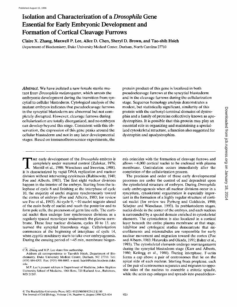
We have previously used P-e lement t ransposon-media ted mutagenesis to genera te lethals deficient in the topo- isomerase I gene, which is located at 13C1 on the X chro- mosome (Lee et al., 1993; see also Fig. 1 A). We have iso- lated addi t ional female sterile and lethal mutants and have carried out genetic crosses to group them into different complementa t ion groups. Al l the lethals form one comple- menta t ion group, which corresponds to deficiencies in topl. One of the female sterile complementa t ion groups, which includes two alleles, fs(1)30 and fs(1)36, appears to affect the early embryonic deve lopment and has been fur- ther characterized. While male flies are not affected by these mutat ions, females with ei ther homozygous or het- erozygous mutat ions in these two alleles develop normal ly and deposi t eggs with normal morphology. However , these embryos fail to develop into the gastrulat ion stage. Fur the r analysis indicates that there are extensive disrup- tions in the actin hexagonal arrays (see later sections in Results), and therefore, these mutants are named dah (dis- cont inuous actin hexagon). These female steriles are clearly in a different complementa t ion group than topl le- thals, since the heterozygous fly like fs(1) dah3°/l(1)top177 is nei ther lethal nor sterile. To locate dah that is affected in these female steriles, we have genera ted germline trans- formants with transgenes of D N A segments in and around the topl locus (Fig. 1 A). A transgene of D N A segment downst ream from the topl gene, p[DSR], can efficiently rescue the female sterile mutants of fs(1) dak ~° and fs(1)dah 36, but not the topl lethals. Conversely, a D N A segment covering topl, p[topl], can rescue topl lethals but not the female steriles. It is therefore likely that dah, which is responsible for the female sterile phenotype in fs(1)dah 3°/36, is located in p[DSR].
To further analyze dah, we have isolated the gene cov- e red in p[DSR]. Initial Nor thern exper iments showed that using the 2.2-kb B a m H I fragment in p[DSR] (Fig. 1 A) as
1. Abbreviation used in this paper: DAPI, 4', 6-diamidino-2-phenylindole.
A
p[topl] p[DSR]
sp I tom
ss[. Rescue Data
Lethals Female Sterlles 77, I l l , 112,113 30, 36
+
+
I I I I I I 5 Kb
B
B B
AUG UGA An
\ \ ] Deletion infs(1)dah 30
\ \ - -~ D e l e t i o n infs(1)dah 36
[ J 1 Kb
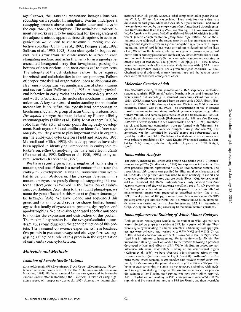
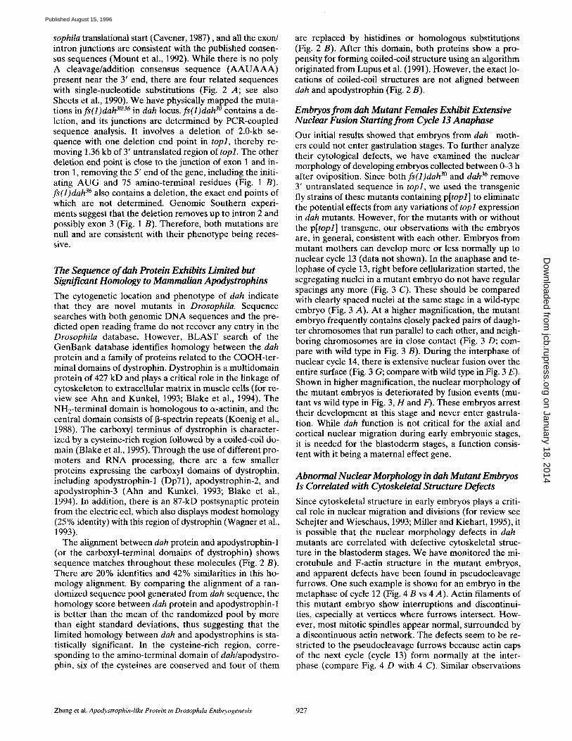
Figure 1. (A) Genetic complementation of lethal and female sterile mutants by an ectopic copy of genomic DNA fragment. Molecular map of the topl and dah region is indicated. The re- striction sites of SpeI (Sp), SalI (S), BamHI (B), and EcoRI (R) are shown. The arrow indicates the insertion site of P-element in fly strain 438, which was used in our mutagenesis experiment. We screened for the mutants generated by imprecise excision events associated with remobilized P-element. Shown here are six mu- tants that have been placed into two complementation groups. Four lethal mutants, 77, 111, 112, and 113, were rescued geneti- cally by an ectopic copy of the topl gene, p[topl]. Two female sterile mutants, 30 and 36, were rescued genetically by an ectopic copy of the dah gene, p[DSR]. The genetic rescue was performed with a transgenic second or third chromosome that was generated by introducing an ectopic copy of genomic DNA fragment, indi- cated as horizontal lines here, using P-element germline transfor- mation. (B) Genomic structure of the dah gene. The exons are shown as filled boxes, and the introns are shown as open boxes. The restriction sites of BamHI and EcoRI are indicated as B and R, respectively. Translational start codon, AUG, and stop codon, UGA, are indicated. An, poly A addition site. Extents of dele- tions in the dah gene for two null mutants, fs(1)dah 3° and fs(1)da# ~, are demarcated as horizontal lines. The broken line at the end indicates the range of uncertainty in the deletion end point.
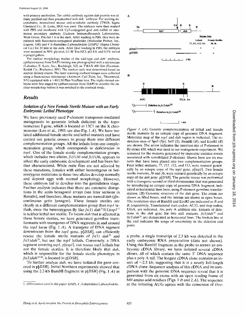
a probe, a single transcript of 2.3 kb was detected in the ear ly embryon ic R N A p r e p a r a t i o n (da ta not shown). Using this B a m H I fragment as the probe to screen an em- bryonic c D N A library, we have isolated several c D N A clones, all of which contain the same 3' D N A sequence plus a poly A tail. The longest c D N A clone contains an in- sert of ~2 .3 kb, suggesting that it is a nearly full-length c D N A clone. Sequence analysis of this c D N A and its com- parison with the genomic D N A sequence reveal that it is genera ted from six exons with an open reading frame of 649 amino acid residues (Figs. 1 B and 2 A). The sequence at the initiating A U G agrees with the consensus of Dro-
Zhang el al. Apodystrophin-like Protein in Drosophila Embo, ogenesis 925
on January 18, 2014jcb.rupress.org
Dow
nloaded from
Published August 15, 1996
A ATAACAATAA ACGCCTGACA AAATGCTGAG ATCGTCGGTG CCCGTGCAGC TGACA~CT 60
M L R S S V P V Q L T K L> 13
GCGAAATAGC GCCGAGGCCG TGGCGTCCAT TGCCCGCGAA AAGGGATCCC AAGGTGGCCA 120
R N S A E A V A S I A R E K G S Q G G Q~ 33
AGGACAAAAG CAACAGGTGG ACAAAGAGCT ATCCACCATC ATGGATGACT TC-CAGCGGGA 180
G Q K Q Q V D K E L S T I M D D 5 Q R D> 53
TTACGGGAAT TACAAGTACA CGGTATACCG GTGCGCCAGT AAATTTGTC-G CGCTGCAAAA 240 Y G N Y K Y T V Y R C A S K F V A L Q K> 73
GATATTTCAC ACTGGAAAAA TCCCCTACAA GCTGGTTTTG GCCATTTTGG ATCGCCATAA 300 I F H T G K I P Y K L V L A I L D R H N> 93
CTq"GAGTC-GT C~CTC~AAC TGATGGTq~CC TCCATTCCAG TTGACCTCCC TGCTC-CACGA 360
L S G G L E L M V P P F Q L T S L L H D> 113
TGTATACTTT GCCTGCGAGA AACTAGGCCA TTTCAACCAG GATGCGAACT ATAAACTGGA 420 V Y F A C E K L G H F N Q D A N Y K L D> 133
CAGGGCAACC GGGCTGT'PGG CCAACTTCTT CTGG/M~TGTC TACGATCCGC AACGCAGACA 480 R A T G L L A N F F W N V Y D P O R R H> 153
CTCCATATCC CTGTTAGAGA TCAGGATCAC CTTCATACTG CTGTGCAAGC TATATAGCAG 540 S I S L L E I R I T F I L L C K L Y S S> 173
CGATCACCTG GTGGGCGACA TCTTTGGCCT GCTGGCCGAT CCCAAGTCGC AGATCATCTC 600 D H L V G D I F G L L A D P K S Q I I S> 193
CAGATATGCA TTCGAACAGC TCCTAGCCAC GCTGAGCAAG TTACTCAGCT ATTTGGGCGA 660
R Y A F E Q L L A T L S K L L S Y L G E> 213
GGCCAAGGCC TATGGCGTCC ACAATCTTCC GTT~TATG GAGCAGTGCT TTGCCCGCTG 720 A K A Y G V H N L P L A M E Q C F A R C> 233
TCCGCATGGC GTTGGTGGAC TGACCGAGCC GCAGTTCCAC AAACTCTGGA CGGGAGCGGG 780 P H G V G G L T E P Q F H K L W T G A G> 253
TGTCCAGACG AGATTCCTGA TCTATGGCAA TCTGCTGGCA CTGGTGAAAC GCATCGAGGA 840 V Q T R F L I Y G N L L A L V K R I E D> 273
CACCGAGCAT TTGATTCACA GTAATTCTTG TGCCGGCTGC CGCAAGGACC ACATTGTGGG 900 T E H L I H S N S C A G C R K E H I V G> 293
CATACGTTTC CGGTGTCAGG TGTGTAGGGA TATATCGTTG TGCCTGCCCT GCTTTGCAGT 960 R F R C Q V C R D I $ L C L P C F A V> 313
GGGCTTCGCT GGTGGTCGCC ACGAGCCGGG TCATCGGATG TGCGAGGTCT TCGTGGAGGA 1020 G F A G G R H E P G H R M C E V F V E D> 333
TCAGCCGCCA TTGCGTTGGA CCCGCCACCT GGCCAGC-CTG TGCGGCTC-GC TGGTGATGCC 1080 Q P P L R W T R H L A R L C G W L V M P> 353
GCGCA~CA CAGGAGGAGG AGCGTCGCGG CTTCTGTAAT GCCCAGGAGA GTC43GCCCGC 1340 R K T Q E E E R R G F C N A Q E $ G 9 A> 373
CTTGGGTCAA TCCGCTGCCA CGCCCATTCC CGCAGAGACG CC-CAGTGTGC GCAGTCAGTG 1200 L G Q S A A T P I P A E T R S V R S Q C> 393
CCAGGAGAAG GACCGCACAG TTGTCCTGCA GCAACAGCAG CAGGAGCTCT GCTCCCTGAC 1260 Q E K D R T V V L Q Q Q Q Q E L C S L T> 413
GGGTCTAGCC AGCGAGGCGT CCAGCAGGCT GCAGTCCATC ATCGATCGAT TGCTTTTGCA 1320 G L A S E A S S R L Q S [ I D R L L L Q> 433
AAATGCGAAG CTGGAAACCC ACCTGCAAAC AGTGGCCACC GCATCTAGTT CGGA~TATC 1380
N A K L E T Q L Q T V A T A S S S E I S> 453
GCAGTTTCTG AGCGCCCATC AGTGCTTTCT GCTGCAGATT ATCGAGGATA TGCGACAGCT 1440 Q F L S A H Q C F L L Q I I E D M R Q L> 473
ATCACATGCC TCAGCGGTAA CTTCGCCGCC ACCTGCGCCA GCTGCCCAAG TGC-CGCCTTC 1500 S H A S A V T S P P P A P A A Q V A P S> 493
AGCTGCGCCC CTAGTCAC~CT CCACGTTCCT TAGCTCCAGC ACGCCTCAGC GACAGAT~CT 1560 A A P L V S $ T F L S S S T P Q R Q M L> 513
t ATTCGACATG TATGCGCCCG TC~TGCCAA TCTCACGCAC TCCATJa.AATG GCGCCGACTT 1620
F D M Y A P V G A N L T H S I N G A D L> 533
GAATCGCAGC TATCTGGAGG CAAACAAATC GGATTACTCG CTCAACGACA TAAGCTTGTG 1680
N R S Y L E A N K S D Y S L N D I S L W> 553
GTTCGACCAG CGTCC-CTCGA GTGTCCAGCC AGGACAAGGA CCTGGTTCCC TGCCGGCTCC 1740
F D Q R R S S V Q P G Q G P G S L P A P> 573
TCTGCCGCTG CCCATGCCGC TGGGTGCTCT ACTGGAACAG CAGCCCCCAA ACGGCGGAGA 1800
L P L P M P L G A L L E Q Q P A N G G D> 593
TGGCCGCGAC ACCGAGATGG TGAACTTTAA GCTGCTGCTG CACAAGGTCA AGGAGATCGT 1860
G R D T E M V N F K L L L H K V K E I V> 613
CGAGGACTCG TACAGCGATA ATGCCGAACT GTCGGCGGCC ACGCAAAATC TGGAGAACGT 1920 E D S Y S D N A E L S A A T Q N L E N V> 633
TCTCGATAGC ATCATCAAGA GCGAGGAC-CA GCAGCGGCGC ATCAGCACGT GAGGATCCGG 1980 L D S • I K S E E Q Q R R I S T *> 649
ATCGGATACC AACTCCCATC GATTCCAGCC AGACCAAGTC CACTTAGCTT TAAGT~TAA 2040 AGTTGTTCTA AAGTAATCGT GACAAAATGA CACAACCGAG TCATCATGCA TTTATTATAT 2100 CCTTCTTCCT TTTCCCATGT GCTTTAACAT TCGCGTAAAT GCTTTAATTT GTAGACTTAC 2160
AACTCTGACA AGAATGCAAT ACAACAAAAT GTTTTTATAA AAATATACAA TC-C 2213
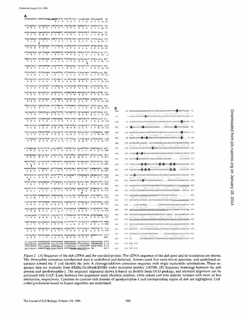
Figure 2. (A) Sequence of the dah c D N A and the encoded protein. The c D N A sequence of the dah gene and its translation are shown. The Drosophila consensus translational start is underlined and italicized. Arrows mark five exon-intron junctions, and underlined se- quences toward the 3' end identify the poly A cleavage/addition consensus sequence with single nucleotide substitutions. These se- quence data are available from EMBL/GenBank/DDBJ under accession number U43786. (B) Sequence homology between the dah protein and apodystrophin-1. The sequence alignment shown is based on BestFit from G C G package, and identical alignment can be produced with GAP. Lines between two sequences mark identical residues, while colons and dots indicate residues with more or less similarities, respectively. Cysteines in cysteine-rich domain of apodystrophin-1 and corresponding region of dah are highlighted. Coil- coiled predictions based on Lupus algorithm are underlined.
The Journal of Cell Biology, Volume 134, 1996 926
on January 18, 2014jcb.rupress.org
Dow
nloaded from
Published August 15, 1996
sophila translational start (Cavener, 1987), and all the exon/ intron junctions are consistent with the published consen- sus sequences (Mount et al., 1992). While there is no poly A cleavage/addition consensus sequence (AAUAAA) present near the 3' end, there are four related sequences with single-nucleotide substitutions (Fig. 2 A; see also Sheets et al., 1990). We have physically mapped the muta- tions in fs(1)dah 30/36 in dah locus, fs(1)dah 3° contains a de- letion, and its junctions are determined by PCR-coupled sequence analysis. It involves a deletion of 2.0-kb se- quence with one deletion end point in top1, thereby re- moving 1.36 kb of 3' untranslated region of top1. The other deletion end point is close to the junction of exon 1 and in- tron 1, removing the 5' end of the gene, including the initi- ating AUG and 75 amino-terminal residues (Fig. 1 B). fs(1)dah 36 also contains a deletion, the exact end points of which are not determined. Genomic Southern experi- ments suggest that the deletion removes up to intron 2 and possibly exon 3 (Fig. 1 B). Therefore, both mutations are null and are consistent with their phenotype being reces- sive.
The Sequence of dah Protein Exhibits Limited but Significant Homology to Mammalian Apodystrophins
The cytogenetic location and phenotype of dah indicate that they are novel mutants in Drosophila. Sequence searches with both genomic DNA sequences and the pre- dicted open reading frame do not recover any entry in the Drosophila database. However, BLAST search of the GenBank database identifies homology between the dah protein and a family of proteins related to the COOH-ter- minal domains of dystrophin. Dystrophin is a multidomain protein of 427 kD and plays a critical role in the linkage of cytoskeleton to extracellular matrix in muscle cells (for re- view see Ahn and Kunkel, 1993; Blake et al., 1994). The NH2-terminal domain is homologous to et-actinin, and the central domain consists of 13-spectrin repeats (Koenig et al., 1988). The carboxyl terminus of dystrophin is character- ized by a cysteine-rich region followed by a coiled-coil do- main (Blake et al., 1995). Through the use of different pro- moters and RNA processing, there are a few smaller proteins expressing the carboxyl domains of dystrophin, including apodystrophin-1 (Dp71), apodystrophin-2, and apodystrophin-3 (Ahn and Kunkel, 1993; Blake et al., 1994). In addition, there is an 87-kD postsynaptic protein from the electric eel, which also displays modest homology (25 % identity) with this region of dystrophin (Wagner et al., 1993).
The alignment between dah protein and apodystrophin-1 (or the carboxyl-terminal domains of dystrophin) shows sequence matches throughout these molecules (Fig. 2 B). There are 20% identities and 42% similarities in this ho- mology alignment. By comparing the alignment of a ran- domized sequence pool generated from dah sequence, the homology score between dah protein and apodystrophin-1 is better than the mean of the randomized pool by more than eight standard deviations, thus suggesting that the limited homology between dah and apodystrophins is sta- tistically significant. In the cysteine-rich region, corre- sponding to the amino-terminal domain of dah/apodystro- phin, six of the cysteines are conserved and four of them
are replaced by histidines or homologous substitutions (Fig. 2 B). After this domain, both proteins show a pro- pensity for forming coiled-coil structure using an algorithm originated from Lupus et al. (1991). However, the exact lo- cations of coiled-coil structures are not aligned between dah and apodystrophin (Fig. 2 B).
Embryos from dah Mutant Females Exhibit Extensive Nuclear Fusion Starting from Cycle 13 Anaphase
Our initial results showed that embryos from dah- moth- ers could not enter gastrulation stages. To further analyze their cytological defects, we have examined the nuclear morphology of developing embryos collected between 0-3 h after oviposition. Since both fs(1)dah 30 and dah 36 r e m o v e
3' untranslated sequence in topl, we used the transgenic fly strains of these mutants containing p[topl] to eliminate the potential effects from any variations of topl expression in dah mutants. However, for the mutants with or without the p[topl] transgene, our observations with the embryos are, in general, consistent with each other. Embryos from mutant mothers can develop more or less normally up to nuclear cycle 13 (data not shown). In the anaphase and te- lophase of cycle 13, right before cellularization started, the segregating nuclei in a mutant embryo do not have regular spacings any more (Fig. 3 C). These should be compared with clearly spaced nuclei at the same stage in a wild-type embryo (Fig. 3 A). At a higher magnification, the mutant embryo frequently contains closely packed pairs of daugh- ter chromosomes that run parallel to each other, and neigh- boring chromosomes are in close contact (Fig. 3 D; com- pare with wild type in Fig. 3 B). During the interphase of nuclear cycle 14, there is extensive nuclear fusion over the entire surface (Fig. 3 G; compare with wild type in Fig. 3 E). Shown in higher magnification, the nuclear morphology of the mutant embryos is deteriorated by fusion events (mu- tant vs wild type in Fig. 3, H and F). These embryos arrest their development at this stage and never enter gastrula- tion. While dah function is not critical for the axial and cortical nuclear migration during early embryonic stages, it is needed for the blastoderm stages, a function consis- tent with it being a maternal effect gene.
Abnormal Nuclear Morphology in dah Mutant Embryos Is Correlated with Cytoskeletal Structure Defects
Since cytoskeletal structure in early embryos plays a criti- cal role in nuclear migration and divisions (for review see Schejter and Wieschaus, 1993; Miller and Kiehart, 1995), it is possible that the nuclear morphology defects in dah- mutants are correlated with defective cytoskeletal struc- ture in the blastoderm stages. We have monitored the mi- crotubule and F-actin structure in the mutant embryos, and apparent defects have been found in pseudocleavage furrows. One such example is shown for an embryo in the metaphase of cycle 12 (Fig. 4 B vs 4 A). Actin filaments of this mutant embryo show interruptions and discontinui- ties, especially at vertices where furrows intersect. How- ever, most mitotic spindles appear normal, surrounded by a discontinuous actin network. The defects seem to be re- stricted to the pseudocleavage furrows because actin caps of the next cycle (cycle 13) form normally at the inter- phase (compare Fig. 4 D with 4 C). Similar observations
Zhang et al. Apodystrophin-like Protein in Drosophila Embryogenesis 927
on January 18, 2014jcb.rupress.org
Dow
nloaded from
Published August 15, 1996
The Journal of Cell Biology, Volume 134, 1996 928
on January 18, 2014jcb.rupress.org
Dow
nloaded from
Published August 15, 1996
are also made in mutant embryos between cycles 11 and 13 in the syncytial blastoderm. The normal function of dah may be to stabilize the specialized cytoskeletal structure in these transient furrows. Absence of dah in null mutants re- suits in disruptions of the furrows, especially at the inter- secting points where stresses are expected to be the great- est. The furrow defect eventually leads to apparent nuclei fusion at the end of cycle 13. Since the nuclear density at cycle 13 is higher than at previous cycles, there is an in- creased probability of nuclear collisions without any stable physical barriers.
In addition to the defects in pseudocleavage furrows, cleavage furrows during cellularization are also disrupted in the mutant. For normal embryos at the cellularization stage, invaginating furrows form interconnected hexagons, each of which encloses a tubulin dome (Fig. 4, E and G). In marked contrast, dah- embryos have extensive disrup- tions in microfilament andmicrotubule structures (Fig. 4, F and H). The regularity and continuity of actin network are totally missing. Related to this observation, many fur- rows fail to invaginate properly (Fig. 4 H). The abnormal microtubule structure appears to correlate with nuclear defects: different sizes of tubulin domes cap different num- bers of nuclei that have been fused together (data not shown). Therefore, the nuclear morphology defect ob- served in the cycle 14 interphase, as described earlier (see Fig. 3, G and H), is also related to the disruptions of cy- toskeletal structure at cleavage furrows.
Dah Protein Is Expressed Only in the Early Embryonic Stages
The cytogenetic data suggest that dah gene product is crit- ical for embryonic development during blastodermal stages. To monitor the expression of dah protein during Drosophila embryogenesis and to localize its distribution, we have expressed dah in bacteria and used it to generate polyclonal antibody. The isolated 71-kD recombinant dah protein also served as a ligand to purify rabbit antibody by affinity chromatography. The specificity of dah antibody was examined by immunoblots of embryo extracts. Protein extracts were prepared from the following three embryo samples: wild type, dah mutant, and dah mutant with p[DSR] to rescue dah phenotype. Identical amounts of protein were loaded on an SDS polyacrylamide gel (Coo- massie-stained portion; Fig. 4 A). From the immunoblots of the same gel, dah antibody specifically detects a single band with a size of 71 kD in the wild-type embryos (Fig. 5 B, lane 6). This signal is missing in dab- embryos (Fig. 5 B, lane 7) and restored in embryos from dah- mother with p[DSR] transgene (Fig. 5 B, lane 8). When other proteins, DNA topoisomerases 1 and IT, are examined in the same set of samples, all three samples yield comparable amounts of signal (Fig. 5, C and D). In addition to demonstrating the specificity of dah antibody, these results also confirm
that the defects in dah- embryos are caused by the ab- sence of dah expression.
We have monitored dah expression during various de- velopmental stages to further understand the function of dah protein during Drosophila development. Both the Northern analysis and immunoblots (Fig. 6, A and B) show that dah expression is primarily in the embryos with ages spanning 0-6 h. While dah protein expression is only detected in the embryos at the early stages of develop- ment, dah message is observed both in early embryos and adult flies (Fig. 6, lanes 9 in A and B). The adult expres- sion of dah message is primarily limited to the ovaries (data not shown), a result consistent with maternal func- tion of dah gene product. However, no protein expression could be detected in ovaries (data not shown). To further define the expression pattern of dah protein during early embryonic development, we prepared the extracts from embryos that were staged hourly between 0 and 6 h. The expressions of dah protein in these embryos were analyzed by quantifying the immunoblot data (Fig. 6 C). The maxi- mal expression is in the embryos of 2-3-h-old, and the signal rapidly declines beyond this point. This period of embry- onic development corresponds to the transition between syncytial blastoderm (cycle 13) and cellularization in cycle 14 (for review see Foe et al., 1993). We have also moni- tored the stage of development in our embryo collection by the nuclear staining with DAPI, and they are within these nuclear cycles. The cytological studies of the dah- mutants indicate that dah gene is essential for embryonic development from cycle 13 to cycle 14. The maximal ex- pression of dah protein therefore appears to coincide with the period when dah gene is critically required.
Localization of dah Protein in the Pseudocleavage and Cleavage Furrows
The mutant phenotype studies suggest that dah may be in- volved in setting up pseudocleavage furrows in the syncy- tial blastoderm and cleavage furrows in the cellular blasto- derm. To address this question, we have localized dah protein and a known cytoskeletal protein, F-actin, during the early development. Colocalization of these two pro- teins will give rise to yellow/orange signals in the overlays of the immunofluorescence images shown in Fig. 7.
Preblastoderm. Before cycle 10, all nuclear divisions oc- cur in the interior of the embryos. In these preblastoderm stages, a number of cytoskeletal components including ac- tin, tubulin, ~/13 H spectrin isoforms, and myosin colocalize in a cortical layer underneath the cytoplasmic membrane of the embryos (Warn et al., 1984; Karr and Alberts, 1986; Pesacreta et al., 1989; Young et al., 1991; Thomas and Kie- hart, 1994). Dah protein signal, also found in the cortex (Fig. 7 A), colocalizes with actin. Since dah- embryos show no apparent abnormalities at this stage and can de-
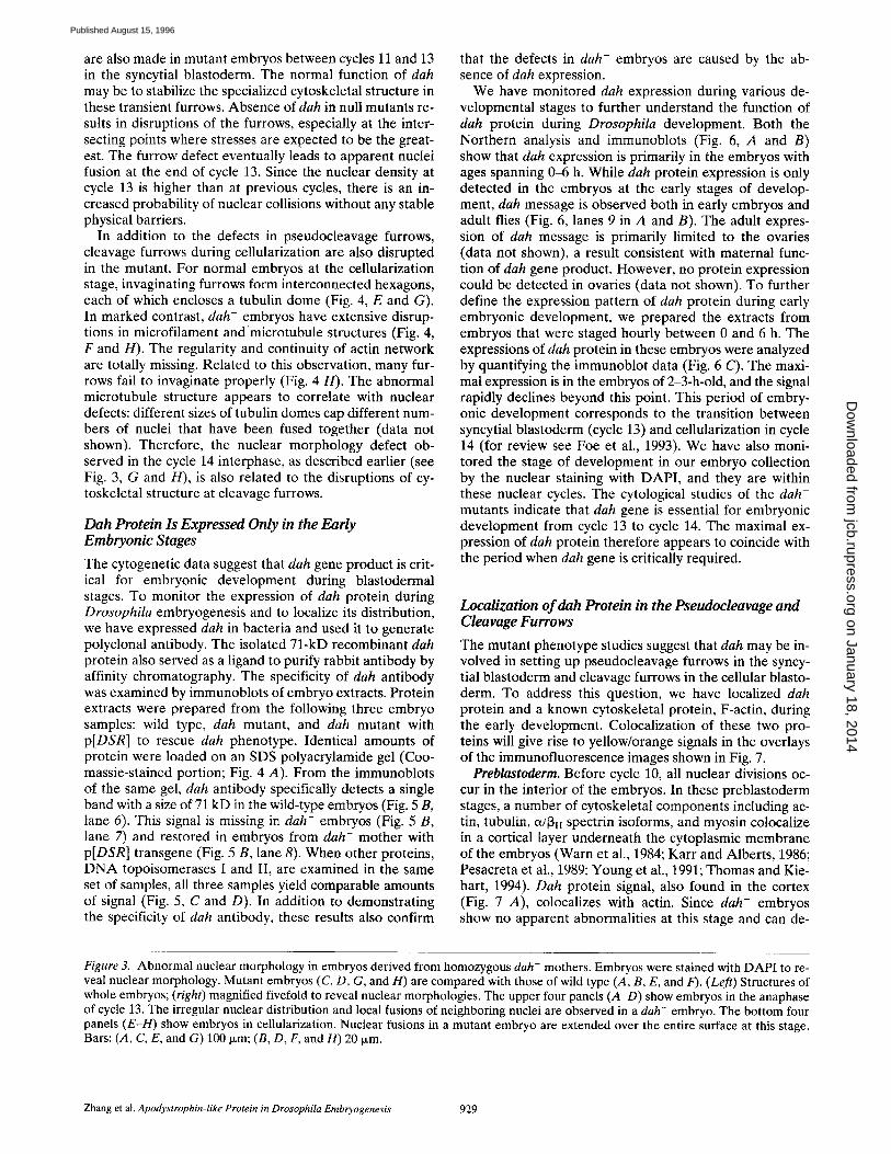
Figure 3. Abnormal nuclear morphology in embryos derived from homozygous dah- mothers. Embryos were stained with DAPI to re- veal nuclear morphology. Mutant embryos (C, D, G, and H) are compared with those of wild type (A, B, E, and F). (Left) Structures of whole embryos; (right) magnified fivefold to reveal nuclear morphologies. The upper four panels (A-D) show embryos in the anaphase of cycle 13. The irregular nuclear distribution and local fusions of neighboring nuclei are observed in a dah- embryo. The bottom four panels (E-H) show embryos in cellularization. Nuclear fusions in a mutant embryo are extended over the entire surface at this stage. Bars: (A, C, E, and G) 100 Ixm; (B, D, F, and H) 20 Ixm.
Zhang et al. Apodystrophin-like Protein in Drosophila Embryogenesis 929
on January 18, 2014jcb.rupress.org
Dow
nloaded from
Published August 15, 1996
velop into blas toderm, dah is not l ikely to play an impor- tant role in the early nuclear divisions and migration. The cortical dis tr ibut ion of dah in the p reb las toderm is proba- bly for s torage and for the prepara t ion of the arrival of nu- clei at the cortex during the b las toderm stage.
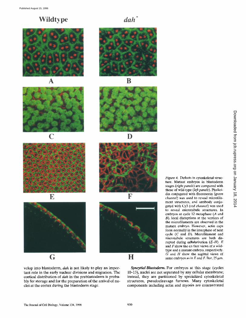
Figure 4. Defects in cytoskeletal struc- ture. Mutant embryos in blastoderm stages (right panels) are compared with those of wild type (left panels). Phalloi- din conjugated with fluorescein (green channel) was used to reveal microfila- ment structures, and antibody conju- gated with Cy3 (red channel) was used to reveal microtubule structures. In embryos at cycle 12 metaphase (A and B), local disruptions at the vertices of the microfilaments are observed in the mutant embryo. However, actin caps form normally in the interphase of next cycle (C and D). Microfilament and microtubule structures are both dis- rupted during cellularization (E-H). E and Fshow the en face views of a wild- type and a mutant embryo, respectively. G and H show the sagittal views of same embryos as in E and F. Bar, 25 Ixm.
Syncytial Blastoderm. For embryos at this stage (cycles 10-13), nuclei are not separated by any cellular membrane; instead, they are part i t ioned by specialized cytoskeletal structures, pseudocleavage furrows. Many cytoskeletal components including actin and myosin are concentra ted
The Journal of Cell Biology, Volume 134, 1996 930
on January 18, 2014jcb.rupress.org
Dow
nloaded from
Published August 15, 1996
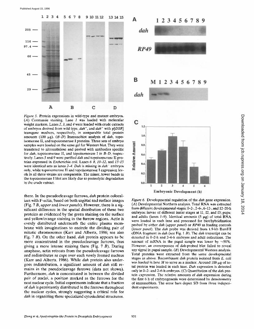
Figure 5. Protein expressions in wild-type and mutant embryos. (A) Coomassie staining. Lane 1 was loaded with molecular weight markers. Lanes 2, 3, and 4 were loaded with crude extracts of embryos derived from wild type, dah-, and dah- with p[DSR] transgene mothers, respectively, in comparable total protein amounts (100 p.g). (B-D) Immunoblot analysis of dah, topo- isomerase II, and topoisomerase I proteins. Three sets of embryo samples were loaded on the same gel for Western blot. They were transfered to nitrocellulose and probed with antibodies specific for dah, topoisomerase II, and topoisomerase I in B-D, respec- tively. Lanes 5 and 9 were purified dah and topoisomerase If pro- teins expressed in Escherichia coli. Lanes 6-8, 10-12, and 13-15 were identical sets as lanes 2--4. Dah is missing in dah- embryos only, while topoisomerase II and topoisomerase I expression lev- els in all three strains are comparable. The minor, lower bands in the topoisomerase I blot are likely due to proteolyfic degradation in the crude extract.
there. In the pseudocleavage furrows, dah prote in colocal- izes with F-actin, based on both sagittal and surface images (Fig. 7 B, upper and lower panels). However , there is a sig- nificant difference in the spatial dis tr ibut ion of these two prote ins as evidenced by the green staining on the surface and yel low/orange staining in the furrow regions. Act in is evenly dis t r ibuted undernea th the ent ire p lasma mem- brane with invaginations to encircle the dividing pair of mitot ic chromosomes (Karr and Alber ts , 1986; see also Fig. 7 B). On the o ther hand, dah prote in appears to be more concent ra ted in the pseudocleavage furrows, thus giving a more intense staining there (Fig. 7 B). Dur ing anaphase, actin re t reats from the pseudocleavage furrows and redistr ibutes as caps over each newly formed nucleus (Karr and Alber ts , 1986). Whi le dah prote in also under- goes redis t r ibut ion, a significant amount of dah still re- mains in the pseudocleavage furrows (data not shown). Fur thermore , dah is concent ra ted in be tween the divided pair of nuclei, a posi t ion marked as the furrows for the next nuclear cycle. Initial experiments indicate that a fraction of dah is persis tent ly dis t r ibuted in the furrows throughout the nuclear cycles, strongly suggesting a critical role for dah in organizing these specialized cytoskeletal structures.
Figure 6. Developmental regulation of the dah gene expression. (A) Developmental Northern analysis. Total RNA was extracted from different developmental stages: 0--2-, 2-6-, 6-12-, and 12-20-h embryos; larvae of different instar stages at I1, I2, and I3; pupa; and adults (lanes 1-9). Identical amounts (5 Ixg) of total RNA were loaded in each lane and processed for blot/hybridization probed by either dah (upper panel) or RP49 as loading controls (lower panel). The dah probe was derived from 1.9-kb BamHI eDNA fragment in dah (see Fig. 1 B). The dah transcript can be detected in 0-2-h and 2-6-h embryos and adult collections. The amount of mRNA in the pupal sample was lower by ~50%. However, an overexposure of dah-probed blot failed to reveal any signal in pupal sample. (B) Developmental Western analysis. Total proteins were extracted from the same developmental stages as above. Recombinant dah protein isolated from E. coli was loaded in lane M to serve as a marker. Around 100 ~g of to- tal protein was loaded in each lane. Dah expression is detected only in 0-2- and 2~-h embryos. (C) Quantitation of the dah pro- tein expression. The relative amounts of dah expression during the first 6 h of embryogenesis were determined by densitometry of immunoblots. The error bars depict SD from three indepen- dent experiments.
Zhang et al. Apodystrophin-like Protein in Drosophila Embryogenesis 931
on January 18, 2014jcb.rupress.org
Dow
nloaded from
Published August 15, 1996
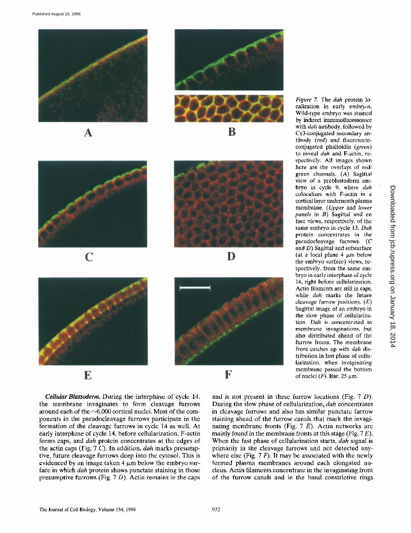
Figure 7. The dah protein lo- calization in early embryos. Wild-type embryo was stained by indirect immunofluorescence with dab antibody, followed by Cy3-conjugated secondary an- tibody (red) and fluorescein- conjugated phalloidin (green) to reveal dah and F-actin, re- spectively. All images shown here are the overlays of red/ green channels. (A) Sagittal view of a preblastoderm em- bryo in cycle 9, where dah colocalizes with F-actin in a cortical layer underneath plasma membrane. (Upper and lower panels in B) Sagittal and en face views, respectively, of the same embryo in cycle 13. Dah protein concentrates in the pseudocleavage furrows. (C and D) Sagittal and subsurface (at a focal plane 4 i~m below the embryo surface) views, re- spectively, from the same em- bryo in early interphase of cycle 14, right before cellularization. Actin filaments are still in caps, while dah marks the future cleavage furrow positions. (E) Sagittal image of an embryo in the slow phase of cellulariza- tion. Dab is concentrated in membrane invaginations, but also distributed ahead of the furrow fronts. The membrane front catches up with dah dis- tribution in fast phase of cellu- larization, when invaginating membrane passed the bottom of nuclei (F). Bar, 25 ~m.
Cellular Blastoderm. During the interphase of cycle 14, the membrane invaginates to form cleavage furrows around each of the ~6,000 cortical nuclei. Most of the com- ponents in the pseudocleavage furrows participate in the formation of the cleavage furrows in cycle 14 as well. At early interphase of cycle 14, before cellularization, F-actin forms caps, and dah protein concentrates at the edges of the actin caps (Fig. 7 C). In addition, dah marks presump- tive, future cleavage furrows deep into the cytosol. This is evidenced by an image taken 4 txm below the embryo sur- face in which dah protein shows punctate staining in those presumptive furrows (Fig. 7 D). Actin remains in the caps
and is not present in these furrow locations (Fig. 7 D). During the slow phase of cellularization, dah concentrates in cleavage furrows and also has similar punctate furrow staining ahead of the furrow canals that mark the invagi- nating membrane fronts (Fig. 7 E). Actin networks are mainly found in the membrane fronts at this stage (Fig. 7 E). When the fast phase of cellularization starts, dah signal is primarily in the cleavage furrows and not detected any- where else (Fig. 7 F). It may be associated with the newly formed plasma membranes around each elongated nu- cleus. Actin filaments concentrate in the invaginating front of the furrow canals and in the basal constrictive rings
The Journal of Cell Biology, Volume 134, 1996 932
on January 18, 2014jcb.rupress.org
Dow
nloaded from
Published August 15, 1996
(Fig. 7 F). Dah protein signal remains after cellularization finishes, and eventually disappears before the stage of slow germ band extension (data not shown).
Discussion
Drosophila early embryos provide a unique system to study the functions of the cytoskeleton in nuclear and cell division (Schejter and Wieschaus, 1993; Miller and Kie- hart, 1995). The pseudocleavage furrows in the syncytial blastoderm, cleavage furrows during cellularization, and contractile rings in cytokinesis share some common cy- toskeletal organization, although each has its distinct ele- ments. All the components in the pseudocleavage furrows are endowed by the mother in the form of either RNA or protein. Cleavage furrows inherit and share most compo- nents from the pseudocleavage furrows, with the addition of three zygotic genes, nullo, serendipitymt, and bottleneck (Merrill et al., 1988; Wieschaus and Sweeton, 1988; for re- view see Schejter and Wieschaus, 1993). It has been pro- posed that the maternal genes set up the furrow structure initially, and these zygotic gene products modify and re- model the cytoskeleton for the specific roles during cellu- larization (Schejter et al., 1992; Postner and Wieschaus, 1994).
We have isolated a new maternal effect mutation and demonstrated its gene product to be essential for the de- velopment of blastoderm. The dah gene product is prima- rily stored in the form of RNA by the mother, and the maximal protein expression is during the transition from syncytial to cellular blastoderm. Cytogenetic data and im- munolocalization experiments presented here show that dah protein is a critical component for both pseudocleav- age furrows in the syncytial blastoderm and cleavage fur- rows during the cellularization process. Furthermore, dah protein is also present in the presumptive furrows ahead of invaginating fronts before the fast phase of cellularization. It is possible that dah plays a critical role in the initiation of the cleavage furrow development. Cytological analysis of several maternal effect mutants has shown that their gene products are involved in the proper formation of pseudocleavage furrows. These maternal mutations, in- cluding sponge (Postner et al., 1992), dal (Sullivan et al., 1990), scrambled, and nuclear-fallout (Sullivan et al., 1993), disrupt cytoskeletal structures in early embryos, re- suiting in abnormal nuclear morphology. Similar to the dah mutant, the nuclear-fallout mutant has disrupted both pseudocleavage and cleavage furrows, while sponge, dal, and scrambled have abnormal furrows in the syncytial blastoderm but form nearly normal cytoskeletal structure at the cellularization stage. It is possible that sponge, dal, and scrambled are distinct elements involved in pseudo- cleavage furrows, while nuclear-fallout and dah are in- volved in both furrow formations.
Sequence search has revealed homology between dah and the COOH terminus of dystrophin and apodystro- phins. The COOH terminus of dystrophin is unique to a family of proteins, including dystrophin, dystrophin-related proteins, and apodystrophins. It is plausible that dah might share the same ancestral origin as the COOH terminus of dystrophin. The function of dystrophin is proposed to link the extracellular matrix through the membrane protein
complex to the actin-based cytoskeleton, therefore stabi- lizing the sarcolemma structure under contractile stress. Both biochemical and cell biological experiments suggest the association of dystrophin with sarcolemma membrane in adult muscles is mediated through glycoprotein com- plexes (for review see Ahn and Kunkel, 1993; Ervasti and Campbell, 1993). The glycoprotein binding sites have been mapped to the COOH-terminal domains by biochemical analysis of calpain-treated dystrophin (Suzuki et al., 1992). Transgenic experiments with apodystrophin-1 (Dp71) demonstrate that the COOH-terminal domains of dystro- phin can associate with sarcolemma membrane and orga- nize the dystroglycan complex formation (Cox et al., 1994). An 87-kD protein isolated from the postsynaptic membrane of the electric organ of electric eel also shows modest homology with apodystrophin-1 (Wagner et al., 1993). This protein is a component of the cytoskeletal structure at the neuromuscular junctions. It may serve to cluster and stabilize nicotinic acetylcholine receptor (But- ler et al., 1992; Wagner et al., 1993). While the homology between dah and the dystrophin COOH terminus is lim- ited, dah is localized in a specialized cytoskeletal structure, and initial fractionation data suggest it is present in the membrane preparations (data not shown). Therefore, the biochemical basis of dah functions might share some fea- tures with dystrophin and the family of apodystrophins. It is possible that dah serves to organize and stabilize the cy- toskeleton associated with embryonic cleavage furrows. Interestingly, in dah mutant embryos, disruptions of meta- phase furrows often happen around the vertices. These are points where one would expect the mechanical stress gen- erated by the dividing nuclei to be the greatest. Therefore, dah seems to be important for stabilizing the actin cytoskel- eton under stress, a role also shared by dystrophin in skeletal muscles.
The dah alleles we have isolated are null mutations. These dah mutants are phenotypically tight; no embryos from homozygous dah- mothers develop beyond cellular blastoderm. Our results demonstrate that dah is a compo- nent participating in the organization and maintenance of cortical furrows. While dah has an essential function in blastoderm development, it does not rule out other poten- tial functions later in development. Missing these other functions may not affect the viability of the fly. However, our immunochemical data suggest that dah protein is not present at significant levels in any developmental stages other than early embryos, thus arguing against this possi- bility. Dah is also unlikely to be critically involved in the conventional cytokinetic contractile rings since pole cells can bud off in the mutant embryos (data not shown), and the homozygous dah- embryo from a mother with a het- erozygous dah mutation can develop into an adult fly. Fu- ture analysis on the precise functions of dah in the cortical metaphase and cellutarization furrows should provide in- sights into the organization and regulation of these spe- cialized cytoskeletal structures.
We thank Prof. R. Fehon (Duke University) for sharing with us his proto- cols for immunofluorescence staining of whole-mount embryos.
This work is supported by a grant from the National Institutes of Health (GM29006).
Received for publication 2 April 1996 and in revised form 28 May 1996.
Zhang et al. Apodystrophin-like Protein in Drosophila Embryogenesis 933
on January 18, 2014jcb.rupress.org
Dow
nloaded from
Published August 15, 1996
References
Ahn, A.H., and L.M. Kunkel. 1993. The structural and functional diversity of dystrophin. Nat. Genet. 3:283-291.
Baker, J., W.E. Theurkauf, and G. Schubiger. 1993. Dynamic changes in micro- tubule configuration correlate with nuclear migration in the preblastoderm Drosophila embryo. J. Cell Biol. 122:113-21.
Blake, D.J., J.M. Tinsley, and K.E. Davies. 1994. The emerging family of dys- trophin-related proteins. Trends Cell Biol. 4:19-23.
Blake, D.J., J.M. Tinsley, K.E. Davies, A.E. Knight, S.J. Winder, and J. Ken- drick-Jones. 1995. Coiled-coil regions in the carboxy-terminal domains of dystrophin and related proteins: potentials for protein-protein interactions. Trends Biochem. Sci. 20:133-135.
Butler, M.H., K. Douville, A.A. Murnane, N.R. Kramarcy, J.B. Cohen, R. Seal- ock, and S.C. Froehner. 1992. Association of the M r 58,000 postsynaptic pro- tein of electric tissue with Torpedo dystrophin and the Mr 87,000 postsynap- tic protein. J. Biol. Chem. 267:6213-6218.
Callaini, G., R. Dallai, and M.G. Riparbelli. 1992. Cytnchalasin induces spindle fusion in the syncytial blastoderm of the early Drosophila embryo. Biol. Cell. 74:24%254.
Cavener, D.R. 1987. Comparison of the consensus sequence flanking transla- tional start sites in Drosophila and vertebrates. Nucleic Acids Res. 15:1353- 1361.
Cox, G.A., Y. Sunada, K.P. Campbell, and J.S. Chamberlain. 1994. Dp71 can restore the dystrophin-associated glycoprotein complex in muscle but fails to prevent dystrophy. Nat. Genet. 8:333-339.
de Cicco, D.V., and A.C. Spradling. 1984. Localization of a cis-acting element responsible for the developmentally regulated amplification of Drosophila chorion genes. Cell. 38:45-54.
Ervasti, J.M., and K.P. Campbell. 1993. Dystrophin and the membrane skele- ton. Curr. Opin. Cell Biol. 5:82--87.
Field, C.M., and B.M. Alberts. 1995. Anillin, a contractile ring protein that cy- cles from the nucleus to the cell cortex. J. Cell Biol. 131:165-178.
Foe, V.E., and B.M. Alberts. 1983. Studies of nuclear and cytoplasmic behavior during the five mitotic cycles that precede gastrulation in Drosophila em- bryogenesis. J. Cell Sci. 61:31-70.
Foe, V.E., G.M. Odell, and B.A. Edgar. 1993. Mitosis and morphogenesis in the Drosophila embryo: point and counterpoint. In The Development of Dro- sophila melanogaster. Vol. 1. M. Bate and A.M. Arias, editors. Cold Spring Harbor Laboratory, Cold Spring Harbor, NY. 149-300.
Fyrberg, E.A., and L.S. Goldstein. 1990. The Drosophila cytoskeleton. Annu. Rev. Cell Biol. 6:559--596.
Hatanaka, K., and M. Okada. 1991. Retarded nuclear migration in Drosophila embryos with aberrant F-actin reorganization caused by maternal mutations and by cytochalasin treatment. Development (Camb.). 111:909-920.
Karess, R.E., XJ. Chang, K.A. Edwards, S. Kulkarni, I. Aguilera, and D.P. Kie- hart. 1991. The regulatory light chain of nonmuscle myosin is encoded by spaghetti-squash, a gene required for cytokinesis in Drosophila. Cell. 65: 1177-1189.
Karr, T.L., and B.M. Alberts. 1986. Organization of the cytoskeleton in early Drosophila embryos. J. Cell Biol. 102:1494-1509.
Kellogg, D.R., T.J. Mitchison, and B.M. Alberts. 1988. Behaviour of microtu- bules and actin filaments in living Drosophila embryos. Development (Camb.). 103:675-686.
Knight, A.E. 1994. The diversity of myosin-like proteins; Ph.D. dissertation. University of Cambridge. 254 pp.
Koenig, M., A.P. Monaco, and L.M. Kunkel. 1988. The complete sequence of dystrophin predicts a rod-shaped cytoskeletal protein. Cell. 53:219-226.
Lee, M.P., S.D. Brown, A. Chen, and T.S. Hsieh. 1993. DNA topoisomerase I is essential in Drosophila melanogaster. Proc. Natl. Acad. Sci. USA. 90:6656- 6660.
Lupas, A., M. Van Dyke, and J. Stock. 1991. Predicting coiled coils from pro- tein sequences. Science (Wash. DC) 252:1162-1164.
Mermall, V., and K.G. Miller. 1995. The 95F unconventional myosin is required for proper organization of the Drosophila syncytial blastoderm. ]. Cell Biol. 129:1575-1588.
Merrill, P.T., D. Sweeton, and E. Wieschaus. 1988. Requirements for autosomal gene activity during preceUular stages of Drosophila melanogaster. Develop- ment (Camb.) 104:495-509.
Miller, K.G., and D.P. Kiehart. 1995. Fly division. J. Cell Biol. 131:1-5. Miller, K.G., C.M. Field, and B.M. Alberts. 1989. Actin-binding proteins from
Drosophila embryos: a complex network of interacting proteins detected by F-actin affinity chromatography. J. Cell Biol. 109:2963-2975.
Mount, S.M., C. Burks, G. Hertz, G.D. Stormo, O. White, and C. Fields. 1992. Splicing signals in Drosophila: intron size, information content, and consen- sus sequences. Nucleic Acids Res. 20:4255-4262.
Nolan, J.M., M.P. Lee, E. Wyckoff, and T.S. Hsieh. 1986. Isolation and charac- terization of the gene encoding Drosophila DNA topoisomerase II. Proc. Natl. Acad. Sci. USA. 83:3664-3668.
Pesacreta, T.C., T.J. Byers, R. Dubreuil, D.P. Kiehart, and D. Branton. 1989. Drosophila spectrin: the membrane skeleton during embryogenesis. Z Cell Biol. 108:1697-1709.
Postner, M.A., and E.F. Wieschaus. 1994. The nullo protein is a component of the actin-myosin network that mediates cellularization in Drosophila melan- ogaster embryos. Z Cell Sci. 107:1863-1873.
Postner, M.A., K.G. Miller, and E.F. Wieschaus. 1992. Maternal effect muta- tions of the sponge locus affect actin cytoskeletal rearrangements in Dro- sophila melanogaster embryos. J. Cell BioL 119:1205-1218.
Rabinowitz, M. 1941. Studies on the cytology and early embryology of the egg of Drosophila melanogaster. J. Morphol. 69:1-49.
Roberts, D.B. 1986. Drosophila: a practical approach. In Practical Approach Series. D. Rickwood and B.D. Hames, editors. IRL Press, Oxford, UK. 59~1.
Robertson, H.M., C.R. Preston, R.W. Phillis, D.M. Johnson-Schlitz, W.K. Benz, and W.R. Engels. 1988. A stable genomic source of P element transposase in Drosophila melanogaster Genetics. 118:461-470.
Sambrook, J., E.F. Fritsch, and T. Maniatis. 1989. Molecular Cloning: A Labo- ratory Manual. Cold Spring Harbor Laboratory, Cold Spring Harbor, NY. 545 pp.
Schejter, E.D., and E. Wieschaus. 1993. Functional elements of the cytoskele- ton in the early Drosophila embryo. Annu. Rev. Cell Biol. 9:67-99.
Schejter, E.D., L.S. Rose, M.A. Postner, and E. Wieschaus. 1992. Role of the zygotic genome in the restructuring of the actin cytoskeleton at the cycle-14 transition during Drosophila embryogenesis. Cold Spring Harbor Syrup. Quant. Biol. 57:653-659.
Sheets, M.D., S.C. Ogg, and M.P. Wickens. 1990. Point mutations in AAUAAA and the poly (A) addition site: effects on the accuracy and effi- ciency of cleavage and polyadenylation in vitro. Nucleic Acids Res. 18:579% 5805.
Studier, F.W., A.H. Rosenberg, JJ. Dunn, and J.W. Dubendorff. 1990. Use of T7 RNA polymerase to direct expression of cloned genes. Methods Enzy- mol. 185:60-89.
Sullivan, W., J.S. Minden, and B.M. Alberts. 1990. daughterless-abo-like, a Drosophila maternal-effect mutation that exhibits abnormal centrosome separation during the late blastoderm divisions. Development (Camb.). 110: 311-323.
Sullivan, W., P. Fogarty, and W. Theurkauf. 1993. Mutations affecting the cy- toskeletal organization of syncytial Drosophila embryos. Development (Camb.). 118:1245-1254.
Suzuki, A., M. Yoshida, H. Yamamoto, and E. Ozawa. 1992. Glycoprotein- binding site of dystrophin is confined to the cysteine-rich domain and the first half of the carboxy-terminal domain. FEBS Lett. 308:154-260.
Thomas, G,H., and D.P. Kiehart. 1994. Beta heavy-spectrin has a restricted tis- sue and subcellular distribution during Drosophila embryogenesis. Develop- ment (Camb.). 120:2039-2050.
Wagner, K.R., J.B. Cohen, and R.L. Huganir. 1993. The 87K postsynaptic membrane protein from Torpedo is a protein-tyrosine kinase substrate ho- mologous to dystrophin. Neuron. 10:511-522.
Warn, R.M., R. Magrath, and S. Webb. 1984. Distribution of F-actin during cleavage of the Drosophila syncytial blastoderm. J. Cell Biol. 98:156-162.
Wieschaus, E., and D. Sweeton. 1988. Requirements for X-linked zygotic gene activity during cellularization of early Drosophila embryos. Development (Camb.). 104:483-493.
Young, P.E., T.C. Pesacreta, and D.P. Kiehart. 1991. Dynamic changes in the distribution of cytoplasmic myosin during Drosophila embryogenesis. Devel- opment (Camb.). 111:1-14.
Zalokar, M. 1976. Autoradiographic study of protein and RNA formation dur- ing early development of Drosophila eggs. Dev. Bibl. 49:425-437.
The Journal of Celt Biology, Volume 134, 1996 934
on January 18, 2014jcb.rupress.org
Dow
nloaded from
Published August 15, 1996