Transit Peptide Cleavage Sites of Integral Thylakoid Membrane Proteins
18
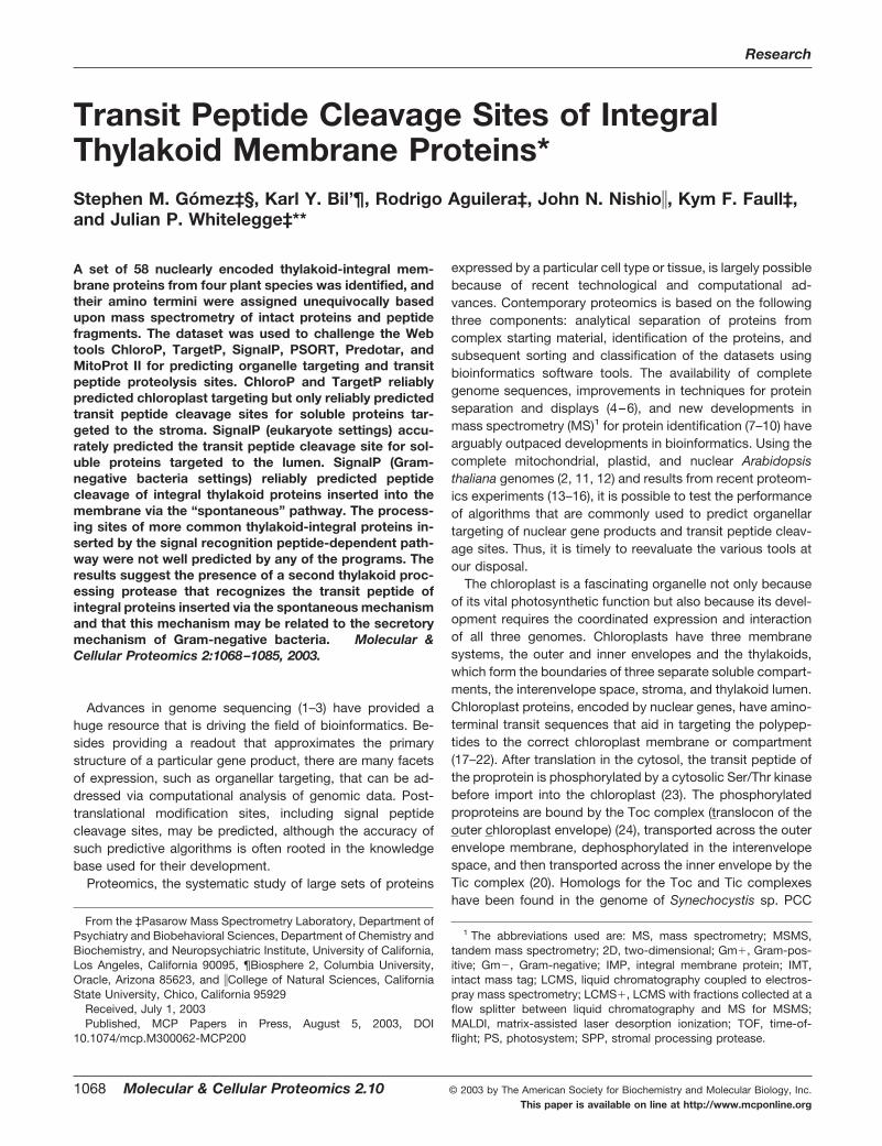
Transit Peptide Cleavage Sites of Integral Thylakoid Membrane Proteins* Stephen M. Go ´ mez‡§, Karl Y. Bil’¶, Rodrigo Aguilera‡, John N. Nishio, Kym F. Faull‡, and Julian P. Whitelegge‡** A set of 58 nuclearly encoded thylakoid-integral mem- brane proteins from four plant species was identified, and their amino termini were assigned unequivocally based upon mass spectrometry of intact proteins and peptide fragments. The dataset was used to challenge the Web tools ChloroP, TargetP, SignalP, PSORT, Predotar, and MitoProt II for predicting organelle targeting and transit peptide proteolysis sites. ChloroP and TargetP reliably predicted chloroplast targeting but only reliably predicted transit peptide cleavage sites for soluble proteins tar- geted to the stroma. SignalP (eukaryote settings) accu- rately predicted the transit peptide cleavage site for sol- uble proteins targeted to the lumen. SignalP (Gram- negative bacteria settings) reliably predicted peptide cleavage of integral thylakoid proteins inserted into the membrane via the “spontaneous” pathway. The process- ing sites of more common thylakoid-integral proteins in- serted by the signal recognition peptide-dependent path- way were not well predicted by any of the programs. The results suggest the presence of a second thylakoid proc- essing protease that recognizes the transit peptide of integral proteins inserted via the spontaneous mechanism and that this mechanism may be related to the secretory mechanism of Gram-negative bacteria. Molecular & Cellular Proteomics 2:1068 –1085, 2003. Advances in genome sequencing (1–3) have provided a huge resource that is driving the field of bioinformatics. Be- sides providing a readout that approximates the primary structure of a particular gene product, there are many facets of expression, such as organellar targeting, that can be ad- dressed via computational analysis of genomic data. Post- translational modification sites, including signal peptide cleavage sites, may be predicted, although the accuracy of such predictive algorithms is often rooted in the knowledge base used for their development. Proteomics, the systematic study of large sets of proteins expressed by a particular cell type or tissue, is largely possible because of recent technological and computational ad- vances. Contemporary proteomics is based on the following three components: analytical separation of proteins from complex starting material, identification of the proteins, and subsequent sorting and classification of the datasets using bioinformatics software tools. The availability of complete genome sequences, improvements in techniques for protein separation and displays (4 – 6), and new developments in mass spectrometry (MS) 1 for protein identification (7–10) have arguably outpaced developments in bioinformatics. Using the complete mitochondrial, plastid, and nuclear Arabidopsis thaliana genomes (2, 11, 12) and results from recent proteom- ics experiments (13–16), it is possible to test the performance of algorithms that are commonly used to predict organellar targeting of nuclear gene products and transit peptide cleav- age sites. Thus, it is timely to reevaluate the various tools at our disposal. The chloroplast is a fascinating organelle not only because of its vital photosynthetic function but also because its devel- opment requires the coordinated expression and interaction of all three genomes. Chloroplasts have three membrane systems, the outer and inner envelopes and the thylakoids, which form the boundaries of three separate soluble compart- ments, the interenvelope space, stroma, and thylakoid lumen. Chloroplast proteins, encoded by nuclear genes, have amino- terminal transit sequences that aid in targeting the polypep- tides to the correct chloroplast membrane or compartment (17–22). After translation in the cytosol, the transit peptide of the proprotein is phosphorylated by a cytosolic Ser/Thr kinase before import into the chloroplast (23). The phosphorylated proproteins are bound by the Toc complex (translocon of the outer chloroplast envelope) (24), transported across the outer envelope membrane, dephosphorylated in the interenvelope space, and then transported across the inner envelope by the Tic complex (20). Homologs for the Toc and Tic complexes have been found in the genome of Synechocystis sp. PCC From the ‡Pasarow Mass Spectrometry Laboratory, Department of Psychiatry and Biobehavioral Sciences, Department of Chemistry and Biochemistry, and Neuropsychiatric Institute, University of California, Los Angeles, California 90095, ¶Biosphere 2, Columbia University, Oracle, Arizona 85623, and College of Natural Sciences, California State University, Chico, California 95929 Received, July 1, 2003 Published, MCP Papers in Press, August 5, 2003, DOI 10.1074/mcp.M300062-MCP200 1 The abbreviations used are: MS, mass spectrometry; MSMS, tandem mass spectrometry; 2D, two-dimensional; Gm, Gram-pos- itive; Gm, Gram-negative; IMP, integral membrane protein; IMT, intact mass tag; LCMS, liquid chromatography coupled to electros- pray mass spectrometry; LCMS, LCMS with fractions collected at a flow splitter between liquid chromatography and MS for MSMS; MALDI, matrix-assisted laser desorption ionization; TOF, time-of- flight; PS, photosystem; SPP, stromal processing protease. Research © 2003 by The American Society for Biochemistry and Molecular Biology, Inc. 1068 Molecular & Cellular Proteomics 2.10 This paper is available on line at http://www.mcponline.org
-
Upload
independent -
Category
Documents
-
view
1 -
download
0
Transcript of Transit Peptide Cleavage Sites of Integral Thylakoid Membrane Proteins
Transit Peptide Cleavage Sites of IntegralThylakoid Membrane Proteins*Stephen M. Gomez‡§, Karl Y. Bil’¶, Rodrigo Aguilera‡, John N. Nishio�, Kym F. Faull‡,and Julian P. Whitelegge‡**
A set of 58 nuclearly encoded thylakoid-integral mem-brane proteins from four plant species was identified, andtheir amino termini were assigned unequivocally basedupon mass spectrometry of intact proteins and peptidefragments. The dataset was used to challenge the Webtools ChloroP, TargetP, SignalP, PSORT, Predotar, andMitoProt II for predicting organelle targeting and transitpeptide proteolysis sites. ChloroP and TargetP reliablypredicted chloroplast targeting but only reliably predictedtransit peptide cleavage sites for soluble proteins tar-geted to the stroma. SignalP (eukaryote settings) accu-rately predicted the transit peptide cleavage site for sol-uble proteins targeted to the lumen. SignalP (Gram-negative bacteria settings) reliably predicted peptidecleavage of integral thylakoid proteins inserted into themembrane via the “spontaneous” pathway. The process-ing sites of more common thylakoid-integral proteins in-serted by the signal recognition peptide-dependent path-way were not well predicted by any of the programs. Theresults suggest the presence of a second thylakoid proc-essing protease that recognizes the transit peptide ofintegral proteins inserted via the spontaneous mechanismand that this mechanism may be related to the secretorymechanism of Gram-negative bacteria. Molecular &Cellular Proteomics 2:1068–1085, 2003.
Advances in genome sequencing (1–3) have provided ahuge resource that is driving the field of bioinformatics. Be-sides providing a readout that approximates the primarystructure of a particular gene product, there are many facetsof expression, such as organellar targeting, that can be ad-dressed via computational analysis of genomic data. Post-translational modification sites, including signal peptidecleavage sites, may be predicted, although the accuracy ofsuch predictive algorithms is often rooted in the knowledgebase used for their development.
Proteomics, the systematic study of large sets of proteins
expressed by a particular cell type or tissue, is largely possiblebecause of recent technological and computational ad-vances. Contemporary proteomics is based on the followingthree components: analytical separation of proteins fromcomplex starting material, identification of the proteins, andsubsequent sorting and classification of the datasets usingbioinformatics software tools. The availability of completegenome sequences, improvements in techniques for proteinseparation and displays (4–6), and new developments inmass spectrometry (MS)1 for protein identification (7–10) havearguably outpaced developments in bioinformatics. Using thecomplete mitochondrial, plastid, and nuclear Arabidopsisthaliana genomes (2, 11, 12) and results from recent proteom-ics experiments (13–16), it is possible to test the performanceof algorithms that are commonly used to predict organellartargeting of nuclear gene products and transit peptide cleav-age sites. Thus, it is timely to reevaluate the various tools atour disposal.
The chloroplast is a fascinating organelle not only becauseof its vital photosynthetic function but also because its devel-opment requires the coordinated expression and interactionof all three genomes. Chloroplasts have three membranesystems, the outer and inner envelopes and the thylakoids,which form the boundaries of three separate soluble compart-ments, the interenvelope space, stroma, and thylakoid lumen.Chloroplast proteins, encoded by nuclear genes, have amino-terminal transit sequences that aid in targeting the polypep-tides to the correct chloroplast membrane or compartment(17–22). After translation in the cytosol, the transit peptide ofthe proprotein is phosphorylated by a cytosolic Ser/Thr kinasebefore import into the chloroplast (23). The phosphorylatedproproteins are bound by the Toc complex (translocon of theouter chloroplast envelope) (24), transported across the outerenvelope membrane, dephosphorylated in the interenvelopespace, and then transported across the inner envelope by theTic complex (20). Homologs for the Toc and Tic complexeshave been found in the genome of Synechocystis sp. PCC
From the ‡Pasarow Mass Spectrometry Laboratory, Department ofPsychiatry and Biobehavioral Sciences, Department of Chemistry andBiochemistry, and Neuropsychiatric Institute, University of California,Los Angeles, California 90095, ¶Biosphere 2, Columbia University,Oracle, Arizona 85623, and �College of Natural Sciences, CaliforniaState University, Chico, California 95929
Received, July 1, 2003Published, MCP Papers in Press, August 5, 2003, DOI
10.1074/mcp.M300062-MCP200
1 The abbreviations used are: MS, mass spectrometry; MSMS,tandem mass spectrometry; 2D, two-dimensional; Gm�, Gram-pos-itive; Gm�, Gram-negative; IMP, integral membrane protein; IMT,intact mass tag; LCMS, liquid chromatography coupled to electros-pray mass spectrometry; LCMS�, LCMS with fractions collected at aflow splitter between liquid chromatography and MS for MSMS;MALDI, matrix-assisted laser desorption ionization; TOF, time-of-flight; PS, photosystem; SPP, stromal processing protease.
Research
© 2003 by The American Society for Biochemistry and Molecular Biology, Inc.1068 Molecular & Cellular Proteomics 2.10This paper is available on line at http://www.mcponline.org

6803 (20), which supports the model that chloroplasts arereduced cyanobacterial endosymbionts (25–28). Once in thestroma the transit peptide is cleaved by a stromal processingprotease (SPP).
Proteins targeted to the thylakoid lumen have bipartite tran-sit peptides. The stromal targeting information is in the amino-proximal portion of the transit peptide, and the carboxyl-proximal portion, similar to signal peptides of secretedproteins in bacteria, contains the information for targeting tothe lumen. Two different pathways for targeting to the lumenhave been characterized. The first is a Sec-dependent path-way related to the SecYEG export mechanism in bacteria (29,30). The second is a �pH-dependent mechanism character-ized by two conserved sequential arginines in the transit pep-tide, the Tat (twin arginine translocase) pathway (31–33). Pro-teins targeted to the lumen via the Sec pathway are generallytranslocated in an unfolded state, whereas proteins importedvia the Tat pathway are translocated in a folded state (22).Proteins imported into the lumen by either pathway are pro-cessed by a thylakoid processing protease that removes thecarboxyl-proximal portion of the transit peptide.
Integral membrane proteins (IMPs) destined for the thyla-koid membrane are targeted by one of two additional trans-location mechanisms. It is thought that most thylakoid-boundproteins are targeted by information contained within the ma-ture protein sequence. This insertion mechanism, which ori-ents the amino terminus of the protein into the stroma, re-quires a signal recognition particle and a putative unidentifiedthylakoid-bound translocation complex (34–37). A few inte-gral thylakoid proteins have bipartite transit peptides that arecleaved first in the stroma and again after membrane inser-tion. These proteins are inserted via a novel signal recognitionparticle-independent mechanism that appears to require noprotein or nucleotide co-factors (the spontaneous pathway)and has the unique characteristic of leaving the amino termi-nus of the mature protein oriented into the lumen (38–42).Chloroplast thylakoids have been used as a model system formembrane proteomics because of the size of the proteome,the limited number of post-translational modifications (16), theease of collecting large amounts of membranes, and theability to easily subfractionate detergent-resistant domains(43).
Proteomics studies using two-dimensional (2D) gel electro-phoresis have provided useful insights for soluble proteins ofthe lumen and stroma as well as peripheral thylakoid proteins(13, 44–47) and integral thylakoid proteins (48), but thesemethods do not allow convenient determination of the aminoterminus where blocked, as is common in the chloroplast. Inthis work, a dataset of 35 nuclear encoded integral thylakoidmembrane proteins from A. thaliana, where the sites of sec-ondary amino-terminal processing were explicitly determinedby MS, was used to challenge several bioinformatics tools.After initial trials the dataset was expanded to include photo-system (PS) II thylakoid-associated proteins from pea and
spinach published previously (16, 49) and a small number oftobacco IMPs, providing a total of 58 nuclear encoded inte-gral thylakoid membrane proteins, each with an experimen-tally determined amino terminus. The results demonstrate thatalthough some programs (ChloroP, TargetP) reliably predicttrafficking to the chloroplast, they variably predict the proc-essing sites of the transit peptides. The results highlight in-adequacies of currently available bioinformatics tools for pre-diction of secondary amino-terminal processing of transitpeptides of integral thylakoid proteins and demonstrate theneed for improvements in the datasets used to train thealgorithms.
MATERIALS AND METHODS
Plant Growth Conditions—A. thaliana var. Columbia seeds weresoaked in water (4 °C) for 1–2 d before sowing. Seeds were sown intrays, and seedlings were transferred to 144 cm2 � 12 cm high pots(�5 plants/pot) containing Scotts Pro-Gro professional potting mix(The Scotts Company, Marysville, OH) approximately 1 week aftergermination. Plants were grown in a growth chamber maintained at23 � 2 °C in the light and 16 � 2 °C in the dark. The plants wereilluminated with a 12-h light period and constant relative humidity of70%. Fluorescent lamps (Sylvania F72T12/CW/VHO, 160 watts) sup-plemented with Sylvania 100-W incandescent bulbs were used as thelight source. Quantum (400–700 nm) flux density was 700–800 �molof photons m�2 s�1 at the level of the leaves. Plants were watered asnecessary and supplemented once per week with 0.25� Hoagland’ssolution (50).
Leaves were harvested from 5-week-old A. thaliana plants, placedin square glass beakers, covered with grinding buffer (51), and groundwith a chilled Polytron homogenizer (Brinkmann Instruments/Kine-matica). The homogenate was filtered over 8 layers of diaper liners(Gerber), and the green filtrate was layered over (40%) Percoll gradi-ents in 1.6-ml microfuge tubes (52). Thylakoids were collected fromthe Percoll pads, diluted in grinding buffer, and pelleted at full speedin a tabletop centrifuge. The green membrane pellets were resus-pended to �1.1 mg/ml chlorophyll in extraction buffer (51) and frozenin liquid nitrogen. This procedure typically required 10–15 min, and allmanipulations were performed on ice or in a 4 °C refrigerator underlow light. Tobacco PS II thylakoid proteins were isolated as describedpreviously (16).
LCMS�—A. thaliana thylakoid protein samples (�66 �g of chloro-phyll) were prepared by acetone precipitation prior to dissolution in60% acetic acid. Reverse-phase chromatography was performed asdescribed previously (5, 49) using a poly(styrene-divinylbenzene) co-polymer (Polymer Labs PLRP/S, 5 �m � 300 Å, 2.1 � 300 mm)stationary-phase column equilibrated in aqueous 0.1% trifluoroaceticacid containing 5% acetonitrile and eluted (100 �l/min at 40 °C) witha stepped increasing concentration of acetonitrile (min/% acetonitrile:0/5, 5/5, 10/25, 130/75, and 150/100) initiated at the moment ofsample injection (100 �l/injection). The absorption of the effluent fromthe column was recorded at 280 nm. The effluent was split, and aportion was sent to a fraction collector (typically about 75%, 2-minfractions); the remainder was directed straight into the MS ion source(LCMS�). Tobacco protein samples were measured by LCMS asdescribed previously (16).
Mass spectra were recorded on a PerkinElmer Life Sciences SciexAPI III triple-quadrupole mass spectrometer with an IonsprayTM
source as described by Whitelegge et al. (5). The instrument wasscanned from m/z 600–2300 (0.3 step size, 1-ms dwell time, 6-s scanspeed, orifice at 65 V). Manufacturer-supplied software was used forthe computations of measured protein molecular weight (MacSpec
Thylakoid Membrane Transit Peptide Cleavage Sites
Molecular & Cellular Proteomics 2.10 1069
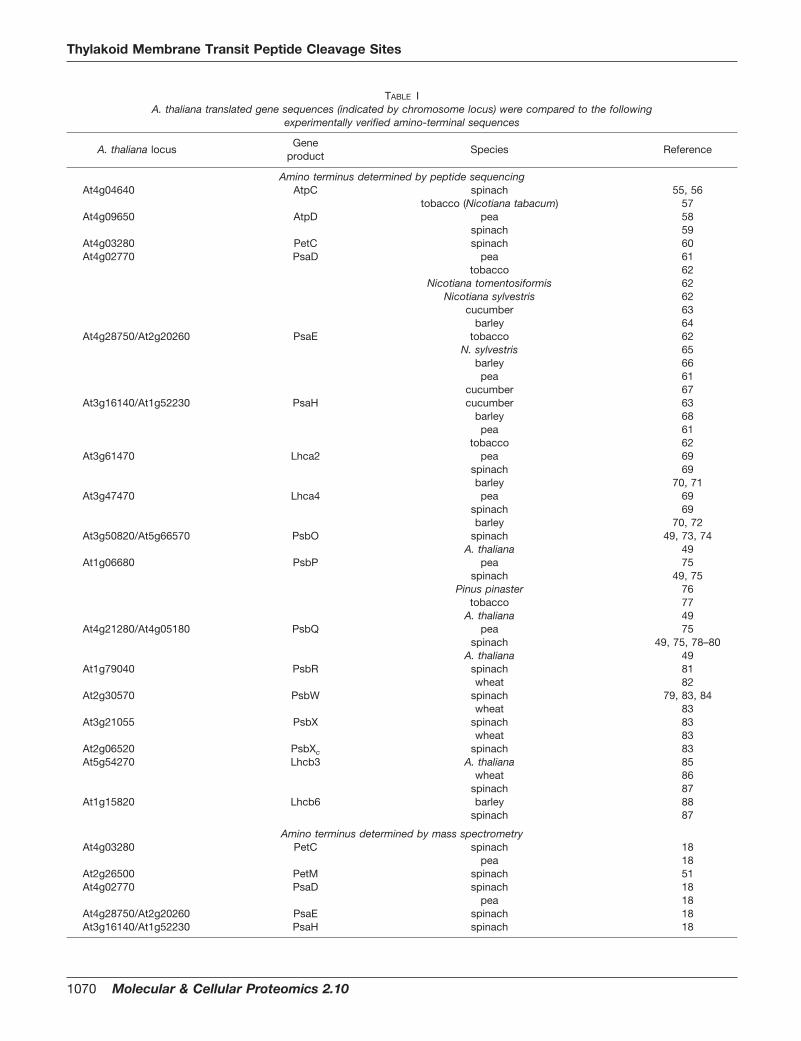
TABLE IA. thaliana translated gene sequences (indicated by chromosome locus) were compared to the following
experimentally verified amino-terminal sequences
A. thaliana locusGene
productSpecies Reference
Amino terminus determined by peptide sequencingAt4g04640 AtpC spinach 55, 56
tobacco (Nicotiana tabacum) 57At4g09650 AtpD pea 58
spinach 59At4g03280 PetC spinach 60At4g02770 PsaD pea 61
tobacco 62Nicotiana tomentosiformis 62
Nicotiana sylvestris 62cucumber 63
barley 64At4g28750/At2g20260 PsaE tobacco 62
N. sylvestris 65barley 66pea 61
cucumber 67At3g16140/At1g52230 PsaH cucumber 63
barley 68pea 61
tobacco 62At3g61470 Lhca2 pea 69
spinach 69barley 70, 71
At3g47470 Lhca4 pea 69spinach 69barley 70, 72
At3g50820/At5g66570 PsbO spinach 49, 73, 74A. thaliana 49
At1g06680 PsbP pea 75spinach 49, 75
Pinus pinaster 76tobacco 77
A. thaliana 49At4g21280/At4g05180 PsbQ pea 75
spinach 49, 75, 78–80A. thaliana 49
At1g79040 PsbR spinach 81wheat 82
At2g30570 PsbW spinach 79, 83, 84wheat 83
At3g21055 PsbX spinach 83wheat 83
At2g06520 PsbXc spinach 83At5g54270 Lhcb3 A. thaliana 85
wheat 86spinach 87
At1g15820 Lhcb6 barley 88spinach 87
Amino terminus determined by mass spectrometryAt4g03280 PetC spinach 18
pea 18At2g26500 PetM spinach 51At4g02770 PsaD spinach 18
pea 18At4g28750/At2g20260 PsaE spinach 18At3g16140/At1g52230 PsaH spinach 18
Thylakoid Membrane Transit Peptide Cleavage Sites
1070 Molecular & Cellular Proteomics 2.10

3.3) and zero-charge molecular weight reconstructions (BioMultiView1.3.1). Calculated average molecular weights were generated fromtranslated gene sequences using PeptideMass (expasy.cbr.nrc.ca/tools/peptide-mass.html) after manually removing the transit peptide.
Protein Identification—Thirty-three intact mass tags (IMTs) from theA. thaliana LCMS experiments were correlated with the predictedmasses calculated by modifying the entries at The Institute forGenomic Research A. thaliana database (www.tigr.org/tdb/e2k1/ath1/) with experimentally verified amino termini of orthologs (Table I).
Cyanogen Bromide Cleavage—Aliquots of fractions (10 �l) col-lected during LCMS� were treated with saturated cyanogen bromide(CNBr) (1 �l, 1 g/ml) for 4 h at room temperature in the dark. Thereaction mixture was either spotted directly (0.3 �l plus 0.5 �l ofmatrix solution) or dried by centrifugal evaporation (SpeedVac). Driedreaction mixtures were redissolved in 5 �l of 70% acetic acid andanalyzed (0.2 �l plus 0.5 �l of matrix) by matrix-assisted laser des-orption ionization (MALDI) coupled to delayed extraction time-of-flightMS in the reflector mode (Voyager DE STR, Applied Biosystems)using �-cyano-4-hydroxycinnamic acid as matrix (10 mg/ml of solu-tion in water/acetonitrile/trifluoroacetic acid 30/70/0.1) and internal/external calibration with bovine insulin. Manufacturer-supplied defaultsettings for a method optimized for peptides less than 6000 Da wereused for all samples. Some proteins have no internal Met or no CNBrpeptides less than 8000 Da (MALDI in the reflectron mode has anupper limit for high resolution data acquisition of approximately 8000Da); consequently some abundant proteins will not be detected dur-ing MALDI analysis, and peptides from low abundance proteins mayappear to be more highly expressed. The MALDI data was comparedwith the fragmentation pattern predicted by the MS-Tag tool in Pro-teinProspector, version 4.0.4 (prospector.ucsf.edu/) (90). MS-Tag cal-culates the predicted fragment masses with the transit peptide se-quence still included. There are very few, if any, amino-terminalpeptide masses for chloroplast-targeted proteins predicted by MS-Tag. The MS-Digest tool was used to predict the fragment massesafter manually removing the transit peptide from each prospectivematch assigned by MS-Tag.
Trypsin Cleavage—Selected fractions collected during LCMS�were reduced, alkylated, and treated with trypsin (Promega sequenc-
ing grade modified by reductive methylation). Dithiothreitol (15 �l, 10mM in 50 mM ammonium bicarbonate; 30 min, 24 °C), iodoacetamide(15 �l, 55 mM in 50 mM ammonium bicarbonate; 20 min, 24 °C), andfinally trypsin (12.5 �l, 6 ng/�l in 50 mM ammonium bicarbonate; 3 h,37 °C) were added to aliquots of fractions (10 �l). After incubation,samples were dried by centrifugal evaporation and stored at �20 °Cprior to analysis by microliquid chromatography-MSMS.
Analysis of Tryptic Peptide Sequence Tags by Tandem Mass Spec-trometry—Samples were analyzed by microliquid chromatography-MSMS with data-dependent acquisition (LCQ-DECA, ThermoFinni-gan, San Jose, CA) after dissolution in 5 �l of 70% acetic acid (v/v).A reverse-phase column (200 �m � 10 cm, PLRP/S 5 �m, 300 Å;Michrom Biosciences, San Jose, CA) was equilibrated for 10 min at1.5 �l/min with 95% A, 5% B (A, 0.1% formic acid in water; B, 0.1%formic acid in acetonitrile) prior to sample injection. A linear gradientwas initiated 10 min after sample injection ramping to 60% A, 40% Bafter 50 min and 20% A, 80% B after 65 min. Column eluent wasdirected to a coated glass electrospray emitter (TaperTip, TT150-50-50-CE-5, New Objective) at 3.3 kV for ionization without nebulizergas. The mass spectrometer was operated in “triple-play” mode witha survey scan (400–1500 m/z), data-dependent zoom scan, andMSMS. Individual sequencing experiments were matched to a cus-tom A. thaliana sequence database using Sequest software (Ther-moFinnigan). “No enzyme” was set such that Sequest considered allpossible peptide sequence permutations rather than just tryptic ones.To identify N-acetylated peptides, a static modification of �42 Dawas set for “amino terminus peptides.”
Data Analysis—ChloroP, version 1.1 (www.cbs.dtu.dk/services/ChloroP/) (91), is a neural network method for identifying probablechloroplast transit peptide sequences and predicting the proteolyticcleavage site of each transit peptide. ChloroP presents its predictionof chloroplast targeting as a “Y” or “N” output based upon thepredicted presence of a chloroplast transit peptide. TargetP, version1.01 (www.cbs.dtu.dk/services/TargetP/) (92), is a layered neural net-work method for predicting subcellular targeting based upon the typeof targeting/transit peptide predicted to be at the amino terminus ofeach protein. TargetP predicts whether the protein in question istrafficked to the chloroplast, mitochondria, secretory pathway, or
TABLE I—continued
A. thaliana locusGene
productSpecies Reference
At5g64040 PsaN pea 14At3g50820/At5g66570 PsbO pea 18
spinach 18At1g06680 PsbP pea 18
spinach 18At4g21280/At4g05180 PsbQ spinach 18At1g79040 PsbR spinach 18At1g44575 PsbS spinach 18At2g30570 PsbW spinach 18, 84At3g21055 PsbX spinach 18At1g29910/At1g29920/At1g29930/ Lhcb1 spinach 18, 89
At2g34420/At2g34430 petunia 90tomato 90
pea 18At2g05070/At2g05100/ Lhcb2 spinach 18, 89
At3g27690 pea 18At5g54270 Lhcb3 pea 18
tomato 90spinach 91
At1g15820 Lhcb6 spinach 18tomato 90
Thylakoid Membrane Transit Peptide Cleavage Sites
Molecular & Cellular Proteomics 2.10 1071

“other” subcellular location. PSORT (World Wide Web version, Oct 8,1999, psort.ims.u-tokyo.ac.jp/) (93) is an expert system using aknowledge-base setup as an “if-then” cascade. PSORT predicts sub-cellular localization with much finer resolution than any of the otherprograms examined. The four subcellular/suborganellar localizationswith highest scores are ranked in order. The output of PSORT is listedin the footnotes to Table IV. PSORT includes a hydrophobic momentanalysis for chloroplast proteins as one of its expert analysis pro-
grams. The usefulness of this calculation is based on the assumptionthat all chloroplast proteins have a similar stromal targeting domain inthe amino-terminal targeting peptide. The hydrophobic moment anal-ysis in PSORT distinguishes chloroplast protein status as negative,positive, or undetermined. Predotar, version 0.5 (www.inra.fr/pre-dotar/), is a program still under development designed to be a Web-based method for distinguishing chloroplast- from mitochondria-tar-geting sequences. Predotar predicts localization to the chloroplast,
TABLE IIExperimental mass measurements of nuclear encoded thylakoid-associated proteins from A. thaliana
Proteina A. thalianalocus
Retentiontime
Intact masstagb
Calculatedintact massc
Amino-terminalCNBr or MSMS tagd
Calculatedamino-terminal
CNBr tage
InternalCNBr tagf
min Da Da Da Da
PsbXIII At3g21055 20.6 3347.6 3345.94 N/DPsaEII At2g20260 26.4 10546.4 10546.93 XPsaEIV At4g28750 29.3 10469.8 10469.76 XPsaNV At5g64040 38.1 9702.7 9705.03 XPsbOIII At3g50820 Fraction 44 26571.71 2082.01 2082.05 YPsaDIV At4g02770 45.1 17848.7 17847.39 YPsbOV At5g66570 45.6 26566.7 26565.70 2082.01 2082.05 YPsbPI At1g06680 47.1 20212.7 20212.41 6892.25 6890.38 YPsbQIV1 At4g21280 48.7 16310.2 16309.57 XPsbQIV� At4g05180 50.6 16349.2 16349.51 XPsaHIII At3g16140 57.6 10355.0 10355.82 XPsaHI At1g52230 57.6 10355.0 10354.79 XPetCIV At4g03280 58.8 19000.9 19000.68 1210.65 1210.61 YAtpDIV At4g09650 72.4 20573.7 20570.59 YPsbRI At1g79040 72.9 10340.9 10341.64 1859.00 1859.04 YAtpCIV At4g04640 73.8 35709.3 35709.19 2626.58 2626.43 YLhca3I At1g61520 74.9 25058.6 25056.64 YLhcb1II2 At2g34420 Fraction 76 24916.20 7830.7 7830.59 YLhcb1II1 At2g34430 Fraction 76 24815.97 YLhca4III At3g47470 76.6 22256.0 22255.36 N/DLhcb1I� At1g29930 77.0 24906.9 24905.18 YLhcb1I� At1g29920 77.0 24906.9 24905.18 YLhcb1I� At1g29910 77.0 24906.9 24905.18 YLhcb2II� At2g05100 79.1 24935.0 24930.18 7890.6 7890.72 YLhcb2III At3g27690 79.1 24935.0 24932.26 7890.6 7890.72 YLhca2III At3g61470 80.3 23201.8 23200.47 6250.4 6247.98 YLhcb6I At1g15820 80.8 23107.8 23106.32 YLhcb3II At5g54270 83.1 24280.6 24280.65 6845.8 6845.57 YLhcb2II� At2g05070 83.6 24945.0 24944.25 7890.6 7890.72 YLhcb5IV At4g10340 83.8 26449.0 26447.35 9630.9 9632.84 YLhcb4V At5g01530 83.9 28212.7 28212.07 acVFGFGK YPsbXc
II At2g06520 84.4 4183.7 4183.94 4182.36 4182.39 YPetMII At2g26500 98.7 4188.6 4188.89 YPsbSI At1g44575 100.6 22457.6 22457.23 4900.75 4900.75 YPsbWII At2g30570 124.7 6004.0 6007.76 N/D
a A. thaliana protein annotation using the standard gene names. Superscript roman numerals indicate the chromosome where the gene islocated. For paralogs on the same chromosome, Arabic numerals indicate the long arm of the chromosome with 1 closest to the centromere,and lowercase Greek letters indicate the short arm of the chromosome with � closest to the centromere. See footnote 2 in the text concerningconfusion about the use of psbX as a gene name.
b Intact mass tag of zero charge average mass determined by LCMS.c Average zero charge mass predicted from translations of the indicated gene sequences using PeptideMass (us.expasy.org/tools/peptide-
mass.html).d Amino-terminal CNBr mass tags. All masses are monoisotopic unless underlined to indicate average mass. X indicates proteins that have
no internal Met or have no CNBr peptides smaller than 10,000 Da. N/D, not detected. Amino termini confirmed by MSMS sequence data arelisted.
e Predicted CNBr peptide mass calculated using MS-Digest program in ProteinProspector (prospector.ucsf.edu/ucsfhtml4.0/msdigest.htm).All masses are monoisotopic unless underlined to indicate average mass.
f Intact mass tags confirmed by internal CNBr mass tags other than or in addition to the amino-terminal CNBr mass tag.
Thylakoid Membrane Transit Peptide Cleavage Sites
1072 Molecular & Cellular Proteomics 2.10

mitochondria, both organelles, or neither organelle. MitoProt II, ver-sion 1.0a4 (www.mips.biochem.mpg.de/cgi-bin/proj/medgen/mitofil-ter) (94), is a computational method for predicting mitochondrial tar-geting sequences and for predicting the proteolytic cleavage sites ofthe targeting peptides. MitoProt predicts targeting based upon acalculated probability score that the protein being examined is local-ized to the mitochondria. SignalP (www.cbs.dtu.dk/services/SignalP/)(95) allows discrimination between eukaryotic, Gram-positive bacte-rial and Gram-negative bacterial signal peptides. All three cases weretested with the dataset. Four parameters are calculated for a yes/noprediction of the presence of a signal peptide. Each organism grouptested gave a predicted cleavage site.
Our dataset was used to test each of the programs listed above.Default settings were used for ChloroP, MitoProt, and Predotar. User-defined settings for TargetP were selected as follows: origin of se-quences, plant, perform cleavage site predictions, no cutoff, andwinner-takes-all. PSORT was set to plant source of input sequence.SignalP was set to the default analysis by all three prediction routines.
RESULTS
Assembly of the Dataset
A dataset was assembled from 58 nuclear encoded thyla-koid-associated proteins, each with experimentally verifiedamino termini. The dataset includes 18 proteins from pea andspinach that have been described already (16, 49), 35 proteinsfrom A. thaliana (Table II), and five proteins from tobacco(Table III). The majority are integral membrane proteins iso-lated from thylakoids. For comparative purposes the set in-cludes a small number of peripheral proteins localized to thestromal or luminal surface.
Assignment of Proteins and Post-translationalModifications
The 35 nuclear encoded proteins from A. thaliana thylakoidswere defined by IMTs, and the identities were confirmed byCNBr peptide mass tags and/or MSMS sequencing of trypticfragments (Table II). IMTs for chloroplast-encoded proteinstargeted to the thylakoid will be presented independently. Theresults show that the amino termini of the Lhcb4 (LHCIIa/CP29) and Lhcb5 (LHCIIc/CP26) chlorophyll a/b-binding light-harvesting proteins are N-acetylvaline at position 33 (initiatingMet is residue 1) and N-acetylleucine at position 38, respec-
tively. The amino terminus of Lhcb4 was confirmed by MSMSsequencing of the amino-terminal tryptic peptide (acetyl-33VFGFGK38) from a fraction collected during LCMS� con-comitant with elution of the intact protein of 28,212.7 Da(LCMS�) (Fig. 1 and Table II) consistent with previous pre-dictions (96). Note that this amino terminus gives a calculatedmass of 28,212.07 Da for the gene product such that fullagreement between measured and calculated mass (withinmeasurement error, 0.01% or 2.8 Da) has been achieved.
CNBr peptide mass tags (Fig. 2A) show that the amino-terminal amino acid of processed Lhcb5 is (N-acetyl) Leu38
rather than Lys41, which was predicted previously (96). Theassignment of Lhcb5 was confirmed by MSMS sequence oftryptic fragments in the appropriate liquid chromatographyfraction (Fig. 2B).
The peptide at m/z 4900.7 (Fig. 3) is assigned as the acety-lated amino terminus of A. thaliana PsbS providing full agree-ment of measured and calculated mass (Table II) in confirma-tion of the previous identification of a 22,457.6-Da IMT asPsbS in spinach (16). Thus in A. thaliana the amino-terminalresidue of PsbS is N-acetylleucine at position 53 (Fig. 3) ratherthan Ala60 (Swiss-Prot accession number Q9XF91) or Ala61
(96).Three proteins (Lhcb1II1, Lhcb1II2, and PsbOIII) were iden-
tified by CNBr peptide mass tags but not detected as IMTsduring LCMS. Each of these proteins is encoded by one copy
TABLE IIIIntact mass tags obtained from tobacco PS II membrane preparations
by LCMS
ProteinaN. tabacum
GenBank™/EBIaccession numbers
Intactmass tagb
Calculatedintact massc
PsbR X70088 10319.8 10319.63Lhcb1–21 X52743 24749.6 24749.15Lhcb1–7 X58229 24949.4 24950.38Lhcb1–40 X52744 24913.2 24912.38Lhcb1–50 X52742 24987.2 24988.43
a Tobacco protein annotation using the standard gene names.b Intact mass tag of zero charge average mass determined by
LCMS as described in Table II.c Average zero charge mass predicted as described in Table II.
FIG. 1. MSMS spectrum of the tryptic peptide sequence tag ofthe amino terminus of Lhcb4. The complete b- and y-ion series areindicated on the spectrum, but only the observed ions are shown inthe inset. The peptide sequence acVFGVGKc (ac, acetyl amino ter-minus) was assigned with an Xcorr value of (2.108) when the databasewas modified to include the correct Lhcb4 amino terminus and staticmodification of the amino terminus was set to �42 Da (acetylation).Identities of the b- and y-ion series are indicated below the spectra forease of alignment. The presence of Lhbc4 in the sample was con-firmed by the presence of several additional internal peptides (86–103, 3.779; 104–113, 3.317; 121–135, 3.815; 222–234, 3.482; and235–244, 3.078 (peptides numbered from Meti, Xcorr score)).
Thylakoid Membrane Transit Peptide Cleavage Sites
Molecular & Cellular Proteomics 2.10 1073

of a highly conserved multigene family and is present in lowabundance compared with its respective paralog. The paral-ogs exhibit similar masses and similar retention times due totheir high level of sequence conservation and therefore highconservation of biochemical/biophysical properties. Hence,the low abundance paralogs were “masked” by their higherabundance paralog(s) using LCMS (Fig. 4A). However, uniqueCNBr mass tags from each paralog could be detected infractions collected during LCMS�. As an example, in A. thali-ana the largest subunit of the oxygen-evolving enzyme (OEE1or PsbO) is encoded on two paralogous genes on chromo-somes III and V. The mature proteins encoded by these genesco-elute and differ by only 0.02% or 6 Da (26,571.7 Da(PsbOIII) versus 26,565.7 Da (PsbOV)). Molecular mass spectraof PsbO show a mass peak at 26,566.7 Da that correspondsto PsbOV and a possible shoulder at 26,570 Da that maycorrespond to PsbOIII (Fig. 4A). When the LCMS� data wasanalyzed unique internal CNBr fragments enabled discrimina-tion between the two paralogs (Fig. 4B). Assuming similarionization efficiency, it is estimated that PsbOV is present in
FIG. 2. Assignment of proteolytic fragments from LCMS frac-tion 77 (77–78 min). A, MALDI-TOF spectrum of CNBr fragments. Toaccommodate the mass range of interest the spectrum was collectedin the linear mode; thus the 13C isotopes of the peptide are notresolved, and average instead of monoisotopic masses were re-corded. Identified peaks are labeled with letters, and unassignedpeaks are labeled with their respective mass. The sample was spikedwith an insulin standard for internal calibration. Underlined peak la-bels indicate carboxyl-terminal peptides, and boxed labels indicateamino-terminal peptides. The identified peaks are as follows: Peak A(measured mass (meas. mass) � 3885.8 Da, calculated mass (calc.mass) � 3885.4 Da, assignment (assgn.) � Lhca2III peptide 222–257(peptides numbered from Meti)), Peak B (meas. mass � 3946.5 Da,calc. mass � 3945.4 Da, assgn. � Peak L2�), Peak C (meas. mass �4161.7 Da, calc. mass � 4158.7 Da, assgn. � Lhcb4V peptide 251–290), Peak D (meas. mass � 4186.1 Da, calc. mass � 4185.0 Da,assgn. � PsbXc
II peptide 75–117) Peak E (meas. mass � 4390.5 Da,calc. mass � 4390.0 Da, assgn. � Lhcb1I�,I�,I�,II2 peptide 227–267),Peak F (meas. mass � 4442.0 Da, calc. mass � 4441.0 Da, assgn. �Lhcb2II� peptide 225–265), Peak G (meas. mass � 4455.2 Da, calc.mass � 4455.1 Da, assgn. � Lhcb2II� peptide 225–265), Peak H(meas. mass � 4646.1 Da, calc. mass � 4644.3 Da, assgn � Lhcb5IV
peptide 237–280), Peak I (meas. mass � 4818.9 Da, calc. mass �4816.4 Da, assgn. � Peak M2�), Peak J (meas. mass � 5575.2 Da,calc. mass � 5573.3 Da, assgn. � Lhcb2II�,II�,III peptide 169–221),Peak K (meas. mass � 6251.0 Da, calc. mass � 6248.0 Da, assgn. �Lhca2III peptide 46–102), Peak L (meas. mass � 7888.6 Da, calc.mass � 7890.7 Da, assgn. � Lhcb2II�,II�,III � acetyl peptide 38–106),and Peak M (meas. mass � 9628.6 Da, calc. mass � 9632.9 Da,assgn. � Lhcb5IV � acetyl peptide 38–122). B, MSMS spectrum of atryptic peptide sequence tag of Lhcb5. The tryptic fragment corre-sponds to an internal peptide in Peak H above. The complete b- and
FIG. 3. MALDI-TOF spectrum of the amino-terminal CNBr pep-tide of PsbS. The spectrum was recorded in reflectron mode allowingresolution of 13C isotopes, and all masses are monoisotopic. Themeasured mass of the peptide (4900.75 Da) is within 1 part/million ofthat calculated for amino-terminal peptide after cleavage of the signalsequence after position 52 and N-acetylation of residue 53. Thus thiscleavage site is confirmed by intact protein mass and amino-terminalCNBr peptide mass as well as positive identification of PsbS bymicroliquid chromatography-MSMS (data not shown).
y-ion series are indicated on the spectrum, but only the observed ionsare shown in the inset. The peptide sequence HLSDPFGNNLLTVIAG-DTER was assigned with an Xcorr value of (5.382). Identities of the b-and y-ion series are indicated below the spectra for ease ofalignment.
Thylakoid Membrane Transit Peptide Cleavage Sites
1074 Molecular & Cellular Proteomics 2.10

steady-state amounts about 5� that of PsbOIII under thedescribed growth conditions.
In addition, five membrane proteins from tobacco (Nicoti-ana tabacum) PS II membrane preparations were identified bytheir corresponding IMTs (Table III). The lack of sequencedata from the nuclear genome complicates assignment oftobacco IMTs. Tentative identifications of these IMTs werebased upon coincidence of measured and predicted massafter removal of the transit peptide (plus possible amino-terminal acetylation), and confidence in the assignmentscame from similarity between their liquid chromatography
retention times and the retention times of orthologs from otherplants (16).
Tests of Web-based Protein Analysis Programs
Chloroplast Targeting—The targeting predictions of fivepublicly available software packages listed on the ExPasyproteomic tools Web page (ca.expasy.org/tools/; ChloroP,TargetP, PSORT, Predotar, and MitoProt II) were tested withthe 58-protein dataset (Table IV). ChloroP and TargetP cor-rectly predicted chloroplast targeting in 57 and 56 instances,respectively. Previous proteomic surveys of soluble chloro-plast proteins using 2D gel separation followed by trypticdigestion and assignment of the peptide fragments by matrix-assisted laser desorption ionization time-of-flight (MALDI-TOF) MS are in agreement with these results. ChloroP pre-dicted that 31 of 34 (13) and TargetP predicted that 74 of 75(46) chloroplast proteins identified by internal mass or se-quence tags are targeted to the chloroplast. However, withoutan equivalent dataset of non-chloroplast proteins with explic-itly determined amino termini, it is not possible to test for falsepositives using the same predictive criteria. A recent study of80 proteins identified from the A. thaliana mitochondria pro-teome by 2D gel separation/MALDI-MS showed that TargetPpredicted 54 to be targeted to the mitochondria. The remain-ing 16 (20%) proteins were predicted to be chloroplast-tar-geted, including mitochondrial proteins such as succinyl-CoAligase, cytochrome c oxidase, and the mitochondrial elonga-tion factor Tu (14).
Predotar predicted that 40 (69%) of the proteins in ourdataset are targeted to the chloroplast, 2 are targeted to themitochondria, 1 is targeted to both organelles, and 15 aretargeted to neither. Predotar had similar success in predict-ing plastid targeting (58/75, 77%) when analyzing solublechloroplast proteins identified by 2D gel/MALDI-MS (46).From our dataset, each protein predicted by Predotar not tobe targeted to either organelle had a closely related paralogor ortholog (except for PsaH) predicted to be plastid-tar-geted. The proteins in this “neither” category appear to haveno species bias. It is of interest that A. thaliana Lhcb1Ia andLhcb1Ib were not predicted to be targeted to the samelocation despite having identical amino acid sequences ex-cept at position 20 in the transit peptide (Lys20 in Lhcb1Ia
and Asn20 in Lhcb1Ib). In this case an additional positivecharge (3 versus 2) in the transit peptide is apparentlysufficient to predict an alternate location for the chloroplastprotein.
PSORT attempts a more ambitious prediction of proteinlocalization by assigning a suborganellar destination for nu-clear encoded proteins. PSORT does not make a single pre-diction but ranks the four suborganellar compartments withthe highest scores. One of the expert programs of PSORT isa hydrophobic moment analysis for predicting chloroplastproteins. The PSORT results can be interpreted in several
FIG. 4. Resolution of PsbO isoforms by LCMS�. A, reconstructedmolecular mass spectrum of intact proteins recorded by electrosprayionization-MS. B, MALDI-TOF spectra recorded in reflectron mode offraction 48 (48–50 min) CNBr peptides showing the presence of bothPsbO isoforms: 103EVKGTGTANQCPTIDGGSETFSFKAGKYTG-KKFCFEPTSFTVKADSVSKNAPPDFQNTKLh163, PsbOIII versus104EVKGTGTANQCPTIDGGSETFSFKPGKYAGKKFCFEPTSFTVKA-DSVSKNAPPEFQNTKLh164, PsbOV (0.15% difference; h, homoserinelactone; sequence differences underlined). Note that analysis ofLCMS� fractions, in this case by CNBr treatment and MALDI-TOF,provided information that complemented the intact mass profilesgenerated in the original experiment.
Thylakoid Membrane Transit Peptide Cleavage Sites
Molecular & Cellular Proteomics 2.10 1075

TABLE IVPredicted organelle targeting by Web-based programs
Proteina Speciesb Localizationc ChloroP TargetP PSORTd Predotar MitoProt
PsaEIV At S Y C O:V:W:E/� P 0.95PsaEII At S Y C W:E:O:X/? P 0.95PsaE So S Y C S:M:T:L/? M 0.80PsaDIV At S Y C N:S:M:X/? P 0.96PsaD So S Y C N:S:M:T/? P 0.94
Lhcb1 So T� N C X:S:M:T/? N 0.45PetC Ps T� Y C P:T:S:L/� P 0.36Lhcb1*4 Ps T� Y C S:X:T:L/? P 0.01Lhcb1*2 Ps T� Y C S:T:L:X/� N 0.81Lhcb4V At T� Y C I:X:R:M/? P 0.99Lhcb5IV At T� Y C T:I:P:X/? P 0.68PsbSI At T� Y C T:P:G:I/� N 0.92Lhca3I At T� Y C S:M:T:X/� P 0.96Lhcb1–7 Nt T� Y C S:M:T:X/� N 0.86Lhcb1–40 Nt T� Y C S:T:L:X/� P 0.70Lhcb1II2 At T� Y C S:X:T:L/� N 0.85Lhcb1–21 Nt T� Y C S:X:T:M/� N 0.86Lhcb2II� At T� Y C T:X:S:L/� P 0.86Lhca4III At T� Y C X:S:M:T/? P 0.97AtpDIV At T� Y C S:T:L:M/? P 0.88PsbRI At T� Y C P:I:T:X/� P 0.86PetCIV At T� Y C T:S:R:L/� P 0.63PsbR So T� Y C T:P:I:S/� P 0.64PsbR Nt T� Y C P:T:I:X/? N 0.78Lhca2III At T� Y C X:O:W:E/� P 0.98Lhcb6I At T� Y M X:O:W:E/? P 0.99Lhcb6 So T� Y C S:X:T:L/� P 0.97PetMII At T� Y C I:T:P:M/? P 0.01PsbS So T� Y C P:G:W:E/� P 0.60Lhcb2 Ps T� Y C N:X:T:S/� N 0.93PsaHIII At T� Y C S:X:T:L/� M 0.84PsaHI At T� Y C S:X:T:L/� N 0.93PsaH So T� Y C C:M:X:T/� P 0.74Lhcb2II� At T� Y C T:X:S:L/� P 0.93PetC So T� Y C L:S:T:M/� P 0.16AtpCIV At T� Y C S:T:L:M/� P 0.80Lhcb3 Ps T� Y C I:X:P:M/� B 0.70Lhcb2III At T� Y C W:X:T:P/? P 0.96Lhcb1I� At T� Y C S:X:T:L/� N 0.88Lhcb1I� At T� Y C S:T:X:L/� P 0.83Lhcb1I� At T� Y C S:T:X:L/� P 0.83Lhcb1II1 At T� Y C X:S:M:T/? P 0.84Lhcb1–50 Nt T� Y C S:T:L:M/� N 0.81Lhcb3V At T� Y C T:I:X:S/� P 0.32PsbW So T� Y C T:I:S:L/� P 0.93PsbXc
II At T� Y C I:T:P:R/? P 0.84PsbWII At T� Y C I:T:P:R/� P 0.66
PsbO Ps Ls Y M L:R:S:M/� N 0.45PsbOIII At Ls Y C L:R:S:T/� P 0.85PsbOV At Ls Y C L:S:R:T/� P 0.30PsaN At Lt Y C P:W:G:E/? P 0.29PsbQ So Lt Y C L:R:S:T/� N 0.54PsbQIV1 At Lt Y C L:C:S:X/? P 0.24PsbQIV� At Lt Y C L:R:S:M/? P 0.18PsbXIII At Lt Y C R:M:I:Q/� P 0.45PsbP So Lt Y C L:S:T:N/� N 0.39PsbPI At Lt Y C L:S:R:T/� P 0.63PsbP Ps Lt Y C R:C:M:T/� N 0.44
Thylakoid Membrane Transit Peptide Cleavage Sites
1076 Molecular & Cellular Proteomics 2.10
ways depending upon the criteria used for determining a “hit.”Under the strictest conditions, namely that the highest scoremust match the actual suborganellar destination and that thehydrophobic moment must predict that it is a chloroplastprotein, PSORT does poorly, predicting 0/5 in the stroma,6/42 in the thylakoid, and 6/11 in the lumen. If the requirementis that only one of the four highest scores matches the correctdestination, then PSORT does much better (stroma, 3/5; thy-lakoid, 37/42; and lumen, 8/11). If the criteria for PSORT aresimilar to ChloroP and Predotar (chloroplast targeting or not),then the results are comparable with Predotar. PSORT cor-rectly predicts 49/58 if at least one of the four highest scoresis a chloroplast domain, 39/58 if at least two of the four highscoring results are to the chloroplast, and 25/58 if at leastthree of the four are in the chloroplast. PSORT was usedpreviously to test a smaller set of soluble chloroplast proteinsidentified by mass and sequence tags. In this study PSORTpredicted 13/34 proteins target to the chloroplast if only oneof the four predicted destinations is in the chloroplast, 10/34if two of the four are chloroplast domains, and 1/34 if three ofthe four predicted targets are chloroplast domains (13). Lessstrict interpretations are reliable if the suborganellar destina-tion of the protein being examined is already known. It istherefore suggested that assignment of an unknown/hypo-thetical protein as a chloroplast protein should only be con-sidered when three of the four PSORT suborganellar predic-tions are to chloroplast compartments. Furthermore, thetentative assignment should be confirmed by furtherexperimentation.
MitoProt was initially used as a negative control, butsurprisingly it predicted that a large number of stromalproteins and thylakoid IMPs (16/47, cutoff �0.90; 32/47,cutoff �0.80) would be targeted to the mitochondria (TableIV). MitoProt predicted that proteins targeted to the lumenwould not go to the mitochondria. Although the size of thedatasets for stromal and luminal proteins is small, the trendis striking. MitoProt identified 53 of 80 (14) and 39 of 48 (15)confirmed mitochondrial proteins as being trafficked to themitochondria using a cutoff of �0.85. The presence ofchloroplast proteins in tests of MitoProt reduced the reli-ability of the predictions (94). Here it is demonstrated thatfor certain chloroplast proteins the probability of MitoProtincorrectly predicting a mitochondrial destination is high,and thus one recommendation that emerged from these
tests is that MitoProt results from plastid-containing eu-karyotes be screened against ChloroP/TargetP to help reducefalse positive predictions.
Transit Peptide Cleavage Prediction—The transit peptide iscleaved by SPP after translocation across the chloroplastenvelope into the stroma. Proteins destined for the lumenhave a bipartite transit peptide that is removed by TPP (22).ChloroP, TargetP, and MitoProt each contain transit peptidecleavage predictions. TargetP uses the ChloroP cleavage as-signment if it predicts the protein is chloroplast-targeted. Onlytwo of the test proteins were predicted by TargetP to go to themitochondria, therefore only the ChloroP results are pre-sented (Table V). SignalP was tested because the two target-ing pathways to the lumen, Sec-dependent and �pH-de-pendent, have analogous systems in eukaryotic, Gram-positive (Gm�), and Gram-negative (Gm�) bacterial secretorypathways (22, 46).
The original test results suggested that 60% of the nuclearencoded chloroplast proteins tested by ChloroP have a pre-dicted cleavage site within 2 amino acid residues of the actualcleavage site (91). We used this 2-amino acid threshold todetermine whether a peptide cleavage prediction was “cor-rect” or not (boxes in Table V). Using this cutoff and separat-ing the proteins into stroma, thylakoid, and lumen domains,the successes of predicting the cleavage sites of stromalproteins were as follows: ChloroP, 5/5 (100%); MitoProt, 2/5(40%); SignalP (Gm�), 1/5 (20%); and SignalP (eukaryotic/Gm�), 0/5 (0%). Predictions of the cleavage sites for luminalproteins were as follows: SignalP (eukaryotic/Gm�), 11/11(100%); Signal P (Gm�), 9/11 (82%); and ChloroP and Mito-Prot, 0/11 (0%). The following are predictions for the thylakoidIMPs: MitoProt, 18/42 (43%); ChloroP, 9/42 (21%); SignalP(Gm�), 3/42 (7%); SignalP (Gm�), 1/42 (2%); and SignalP(eukaryotic), 0/42 (0%). The data for stromal and luminalproteins suggest that ChloroP is best for predicting the cleav-age site of the transit peptide by SPP, whereas the eukaryoticsettings of SignalP are best for predicting the processing siteby thylakoid processing protease.
None of the programs adequately predict the transit pep-tide processing site of the membrane-spanning proteins in-serted into the thylakoid via the signal recognition particle-de-pendent pathway. ChloroP correctly predicted the transitpeptide cleavage site for one thylakoid IMP (A. thaliana Pet-CIV, the cytochrome b6/f complex Rieske Fe-S protein), but
a A. thaliana and tobacco protein annotations as described in Tables II and III. Pea and spinach annotations using standard genes.b At, A. thaliana; Ps, Pisum sativum; So, Spinacia oleracea; Nt, N. tabacum.c Chloroplast localization. S, stroma; T�, thylakoid membrane via the signal recognition particle-dependent pathway; T�, thylakoid
membrane via the spontaneous pathway; Ls, lumen via the Sec pathway; Lt, lumen via the Tat pathway.d Prediction of protein targeting using PSORT. E, endoplasmic reticulum lumen; W, endoplasmic reticulum membrane; X, peroxisome; P,
plasma membrane; N, nucleus; Q, mitochondria outer membrane; I, mitochondria inner membrane; R, mitochondria intermembrane space; M,mitochondria lumen; S, chloroplast stroma; T, chloroplast thylakoid membrane; L, chloroplast lumen; O, secreted; V, vacuole; C, cytosol; G,Golgi body. The correct localizations are in bold. The PSORT hydrophobic moment analysis for chloroplast proteins output is indicated bypositive (�), negative (�), or unknown (?).
Thylakoid Membrane Transit Peptide Cleavage Sites
Molecular & Cellular Proteomics 2.10 1077

for the spinach and pea orthologs it predicted a cleavage site7 amino acids carboxyl-proximal and 10 amino acids amino-proximal, respectively (Table V). The majority of the correctpredictions for MitoProt are for the Lhcb1 and Lhcb2 proteins.
If MitoProt predicted the presence of a mitochondrial transitpeptide, then it consistently predicted a cleavage site 2 aminoacids carboxyl-proximal from the actual site in the Lhcb1 andLhcb2 proteins. ChloroP predictions ranged from 11 amino
TABLE VTransit peptide cleavage prediction by Web-based programs
Thylakoid Membrane Transit Peptide Cleavage Sites
1078 Molecular & Cellular Proteomics 2.10

acids amino-proximal to 12 amino acids carboxyl-proximal tothe actual site for the same set of proteins (Table V). Theappearance that MitoProt does better at predicting the transitpeptide cleavage site of thylakoid IMPs is more likely due toits consistency at predicting the same cleavage site for or-thologous proteins than due to recognition of some specificthylakoid-localizing domain. It is somewhat surprising that theMitoProt algorithm, which was not designed to work withchloroplast-targeted proteins, is much more consistent in itspredictions of where a putative cleavage site is located thanChloroP, which was specifically created to analyze chloro-plast proteins.
PsbW and PsbXc2 are the only thylakoid-bound proteins in
the dataset that have amino termini oriented into the thylakoidlumen (39, 41). They are also the only proteins in the datasetthat are targeted to the thylakoid by the spontaneous insertionpathway (42). The Gram-negative SignalP program correctly
predicted the transit peptide processing sites for the twoPsbW and single PsbXc examples in the study. We also testedthe two other membrane proteins, PsbY and AtpG, which areinserted into the thylakoid via the spontaneous mechanism(42). Two PsbY proteins (spinach and A. thaliana, Swiss-Protaccession numbers P80470 and O49347, respectively) andtwo AtpG proteins (subunit II of the CF0 ATPase; spinach,Swiss-Prot accession number P31853; and A. thaliana,Swiss-Prot accession number Q42139) are listed in the SwissProtein/TrEMBL databases in addition to the single PsbXc andtwo PsbW proteins in the dataset. Bacterial signal peptidesare cleaved according to the (�3, �1) rule where the �3 and�1 positions before the cleavage site are small neutral aminoacids (usually Ala) (95, 97). Eukaryotic and Gm� signal pep-tide consensus sequences generally have hydrophobic aminoacids in the region between �16 and �25, whereas Gm�
signal peptides are more likely to have hydrophilic or posi-tively charged amino acids in this region, like the transit pep-tides of thylakoid proteins imported via the spontaneous path.The Gm� SignalP program correctly predicted the processingsite of all seven proteins that are imported into the thylakoidmembrane by this mechanism (data not shown). This unex-pected observation suggests that the spontaneous thylakoidimport mechanism may be related to the secretory mecha-nism of Gram-negative bacteria.
DISCUSSION
There is clearly a need for reliable bioinformatics softwarefor predicting protein destinations and processing sites inlarge proteomics datasets. However, the requirement for ex-
2 psbT is the name of the chloroplast gene encoding the PS IIreaction center T protein. psbT was also used as the name for thenuclear gene encoding the soluble PS II Mr 5000 oxygen-encodingenzyme protein targeted to the lumen, later renamed psbX. Unfortu-nately, psbX also was used for a gene encoding a membrane-span-ning PS II protein of unclear function in cyanobacteria and the plastidsof rhodophyta and cryptophyta. Orthologs of this cyanobacterial genehave been found in A. thaliana (98) and rice (Sasaki, T., Matsumoto,T., and Yamamoto, K. (2001) GenBankTM/EBI accession numberAP004300), and this gene is UV-B-repressed in pea (Liu, L., White,M. J., and MacRae, T. H. (2001) GenBankTM/EBI accession numberAY065654). We label this gene as psbXc. The use of PsbX for unre-lated soluble and membrane-bound PS II proteins has created con-siderable confusion.
TABLE V—continued
1 A. thaliana and tobacco protein annotations as in Tables II and III. Pea and spinach annotations using standard gene names.2 Species abbreviations as in Table IV.3 Chloroplast localization as in Table IV.4 �, difference between observed and predicted amino-terminal residue. Positive numbers indicate the predicted cleavage site is amino-
proximal to the observed amino terminus. Negative numbers indicate the predicted cleavage site is carboxyl-proximal to the observedamino-terminus. Boxes indicate that the predicted amino terminus is within � 2 amino acids of the amino-terminal amino acid observed by MS.Gray boxes indicate program that best predicts the cleavage site for proteins in the stroma and the lumen. The special case of PsbW and PsbXc
is discussed in the text. Euk, eukaryotic.5 Amino terminus determined by LCMS. n, unblocked alpha amine; ac, acetylated alpha amine.
Thylakoid Membrane Transit Peptide Cleavage Sites
Molecular & Cellular Proteomics 2.10 1079

perimentally verified amino and carboxyl termini of the matureproteins is rarely met because of the typically incompleteprotein coverage obtained in standard peptide mass or se-quence tag protein identification experiments. Thus, theLCMS� approach is indispensable because it provides intactprotein mass information as well as eliminating peptide lossesassociated with recovery of peptides from gels. The intactmass usually yields the amino-terminal cleavage site, al-though incorrect assignment of the carboxyl terminus couldpotentially confound assignments based upon the intact massalone. Finding the amino-terminal peptide in digested LCMS�
fractions using standard software packages remains chal-lenging because protein masses are generally calculated fromtranslated mRNA and include the transit peptide in the masscalculation. By not specifying a particular digest it is possibleto detect non-tryptic peptides potentially representing theamino terminus unless they are acetylated at the amino ter-minus, in which case it is necessary to modify all peptideamino termini for searching. Although a non-tryptic cleavageat the amino terminus of a peptide near the predicted cleav-age site may locate the amino terminus, this is hypotheticalunless it is also N�-acetylated or in agreement with the meas-ured intact mass. The current state of the art does not permitrapid prediction of the mass of putative chloroplast proteinsafter transit peptide cleavage, and there is no method forpredicting the acetylation of the amino terminus that is com-mon for thylakoid-bound proteins.
All the programs that were tested were deficient in one ormore of their predictive algorithms, and none adequately pre-dict the transit peptide processing site of the membrane-spanning proteins in the thylakoid. The training dataset usedfor the neural networks of ChloroP is the only one readilyavailable for examination (www.cbs.dtu.dk/services/ChloroP/pages/datasets.html). This dataset was created by extractingfrom Swiss-Prot release 35 all entries with an FT line contain-
ing the key word “TRANSIT” and description word “CHLO-ROPLAST.” Entries with a chloroplast transit peptide marked“BY SIMILARITY,” “PROBABLE,” or “POTENTIAL” were re-moved. The remaining entries were screened with SignalP,and those that showed the presence of the bipartite transitpeptide for trafficking to the thylakoid or lumen were removed.A final set of 75 proteins was obtained after homology reduc-tion and removal of a few mistargeted proteins (91). Theremoval of thylakoid- and lumen-targeted proteins essentiallymakes ChloroP a stroma-targeting and processing predictionalgorithm (Table VI). Because the thylakoid- and lumen-local-ized proteins must pass through the chloroplast envelope tothe stroma the chloroplast-targeting predictions are correct,but the transit peptide cleavage predictions of ChloroP areonly for proteins cleaved by SPP. Thus the ChloroP Web pageis somewhat misleading in presenting results of the transitpeptide cleavage prediction as the amino terminus for allchloroplast-targeted proteins rather than just the stromalones.
Our re-examination of the ChloroP dataset (Table VI) showsseveral problems that are probably common to all such train-ing/testing datasets. Of the 75 proteins in the dataset only 43(57%) had explicitly determined transit peptide cleavage sites.Nine of the remaining sequences had a cleavage site thatshould have been labeled in the Swiss-Prot entry “BY SIMI-LARITY” because the amino terminus indicated in the entrywas predicted from an ortholog that had an experimentallyverified amino terminus. There were 15 entries where no ex-perimental evidence confirming the cleavage site had beenobtained from any plant or algae and where the annotation“POTENTIAL” or “PROBABLE” was omitted. Five more en-tries that should have had the transit peptide labeled “PROB-ABLE” have been shown previously by experimental peptidesequence data to be cleaved at a site different from theannotation in the Swiss Protein Database (accession numbers
TABLE VIAnalysis of the teaching dataset used by ChloroP (www.cbs.dtu.dk/services/ChloroP/pages/datasets.html)
Examination of the ChloroP training datasetAmino terminus explicitly determined for the Swiss-Prot entry 49Amino terminus determined in orthologous protein, “By Similarity” annotation omitted in Swiss-Prot 9Amino terminus not experimentally determined, “Probable” or “Potential” omitted in Swiss-Prot 8Amino terminus annotation is incorrect in Swiss-Prot when compared to the literature 6Amino terminus from translation products imported into heterologous plastids or from transformed E. coli 3
Total 75
ChloroP training dataset is biased to proteins targeted to the stromaStroma 62Thylakoid 12Envelope 1Lumen 0
ChloroP training dataset is biased towards two species and has a significant number of algal sequencesStreptophyta 62
Spinach 18Pea 15
Chlorophyta 12Chlamydomonas reinhardtii 8
Rhodophyta 1
Thylakoid Membrane Transit Peptide Cleavage Sites
1080 Molecular & Cellular Proteomics 2.10

P11893, P17067, P09195, P12360, and P07370). Three of thefive entries listed above had experimental confirmation of theamino terminus from orthologous proteins published severalyears prior to Swiss-Prot release 35 and are still incorrectlyannotated (70, 87, 99). Of the three remaining entries, twowere cleavage sites determined from proteins made in cell-free extracts and imported into chloroplasts of heterologousspecies (Swiss-Prot accession numbers P15102 andP00873), and one was based on the amino terminus foundafter recovery of protein expressed in transgenic Escherichiacoli (Swiss-Prot accession number P12629).
Examples of misannotations include the entries for the to-mato chloroplast biosynthetic threonine dehydratase (Swiss-Prot accession number P25306), the barley chloroplast pho-tosystem I reaction center subunit XI (PsaL) (Swiss-Protaccession number P23993), and the safflower chloroplastacyl-[acyl-carrier protein] desaturase (Swiss-Prot accessionnumber P22243). The tomato threonine dehydratase hasLys52 annotated as the experimentally confirmed amino ter-minus of the processed preprotein. The work referenced forthis annotation did not include protein sequencing, and the10-amino acid sequence annotated as the amino-terminalprotein sequence in the Swiss Protein Database was from afigure where a 10-amino acid sequence was underlined toindicate the proposed region for the transit peptide cleavagesite (100). The barley PsaL protein is blocked at the aminoterminus of the processed protein, and the annotated aminoterminus (Ala41) found in the Swiss Protein Database was oneof two proposed sites for the transit peptide cleavage site(101). The amino terminus of the processed PsaL protein hasnot been verified in vascular plants and should have beenannotated “POTENTIAL.”
The amino terminus of the safflower acyl-[acyl-carrier pro-tein] desaturase is also blocked (102), and the annotationindicating an experimentally determined amino terminus(Ala34) is from two overlapping peptide sequences at theamino-proximal end of a peptide map that begins with thesame amino acid. This transit peptide should have been an-notated as “PROBABLE.” It is not surprising that the gargan-tuan task of annotating the Swiss Protein Database results infrequent unclear or misannotated entries. A suggested im-provement in the Swiss Protein Database would be to link the“BY SIMILARITY” annotation to the entry to which it is refer-ring. Although this would not solve the problems encounteredwhen the annotation is missing, it would greatly speed up theability to cross-reference predicted modifications and func-tional groups. The current situation requires time-consuming,careful, and diligent confirmation of the annotation of eachentry before attempting to correlate intact protein mass tagswith translated genomic sequence information, especially forproteins that have amino-terminal trafficking peptides. Thesame sort of diligence should also be utilized when creatingsoftware training/testing datasets, thereby minimizing ambi-guities or errors that decrease confidence in the bioinformat-
ics tools trained/tested with these datasets.The range of species used in the ChloroP training set is
narrow due to the lack of experimental verification of post-translational modifications for most proteins (see Table I), andthe dataset is skewed toward proteins from pea and spinach.The training set also has a significant number of algal se-quences (Table VI), but algal transit peptides are 32 aminoacids shorter, on average, than those from vascular plants(103). Where direct comparisons between orthologous pro-teins are possible, the transit peptide cleavage sites are notconserved between the algae and vascular plants. It is rea-sonable that separate training datasets are needed for vas-cular plants and green algae, as is done in SignalP for theeukaryotes, Gram-positive bacteria, and Gram-negative bac-teria. Additionally, it seems reasonable to use separate train-ing sets for each suborganellar compartment or training setsfor each characterized translocation mechanism. In this waythe training sets would narrow the focus of the neural netprograms and help to avoid the confusion created by attempt-ing to find a generalized transit peptide cleavage motif thatmay not be universally applicable for all photosyntheticorganisms.
The results in Table V suggest that there may be at leastfour proteases in the chloroplast involved in removing transitpeptides: a stromal processing protease, for which the cleav-age site can be predicted by ChloroP for proteins targeted tothe stroma; a lumen processing protease, for which the cleav-age site can be predicted by SignalP (eukaryotic) for proteinsimported into the lumen via the Sec or Tat mechanisms; athylakoid processing protease, for which the cleavage site ispredicted by SignalP (Gm�) for proteins inserted into themembrane via the spontaneous mechanism; and a thylakoidprotease, for which no reliable cleavage prediction programexists for proteins inserted into the membrane via a signalrecognition particle-dependent mechanism. Attempts to iden-tify common features of either transit peptides or matureproteins that might be useful for such predictions have thusfar been unfruitful.
Previous proteomic studies of A. thaliana organelles thattested the trafficking predictions of various predictive pro-grams (13, 14) are in general agreement with the results weobserve for trafficking. However, because the earlier studiesidentified proteins based upon internal sequence or peptidemass tags, they were unable to assess the cleavage predic-tion routines. Subsequent studies of luminal proteins in A.thaliana analyzed the cleavage prediction routines of TargetPand SignalP (46, 47). Ten proteins overlap between thesestudies and ours: six lumen (PsbQVI1, PsbQVI�, PsbPI, PsaNV,PsbOV, and PsbOIII with The Institute for Genomic Research(TIGR) chromosomes locus numbers t4g21280, At4g05180,At1g06680, At5g64040, At5g66570, and At3g50820, respec-tively), two stroma (PsaDIV and PsaEIV with TIGR chromo-some locus numbers At4g02770 and At4g28570, respective-ly), and two thylakoid-integral (AtpCIV and AtpDIV with TIGR
Thylakoid Membrane Transit Peptide Cleavage Sites
Molecular & Cellular Proteomics 2.10 1081

chromosome locus numbers At4g04640 and At4g09650, re-spectively). SignalP was better at predicting the amino termi-nus of luminal proteins than TargetP (46), which is in agree-ment with our observations; however, only three (PsbPI,PsbOV, and PsbOIII) of the luminal proteins that overlappedwith our study used the correct amino terminus when testingthe programs. In contrast, our results agree completely withthe experimentally determined amino termini for the oxygen-evolving enzyme proteins in the second study (47).
Integral membrane proteins are predicted to account for atleast one third of the open reading frames in the A. thalianagenome (104). The 2D methods currently in use are biasedagainst low abundance proteins (105, 106) and IMPs. How-ever, IMPs with masses up to and exceeding 100,000 Da andcontaining up to 15 membrane-spanning �-helices have nowbeen successfully characterized by LCMS using intact masstags (5, 16, 49, 88, 107–111).3 LCMS� allows us to subjectfractions to off-line digestion to generate peptide fragmentsfor mass tag (112–115) and sequence tag (116, 117) experi-ments to confirm the IMT data (49, 118, 119). Characterizinga protein based upon its intact mass tag allows us to simul-taneously determine secondary post-translational modifica-tions for several proteins. LCMS� is the only method cur-rently available to rapidly determine such modifications for alarge number of proteins. Consequently, LCMS� should bethe method of choice for obtaining datasets to train/test pre-dictive bioinformatics tools for trafficking and post-transla-tional processing.
* This work was supported by National Institutes of Health GrantsAI-12601 and AI-29733 (to J. P. W.) and United States Department ofEnergy Grant DE-FG03-01ER15253 (to J. P. W. and K. F. F.). TheNational Institutes of Health, the Pasarow Foundation, and the W. M.Keck Foundation provided funds toward instrument purchases forthis study. The costs of publication of this article were defrayed in partby the payment of page charges. This article must therefore be herebymarked “advertisement” in accordance with 18 U.S.C. Section 1734solely to indicate this fact.
§ Present address: Pocagua Agricultural Systems, 718 10th St.SW, Albuquerque, NM 87102.
** To whom correspondence should be addressed. Tel.: 310-794-5156; Fax: 310-206-2616; E-mail: [email protected].
REFERENCES
1. The C. elegans Sequencing Consortium (1998) Genome sequence of thenematode C. elegans: a platform for investigating biology. Science 282,2012–2018
2. The Arabidopsis Genome Initiative (2000) Analysis of the genome se-quence of the flowering plant Arabidopsis thaliana. Nature 408,796–815
3. International Human Genome Sequencing Consortium (2001) Initial se-quencing and analysis of the human genome. Nature 409, 860–921
4. Laemmli, U. K. (1970) Cleavage of structural proteins during the assemblyof the head of the bacteriophage T4. Nature 227, 680–685
5. Whitelegge, J. P., Gundersen, C. B., and Faull, K. F. (1998) Electrospray-ionization mass spectrometry of intact intrinsic membrane proteins.Protein Sci. 7, 1423–1430
6. Washburn, M. P., Wolters, D., and Yates, J. R., III (2001) Large-scaleanalysis of the yeast proteome by multidimensional protein identifica-tion technology. Nat. Biotechnol. 19, 242–247
7. Whitehouse, C. M., Dreyer, R. N., Yamshita, M., and Fenn, J. B. (1985)Electrospray interface for liquid chromatographs and mass spectrom-eters. Anal. Chem. 57, 675–679
8. Tanaka, K., Waki, H., Ido, Y., Akita, S., Yoshida, Y., and Yoshida, T. (1988)Protein and polymer analysis up to m/z 100,000 by laser ionizationtime-of-flight mass spectrometry. Rapid Commun. Mass Spectrom. 2,151–153
9. Karas, M., and Hillenkamp, F. (1988) Laser desorption ionization of pro-teins with molecular masses exceeding 10,000 daltons. Anal. Chem. 60,2299–2301
10. Fenn, J. B., Mann, M., Meng, C. K., Wong, S. F., and Whitehouse, C. M.(1989) Electrospray ionization for mass spectrometry of large biomol-ecules. Science 246, 64–71
11. Marienfeld, J., Unseld, M., Brandt, P., and Brennicke, A. (1996) Genomicrecombination of the mitochondrial atp6 gene in Arabidopsis thaliana atthe protein processing site creates two different presequences. DNARes. 3, 287–290
12. Sato, S., Nakamura, Y., Kaneko, T., Asamizu, E., and Tabata, S. (1999)Complete structure of the chloroplast genome of Arabidopsis thaliana.DNA Res. 6, 283–290
13. Peltier, J.-B., Friso, G., Kalume, D. E., Roepstorff, P., Nilsson, F., Adam-ska, I., and van Wijk, K. J. (2000) Proteomics of the chloroplast: sys-tematic identification and targeting analysis of lumenal and peripheralthylakoid proteins. Plant Cell 12, 319–341
14. Millar, A. H., Sweetlove, L. J., Giege, P., and Leaver, C. J. (2001) Analysisof the Arabidopsis mitochondrial proteome. Plant Physiol. 127,1711–1727
15. Kruft, V., Eubel, H., Jansch, L., Werhahn, W., and Braun, H.-P. (2001)Proteomic approach to identify novel mitochondrial proteins in Arabi-dopsis. Plant Physiol. 127, 1694–1710
16. Gomez, S. M., Nishio, J. N., Faull, K. F., and Whitelegge, J. P. (2002) Thechloroplast grana proteome defined by intact mass measurements fromliquid chromatography mass spectrometry. Mol. Cell. Proteomics 1,46–59
17. Cline, K., and Henry, R. (1996) Import and routing of nucleus-encodedchloroplast proteins. Annu. Rev. Cell Dev. Biol. 12, 1–26
18. Fuks, B., and Schnell, D. J. (1997) Mechanism of protein transport acrossthe chloroplast envelope. Plant Physiol. 114, 405–410
19. Lubeck, J., Heins, L., and Soll, J. (1997) Protein import into chloroplasts.Physiol. Plant. 100, 53–64
20. Heins, L., Collinson, I., and Soll, J. (1998) The protein translocation appa-ratus of chloroplast envelopes. Trends Plant Sci. 3, 56–61
21. Dalbey, R. E., and Robinson, C. (1999) Protein translocation into andacross the bacterial plasma membrane and the plant thylakoid mem-brane. Trends Biochem. Sci. 24, 17–22
22. Robinson, C., Thompson, S. J., and Woolhead, C. (2001) Multiple path-ways used for the targeting of thylakoid proteins in chloroplasts. Traffic2, 245–251
23. Waegemann, K., and Soll, J. (1996) Phosphorylation of the transit se-quence of chloroplast precursor proteins. J. Biol. Chem. 271,6545–6554
24. Schnell, D. J., Blobel, G., Keegstra, K., Kessler, F., Ko, K., and Soll, J.(1997) A consensus nomenclature for the protein-import components ofthe chloroplast envelope. Trends Cell Biol. 7, 303–304
25. Mereschkowsky, C. (1910) Theorie der zwei Plasmaarten als Grundiageder Symbiogenesis, einer neuen Lehre von der Entstehung der Organ-ismen. Biol. Zentralbl. 30, 278–303, 321–347, 353–367
26. Goksøyr, J. (1967) Evolution of eukaryotic cells. Nature 211, 116127. Margulis, L. (1970) Origin of Eukaryotic Cells, pp. 276–293, Yale University
Press, New Haven, CT28. Flavell, R. (1972) Mitochondria and chloroplasts as descendants of pro-
karyotes. Biochem. Genet. 6, 275–29129. Yuan, J., Henry, R., McCaffery, M., and Cline, K. (1994) SecA homolog in
protein transport within chloroplasts: evidence for endosymbiont-de-rived sorting. Science 266, 796–798
30. Schuenemann, D., Amin, P., Hartmann, E., and Hoffman, N. E. (1999)Chloroplast SecY is complexed to SecE and involved in the transloca-tion of the 33-kDa, but not the 23-kDa subunit of the oxygen-evolving3 J. P. Whitelegge and S. J. Karlish, unpublished data.
Thylakoid Membrane Transit Peptide Cleavage Sites
1082 Molecular & Cellular Proteomics 2.10

complex. J. Biol. Chem. 274, 12177–1218231. Mould, R. M., and Robinson, C. (1991) A proton gradient is required for the
transport of two lumenal oxygen-evolving proteins across the thyalkoidmembrane. J. Biol. Chem. 266, 12189–12193
32. Cline, K., Ettinger, W. F., and Theg, S. (1992) Protein-specific energyrequirements for protein transport across or into thylakoid membranes.Two lumenal proteins are transported in the absence of ATP. J. Biol.Chem. 267, 2688–2696
33. Brink, S., Bogsch, E. G., Edwards, W. R., Hynds, P. J., and Robinson, C.(1998) Targeting of thylakoid proteins by the �pH-driven twin-argininetranslocation pathway requires a specific signal in the hydrophobicdomain in conjunction with the twin-arginine motif. FEBS Lett. 434,425–430
34. Lamppa, G. K. (1988) The chlorophyll a/b-binding protein inserts into thethylakoids independent of its cognate transit peptide. J. Biol. Chem.263, 14996–14999
35. Li, X., Henry, R., Yuan, J., Cline, K., and Hoffman, N. E. (1995) A chloro-plast homologue of the signal recognition particle subunit SRP54 isinvolved in the post-translational integration of a protein into thylakoidmembranes. Proc. Natl. Acad. Sci. U. S. A. 92, 3789–3793
36. Tu, C. J., Schuenemann, D., and Hoffman, N. E. (1999) Chloroplast FtsY,chloroplast signal recognition particle, and GTP are required to recon-stitute the soluble phase of light-harvesting chlorophyll protein transportinto the thylakoid membrane. J. Biol. Chem. 274, 27219–27224
37. Kogata, N., Nishio, K., Hirohashi, T., Kikuchi, S., and Nakai, M. (1999)Involvement of a chloroplast homologue of the signal recognition par-ticle receptor protein, FtsY, in protein targeting to thylakoids. FEBS Lett.329, 329–333
38. Michl, D., Robinson, C., Shackleton, J. B., Herrmann, R. G., and Klosgen,R. B. (1994) Targeting of proteins to the thylakoids by bipartite prese-quences: CFoII is imported by a novel, third pathway. EMBO J. 13,1310–1317
39. Lorkovic, Z. J., Schroder, W. P., Pakrasi, H. B., Irrgang, K.-D., Herrmann,R. G., and Oelmuller, R. (1995) Molecular characterisation of PSII-W, theonly nuclear-encoded component of the photosystem II reaction centre.Proc. Natl. Acad. Sci. U. S. A. 92, 8930–8934
40. Robinson, D., Karnauchov, I., Herrmann, R. G., Klosgen, R. B., and Rob-inson, C. (1996) Protease-sensitive thylakoidal import machinery for theSec-, �pH-, and signal recognition particle-dependent protein targetingpathways, but not for CFoII integration. Plant J. 10, 149–155
41. Kim, S. J., Robinson, C., and Mant, A. (1998) Sec/SRP-independentinsertion of two thylakoid membrane proteins bearing cleavable signalpeptides. FEBS Lett. 424, 105–108
42. Tissier, C., Woolhead, C. A., and Robinson, C. (2002) Unique structuraldeterminants in the signal peptides of “spontaneously” inserting thyla-koid membrane proteins. Eur. J. Biochem. 269, 3131–3141
43. Berthold, D. A., Babcock, G. T., and Yocum, C. F. (1981) A highly resolved,oxygen-evolving photosystem II preparation from spinach thylakoidmembranes. FEBS Lett. 134, 231–234
44. Kieselbach, T., Hagman, Å., Andersson, B., and Schroder, W. P. (1998)The thylakoid lumen of chloroplasts. J. Biol. Chem. 273, 6710–6716
45. Kieselbach, T., Bystedt, M., Hynds, P., Robinson, C., and Schroder, W. P.(2000) A peroxidase homologue and novel plastocyanin located byproteomics to the Arabidopsis thyalkoid lumen. FEBS Lett. 480,271–276
46. Peltier, J.-B., Emanuelsson, O., Kalume, D. E., Ytterberg, J., Friso, G.,Rudella, A., Liberles, D. A., Soderberg, L., Roepstorff, P., von Heijne, G.,and van Wijk, K. J. (2002) Central functions of the lumenal and periph-eral thylakoid proteome of Arabidopsis determined by experimentationand genome-wide prediction. Plant Cell 14, 211–236
47. Schubert, M., Petersson, U. A., Haas, B. J., Funk, C., Schroder, W. P., andKieselbach, T. (2002) Proteome map of the chloroplast lumen of Arabi-dopsis thaliana. J. Biol. Chem. 277, 8354–8365
48. Hippler, M., Klein, J., Fink, A., Allinger, T., and Hoerth, P. (2001) Towardsfunctional proteomics of membrane protein complexes: analysis ofthylakoid membranes from Chlamydomonas reinhardtii. Plant J. 28,595–606
49. Whitelegge, J. P., Zhang, H., Aguilera, R., Taylor, R. M., and Cramer, W. A.(2002) Full subunit coverage liquid chromatography electrospray ioni-
zation mass spectrometry (LCMS�) of an oligomeric membrane protein:cytochrome b6f complex from spinach and the cyanobacterium Mas-tigocladus laminosus. Mol. Cell. Proteomics 1, 816–827
50. Hoagland, D. R., and Arnon, D. I. (1950) The water culture method forgrowing plants without soil. California Agriculture Experiment StationCircular 347, University of California, Berkeley, CA
51. Peter, G. F., and Thornber, J. P. (1991) Biochemical composition andorganization of higher plant photosystem II light-harvesting pigment-proteins. J. Biol. Chem. 266, 16745–16754
52. Nishio, J. N., and Whitmarsh, J. (1991) Dissipation of the proton electro-chemical potential in intact and lysed chloroplasts. I. The electric po-tential. Plant Physiol. 95, 522–528
53. Hamasur, B., and Glaser, E. (1992) Plant mitochondrial F0F1 ATP syn-thase. Identification of the individual subunits and properties of thepurified spinach leaf mitochondrial ATP synthase. Eur. J. Biochem. 205,409–416
54. Hightower, K. E., and McCarty, R. E. (1996) Proteolytic cleavage within aregulatory region of the gamma subunit of chloroplast coupling factor 1.Biochemistry 35, 4846–4851
55. Larsson, K. H., Napier, J. A., and Gray, J. C. (1992) Import and processingof the precursor form of the gamma subunit of the chloroplast ATPsynthase from tobacco. Plant Mol. Biol. 19, 343–349
56. Hoesche, J. A., and Berzborn, R. J. (1992) Cloning and sequencing of acDNA for the delta-subunit of photosynthetic ATP-synthase (EC3.6.3.14) from pea (Pisum sativum). Biochim. Biophys. Acta 1171,201–204
57. Berzborn, R. J., Finke, W., Otto, J., and Meyer, H. E. (1987) Proteinsequence and structure of N-terminal amino acids of subunit delta ofspinach photosynthetic ATP-synthase CF1. Z. Naturforsch. 42,1231–1238
58. Pfefferkorn, B., and Meyer, H. E. (1986) N-terminal amino acid sequenceof the Rieske iron-sulfur protein from the cytochrome b6/f-complex ofspinach thylakoids. FEBS Lett. 206, 233–237
59. Dunn, P. P. J., Packman, L. C., Pappin, D., and Gray, J. C. (1988)N-terminal amino acid sequence analysis of the subunits of pea pho-tosystem I. FEBS Lett. 228, 157–161
60. Obokata, J., Mikami, K., Hayashida, N., Nakamura, M., and Sugiura, M.(1993) Molecular heterogeneity of photosystem I. psaD, psaE, psaF,psaH and psaL are all present in isoforms in Nicotiana spp. PlantPhysiol. 102, 1259–1267
61. Iwasaki, Y., Sasaki, T., and Takabe, T. (1990) Sequencing and expressionof the gene that encodes a 20-kDa polypeptide of the PS I complex fromcucumber cotyledon. Plant Cell Physiol. 31, 871–879
62. Scheller, H. V., Hoej, P. B., Svendsen, I., and Moeller, B. L. (1988) Partialamino acid sequence of two nuclear-encoded photosystem I polypep-tides from barley. Biochim. Biophys. Acta 933, 501–505
63. Obokata, J., Mikami, K., Yamamoto, Y., and Hayashida, N. (1994) Micro-heterogeneity of PSI-E subunit of photosystem I in Nicotaia sylvestris.Plant Cell Physiol. 35, 203–209
64. Anandan, S., Vainstein, A., and Thornber, J. P. (1989) Correlation of somepublished amino acid sequences for photosystem I polypeptides to a 17kDa LHCI pigment-protein and to subunits III and IV of the core com-plex. FEBS Lett. 256, 150–154
65. Iwasaki, Y., Ishikawa, H., Hibino, T., and Takabe, T. (1991) Characteriza-tion of genes that encode subunits of cucumber PS I complex byN-terminal sequencing. Biochim. Biophys. Acta 1059, 141–148
66. Okkels, J. S., Scheller, H. V., Jepsen, L. B., and Moeller, B. L. (1989) AcDNA clone encoding the precursor for a 10.2 kDa photosystem Ipolypeptide of barley. FEBS Lett. 250, 575–579
67. Ikeuchi, M., Hirano, A., and Inoue, Y. (1991) Correspondence or apopro-teins of light-harvesting chlorophyll a/b complexes associated withphotosystem 1 to cab genes: Evidence for a novel type 4 apoprotein.Plant Cell Physiol. 32, 103–112
68. Knoetzel, J., Svendsen, I., and Simpson, D. J. (1992) Identification of thephotosystem I antenna polypeptides in barley. Isolation of three pig-ment-binding antenna complexes. Eur. J. Biochem. 206, 209–215
69. Welty, B. A., and Thornber, J. P. (1992) in Research in Photosynthesis(Murata, N., ed) Vol. 1, pp. 323–326, Kluwer Academic Publishers,Dordrecht, The Netherlands
70. Anandan, S., Morishige, D. T., and Thornber, J. P. (1993) Light-inducedbiogenesis of light-harvesting complex I (LHC I) during chloroplast
Thylakoid Membrane Transit Peptide Cleavage Sites
Molecular & Cellular Proteomics 2.10 1083
development in barley (Hordeum vulgare). Studies using cDNA clones ofthe 21- and 20-kilodalton LHC I apoproteins. Plant Physiol. 101, 227–236
71. Yamamoto, Y., Hermodson, M. A., and Krogmann, D. W. (1986) Improvedpurification and N-terminal sequence of the 33-kDa protein in spinachPS II. FEBS Lett. 195, 155–158
72. Oh-Oka, H., Tanaka, S., Wada, K., Kuwabara, T., and Murata, N. (1986)Complete amino acid sequence of 33 kDa protein isolated from spinachphotosystem II particles. FEBS Lett. 197, 63–66
73. Murata, N., Kajiura, H., Fujimura, Y., Miyao, M., Murata, T., Watanabe, A.,and Shinozaki, K. (1987) in Progress in Photosynthesis Research(Biggins, J., ed) Vol. 1, pp. 701–704, Martinus Nijhoff Publishers,Dordrecht, The Netherlands
74. Costa, P., Pionneau, C., Bauw, G., Dubos, C., Bahrman, N., Kremer, A.,Frigerio, J.-M., and Plomion, C. (1999) Separation and characterizationof needle and xylem maritime pine proteins. Electrophoresis 20,1098–1108
75. Takahashi, H., Ehara, Y., and Hirano, H. (1991) A protein in the oxygen-evolving complex in the chloroplast is associated with symptom expres-sion on tobacco leaves infected with cucumber mosaic virus strain Y.Plant Mol. Biol. 16, 689–698
76. Kuwabara, T., Murata, T., Miyao, M., and Murata, N. (1986) Partial deg-radation of the 18-kDa protein of the photosynthetic oxygen-evolvingcomplex: a study of a binding site. Biochim. Biophys. Acta 850,146–155
77. Schroder, W. P., Henrysson, T., and Aakerlund, H.-E. (1988) Character-ization of low molecular mass proteins of photosystem II by N-terminalsequencing. FEBS Lett. 235, 289–292
78. Soncini, F. C., and Vallejos, R. H. (1989) The chloroplast reductase-binding protein is identical to the 16.5-kDa polypeptide described as acomponent of the oxygen-evolving complex. J. Biol. Chem. 264,21112–21115
79. Lautner, A., Klein, R., Ljungberg, U., Reilaender, H., Bartling, D., Anders-son, B., Reinke, H., Beyreuther, K., and Herrmann, R. G. (1988) Nucle-otide sequence of cDNA clones encoding the complete precursor forthe “10-kDa” polypeptide of photosystem II from spinach. J. Biol.Chem. 263, 10077–10081
80. Webber, A. N., Packman, L. C., and Gray, J. C. (1989) A 10 kDa polypep-tide associated with the oxygen-evolving complex of photosystem II hasa putative C-terminal non-cleavable thylakoid transfer domain. FEBSLett. 242, 435–438
81. Ikeuchi, M., Takio, K., and Inoue, Y. (1989) N-terminal sequencing ofphotosystem II low-molecular-mass proteins. 5 and 4.1 kDa compo-nents of the O2-evolving core complex from higher plants. FEBS Lett.242, 263–269
82. Zheleva, D., Sharma, J., Panico, M., Morris, H. R., and Barber, J. (1998)Isolation and characterization of monomeric and dimeric CP47-reactioncenter photosystem II complexes. J. Biol. Chem. 273, 16122–16127
83. Morishige, D. T., and Thornber, J. P. (1991) Correlation of apoproteins withthe genes of the major chlorophyll a/b binding protein of photosystem IIin Arabidopsis thaliana. Confirmation for the presence of a third memberof the LHC IIb gene family. FEBS Lett. 293, 183–187
84. Webber, A. N., and Gray, J. C. (1989) Detection of calcium binding byphotosystem II polypeptides immobilised onto nitrocellulose mem-brane. FEBS Lett. 249, 79–82
85. Zolla, L., Timperio, A.-M., Testi, M. G., Bianchetti, M., Bassi, R., Manera,F., and Corradini, D. (1999) Isolation and characterization of chloroplastphotosystem II antenna of spinach by reversed-phase liquid chroma-tography. Photosynth. Res. 61, 281–290
86. Morishige, D. T., Anandan, S., Jaing, J. T., and Thornber, J. P. (1990)Amino-terminal sequence of the 21 kDa apoprotein of a minor light-harvesting pigment-protein complex of the Photosystem II antenna(LHC IId/CP24). FEBS Lett. 264, 239–242
87. Michel, H., Griffin, P. R., Shabanowitz, J., Hunt, D. F., and Bennett, J.(1991) Tandem mass spectrometry identifies sites of three post-trans-lational modificaions of spinach light-harvesting chlorophyll-protein II.J. Biol. Chem. 266, 17584–17591
88. Huber, C. G., Timperio, A.-M., and Zolla, L. (2001) Isoforms of photosys-tem II antenna proteins in different plant species revealed by liquidchromatography-electrospray ionization mass spectrometry. J. Biol.Chem. 276, 45755–45761
89. Ouellette, A. J. A., and Barry, B. A. (2002) Tandem mass spectrometric
identification of spinach photosystem II light-harvesting components.Photosynth. Res. 72, 159–173
90. Clauser, K. R., Baker, P. R., and Burlingame, A. L. (1999) Role of accuratemass measurement (�/� 10 ppm) in protein identification strategiesemploying MS or MS/MS and database searching. Anal. Chem. 71,2871–2882
91. Emanuelsson, O., Nielsen, H., and von Heijne, G. (1999) ChloroP, a neuralnetwork-based method for predicting chloroplast transit peptides andtheir cleavage sites. Protein Sci. 8, 978–984
92. Emanuelsson, O., Nielsen, H., Brunak, S., and von Heijne, G. (2000)Predicting subcellular localization of proteins based on their N-terminalamino acid sequence. J. Mol. Biol. 300, 1005–1016
93. Nakai, K., and Kanehisa, M. (1992) A knowledge base for predictingprotein localization sites in eukaryotic cells. Genomics 14, 897–911
94. Claros, M. G., and Vincens, P. (1996) Computational method to predictmitochondrially imported proteins and their targeting sequences. Eur.J. Biochem. 241, 779–786
95. Nielsen, H., Engelbrecht, J., Brunak, S., and von Heijne, G. (1997) Identi-fication of prokaryotic and eukaryotic signal peptides and prediction oftheir cleavage sites. Protein Eng. 10, 1–6
96. Jansson, S. (1999) A guide to the Lhc genes and their relatives in Arabi-dopsis. Trends Plant Sci. 4, 236–240
97. von Heijne, G. (1985) Signal sequences. The limits of variation. J. Mol. Biol.184, 99–105
98. Kim, S. J., Robinson, D., and Robinson, C. (1999) An Arabidopsis thalianacDNA encoding PSII-X, a 4.1 kDa component of photosystem II: abipartite presequence mediates SecA/�pH-independent targeting intothylakoids. FEBS Lett. 390, 175–178
99. Carol, P., Li, Y. F., and Mache, R. (1991) Conservation and evolution of thenucleus-encoded and chloroplast-specific ribosomal proteins in peaand spinach. Gene (Amst.) 103, 139–145
100. Samach, A., Hareven, D., Gutfinger, T., Ken-Dror, S., and Lifschitz, E.(1991) Biosynthetic threonine deaminase gene of tomato: isolation,structure, and upregulation in floral organs. Proc. Natl. Acad. Sci.U. S. A. 88, 2678–2682
101. Okkels, J. S., Scheller, H. V., Svendsen, I., and Møller, B. L. (1991)Isolation and characterization of a cDNA encoding an 18-kDa hydro-phobic photosystem I subunit (PSI-L) from barley (Hordeum vulgare L.).J. Biol. Chem. 266, 6767–6773
102. Thompson, G. A., Scherer, D. E., Foxall-Van Aken, S., Kenny, J. W.,Young, H. L., Shintani, D. K., Kridl, J. C., and Knauf, V. C. (1991) Primarystructures of the precursor and mature forms of stearoyl-acyl carrierprotein desaturase from safflower embryos and requirement of ferre-doxin for enzyme activity. Proc. Natl. Acad. Sci. U. S. A. 88, 2578–2582
103. Bruce, B. D. (2001) The paradox of the plastid transit peptides: conser-vation of function despite divergence in primary structure. Biochim.Biophys. Acta 1541, 2–21
104. Liu, Y., Engelman, M., and Gerstein, M. (2002) Genomic analysis ofmembrane protein families: abundance and conserved motifs. GenomeBiol. 3, 1–12
105. Aebersold, R., Rist, B., and Gygi, S. P. (2000) Quantitative proteomeanalysis: methods and applications. Ann. N. Y. Acad. Sci. 919, 33–47
106. Gygi, S. P., Corthals, G. L., Zhang, Y., Rochon, Y., and Aebersold, R.(2000) Evaluation of two-dimensional gel electrophoresis-based pro-teome analysis technology. Proc. Natl. Acad. Sci. U. S. A. 97, 9390–9395
107. Whitelegge, J. P., le Coutre, J., Lee, J. C., Engel, C. K., Prive, G. G., Faull,K. F., and Kaback, H. R. (1999) Toward the bilayer proteome, electro-spray ionization-mass spectrometry of large, intact transmembraneproteins. Proc. Natl. Acad. Sci. U. S. A. 96, 10695–10698
108. le Coutre, J., Whitelegge, J. P., Gross, A., Turk, E., Wright, E. M., Kaback,H. R., and Faull, K. F. (2000) Proteomics on full-length membraneproteins using mass spectrometry. Biochemistry 39, 4237–4242
109. Turk, E., Kim, O., le Coutre, J., Whitelegge, J. P., Eskandari, S., Lam, J. T.,Kreman, M., Zampighi, G., Faull, K. F., and Wright, E. M. (2000) Molec-ular characterization of Vibrio parahaemolyticus vSGLT: a model forsodium-coupled sugar cotransporters. J. Biol. Chem. 275, 25711–25716
110. Corradini, D., Huber, C. G., Timperio, A. M., and Zolla, L. (2000) Resolutionand identification of the protein components of the photosystem IIantenna system of higher plants by reversed-phase liquid chromatog-raphy with electrospray-mass spectrometric detection. J. Chromatogr.A 886, 111–121
Thylakoid Membrane Transit Peptide Cleavage Sites
1084 Molecular & Cellular Proteomics 2.10
111. Zolla, L., and Timperio, A. M. (2000) High performance liquid chromatog-raphy-electrospray mass spectrometry for the simultaneous resolutionand identification of intrinsic thylakoid membrane proteins. Proteins 41,398–406
112. Henzel, W. J., Billeci, T. M., Stults, J. T., Wong, S. C., Grimley, C., andWatanabe, C. (1993) Identifying proteins from two-dimensional gels bymolecular mass searching of peptide fragments in protein sequencedatabases. Proc. Natl. Acad. Sci. U. S. A. 90, 5011–5015
113. James, P., Quadroni, M., Carafoli, E., and Gonnet, G. (1993) Proteinidentification by mass profile fingerprinting. Biochem. Biophys. Res.Commun. 195, 58–64
114. Mann, M., Hojrup, P., and Roepstorff, P. (1993) Use of mass spectrometricmolecular weight information to identify proteins in sequence data-bases. Biol. Mass Spectrom. 22, 338–345
115. Yates, J. R., III, Speicher, S., Griffin, P. R., and Hunkapiller, T. (1993)Peptide mass maps: a highly informative approach to protein identifi-
cation. Anal. Biochem. 214, 397–408116. Yates, J. R., III, Eng, J. K., and McCormack, A. L. (1995) Mining
genomes: correlating tandem mass spectra of modified and unmod-ified peptides to sequences in nucleotide databases. Anal. Chem. 67,3202–3210
117. Dongre, A. R., Eng, J. K., and Yates, J. R., III (1997) Emerging tandem-mass-spectrometry techniques for the rapid identification of proteins.Trends Biotechnol. 15, 418–425
118. Zhang, H., Whitelegge, J. P., and Cramer, W. A. (2001) Ferredoxin:NADP�
oxidoreductase is a subunit of the chloroplast cytochrome b6f complex.J. Biol. Chem. 276, 38159–38165
119. Whitelegge, J. P., Gomez, S. M., Aguilera, R., Roberson, R. W., Vermaas,V. F., Crother, T. R., Champion, C. I., Nally, J. E., Blanco, D. R., Lovett,M. A., Miller, J. N., and Faull, K. F. (2002) Identification of proteins andintact mass measurements in proteomics. Appl. Genomics Proteomics1, 13–22
Thylakoid Membrane Transit Peptide Cleavage Sites
Molecular & Cellular Proteomics 2.10 1085