Analysis of mutant and total huntingtin expression in ... - Nature

Molecular Biology of the CellVol. 17, 1652–1663, April 2006
Involvement of Mitochondrial Complex II Defects inNeuronal Death Produced by N-Terminus Fragmentof Mutated HuntingtinAlexandra Benchoua,* Yael Trioulier,* Diana Zala,† Marie-Claude Gaillard,‡Nathalie Lefort,§ Noelle Dufour,* Frederic Saudou,� Jean-Marc Elalouf,‡Etienne Hirsch,¶ Philippe Hantraye,* Nicole Deglon,* and Emmanuel Brouillet*
*URA CEA-CNRS 2210, Service Hospitalier Frederic Joliot, MIRCen Program, Departement de RecherchesMedicales, Direction des Sciences du Vivant, Commissariat a l’Energie Atomique (CEA), 91401 Orsay Cedex,France; †Swiss Federal Institute of Technology Lausanne, Institute of Neurosciences, EPFL, CH 1015Lausanne, Switzerland; ‡Service de Biochimie et de Genetique Moleculaire, Departement de Biologie Joliot-Curie, CEA Saclay, 91191 Gif-sur-Yvette Cedex, France; §INSERM U421, Faculte de Medecine, Creteil 94010Cedex, France; �UMR 146, Centre National de la Recherche Scientifique/Institut Curie, Centre Universitaire,91405 Orsay Cedex, France; and ¶INSERM U679 (formerly U289), Neurologie et TherapeutiqueExperimentale, Hopital de la Salpetriere, 75651 Paris Cedex 13, France
Submitted July 7, 2005; Accepted January 23, 2006Monitoring Editor: Donald Newmeyer
Alterations of mitochondrial function may play a central role in neuronal death in Huntington’s disease (HD). However,the molecular mechanisms underlying such functional deficits of mitochondria are not elucidated yet. We herein showedthat the expression of two important constituents of mitochondrial complex II, the 30-kDa iron-sulfur (Ip) subunit and the70-kDa FAD (Fp) subunit, was preferentially decreased in the striatum of HD patients compared with controls. We alsoexamined several mitochondrial proteins in striatal neurons that were infected with lentiviral vectors coding for theN-terminus part of huntingtin (Htt) with either a pathological (Htt171-82Q) or physiological (Htt171-19Q) polyglutaminetract. Compared with Htt171-19Q, expression of Htt171-82Q preferentially decreased the levels of Ip and Fp subunits andaffected the dehydrogenase activity of the complex. The Htt171-82Q–induced preferential loss of complex II was notassociated with a decrease in mRNA levels, suggesting the involvement of a posttranscriptional mechanism. Importantly,the overexpression of either Ip or Fp subunit restored complex II levels and blocked mitochondrial dysfunction andstriatal cell death induced by Htt171-82Q in striatal neurons. The present results strongly suggest that complex II defectsin HD may be instrumental in striatal cell death.
INTRODUCTION
Huntington’s disease (HD) is a progressive neurodegenera-tive disorder caused by an abnormal expansion of a CAGrepeat located in exon 1 of the gene encoding for the Hun-tingtin protein (Htt; The Huntington’s Disease CollaborativeResearch Group, 1993). The mutation induces at least in parta loss of function, since wild-type Htt plays an importantrole in cell survival (Zuccato et al., 2003). In addition, theCAG repeat expansion leads to an abnormal polyglutamine(polyQ) tract that confers a new toxic function to full-lengthmutated Htt and/or short N-terminus fragments of the pro-tein (Wellington et al., 2002; Li and Li, 2004). However, the
mechanisms underlying the neuronal death induced by thepolyQ expansion remain elusive.
One hypothesis is that mitochondrial dysfunction is in-volved in striatal cell death in HD. HD patients display earlystriatal hypometabolism (for review, Beal, 1992), increase inlactate brain concentrations (Koroshetz et al., 1997; Jenkins et al.,1998), and reduced production of ATP in muscles (Lodi et al.,2000). Mitochondria isolated from cell expressing mutatedHtt show decreased membrane potential, a defect in Ca2�
homeostasis, and higher susceptibility to Ca2�-induced perme-ability transition (Panov et al., 2002; Choo et al., 2004).
The mitochondrial hypothesis is also supported by theobservation that systemic administration of the complex IIinhibitor 3-nitropropionic acid (3NP) produces in rats and innonhuman primates preferential degeneration of the stria-tum, abnormal movements, and frontal type cognitive def-icits that are highly reminiscent of HD (for review Brouilletet al., 1999). In addition, complex II activity is severely im-paired in postmortem brain extracts of HD patients (Gu etal., 1996; Browne et al., 1997; Tabrizi et al., 1999).
The main component of complex II is the enzyme succi-nate dehydrogenase (SDH; Ackrell, 2000). Complex II/SDHis composed of four nuclear-encoded subunits: the 70-kDaFp subunit that catalyzes the oxidation of succinate, the
This article was published online ahead of print in MBC in Press(http://www.molbiolcell.org/cgi/doi/10.1091/mbc.E05–07–0607)on February 1, 2006.
Address correspondence to: Emmanuel Brouillet ([email protected]).
Abbreviations used: HD, Huntington’s disease; Htt, huntingtin; IRE,iron responsive element; mPTP, mitochondrial permeability transi-tion pore; 3NP, 3-nitropropionic acid; polyQ, polyglutamine; SDH,succinate dehydrogenase; UTR, untranslated region.
1652 © 2006 by The American Society for Cell Biology

30-kDa Ip subunit that transfers electrons to the ubiquinonevia its iron centers and two small subunits (SDH-D andSDH-C) that anchor the complex to the internal mitochon-drial membrane. Thus, complex II/SDH plays a central rolein the respiratory chain, the tricarboxylic acid cycle andprobably the control of free radical production (Ackrell,2000; Rustin et al., 2002).
The main objective of the present study was, therefore,to determine whether complex II defects could play acausal role in degeneration of striatal cells with HD re-lated conditions. However, investigation of the mitochon-drial function in neuronal cell models of polyQ diseaseswas limited by the need of large amount of materialnecessary to perform biochemical analysis. Models ob-tained by transfection in postmitotic neurons, leading totransgene expression in only 5–10% of cells, do not allowsuch kind of study. We therefore used a lentiviral vectorexpressing a mutant huntingtin protein (Htt171-82Q) togenerate a chronic model of HD in rat primary striatalcultures. Our previous work showed that in this model,the majority of neurons expressed the transgene so that,apart from immunocytological studies, Western blot anal-ysis and flow cytometry measurement could be per-formed to establish potential sequential links betweenbiochemical alterations and cell death (Zala et al., 2005).Striatal neurons infected with lenti-Htt171-82Q show so-matodendritic aggregates and intranuclear inclusions 4wk after infection and significant loss of neuronal markers(such as NeuN) is detected on the eighth week afterinfection. Cell death is not seen with vectors coding forthe same protein with a polyQ in the physiological range(lenti-Htt171-19Q). The huntingtin fragment used in thisstudy codes for the first 171 amino acids of the protein; itsrelevance to HD pathogenesis is supported by several invitro and in vivo observations (de Almeida et al., 2002;Zala et al., 2005). In particular, expression of this constructin transgenic mice leads to motor abnormalities and HD-like neuropathological changes affecting preferentially thestriatum (Schilling et al., 1999). Another important aspect ofthis Htt fragment is that it produces cell death in lentiviral-infected primary striatal cells, whereas in cortical neurons, itis inefficient to trigger degeneration (Zala et al., 2005). Thus,although this Htt fragment is relatively short, it retains cellspecificity to trigger striatal cell death.
Thus, using this lentiviral vector-based model that reca-pitulates several cellular aspects of HD, we examined thelevels of expression of the molecular constituents of complexII/SDH along with indices of mitochondrial dysfunctionand neurodegeneration in cultured striatal neurons. We alsostudied complex II subunit expression in the striatum of HDpatients. In addition, we asked whether increasing complexII expression using lentiviral vectors could protect striatalcultures against the toxicity of mutated Htt.
MATERIALS AND METHODS
Human SamplesHuman brain samples were obtained from INSERM U289 brain bank at theSalpetriere hospital (Paris) according to standard legislation. Cerebral cortex,caudate, and putamen samples were obtained from 14 controls with nohistory of neurological or psychiatric disorders (6 males; 8 females; mean �SD, age: 75.0 � 22.2 years; postmortem delay: 13.9 � 6.4 h) and 6 symptomaticHD patients clinically and neuropathologically diagnosed and assessed asgrade 1, 2, or 2/3 according to the classification of Vonsattel and colleagues(Vonsattel et al., 1985; 2 males, 4 females; mean � SD, age: 64.8 � 8.3 years;postmortem delay: 25.7 � 14.9 h). We could also analyze the cerebellum of 3controls and 3 HD patients. Six of the HD patients displayed choreic move-ments at the end of their life. One choreic HD patient also displayed signs of
amyotrophic lateral sclerosis. Tissue samples were prepared as previouslydescribed (Gourfinkel-An et al., 2002).
Primary Embryonic Striatal NeuronsCultures were prepared as previously described (Zala et al., 2005). Primarystriatal neurons were obtained from embryonic day 14 (E14)–E15 embryos.Time pregnant Sprague Dawley rats (Janvier, Le Genest-St-Isle, France) werekilled by injection of an overdose of pentobarbital and embryos were re-moved quickly and dissected on ice in Hanks’ balanced sodium salts (withoutCa2� and Mg2�, Sigma, Fallavier, France). Ganglionic eminences were iso-lated and incubated 15 min at 37°C in 0.3 mg/ml DNAse I (Sigma). Tissueswere mechanically dissociated with a fire-polished Pasteur pipette, and debriswere removed after decantation of the suspension. Cells were finally concen-trated by centrifugation (20°C, 5 min, 1500 � g) and resuspended in serum-free culture Neurobasal medium completed with 2% B27 supplement (In-vitrogen, Carlsbad, CA), 1% antibiotic mix (Sigma), and 0.5 mM l-glutamine.Cells were plated at a density of 150,000 cells/cm2 in Costar multiwell plates,coated with 50 �g/ml poly-l-lysine (Sigma). The cultures were kept in ahumid incubator (5% CO2, 37°C), and half of the medium was changed oncea week.
Lentivirus Construction, Production, and InfectionThe cDNA coding for the first 171 amino acids of the human huntingtincontaining 19 or 82 CAG repeats (Htt171-19Q/82Q) were cloned in theSIN-W-PGK transfer vector (de Almeida et al., 2002) or in a SIN-W transfervector containing the Tet-response element (TRE; BD Bioscience Clontech,Mountain View, CA) with seven direct repeats of the TetO operator sequence,upstream of a minimal CMV promoter (Regulier et al., 2003). Lentiviralvectors coding for the tetracycline-controlled transactivator tTA1 (Regulier etal., 2002; tetracycline repressor tetR fused to four copies (4F) of the minimaltranscriptional activation domain of VP16 under the control of the PGKpromoter; Gossen and Bujard, 1992) and a tetracycline-regulated green fluo-rescent protein (TRE-EGFP; BD Bioscience Clontech) were used for doxycy-cline-regulated expression of transgenes. DNA coding for human SDH-A andSDH-B was isolated from IMAGE clones 3051442 and 4124737, respectively(Open Biosystems, Huntsville, AL) by PCR using the following primers:SDH-A 5�CAC Cgg ATC CAC CAT gTC ggg ggT CCg ggg CC 3� and 5� CTCgAg TCA gTA ggA gCg AAT ggC TG 3�, SDH-B: 5� CAC Cgg ATC CAC CATggC ggC ggT ggT CgC AC 3� and 5� CTC gAg TTA AAC TgA AgC TTT CTTCTC 3�. PCR products were then cloned into the entry vector pENTR/d-TOPO (Gateway Technology Invitrogen). An LR clonase reaction was thenperformed with the destination vector SIN-W-PGK-CASSRFA according tothe manufacturer’s instruction. The SIN-W-PGK-CASSRFA was obtained byinserting a Gateway conversion cassette with attR recombination sites flank-ing ccdB genes and a chloramphenicol-resistance gene in the SIN-W-PGKtransfer vector (Deglon et al., 2000). Viral production was performed in 293Tcells with a four-plasmid system. The viruses were resuspended in phos-phate-buffered saline (PBS) with 1% BSA and matched for particle concentra-tion after measuring of p24 antigen content. Twenty-four hours after plating,the cell cultures were exposed to lentiviral vectors at a concentration of 10 ngp24/105 cells. For the experiments using the lenti-TRE-Htt171-19Q/82Q, cellswere simultaneously coinfected (1:1 ratio) with the lentiviral vector coding forthe transactivator (tTA1) and tetracycline-regulated transgenes. At 2 day invitro (DIV), half of the media was replaced with freshly prepared culturemedium. Previous quantitative RT-PCR analyses at 3 wk have shown that thetranscriptional levels of Htt171-82Q and Htt171-19Q are similar when lenti-viral vectors are matched for p24 antigen levels (Zala et al., 2005). For coin-fections, Htt encoding vectors were used as described above and the secondinfection was performed 1 wk later with one of the SDH-encoding vectors.Analysis of the levels of SDH Ip and Fp proteins by Western blot to verifyefficiency of viral vectors coding SDH subunits was performed at 2 wkpostinfection in 30 DIV neuron cultures.
Flow Cytometry AnalysisFor TUNEL staining, 3 � 105 infected cells were trypsinized for 5 min, and asolution of 10% fetal bovine serum was added to stop the reaction. The cellswere collected and washed by centrifugation at 1500 � g for 10 min. Cellswere fixed in 200 �l of freshly prepared paraformaldehyde 2% solution for 10min on ice, washed by centrifugation, and permeabilized with a 0.1% solutionof saponine. TUNEL reaction was performed using the Cell Death FITC kit(Roche Pharma, Basel, Switzerland) following the manufacturer’s instruction,washed by centrifugation, and resuspended in 500 �l of PBS for analysis. Onepart of the cells was processed for TUNEL reaction without transferase(negative control) or preincubated with DNAse I (positive control) in order todetermine the relevant gate (fluorescence intensity thresholds) for FACSanalysis.
Cell counts were performed on FACScalibur analyzer on four sister culturesusing CellQuest software (BD Bioscience, Franklin Lakes, NJ). The gate usedto determine the number of cells considered TUNEL-positive (i.e., with highFITC fluorescence) in samples to be analyzed was set to exclude all cells of thenegative control (TUNEL reaction without the transferase enzyme) showingvery low (endogenous) fluorescence and include cells of the positive control
Mitochondrial Toxicity of Mutated Huntingtin
Vol. 17, April 2006 1653
(incubated with DNase to produce DNA cleavage) showing high accumula-tion of FITC. For each sample (one plating well) FACS count was stopped at10,000 cells. Four samples per condition were analyzed. The number ofpositive cells was expressed as percentage of total cells.
For MitoTracker red staining, cells were exposed to 200 nM Mitotracker redCMX dye (Molecular Probes, Eugene, OR) 30 min at 37°C before trypsiniza-tion. Cells were fixed in 4% paraformaldehyde in PBS for 15 min, rinsed inPBS, and analyzed. Cultures that were not incubated with the MitotrackerRed dye were used as a “negative” control to determine the FACS gating.Cells with higher levels of fluorescence were “positive.” Mean fluorescenceintensity per sample was determined from the intensity of cells that wereconsidered positive (i.e., with fluorescence intensity higher than that detectedin cells not incubated with Mitotracker Red). The number of cells incubatedwith Mitotracker Red that were considered negative was low (�5%) in controlcells at 6 wk.
Western Blot AnalysisCultures were trypsinized and collected by centrifugation (1000 � g, 10 min).The pellets were incubated on ice in lysis buffer (150 mM NaCl, 50 mM Tris,pH 8.0, 5 mM EDTA, 0.5% Triton, 1% NP 40, and 0.5% protease inhibitorcocktail; Sigma) for 30 min with vortexing every 10 min. Homogenates werecentrifuged at 13,000 � g for 30 min at 4°C and supernatants were stored at�80°C. Protein concentrations were determined with the BCA protein Assay(Pierce, Rockford, IL). Ten micrograms of protein was resolved on a 12%SDS-PAGE gel and transferred onto polyvinylidene difluoride membranes.Membranes were incubated in 5% nonfat dry milk in tris-buffered salinecontaining 0.1% Tween 20 (T-tris–buffered saline) for 2 h at room temperaturewith the following primary mouse monoclonal antibodies (Molecular Probes):anti-70-kDa subunit of complex II, 1/3000; anti-30-kDa subunit of complex II,1/3000; and anti-cytochrome oxidase (complex IV) subunit IV, 1/500. Themembranes were also probed with monoclonal anti-calbindin D-28K (1/500,Sigma, clone CB-955), monoclonal anti-cytochrome c (1/10,000, clone 7H8-2C12, BD PharMingen, San Jose, CA), and polyclonal anti-BclXL (H62, 1/500,Santa Cruz Biotechnology, Santa Cruz, CA). After a 1-h incubation with ananti-mouse or anti-rabbit IGg HRP-coupled secondary antibodies, antigenswere revealed using Enhanced Chemiluminescence Reaction (ECL�, Amer-sham Pharmacia Biotech, Les Ulis, France). Blots were routinely stripped in adenaturating solution (Tris-HCl 0.5 M, pH 6.8, SDS 10%, beta-mercaptoetha-nol 0.8%) and reprobed with a monoclonal anti-�-subunit of mitochondrialcomplex V (F1-ATPase, Molecular Probes). The preservation of �-subunit ofcomplex V in the HD striatum and cells infected with lenti-Htt171-82Q wasalso found when membranes were probed directly with the antibody for�-subunit of complex V without stripping procedure. Coomassie blue wasused as an absolute control for total protein loading. Quantification of signalintensity was performed using TotaLab software (Amersham) and normal-ized to Coomassie values (Benchoua et al., 2001, 2002, 2004).
ImmunostainingCell cultures were washed with cold PBS and fixed in 4% paraformaldehydefor 20 min at 37°C. Cultures were subsequently washed with PBS and thenincubated in a blocking solution of PBS supplemented with 10% normal goatserum (NGS) and 0.03% Triton X-100 (Sigma) after permeabilization in TritonX-100 0.2% for 20 min. The cells were then incubated for 3 h at roomtemperature in blocking solution containing a primary antibody and then for2 h at room temperature with secondary antibodies coupled to fluorophores(FluoProbes, Interchim, Lyon, France). The antibodies and dilutions were used:rabbit polyclonal Ubiquitin, 1/500 (Dako, Trappes, France), DARPP32, 1/5000(Chemicon International, Temecula, CA), and the monoclonal anti-huntingtinEM48, 1/500 (MAB5374, Chemicon International). Before mounting in Mowiol(Calbiochem-Merck KGaA, Darmstadt, Germany) on microscope slides, cov-erslips were incubated in 2 �g/ml Hoechst 33258 (bis-benzimide; Sigma) forlabeling nuclear DNA.
Cell cultures stained with ubiquitin and DARPP32 and Hoescht wereanalyzed by counting the number of aggregates/inclusions and positiveneurons respectively, using an Olympus microscope (Alberstland; Danemark)at a magnification of �20. For detection and counts of neuronal nuclei, imageacquisition was performed with a �20 objective using a motorized ZeissAxioplan2 imaging microscope (Carl Zeiss France, Vesinet, France). Imageanalysis using the Morphostar software (Morphostar 6.0, IMSTAR, Paris,France) consisted in thresholding the image to select all nuclei, and afterfiltering of the size, mean pixels intensity, shape/rotondity index, of all nucleiwere segmented and identified (Bizat et al., 2003). Distribution analysisshowed that in control cultures, nuclei presenting neuronal morphologyshowed cross-section areas in the 30–60-�m2 range. They represented �40%of total cells at 4 wk in our conditions. At least five randomly chosen fields ofview were counted for each of the samples (one well of 24-well plates) stainedwith a given antibody or Hoescht and the mean number of cells was calcu-lated. For each experimental condition, 6–8 coverslips were analyzed. Resultsare expressed as mean (�SEM) number of cells per field of view.
Effector Caspase Activity AssayEffector caspase activity (including caspase-3 and -7 and to a lesser extendcaspase-6), was measured as previously described (Benchoua et al., 2002,
2004). Thirty micrograms of protein, obtained as described above, was dilutedto a final volume of 90 �l in caspase assay buffer (50 mM HEPES, pH 7.4, 100mM NaCl, 1 mM EDTA, 10 mM DTT). The reaction was started by theaddition of 10 �l of a 2 mM stock solution of fluoro-linked caspase substrateac-DEVD-AFC (Calbiochem). After incubation at 37°C for 2 h, the reactionwas stopped, and free AFC was measured at an excitation wavelength of 400nm and an emission wavelength of 505 nm with a Fusion fluorimeter (PerkinElmer-Cetus, Les Ulis, France). Fluorescence units were converted intonmoles of AFC released per min and per mg of proteins using a standardcurve of free AFC (Sigma) and relative activation was obtained comparedwith noninfected cells.
SDH Activity AssaySDH activity was measured as described in Munujos et al. (1993). Cells, 1.5 �106, were homogenized using a glass dounce homogenizer in the followingmitochondrial buffer: 20 mM Tris-HCl, pH 7.2, 250 mM saccharose, 2 mMEGTA, 40 mM KCl, 1 mg/ml BSA (Gimenez-Roqueplo et al., 2001). Homog-enates were centrifugated at 1500 � g, 5 min at 4°C and supernatent imme-diately processed for SDH activity or stored at �80°C. Twenty microliters ofhomogenate were diluted in 2� activity buffer (100 mM Tris-HCl, pH 8.3, 1mM EDTA) containing 20 mM succinate. Reaction was started by adding 2mM of iodonitrotetrazolium chloride (INT, Sigma) and incubated 90 min at37°C. The formation of a red formazan produced by INT reduction wasmeasured at 490 nm on a spectrometer (Bio-Rad). OD values were thennormalized to the quantity of proteins.
Real-Time RT-PCRTotal RNAs were extracted from 1.5 � 106 cells using a guanidium thiocya-nate/phenol method (Chomczynski and Sacchi, 1987) followed by digestionwith RQ1 DNase (Promega, Charbonniere, France). RT reaction was per-formed on 200 ng of total RNA using Superscript II reverse transcriptase(Invitrogen) followed by treatment with RNase H. Two nanograms of ran-dom-primed cDNAs was processed for real-time quantitative PCR using anABI 7000 Sequence Detection System (Applied Biosystems, Foster City, CA)and PCR products were quantified by measuring SYBR green fluorescent dyeincorporation. Value obtained for mRNA SDH were normalized to theircorresponding value of the reference mRNA cyclophilin A. Primers usedwere designed Oligo 6.3 software.
Statistical AnalysisFor studies in cell culture, each experiment was performed in quadruplicate(four sister cultures) or more and reproduced at least on two independentdissections. Two groups striatal cells were analyzed each time: neuronsexpressing the nonpathological N-terminus Htt fragment with 19 glutamines(Htt171-19Q) and neurons expressing the N-terminus fragment of Htt withthe pathological polyQ expansion (Htt171-82Q). These two groups were alsocompared with wild-type, noninfected neurons (NI). Data were expressed asmean � SEM. One-way ANOVA was performed to compare differencesbetween groups, followed by a Fischer PLSD post hoc test. Unpaired Stu-dent’s t test was used for comparison between HD patients and controls.A p value �0.05 was considered significant.
RESULTS
Reduced Expression of Complex II Subunits in theStriatum of HD PatientsBecause it was reported that activity of complex II was de-creased in the HD striatum, we first examined whether thelevels of the main constituents of the complex were altered inthe striatum of HD patients. Postmortem brain extracts fromGrade 1–2/3 HD patients were assessed by Western blot anal-ysis. An equal amount of proteins was loaded for comparison.For quantitative analysis of Western blot, the levels of expres-sion of Ip and Fp subunits were normalized to the actualamount of protein detected in the corresponding lane. Thisapproach avoids normalization to a single housekeeping pro-tein with expression levels potentially affected by pathologicalconditions (Benchoua et al., 2001, 2002). Both Ip and Fp subunitlevels were significantly reduced in the striatum of all HD-affected patients we analyzed (Figure 1, A and B). Depletionwas slightly more severe for the Ip subunit. Reduced levels ofFp and Ip proteins were detected in both the caudate and theputamen nuclei (Figure 1, A and B). We also observed that inthe cerebellum and cerebral cortex of HD patients, SDH levelswere not substantially decreased compared with those of con-
A. Benchoua et al.
Molecular Biology of the Cell1654

trols (Figure 1, C and D). Thus SDH defects preferentially affectthe striatum in HD patients.
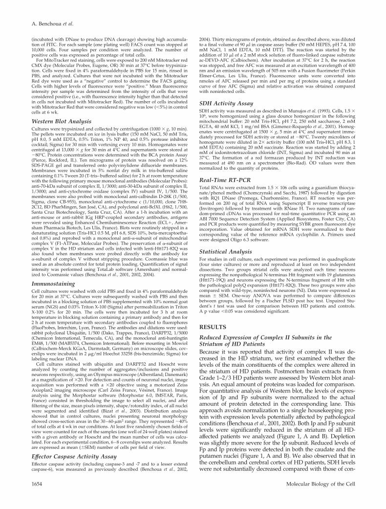
To examine whether the complex II/SDH changes resultedfrom striatal cell degeneration, we evaluated in the putamen ofHD patients and control subjects the levels of calbindin, amarker of medium-size spiny neurons. The expression ofcalbindin as assessed by Western blot was slightly re-duced in the striatum but the apparent loss was lesspronounced than that seen for SDH proteins (Figure 2). Toexamine whether the complex II/SDH changes resultedfrom a general loss of mitochondrial proteins, we reeval-uated in the putamen of HD patients and control subjectsthe levels of SDH proteins and several mitochondrialproteins that are nuclear encoded (Figure 2). Interestingly,no significant modifications were observed for �-subunitof complex V (F1-ATPase), a macromolecular complex,which like SDH is attached to the inner mitochondrialmembrane. Although variable from one control individ-ual to the other, levels of subunit IV of cytochrome oxi-dase (mitochondrial complex IV), another complex of theinner membrane were slightly decreased in the striatumof grade III/II HD patients. Levels of cytochrome c, whichis localized in the intermembrane space, were not changed. Thelevels of the outer membrane protein BclXL also remainedunchanged compared with controls. These results supportedthe view that the loss of complex II subunits in the HD striatumdid not simply result from depletion in mitochondria.
These results indicated that the reduced activity of com-plex II in the HD striatum that was reported by others (Guet al., 1996; Browne et al., 1997; Tabrizi et al., 1999) likelyresults form a reduced expression of complex II constituents.
Time Course of Degeneration in Striatal Cells ExpressingN-Terminus Fragment of Mutated Htt Using Lentivirus-mediated Gene TransferBecause results in HD patients could not indicate whetheralterations of complex II expression were a cause or a con-sequence of striatal cell death, we decided to study complexII in a new model of HD in primary striatal neurons inculture (Zala et al., 2005).
In the present study we further evaluated the time courseof degeneration induced by lenti-Htt171-82Q. Previous workshowed that a 20 and 50% reduction in the number ofneurons expressing the neuronal marker NeuN occurred at6 and 8 wk postinfection, respectively (Zala et al., 2005). Inline with this, immunofluorescence analyses performed inthe present study at 8 wk postinfection showed that lenti-Htt171-82Q produced a significant loss (�94%, p � 0.0001)of neurons expressing the striatal marker DARPP32 as com-pared with lenti-Htt171-19Q. Because mutated Htt producedsevere alteration in transcription, this loss of neuronalmarker may be due to a decrease in expression of the markerand/or actual degeneration (i.e., cell death). To determinewhether our model was characterized by actual cell degen-eration, we therefore took advantage of the fact that �90% ofthe neurons steadily expressed the transgene, to performbiochemical and FACS analyses. We first analyzed the po-tential activation of the effector caspases, the proteasesknown to play a role in the toxicity of short N-terminusfragments of mutated Htt in cultured neurons (Saudou et al.,1998). Biochemical assay using the fluorogenic caspase sub-strate ac-DEVD-AFC indicated that, at 8 wk postinfection,
Figure 1. Preferential depletion of complexII/SDH subunits in the striatum of HD pa-tients. Levels of Ip and Fp subunits of com-plex II/SDH were quantified by Western-blotin caudate (A), putamen (B), cerebral cortex(C), and cerebellum (D) of controls and symp-tomatic HD patients after normalization tototal protein content measured in each lane.Neuropathological grade of each HD patientis indicated above the lanes. Note that onlythe striatum shows marked depletion of SDHconstituents. Data represent mean � SEM*p � 0.05 by unpaired Student’s t test.
Mitochondrial Toxicity of Mutated Huntingtin
Vol. 17, April 2006 1655
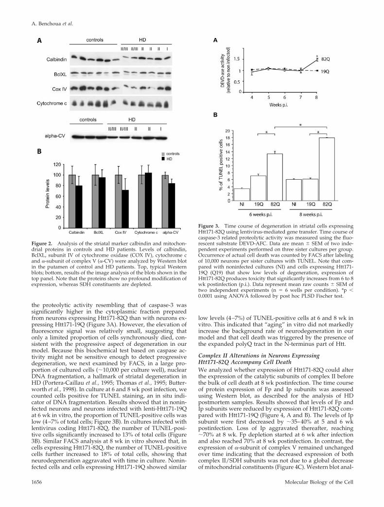
the proteolytic activity resembling that of caspase-3 wassignificantly higher in the cytoplasmic fraction preparedfrom neurons expressing Htt171-82Q than with neurons ex-pressing Htt171-19Q (Figure 3A). However, the elevation offluorescence signal was relatively small, suggesting thatonly a limited proportion of cells synchronously died, con-sistent with the progressive aspect of degeneration in ourmodel. Because this biochemical test based on caspase ac-tivity might not be sensitive enough to detect progressivedegeneration, we next examined by FACS, in a large pro-portion of cultured cells (�10,000 per culture well), nuclearDNA fragmentation, a hallmark of striatal degeneration inHD (Portera-Caillau et al., 1995; Thomas et al., 1995; Butter-worth et al., 1998). In culture at 6 and 8 wk post infection, wecounted cells positive for TUNEL staining, an in situ indi-cator of DNA fragmentation. Results showed that in nonin-fected neurons and neurons infected with lenti-Htt171-19Qat 6 wk in vitro, the proportion of TUNEL-positive cells waslow (4–7% of total cells; Figure 3B). In cultures infected withlentivirus coding Htt171-82Q, the number of TUNEL-posi-tive cells significantly increased to 13% of total cells (Figure3B). Similar FACS analysis at 8 wk in vitro showed that, incells expressing Htt171-82Q, the number of TUNEL-positivecells further increased to 18% of total cells, showing thatneurodegeneration aggravated with time in culture. Nonin-fected cells and cells expressing Htt171-19Q showed similar
low levels (4–7%) of TUNEL-positive cells at 6 and 8 wk invitro. This indicated that “aging” in vitro did not markedlyincrease the background rate of neurodegeneration in ourmodel and that cell death was triggered by the presence ofthe expanded polyQ tract in the N-terminus part of Htt.
Complex II Alterations in Neurons ExpressingHtt171-82Q Accompany Cell DeathWe analyzed whether expression of Htt171-82Q could alterthe expression of the catalytic subunits of complex II beforethe bulk of cell death at 8 wk postinfection. The time courseof protein expression of Fp and Ip subunits was assessedusing Western blot, as described for the analysis of HDpostmortem samples. Results showed that levels of Fp andIp subunits were reduced by expression of Htt171-82Q com-pared with Htt171-19Q (Figure 4, A and B). The levels of Ipsubunit were first decreased by �35–40% at 5 and 6 wkpostinfection. Loss of Ip aggravated thereafter, reaching�70% at 8 wk. Fp depletion started at 6 wk after infectionand also reached 70% at 8 wk postinfection. In contrast, theexpression of �-subunit of complex V remained unchangedover time indicating that the decreased expression of bothcomplex II/SDH subunits was not due to a global decreaseof mitochondrial constituents (Figure 4C). Western blot anal-
Figure 2. Analysis of the striatal marker calbindin and mitochon-drial proteins in controls and HD patients. Levels of calbindin,BclXL, subunit IV of cytochrome oxidase (COX IV), cytochrome cand �-subunit of complex V (�-CV) were analyzed by Western blotin the putamen of control and HD patients. Top, typical Westernblots; bottom, results of the image analysis of the blots shown in thetop panel. Note that the proteins show no profound modification ofexpression, whereas SDH constituents are depleted.
Figure 3. Time course of degeneration in striatal cells expressingHtt171-82Q using lentivirus-mediated gene transfer. Time course ofcaspase-3 related proteolytic activity was measured using the fluo-rescent substrate DEVD-AFC. Data are mean � SEM of two inde-pendent experiments performed on three sister cultures per group.Occurrence of actual cell death was counted by FACS after labelingof 10,000 neurons per sister cultures with TUNEL. Note that com-pared with noninfected cultures (NI) and cells expressing Htt171-19Q (Q19) that show low levels of degeneration, expression ofHtt171-82Q produces toxicity that significantly increases from 6 to 8wk postinfection (p.i.). Data represent mean raw counts � SEM oftwo independent experiments (n � 6 wells per condition). *p �0.0001 using ANOVA followed by post hoc PLSD Fischer test.
A. Benchoua et al.
Molecular Biology of the Cell1656

ysis of the levels of BclXL, cytochrome c, and subunit IV ofcytochrome oxidase (Cox IV) also supported the preferentialaspect of the loss of SDH compared with other mitochon-drial proteins (Figure 4, D and E). Thus, the preferential lossof complex II/SDH subunits and actual degeneration seemto progress concurrently in cells expressing Htt171-82Q.
Complex II/SDH Alterations Are Not Associated with anAltered Transcription of SDH GenesAbnormal transcription is an early feature of HD (Sugarsand Rubinsztein, 2003). Because the four subunits of thecomplex II/SDH are nuclear-encoded, we reasoned thatHtt171-82Q may act at the nuclear level and could represstranscription of the SDH genes. Quantification of SDHsubunit mRNA content using real-time RT-PCR was per-formed 6 wk after infecting striatal cultures. Culturesexpressing Htt171-82Q showed loss of Ip and Fp proteinsat this time point. However, no profound changes inmRNAs for SDH subunits were found in cultures express-ing m-Htt82Q compared with mock- and Htt171-19Q–infected cells (Table 1). This indicated that, at least at the
beginning of the process (6 wk), the marked depletion inSDH protein was not associated with proportionate alter-ations of SDH gene transcription.
Decreased Succinate Oxidation and MitochondrialMembrane Potential in Neurons Expressing Htt171-82QWe studied whether loss of complex II subunits could lead tofunctional change in the mitochondria. Functional analysis ofcomplex II activity was performed at 6 wk postinfection, whenboth subunits were decreased in cells expressing Htt171-82Q.We tested the capability of crude mitochondrial preparations tooxidize saturating concentrations of succinate in the presenceof the electron acceptor INT (iodo-nitroblue tetrazolium). Re-sults demonstrated that SDH catalytic activity (Vmax) wassignificantly reduced by 30% in cells expressing Htt171-82Qcompared with Htt171-19Q (Figure 5A), consistent with the�40% loss of Ip and Fp proteins seen by Western blot at thesame time point. This confirmed that the partial depletion incomplex II/SDH catalytic subunits detected by Western blotwas sufficient to induce a functional impairment of complex II.
Figure 4. Expression of Htt171-82Q expres-sion of complex II/SDH catalytic subunits.Representative immuno-blots showing Fp,Ip, and �-subunit of complex V (alpha-CV)expression in Htt171-19Q (19Q)- and Htt171-82Q (82Q)-expressing cells after 5, 6, and 8 wkin culture. Levels of expression of Fp and Ipproteins were quantified by Western blot atdifferent time points in control cells (graystars), and neurons infected with either lenti-Htt171-82Q (filled triangle), or lenti-Htt19Q(filled square). Expression of alpha-CV wasalso quantified indicating only minorchanges. Typical Western blot showing theexpression of SDH subunits compared withother mitochondrial proteins at 6 wk postin-fection is shown in D and correspondingquantification in E. Note that SDH Ip and Fpshow decreased expression, whereas theother proteins are relatively preserved. Datarepresent mean � SEM of at least two inde-pendent experiments performed on three sis-ter cultures per group for each time point(n � 6 wells per condition). Loss of SDHsubunits at 6 wk has been observed in morethan four independent experiments. *p � 0.05by ANOVA and post hoc PLSD Fischer test.
Mitochondrial Toxicity of Mutated Huntingtin
Vol. 17, April 2006 1657

We next assessed whether Htt171-82Q–induced defectsin the respiratory chain could produce alterations in mi-tochondrial homeostasis. We hypothesized that complexII defect could modify mitochondrial membrane potential.To examine this hypothesis, we carried out FACS analysisof the fluorescence resulting from Mitotracker red (CMX-ROS) accumulation in living striatal cell cultures. Wechose this dye because it can be fixed using paraformal-dehyde, and for biosafety reasons we had to treat infectedcells with a fixative to inactivate the viral particles poten-tially remaining in the cultures before FACS analysis.Chloromethyl-X-Rosamine (CMX-ROS) dye accumulationin active mitochondria was demonstrated to be dependenton mitochondrial membrane potential (Pendergrass et al.,2004) and the changes of fluorescence intensity resultingfrom mitochondrial membrane potential variations can bemeasured by FACS with greater sensitivity than with spec-trofluorimetric techniques (Kalbacova et al., 2003). However,fixative can, in certain conditions, affect the intracellular distri-bution of MitoTracker red (Gilmore and Wilson, 1999). For thisreason, we checked in (noninfected) cultured striatal neu-rons that the intensity of accumulated fluorescence as afunction of incubation time was only slightly (�15%)decreased by 4% paraformaldehyde fixation (Figure 5B).Even after a 30-min incubation, cells were not saturated
and fluorescence signal increased, suggesting absence ofmajor quenching effects. We also checked that accumula-tion of Mitotracker Red in striatal cells was related tomitochondrial membrane potential by showing that a5-min preincubation with 50 �M FCCP significantly re-duced MitoTracker accumulation by 70% (Figure 5B).FACS analysis showed that at 6 wk postinfection, cellsexpressing either Htt171-82Q or Htt171-19Q accumulatedMitotracker red dye (Figure 5C). However, cells express-ing Htt171-82Q exhibited a significant reduction of thefluorescence mean intensity related to Mitotracker incor-poration when compared with mock or lenti-Htt171-19Q–infected cells (Figure 5, C and D). This indicated thatexpression of N-terminus fragment of mutated Htt couldinduce a functional alteration of mitochondrial membranepotential, possibly as a consequence of the complex II/SDH defect.
Overexpression of SDH Subunits Restores MitochondrialMembrane Potential and Blocks Neuronal Death Inducedby Htt171-82QWe next wanted to determine whether complex II/SDHdefects were solely a consequence of degeneration or couldplay a causal role in neurodegeneration produced bymutated N-terminal fragment of Htt. We reasoned that if
Figure 5. Expression of Htt171-82Q inducesmitochondrial dysfunction. Dehydrogenaseactivity of complex II/SDH was measured inHtt177-19Q– and Htt171-82Q–expressingcells at 6 wk postinfection. Activity of com-plex II (succinate oxidation) was evaluatedusing the colorimetric substrate INT. Datarepresent mean � SEM of two independentexperiments with three sister cultures pergroup. (B) Fluorescence of MitoTracker red inliving cells (�) or after PFA fixation of cul-tures (�) as a function of incubation time inpresence of the dye. For these methodologicalcontrols, fluorescence accumulated in cellswas determined using a plate reader. Incuba-tion with the ionophore FCCP (50 �M) pro-duces marked reduction of the accumulationof Mitotracker red, demonstrating that accu-mulation of the dye is highly sensitive to theloss of mitochondrial membrane potential.(C) Detection of MitoTracker red levels inindividual cells after PFA fixation was per-formed by FACS, showing the typical distri-bution of fluorescence levels in cells express-ing Htt171-82Q (82Q) and Htt171-19Q at 6 wkpostinfection. Note the shift to the left of flu-orescence distribution in cells expressingHtt171-82Q, indicating reduced fluorescence
mean intensity (FMI) in the culture. (D) Histograms representing the quantification of FMI measured in two independent experiments (n �6 wells per condition). Data are mean � SEM. *p � 0.05 by ANOVA and post hoc PLSD Fischer test.
Table 1. Expression of the four mRNAs encoding SDH subunits in striatal cells expressing Htt171-19Q or Htt171-82Q
Transcript SDH-A SDH-B SDH-C SDH-D
Mock 100.03 � 5.05 100.38 � 5.00 100.26 � 7.02 100.41 � 8.16Htt-19Q 94.61 � 9.15 77.41 � 5.71 93.49 � 0.32 85.44 � 9.29m-Htt-82Q 103.22 � 10.46 77.05 � 15.39 108.66 � 7.83 95.71 � 4.11
Levels of SDH mRNAs were assessed using real time RT-PCR. Data are expressed as mean � SEM of percentage of expression relative tomock-infected cells. Note that no significant changes were produced by Htt171-82Q.
A. Benchoua et al.
Molecular Biology of the Cell1658

depletion of SDH subunits was causal in HD associatedneuronal death, restoring SDH levels using lentivirus-medi-ated gene overexpression should protect striatal neuronsagainst Htt171-82Q toxicity. To test this hypothesis, we de-signed two lentiviral vectors encoding either human Fpsubunit (lenti-SDH-A) or human Ip subunit (lenti-SDH-B).All potential posttranscriptional regulatory elements (UTRs)were deleted from human SDH-A and SDH-B encodingcDNA before insertion in the lentivirus transfer construct.Western blot analysis of striatal neurons showed that 2 wkafter infection (Figure 6A), both viral vectors were able totransduce expression of exogenous Fp subunit (lenti-SDH-A) or Ip subunit (lenti-SDH-B). The antibodies we usedto detect SDH Ip and Fp subunits similarly recognize thehuman and rat proteins so that we could not separatelydetect recombinant and endogenous SDH proteins. How-ever, Western blot analysis showed that the overall levels ofSDH subunits (endogenous plus recombinant) were in-creased in neurons transduced with lenti-SDH-A and lenti-SDH-B. Interestingly, lentivirus-mediated expression of only
one of the two exogenous subunits was sufficient to producean increased expression of the other endogenous subunit ofthe complex II/SDH complex.
We next addressed whether restoring the levels of SDHcatalytic subunits was able to preserve the mitochondrialmembrane potential in neurons expressing Htt171-82Q. Inthese experiments, neurons were first infected with lenti-Htt171-82Q or lenti-Htt171-19Q or were mock-infected, and1 wk later, they were mock-infected or exposed to lenti-SDH-A or lenti-SDH-B. In cells expressing Htt171-19Q,MitoTracker incorporation remained unchanged comparedwith mock-infected cells (Figure 6B). The double infection ofneurons expressing Htt171-19Q with lenti-SDH-A or lenti-SDH-B did not modify dye incorporation either. In cellsexpressing Htt171-82Q, a significant reduction of Mito-Tracker incorporation was detected at 6 wk postinfection asseen in the experiment described above (Figure 6B). Impor-tantly, the reduction of dye incorporation in cell expressingHtt171-82Q was totally abolished by overexpressing eitherIp or Fp SDH subunits. This indicated that SDH depletioncould be causal in Htt171-82Q–induced loss of mitochon-drial membrane potential.
We finally asked whether this mitochondrial improvementwas associated with a reduction of the toxic properties ofHtt171-82Q in neurons. Consistent with experiments reportedabove, a significant elevation in the number of TUNEL positivecells (i.e., 3 times the levels seen in controls) was detected inneurons expressing Htt171-82Q at 8 wk postinfection (Figure6C). In sister cultures that were also infected 1 wk later withlenti-SDH-A and lenti-SDH-B, the Htt171-82Q produced noelevation in the number of TUNEL-positive cells. Thus, over-expressing SDH subunits could block cell death induced byHtt171-82Q. This data suggests that complex II/SDH depletionplays an instrumental role in the slow toxicity of the N-termi-nus fragment of mutated Htt expressed under the PGK pro-moter.
Analysis of cell death and neuroprotection at later timepoints in vitro (e.g., 10 wk) could not be easily performed inour lentivirus-based model using the PGK promoter becausecultures tend to become exquisitely vulnerable after 9–10 wkin vitro. Therefore, to check that Htt171-82Q could producedegeneration independently of “aging” of culture in vitroand that SDH-A and SDH-B could be neuroprotective in amore rapid model of degeneration, we used tetracycline-regulated (TRE) Htt171-19/82Q vectors (Regulier et al.,2003). This system leads to higher expression levels of thetransgene compared with PGK and therefore more rapiddegeneration (Regulier et al., 2003). We examined cultures atdifferent time points after infection with both the lentiviruscoding for the transactivator protein tTA1 and lentiviralvectors expressing TRE-Htt171-82Q, TRE-Htt171-19Q, or thegreen fluorescent protein (TRE-GFP). The number of EM48-positive huntingtin-containing inclusions/aggregates in-creased rapidly from 2 to 3 wk postinfection in cells express-ing Htt171-82Q under the TRE promoter and plateauedthereafter. On the fourth week postinfection, numerousubiquitin- and EM48-positive intranuclear inclusions wereseen (Figure 7, A and B). The number of DARPP32-positivecells was decreased by more than 90% in cultures infectedwith lenti-TRE-Htt171-82Q compared with lenti-TRE-Htt171-19Q (Figure 7C). The number of neuronal nuclei wasdecreased by 35% in cultures infected with lenti-TRE-Htt171-82Q compared with lenti-TRE-Htt171-19Q and lenti-TRE-GFP (Figure 7D). Thus, expression of Htt171-82Q underthe tetracycline-regulated promoter produced neurodegen-eration more rapidly compared with Htt171-82Q under thePKG promoter.
Figure 6. Overexpression of complex II/SDH subunits restoresmitochondrial membrane potential and rescues neuronal death in-duced by Htt171-82Q. (A) Representative Western blots of 30 DIVneurons transduced with SDH encoding lentiviral vectors 2 wk afterinfection. (B) Overexpression of Fp (SDH-A virus) or Ip (SDH-Bvirus) suppresses mitochondrial membrane potential alteration in-duced by Htt171-82Q expression at 6 wk postinfection. (C) Deter-mination of the number of TUNEL-positive cells at 8 wk in cultureindicates that both infection with lentiviral vectors coding SDHsubunits significantly reduces Htt171-82Q–induced cell death. Dataare mean � SEM of two independent experiments performed onthree sister cultures per group (n � 6 wells per condition). *p � 0.05by ANOVA and post hoc PLSD Fischer test.
Mitochondrial Toxicity of Mutated Huntingtin
Vol. 17, April 2006 1659

We next tested the effects of lenti-SDH-A and lenti-SDH-Bagainst the neurotoxicity of lenti-TRE-Htt171-82Q. Overex-pression of SDH-A nor SDH-B did modify neither the num-ber of ubiquitin-containing inclusions (Figure 7B) nor theHtt171-82Q–induced decrease in DARPP32. However, thereduction of the number of neuronal nuclei in cultures ex-pressing Htt171-82Q was not seen in cultures coinfectedwith Htt171-82Q plus SDH-A or SDH-B.
Our results further support the view that restoring com-plex II/SDH levels did not modify early signs of degenera-tion/dysfunction (e.g., loss of DARPP32 expression, aggre-gates formation) but could efficiently block the executionphase of cell death induced by the N-terminus fragment ofmutated Htt.
DISCUSSION
There are several hypothetical mechanisms that could un-derlie striatal cell death in HD. Main hypotheses includeabnormal transcription (Sugars and Rubinsztein, 2003), in-creased transglutaminase activity (Lesort et al., 2002), earlyaxonal transport dysfunction (Li and Li, 2004), and disrup-tion of Ca2� homeostasis (Bezprozvanny and Hayden, 2004).Protein misfolding, reduction of autophagy, and proteasomedysfunction play important roles in HD (Meriin and Sher-man, 2005). In addition, mitochondrial dysfunction couldalso contribute to neurodegeneration (Brouillet et al., 2005).However, there existed no direct evidence that mitochon-drial change could play a causal role in HD.
In the present study, we hypothesized that an early de-crease in the levels of one or more of the four subunitsconstituting complex II/SDH could, at least in part, be re-sponsible for mitochondrial dysfunction in HD. Taking ad-vantage of a newly developed model of progressive striataldegeneration in primary cultures (Zala et al., 2005), we ex-amined this hypothesis using biochemical and cytologicalmethods.
Our results show that expressing the N-terminus part ofHtt with a pathological polyglutamine expansion in cul-tured striatal primary neurons reduced the levels of complexII/SDH subunits Ip and Fp and the dehydrogenase activityof the complex. We also found similar molecular defects incomplex II/SDH in the HD striatum. No major changes inthe expression of SDH subunits were found in the cerebralcortex and cerebellum, two regions less vulnerable to degen-eration in HD patients. Our data are consistent with thereduced activity of complex II-III in HD patients reported inthe literature. A decrease in succinate oxidation rangingfrom 39 to 59% has been observed in the caudate nucleus ofHD patients (Butterworth et al., 1985; Brennan et al., 1985;Mann et al., 1990; Gu et al., 1996; Browne et al., 1997; Tabriziet al., 1999). In the putamen, succinate oxidation showed anaverage 69% decrease (Browne et al., 1997). In line with this,striatal cell lines expressing the full-length Htt with 111polyglutamine (Knock-in 111) were found highly vulnerableto the complex II inhibitor 3NP (Ruan et al., 2004). In thepresent study, neither striatal cells expressing Htt171-82Qnor the HD striatum showed profound alterations in �-sub-unit of complex V, cytochrome c, and BclXL. Levels ofsubunit IV of cytochrome oxidase (complex IV) and calbi-ndin were slightly reduced in III/II HD patients, consistentwith striatal degeneration. However, depletion of these pro-teins appeared less pronounced than that of SDH subunits.In cell expressing Htt171-82Q at 6 wk postinfection, wefound no modification of subunit IV of cytochrome oxidase(complex IV). These observations support the view that HDmay be preferentially associated with complex II defects.Our results obtained in cell culture also suggest that theeffect of the short fragment of mutated Htt is likely not anonspecific toxic effect but has some relevance to the humandisorder. In addition, SDH/complex II alterations are prob-ably not simply the consequence of neuronal loss becausethe severity of reduction of the complex is disproportionate
Figure 7. Overexpression of complex II/SDHsubunits protects striatal cell loss rapidly inducedby expression of Htt171-82Q under the tetracy-cline-regulated promoter. The neuroprotective ef-fect of lenti-SDH-A and lenti-SDH-B was tested ina model of degeneration induced by infectionwith lentiviral vectors coding for Htt-171-82Q un-der the tetracycline-regulated promoter (TRE).(A) Time course of accumulation of Htt-contain-ing inclusions/aggregates detected by immuno-fluorescence with the EM48 antibody. Note thatinclusion accumulation culminates at 3–4 wkpostinfection (p.i.). (B) Inclusions/aggregates incells expressing TRE-Htt-171-82Q at 4 wk postin-fection were detected by immunofluorescencewith an anti-ubiquitin antibody. (C) Loss ofDARPP32-positive cells at 4 wk postinfectionwith lenti-TRE-Htt-171-82Q compared with lenti-TRE-Htt171-19Q. The coinfection with lenti-SDH-A and lenti-SDH-B did not markedly mod-ify the density of inclusions and the density ofDARPP32 cells. (D) Neuronal nuclei were identi-fied and counted after DNA staining at 4 wkpostinfection with lenti-TRE-Htt-171-82Q, lenti-TRE-Htt171-19Q, and lenti-TRE-GFP. When indi-cated, cultures were coinfected with lenti-SDH-Aor lenti-SDH-B. Note that the significant cell lossevidenced in cultures expressing Htt171-82Q isblocked by expression of SDH-A and SDH-B.*p � 0.05 compared with all other groups.
A. Benchoua et al.
Molecular Biology of the Cell1660

in comparison with the extent of cell degeneration (�20%range; see present data and Zala et al., 2005).
The mechanisms that could underlie the loss of complexII/SDH in our in vitro model have not been elucidated.Interestingly, the present results indicate that the shortN-terminus fragment of mutated Htt alone is sufficient toproduce complex II/SDH defects. This is in agreementwith the hypothesis that, at least in part, N-terminusfragments of mutated Htt generated from the cleavage ofthe full-length protein contribute to the neurotoxicitycaused by the mutation (Li and Li, 2004; Wellington et al.,2002). The present study shows that although proteinlevels and activity of complex II/SDH were markedlyaltered, the levels of mRNA of subunits were not de-creased, suggesting the involvement of posttranscrip-tional regulation. Our results suggest that the loss of Ipprotein precedes the loss of Fp protein. In human, muta-tions in any of the SDH subunits cause the complex II tofully disassemble (Rustin et al., 2002). Thus, it is possiblethat short fragments of mutated Htt produce their effectpreferentially on the Ip subunit, triggering destabilizationof the entire complex, and in turn the loss of the Fpprotein. Given that mutated Htt is located in the cyto-plasm and can bind the outer mitochondrial membrane(Panov et al., 2002; Choo et al., 2004), many speculativemechanisms can be proposed to explain how mutated Httreduces posttranscriptional Ip levels. For instance, mu-tated Htt could induce a decrease in the import of Ip intothe mitochondria, an increase in degradation, or an ab-normal assembly. One interesting mechanism may be re-lated to p53. A role for the accumulation of p53 in mito-chondrial abnormalities and degeneration in HD has beenrecently demonstrated (Bae et al., 2005). The accumulationof p53 could possibly result from proteasome dysfunctioninduced by short fragments of mutated Htt (Jana et al.,2001; Waelter et al., 2001). Apart from its role as a tran-scription factor, p53 interacts in the cytosol with proteins(e.g., Bax and BclXL) involved in regulation of the mito-chondrial pathway of apoptosis (Chipuk et al., 2005). p53might be even localized within the mitochondria (Heyneet al., 2004). p53 is also involved in oxidative stress (Culm-see and Mattson, 2005), and SDH Ip is often consideredas a “sensor” of the ubiquinone pool, a major source ofreactive oxygen species in the cells (Rustin et al., 2002).Thus, it is possible that p53 could be involved in the reductionof complex II/SDH levels induced by mutated Htt.
Another interesting possibility is related to the Ip proteinmRNA structure. Ip mRNA possesses an UTR named IRE (ironresponsive element) sequence to which specific proteins (IRP-1and IRP-2) can bind, regulating translation (Gray et al., 1996).The presence of mutated Htt could interfere at this level tomodify posttranscriptionally SDH expression. Supporting thishypothesis, huntingtin has been implicated in regulation ofiron homeostasis (Hilditch-Maguire et al., 2000). Of interest,IRP-2 levels are regulated by the ubiquitin-proteasome path-way (Guo et al., 1995). Accumulation of IRP2 might reducetranslation of mRNAs coding SDH Ip subunit, leading to de-pletion of the protein. Ongoing experiments will examine therole of IRPs.
The present results show that loss of complex II/SDH sub-units and activity was accompanied by a significant decrease inmitochondrial membrane potential in striatal neurons express-ing Htt171-82Q. Our observation is in agreement with theresults obtained in mitochondria of HD patients and transgenicYAC72 mouse HD model that showed abnormal mitochon-drial transmembrane potential (Sawa et al., 1999; Panov et al.,2002). Noticeably, Bae et al. (2005) showed reduced accumula-
tion of Mitotracker Red in striatal neuronal cells expressingmutated Htt, along with changes in JC-1 fluorescence, indicat-ing anomalies in mitochondrial membrane potential. Reduc-tion of SDH activity could reduce the proton gradient pro-duced by complex III and complex IV across the innermitochondrial membrane. Interestingly, we found that overex-pressing Ip or Fp proteins using lentivirus could preventHtt171-82Q–induced loss of mitochondrial membrane poten-tial. This suggests that loss of mitochondrial membrane poten-tial in neurons expressing short N-terminus fragment of mu-tated Htt could be, at least in part, a direct consequence ofcomplex II/SDH defects.
We showed that correcting the molecular defect of SDHcomplex blocked death of striatal cells induced by express-ing low levels of the N-terminus part of mutated Htt. WhenHtt171-82Q expression levels were increased using the tet-racycline-regulated promoter, overexpression of SDH-A orSDH-B modified neither the reduction of DARPP32 expres-sion in striatal cultures nor the levels of ubiquitin-positiveinclusions. However, the rescuing effect of SDH-A andSDH-B overexpression from cell death was seen. These re-sults indicate that loss of complex II/SDH is probably notresponsible for loss of a number of striatal markers and doesnot play a direct effect on sequestration/detoxification ofmutated Htt fragment. Importantly our results provide ev-idence that complex II/SDH subunits are critical for theexecution of cell death induced by toxicity of N-terminusfragment of mutated Htt.
The observation that overexpression of one SDH subunitleads to an increased expression of the entire complex maysound surprising at first. It is probable that this phenomenonis due to an increased “stabilization” of the SDH complex. Inhumans, mutations in any of the SDH subunits cause thecomplex II to fully disassemble (Rustin et al., 2002). Oneelegant study showed that, in lymphoblast cells isolatedfrom a patient with homozygous Fp mutations leading tomajor loss of SDH (Bourgeron et al., 1995), overexpression ofa wild-type Fp protein led to a fully functional complex(Parfait et al., 2000).
In conclusion, the present study shows preferential loss ofcomplex II/SDH subunits in the HD striatum and a cellularmodel of the disease and provides the first proof of conceptthat regulating complex II/SDH expression may be of ther-apeutic interest to slow down striatal degeneration in HD.Animal experiments are awaited to determine whether suchan experimental therapy could be effective in vivo.
ACKNOWLEDGMENTS
We thank Pierre Rustin and Sandrine Humbert for having shared with ustheir scientific expertise. We thank Carl Johnson of the Hereditary DiseaseFoundation for advice and encouragement. We are grateful to Marc Peschan-ski for his constructive comments on the manuscript. We thank Dr JasonWray for critical reading of the manuscript and language corrections. Thiswork has been supported by Commissariat a l’Energie Atomique (CEA) andCentre National de la Recherche Scientifique. A.B. is a recipient of postdoc-toral fellowship from CEA. Y.T. holds a postdoctoral fellowship from theHereditary Disease Foundation.
REFERENCES
Ackrell, B. A. (2000). Progress in understanding structure-function relation-ships in respiratory chain complex II. FEBS Lett. 466, 1–5.
Bae, B. I. et al. (2005). p53 mediates cellular dysfunction and behavioralabnormalities in Huntington’s disease. Neuron 47, 29–41.
Beal, M. F. (1992). Does impairment of energy metabolism result in excitotoxicneuronal death in neurodegenerative illnesses? Ann. Neurol. 31, 119–130.
Mitochondrial Toxicity of Mutated Huntingtin
Vol. 17, April 2006 1661

Benchoua, A., Braudeau, J., Reis, A., Couriaud, C., and Onteniente, B. (2004).Activation of proinflammatory caspases by cathepsin B in focal cerebralischemia. J. Cereb. Blood Flow Metab. 24, 1272–1279.
Benchoua, A., Couriaud, C., Guegan, C., Tartier, L., Couvert, P., Friocourt, G.,Chelly, J., Menissier-de Murcia, J., and Onteniente, B. (2002). Active caspase-8translocates into the nucleus of apoptotic cells to inactivate poly(ADP-ribose)polymerase-2. J. Biol. Chem. 277, 34217–34222.
Benchoua, A., Guegan, C., Couriaud, C., Hosseini, H., Sampaio, N., Morin, D.,and Onteniente, B. (2001). Specific caspase pathways are activated in the twostages of cerebral infarction. J. Neurosci. 21, 7127–7134.
Bezprozvanny, I., and Hayden, M. R. (2004). Deranged neuronal calciumsignaling and Huntington disease. Biochem. Biophys. Res. Commun. 322,1310–1317.
Bizat, N., Hermel, J. M., Boyer, F., Jacquard, C., Creminon, C., Ouary, S.,Escartin, C., Hantraye, P., Kajewski, S., Brouillet, E. (2003). Calpain is a majorcell death effector in selective striatal degeneration induced in vivo by 3-ni-tropropionate: implications for Huntington’s disease. J. Neurosci. 23, 5020–5030.
Bourgeron, T., Rustin, P., Chretien, D., Birch-Machin, M., Bourgeois, M.,Viegas-Pequignot, E., Munnich, A., and Rotig, A. (1995). Mutation of a nuclearsuccinate dehydrogenase gene results in mitochondrial respiratory chaindeficiency. Nat. Genet. 11, 144–149.
Brennan, W. A., Jr., Bird, E. D., and Aprille, J. R. (1985). Regional mitochon-drial respiratory activity in Huntington’s disease brain. J. Neurochem. 44,1948–1950.
Brouillet, E., Conde, F., Beal, M. F., and Hantraye, P. (1999). ReplicatingHuntington’s disease phenotype in experimental animals. Prog. Neurobiol.59, 427–468.
Brouillet, E., Jacquard, C., Bizat, N., and Blum, D. (2005). 3-Nitropropionicacid: a mitochondrial toxin to uncover physiopathological mechanisms un-derlying striatal degeneration in Huntington’s disease. J. Neurochem. 95,1521–1540.
Browne, S. E., Bowling, A. C., MacGarvey, U., Baik, M. J., Berger, S. C., Muqit,M. M., Bird, E. D., and Beal, M. F. (1997). Oxidative damage and metabolicdysfunction in Huntington’s disease: selective vulnerability of the basal gan-glia. Ann. Neurol. 41, 646–653.
Butterworth, N. J., Willians, L., Bullock, J. Y., Love, D. R., Faull, R.L.M., andDragunow, M. (1998). Trinucleotide (CAG) repeat length is positively corre-lated with the degree of DNA fragmentation in Huntington’s disease stria-tum. Neuroscience 87, 49–53.
Butterworth, J., Yates, C. M., and Reynolds, G. P. (1985). Distribution ofphosphate-activated glutaminase, succinic dehydrogenase, pyruvate dehy-drogenase and gamma-glutamyl transpeptidase in post-mortem brain fromHuntington’s disease and agonal cases. J. Neurol. Sci. 67, 161–171.
Chipuk, J. E., Bouchier-Hayes, L., Kuwana, T., Newmeyer, D. D., and Green,D. R. (2005). PUMA couples the nuclear and cytoplasmic proapoptotic func-tion of p53. Science 309, 1732–1735.
Chomczynski, P., and Sacchi, N. (1987). Single-step method of RNA isolationby acid guanidinium thiocyanate-phenol-chloroform extraction. Anal. Bio-chem. 162, 156–159.
Choo, Y. S., Johnson, G. V., MacDonald, M., Detloff, P. J., and Lesort, M.(2004). Mutant huntingtin directly increases susceptibility of mitochondria tothe calcium-induced permeability transition and cytochrome c release. Hum.Mol. Genet. 13, 1407–1420.
Culmsee, C., and Mattson, M. P. (2005). p53 in neuronal apoptosis. Biochem.Biophys. Res. Commun. 331, 761–777.
de Almeida, L. P., Ross, C. A., Zala, D., Aebischer, P., and Deglon, N. (2002).Lentiviral-mediated delivery of mutant huntingtin in the striatum of ratsinduces a selective neuropathology modulated by polyglutamine repeat size,huntingtin expression levels, and protein length. J. Neurosci. 22, 3473–3483.
Deglon, N., Tseng, J. L., Bensadoun, J. C., Zurn, A. D., Arsenijevic, Y., deAlmeida, L., Zufferey, R., Trono, D., and Aebischer, P. (2000). Self-inactivatinglentiviral vectors with enhanced transgene expression as potential gene trans-fer system in Parkinson’s disease. Hum. Gene. Ther. 11, 179–190.
Gilmore, K., and Wilson, M. (1999). The use of chloromethyl-X-rosamine(Mitotracker red) to measure loss of mitochondrial membrane potential inapoptotic cells is incompatible with cell fixation. Cytometry 36, 355–358.
Gimenez-Roqueplo, A. P., Favier, J., Rustin, P., Mourad, J. J., Plouin, P. F.,Corvol, P., Rotig, A., and Jeunemaitre, X. (2001). The R22X mutation of theSDHD gene in hereditary paraganglioma abolishes the enzymatic activity ofcomplex II in the mitochondrial respiratory chain and activates the hypoxiapathway. Am. J. Hum. Genet. 69, 1186–1197.
Gossen, M., and Bujard, H. (1992). Tight control of gene expression in mam-malian cells by tetracycline-responsive promoters. Proc. Natl. Acad. Sci. USA89, 5547–5551.
Gourfinkel-An, I., Vila, M., Faucheux, B., Duyckaerts, C., Viallet, F., Hauw,J. J., Brice, A., Agid, Y., and Hirsch, E. C. (2002). Metabolic changes in the basalganglia of patients with Huntington’s disease: an in situ hybridization studyof cytochrome oxidase subunit I mRNA. J. Neurochem. 80, 466–476.
Gray, N. K., Pantopoulos, K., Dandekar, T., Ackrell, B. A., and Hentze, M. W.(1996). Translational regulation of mammalian and Drosophila citric acid cycleenzymes via iron-responsive elements. Proc. Natl. Acad. Sci. USA 93, 4925–4930.
Gu, M., Gash, M. T., Mann, V. M., Javoy-Agid, F., Cooper, J. M., and Schapira,A. H. (1996). Mitochondrial defect in Huntington’s disease caudate nucleus.Ann. Neurol. 39, 385–389.
Guo, B., Phillips, J. D., Yu, Y., and Leibold, E. A. (1995). Iron regulates theintracellular degradation of iron regulatory protein 2 by the proteasome.J. Biol. Chem. 270, 21645–21651.
Heyne, K., Mannebach, S., Wuertz, E., Knaup, K. X., Mahyar-Roemer, M., andRoemer, K. (2004). Identification of a putative p53 binding sequence withinthe human mitochondrial genome. FEBS Lett. 578, 198–202.
Hilditch-Maguire, P., Trettel, F., Passani, L. A., Auerbach, A., Persichetti, F.,and MacDonald, M. E. (2000). Huntingtin: an iron-regulated protein essentialfor normal nuclear and perinuclear organelles. Hum. Mol. Genet. 9, 2789–2797.
Jana, N. R., Zemskov, E. A., Wang, G., and Nukina, N. (2001). Alteredproteasomal function due to the expression of polyglutamine-expanded trun-cated N-terminal huntingtin induces apoptosis by caspase activation throughmitochondrial cytochrome c release. Hum. Mol. Genet. 10, 1049–1059.
Jenkins, B.G., Rosas, H. D., Chen, Y. C., Makabe, T., Myers, R., MacDonald,M., Rosen, B. R., Beal, M. F., and Koroshetz, W. J. (1998). 1H NMR spectros-copy studies of Huntington’s disease: correlations with CAG repeat numbers.Neurology 50, 1357–1365.
Kalbacova, M., Vrbacky, M., Drahota, Z., and Melkova, Z. (2003). Comparisonof the effect of mitochondrial inhibitors on mitochondrial membrane potentialin two different cell lines using flow cytometry and spectrofluorometry.Cytometry A 52, 110–116.
Koroshetz, W. J., Jenkins, B. G., Rosen, B. R., and Beal, M. F. (1997). Energymetabolism defects in Huntington’s disease and effects of coenzyme Q10.Ann. Neurol. 41, 160–165.
Lesort, M., Chun, W., Tucholski, J., and Johnson, G. V. (2002). Does tissuetransglutaminase play a role in Huntington’s disease? Neurochem. Int. 40,37–52.
Li, S. H., and Li, X. J. (2004). Huntingtin and its role in neuronal degeneration.Neuroscientist 10, 467–475.
Lodi, R., Schapira, A. H., Manners, D., Styles, P., Wood, N. W., Taylor, D. J.,and Warner, T. T. (2000). Abnormal in vivo skeletal muscle energy metabo-lism in Huntington’s disease and dentatorubropallidoluysian atrophy. Ann.Neurol. 48, 72–76.
Mann, V. M., Cooper, J. M., Javoy-Agid, F., Agid, Y., Jenner, P., and Schapira,A. H. (1990). Mitochondrial function and parental sex effect in Huntington’sdisease. Lancet 336, 749.
Meriin, A. B., and Sherman, M. Y. (2005). Role of molecular chaperones inneurodegenerative disorders. Int. J. Hyperthermia 21, 403–419.
Munujos, P., Coll-Canti, J., Gonzalez-Sastre, F., and Gella, F. J. (1993). Assayof succinate dehydrogenase activity by a colorimetric-continuous methodusing iodonitrotetrazolium chloride as electron acceptor. Anal. Biochem. 212,506–509.
Panov, A. V., Gutekunst, C. A., Leavitt, B. R., Hayden, M. R., Burke, J. R.,Strittmatter, W. J., and Greenamyre, J. T. (2002). Early mitochondrial calciumdefects in Huntington’s disease are a direct effect of polyglutamines. Nat.Neurosci. 5, 731–736.
Parfait, B., Chretien, D., Rotig, A., Marsac, C., Munnich, A., and Rustin, P.(2000). Compound heterozygous mutations in the flavoprotein gene of therespiratory chain complex II in a patient with Leigh syndrome. Hum. Genet.106, 236–243.
Pendergrass, W., Wolf, N., and Poot, M. (2004). Efficacy of MitoTracker Greenand CMXrosamine to measure changes in mitochondrial membrane poten-tials in living cells and tissues. Cytometry A 61, 162–169.
Portera-Caillau, C., Hedreen, J. C., Price, D. L., and Koliatsos, V. E. (1995).Evidence for apoptotic cell death in Huntington disease and excitotoxicanimal models. J. Neurosci. 15, 3775–3787.
Regulier, E., Pereira de Almeida, L., Sommer, B., Aebischer, P., and Deglon, N.(2002). Dose-dependent neuroprotective effect of ciliary neurotrophic factor
A. Benchoua et al.
Molecular Biology of the Cell1662

delivered via tetracycline-regulated lentiviral vectors in the quinolinic acid ratmodel of Huntington’s disease. Hum. Gene Ther. 13, 1981–1990.
Regulier, E., Trottier, Y., Perrin, V., Aebischer, P., and Deglon, N. (2003). Earlyand reversible neuropathology induced by tetracycline-regulated lentiviraloverexpression of mutant huntingtin in rat striatum. Hum. Mol. Genet. 12,2827–2836.
Ruan, Q., Lesort, M., MacDonald, M. E., and Johnson, G. V. (2004). Striatalcells from mutant huntingtin knock-in mice are selectively vulnerable tomitochondrial complex II inhibitor-induced cell death through a non-apop-totic pathway. Hum. Mol. Genet. 13, 669–681.
Rustin, P., Munnich, A., and Rotig, A. (2002). Succinate dehydrogenase andhuman diseases: new insights into a well-known enzyme. Eur. J. Hum. Genet.10, 289–291.
Saudou, F., Finkbeiner, S., Devys, D., and Greenberg, M. E. (1998). Huntingtinacts in the nucleus to induce apoptosis but death does not correlate with theformation of intranuclear inclusions. Cell 95, 55–66.
Sawa, A., Wiegand, G. W., Cooper, J., Margolis, R. L., Sharp, A. H., Lawler,J. F., Jr., Greenamyre, J. T., Snyder, S. H., and Ross, C. A. (1999). Increasedapoptosis of Huntington disease lymphoblasts associated with repeat length-dependent mitochondrial depolarization. Nat. Med. 5, 1194–1198.
Schilling, G. et al. (1999). Intranuclear inclusions and neuritic aggregates intransgenic mice expressing a mutant N-terminal fragment of huntingtin.Hum. Mol. Genet. 8, 397–407.
Sugars, K. L., and Rubinsztein, D. C. (2003). Transcriptional abnormalities inHuntington disease. Trends Genet. 19, 233–238.
Tabrizi, S. J., Cleeter, M. W., Xuereb, J., Taanman, J. W., Cooper, J. M., andSchapira, A. H. (1999). Biochemical abnormalities and excitotoxicity in Hun-tington’s disease brain. Ann. Neurol. 45, 25–32.
The Huntington’s Disease Collaborative Research Group. (1993). A novelgene containing a trinucleotide repeat that is expanded and unstable onHuntington’s disease chromosomes. Cell 72, 971–983.
Thomas, L. B., Gates, D. J., Richfield, E. K., O’Brien, T. F., Schweitzer, J. B., andSteindler, D. A. (1995). DNA end labeling (TUNEL) in Huntington’s diseaseand other neuropathological conditions. Exp. Neurol. 133, 265–272.
Vonsattel, J. P., Myers, R. H., Stevens, T. J., Ferrante, R. J., Bird, E. D., andRichardson, E. P., Jr. (1985). Neuropathological classification of Huntington’sdisease. J. Neuropathol. Exp. Neurol. 44, 559–577.
Waelter, S., Boeddrich, A., Lurz, R., Scherzinger, E., Lueder, G., Lehrach, H.,and Wanker, E. E. (2001). Accumulation of mutant huntingtin fragments inaggresome-like inclusion bodies as a result of insufficient protein degradation.Mol. Biol. Cell 12, 1393–1407.
Wellington, C. L. et al. (2002). Caspase cleavage of mutant huntingtin precedesneurodegeneration in Huntington’s disease. J. Neurosci. 22, 7862–7872.
Zala, D., Benchoua, A., Brouillet, E., Zurn, A. D., Aebischer, P., and Deglon,N. (2005). Progressive and selective striatal degeneration in primary neuronalcultures using lentiviral vector coding mutant huntingtin fragment. Neuro-biol. Dis. 20, 785–798.
Zuccato, C. et al. (2003). Huntingtin interacts with REST/NRSF to modulatethe transcription of NRSE-controlled neuronal genes. Nat. Genet. 35, 76–83.
Mitochondrial Toxicity of Mutated Huntingtin
Vol. 17, April 2006 1663
Copyright © 2022 FDOKUMEN