Intrachromosomal amplification of chromosome 21 (iAMP21) may arise from a breakage–fusion–bridge...
9
Intrachromosomal Amplification of Chromosome 21 (iAMP21) May Arise from a Breakage–Fusion– Bridge Cycle Hazel M. Robinson, 1 Christine J. Harrison, 1 * Anthony V. Moorman, 1 Ilse Chudoba, 2 and Jonathan C. Strefford 1 1 Leukaemia Research Cytogenetics Group,Cancer Sciences Division,University of Southampton, Southampton,UK 2 MetaSystems, Altlussheim,Germany Intrachromosomal amplification of chromosome 21 (iAMP21), involving amplification of the RUNX1 gene and duplication of chromosome 21, dup(21q), defines a new cytogenetic subgroup in B-lineage acute lymphoblastic leukemia (ALL) with a poor prognosis. Characterization of this abnormality has become vital to ensure that the most accurate detection method is used. We have previously defined common regions of amplification and deletion of chromosome 21 in these patients, although the level and extent of amplification within the amplicon was highly variable. This study, using interphase fluorescence in situ hybridization (FISH) with chromosome 21 locus specific probes, substantiated these findings in a large series of patients and confirmed that the amplicon always included RUNX1. Thus, FISH with probes directed to the RUNX1 gene remains the most reliable detection method. Metaphase FISH, supported by G- and multiple color chromosomal banding (mBAND) revealed the patient specific morphology and genetic profile of the dup(21q) chromosomes, as well as the complexity of the intrachromoso- mal changes giving rise to them. These findings suggested that iAMP21 had arisen from a breakage–fusion–bridge cycle: a mechanism previously described in tumors, which we report for the first time in ALL. V V C 2007 Wiley-Liss, Inc. INTRODUCTION Acquired chromosomal abnormalities are impor- tant disease-specific markers in hematological malig- nancies. They have contributed significantly toward the understanding of the mechanisms leading to leu- kemogenesis. Probably, the most important feature is the independent prognostic significance attributed to a number of them, which form the basis of risk stratification for treatment. Chromosome 21 is often gained in acute myeloid leukemia (AML) (United Kingdom Cancer Cytogenetics Group, 1992) and acute lymphoblastic leukemia (ALL), in which it is the hallmark of the good-risk high hyperdiploid group (Moorman et al., 2003). One gene, RUNX1 (AML1) at 21q22, is frequently involved in chromo- somal rearrangements of different, lineage-specific subtypes of leukemia. For example, the transloca- tion, t(8;21)(q22;q22), gives rise to the RUNX1- RUNX1T1 fusion in AML of favorable risk; t(12;21)(p13;q22) results in the RUNX1-ETV6 fusion in childhood ALL; while t(3;21)(q26;q22) produces the RUNX1-MDS1 fusion in myelodysplastic syn- drome (MDS) and the blastic phase of chronic mye- loid leukemia (Miyoshi et al., 1991; Nucifora and Rowley, 1995; Romana et al., 1995). In addition to rearrangements of the gene, amplification involving RUNX1 has been reported in both myeloid and lymphoid leukemia. Although rarely described in AML and MDS (Hilgenfeld et al., 2001; Andersen et al., 2005), there are a num- ber of reports in ALL (Niini et al., 2000; Busson-Le Coniat et al., 2001; Dal Cin et al., 2001; Mikhail et al., 2002; Morel et al., 2002; Penther et al., 2002; Harewood et al., 2003; Robinson et al., 2003; Sou- lier et al., 2003), from which it has been defined as a rare cytogenetic subgroup (Robinson et al., 2003; Soulier et al., 2003). In these ALL studies, addi- tional copies of the RUNX1 gene were found while screening for the presence of the ETV6-RUNX1 fusion by fluorescence in situ hybridization (FISH) (Harrison et al., 2005). Although negative for the fusion, multiple copies of RUNX1 were found to be arranged in a ladder-like fashion on a single duplicated chromosome 21, dup(21q) (Harewood et al., 2003). The size and morphology of the *Correspondence to: Professor C. J. Harrison, Leukaemia Re- search Cytogenetics Group, Cancer Sciences Division, MP822 Duthie Building, Southampton General Hospital, Southampton SO16 6YD, UK. E-mail: [email protected] Supported by: Leukaemia Research, UK. Received 30 August 2006; Accepted 15 November 2006 DOI 10.1002/gcc.20412 Published online 22 January 2007 in Wiley InterScience (www.interscience.wiley.com). V V C 2007 Wiley-Liss, Inc. GENES, CHROMOSOMES & CANCER 46:318–326 (2007)
Transcript of Intrachromosomal amplification of chromosome 21 (iAMP21) may arise from a breakage–fusion–bridge...

Intrachromosomal Amplification of Chromosome21 (iAMP21) May Arise from a Breakage–Fusion–Bridge Cycle
Hazel M. Robinson,1 Christine J. Harrison,1* Anthony V. Moorman,1 Ilse Chudoba,2 and Jonathan C. Strefford1
1Leukaemia Research Cytogenetics Group,Cancer Sciences Division,Universityof Southampton,Southampton,UK2MetaSystems,Altlussheim,Germany
Intrachromosomal amplification of chromosome 21 (iAMP21), involving amplification of the RUNX1 gene and duplication of
chromosome 21, dup(21q), defines a new cytogenetic subgroup in B-lineage acute lymphoblastic leukemia (ALL) with a poor
prognosis. Characterization of this abnormality has become vital to ensure that the most accurate detection method is used.
We have previously defined common regions of amplification and deletion of chromosome 21 in these patients, although the
level and extent of amplification within the amplicon was highly variable. This study, using interphase fluorescence in situ
hybridization (FISH) with chromosome 21 locus specific probes, substantiated these findings in a large series of patients and
confirmed that the amplicon always included RUNX1. Thus, FISH with probes directed to the RUNX1 gene remains the most
reliable detection method. Metaphase FISH, supported by G- and multiple color chromosomal banding (mBAND) revealed the
patient specific morphology and genetic profile of the dup(21q) chromosomes, as well as the complexity of the intrachromoso-
mal changes giving rise to them. These findings suggested that iAMP21 had arisen from a breakage–fusion–bridge cycle: a
mechanism previously described in tumors, which we report for the first time in ALL. VVC 2007 Wiley-Liss, Inc.
INTRODUCTION
Acquired chromosomal abnormalities are impor-
tant disease-specific markers in hematological malig-
nancies. They have contributed significantly toward
the understanding of the mechanisms leading to leu-
kemogenesis. Probably, the most important feature
is the independent prognostic significance attributed
to a number of them, which form the basis of risk
stratification for treatment. Chromosome 21 is often
gained in acute myeloid leukemia (AML) (United
Kingdom Cancer Cytogenetics Group, 1992) and
acute lymphoblastic leukemia (ALL), in which it is
the hallmark of the good-risk high hyperdiploid
group (Moorman et al., 2003). One gene, RUNX1(AML1) at 21q22, is frequently involved in chromo-
somal rearrangements of different, lineage-specific
subtypes of leukemia. For example, the transloca-
tion, t(8;21)(q22;q22), gives rise to the RUNX1-RUNX1T1 fusion in AML of favorable risk;
t(12;21)(p13;q22) results in the RUNX1-ETV6 fusion
in childhood ALL; while t(3;21)(q26;q22) produces
the RUNX1-MDS1 fusion in myelodysplastic syn-
drome (MDS) and the blastic phase of chronic mye-
loid leukemia (Miyoshi et al., 1991; Nucifora and
Rowley, 1995; Romana et al., 1995).
In addition to rearrangements of the gene,
amplification involving RUNX1 has been reported
in both myeloid and lymphoid leukemia. Although
rarely described in AML and MDS (Hilgenfeld
et al., 2001; Andersen et al., 2005), there are a num-
ber of reports in ALL (Niini et al., 2000; Busson-Le
Coniat et al., 2001; Dal Cin et al., 2001; Mikhail
et al., 2002; Morel et al., 2002; Penther et al., 2002;
Harewood et al., 2003; Robinson et al., 2003; Sou-
lier et al., 2003), from which it has been defined as
a rare cytogenetic subgroup (Robinson et al., 2003;
Soulier et al., 2003). In these ALL studies, addi-
tional copies of the RUNX1 gene were found while
screening for the presence of the ETV6-RUNX1fusion by fluorescence in situ hybridization (FISH)
(Harrison et al., 2005). Although negative for the
fusion, multiple copies of RUNX1 were found to
be arranged in a ladder-like fashion on a single
duplicated chromosome 21, dup(21q) (Harewood
et al., 2003). The size and morphology of the
*Correspondence to: Professor C. J. Harrison, Leukaemia Re-search Cytogenetics Group, Cancer Sciences Division, MP822 DuthieBuilding, Southampton General Hospital, Southampton SO16 6YD,UK. E-mail: [email protected]
Supported by: Leukaemia Research, UK.
Received 30 August 2006; Accepted 15 November 2006
DOI 10.1002/gcc.20412
Published online 22 January 2007 inWiley InterScience (www.interscience.wiley.com).
VVC 2007 Wiley-Liss, Inc.
GENES, CHROMOSOMES & CANCER 46:318–326 (2007)

dup(21q) was highly variable between patients
(Harewood et al., 2003); therefore, it could not be
reliably classified by cytogenetics alone. Array-
based comparative genomic hybridization (aCGH)
defined a common region of amplification (CRA)
(genomic position, 33.2–39.8 Mb) and deletion
(CRD) (genomic position, 43.7–47 Mb, including
subtelomeric sequences) on chromosome 21 (Stref-
ford et al., 2006). This study also demonstrated
that, although the CRA always contained RUNX1,the amplicon showed considerable variation in
extent and level of amplification between patients,
as observed in earlier FISH investigations (Le
Coniat et al., 1995; Busson-Le Coniat et al., 2001).
Thus, the abnormality was renamed to more accu-
rately reflect these features, as intrachromosomal
amplification of chromosome 21 (iAMP21) (Stref-
ford et al., 2006). It occurs at an incidence of
approximately 2% in childhood ALL, with a higher
frequency in older children (Harewood et al., 2003;
Soulier et al., 2003). All patients have a common/
precursor-B immunophenotype, generally with a
low presenting WBC count and, most significantly,
an adverse outcome (Robinson et al., 2003; Moor-
man et al., 2006). In view of these findings, it has
been recommended in the United Kingdom that
childhood patients are treated as high-risk on cur-
rent protocols. Thus, characterization of this abnor-
mality has become vital to ensure that the most
accurate detection method is applied.
Our recent studies have contributed toward an
improved definition. In addition to depicting the
genomic features, we have described the gene
expression profiles. In eight iAMP21 patients,
RUNX1 was not differentially over expressed when
compared to other ALL patients in the cohort
described by Van Delft et al. (2005). No putative
genes were highlighted within the CRA that might
be driving leukemia pathogenesis in these patients
(Strefford et al., 2006). Although point mutations
in the Runt domain coding exons of RUNX1 have
been detected in AML and MDS (Osato et al.,
1999; Roumier et al., 2003; Cameron and Neil,
2004; Mikhail et al., 2006), sequence analysis did
not detect such mutations in iAMP21 patients
(Busson-Le Coniat et al., 2001; Penther et al.,
2002). Therefore, whether RUNX1, or any other
gene within the CRA, is the target of this chromo-
somal subgroup has yet to be established.
To increase further our understanding of this ab-
normality, a combinatorial approach based upon
the original aCGH study, using FISH, conven-
tional cytogenetics, and multiple color chromo-
somal banding (mBAND), has been performed in a
large group of iAMP21 patients. Comparison of
amplicon size, level of amplification, FISH signal
distribution in metaphase, and chromosomal mor-
phology have revealed unexpected findings, point-
ing to a potentially distinct mechanism giving rise
to the formation of iAMP21.
MATERIALS ANDMETHODS
Patients
A total of 46 ALL patients with iAMP21 entered
to one of the United Kingdom treatment trials
(ALL97 or ALL2003 for children aged 1–18 years
and UKALLXII for adults aged 15–55 years) were
included in this study. All patients with meta-
phases were identified as iAMP21 by FISH and
conventional cytogenetics, according to our previ-
ous description: patients with three or more extra
copies of RUNX1 on a single abnormal chromo-
some 21 (Harewood et al., 2003). Patients with no
metaphases available were classified according to
the definition: within interphase cells, RUNX1 sig-
nals were present as a group, gathered closely to-
gether in a cluster, characteristic of amplification
on a single chromosome; while a single signal,
assumed to represent the normal chromosome 21,
was usually located apart.
Cytogenetics
Conventional cytogenetic analysis was under-
taken by the United Kingdom regional cytogenet-
ics laboratories on diagnostic bone marrow and/or
peripheral blood samples. Chromosomal abnormal-
ities were further characterized by FISH where
possible. Karyotypes of the iAMP21 patients were
reviewed by the Leukemia Research Cytogenetic
Group (Harrison et al., 2001) and described accord-
ing to the International System of Human Cytoge-
netic Nomenclature (ISCN, 2005). The abnormal
chromosome 21 was classified according to mor-
phology (Harewood et al., 2003).
FISH
FISH was performed on the same fixed cell sus-
pensions as used for cytogenetic analysis. Initial
FISH screening, to identify iAMP21 patients, was
carried out using the LSI1 TEL/AML1 ES Dual
Color Translocation probe (Abbott Diagnostics,
United Kingdom). RUNX1 copy number was deter-
mined from the number of signals in 200 inter-
phase cells.
Genes, Chromosomes & Cancer DOI 10.1002/gcc
319BREAKAGE–FUSION–BRIDGE CYCLE IN iAMP21

Further FISH analysis was performed using five
chromosome 21 locus-specific probes generated
from the same BAC clones as found on the aCGH
BAC array (Genosystems, France), as described pre-
viously (Strefford et al., 2006), together with a 21q
subtelomeric probe (Tel21q) (QBiogene, United
Kingdom). The genomic positions of the BAC clones
were determined using the National Center for Bio-
technology Information (NCBI) MapViewer for
Homo Sapiens, Build 35, version 1 (www.ncbi.nlm.
nih.gov/mapview). They were selected to represent
those regions flanking the CRA, which had been pre-
viously shown to be either gained or lost by aCGH
(Strefford et al., 2006). These probes, including
RUNX1, were denoted A to G (A: RP11-13J15;
B: RP11-30N6; C: RP11-147H1; D: RUNX1; E:
AF121782; F: RP11-88N2; G: Tel21q). Their
genomic positions and chromosomal localization are
provided in Figure 1. Dual probe, dual color experi-
ments were carried out, with the same probe combi-
nations used in all patients. These probes were la-
beled with either Spectrum Green or Spectrum Red
(Abbott Diagnostics), as indicated in Figure 1, and
hybridized according to standard methodologies.
The copy number for each probe was determined by
scoring 100 abnormal interphase cells. The clustering
of signals in interphase resulted in their close apposi-
tion, often making accurate enumeration difficult. If
a variation in copy number was observed between
cells of the same patient, it was recorded as a range.
Our previous study (Harewood et al., 2003) identified
that the additional signals were usually present on
the dup(21q), thus it was assumed that five or more
clustered signals (gain of three or more) seen in inter-
phase represented amplification on dup(21q), while
three or four signals (gain of one or two) represented
a gain or a high level gain, respectively. The close
association of the signals in interphase differentiated
this abnormality from gain of signals resulting from
multiple copies of chromosome 21 as seen, for exam-
ple, in high hyperdiploidy. The presence of a single
signal indicated a deletion.
In samples with available metaphases, the num-
bers and positions of signals from each probe were
mapped to dup(21q) in relation to each other. A
peptide nucleic acid (PNA) probe, which hybrid-
izes to the telomeric TTAGGG repeats (Telomere
PNA FISH Kit/CY3, Dako Cytomation, Den-
mark), was used to demonstrate the presence of a
telomere on dup(21q). Although it did not identify
the chromosomal origin of the telomeric sequen-
ces, it hybridized distal to Tel21q. Whole chromo-
some paint (wcp) 21 (QBiogene) was applied
sequentially to metaphases hybridized with the
latter two probes to confirm their location to
dup(21q). These sequential studies were carried
out on five patients (3131, 3956, 4623, 6937, and
6788). The same five patients were studied further
with a chromosome 21 mBAND paint (Xcyte 21,
MetaSystems, Germany) using different classifiers,
as previously described (Chudoba et al., 2004).
RESULTS
Conventional Cytogenetics
Karyotypes of the 37 patients with a successful
cytogenetic result are provided in Table 1. In six
patients, dup(21q) was the sole chromosomal
change. The chromosome number ranged from 45
to 47, with subpopulations of two patients having 48
and 51 chromosomes. Conventional and molecular
cytogenetic analysis showed no established chromo-
somal abnormalities, previously defined as charac-
teristic of certain diagnostic/prognostic subgroups.
Apart from the gain of an X chromosome in seven
patients, the other associated abnormalities were
nonrecurrent. The dup(21q) were classified accord-
ing to their morphology, as previously described
(Harewood et al., 2003): large metacentric (LM),
large acrocentric (LA), ring (R), small acrocentric
(SA), submetacentric (SM), and normal (N) chromo-
Figure 1. Metaphase FISH, G- and mBAND of five cases of iAMP21.The chromosomal band locations and genomic positions of the sevenlocus-specific probes, A–G, are shown in the chromosome 21 idiogram.The red and green boxes indicate the labeling of the probes with Spec-trum Red and Spectrum Green, repectively. Metaphase FISH results ofthe seven probes, mBAND and G-banding of dup(21q) (columns) fromfive patients (3131, 6788, 4623, 6937, and 3956) (rows). In patient6937, the mBAND classifier assigned the centromeric region brownand the telomeric region green; the opposite of the other four patients.The variable number, size, and intensity of signals are shown. The rela-tive positions of the signals can be deduced by comparison between col-umns and the differences between patients by comparison of rows.[Color figure can be viewed in the online issue, which is available atwww.interscience.wiley.com.]
Genes, Chromosomes & Cancer DOI 10.1002/gcc
320 ROBINSON ETAL.
somes, as indicated in Tables 1 and 2. Apart from
Case 5898, which showed variation from cell to cell,
the morphology of the dup(21q) was consistent in
all metaphases from the same patient.
Interphase FISH
Interphase FISH results from the 46 iAMP21
patients are shown in Table 2. Results from two or
more probes were available for all patients. The
number of signals for each probe varied between
patients. In all cases the amplicon included the
RUNX1 gene (probe D), for which the copy num-
ber ranged from 4 to 14 signals. The extent of the
amplified region was variable, resulting in a unique
pattern of imbalance for each patient. On the basis
of our aCGH study (Strefford et al., 2006), if ampli-
fication of two consecutively positioned probes was
observed, it was assumed that the area between
them was also amplified. In the majority of
patients, the amplicon extended proximally and
distally, to include probes C and E, spanning a
region of �10 Mb. In one patient (6788), the entire
length of 21q, including probes A to G, was ampli-
fied, while in five patients (3131, 7255, 5607, 5655,
TABLE 1. Karyotypes of 35 iAMP21 Patients
Patient no.Morphologyof dup(21q) Karyotype
3,131a LM 46,XY,t(1;16)(q23;p13),ider(21)(q10)dup(21)(q?)[3]/51,idem,+X,+3,+10,+14,+21 [3]5,754a LM 46,XY,ider(21)(q10)dup(21)(q?)[6]5,898b,c LM 47,XY,+X,del(16)(q13),i(17)(q10),ider(21)(q10)dup(21)(q?)[3]/47,idem,add(7)(p1?)[3]6,020c LM 46,XY,del(7)(q22q36),ider(21)(q10)dup(21)(q?)[cp3]6,788c LM 46,XY,add(13)(q?),ider(21)(q10)dup(21)(q?)[2]/48,idem,+8,+14[5]6,957c LM 46,XX,ider(21)(q10)dup(21)(q?)[13]7,045 LM 47,XX,+X,del(9)(p?),�10,del(11)(q13),ider(21)(q10)dup(21)(q?),+mar[cp11]7,829 LM 48,XY,+X,inv(1)(p13q?32),ider(21)(q10)dup(21)(q?),+mar[11]3,956a LA 45,XY,dic(8;16)(p1?;p1?),del(13)(q1?4),dup(21)(q?)[6]/46,idem,der(Y)t(Y;13)(q1?;q1?4),
+dic(8;16)(p1?;p1?),del(13)(q1?4)[2]4,134a LA 46,XY,dup(21)(q?)[13]4,178a LA 46,XX,del(7)(q22),t(14;22)(q32;q11),dup(21)(q?)[10]4,623a LA 46,XX,dup(21)(q?)[11]6,008 LA 46,XY,t(2;8)(p12;q24),del(9)(p2?1),del(13)(q1?),dup(21)(q?)[9]6,937 LA 46,XX,dup(21)(q?)[10]7,219c LA 46,XY,dup(21)(q?),inc[3]7,255c LA 45,XX,�21,+mar1,inc[2]8,743 LA 46,XX,t(7;9;17)(q22;p1?;p1?),del(11)(q23q2?5),dup(21)(?)[7]2,776a R 47,XX,+X,der(21)r(21)(q?)dup(21)(q?)[3]3,743a R 45,XX,add(8)(p?),�11,der(15)t(11;15)(?;q24),der(21)r(21)(q?)dup(21)(q?)[2]3,970a R 47,XX,add(7)(q2),+10,der(21)r(21)(q?)dup(21)(q?)[7]/47,idem,del(12)(p13)[4]4,405a R 45,Y,t(X;15)(q2?1;q2?4),dic(12;17)(p1?;p1?),der(21)r(21)(q?)dup(21)(q?)[4]4,444a R 46,XY,der(21)r(21)(q?)dup(21)(q?)[3]5,607a R 46,XY,t(8;11)(p1?;q21),del(11)(q2?1),der(21)r(21)(q?)dup(21)(q?)[8]5,674b R 47,XY,+X,dup(21)(q?)[7]5,809 R 45,XY,add(1)(p36),�2,add(4)(q35),�7,del(12)(p12),del(16)(q2?),
�19,der(21)r(21)(q?)dup(21)(q?),+4mar,inc[cp6]7,583 R 47,XY,+X,der(21)r(21)(q?)dup(21)(q?)[4]7,650 R 47,XY,+9,der(21)r(21)(q?)dup(21)(q?)3,527a SA 45,XX,�7,del(12)(p12),dup(21)(q?)[5]4,780b SA 45,XY,�11,del(12)(p1?2),der(20)t(11;20)(q?;q?),dup(21)(q?)[21]7,45a SM 46,XY,dup(21)(q?)[4]/46,idem,del(5)(q?),der(15)t(5;15)(q?;q?)[3]
4,135a SM 46,XX,t(12;16)(q24;p11),del(15)(q24q26),t(17;20)(p1?3;q11),dup(21)(q?)[6]4,237a SM 46,XX,dup(21)(q?)[3]/46,idem,del(16)(q1?)[7]4,279a,c SM 46–47,XY,dup(21)(q?),inc[12]5,655b SM 46,XX,del(1)(q4?),del(6)(q1?5),del(7)(q2?2q3?1),dup(21)(q?)[3]/46,idem,add(6)(q2?)[12]6,868 SM 47,XY,+X,�5,�9,add(20)(q),+21,dup(21)(q?),+mar,inc[7]4,316 N 46,XX[24]7,828 N 46,XY[20]
LM, large metacentric; LA, large acrocentric; R, ring; SA, small acrocentric; SM, sub-metacentric; N, normal. The normal population has been omitted
from the abnormal karyotypes.aPreviously reported in Harewood et al. (2003).bPreviously reported in Robinson et al. (2003).cPreviously reported in Strefford et al. (2006).
Genes, Chromosomes & Cancer DOI 10.1002/gcc
321BREAKAGE–FUSION–BRIDGE CYCLE IN iAMP21

TABLE 2. Interphase FISH Results of the 46 iAMP21 Patients (Rows)
Results for each patient are given in rows. The chromosomal morphology of the dup(21q) is indicated in column 2: LM, large metacentric; LA,
large acrocentric; R, ring; SA, small acrocentric; SM, small metacentric; N, normal; F, failed cytogenetics. The results are grouped according to the
morphology of the dup(21 q). The boxes relate to the results from individual probe hybridizations for each patient (columns A–G). The number
inside the boxes indicates the copy number for each probe; FAIL, FISH failed to produce a result. [Color table can be viewed in the online issue,
which is available at www.interscience.wiley.com.]
Genes, Chromosomes & Cancer DOI 10.1002/gcc
322 ROBINSON ETAL.

and 6092), the level of amplification was variable
within the amplicon. In two patients (6996 and
5858), interphase FISH showed two clusters of sig-
nals, with a single signal apart, indicating that, in
addition to the normal chromosome 21, two copies
of dup(21q) were present. Unfortunately, no meta-
phases were available to confirm these findings.
Deletions of probe G, covering the subtelomeric
region, were observed in 34/44 (77%) patients. In
20 (59%) of them, the deletion extended proxi-
mally to include probe F, defining a deleted region
of up to �3.5 Mb. Probe G was gained in six
patients (3131, 6788, 3743, 4316, 5858, and 5809)
and showed a normal copy number in five (7255,
3527, 4780, 6092, and 7100). In three patients
(5674, 5655, and 7024), deletions were observed at
sites proximal to RUNX1.
Comparison of dup(21q) and Amplicon Size
Comparison of the dup(21q) morphology with
the FISH data (Table 2) provided the opportunity
to determine the relationship between the level
and extent of amplification and the size and form
of the abnormal chromosome. The two patients
(3527 and 4780) with the smallest dup(21q) chro-
mosomes were the only ones to have SA morphol-
ogy. They had the smallest amplified regions, cov-
ering probes C and D only, with the lowest level of
gain indicated by three additional signals. They
showed no deletion of the subtelomeric region by
FISH. In the remaining 33 cases, dup(21q) was
larger, which corresponded to an increased ampli-
con size by FISH. However, the different patterns
of imbalance did not correlate with a particular
morphological type. In view of the level and extent
of the amplified regions in the two cases with nor-
mal chromosomes 21 and the lack of abnormal
metaphases seen by FISH, it is likely that the
abnormal population was nondividing.
Metaphase FISH Analysis
In five cases (3131, 6788, 4623, 6937, and 3956),
metaphase FISH was successful with the seven
locus-specific probes. The signals were located to
the normal and dup(21q) chromosome, as shown in
Figure 1. There was a high level of heterogeneity
in number, size, and intensity of signals between
probes on the dup(21q) chromosomes, with consid-
erable variation between patients. Signals fre-
quently appeared larger and brighter, suggesting
amplification of the regions spanned by the probes.
Most significantly, signals were often in unex-
pected locations in relation to each other, indicat-
ing that a series of complex intrachromosomal rear-
rangements had occurred. For example, patient
3131 showed four signals for probe A and two for
probe B. The orientation of these probes relative
to each other provided evidence of an inversion in
each arm. The multiple signals of probes C, D, and
E were distributed in a ladder-like fashion along
dup(21q), consistent with a high level of chromo-
somal breakage within these regions. The subtelo-
meric signals (probe G) were located to the center
of each chromosome arm. As the signals for probe
F appeared to be located distal to the subtelomeric
ones, this provided additional evidence of an inver-
sion. In Case 6788, the signals were distributed
unevenly between the two arms. Patients 4623 and
6937 showed loss of signals for probes F and G,
while 3956 showed loss of the probe G signal only,
indicating variability in the extent of the subtelo-
meric deletion. Signals from the probe localized to
the highly amplified region (D) were distributed
along the length of the dup(21q), indicating multi-
ple breaks and inversions. The mBAND and G-
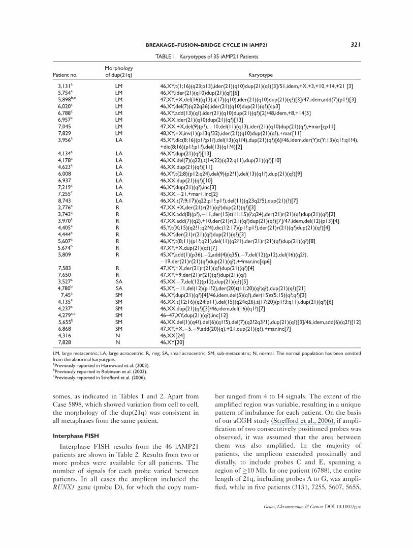
Figure 2. High resolution mBAND image of a normal chromosome21 (a) compared to the dup(21q) from patient 6937 (b). The duplicatedand inverted regions are clearly shown. [Color figure can be viewed inthe online issue, which is available at www.interscience.wiley.com.]
Genes, Chromosomes & Cancer DOI 10.1002/gcc
323BREAKAGE–FUSION–BRIDGE CYCLE IN iAMP21
banding images confirmed the complexity of the
rearrangements giving rise to dup(21q), as well as
the variability between patients, even when classi-
fied into the same morphological group (LM,
patients 3131 and 6788; LA, patients 4623, 6937,
and 3956). Figure 2 shows a higher resolution
mBAND image from patient 6937, which indicates
the duplicated and inverted segments in compari-
son to the normal chromosome 21. Although the or-
igin of the telomere was unknown, pantelomeric
probes indicated the presence of telomeric sequen-
ces on the dup(21q) chromosomes of three patients
with loss of their subtelomeres (3956, 4623, and
6937).
DISCUSSION
This study reports detailed analysis of a large
group of ALL patients with iAMP21 by FISH and
conventional cytogenetics, from which a clearer
understanding of the mechanisms giving rise to the
dup(21q) have emerged. This approach provided
conclusive evidence that the abnormality is highly
complex, with each patient having a unique
dup(21q) genomic profile. This was further sup-
ported by the range of morphological forms
observed at the chromosomal level. It demon-
strated that the amplicon was usually large, with
considerable variability in the level of amplifica-
tion, both within the amplicon and between
patients. FISH confirmed that the highly amplified
region of 21q always included the RUNX1 gene,
which corresponded to the CRA indicated in our
previous study. In 77% of patients, an accompany-
ing deletion of variable size around the subtelo-
meric region was present, corresponding to the pre-
viously identified CRD (Strefford et al., 2006).
The distribution of signals from the individual
locus-specific probes observed in metaphase indi-
cated that the dup(21q) was formed as a result of
multiple breaks and reunions, leading to complex
intrachromosomal rearrangements, including dele-
tions, duplications, inversions, and amplifications.
The complexity of the rearrangements was high-
lighted by the variable mBAND and G-banding
patterns. There was some evidence from inter-
phase FISH that the dup(21q) chromosome may
be duplicated in some patients, but it was not pos-
sible to confirm this in metaphase.
From solid tumor studies, there is increasing evi-
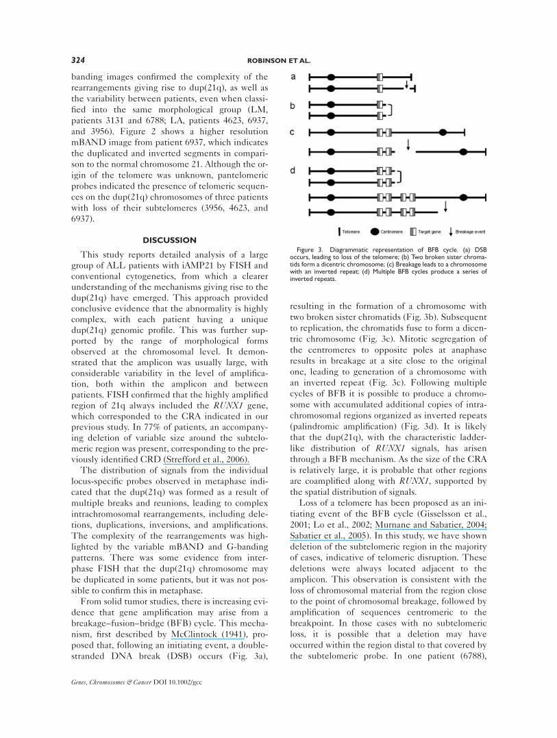
dence that gene amplification may arise from a
breakage–fusion–bridge (BFB) cycle. This mecha-
nism, first described by McClintock (1941), pro-
posed that, following an initiating event, a double-
stranded DNA break (DSB) occurs (Fig. 3a),
resulting in the formation of a chromosome with
two broken sister chromatids (Fig. 3b). Subsequent
to replication, the chromatids fuse to form a dicen-
tric chromosome (Fig. 3c). Mitotic segregation of
the centromeres to opposite poles at anaphase
results in breakage at a site close to the original
one, leading to generation of a chromosome with
an inverted repeat (Fig. 3c). Following multiple
cycles of BFB it is possible to produce a chromo-
some with accumulated additional copies of intra-
chromosomal regions organized as inverted repeats
(palindromic amplification) (Fig. 3d). It is likely
that the dup(21q), with the characteristic ladder-
like distribution of RUNX1 signals, has arisen
through a BFB mechanism. As the size of the CRA
is relatively large, it is probable that other regions
are coamplified along with RUNX1, supported by
the spatial distribution of signals.
Loss of a telomere has been proposed as an ini-
tiating event of the BFB cycle (Gisselsson et al.,
2001; Lo et al., 2002; Murnane and Sabatier, 2004;
Sabatier et al., 2005). In this study, we have shown
deletion of the subtelomeric region in the majority
of cases, indicative of telomeric disruption. These
deletions were always located adjacent to the
amplicon. This observation is consistent with the
loss of chromosomal material from the region close
to the point of chromosomal breakage, followed by
amplification of sequences centromeric to the
breakpoint. In those cases with no subtelomeric
loss, it is possible that a deletion may have
occurred within the region distal to that covered by
the subtelomeric probe. In one patient (6788),
Figure 3. Diagrammatic representation of BFB cycle. (a) DSBoccurs, leading to loss of the telomere; (b) Two broken sister chroma-tids form a dicentric chromosome; (c) Breakage leads to a chromosomewith an inverted repeat; (d) Multiple BFB cycles produce a series ofinverted repeats.
Genes, Chromosomes & Cancer DOI 10.1002/gcc
324 ROBINSON ETAL.
amplification of the subtelomeric region was
observed, consistent with breakage in the adjacent
distally located region, in a manner similar to that
reported for mouse embryonic stem cells (Lo et al.,
2002). Further support comes from the dup(21q) of
patient (3131), in whom the subtelomeric signal
was found in an unexpected location, indicating
that a break must have occurred within a region
distal to this probe. In five patients with no subte-
lomeric deletion and three with gain of probe G, it
was not possible to confirm the location of these
signals in metaphase. Thus, it cannot be ruled out
that an additional copy(ies) of a chromosome 21
intact telomeric region was present, as observed in
two patients (3131 and 3745) with subpopulations
including an extra copy of chromosome 21 seen by
conventional cytogenetics.
Although loss or dysfunction of the telomeric
region may prove to be the initiating event, the
complexity of the dup(21q) indicated that break-
age must also have occurred in other regions of the
chromosome. Previous studies have indicated that
amplification of genes may result from breakage
within locations in close proximity to fragile sites
(Ciullo et al., 2002). Although there are no reports
of fragile sites located to chromosome 21, Hattori
et al. (2000) reported duplicated sequences on
chromosome 21, which may be susceptible to DSB
(Kolomietz et al., 2002).
The BFB cycle usually ends when the unstable
chromosome acquires a new telomere. Acquisition
of a new telomere may result from a number of
mechanisms including: nonreciprocal translocations,
breakage induced replication, direct addition by
telomerase, the formation of a dicentric or ring chro-
mosome (Murnane and Sabatier, 2004). Thus, the
loss of one telomere may directly affect the stability
of the whole genome (Hattori et al., 2000; Ciullo
et al., 2002; Gisselsson, 2005; Sabatier et al., 2005). It
is likely that the more efficient the cell is at telomere
acquisition the less complex the karyotype and the
lower the level of amplification. It was of note that
the level of amplification in the iAMP21 patients
was relatively low (5–14 copies), raising the possibil-
ity that stabilization of this abnormality occurs early,
thus inhibiting high level amplification.
Firm evidence to support this hypothesis of a
BFB cycle giving rise to iAMP21 would be convinc-
ingly supported by the observation of anaphase
bridges in bone marrow smears or biopsies. Although
no such material was available for these patients, it
is unlikely that bridges would have been seen
because, apart from a single case, all dup(21q) chro-
mosomes were identical between cells of the same
patient. This implicates that the cycle may have ter-
minated and the chromosome become stable by the
time the diagnostic sample had been taken.
In this study, we have used a FISH approach in
an attempt to define further the mechanism giving
rise to iAMP21. A high level of chromosomal insta-
bility and subsequent karyotype complexity of the
dup(21q) chromosome has indicated that it has
arisen from a BFB cycle. The initiating event giv-
ing rise to this mechanism remains, as yet, unre-
solved. This study has confirmed that RUNX1 is
always located within the amplicon. Therefore,
FISH with probes directed to the RUNX1 gene
remains the most reliable detection method for
this high-risk abnormality: iAMP21.
ACKNOWLEDGMENTS
The authors thank the United Kingdom Cancer
Cytogenetics Group laboratories for contribution of
fixed cell suspensions, the Leukemia Research Cy-
togenetics Group for technical support and discus-
sion. This study could not have been performed
without the dedication of the United KingdomMed-
ical Research Council Childhood and Adult leuke-
mia Working Parties and their members, who have
designed and coordinated the clinical trials through
which these patients were identified and treated.
REFERENCES
Andersen MK, Christiansen DH, Pedersen-Bjergaard J. 2005.Amplification or duplication of chromosome band 21q22 withmultiple copies of the AML1 gene and mutation of the TP53gene in therapy-related MDS and AML. Leukemia 19:197–200.
Busson-Le Coniat M, Nguyen KF, Daniel MT, Bernard OA, BergerR. 2001. Chromosome 21 abnormalities with AML1 amplificationin acute lymphoblastic leukemia. Genes Chromosomes Cancer32:244–249.
Cameron ER, Neil JC. 2004. The Runx genes: Lineage-specificoncogenes and tumor suppressors. Oncogene 23:4308–4314.
Chudoba I, Hickmann G, Friedrich T, Jauch A, Kozlowski P, SengerG. 2004. mBAND: A high resolution multicolor banding tech-nique for the detection of complex intrachromosomal aberrations.Cytogenet Genome Res 104:390–393.
Ciullo M, Debily MA, Rozier L, Autiero M, Billault A, Mayau V, ElMarhomy S, Guardiola J, Bernheim A, Coullin P, Piatie-TonneauD, Debatisse M. 2002. Initiation of the breakage-fusion-bridgemechanism through common fragile site activation in humanbreast cancer cells: The model of PIP gene duplication from abreak at FRA7I. Hum Mol Genet 11:2887–2894.
Dal Cin P, Atkins L, Ford C, Ariyanayagam S, Armstrong SA,George R, Cleary A, Morton CC. 2001. Amplification of AML1 inchildhood acute lymphoblastic leukemias. Genes ChromosomesCancer 30:407–409.
Gisselsson D. 2005. Mitotic instability in cancer: Is there method inthe madness? Cell Cycle 4:1007–1010.
Gisselsson D, Jonson T, Petersen A, Strombeck B, Dal Cin P,Hoglund M, Mitelman F, Mertens F, Mandahl N. 2001. Telomeredysfunction triggers extensive DNA fragmentation and evolutionof complex chromosome abnormalities in human malignanttumors. Proc Natl Acad Sci USA 98:12683–12688.
Harewood L, Robinson H, Harris R, Al Obaidi MJ, Jalali GR, Marti-neau M, Moorman AV, Sumption N, Richards S, Mitchell C, Har-rison CJ. 2003. Amplification of AML1 on a duplicated chromo-some 21 in acute lymphoblastic leukemia: A study of 20 cases.Leukemia 17:547–553.
Genes, Chromosomes & Cancer DOI 10.1002/gcc
325BREAKAGE–FUSION–BRIDGE CYCLE IN iAMP21
Harrison CJ, Martineau M, Secker-Walker LM. 2001. The Leukae-mia Research Fund/United Kingdom Cancer Cytogenetics GroupKaryotype Database in acute lymphoblastic leukaemia: A valua-ble resource for patient management. Br J Haematol 113:3–10.
Harrison CJ, Moorman AV, Barber KE, Broadfield ZJ, Cheung KL,Harris RL, Jalali GR, Robinson HM, Strefford JC, Stewart A,Wright S, Griffiths M, Ross FM, Harewood L, Martineau M.2005. Interphase molecular cytogenetic screening for chromo-somal abnormalities of prognostic significance in childhood acutelymphoblastic leukaemia: A UK Cancer Cytogenetics GroupStudy. Br J Haematol 129:520–530.
Hattori M, Fujiyama A, Taylor TD, Watanabe H, Yada T, Park HS,Toyoda A, Ishii K, Totoki Y, Choi DK, Groner Y, Soeda E, Ohki M,Takagi T, Sakaki Y, Taudien S, Blechschmidt K, Polley A, MenzelU, Delabar J, Kumpf K, Lehmann R, Patterson D, Reichwald K,Rump A, Schillhabel M, Schudy A, Zimmermann W, Rosenthal A,Kudoh J, Schibuya K, Kawasaki K, Asakawa S, Shintani A, Sasaki T,Nagamine K, Mitsuyama S, Antonarakis SE, Minoshima S, ShimizuN, Nordsiek G, Hornischer K, Brant P, Scharfe M, Schon O, DesarioA, Reichelt J, Kauer G, Blocker H, Ramser J, Beck A, Klages S,Hennig S, Riesselmann L, Dagand E, Haaf T, Wehrmeyer S,Borzym K, Gardiner K, Nizetic D, Francis F, Lehrach H, ReinhardtR, Yaspo ML. 2000. The DNA sequence of human chromosome21. Nature 405:311–319.
Hilgenfeld E, Padilla-Nash H, McNeil N, Knutsen T, Montagna C,Tchinda J, Horst J, Ludwig WD, Serve H, Buchner T, Berdel WE,Schrock E, Ried T. 2001. Spectral karyotyping and fluorescence insitu hybridization detect novel chromosomal aberrations, a recurringinvolvement of chromosome 21 and amplification of the MYC onco-gene in acutemyeloid leukaemiaM2. Br JHaematol 113:305–317.
ISCN. 2005. An International System for Human CytogeneticNomenclature. Basel, Switzerland: Karger, pp. 1–130.
Kolomietz E, Meyn MS, Pandita A, Squire JA. 2002. The role of Alurepeat clusters as mediators of recurrent chromosomal aberrationsin tumors. Genes Chromosomes Cancer 35:97–112.
Le Coniat M, Romana SP, Berger R. 1995. Partial chromosome 21amplification in a child with acute lymphoblastic leukemia. GenesChromosomes Cancer 14:204–209.
Lo AW, Sprung CN, Fouladi B, Pedram M, Sabatier L, Ricoul M,Reynolds GE, Murnane JP. 2002. Chromosome instability as aresult of double-strand breaks near telomeres in mouse embryonicstem cells. Mol Cell Biol 22:4836–4850.
McClintock B. 1941. The stability of broken ends of chromosomesin Zea mays. Genetics 41:234–282.
Mikhail FM, Serry KA, Hatem N, Mourad ZI, Farawela HM, ElKaffash DM, Coignet L, Nucifora G. 2002. AML1 gene over-expression in childhood acute lymphoblastic leukemia. Leukemia16:658–668.
Mikhail FM, Sinha KK, Saunthararajah Y, Nucifora G. 2006. Normaland transforming functions of RUNX1: A perspective. J CellPhysiol 207:582–593.
Miyoshi H, Shimizu K, Kozu T, Maseki N, Kaneko Y, Ohki M.1991. t(8;21) breakpoints on chromosome 21 in acute myeloid leu-kemia are clustered within a limited region of a single gene,AML1. Proc Natl Acad Sci USA 88:10431–10434.
Moorman AV, Richards SM, Martineau M, Cheung KL, RobinsonHM, Jalali GR, Broadfield ZJ, Harris RL, Taylor KE, Gibson BE,Hann IM, Hill FG, Kinsey SE, Eden TO, Mitchell CD, HarrisonCJ. 2003. Outcome heterogeneity in childhood high-hyperdiploidacute lymphoblastic leukemia. Blood 102:2756–2762.
Moorman AV, Richards SM, Robinson HM, Strefford JC, GibsonBE, Kinsey SE, Eden OB, Vora AJ, Mitchell CD, Harrison CJ.Prognosis of children with acute lymphoblastic leukaemia (ALL)
and intra-chromosomal amplification of chromosome 21 (iAMP21).DOI: 10.1182/BLOOD-2006-08-040436 (online advance publica-tion).
Morel F, Herry A, Le Bris M-J, Douet-Guilbert N, Le Calvez G,Marion V, Berthou C, De Braekeeler M. 2002. AML1 amplifica-tion in a case of childhood acute lymphoblastic leukemia. CancerGenet Cytogenet 137:142–145.
Murnane JP, Sabatier L. 2004. Chromosome rearrangements result-ing from telomere dysfunction and their role in cancer. Bioessays26:1164–1174.
Niini T, Kanerva J, Vettenranta K, Saarinen-Pihkala UM, KnuutilaS. 2000. AML1 gene amplification: A novel finding in childhoodacute lymphoblastic leukemia. Haematologica 85:362–366.
Nucifora G, Rowley JD. 1995. AML1 and the 8;21 and 3;21 translo-cations in acute and chronic myeloid leukemia. Blood 86:1–14.
Osato M, Asou N, Abdalla E, Hoshino K, Yamasaki H, Okubo T,Suzushima H, Takatsuki K, Kanno T, Shigesada K, Ito Y. 1999.Biallelic and heterozygous point mutations in the runt domain ofthe AML1/PEBP2aB gene associated with myeloblastic leuke-mias. Blood 93:1817–1824.
Penther D, Preudhomme C, Talmant P, Roumier C, Godon A,Mechinaud F, Milpied N, Bataille R, Avet-Loiseau H. 2002.Amplification of AML1 gene is present in childhood acute lym-phoblastic leukemia but not in adult, and is not associated withAML1 gene mutation. Leukemia 16:1131–1134.
Robinson HM, Broadfield ZJ, Cheung KL, Harewood L, Harris RL,Jalali GR, Martineau M, Moorman AV, Taylor KE, Richards S,Mitchell C, Harrison CJ. 2003. Amplification of AML1 in acutelymphoblastic leukemia is associated with a poor outcome. Leu-kemia 17:2249–2250.
Romana SP, Poirel H, Leconiat M, Flexor MA, Mauchauffe M, Jon-veaux P, Berger R, Bernard OA. 1995. High frequency of t(12;21)in childhood B-lineage acute lymphoblastic leukemia. Blood86:4263–4269.
Roumier C, Fenaux P, Lafage M, Imbert M, Eclache V, Preud-homme C. 2003. New mechanisms of AML1 gene alteration inhematological malignancies. Leukemia 17:9–16.
Sabatier L, Ricoul M, Pottier G, Murnane JP. 2005. The loss of a sin-gle telomere can result in instability of multiple chromosomes ina human tumor cell line. Mol Cancer Res 3:139–150.
Soulier J, Trakhtenbrot L, Najfeld V, Lipton JM, Mathew S, Avet-Loiseau H, De Braekeleer M, Salem S, Baruchel A, RaimondiSC, Raynaud SD. 2003. Amplification of band q22 of chromosome21, including AML1, in older children with acute lymphoblasticleukemia: An emerging molecular cytogenetic subgroup. Leuke-mia 17:1679–1682.
Strefford JC, Van Delft FW, Robinson HM, Worley H, YiannikourisO, Selzer R, Richmond T, Hann I, Bellotti T, Raghavan M, YoungBD, Saha V, Harrison CJ. 2006. Complex genomic alterations andgene expression in acute lymphoblastic leukemia with intrachro-mosomal amplification of chromosome 21. Proc Natl Acad SciUSA 103:8167–8172.
United Kingdom Cancer Cytogenetics Group. 1992. Primary, single,autosomal trisomies associated with haematological disorders.United Kingdom Cancer Cytogenetics Group (UKCCG). LeukRes 16:841–851.
Van Delft FW, Bellotti T, Luo Z, Jones LK, Patel N, YiannikourisO, Hill AS, Hubank M, Kempski H, Fletcher D, Chaplin T, FootN, Young BD, Hann IM, Gammerman A, Saha V. 2005. Prospec-tive gene expression analysis accurately subtypes acute leukaemiain children and establishes a commonality between hyperdiploidyand t(12;21) in acute lymphoblastic leukaemia. Br J Haematol130:26–35.
Genes, Chromosomes & Cancer DOI 10.1002/gcc
326 ROBINSON ETAL.