Interfacial tension for various organic-water systems and study ...

FEBS Letters 586 (2012) 526–535
journal homepage: www.FEBSLetters .org
Review
Interfacial electrochemical electron transfer in biology – Towards the levelof the single molecule
Jingdong Zhang, Qijin Chi, Allan G. Hansen, Palle S. Jensen, Princia Salvatore, Jens Ulstrup ⇑Department of Chemistry, DTU Chemistry, Building 207, Kemitorvet, Technical University of Denmark, DK 2800 Kongens Lyngby, Denmark
a r t i c l e i n f o a b s t r a c t
Article history:Received 8 August 2011Revised 4 October 2011Accepted 11 October 2011Available online 20 October 2011
Edited by Miguel Teixeira andRicardo O. Louro
Keywords:Protein film voltammetryScanning tunneling microscopySingle-crystal electrode surfacesMetalloproteins
0014-5793/$36.00 � 2011 Federation of European Biodoi:10.1016/j.febslet.2011.10.023
⇑ Corresponding author.E-mail address: [email protected] (J. Ulstrup).
Physical electrochemistry has undergone a remarkable evolution over the last few decades, inte-grating advanced techniques and theory from solid state and surface physics. Single-crystal elec-trode surfaces have been a core notion, opening for scanning tunnelling microscopy directly inaqueous electrolyte (in situ STM). Interfacial electrochemistry of metalloproteins is presently goingthrough a similar transition. Electrochemical surfaces with thiol-based promoter molecular mono-layers (SAMs) as biomolecular electrochemical environments and the biomolecules themselves havebeen mapped with unprecedented resolution, opening a new area of single-molecule bioelectro-chemistry. We consider first in situ STM of small redox molecules, followed by in situ STM ofthiol-based SAMs as molecular views of bioelectrochemical environments. We then address electrontransfer metalloproteins, and multi-centre metalloenzymes including applied single-biomolecularperspectives based on metalloprotein/metallic nanoparticle hybrids.� 2011 Federation of European Biochemical Societies. Published by Elsevier B.V. All rights reserved.
1. Introduction
Interfacial electrochemistry of biological molecules and macro-molecules such as redox metalloproteins, their amino acid buildingblocks, DNA components, biomimetic lipid membranes, and bioin-organic ‘‘hybrids’’ of metallic nanoparticles and metalloproteins, ismoving towards new levels of understanding. Key notions areimaging of the biomolecules at the electrochemical interface, andelectron transfer (ET) and even enzyme processes followed to-wards the single molecule level of resolution.
Interfacial electrochemical ET in biology towards unprece-dented levels of resolution follows fruitful interactions among sev-eral areas, rooted in physical electrochemistry closely associatedwith surface and condensed matter physics, biotechnology, andnew instrumentation. The latter includes scanning tunnelling andatomic force microscopy, operating directly in aqueous biologicalmedia under electrochemical potential control (in situ STM andAFM) [1–3]. In situ STM/AFM now extend well beyond imaging(which remains a daunting challenge for large and fragile biomol-ecules) and has also been brought to map and control biomolecularfunction such as ET of even large, multi-centre metalloproteins, orprotein unfolding and DNA unzipping. The introduction of single-crystal, (‘‘well-defined’’) atomically planar electrode surfaces wasa major breakthrough that also laid the foundation for other elec-trochemical technology such as a range of spectroscopic surface
chemical Societies. Published by E
techniques as well as statistical mechanical and electronic struc-tural theories [4–7]. In addition, in situ imaging of single biomole-cules rests on biotechnology such as mutant and syntheticmetalloproteins. Altogether this has led bioelectrochemistry to-wards similar boundary-traversing results as previously in physi-cal electrochemistry.
An essential pre-requisite in protein monolayer voltammetry(PMV) and single-biomolecular ET is understanding of the underly-ing molecular ET (and proton transfer) processes. Molecular chargetransport theory has continued to develop [8–13], more recently asa theoretical frame for in situ STM and other condensed matter sin-gle-molecule conductivity phenomena [14]. Single-molecule(in situ) STM mapping rests on molecular tunnelling conductivity,rather than topographic shape. At least two-fold theoretical sup-port is therefore needed. Electronic structure computations have,first disclosed sometimes unexpected, details in the STM contrastsof ‘‘small’’ (bio)molecules such as single amino acids and relatedmolecules [15,16]. Solvation is, further crucial and computationallydemanding for in situ STM of electrostatically charged moleculessuch as functionalized alkanethiol SAMs [17]. Secondly, othernew challenges arise for large redox molecules and biological(macro)molecules such as metalloproteins where molecular ETtheory has been a powerful tool [14]. By their analytical form, these‘‘phenomenological’’ theories have offered immediate insight intocurrent/overpotential and other in situ tunnelling correlationsand disclosed even new ET phenomena. The combination ofprotein biotechnology with well-defined (single-crystal) electro-chemical interfaces and in situ STM/AFM offer other perspectives
lsevier B.V. All rights reserved.

Fig. 1. Left: Schematic view of redox molecules enclosed between a STM working electrode and tip or between a pair of nanogap electrodes. Right: Electronic energy schemefor the redox molecule initially in the oxidized state.
J. Zhang et al. / FEBS Letters 586 (2012) 526–535 527
in bioelectrochemical signal transfer between target molecules andexternal biosensing electrochemical circuitry.
2. Interfacial ET in molecular and protein monolayervoltammetry (PMV)
In situ STM maging and image interpretation rest funda-mentally on interfacial electrochemical ET processes. PMV iscrucial to control conditions for optimal immobilized proteinfunction. Condensed matter molecular charge transfer theory[8–10,12,13,18] offers comprehensive conceptual support for bothbioelectrochemistry of redox proteins, and for in situ STM of com-plex molecules. The theory addresses, overarchingly: (a) electrontunnelling between the electrode and donor or acceptor groupsin the biomolecules through intermediate protein ‘‘matter’’, and(b) the nuclear environmental effects from local, collective proteinor DNA, and solvent nuclear modes. In situ STM [10,18,19] offersnew theoretical challenges.
2.1. Theoretical notions of interfacial chemical and bioelectrochemicalET
Views of the electrochemical ET process carry over to (bio)elec-trochemical nanoscale systems. Notions in focus are illuminated bythe following cathodic current density form (with an analogousanodic form) broadly valid but with recognized limitations [8,9]:
Fig. 2. Left: Electrochemical control of the energy scheme in Fig. 1. The continuous electshifted by the bias voltage energy. Right: Current/overpotential relation on parallel vari
jðe; gÞ ¼ eC12oxC
12redjeff
xeff
2pexp �ðER þ egÞ2
4ERkBT
" #ð1Þ
Cox and Cred are the populations of the oxidized and reduced stateof the (bio)molecule at the electrode surface, e the electroniccharge, ER the nuclear reorganization free energy, g the overpoten-tial, xeff the effective nuclear vibrational frequency of all thenuclear modes reorganized, kB Boltzmann’s constant and T the tem-perature. jeff is the electronic transmission coefficient the mostimportant part of which is the electron exchange energy, whichcouples the molecular acceptor level (A) with metallic electroniclevels around the Fermi energy eF. Eq. (1) holds an electronic tun-nelling factor, jeff, and a nuclear activation factor that includes con-tributions from all the protein conformational, and external solventpolarization modes, and the driving force (eg). Molecular chargetransfer theory has developed into much more powerful framesthan implied by this simple formalism. A number of these areimportant in interfacial ET processes of biological macromoleculesand covered in a comprehensive literature, overviewed e.g. in[8–10,12,13,18,20,21].
2.2. The reorganization free energy and the electronic tunnelling factor
The reorganization free energy, ER holds an intramolecular andan environmental contribution. Modified forms of the quadratic
ronic energy spectra of the enclosing electrodes are indicated. The Fermi levels areation of the substrate and tip electrode potentials.
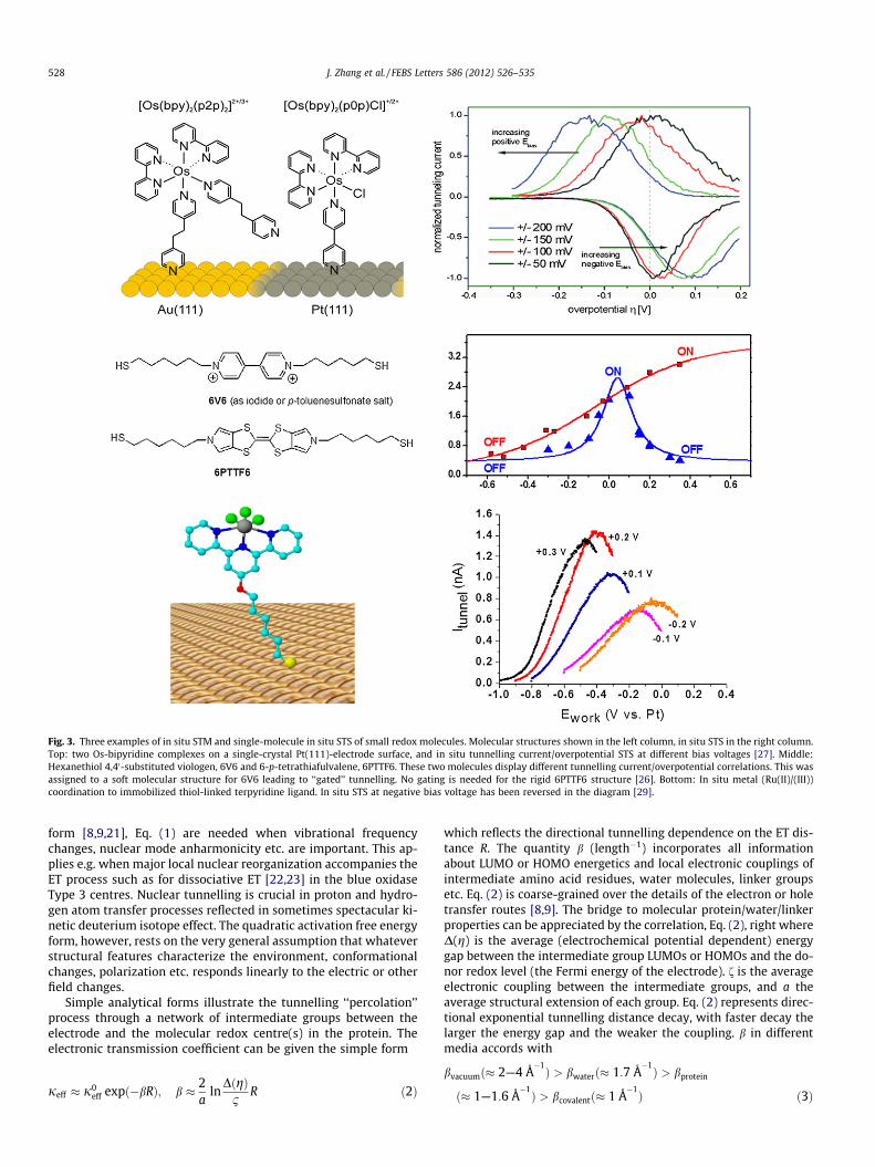
Fig. 3. Three examples of in situ STM and single-molecule in situ STS of small redox molecules. Molecular structures shown in the left column, in situ STS in the right column.Top: two Os-bipyridine complexes on a single-crystal Pt(111)-electrode surface, and in situ tunnelling current/overpotential STS at different bias voltages [27]. Middle:Hexanethiol 4,40-substituted viologen, 6V6 and 6-p-tetrathiafulvalene, 6PTTF6. These two molecules display different tunnelling current/overpotential correlations. This wasassigned to a soft molecular structure for 6V6 leading to ‘‘gated’’ tunnelling. No gating is needed for the rigid 6PTTF6 structure [26]. Bottom: In situ metal (Ru(II)/(III))coordination to immobilized thiol-linked terpyridine ligand. In situ STS at negative bias voltage has been reversed in the diagram [29].
528 J. Zhang et al. / FEBS Letters 586 (2012) 526–535
form [8,9,21], Eq. (1) are needed when vibrational frequencychanges, nuclear mode anharmonicity etc. are important. This ap-plies e.g. when major local nuclear reorganization accompanies theET process such as for dissociative ET [22,23] in the blue oxidaseType 3 centres. Nuclear tunnelling is crucial in proton and hydro-gen atom transfer processes reflected in sometimes spectacular ki-netic deuterium isotope effect. The quadratic activation free energyform, however, rests on the very general assumption that whateverstructural features characterize the environment, conformationalchanges, polarization etc. responds linearly to the electric or otherfield changes.
Simple analytical forms illustrate the tunnelling ‘‘percolation’’process through a network of intermediate groups between theelectrode and the molecular redox centre(s) in the protein. Theelectronic transmission coefficient can be given the simple form
jeff � j0eff expð�bRÞ; b � 2
aln
DðgÞ1
R ð2Þ
which reflects the directional tunnelling dependence on the ET dis-tance R. The quantity b (length�1) incorporates all informationabout LUMO or HOMO energetics and local electronic couplings ofintermediate amino acid residues, water molecules, linker groupsetc. Eq. (2) is coarse-grained over the details of the electron or holetransfer routes [8,9]. The bridge to molecular protein/water/linkerproperties can be appreciated by the correlation, Eq. (2), right whereD(g) is the average (electrochemical potential dependent) energygap between the intermediate group LUMOs or HOMOs and the do-nor redox level (the Fermi energy of the electrode). f is the averageelectronic coupling between the intermediate groups, and a theaverage structural extension of each group. Eq. (2) represents direc-tional exponential tunnelling distance decay, with faster decay thelarger the energy gap and the weaker the coupling. b in differentmedia accords with
bvacuumð� 2—4 Å�1Þ > bwaterð� 1:7 Å
�1Þ > bprotein
ð� 1—1:6 Å�1Þ > bcovalentð� 1 Å
�1Þ ð3Þ
J. Zhang et al. / FEBS Letters 586 (2012) 526–535 529
indicating that protein is a more facile tunnelling medium thanwater, in turn more facile than vacuum. bprotein also spans a rangeof values, with ba-helix
protein > b-proteinb�sheet . Eq. (2) applies, further onlywhen the tunnelling barrier is significant, DðgÞ=f� 1. At smallDðgÞ=f, tunnelling is replaced by ‘‘hopping’’ through intermediatelevels now populated physically with temporarily trapped electronsor holes. This opens a range of ET channels including vibrationallyfully or partially relaxed (‘‘dynamically populated’’) intermediatestates [8,9]. The four-heme cyt c3 class [24] is a possible examplefor which this notion may hold clues to ultra-fast ET between theclosely spaced heme groups.
3. Theoretical notions in bioelectrochemistry towards thesingle-molecule level
3.1. Some theoretical concepts
We address specifically in situ STM of redox (bio)molecules butconcepts and formalism carry over to other metallic nanogap con-figurations. In addition to the substrate and tip, a third electrodeserves as reference electrode. The three-electrode configuration isthe basis for two kinds of tunnelling ‘‘spectroscopy’’ unique toin situ STM [14]. One is the current–bias voltage relation as inSTM in air or vacuum, but with the substrate (over)potential keptconstant. The other one is the current-overpotential relation atconstant bias voltage, i.e. the substrate and tip potentials are variedin parallel relative to the reference electrode.
Fig. 1 shows a schematic view of a redox molecule in a molec-ular scale gap between two electrochemical metallic electrodes.The Fermi levels of the two electrode surfaces are separated bythe bias voltage, eVbias, at given overpotential g. The molecular re-dox level, say the oxidized, electronically ‘‘empty’’ level is firstlocated above the Fermi energy of the working electrode. Fig. 1shows explicitly the configuration for positive bias voltage, so thatthe tip Fermi level is lower than the working electrode Fermi level.If all energies are counted from the Fermi energy of the workingelectrode, eFsubstr, the empty redox level, eox, is lowered withincreasing negative g. The redox site is exposed to part of the biasvoltage and also shifted relative to eFsubstr on bias voltage variation.Conformational and solvent polarization fluctuations as in electro-chemical ET are, finally, crucial.
3.2. New interfacial (bio)electrochemical ET phenomena
The in situ STM results are most transparent when the biasvoltage is small, i.e. [10,14,18]:
cjeVbiasj < ER � eg ð4Þ
c is the fraction of the bias voltage at the site of the redox centre.The bias voltage is a ‘‘probing energy tip’’, and the ‘‘spectral resolu-tion’’ better, the narrower the ‘‘probing tip’’. As the overpotential israised, at fixed bias voltage, the cathodic current first rises due to
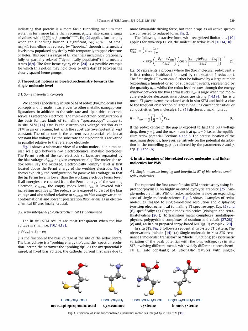
Fig. 4. Overview of some functionalized alkanet
more favourable driving force, but then drops as all active speciesare converted to reduced form, Fig. 2.
The following attractive form, with recognized limitations [19]applies for two-step ET via the molecular redox level [10,14,18]:
isymmtunn ¼
12
eno=rxeff
2p
� exp �ER � eVbias
4kBT
� �cosh
ð12� cÞeVbias � neg2kBT
� �� ��1
ð5Þ
Eq. (5) represents a process where the (bio)molecular redox centreis first reduced (oxidized) followed by re-oxidation (-reduction).The first single-ET event can, further be followed by a large number(exceeding a hundred or so) of subsequent events, represented bythe quantity no/r, whilst the redox level relaxes through the energywindow between the two Fermi levels. no/r is large when the mole-cule-electrode electronic interactions are strong [14,19]. This is anovel ET phenomenon associated with in situ STM and holds a clueto the frequent observation of large tunnelling current densities, orconductivities (per molecule). Eq. (5) discloses a maximum at
g ¼ gmax ¼1n
12� c
� �Vbias ð6Þ
If the redox centre in the gap is exposed to half the bias voltagedrop, then c ¼ 1
2, and the maximum is at gmax = 0, i.e. at the equilib-rium redox potential, Sections 4 and 5. The precise location of themaximum depends, however, sensitively on the potential distribu-tion in the tunnelling gap, as reflected by the parameters n and c,Eqs. (5) and (6).
4. In situ imaging of bio-related redox molecules and linkermolecules for PMV
4.1. Single-molecule imaging and interfacial ET of bio-related smallredox molecules
Tao reported the first case of in situ STM spectroscopy using Fe-protoporphyrin IX on highly oriented pyrolytic graphite [25]. Sin-gle-molecule in situ STM of redox molecules is now an expandingarea of single-molecule science. Fig. 3 shows examples of redoxmolecules imaged to single-molecule resolution and displayingtwo-step electrochemical tunnelling ET spectroscopy, Eqs. (5) and(6), specifically: (a) Organic redox molecules (viologen and tetra-thiafulvalene [26]); (b) transition metal complexes (metallopor-phyrins, polypyridine complexes of osmium and cobalt [27,28]);(c) and, an in situ prepared terpy-based Ru(II)/(III) complex [29].
In situ STS, Fig. 3 follows a sequential two-step ET pattern. Theobservations include [14]: (a) Single-molecule in situ STS reso-nance (‘‘molecular transistor’’ or ‘‘diode’’ function); (b) systematicvariation of the peak potential with the bias voltage; (c) in situSTS involving different metals with widely different electrochemi-cal ET rate constants; (d) stochastic features with single-,
hiol molecules imaged by in situ STM [30].

530 J. Zhang et al. / FEBS Letters 586 (2012) 526–535
double-, triple- etc. molecular conductivity; and (e) coherentmulti-ET in a single in situ STS event.
4.2. Functionalized alkanethiols
The two classes of biological chemical building blocks, aminoacids and nucleobases have been prominent targets in single-molecule imaging of biological molecules. Functionalized alkan-ethiols and cysteine (Cys) have been particularly important alsoas linker groups in PMV.
4.2.1. Functionalized alkanethiols as linkers in PMVThe class of straight or branched, pure or functionalized alkan-
ethiols are core molecular targets in single-molecule in situ STM,Fig. 4. Imaging has been closely tied to electrochemical ET of or-dered alkanethiol-based SAMs where the reductive desorptionprocess
Au� S� R þ e� ! Auþ �S� R ð12Þ
is crucial [14,30]. Alkanethiol-based SAMs have also been subject tolarge scale electronic structure computations disentangling STM inminute detail in both vacuum and aqueous environment.
Electrochemically controlled SAMs of the alkanethiol class char-acterized to molecular and sub-molecular in situ STM resolutionhas been reviewed recently [30]. We note here some issues ofimportance to functionalized alkanethiols as linker molecules forgentle immobilization of fully functional redox metalloproteinand metalloenzyme monolayers on single-crystal Au(111)-elec-trode surfaces. With a few exceptions, PMV is unstable or absentunless the electrode is modified by linker molecular SAMs, or the
Fig. 5. High-resolution in situ STM of some alkanethiol-based molecules which are alsoCysteamine [17], mercaptopropionic acid [31] and cysteine [15,16]. Bottom row Au(110)-to show the submolecular resolution, and DFT computed in situ STM image [15]. The lattegroups in the Cys molecule, they reflect the electronic densities (molecular orbitals) rat
protein modified by non-native thiol-based amino acid residues.The thiol-based linker molecules adsorb strongly on the Au-sur-face. The opposite end holds a functional group that interacts‘‘gently’’ with the protein, ascertaining that the latter retains fullfunctional integrity. The interactions are often subtle. Closely re-lated linker molecules can induce widely different voltammetricresponses, and linker groups with no immediate expectable pro-tein compatibility can arouse strong voltammetric signals.
The use of single-crystal electrodes enables surface structuralcharacterization at the atomic and molecular level for pure andmodified electrodes, directly in aqueous solution. Fig. 5 showsin situ STM of Au(111)- and Au(110)-electrode surfaces modifiedby thiol-based linker molecules, all of which form highly orderedmonolayers, controlled by electrostatic and hydrogen bond net-works [16,32]. The adsorption process can also be followed in realtime through several intermediate phases [17,33]. The in situ STMimages shown in Fig. 5 thus offer an unprecedentedly detailedview of the microenvironments for immobilized redox proteins‘‘in voltammetric action’’.
4.2.2. Theoretical computations and STM image simulationsMultifarious patterns of widely different functionalized alkane-
thiol SAMs have been mapped to single-molecule resolution byin situ STM in aqueous electrolyte solution, strongly supportedby electrochemical studies. In situ STM is, however, rooted in elec-tronic conductivity and the quantum mechanical tunnelling effect.Theoretical support is therefore essential in detailed image inter-pretation of the many facets of alkanethiol-based SAM packingand in situ STM contrasts. We refer to recent studies of this impor-tant aspect [14,15,32,34,35].
linker molecules in PMV. Top row Au(111)-electrode surfaces; from left to right:electrode surfaces, from left to right: Cysteine surface lattice, zoom-in on the latticer shows that although the three in situ STM lobes originate from the three functionalher than the position of the O-, N-, and S-atoms.
J. Zhang et al. / FEBS Letters 586 (2012) 526–535 531
5. Electrochemistry and in situ STM of single redoxmetalloprotein molecules
5.1. Metalloprotein voltammetry at bare and modified electrodes
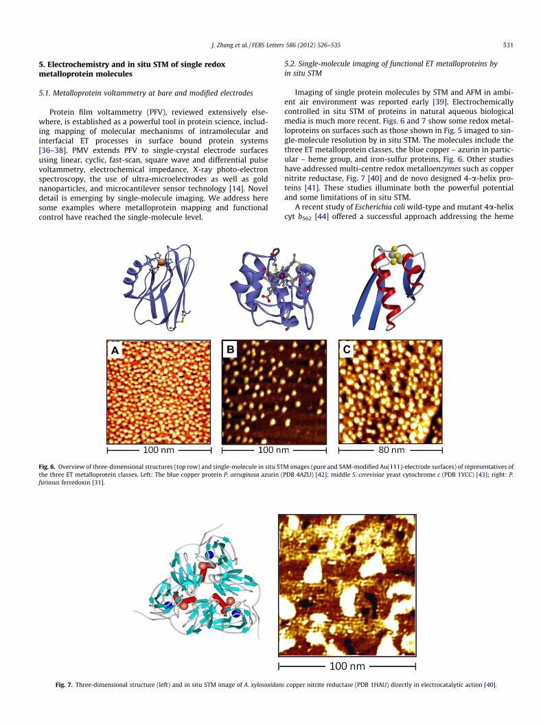
Protein film voltammetry (PFV), reviewed extensively else-where, is established as a powerful tool in protein science, includ-ing mapping of molecular mechanisms of intramolecular andinterfacial ET processes in surface bound protein systems[36–38]. PMV extends PFV to single-crystal electrode surfacesusing linear, cyclic, fast-scan, square wave and differential pulsevoltammetry, electrochemical impedance, X-ray photo-electronspectroscopy, the use of ultra-microelectrodes as well as goldnanoparticles, and microcantilever sensor technology [14]. Noveldetail is emerging by single-molecule imaging. We address heresome examples where metalloprotein mapping and functionalcontrol have reached the single-molecule level.
Fig. 6. Overview of three-dimensional structures (top row) and single-molecule in situ STthe three ET metalloprotein classes. Left: The blue copper protein P. aeruginosa azurin (furiosus ferredoxin [31].
Fig. 7. Three-dimensional structure (left) and in situ STM image of A. xylosoxidans
5.2. Single-molecule imaging of functional ET metalloproteins byin situ STM
Imaging of single protein molecules by STM and AFM in ambi-ent air environment was reported early [39]. Electrochemicallycontrolled in situ STM of proteins in natural aqueous biologicalmedia is much more recent. Figs. 6 and 7 show some redox metal-loproteins on surfaces such as those shown in Fig. 5 imaged to sin-gle-molecule resolution by in situ STM. The molecules include thethree ET metalloprotein classes, the blue copper – azurin in partic-ular – heme group, and iron-sulfur proteins, Fig. 6. Other studieshave addressed multi-centre redox metalloenzymes such as coppernitrite reductase, Fig. 7 [40] and de novo designed 4-a-helix pro-teins [41]. These studies illuminate both the powerful potentialand some limitations of in situ STM.
A recent study of Escherichia coli wild-type and mutant 4a-helixcyt b562 [44] offered a successful approach addressing the heme
M images (pure and SAM-modified Au(111)-electrode surfaces) of representatives ofPDB 4AZU) [42]; middle S. cerevisiae yeast cytochrome c (PDB 1YCC) [43]; right: P.
copper nitrite reductase (PDB 1HAU) directly in electrocatalytic action [40].
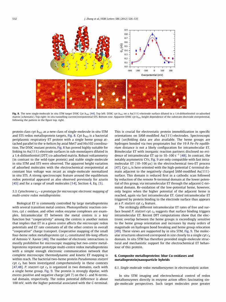
Fig. 8. The new single-molecule in situ STM target D50C Cyt b562 [44]. Top left: D50C cyt b562 on a Au(111)-electrode surface diluted in a 1,4-dithiothreitol co-adsorbedmatrix (schematic). Top right: In situ tunnelling current/overpotential STS. Bottom row: Apparent D50C cyt b562 height dependence of the substrate electrode overpotential,following the pattern in the figure top, right.
532 J. Zhang et al. / FEBS Letters 586 (2012) 526–535
protein class cyt b562 as a new class of single-molecule in situ STMand STS redox metalloprotein targets, Fig. 8. Cyt b562 is a bacterialperiplasmic respiratory ET protein with a single heme group at-tached parallel to the a-helices by axial Met7 and His102 coordina-tion. The D50C mutant protein, Fig. 8 has proved highly suitable forlinking to Au(111)-electrode surfaces in sub-monolayers diluted ina 1,4-dithiothreitol (DTT) co-adsorbed matrix. Robust voltammetry(in contrast to the wild-type protein) and stable single-moleculein situ STM and STS were observed. The apparent height variationof adsorbed molecules with the electrochemical overpotential atconstant bias voltage was recast as single-molecule normalizedin situ STS. A strong spectroscopic feature around the equilibriumredox potential appeared as also observed previously for azurin[45] and for a range of small molecules [14], Section 4, Eq. (5).
5.3. Cytochrome c4 – a prototype for microscopic electronic mapping ofmulti-centre redox metalloproteins
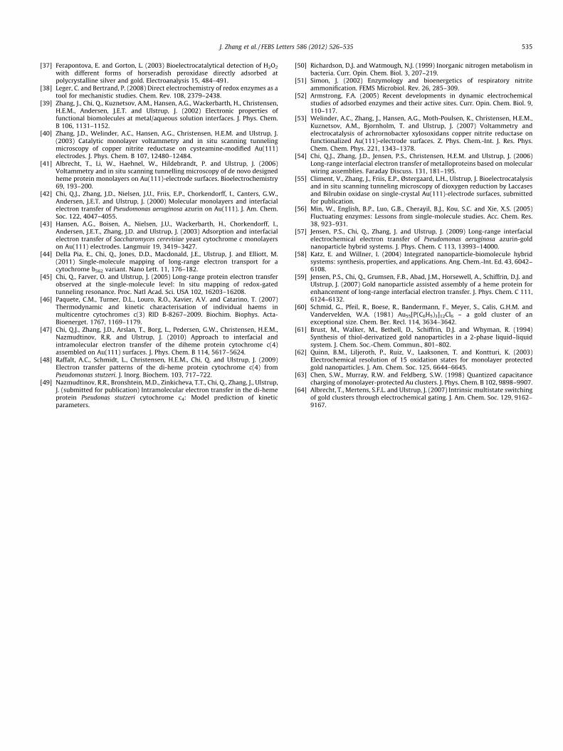
Biological ET is commonly controlled by large metalloproteinswith several transition metal centres. Photosynthetic reaction cen-tres, cyt c oxidase, and other redox enzyme complexes are exam-ples. Intramolecular ET between the metal centres is a keyfunction but ‘‘cooperativity’’ among the centres is another notionthat implies that ET in a given centre affects the microscopic redoxpotentials and ET rate constants of all the other centres in overall‘‘cooperative’’ charge transport. Cooperative mapping of the smallfour-heme redox metalloprotein cyt c3 constituted life-long effortsof Antonio V. Xavier [46]. The number of electronic interactions ismostly prohibitive for microscopic mapping but two-centre metal-loproteins represent prototype multi-centre redox metalloproteinswith a simple enough electronic communication network thatcomplete microscopic thermodynamic and kinetic ET mapping iswithin reach. The bacterial two-heme protein Pseudomonas stutzericyt c4 has been investigated comprehensively in these contexts[47,48]. P. stutzeri cyt c4 is organized in two domains, each witha single heme group, Fig. 9. The protein is strongly dipolar, withexcess positive and negative charge (pH 7) on the C- and N-termi-nal domain, respectively. The redox potential difference is about100 mV, with the higher potential associated with the C-terminal.
This is crucial for electrostatic protein immobilization in specificorientations on SAM-modified Au(111)-electrodes. Spectroscopicand (un)folding data are also available. The heme groups arehydrogen bonded via two propionates but the 19 Å Fe–Fe equilib-rium distance is not a likely configuration for intramolecular ET.Bimolecular ET with inorganic reaction partners disclosed no evi-dence of intramolecular ET up to 10–100 s�1 [48]. In contrast, thenotably asymmetric CVs, Fig. 9 are only compatible with fast intra-molecular ET (10–100 ls) in the electrochemical two-ET process[47]. Cyt c4 is here oriented with the high-potential C-terminal do-main adjacent to the negatively charged SAM-modified Au(111)-surface. This domain is reduced first in a cathodic scan followedby reduction of the remote N-terminal domain at the lower poten-tial of this group, via intramolecular ET through the adjacent C-ter-minal domain. Re-oxidation of the low-potential heme, however,only begins when the higher potential of the adjacent heme isreached, again via fast intramolecular ET. Gated intramolecular ETtriggered by protein binding to the electrode surface thus appearsas a P. stutzeri cyt c4 feature.
The strikingly different intramolecular ET rates of free and sur-face bound P. stutzeri cyt c4 suggests that surface binding triggersintramolecular ET. Recent DFT computations show that the elec-tronic overlap between the heme groups is exceedingly sensitiveto the heme group orientation and increases by many orders ofmagnitude on hydrogen bond breaking and heme group relocation[49]. These views are supported by in situ STM, Fig. 9. The molec-ular structures observed correspond in size closely to a single cyt c4
domain. In situ STM has therefore provided single-molecule struc-tural and mechanistic support for the electrochemical ET behav-iour of this protein.
6. Composite metalloproteins: blue Cu-oxidases andmetalloprotein/nanoparticle hybrids
6.1. Single-molecule redox metalloenzymes in electrocatalytic action
In situ STM imaging and electrochemical control of redoxmetalloenzymes directly in enzyme action offers fascinating sin-gle-molecule perspectives. Such target molecules pose greater

Fig. 9. Left: P. stutzeri cyt c4 (PDB 1EPT) on a Au(111)-electrode surface modified by a mercaptodecanoic acid SAM. The positively charged high-potential C-terminal domainmarked in red, the negatively charged N-terminal domain in blue. Top right: Cyclic voltammogram of P. stutzeri cyt c4. The asymmetric appearance reflects the orientation ofthe molecule and intramolecular ET between the two heme groups as a core feature. Bottom right: In situ STM of P. stutzeri cyt c4 on the mercaptodecanoic acid SAM modifiedAu(111)-electrode surface. The bright spots are individual P. stutzeri cyt c4 molecules. The ‘‘spot’’ size corresponds to that of a single domain and supports the view of theprotein in an upright orientation [47].
J. Zhang et al. / FEBS Letters 586 (2012) 526–535 533
challenges than the smaller ET proteins. Recent – so far very few –studies illuminate these perspectives. The nitrite reductases (NiRs)are central in the biological nitrogen cycle where they catalyze thereduction of nitrite to lower oxidation states [50,51]. Rationales forthe trimeric CuNiRs (each monomer molecular mass � 36 kDa,here represented by Achromobacter Xylosoxidans CuNiR) as single-molecule in situ STM targets are, first that each monomer containsa type I blue copper centre for electron inlet, here from the workingelectrode, and a type II centre for catalytic NO2
� -reduction. Thetwo centres are directly linked, offering facile intramolecular ET,cf. cyt c4. The enzyme substrate nitrite is, secondly a smallmolecule not detectable by in situ STM on the enzyme background.As frequently observed in enzyme voltammetry, binding ofsubstrate induces, however, significant electronic changes in theenzyme [40,52,53], notably in the contact between the electronacceptor centre and the electrode surface. This resembles againcyt c4 and offers prospects for electronic mapping of the enzymein action at the single-molecule level.
A. xylosoxidans CuNiR is electrocatalytically active on SAM-mod-ified Au(111)-electrode surfaces [40,53]. The voltammetric pat-terns are controlled by subtle hydrophilic and hydrophobicsurface combinations of many surface linker molecules tested. Anotable outcome is that the enzyme ‘‘in action’’ on cysteamine-and benzylthiol-modified Au(111)-electrode surfaces can beimaged in action at the single-molecule level, Fig. 7 [40,54]. En-zyme molecules even with the triangular crystallographic CuNiRstructure are observed, and the molecular-scale contrasts only ap-pear when nitrite is present. A recent study has disclosed a similarpattern for fungal laccase (Streptomyces coelicolor) for which
substrate dioxygen also triggers intramolecular ET and strongin situ STM contrasts [55]. In situ STM of CuNiR and the laccaseshas thus opened the area of single-molecule electrochemical redoxmetalloenzyme activity. These novel observations can be com-pared with more established single-molecule fluorescence-basedenzyme kinetics [14,56].
6.2. Electrochemistry of Au-nanoparticle/metalloprotein hybrids
Inorganic particles and other structures have reached the sizerange of biomolecules such as proteins. This is also the rangewhere electronic properties transform from macroscopic to sin-gle-molecule behaviour. The combination of inorganic metallic orsemiconductor structures with comparable-size (bio)moleculesinto ‘‘hybrid’’ structures has become a new core notion [57–59].
Solute monolayer-coated (‘‘capped’’) AuNPs are central in col-loid and surface science. Facile chemical synthesis introduced bySchmid [60] and by Brust and Schiffrin [61] have strongly boostedAuNP and other metal-NP science. The smallest, i.e.61 nm particlesbehave like a similar-size molecule with a discrete electronic spec-trum or a wide HOMO/LUMO gap [62–64]. Intermediate-size AuN-Ps, say 1.6 nm (Au145) to 2.5 nm (many hundred Au-atoms) displayCoulomb charging effects [64] while the Coulomb energy spacingsin larger AuNPs (P 3–5 nm) are too close for discreteness-of-chargeeffects. Protected AuNPs combined with redox metalloproteins into‘‘hybrids’’, are important in bioelectrocatalysis, with links to biolog-ical diagnostics [58]. A recent study of a ‘‘hybrid’’ between a 3 nmAuNP and P. aeruginosa azurin illuminates interfacial electrochem-ical ET and in situ STM of a protein/NP hybrid [57].

534 J. Zhang et al. / FEBS Letters 586 (2012) 526–535
P. aeruginosa azurin can be linked by strong hydrophobic forcesto alkanethiol-protected 3 nm AuNPs in turn immobilized on aAu(111)-electrode surface via an aromatic 4,40-biphenyl-dithiollinker. Notably, the AuNP hydrophobically linked to azurin in-creases the ET rate by at least an order of magnitude comparedto azurin alone on similar alkanethiol-modified surfaces, in spiteof a 4 nm ET distance increase. A two-step, azurin/AuNP andAuNP/electrode ET mechanism accords with the data. The dual vol-tammetric pattern also accords with dual in situ STM contrastswith a weaker, fluctuating contrast assigned to the AuNP/azurinhybrid and a stronger robust contrast to displaced AuNPs or azurinmolecules.
7. Concluding observations and some perspectives
Single-crystal, atomically planar electrode surfaces have pavedthe way for in situ STM and AFM in bioelectrochemistry. In situSTM has increased structural resolution of metalloproteins andsmall biomolecules on (bio)electrochemical electrode surfaces tothe molecular and sometimes sub-molecular levels. Dynamic phe-nomena such as phase transitions and the monolayer formationprocess can also be followed. The image detail in both individualadsorbate molecules and their lateral organization offers under-standing of the interaction of ‘‘biological liquids’’ with solid sur-faces. As in situ STM is based on molecular electronicconductivity, we have also included a short theoretical discussionof single-molecule interfacial ET processes, with focus on ‘‘natural’’biomolecular aqueous solution environment, and electrochemi-cally controlled (bio)molecular function. Single-molecule resolu-tion of the molecules (particularly proteins) fully active in ET orenzyme function is achieved. Not only structural mapping but alsoET, redox enzyme function, and cooperative phenomena can be ad-dressed, illuminated by azurin, cyt b562, cyt c4, and CuNiR. Biomo-lecular ‘‘electronics’’, enzyme electrochemistry, and biologicalsingle-molecule screening are attractive applied perspectives ofthe new bioelectrochemistry. Fundamental bioelectrochemicalinnovation including theoretical support remains, however, crucialpre-requisites.
Acknowledgement
This work is supported by the Danish Research Council forTechnology and Production, the Lundbeck Foundation, and the Vil-lum Kann Rasmussen Foundation.
References
[1] Lustenberger, P., Rohrer, H., Christoph, R. and Siegenthaler, H. (1988) Scanningtunneling microscopy at potential controlled electrode surfaces in electrolyticenvironment. J. Electroanal. Chem. 243, 225–235.
[2] Wiechers, J., Twomey, T., Kolb, D.M. and Behm, R.J. (1988) An in situ scanningtunneling microscopy study of Au(111) with atomic scale resolution. J.Electroanal. Chem. 248, 451–460.
[3] Gewirth, A.A. and Niece, B.K. (1997) Electrochemical applications of in situscanning probe microscopy. Chem. Rev. 97, 1129–1162.
[4] Wieckowski, A., Ed., (1999). Interfacial Electrochemistry. Theory, Experimentand Applications, Marcel Dekker, New York.
[5] Carnie, S.L. and Torrie, G.M. (1984) The statistical-mechanics of the electricaldouble-layer. Adv. Chem. Phys. 56, 141–253.
[6] Kornyshev, A.A. (1988) Solvation of a metal surface in: The Chemical Physics ofSolvation. Part C. Solvation Phenomena in Specific Physical Chemical andBiological Systems (Dogonadze, R.R., Kálmán, E., Kornyshev, A.A. and Ulstrup,J., Eds.), pp. 355–400, Elsevier, Amsterdam.
[7] Kolb, D.M. (2001) Electrochemical surface science. Ang. Chem.-Int. Ed. 40,1162–1181.
[8] Kuznetsov, A.M. (1995) Charge Transfer in Physics, Chemistry and Biology,Gordon & Breach, Reading.
[9] Kuznetsov, A.M. and Ulstrup, J. (1999) Electron Transfer in Chemistry andBiology: An Introduction to the Theory, Wiley, Chichester.
[10] Kuznetsov, A.M. and Ulstrup, J. (2000) Mechanisms of in situ scanningtunnelling microscopy of organized redox molecular assemblies. J. Phys.Chem. A 104, 11531–11540.
[11] Marcus, R.A. (1956) On the theory of oxidation-reduction reactions involvingelectron transfer 1. J. Chem. Phys. 24, 966–978.
[12] Iversen, G., Kharkats, Y.I. and Ulstrup, J. (1998) Simple dielectric image chargemodels for electrostatic interactions in metalloproteins. Mol. Phys. 94, 297–306.
[13] Kuznetsov, A.M. and Ulstrup, J. (1982) On the Theory of Long-Range ElectronHopping in Polar Media. Phys. Status Solidi B: Basic Res. 114, 673–683.
[14] Zhang, J.D., Kuznetsov, A.M., Medvedev, I.G., Chi, Q.J., Albrecht, T., Jensen, P.S.and Ulstrup, J. (2008) Single-molecule electron transfer in electrochemicalenvironments. Chem. Rev. 108, 2737–2791.
[15] Zhang, J.D., Chi, Q.J., Nazmutdinov, R.R., Zinkicheva, T.T. and Bronshtein, M.D.(2009) Submolecular electronic mapping of single cysteine molecules byin situ scanning tunneling imaging. Langmuir 25, 2232–2240.
[16] Zhang, J.D., Chi, Q.J., Nielsen, J.U., Friis, E.P., Andersen, J.E.T. and Ulstrup, J.(2000) Two-dimensional cysteine and cystine cluster networks on Au(111)disclosed by voltammetry and in situ scanning tunneling microscopy.Langmuir 16, 7229–7237.
[17] Zhang, J.D., Bilic, A., Reimers, J.R., Hush, N.S. and Ulstrup, J. (2005) Coexistenceof multiple conformations in cysteamine monolayers on Au(111). J. Phys.Chem. B 109, 15355–15367.
[18] Kuznetsov, A.M. and Ulstrup, J. (2001) Mechanisms of in situ scanningtunnelling microscopy of organized redox molecular assemblies (vol. 104A,pp. 11531, 2000). J. Phys. Chem. A 105, 7494.
[19] Albrecht, T., Guckian, A., Kuznetsov, A.M., Vos, J.G. and Ulstrup, J. (2006)Mechanism of electrochemical charge transport in individual transition metalcomplexes. J. Am. Chem. Soc. 128, 17132–17138.
[20] Kuznetsov, A.M., Vigdorovich, M.D. and Ulstrup, J. (1993) Self-consistentenvironmental fluctuation effects on the electronic tunnel factor and theactivation Gibbs energy in long-range electron-transfer. Chem. Phys. 176,539–554.
[21] Kuznetsov, A.M. and Ulstrup, J. (1999) Simple schemes in chemical electrontransfer formalism beyond single-mode quadratic forms: environmentalvibrational dispersion and anharmonic nuclear motion. Phys. Chem. Chem.Phys. 1, 5587–5592.
[22] Saveant, J.M. (1987) A simple-model for the kinetics of dissociative electron-transfer in polar-solvents – application to the homogeneous andheterogeneous reduction of alkyl-halides. J. Am. Chem. Soc. 109, 6788–6795.
[23] German, E.D. and Kuznetsov, A.M. (1994) Quantum-mechanical theory ofdissociative electron-transfer in polar-solvents. J. Phys. Chem. 98, 6120–6127.
[24] Louro, R., Catarino, T., Paquete, C. and Turner, D. (2004) Distance dependenceof interactions between charged centres in proteins with common structuralfeatures RID B-8267-2009. FEBS Lett. 576, 77–80.
[25] Tao, N.J. (1996) Probing potential-tuned resonant tunneling through redoxmolecules with scanning tunneling microscopy. Phys. Rev. Lett. 76, 4066–4069.
[26] Leary, E., Higgins, S.J., van Zalinge, H., Haiss, W., Nichols, R.J., Nygaard, S.,Jeppesen, J.O. and Ulstrup, J. (2008) Structure-property relationships in redox-gated single molecule junctions – A comparison of pyrrolo-tetrathiafulvaleneand viologen redox groups. J. Am. Chem. Soc. 130, 12204–12205.
[27] Albrecht, T., Moth-Poulsen, K., Christensen, J.B., Guckian, A., Bjornholm, T., Vos,J.G. and Ulstrup, J. (2006) In situ scanning tunnelling spectroscopy of inorganictransition metal complexes. Faraday Discuss. 131, 265–279.
[28] Yoshimoto, S., Tsutsumi, E., Suto, K., Honda, Y. and Itaya, K. (2005) Molecularassemblies and redox reactions of zinc(II) tetraphenylporphyrin and zinc(II)phthalocyanine on Au(111) single crystal surface at electrochemical interface.Chem. Phys. 319, 147–158. and references therein.
[29] Salvatore, P., Hansen, A.G., Moth-Poulsen, K., Bjørnholm, T., Nichols, R.J. andUlstrup, J. (2011) Voltammetry and in situ scanning tunnelling spectroscopy ofosmium, iron, and ruthenium complexes of 2,20:60 ,200-terpyridine covalentlylinked to Au(111)-electrodes. Phys. Chem. Chem. Phys. 13, 14394–14403.
[30] Zhang, J., Welinder, A.C., Chi, Q. and Ulstrup, J. (2011) Electrochemicallycontrolled self-assembled monolayers characterized with molecular and sub-molecular resolution. Phys. Chem. Chem. Phys. 13, 5526–5545.
[31] Zhang, J.D., Christensen, H.E.M., Ooi, B.L. and Ulstrup, J. (2004) In situ STMimaging and direct electrochemistry of Pyrococcus furiosus ferredoxinassembled on thiolate-modified Au(111) surfaces. Langmuir 20, 10200–10207.
[32] Nazmutdinov, R.R., Zhang, J.D., Zinkicheva, T.T., Manyurov, I.R. and Ulstrup,J. (2006) Adsorption and in situ scanning tunneling microscopy of cysteineon Au(111): structure, energy, and tunneling contrasts. Langmuir 22, 7556–7567.
[33] Zhang, J.D., Chi, Q.J. and Ulstrup, J. (2006) Assembly dynamics and detailedstructure of 1-propanethiol monolayers on Au(111) surfaces observed realtime by in situ STM. Langmuir 22, 6203–6213.
[34] Bilic, A., Reimers, J.R. and Hush, N.S. (2005) The structure, energetics, andnature of the chemical bonding of phenylthiol adsorbed on the Au(111)surface. Implications for density-functional calculations of molecular-electronic conduction. J. Chem. Phys. 122, 094708.
[35] Wang, Y., Chi, Q., Hush, N.S., Reimers, J.R., Zhang, J. and Ulstrup, J. (2011) Goldmining by alkanethiol radicals: vacancies and pits in the self-assembledmonolayers of 1-propanethiol and 1-butanethiol on Au(111). J. Phys. Chem. C115, 10630–10639.
[36] Armstrong, F.A. (2002) Insights from protein film voltammetry intomechanisms of complex biological electron-transfer reactions. J. Chem. Soc.-Dalton Trans., 661–671.
J. Zhang et al. / FEBS Letters 586 (2012) 526–535 535
[37] Ferapontova, E. and Gorton, L. (2003) Bioelectrocatalytical detection of H2O2
with different forms of horseradish peroxidase directly adsorbed atpolycrystalline silver and gold. Electroanalysis 15, 484–491.
[38] Leger, C. and Bertrand, P. (2008) Direct electrochemistry of redox enzymes as atool for mechanistic studies. Chem. Rev. 108, 2379–2438.
[39] Zhang, J., Chi, Q., Kuznetsov, A.M., Hansen, A.G., Wackerbarth, H., Christensen,H.E.M., Andersen, J.E.T. and Ulstrup, J. (2002) Electronic properties offunctional biomolecules at metal/aqueous solution interfaces. J. Phys. Chem.B 106, 1131–1152.
[40] Zhang, J.D., Welinder, A.C., Hansen, A.G., Christensen, H.E.M. and Ulstrup, J.(2003) Catalytic monolayer voltammetry and in situ scanning tunnelingmicroscopy of copper nitrite reductase on cysteamine-modified Au(111)electrodes. J. Phys. Chem. B 107, 12480–12484.
[41] Albrecht, T., Li, W., Haehnel, W., Hildebrandt, P. and Ulstrup, J. (2006)Voltammetry and in situ scanning tunnelling microscopy of de novo designedheme protein monolayers on Au(111)-electrode surfaces. Bioelectrochemistry69, 193–200.
[42] Chi, Q.J., Zhang, J.D., Nielsen, J.U., Friis, E.P., Chorkendorff, I., Canters, G.W.,Andersen, J.E.T. and Ulstrup, J. (2000) Molecular monolayers and interfacialelectron transfer of Pseudomonas aeruginosa azurin on Au(111). J. Am. Chem.Soc. 122, 4047–4055.
[43] Hansen, A.G., Boisen, A., Nielsen, J.U., Wackerbarth, H., Chorkendorff, I.,Andersen, J.E.T., Zhang, J.D. and Ulstrup, J. (2003) Adsorption and interfacialelectron transfer of Saccharomyces cerevisiae yeast cytochrome c monolayerson Au(111) electrodes. Langmuir 19, 3419–3427.
[44] Della Pia, E., Chi, Q., Jones, D.D., Macdonald, J.E., Ulstrup, J. and Elliott, M.(2011) Single-molecule mapping of long-range electron transport for acytochrome b562 variant. Nano Lett. 11, 176–182.
[45] Chi, Q., Farver, O. and Ulstrup, J. (2005) Long-range protein electron transferobserved at the single-molecule level: In situ mapping of redox-gatedtunneling resonance. Proc. Natl Acad. Sci. USA 102, 16203–16208.
[46] Paquete, C.M., Turner, D.L., Louro, R.O., Xavier, A.V. and Catarino, T. (2007)Thermodynamic and kinetic characterisation of individual haems inmulticentre cytochromes c(3) RID B-8267–2009. Biochim. Biophys. Acta-Bioenerget. 1767, 1169–1179.
[47] Chi, Q.J., Zhang, J.D., Arslan, T., Borg, L., Pedersen, G.W., Christensen, H.E.M.,Nazmudtinov, R.R. and Ulstrup, J. (2010) Approach to interfacial andintramolecular electron transfer of the diheme protein cytochrome c(4)assembled on Au(111) surfaces. J. Phys. Chem. B 114, 5617–5624.
[48] Raffalt, A.C., Schmidt, L., Christensen, H.E.M., Chi, Q. and Ulstrup, J. (2009)Electron transfer patterns of the di-heme protein cytochrome c(4) fromPseudomonas stutzeri. J. Inorg. Biochem. 103, 717–722.
[49] Nazmudtinov, R.R., Bronshtein, M.D., Zinkicheva, T.T., Chi, Q., Zhang, J., Ulstrup,J. (submitted for publication) Intramolecular electron transfer in the di-hemeprotein Pseudonas stutzeri cytochrome c4: Model prediction of kineticparameters.
[50] Richardson, D.J. and Watmough, N.J. (1999) Inorganic nitrogen metabolism inbacteria. Curr. Opin. Chem. Biol. 3, 207–219.
[51] Simon, J. (2002) Enzymology and bioenergetics of respiratory nitriteammonification. FEMS Microbiol. Rev. 26, 285–309.
[52] Armstrong, F.A. (2005) Recent developments in dynamic electrochemicalstudies of adsorbed enzymes and their active sites. Curr. Opin. Chem. Biol. 9,110–117.
[53] Welinder, A.C., Zhang, J., Hansen, A.G., Moth-Poulsen, K., Christensen, H.E.M.,Kuznetsov, A.M., Bjornholm, T. and Ulstrup, J. (2007) Voltammetry andelectrocatalysis of achrornobacter xylosoxidans copper nitrite reductase onfunctionalized Au(111)-electrode surfaces. Z. Phys. Chem.-Int. J. Res. Phys.Chem. Chem. Phys. 221, 1343–1378.
[54] Chi, Q.J., Zhang, J.D., Jensen, P.S., Christensen, H.E.M. and Ulstrup, J. (2006)Long-range interfacial electron transfer of metalloproteins based on molecularwiring assemblies. Faraday Discuss. 131, 181–195.
[55] Climent, V., Zhang, J., Friis, E.P., Østergaard, L.H., Ulstrup, J. Bioelectrocatalysisand in situ scanning tunneling microscopy of dioxygen reduction by Laccasesand Bilrubin oxidase on single-crystal Au(111)-electrode surfaces, submittedfor publication.
[56] Min, W., English, B.P., Luo, G.B., Cherayil, B.J., Kou, S.C. and Xie, X.S. (2005)Fluctuating enzymes: Lessons from single-molecule studies. Acc. Chem. Res.38, 923–931.
[57] Jensen, P.S., Chi, Q., Zhang, J. and Ulstrup, J. (2009) Long-range interfacialelectrochemical electron transfer of Pseudomonas aeruginosa azurin-goldnanoparticle hybrid systems. J. Phys. Chem. C 113, 13993–14000.
[58] Katz, E. and Willner, I. (2004) Integrated nanoparticle-biomolecule hybridsystems: synthesis, properties, and applications. Ang. Chem.-Int. Ed. 43, 6042–6108.
[59] Jensen, P.S., Chi, Q., Grumsen, F.B., Abad, J.M., Horsewell, A., Schiffrin, D.J. andUlstrup, J. (2007) Gold nanoparticle assisted assembly of a heme protein forenhancement of long-range interfacial electron transfer. J. Phys. Chem. C 111,6124–6132.
[60] Schmid, G., Pfeil, R., Boese, R., Bandermann, F., Meyer, S., Calis, G.H.M. andVandervelden, W.A. (1981) Au55[P(C6H5)3]12Cl6 – a gold cluster of anexceptional size. Chem. Ber. Recl. 114, 3634–3642.
[61] Brust, M., Walker, M., Bethell, D., Schiffrin, D.J. and Whyman, R. (1994)Synthesis of thiol-derivatized gold nanoparticles in a 2-phase liquid–liquidsystem. J. Chem. Soc.-Chem. Commun., 801–802.
[62] Quinn, B.M., Liljeroth, P., Ruiz, V., Laaksonen, T. and Kontturi, K. (2003)Electrochemical resolution of 15 oxidation states for monolayer protectedgold nanoparticles. J. Am. Chem. Soc. 125, 6644–6645.
[63] Chen, S.W., Murray, R.W. and Feldberg, S.W. (1998) Quantized capacitancecharging of monolayer-protected Au clusters. J. Phys. Chem. B 102, 9898–9907.
[64] Albrecht, T., Mertens, S.F.L. and Ulstrup, J. (2007) Intrinsic multistate switchingof gold clusters through electrochemical gating. J. Am. Chem. Soc. 129, 9162–9167.
Copyright © 2022 FDOKUMEN