The Potential for Regenerated Protein Fibres within a Circular ...
Upload
independentCategory
view
3download
0
Interaction of Interceed oxidized regenerated cellulose with macrophages: A potential mechanism by which Interceed may prevent adhesions
Sujatha Reddy, MD, Nalini Santanam, PhD, P. Pravin Reddy, MD, John A. Rock, MD, Aria A. Murphy, MD, and Sampath Parthasarathy, PhD
Atlanta, Georgia
OBJECTIVE: The objective of the study was to determine whether Interceed oxidized regenerated cellulose (Johnson & Johnson Medical, Arlington, Tex.), because of its polyanionic nature, may compete for the macrophage scavenger receptor. STUDY DESIGN: RAW macrophages were incubated with Interceed oxidized regenerated cellulose and known scavenger receptor ligands. The production of interleukin-113 by mouse peritoneal macrophages was measured in the presence of Interceed cellulose. RESULTS" When macrophages were incubated with Interceed cellulose, increasing concentrations inhibited the uptake of fluorescent acety[ low-density lipoprotein. In the presence of Interceed cellulose there was a decrease in the production of interleukin-113 by mouse macrophages. CONCLUSION: These results suggest that the interaction of Interceed oxidized regenerated cellulose with macrophages with scavenger receptors may result in a decreased secretion of matrix components, inflammatory mediators, and cellular growth factors. Thus Interceed cellulose may function as a biologic barrier in preventing adhesions. (Am J Obstet Gynecol 1997;177:1315-21 .)
Key words: Scavenger receptor, solubilized Interceed oxidized regenerated cellulose, po!yanion, acetyl low-density lipoprotein, interleukin-1
Postoperative adhesion formation results from the fibroproliferative end products of the inflammatory pro-
cess. Macrophages are crucial to this process. Approxi-
mately 3% of all laparotomies are performed for adhe- sion-induced intestinal obstructions. ~'2 A great deal of
research has been directed toward the reduction and prevention of adhesions because of the morbidity and cost associated with adhesions. In 1988 the cost of
abdominopelvic adhesiolysis was ~/1179.9 million. 3 Re-
cently, a number of products, such as Interceed oxidized
regenerated cellulose (Johnson & Johnson Medical, Ar- lington, Tex.), for preventing adhesions intraoperatively
have become available. < 5 Interceed cellulose has been shown to decrease postoperative adhesions. < 7 The exact
mechanism of action of Interceed cellulose is unknown,
but it i s felt that it functions as a physical barrier
separating opposing surfaces. 8 However, Interceed cellu-
lose rapidly dissolves from its mesh form into a soft
gelatinous mass in vivo. This occurs in about 2 days. Therefore it is difficult to attribute the efficacy solely to its
From the Department of Obstetrics and @necology, Emo U University. Second Prize Presidential Resident~Fellow Paper; presented at the Twenty-third Annual Meeting of the ,Society of Gynecologic Surgeons, New Orle, ans, Louisiana, Februa U 24-26, 199Z Reprint requests: Sampath Parthasarathy, PhD, Department of Obstet- rics and Gynecology, Emo~3~ Universiiy, Atlanta, GA 30322. Copyright © 1997 by Mosby-Year Book, Inc. 0002-9378/97 $5.00 + 0 6/6/85176
ability to act as a physical barrier, suggesting it may also
have biologic activity that may account for its ability to
prevent adhesions.
Adhesion formation occurs as a result of the inflam-
mation, granulation, and scar formation that follow
trauma. A variety of stimuli are known to induce injury
and adhesions. These include surgical trauma, infection,
ischemia, and radiation. After injury and the inflamma-
tory reaction, fibrin arrives at the site of injury through
exudation. Fibrin acts as a g]ue between structures. If the fibrin is not removed through fibrinolytic action, the
temporary adhesions become permanent fibrous adhe- sions. Through granulation, macrophages and fibro- blasts invade the fibrin network, and collagen and other
connective tissue elements are laid down. Macrophages
present in the human peritoneal cavity are potent medi- ators of inflammation. They are capable of generating
inflammatory cytokines and growth factors for fibro-
blasts. They also are intricately involved in the produc- tion and dissolution of extracellular matrix components such as collagen.
In this study we tested the ability of Interceed cellulose to compete for the macrophage scavenger receptor. The attachment of macrophages to matrix components by
way of its scavenger receptor(s) has been a topic of novel interest.9, 10 Macrophage scavenger receptors bind and internalize a number of polyanionic molecules such as
1315
1316 Reddy et al. December 1997 Am J Obstet Gynecol
acetylated low-density lipoproteins (LDL), fucoidin, polyinosinic acid, and oxidized LDL. 11' 12 These ligands
and their components are potent activators of the mono-
cyte-macrophage inflammatory process and induce the
synthesis and secretion of cytokines such as interleu- kin-l]3 (IL-I[3)} 316
Material and methods
Interceed oxidized regenerated cellulose was obtained
from Johnson & Johnson Medical. DiI (3,3'-dioctade-
cylindocarbocyanine) was obtained from Molecular
Probes, Plano, Texas. RPMI 1640 medium was purchased
from Mediatech, Herndon, Virginia. 1-Carbon-14-1a-
beled oleic acid was purchased from New England Nu-
clear, Boston.
Preparation of LDL and aeetylated LDL. Blood was
collected from the healthy donor and LDL was prepared
from the plasma by ultracentrifugation as described in
previous publications} 7 DiI-labeled acetylated LDL was
prepared as described by Pitas et al} s
Solubilization of Interceed cellulose. Approximately
100 mg of Interceed mesh was treated with ]00 Ixl of
ammonium hydroxide in 2 ml of phosphate-buffered
saline solution. The solution was vigorously mixed with
mild warming. Interceed mesh readily goes into solution
under these conditions. The control tube contained only ammonium hydroxide. Nitrogen gas was gently bubbled
through the tubes to remove excess ammonia. The pH of
the solution was neutral. Interceed and control solutions
were filter sterilized by passage through a 0.45 Ixm filter.
Fluorescence measurement. RAW macrophages were
cultured on 24-well plates. The cells were washed and
then incubated with increasing concentrations of solubi-
lized Interceed cellulose (0 to 100 ~g per well) in the
presence of fluorescent DiI-acetylated LDL (5 ~g) for 48
hours. At the end of incubation the cells were washed,
and the fluorescence was visualized with use of the Nikon
fluorescent microscope. The fluorescent material from the cells was then extracted with methanol and quanti-
tated with use of a Shimadzu Spectro Fluorometer (ex- citation 590 nm, emission 570 nm). is
14C-labeled oleate incorporation. To determine the
uptake and degradation of acetyl LDL by cells, the incor-
poration of 14C-labeled oleate into cellular cholesteryl esters was measured in the presence of solubilized Interceed
cellulose. RAW macrophages were incubated with 25 p~g of ace@ LDL and 100 nmol of 14C-labeled oleate (9000
disintegrations/min/nmol) in the presence of 125 ~g of solubilized Interceed cellulose for 24 hours at 37 ° C. After
the cells were washed, cellular lipids were exn-acted and separated by thin-layer chromatography. Spots correspond- ing to cholesteryl esters were visualized and the radioactivity was determined. 19
Interleukin-1 (IL-1) production. Mouse peritoneal macrophages were harvested 19 and plated in culture
dishes at 2 × 10⁵ cells per well in RPMI 1640 medium
containing 10% fetal calf serum. After 24 hours cells
were washed and incubated with 100 txg of solubilized
Interceed cellulose in the presence or absence of either
10 nmol of 13-hydroperoxy linoleate (generated by the
action of soybean lipoxygenase on linoleic acid) or 25 peg
of oxidized LDL or 1 txg of lipopolysaccharide (known
stimulants of IL-1 production). Cells were incubated for
24 hours and the amount of IL-113 released into the
culture supernatant was quantified with use of a commer-
cial kit for mouse IL-1[3 (Intertest-l[3X, available from
Genzyme Diagnostics, Cambridge, Mass.). The commer-
cial kit is a solid-phase enzyme-linked immunosorbent
assay (ELISA) kit that uses the multiple-antibody sand-
wich principle. Briefly, 100 Ixl of the culture supernatants
was added to each well in the 96-well microtiter plate
precoated with monoclonal antimouse IL-113 and incu-
bated for 1 hour. After the unbound material was
washed, biotinylated polyclonal antimonoclonal IL-l[3,
which binds to the captured monoclonal IL-l[3 was
added. After incubation and washing, the polyclonal
antibody was removed and the streptavidin conjugated to
horseradish peroxidase was added to the wells and
incubated. The plate was washed again and then incu-
bated with the substrate solution, which initiated a
peroxidase-catalyzed color change that was measured at
an absorbance of 450 nm with use of an ELISA plate
reader. The concentration of the unknown antigen is
quantitated with use of the standard curve obtained from known standards provided by the manufacturer.
Results
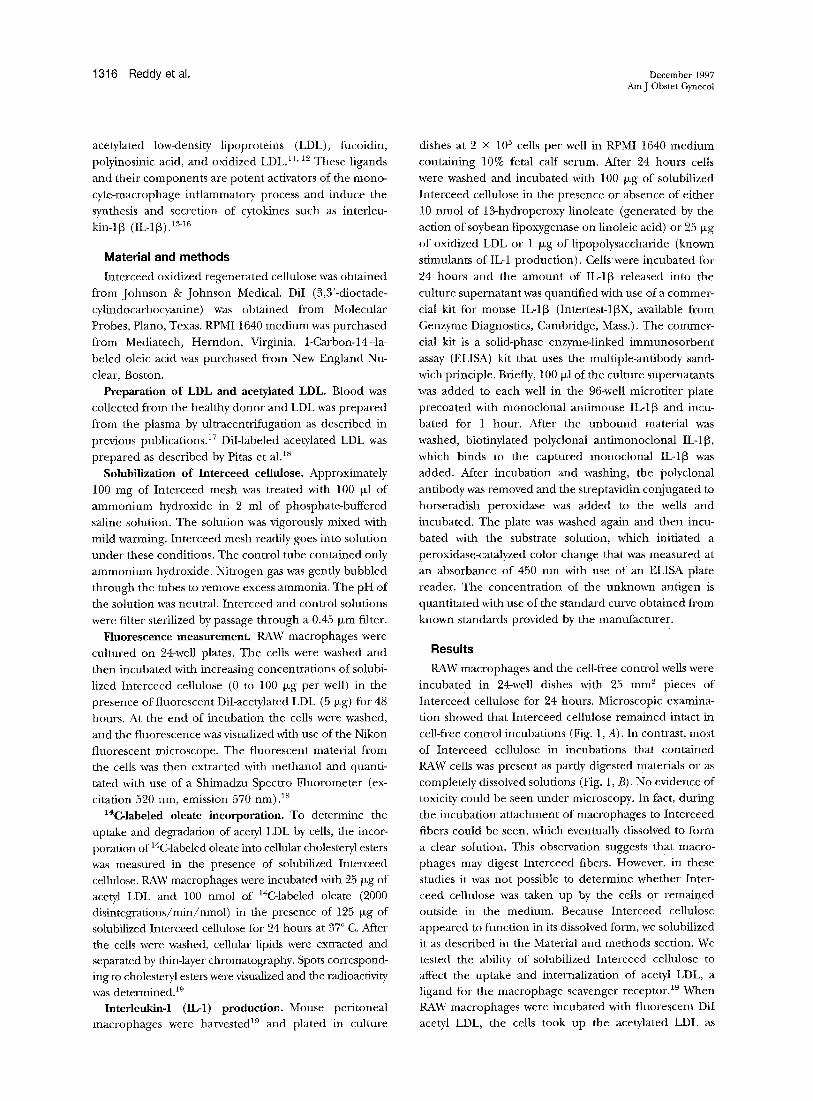
RAW macrophages and the cell-free control wells were
incubated in 24-well dishes with 25 mm z pieces of
Interceed cellulose for 24 hours. Microscopic examina-
tion showed that Interceed cellulose remained intact in
cell-free control incubations (Fig. 1, A). In contrast, most
of Interceed cellulose in incubations that contained RAW cells was present as partly digested materials or as
completely dissolved solutions (Fig. 1, B). No evidence of
toxicity could be seen under microscopy. In fact, during the incubation attachment of macrophages to Interceed
fibers could be seen, which eventually dissolved to form
a clear solution. This observation suggests that macro- phages may digest Interceed fibers. However, in these
studies it was not possible to determine whether Inter- ceed cellulose was taken up by the cells or remained outside in the medium. Because Interceed cellulose
appeared to function in its dissolved form, we solubilized it as described in the Material and methods section. We tested the ability of solubilized Interceed cellulose to affect the uptake and internalization of acetyl LDL, a ligand for the macrophage scavenger receptor} 9 When RAW macrophages were incubated with fluorescent DiI ace@ LDL, the cells took up the acetylated LDL as
Volume 177, Number 6 Reddy et ai. 1317 Am J Obstet Gynecol
Fig. 1. RAW macrophages and no-cell controls were cultured in 24-well dishes in presence and absence of 5 mm 2 pieces of Interceed cellulose. After 24-hour incubation period, cells were observed with Nikon light microscope. A, Interceed cellulose, in no-ceil control well. Fibers remained intact after 24 hours. B, Interceed cellulose, when incubated with RAW macrophages, appeared either partly digested or completely dissolved. Macrophages can be seen aligning along Interceed fiber.
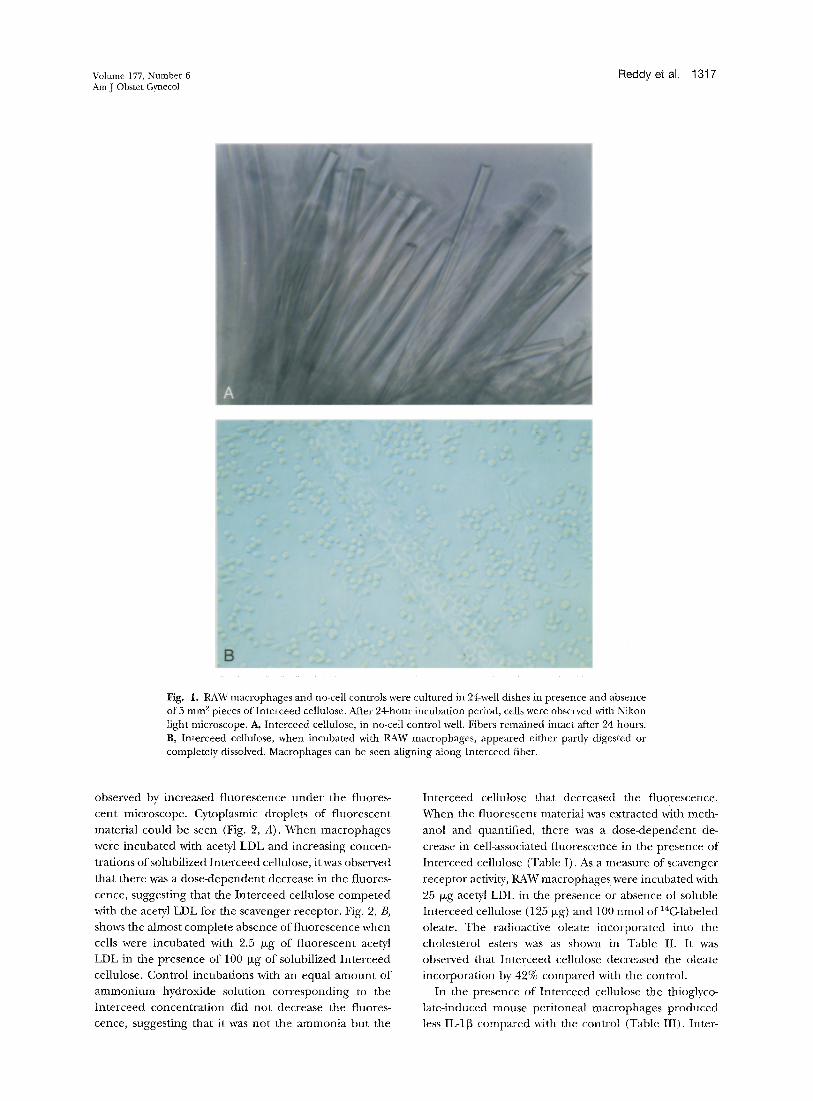
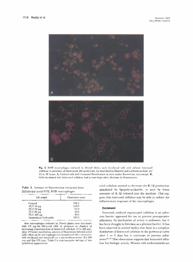
observed by increased f luorescence unde r the fluores-
cent microscope. Cytoplasmic droplets of f luorescent
material could be seen (Fig. 2, A). When macrophages
were incubated with ace@ LDL and increasing concen-
trations o f solubilized In te rceed cellulose, it was observed
that there was a dose -dependen t decrease in the fluores-
cence, suggesting that the In te rceed cellulose compe ted
with the acetyl LDL for the scavenger receptor . Fig. 2, B,
shows the almost comple te absence of f luorescence when
cells were incubated with 2.5 txg of f luorescent acetyl
LDL in the presence of 100 txg of solubilized In te rceed
cellulose. Contro l incubat ions with an equal a m o u n t of
a m m o n i u m hydroxide solution cor responding to the
In te rceed concent ra t ion did no t decrease the fluores-
cence, suggesting that it was no t the ammon ia but the
In te rceed cellulose that decreased the f luorescence.
When the f luorescent material was extracted with meth-
anol and quantified, there was a dose-dependen t de-
crease in cell-associated f luorescence in the presence of
In te rceed cellulose (Table I). As a measure of scavenger
receptor activity, RAW macrophages were incubated with
25 b~g acetyl LDL in the presence or absence of soluble
In te rceed cellulose (125 b~g) and 100 nmol of 14C-labeled
oleate. The radioactive oleate incorpora ted into the
cholesterol esters was as shown in Table II. It was
observed that In te rceed cellulose decreased the oleate
incorpora t ion by 42% compared with the control.
In the presence of In te rceed cellulose the thioglyco-
la teqnduced mouse per i toneal macrophages p roduced
less IL-113 compared with the control (Table III). Inter-
1318 l=leclcly el al. December 1997 Am J Obstet Gynecol
A
B
Fig. 2. RAW macrophages cultured in 24-well dishes were incubated with and without Interceed cellulose in presence of fluorescent Dil ace~l LDL (as described in Material and methods section) for 24 to 48 hours. A, Control cells had increased fluorescence as seen under fluorescent microscope. B, Cells incubated with Interceed cellulose had a dose-dependent decrease in fluorescence.
Tab le I. A m o u n t of f luorescence ex t rac ted f rom
DiI- labeled acetyl LDL RAW m a c r o p h a g e s
Cell sample l bTuorescence units
Control 162.4 TC-7 10 Ixg lt2.0 TC-7 20 Ixg 55.0 TC-7 50 >g 44.0 TC-7 100 ixg 30.0 Ammonium hydroxide 187.0
Raw macrophages cultured in 24-well plates were incubated with 2.5 Ixg/ml DiI-acetyl LDL in presence or absence of increasing concentrations of Interceed cellulose (0 to 100 p,g). After 24 hours' incubation, amount of fluorescent-labeled acetyl LDL taken up by macrophages was quantitated after extraction with methanol and measured on a spectrofluorometer (Ex 520 nm and Em 570 nm). Table I is representative average of two individual experiments.
ceed cellulose s e e m e d to decrease the IL-I[3 p r o d u c t i o n
s t imula ted by l ipopolysacchar ide , as seen by lower
a m o u n t s of IL-1[3 re leased in to the med i u m. This sug-
gests tha t I n t e r c e e d cellulose may be able to r educe the
i n f l a m m a t o w response of the macrophages .
Comment
In t e r ceed oxidized r e g e n e r a t e d cellulose is an adhe-
sion b a r r i e r app roved for use to p r ev en t pos topera t ive
adhesions . Its m e c h a n i s m of ac t ion is u n k n o w n , bu t it
has b e e n t h o u g h t to func t ion as a physical barr ier . I t has
b e e n observed in an imal studies tha t the re is a comple t e
dissolut ion of I n t e r c e e d cellulose in the pe r i tonea l cavity
af ter 1 to 2 days b u t it con t inues to p r ev en t adhe-
sions.20.2~ This observa t ion suggests tha t I n t e r c e e d cellu-
lose has biologic activity. W o m e n with endomet r io s i s are
Volume 177, Number 6 Fleddy et at. ] 319 Am J Obstet Gynecol
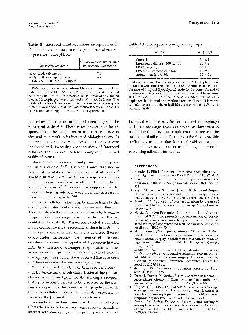
Table II. In te rceed cellulose inhibits incorpora t ion of
I4C-labeled oleate into macrophage cholesterol esters
in presence of acetyl LDL
14C-labeled oleate incorporated Incubation conditions in cholesterol ester (nmol)
Acetyl LDL (25 ~xg/ml) 7.2 Acetyl LDL (25 ~xg/ml) plus 4.2
Interceed cellulose (125 p~g/ml)
RAW macrophages were cultured in 6-well plates and incu- bated with acetyl LDL (25 b~g/ml) with and without Interceed cellulose (125 Ixg/ml), in presence of 100 nmol of 14C-labeled oleate. Macrophages were incubated at 37 ° C for 24 hours. The 14C-labeled oIeate incorporated into cholesterol ester was quan- titated as described in Material and Methods section. Table II is representative average of two individual experiments.
felt to have an increased n u m b e r of macrophages in the
per i toneal cavity32'2~ These macrophages may be re-
sponsible for the dissolution of In te rceed cellulose in
vivo and may result in its increased biologic activity. As
observed in our study, when RAW macrophages were
incubated with increasing concentra t ions of In te rceed
cellulose, the In te rceed cellulose completely dissolved
,vithin 48 hours.
Macrophages play an impor tan t pro inf lammatory role
in various diseases. 24' 25 It is well known that macro-
phages play a vital role in the format ion of adhesions. 26
These cells take up various anionic compounds such as
fucoidin, polydnosinic acid, oxidized LDL, etc., by its 11 12 scavenger receptors. ' Studies have suggested that the
uptake of these ligands by macr0phages may increase its
p ro inf lammatory capacity. ~3-16
In te rceed cellulose is taken up by macrophages by the
scavenger receptors and thereby may prevent adhesions.
To establish whether In te rceed cellulose affects macro-
phage uptake of scavenger ligands, we also used fluores-
cent-labeled acetyl LDL. Fluorescent- labeled acetyl LDL
is a l igand for scavenger receptors. As these ligands bind
to receptors the cells take on a characteristic fluores-
cence under microscopy. The presence of In te rceed
cellulose decreased the uptake of f luorescent- labeled
LDL. As a measure of scavenger receptor activity, radio-
active oleate incorpora t ion into the cholesterol ester in
macrophages was studied. It was observed that In te rceed
cellulose decreased the oleate incorporat ion.
We next s tudied the effect of In te rceed cellulose on
cellular b iochemica l product ion . Bacterial lipopolysac-
char ide is a known l igand for the scavenger receptor .
IL-113 produc t ion is known to be media ted by the scav-
enger receptor . In the presence of l ipopolysaccharide
In t e rceed cellulose seemed to block the expec ted in-
crease in IL-113 caused by l ipopolysaccharide.
In conclusion, we have shown that In te rceed cellulose
affects the ability of known scavenger receptor ligands to
interact with macrophages . The primary interact ion of
Table III. IL-I[3 produc t ion by macrophages
Sample IL-l[3 (pg)
Control 134 ± 11 Interceed cellulose (100 Ixg/ml) 116 _+ 8 LPS (1 ixg/ml) 155 2 37 LPS plus Interceed cellulose 118 -~ 8 Ammonium hydroxide 137 + 21
Mouse peritoneal macrophages grown on 24-well plates were incubated with Interceed cellulose (10O Izg/ml) in presence or absence of 1 brg/ml lipopolysaccharide for 24 hours. At end of incubation, 100 Ixl of culture supernatant was used to measure IL-1[3 released with use of commercially available ELISA kit as explained in Material and Methods section. Table III is repre- sentative average of three individual experiments. LPS, Lipo- polysaccharide.
In te rceed cellulose may be on activated macrophages
and their scavenger receptors, which are impor tan t in
p romot ing the growth of ectopic e n d o m e t r i u m and the
format ion of adhesions. This study is the first to provide
prel iminary evidence that In te rceed oxidized regener-
ated cellulose may funct ion as a biologic barr ier in
prevent ing adhesion formation.
REFERENCES
1. Menzies D, Ellis H. Intestinal obstruction from adhesions-- how big is the problem? Ann R Coil Surg Ing 1990;72:60-3.
2. Ellis H. The cause and prevention of postoperative intra- peritoneal adhesions. Surg Gynecol Obstet 1971;133:497- 511.
3. Ray NF, LarsenJW, Stillman RJ,Jacobs RJ. Economic impact of hospitalization for lower abdominal adhesiolysis in the United States in 1988. Surg Gynecol Obstet 1993;176:271-6.
4. Franklin RR. Reduction of ovarian adhesions by the use of Interceed: Ovarian Adhesion Study Group. Obstet Gynecol 1995;86:335-40.
5. Nordic Adhesion Prevention Study Group. The efficacy of Interceed(TC7)* for prevention of refmxnation of postop- erative adhesions on ovaries, fallopian tubes, and fimbriae in microsurgical operations for fertility: a multicenter study. FertiI Steril 1995;63:709-14.
6. Mais V, Ajossa S, Marongiu D, Peiretti RF, Guerriero S, Melis GB. Reduction of adhesion reformation after laparoscopic endometriosis surgmT: a randomized trial with an oxidized regenerated cellulose absorbable barrier. Obstet Gynecol 1995;86:512-5.
7. Sekiba K. Use of Interceed (TC7) absorbable adhesion barrier to reduce postoperative adhesion reformation in infertility and endometriosis surgery: the Obstetrics and Gynecology Adhesion Prevention Committee. Obstet Gy- necol 1992;79:518-22.
8. diZerega GS. Contemporary adhesion prevention. Fertil Steril 1994;61:219-35.
9. Fraser I, Hughes D, Gordon S. Divalent cation-independent macrophage adhesion inhibited by monoclonal antibody to murine scavenger receptor. Nature 1993;364:343-6.
10. Hughes DA, Fraser IP, Gordon S. Murine macrophage scavenger receptor: in vivo expression and function as receptor for macrophage adhesion in lymphoid and non- lymphoid organs. EurJ Immunol 1995;25:466-73.
11. Pearson AM, Rich A, Krieger M. Polynucleotide binding to macrophage scavenger receptors depends on the formation of base-quartet-stabilized four-stranded helices. J Biol Chem 1993;268:3546-54.
1320 Reddy et al. December 1997 _~:a J Obstet GynecoI
12. Parthasarathy S. Modified lipoproteins in the pathogenesis of atherosclerosis. Austin (TX): RG Landes; 1994.
13. Palkama T, Majuri ML, Mattila P, Hurme M, Renkonen R. Regulation of endothelial adhesion molecules by" ligands binding to the scavenger receptor. Clin Exp Immunol 1993;92:353-60.
14. Palkama T. Induction of interleukin-1 production by li- gands binding to the scavenger receptor in human mono- cytes and the THP-1 cell line. Imnmnology 1991;74:432-8.
15. Thomas CE,Jackson RL, Ohlweiler DF, Ku G. Multiple lipid oxidation products in low density lipoproteins induce inter- lenkin-1 beta release from human blood mononuclear cells. J Lipid Res 1994;35:417-27.
16. Ku G, Thomas CE, Akeson AL, Jackson RL. Induction of interleukin 1 beta expression from human peripheral blood monocyte derived macrophages by 9-hydroxyoctadecadi- enoic acid. J Biol Chem 1992;267:14183-8.
17. Santanam N, Parthasarathy S. Paradoxical actions of anti- oxidants in the oxidation of low density lipoprotein by peroxidases. J Ctin Invest 1995;95:2594-600.
18. Dejager S, Mietus Snyder M, Friera A, Pitas RE. Dominant negative mutations of the scavenger receptor: native recep- tor inactivation by expression of truncated variants. J Clin Invest 1993;99:894-902.
19. Parthasarathy S, Printz DJ, Boyd D, Joy L, Steinberg D. Macrophage oxidation of low density lipoprotein generates a modified form recognized by the scavenger receptor. Arteriosclerosis 1986;6:505-10.
20. Haney AF, Doty E. Comparison of the peritoneal cells elicited by oxidized regenerated cellulose (Interceed) and expanded polytetrafluoroethylene (Gore-Tex Surgical Membrane) in a nmrine model..~n J Obstet Gynecol 1992;166:1137-49:
91. Diamond MP, Linsky CB, Cunningham T, Kamp L, Pines E, DeCherney AH, et al. Synergistic effects of INTERCEED (TCT) and heparin in reducing adhesion formation in the rabbit uterine horn model. Fertil Steril 1991;55:389-94.
99 HalmeJ, Becker S, Wing R. Accentuated cyclic activation of peritoneal macrophages in patients with endometriosis. Am J Obstet Gynecol 1984;148:85-90.
23. Uda S. The role of the peritoneal macrophage on the infertility associated with endometriosis. Osaka City Med J 1989;38:11-26.
24. Nathan CF, Tsunawaki S. Secretion of toxic oxygen prod- ucts by macrophages: regulatory cytokines and their effects on the oxidase. Ciba Found Syrup 1986;118:211-30.
25. Yamamoto N, Willett NP, Lindsay DD. Participation of serum proteins in the inflammation-primed activation of macrophages. Inflammation 1994;18:311-92.
26. Haney AF. Endometriosis, macrophages, and adhesions [review]. Prog Clin Biol Res 1993;381:19-44.
Discussion
Dl~. MICHAEL P. DIAMOND, Detroit, Michigan. Preven- tion of adhesions are a major problem each of us faces in every surgical procedure we perform because of the potential consequences of pain, infertility, small bowel obstruction, and difficult reoperative procedures. None- theless, the frequency with which adhesions develop has often been underappreciated, in part because a second operative procedure is used to identify them, yet there is often no clinical reason to justify such a procedure.
We have previously reported that among women un- dergoing laparotomy for infertility adhesions were iden- tified in 91 of 106 or 86% of subjects at second-look laparoscopy in spite of application of microsurgical prin- ciples and use of the carbon dioxide laser.
More recently, we have also demonstrated that after laparoscopic adhesiolyses adhesions developed in 66 of
68, or 97%, of women. Thus laparoscopic surgery is not a panacea for prevention of adhesions. Unfortunately, these observations are not unique to infertility, patients and the procedures they undergo. A study of men and women undergoing colectomy identified that 94% of control subjects had adhesions to the anterior abdominal wall incision line.
Thus clearly there is a place for adjuvants to try to reduce postoperative adhesion development and tile clinical consequences. Currently there are two products approved by the Food and Drug Administration for the reduction of postoperative adhesions, Interceed and Seprafilm, each which are regulated as devices on the basis of the concept that they separate potentially oppos- ing surfaces during the brief time period of days during which reperitonealization occurs.
Dr. Reddy and colleagues are to be congratulated for this challenging hypothesis, that Interceed cellulose ex- erts its efficacy solely by this barrier mechanism. It has been known that this material, which is composed of oxidized regenerated cellulose, gelates after placement. Interceed is degraded into oligomers of varying length that get smaller in time as has been shown by high- pressure liquid chromatography. This process occurs by both enzymatic and macrophage-dependent processes. In studies in which lead-labeled Interceed has been ingested, a peritoneal macrophage scanning electron micrographs have shown Interceed breakdown products within the cytoplasm in vacuoles and associated with cellular membranes.
This article has provided evidence that the breakdown products may be biologically active. In this study solubi- lized Interceed inhibited uptake of acetylated LDL and fucoidin. Because each of these molecules normally serves as potent activators of the inflammatory process, including the synthesis and secretion of growth factors and cytokines, their inhibition may lessen inflammatory adhesion development.
The clinical significance of this observation is that, if Interceed's efficacy is due to the breakdown products rather than to action as a barrier, then benefit may be realized at other sites throughout the abdominopehdc cavity distant from where it has been placed. Furthe> more, variation in efficacy may be the result of expression of function at scavenger receptors.
In closing, I would like to ask three questions. (1) What limitations should be considered in view of having performed these studies in RAW macrophages, a celt line derived from rodents, as opposed to individually ha> vested macrophages from humans and have you assessed this issue? (2) Have you had the opportunity to assess the effect of blocking the scavenger receptor with Interceed on inflammatory process markers such as growlh factor or cytokine secretion? I too have thought that Interceed's action may in part be as a biologically active molecule rather than solely as a barrier; however, I had envisioned other possible actions. This raises the question of which biologic activity is associated with its efficacy. I wonder whether you have had the opportuni ty to statistically
Volume 177, Number 6 gleddy et al. 1321 AmJ Obstet Gynecol
correlate whether animals in which efficacy is shown exhibit blockage of the scavenger receptors. In other words, do you have thoughts on how you might try to differentiate Interceed's efficacy as a barrier as opposed to biologically active membrane affecting scavenger re- ceptor or other mechanisms?
Dr. Reddy (Closing). I am looking forward to compar- ing cultured RAW macrophages to freshly collected cells from donors. I anticipate that the freshly gathered mac- rophages will have a more intense response than will the cultured cells. The cultured macrophages probably man- ifest a blunted response. As far as animal studies, we hope
to prove that Interceed functions in its soluble form in mice. We would do this by producing tissue injury and then inserting soluble and intact Interceed and then comparing the number and type of adhesions at second look.
We also plan to determine how Interceed affects IL-I~ production. We are considering experiments with triti- ated proline suture to determine the effect of Interceed on collagen formation. By showing that Interceed affects the production of these biochemical substances we would have further evidence that Interceed functions as a biologic barrier.
Copyright © 2022 FDOKUMEN