Arthroscopic Lysis of Adhesions and Anterior Interval
7
Technical Note Arthroscopic Lysis of Adhesions and Anterior Interval Release With Manipulation Under Anesthesia for Severe Post-traumatic Knee Stiffness: A Simple and Reproducible Step-by-Step Guide Mohit Kukreja, M.D., Jeansol Kang, M.D., Emily J. Curry, B.A., and Xinning Li, M.D. Abstract: Post-traumatic knee stiffness can present after injuries around the knee and surgery. Management is guided by the type of initial injury, amount of range-of-motion loss, time since injury, and cartilage status. Cases refractory to conservative management may conventionally be treated with manipulation under anesthesia (MUA), arthroscopic lysis of adhesions, or open quadricepsplasty. We describe our arthroscopic technique of lysis of adhesions with anterior interval release and intraoperative MUA, which has been shown to provide sustainable range-of-motion improvement in a subset of patients with severe knee arthrofibrosis. Although technically demanding, this technique benefits from being minimally invasive, allows for direct visualization of intra-articular structures, and allows all-round arthroscopic release of adhesions to improve patellar mobility and decrease the risk of fracture prior to MUA. A rigorous postoperative formal physical therapy protocol and patient compliance are imperative to achieve good outcomes. P ost-traumatic knee stiffness is common after in- juries around the knee and surgery. Restricted range of motion (ROM) can be classified as extra- articular, intra-articular, or both. Management options depend on the type of initial injury (extra- or intra- articular), type of ROM loss (flexion, extension, or both), time since injury, and cartilage status. It is imperative to identify the specific cause of the knee stiffness to direct optimal surgical treatment. Arthrofibrotic knee stiffness most commonly results from intra-articular scarring, fibrotic adhesions, tissue remodeling, periarticular tissue retraction, or bony impingement. 1 Conservative treatment using anti- inflammatory medications, cortisone injections, and physical therapy (PT) has shown limited success in isolation, especially in the setting of post-traumatic knee stiffness. Manipulation under anesthesia (MUA) can be effective but may result in complications such as fracture, failure of fracture fixation, or chondral damage. Arthroscopic lysis of adhesions with anterior interval release supplemented by intraoperative MUA has been accepted as a successful treatment modality for this condition and has provided sustainable ROM improvement in certain subsets of patients with post- traumatic stiffness. 2-5 A step-by-step approach is crucial and involves sequential release of scars, adhe- sions, and fibrotic structures in different regions of the knee. We present the arthroscopic surgical technique of the senior author (X.L.) to address severe post- traumatic or postsurgical knee stiffness (Video 1). Technique Setup and Patient Positioning The indications for the described procedure include intra-articular knee arthrofibrosis refractory to conser- vative management including cortisone injections, anti-inflammatory medications, and prolonged PT. In addition, in patients with osteoporosis who have an increased risk of fracture with MUA alone, performing From the Department of Orthopaedic Surgery, Boston University School of Medicine, Boston, Massachusetts, U.S.A. The authors report the following potential conflicts of interest or sources of funding: X.L. receives equity and is on the editorial board of the Journal of Medical Insight. Full ICMJE author disclosure forms are available for this article online, as supplementary material. Received September 26, 2018; accepted January 6, 2019. Address correspondence to Xinning Li, M.D., Sports Medicine and Shoulder Service, Department of Orthopaedic Surgery, Boston University School of Medicine, Boston Medical Center, 850 Harrison Ave, Dowling 2-North, Bos- ton, MA 02118, U.S.A. E-mail: [email protected] Ó 2019 by the Arthroscopy Association of North America. Published by Elsevier. This is an open access article under the CC BY-NC-ND license (http:// creativecommons.org/licenses/by-nc-nd/4.0/). 2212-6287/181191 https://doi.org/10.1016/j.eats.2019.01.005 Arthroscopy Techniques, Vol 8, No 5 (May), 2019: pp e429-e435 e429
-
Upload
khangminh22 -
Category
Documents
-
view
0 -
download
0
Transcript of Arthroscopic Lysis of Adhesions and Anterior Interval

Technical Note
From theMedicine, Bo
The authofunding: X.LMedical Insarticle online
Received SAddress co
Service, DepMedicine, Boton, MA 021
� 2019 bElsevier. Thicreativecomm
2212-6287https://doi
Arthroscopic Lysis of Adhesions and Anterior IntervalRelease With Manipulation Under Anesthesia for
Severe Post-traumatic Knee Stiffness: A Simple andReproducible Step-by-Step Guide
Mohit Kukreja, M.D., Jeansol Kang, M.D., Emily J. Curry, B.A., and Xinning Li, M.D.
Abstract: Post-traumatic knee stiffness can present after injuries around the knee and surgery. Management is guided bythe type of initial injury, amount of range-of-motion loss, time since injury, and cartilage status. Cases refractory toconservative management may conventionally be treated with manipulation under anesthesia (MUA), arthroscopic lysisof adhesions, or open quadricepsplasty. We describe our arthroscopic technique of lysis of adhesions with anterior intervalrelease and intraoperative MUA, which has been shown to provide sustainable range-of-motion improvement in a subsetof patients with severe knee arthrofibrosis. Although technically demanding, this technique benefits from being minimallyinvasive, allows for direct visualization of intra-articular structures, and allows all-round arthroscopic release of adhesionsto improve patellar mobility and decrease the risk of fracture prior to MUA. A rigorous postoperative formal physicaltherapy protocol and patient compliance are imperative to achieve good outcomes.
ost-traumatic knee stiffness is common after in-
Pjuries around the knee and surgery. Restrictedrange of motion (ROM) can be classified as extra-articular, intra-articular, or both. Management optionsdepend on the type of initial injury (extra- or intra-articular), type of ROM loss (flexion, extension, orboth), time since injury, and cartilage status. It isimperative to identify the specific cause of the kneestiffness to direct optimal surgical treatment.Arthrofibrotic knee stiffness most commonly resultsfrom intra-articular scarring, fibrotic adhesions, tissueremodeling, periarticular tissue retraction, or bony
Department of Orthopaedic Surgery, Boston University School ofston, Massachusetts, U.S.A.rs report the following potential conflicts of interest or sources of. receives equity and is on the editorial board of the Journal ofight. Full ICMJE author disclosure forms are available for this, as supplementary material.eptember 26, 2018; accepted January 6, 2019.rrespondence to Xinning Li, M.D., Sports Medicine and Shoulderartment of Orthopaedic Surgery, Boston University School ofston Medical Center, 850 Harrison Ave, Dowling 2-North, Bos-18, U.S.A. E-mail: [email protected] the Arthroscopy Association of North America. Published bys is an open access article under the CC BY-NC-ND license (http://ons.org/licenses/by-nc-nd/4.0/)./181191.org/10.1016/j.eats.2019.01.005
Arthroscopy Techniques, Vol 8, No
impingement.1 Conservative treatment using anti-inflammatory medications, cortisone injections, andphysical therapy (PT) has shown limited success inisolation, especially in the setting of post-traumatic kneestiffness. Manipulation under anesthesia (MUA) can beeffective butmay result in complications such as fracture,failure of fracture fixation, or chondral damage.Arthroscopic lysis of adhesions with anterior interval
release supplemented by intraoperative MUA has beenaccepted as a successful treatment modality for thiscondition and has provided sustainable ROMimprovement in certain subsets of patients with post-traumatic stiffness.2-5 A step-by-step approach iscrucial and involves sequential release of scars, adhe-sions, and fibrotic structures in different regions of theknee. We present the arthroscopic surgical techniqueof the senior author (X.L.) to address severe post-traumatic or postsurgical knee stiffness (Video 1).
Technique
Setup and Patient PositioningThe indications for the described procedure include
intra-articular knee arthrofibrosis refractory to conser-vative management including cortisone injections,anti-inflammatory medications, and prolonged PT. Inaddition, in patients with osteoporosis who have anincreased risk of fracture with MUA alone, performing
5 (May), 2019: pp e429-e435 e429

Fig 1. The patient is in thesupine position, and this isthe right knee. Preoperativerange of motion shows fullextension (A) and 20� offlexion (B) with a mechan-ical block to range ofmotion.
e430 M. KUKREJA ET AL.
this technique first with arthroscopic lysis of adhesionsalong with capsular and anterior interval release wouldminimize their risk of complications.The patient is placed in the standard supine position
with lateral thigh support. General anesthesia is used inall of our patients; however, regional epidural spinalanesthesia with an indwelling catheter may be analternative for intraoperative and postoperative painmanagement. Preoperative ROM is recorded with thepatient under anesthesia with photographs taken ofknee flexion and extension (with the patient’s consent)so that progress can be easily monitored postoperatively(Fig 1).
Arthroscopic Portal PositioningThe anterolateral (AL), anteromedial (AM), super-
omedial (SM), and superolateral (SL) portals are usedfor both the working portals and outflow cannula (Fig2A). The AL and AM portals are established throughthe standard AL and AM soft spots (standard kneearthroscopy portal sites), whereas the SM portal isestablished 2 cm superior and 2 cm medial to the SMedge of the patella. The SL portal is established about2 cm superior and 2 cm lateral to the SL edge of thepatella. Both the SM and SL portals will serve as theviewing or working portals for this arthroscopic tech-nique. These 2 portals are unrestricted and help withgaining unrestricted access to the medial or lateralretinaculum for arthroscopic release. In the senior au-thor’s experience, the first step in the arthroscopic lysisof adhesions will always start with the SL portal as theviewing portal and the SM portal as the working portalbecause of the limitations of knee flexion (Fig 2B). It isvery difficult to start with the arthroscope in thetraditional AL viewing portal when the knee is unableto be flexed past 90�. The posteromedial and postero-lateral accessory portals are used only in cases in whichthere is a persistent extension deficit due to tight
posterior capsular structures despite anterior interval aswell as medial and lateral capsular release with MUA.
Step-by-Step Approach
Debridement of Patellofemoral and SuprapatellarCompartments. A diagnostic arthroscopy using a stan-dard 30� arthroscope (Smith & Nephew, Andover, MA)in the anterior compartment can be performed throughtheALportal. However, if kneeflexion is restricted to lessthan60�, the viewing arthroscope should be inserted intothe patellofemoral joint through the SL portal (viewingportal) and then the SM working portal is establishedwith the knee in extension (Fig 2). To help visualize theintra-articular structures and aid with lysis of adhesionsthrough capsular distension, 100 to 150 mL of salinesolution can also be injected into the joint.A standard arthroscopic shaver (4.5-mm Dyonics
Incisor Plus Platinum; Smith & Nephew) and radio-frequency (RF) ablation device (Super TurboVac 90Coblation wand; Smith & Nephew) are used to debridethe patellofemoral joint and the suprapatellar pouchwith the knee in extension, releasing all adhesions,hypertrophied synovium, and intra-articular bands(Fig 3). The deep quadriceps muscle bulk should bevisualized to confirm that the releases are adequate.The suprapatellar pouch usually extends up to 3 cmproximal to the superior edge of the patella. Adhesionsshould be liberally released in the patellofemoralcompartment to help with the patellar excursion.
Medial and Lateral Gutter Synovectomy and Lysis ofAdhesions. All adhesions in the medial and lateral par-apatellar gutters should be cleared using the AL viewingportal and a combination of the shaver and ablator fromthe AM working portal (Fig 3). The inner aspect of theknee capsule is typically adhered to the medial andlateral aspects of the femoral condyle, and clearing thisscarring helps patellar mobility on the trochlea.
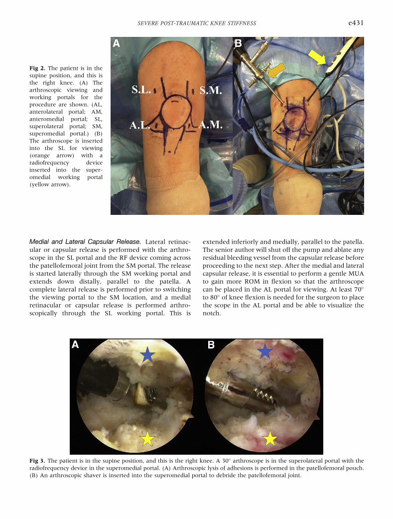
Fig 2. The patient is in thesupine position, and this isthe right knee. (A) Thearthroscopic viewing andworking portals for theprocedure are shown. (AL,anterolateral portal; AM,anteromedial portal; SL,superolateral portal; SM,superomedial portal.) (B)The arthroscope is insertedinto the SL for viewing(orange arrow) with aradiofrequency deviceinserted into the super-omedial working portal(yellow arrow).
SEVERE POST-TRAUMATIC KNEE STIFFNESS e431
Medial and Lateral Capsular Release. Lateral retinac-ular or capsular release is performed with the arthro-scope in the SL portal and the RF device coming acrossthe patellofemoral joint from the SM portal. The releaseis started laterally through the SM working portal andextends down distally, parallel to the patella. Acomplete lateral release is performed prior to switchingthe viewing portal to the SM location, and a medialretinacular or capsular release is performed arthro-scopically through the SL working portal. This is
Fig 3. The patient is in the supine position, and this is the right kradiofrequency device in the superomedial portal. (A) Arthroscop(B) An arthroscopic shaver is inserted into the superomedial port
extended inferiorly and medially, parallel to the patella.The senior author will shut off the pump and ablate anyresidual bleeding vessel from the capsular release beforeproceeding to the next step. After the medial and lateralcapsular release, it is essential to perform a gentle MUAto gain more ROM in flexion so that the arthroscopecan be placed in the AL portal for viewing. At least 70�
to 80� of knee flexion is needed for the surgeon to placethe scope in the AL portal and be able to visualize thenotch.
nee. A 30� arthroscope is in the superolateral portal with theic lysis of adhesions is performed in the patellofemoral pouch.al to debride the patellofemoral joint.

Fig 4. The patient is in the supine position, and this is the right knee. (A) The arthroscope is in the anterolateral viewing portalwith the radiofrequency (RF) device in the anteromedial (AM) portal. The tibial plateau (star) is seen here, and the anteriorinterval is labeled with an arrow. The RF device is inserted into the AM portal to start the anterior interval release. (B) With thesame viewing and working portals, the hooked RF device is inserted into the AM portal to complete the anterior interval release,working from the medial to lateral direction and 2 cm under the tibial plateau.
e432 M. KUKREJA ET AL.
Anterior Interval Release. In cases of severe arthrofib-rosis or deficits in knee flexion, it is important to releasethe anterior interval of the knee between the anteriortibial plateau and the patellar tendon. With thearthroscope in the AL viewing portal and the arthro-scopic shaver in the AM portal, the hypertrophic scar-red infrapatellar (Hoffa) fat pad and adhesions in thepretibial recess are released. This is a vascular area;therefore, a combination of the shaver and electro-cautery should be used when excising adhesions in thisregion (Fig 4).A hooked RF device (Dyonics RF Hook; Smith &
Nephew) or standard RF device is used to release theanterior interval between the tibia and the patellartendon. The release should be performed from themedial to lateral direction and down 2 cm inferior to the
tibial plateau surface (Fig 4B). The hooked RF devicehelps to easily dissect the scar and fat pad between theplane of the patellar tendon and the tibial plateau. Thearthroscope is then switched to the AM portal, and theanterior interval release is completed at this time fromthe lateral tomedial direction 2 cmbelow theplateau. It isessential to shut off the pump at this time to make surethere is no residual bleeding from this area.
Intercondylar Notch Examination andDebridement. After anterior interval release, the scartissue in the joint is sequentially debrided, including theintercondylar notch and the medial and lateral femoralcondyle, using a combination of the shaver and ablator.An arthroscopic burr is used for any bony overgrowthsor spurs that may be encountered owing to the
Fig 5. The patient is in thesupine position, and this isthe right knee. After thearthroscopic lysis of adhe-sions, medial and lateralcapsular release, and ante-rior interval release, gentlemanipulation under anes-thesia is performed to breakup the adhesions. Fullextension (A) and 135� offlexion (B) are obtained inthis patient with a very stiffpreoperative knee.
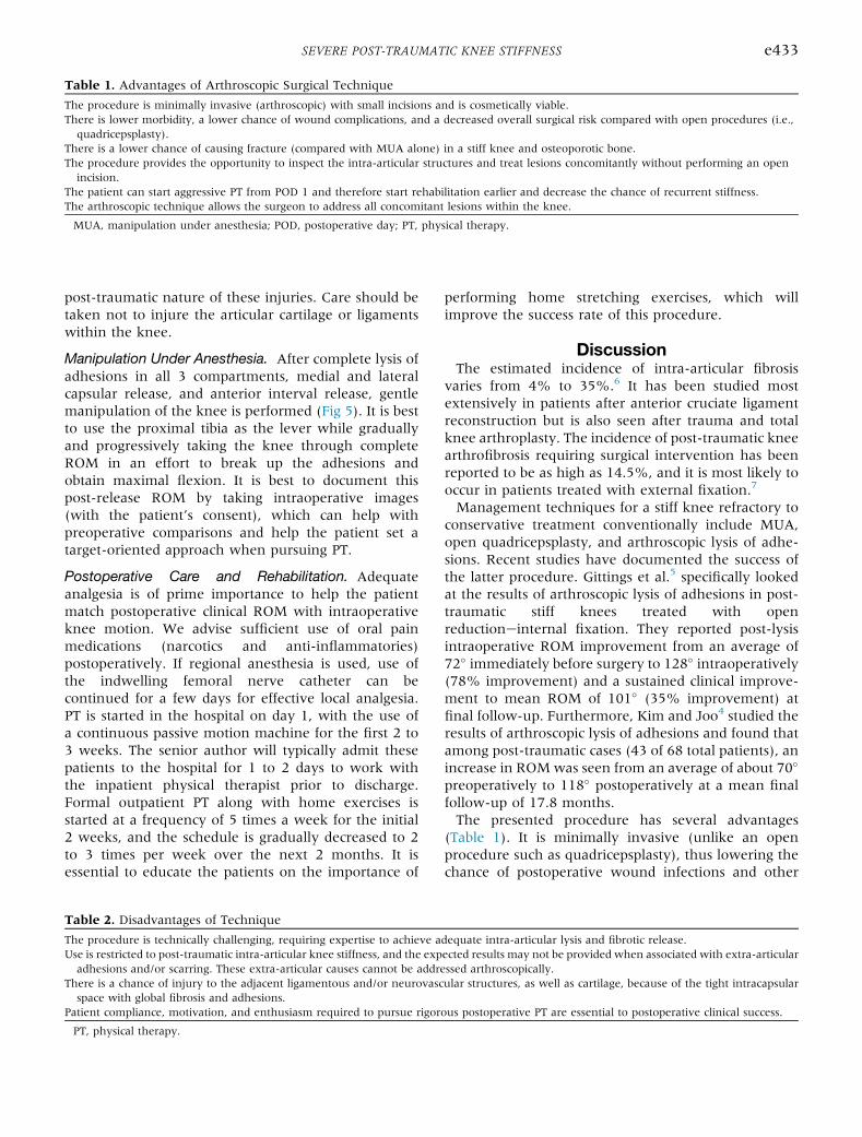
Table 1. Advantages of Arthroscopic Surgical Technique
The procedure is minimally invasive (arthroscopic) with small incisions and is cosmetically viable.There is lower morbidity, a lower chance of wound complications, and a decreased overall surgical risk compared with open procedures (i.e.,
quadricepsplasty).There is a lower chance of causing fracture (compared with MUA alone) in a stiff knee and osteoporotic bone.The procedure provides the opportunity to inspect the intra-articular structures and treat lesions concomitantly without performing an open
incision.The patient can start aggressive PT from POD 1 and therefore start rehabilitation earlier and decrease the chance of recurrent stiffness.The arthroscopic technique allows the surgeon to address all concomitant lesions within the knee.
MUA, manipulation under anesthesia; POD, postoperative day; PT, physical therapy.
SEVERE POST-TRAUMATIC KNEE STIFFNESS e433
post-traumatic nature of these injuries. Care should betaken not to injure the articular cartilage or ligamentswithin the knee.
Manipulation Under Anesthesia. After complete lysis ofadhesions in all 3 compartments, medial and lateralcapsular release, and anterior interval release, gentlemanipulation of the knee is performed (Fig 5). It is bestto use the proximal tibia as the lever while graduallyand progressively taking the knee through completeROM in an effort to break up the adhesions andobtain maximal flexion. It is best to document thispost-release ROM by taking intraoperative images(with the patient’s consent), which can help withpreoperative comparisons and help the patient set atarget-oriented approach when pursuing PT.
Postoperative Care and Rehabilitation. Adequateanalgesia is of prime importance to help the patientmatch postoperative clinical ROM with intraoperativeknee motion. We advise sufficient use of oral painmedications (narcotics and anti-inflammatories)postoperatively. If regional anesthesia is used, use ofthe indwelling femoral nerve catheter can becontinued for a few days for effective local analgesia.PT is started in the hospital on day 1, with the use ofa continuous passive motion machine for the first 2 to3 weeks. The senior author will typically admit thesepatients to the hospital for 1 to 2 days to work withthe inpatient physical therapist prior to discharge.Formal outpatient PT along with home exercises isstarted at a frequency of 5 times a week for the initial2 weeks, and the schedule is gradually decreased to 2to 3 times per week over the next 2 months. It isessential to educate the patients on the importance of
Table 2. Disadvantages of Technique
The procedure is technically challenging, requiring expertise to achieve aUse is restricted to post-traumatic intra-articular knee stiffness, and the exp
adhesions and/or scarring. These extra-articular causes cannot be addreThere is a chance of injury to the adjacent ligamentous and/or neurovasc
space with global fibrosis and adhesions.Patient compliance, motivation, and enthusiasm required to pursue rigor
PT, physical therapy.
performing home stretching exercises, which willimprove the success rate of this procedure.
DiscussionThe estimated incidence of intra-articular fibrosis
varies from 4% to 35%.6 It has been studied mostextensively in patients after anterior cruciate ligamentreconstruction but is also seen after trauma and totalknee arthroplasty. The incidence of post-traumatic kneearthrofibrosis requiring surgical intervention has beenreported to be as high as 14.5%, and it is most likely tooccur in patients treated with external fixation.7
Management techniques for a stiff knee refractory toconservative treatment conventionally include MUA,open quadricepsplasty, and arthroscopic lysis of adhe-sions. Recent studies have documented the success ofthe latter procedure. Gittings et al.5 specifically lookedat the results of arthroscopic lysis of adhesions in post-traumatic stiff knees treated with openreductioneinternal fixation. They reported post-lysisintraoperative ROM improvement from an average of72� immediately before surgery to 128� intraoperatively(78% improvement) and a sustained clinical improve-ment to mean ROM of 101� (35% improvement) atfinal follow-up. Furthermore, Kim and Joo4 studied theresults of arthroscopic lysis of adhesions and found thatamong post-traumatic cases (43 of 68 total patients), anincrease in ROMwas seen from an average of about 70�
preoperatively to 118� postoperatively at a mean finalfollow-up of 17.8 months.The presented procedure has several advantages
(Table 1). It is minimally invasive (unlike an openprocedure such as quadricepsplasty), thus lowering thechance of postoperative wound infections and other
dequate intra-articular lysis and fibrotic release.ected results may not be provided when associated with extra-articularssed arthroscopically.ular structures, as well as cartilage, because of the tight intracapsular
ous postoperative PT are essential to postoperative clinical success.

Table 3. Preoperative and Intraoperative Pearls
Preoperative1. The diagnosis of intra-articular arthrofibrosis should be supported by a thorough clinical examination and ancillary investigations to rule outother causes of knee stiffness (extra-articular). The best outcomes will be obtained only in this subset of patients.
2. ROM restriction may occur in flexion (patellofemoral, suprapatellar, intercondylar, and/or anterior interval), extension (posterior capsule),or a combination of both. A detailed stepwise evaluation and systematic approach are important and should be planned preoperatively.
3. Preoperative patient counseling should reinforce the importance of postoperative PT.4. Documenting preoperative and intraoperative ROM under anesthesia helps in monitoring patients’ progress. It also helps patients set targetsand work toward set goals.
Intraoperative1. In a knee that is severely stiff, the viewing portal should start in the superolateral location with the working portal in the superomediallocation.
2. Both medial and lateral capsular releases are performed first to allow a gentle MUA to gain knee flexion. The pump must be turned off toevaluate for bleeding in the retinacular area.
3. Once knee flexion around 70� to 80� is obtained, the anterolateral portal is used for viewing and the anteromedial portal may be used as theworking portal.
4. Releasing the anterior interval and re-establishing the pretibial recess help patellar excursion and mobility. Use of the hooked RF device isideal to achieve this. Release is performed from the medial to lateral direction between the tibial plateau and the patellar tendon about 2 cmbelow the tibial surface. It is essential to shut off the pump and evaluate for bleeding after the completion of the anterior interval release.
5. Lysis of adhesions should be performed in all 3 compartments including the patellofemoral pouch.6. MUA should be performed after release, lysis of adhesions, and anterior interval release, and the proximal two-thirds of the tibia should beused as the primary lever to avoid intraoperative fracture.
7. Posterior releases (arthroscopic or open) should be added whenever there is an extension deficit despite a complete anterior release withMUA.
8. A combination of an arthroscopic shaver and RF electrocautery device is needed to achieve a bloodless intraoperative field. Closure shouldbe preceded by dry arthroscopy to check and confirm complete hemostasis.
9. Postoperative pain management with multiple modalities (regional anesthesia, short-acting opioids, and NSAIDs) will alleviate pain andmuscle stiffness and help the patient continue with a scheduled aggressive rehabilitation protocol.
10. The senior author prefers to admit the patient to the hospital for 1 to 2 days to work with the physical therapist right after surgery. FormalPT is started with 5 d/wk for the first 2 wk and then trending down to 2 to 3 times per week in 2 mo.
MUA, manipulation under anesthesia; NSAIDs, nonsteroidal anti-inflammatory drugs; PT, physical therapy; RF, radiofrequency; ROM, range ofmotion.
e434 M. KUKREJA ET AL.
incision-related complications and offering lessmorbidity and faster recovery. Patients can start activePT from postoperative day 1, minimizing the expectedloss of ROM when comparing intraoperative ROM and1-year postsurgical clinically sustained ROM.5,8 Thearthroscopic technique also allows for all-roundrelease of adhesions that will provide greater patellarmobility and decrease the risk of fracture prior to MUA,especially in older patients with osteoporotic bone,compared with MUA performed alone. Moreover, thisprocedure provides an opportunity to directly inspectand evaluate intra-articular structures (ligaments,menisci, cartilage).There are some drawbacks to this technique (Table 2).
The procedure is technically demanding, requiring
Table 4. Pitfalls and Limitations
Functional outcomes are dependent on the time lapse between index postare obtained when the procedure is performed within 6-7 mo after theprognostic factor, and the expected results may not be provided in case
Establishing the right expectations with the patient is very important. Inalways be higher than the final motion at the final follow-up.
In cases of severe knee stiffness, the patient will never obtain full knee ROsurgery.
Inadequate pain management in the immediate postsurgical phase can hiprogram. Postoperative pain management is important to help translategain of motion. All efforts should be made to help decrease the chance
MUA, manipulation under anesthesia; ROM, range of motion.
advanced arthroscopic expertise and accurate portalplacement to attain complete lysis of adhesions. In se-vere cases, minimal flexion at the knee or extensiveintercondylar notch scarring and fibrosis may presentdifficulty in accessing the knee joint in flexion. Thus,the senior author uses the SL and SM portals with theknee in extension as the first step in the procedure.Subsequently, medial and lateral retinacular or capsularreleases are performed along with a gentle MUA to gainknee flexion, which allows the scope to be placed in theAL portal. If a posterior capsular release is warranted,advanced arthroscopic skills are required to establishposterior accessory portals and negotiate the arthro-scope into the posterior compartment for arthroscopicrelease. There is also a risk of damaging the ligamentous
-traumatic surgery and arthroscopic lysis of adhesions. The best resultsindex operation. Therefore, time since first surgical procedure is as of chronic stiffness or regional muscle contracture.the senior author’s experience, intraoperative motion after MUA will
M after surgery. A reasonable expectation is about 100� to 110� after
nder patient compliance in following an aggressive rehabilitationthe intraoperative gain of ROM into a sustained clinical postoperativeof postoperative hemarthrosis, muscle spasms, and inflammation.

SEVERE POST-TRAUMATIC KNEE STIFFNESS e435
or neurovascular structures. If the cause of stiffness hasan extra-articular component, this surgical interventionmay not suffice and might require an open quad-ricepsplasty and/or open posterior release. Finally, theresults of this intervention are highly dependent onpatient motivation and compliance in carrying out arigorous formal PT protocol postoperatively.There are certain pearls advocated by the senior
author to help obtain optimal outcomes from thistechnique and achieve ideal postoperative results(Table 3). Limitations of this technique are listed inTable 4. The timing of surgical intervention is impor-tant. The best results are obtained when the procedureis performed within 6 to 7 months of the inciting event(ideally between 3 and 6 months), before there ischronic contracture of the adjacent musculature.4
Appropriate management of immediate postoperativepain with multimodal techniques is vital to decreaseinflammation and muscle spasms and to preventrecurrent stiffness, which will also allow the patient tocomply with PT exercises.
References1. Mariani PP, Santori N, Rovere P, Della Rocca C, Adriani E.
Histological and structural study of the adhesive tissue in
knee fibroarthrosis: A clinical-pathological correlation.Arthroscopy 1997;13:313-318.
2. Reider B, Belniak RM, Preiskorn D. Arthroscopic arthrol-ysis for flexion contracture following intraarticular recon-struction of the anterior cruciate ligament. Arthroscopy1996;12:165-173.
3. Sprague NF, O’Connor RL, Fox JM. Arthroscopic treatmentof postoperative knee fibroarthrosis. Clin Orthop Relat Res1982;166:165-172.
4. Kim YM, Joo YB. Prognostic factors of arthroscopic adhe-siolysis for arthrofibrosis of the knee. Knee Surg Relat Res2013;25:202-206.
5. Gittings D, Hesketh P, Dattilo J, Zgonis M, Kelly J,Mehta S. Arthroscopic lysis of adhesions improves kneerange of motion after fixation of intra-articular fracturesabout the knee. Arch Orthop Trauma Surg 2016;136:1631-1635.
6. Lindenfeld TN, Wojtvs EM, Husain A. Surgical treatment ofarthrofibrosis of the knee. Instr Course Lect 2000;49:211-221.
7. Haller JM, Holt DC, McFadden ML, Higgins TF, Kubiak EN.Arthrofibrosis of the knee following a fracture of the tibialplateau. Bone Joint J 2015;97-B:109-114.
8. Sassoon AA, Adigweme OO, Langford J, Koval KJ,Haidukewych GJ. Manipulation under anesthesia: A safeand effective treatment for posttraumatic arthrofibrosis ofthe knee. J Orthop Trauma 2015;29:464-468.