Ligand Contributions to the Electronic Structures of the Oxidized Cobalt(II) salen Complexes
15
Ligand Contributions to the Electronic Structures of the Oxidized Cobalt(II) salen Complexes Ame ́ lie Kochem, † Hussein Kanso, † Benoit Baptiste, † Himanshu Arora, † Christian Philouze, † Olivier Jarjayes, † Herve ́ Vezin, ‡ Dominique Luneau, § Maylis Orio, ‡ and Fabrice Thomas* ,† † Equipe de Chimie Inorganique Redox Biomime ́ tique, De ́ partement de Chimie Molé culaire, Universite ́ Joseph Fourier, 38041 Grenoble Cedex 9, France ‡ Laboratoire de Spectrochimie Infrarouge et Raman, Universite ́ des Sciences et Technologies de Lille, UMR CNRS 8516, 59655 Villeneuve d′Ascq Cedex, France § Laboratoire des Multimate ́ riaux et Interfaces, Groupe de Crystallographie et Ingé nierie Mole ́ culaire, Universite ́ Claude Bernard Lyon 1, UMR CNRS 5615, Bâ timent Berthollet-Domaine Scientifique de la Doua, 69622 Villeurbanne Cedex, France * S Supporting Information ABSTRACT: Square planar cobalt(II) complexes of salen ligands N,N′-bis(3-tert-butyl-5R-salicylidene)-1,2-cyclohexane- diamine), where R = OMe (1) and tert-butyl (2), were prepared. 1 and 2 were electrochemically reversibly oxidized into cations [1-H 2 O] + and [2-H 2 O] + in CH 2 Cl 2 . The chemically generated [1-H 2 O](SbF 6 )·0.68 H 2 O·0.82CH 2 Cl 2 and [2-H 2 O](SbF 6 )·0.3H 2 O·0.85CH 2 Cl 2 were characterized by X-ray diffraction and NIR spectroscopy. Both complexes are paramagnetic species containing a square pyramidal cobalt ion coordinated at the apical position by an exogenous water molecule. They exhibit remarkable NIR bands at 1220 (7370 M −1 cm −1 ) and 1060 nm (5560 M −1 cm −1 ), respectively, assigned to a CT transition. DFT calculations and magnetic measurements confirm the paramagnetic (S = 1) ground spin state of the cations. They show that more than 70% of the total spin density in [1- H 2 O] + and [2-H 2 O] + is localized on the metal, the remaining spin density being distributed over the aromatic rings (30% phenoxyl character). In the presence of N-methylimidazole 1 and 2 are irreversibly oxidized by air into the genuine octahedral cobalt(III) bis(phenolate) complexes [1-im 2 ] + and [2-im 2 ] + , the former being structurally characterized. Neither [1-im 2 ] + nor [2-im 2 ] + exhibits a NIR feature in its electronic spectrum. 1 and 2 were electrochemically two-electron oxidized into [1] 2+ and [2] 2+ . The cations were identified as Co(III)−phenoxyl species by their characteristic absorption band at ca. 400 nm in the UV− vis spectrum. Coordination of the phenoxyl radical to the cobalt(III) metal ion is evidenced by the EPR signal centered at g = 2.00. ■ INTRODUCTION The coordination chemistry of transition-metal ions with pro- radical phenolate ligands has attracted considerable research interest since the elucidation of the electronic structure of the active site of galactose oxidase (GO). 1 GO uses an active site constituted of a single copper ion to catalyze the two-electron oxidation of several alcohols into aldehydes. Initially, it was proposed that the copper ion was at its +III oxidation state and acting as a two-electron oxidant toward substrates. 2 Later, exhaustive spectroscopic characterization ruled out this hypothesis, and it is now established that the active site is rather a tyrosyl radical coordinated to a copper(II) ion. 3 Both the metal and the radical centers, which usually support one- electron redox chemistry when they are alone, work here in synergy to promote a very efficient two-electron oxidative chemistry. This fascinating bioinorganic chemistry was a source of inspiration for chemists who subsequently synthesized many phenoxyl radical complexes, especially from tetradentate Schiff base ligands. 4 During the past decade a number of salen-radical complexes involving first-row transition metals (mainly copper, 5,6 nickel, 6,7 zinc, 8 and iron 9 ) were developed, but surprisingly, very few examples of cobalt radical salen complex were reported. 10 Recently, we focused our attention on the oxidative chemistry of cobalt−tetrahydrosalen complexes and established that one-electron oxidation of the phenolate−cobalt(II) entity was metal centered, affording the phenolate−cobalt(III) complexes. 11 We herein extend our investigation to cobalt complexes involving tetradentate Schiff bases. This study was motivated by the fact that this class of cobalt complexes is of widespread importance in catalysis, 12 for instance, in the ring opening of epoxides 13 and oxidation of alcohols. 14 In both reactions the active species is the monocationic form of the complex or its neutral form if an additional anionic exogenous ligand (chloride, acetate) is axially ligated. The catalyst for ring Received: April 13, 2012 Published: September 26, 2012 Article pubs.acs.org/IC © 2012 American Chemical Society 10557 dx.doi.org/10.1021/ic300763t | Inorg. Chem. 2012, 51, 10557−10571
-
Upload
univ-lille1 -
Category
Documents
-
view
0 -
download
0
Transcript of Ligand Contributions to the Electronic Structures of the Oxidized Cobalt(II) salen Complexes

Ligand Contributions to the Electronic Structures of the OxidizedCobalt(II) salen ComplexesAmelie Kochem,† Hussein Kanso,† Benoit Baptiste,† Himanshu Arora,† Christian Philouze,†
Olivier Jarjayes,† Herve Vezin,‡ Dominique Luneau,§ Maylis Orio,‡ and Fabrice Thomas*,†
†Equipe de Chimie Inorganique Redox Biomimetique, Departement de Chimie Moleculaire, Universite Joseph Fourier, 38041Grenoble Cedex 9, France‡Laboratoire de Spectrochimie Infrarouge et Raman, Universite des Sciences et Technologies de Lille, UMR CNRS 8516, 59655Villeneuve d′Ascq Cedex, France§Laboratoire des Multimateriaux et Interfaces, Groupe de Crystallographie et Ingenierie Moleculaire, Universite Claude Bernard Lyon1, UMR CNRS 5615, Batiment Berthollet-Domaine Scientifique de la Doua, 69622 Villeurbanne Cedex, France
*S Supporting Information
ABSTRACT: Square planar cobalt(II) complexes of salenligands N,N′-bis(3-tert-butyl-5R-salicylidene)-1,2-cyclohexane-diamine), where R = OMe (1) and tert-butyl (2), wereprepared. 1 and 2 were electrochemically reversibly oxidizedinto cations [1-H2O]+ and [2-H2O]+ in CH2Cl2. Thechemically generated [1-H2O](SbF6)·0.68 H2O·0.82CH2Cl2and [2-H2O](SbF6)·0.3H2O·0.85CH2Cl2 were characterizedby X-ray diffraction and NIR spectroscopy. Both complexes areparamagnetic species containing a square pyramidal cobalt ioncoordinated at the apical position by an exogenous water molecule. They exhibit remarkable NIR bands at 1220 (7370 M−1
cm−1) and 1060 nm (5560 M−1 cm−1), respectively, assigned to a CT transition. DFT calculations and magnetic measurementsconfirm the paramagnetic (S = 1) ground spin state of the cations. They show that more than 70% of the total spin density in [1-H2O]+ and [2-H2O]+ is localized on the metal, the remaining spin density being distributed over the aromatic rings (30%phenoxyl character). In the presence of N-methylimidazole 1 and 2 are irreversibly oxidized by air into the genuine octahedralcobalt(III) bis(phenolate) complexes [1-im2]
+ and [2-im2]+, the former being structurally characterized. Neither [1-im2]
+ nor[2-im2]
+ exhibits a NIR feature in its electronic spectrum. 1 and 2 were electrochemically two-electron oxidized into [1]2+ and[2]2+. The cations were identified as Co(III)−phenoxyl species by their characteristic absorption band at ca. 400 nm in the UV−vis spectrum. Coordination of the phenoxyl radical to the cobalt(III) metal ion is evidenced by the EPR signal centered at g =2.00.
■ INTRODUCTION
The coordination chemistry of transition-metal ions with pro-radical phenolate ligands has attracted considerable researchinterest since the elucidation of the electronic structure of theactive site of galactose oxidase (GO).1 GO uses an active siteconstituted of a single copper ion to catalyze the two-electronoxidation of several alcohols into aldehydes. Initially, it wasproposed that the copper ion was at its +III oxidation state andacting as a two-electron oxidant toward substrates.2 Later,exhaustive spectroscopic characterization ruled out thishypothesis, and it is now established that the active site israther a tyrosyl radical coordinated to a copper(II) ion.3 Boththe metal and the radical centers, which usually support one-electron redox chemistry when they are alone, work here insynergy to promote a very efficient two-electron oxidativechemistry. This fascinating bioinorganic chemistry was a sourceof inspiration for chemists who subsequently synthesized manyphenoxyl radical complexes, especially from tetradentate Schiffbase ligands.4 During the past decade a number of salen-radical
complexes involving first-row transition metals (mainlycopper,5,6 nickel,6,7 zinc,8 and iron9) were developed, butsurprisingly, very few examples of cobalt radical salen complexwere reported.10
Recently, we focused our attention on the oxidativechemistry of cobalt−tetrahydrosalen complexes and establishedthat one-electron oxidation of the phenolate−cobalt(II) entitywas metal centered, affording the phenolate−cobalt(III)complexes.11 We herein extend our investigation to cobaltcomplexes involving tetradentate Schiff bases. This study wasmotivated by the fact that this class of cobalt complexes is ofwidespread importance in catalysis,12 for instance, in the ringopening of epoxides13 and oxidation of alcohols.14 In bothreactions the active species is the monocationic form of thecomplex or its neutral form if an additional anionic exogenousligand (chloride, acetate) is axially ligated. The catalyst for ring
Received: April 13, 2012Published: September 26, 2012
Article
pubs.acs.org/IC
© 2012 American Chemical Society 10557 dx.doi.org/10.1021/ic300763t | Inorg. Chem. 2012, 51, 10557−10571

opening of epoxides is prepared in two steps. The ligand is firstreacted with Co(OAc)2, affording a Co(II)−bis(phenolate)complex. In the second step, the resulting species is one-electron oxidized either with air (in the presence of acetic acid)or a chemical reagent (one-electron oxidant such as cationicferrocenium) to give the active catalyst.13 In principle, one-electron oxidation of a phenolate complex (CoII−L) couldafford either the metal−ligand radical (CoII−L•) or the (CoIII−L) form according to eq 1. Surprisingly, only the cobalt(III)−bis(phenolate) salen, and not the cobalt(II)−phenoxyl form, isconsidered to be the one-electron oxidation product (and thusthe active catalyst).
X Yooo− − −+
+
‐ + + + ·−
‐
M L M L or M Ln n N
e
e ( 1)(1)
Determining the electronic structure of one-electron-oxidized Co(II)−bis(phenolate) salen complexes is of primeimportance for unravelling catalytic pathways and enhanceturnover rates. Recently, Benisvy et al. investigated theoxidative chemistry of tetracoordinated cobalt(II) complexesinvolving two bidentate imidazole−phenolate chelators, i.e.,ligand systems that exhibit strong analogies to the salicylidenefamily (Figure 1a).15 They nicely showed that the one-electron-
oxidized species are comprised of one phenoxyl radical and onephenolate moiety bound to a Co(II) metal ion and not acobalt(III)−bis(phenolate) entity. On the other hand, it hasbeen reported that reaction of cobalt(II) salts with 2 equiv ofsterically hindered aminobenzophenols affords cobalt(II)complexes involving two coordinated o-iminobenzosemiquino-nate radical ligands (Figure 1b).16,17 Interestingly, suchcomplexes could be also isolated in a square pyramidalcobalt(III) form with two coordinated o-iminobenzosemiquin-onate ligands when an axial anionic ligand is present (Figure1c).18,19 Benzenedithiolate and o-phenylenediamine ligands
were also found to undergo one-electron oxidation, affordingthe dithiosemiquinonate and o-diiminobenzosemiquinonateradicals, respectively, when coordinated to cobalt(II).18,20 Inthe corresponding anionic square planar complexes Wieghardtet al. demonstrated that strong metal−ligand orbital mixingoccurs, providing both metal and ligand character to the singlyoccupied molecular orbital (SOMO).20 It should be empha-sized that the radical character of the SOMO in the abovecobalt complexes was evidenced by combined theoretical,structural, and spectroscopic methods. From these studies itappears that a good spectroscopic marker for cobalt(II) systemsinvolving a combination of a quinone-like and a coordinatedsemiquinone-like radical is the presence of a ligand-to-ligandcharge transfer transition in the near-infrared (NIR) region ofthe absorption spectra.18−20
The visible spectrum of the one-electron-oxidized form ofthe Co(II)−bis(phenolate) complex of the Jacobsen ligand (2in Scheme 1) was reported recently.21 Although the complex
was believed to be a Co(III)−bis(phenolate) species it displaysabsorptions in the whole visible region, as well as an intenseabsorption band at >900 nm,21 which resembles the signatureof Co(II) radical complexes. In this article we investigate therelevance of phenoxyl radicals in the one- and two-electron-oxidized forms of two cobalt(II) salen complexes 1 and 2(Scheme 1). We show that the electronic structure of the one-electron-oxidized cobalt(II) salen derivatives [1-H2O]
+ and [2-H2O]
+ is solvent dependent and that radical forms are relevant.In the presence of strong exogenous donors (such as N-methylimidazole) a metal-centered oxidation process isobserved, affording the genuine octahedral Co(III)−bis-(phenolate) complexes [1-im2]
+ and [2-im2]+. When one-
electron oxidation is performed in neat CH2Cl2 the para-magnetic complexes [1-H2O]
+ and [2-H2O]+ are obtained. A
strong metal−ligand orbital mixing occurs in the complexes,which confers both metal and ligand character to the SOMO.The two-electron-oxidized species [1]2+ and [2]2+ are Co(III)−phenoxyl radical complexes irrespective of the medium andphenolate substituent.
■ EXPERIMENTAL SECTIONMaterials and Methods. X-band and Q-band EPR spectra were
recorded on a BRUKER EMX Plus spectrometer controlled with theXenon software and equipped with a Bruker teslameter. A Brukernitrogen flow cryostat connected to a high-sensitivity resonant cavitywas used for 100 K measurements. An Oxford Instrument Helium flowcryostat connected to a dual-mode resonant cavity was used to runexperiments at 10 K. Spectra were simulated using the SIMFONIAsoftware (BRUKER). NMR spectra were recorded on a BRUKER AM300 (1H at 300 MHz, 13C at 75 MHz). Chemical shifts are givenrelative to tetramethylsilane (TMS). Mass spectra were recorded on aThermofinnigen (EI/DCI) apparatus. Microanalyses were performed
Figure 1. Examples of radical cobalt complexes involving iminobenzo-semiquinonate and dithiosemiquinonate ligands.
Scheme 1. Structure of the Initial Neutral Cobalt ComplexesInvestigated in This Study
Inorganic Chemistry Article
dx.doi.org/10.1021/ic300763t | Inorg. Chem. 2012, 51, 10557−1057110558

by the Service Central d′Analyze du CNRS (Lyon, France). UV−vis−NIR spectra at 298 K were recorded on a Perkin-Elmer Lambda 1050spectrophotometer equipped with a temperature controller unit set at298 K. The quartz cell path length was 1 cm. UV−vis spectra at 240 Kwere recorded on a Cary 50 spectrophotometer equipped with aHellma low-temperature immersion probe (1 cm path length quartzcell). Temperature was controlled with a Lauda RK8 KS cryostat.Cyclic voltammetry curves were recorded on a CHI 620 potentiostatin a standard three-electrode cell under argon atmosphere. AnAgNO3/Ag (0.01 M) reference electrode was used. All potentials givenin the text are referenced against the Fc+/Fc couple. A vitrous carbondisk electrode (5 mm diameter) polished with 1 μm diamond pastewas used as working electrode. Electrolysis was performed on a PAR273 potentiostat, under argon atmosphere at −40 °C, using a carbonfelt working electrode.Crystal Structure Analysis. Collected reflections were corrected
for Lorentz and polarization effects but not for absorption in the caseof 1 and [1-H2O](SbF6)·0.68H2O·0.82CH2Cl2. For the otherstructures SADABS-2004/1 was used for absorption correction.Structures were solved by direct methods and refined using theTEXSAN22 and OLEX2 software.23 All non-hydrogen atoms wererefined with anisotropic thermal parameters. Hydrogen atoms weregenerated in idealized positions, riding on the carrier atoms, withisotropic thermal parameters except the hydroxyl ones, which werelocalized on the Fourier map and fixed.Magnetic Measurements. Magnetic susceptibility versus temper-
ature (2−300 K) was measured on a powder sample of [1-H2O](SbF6)·0.68H2O·0.82CH2Cl2 using a SQUID magnetometerfrom Quantum Design model MPMS-XL in an applied magnetic fieldof 0.1 T. Magnetization versus magnetic field (0−0.5 T) was measuredat 2 K. All data were corrected for the contribution of the sampleholder, and the diamagnetism of the samples was estimated fromPascal’s constants.24
Computational Details. All theoretical calculations wereperformed with the ORCA program package.25 Full geometryoptimizations26 were carried out for all complexes using the GGAfunctional BP8627−29 in combination with the TZV/P30 basis set forall atoms and by taking advantage of the resolution of the identity (RI)approximation in the Split-RI-J variant31 with the appropriateCoulomb fitting sets.32 Increased integration grids (Grid4 in ORCAconvention) and tight SCF convergence criteria were used. Solventeffects were accounted for according to the experimental conditions.For that purpose, we used the CH2Cl2 (ε = 9.08) solvent within theframework of the conductor-like screening (COSMO) dielectriccontinuum approach.33 Relative energies were obtained from single-point calculations using the B3LYP34,35 functional together with theTZV/P30 basis set. They were computed from the gas-phase-optimizedstructures as a sum of electronic energy, thermal corrections to freeenergy, and free energy of solvation. Optical properties were alsoobtained from single-point calculations using the hybrid functionalB3LYP34,35 and the TZV/P30 basis set. Electronic transition energiesand dipole moments for all models were calculated using time-dependent DFT (TD-DFT)36−38 within the Tamm−Dancoffapproximation.39,40 To increase computational efficiency, the RIapproximation41 was used in calculating the Coulomb term and atleast 30 excited states were calculated in each case. Both g tensors andhyperfine coupling constants were obtained from single-pointcalculations employing the hybrid functional B3LYP.34,35 The triplypolarized core property basis set CP(PPP)42 was applied for the metal,while the EPR-II43 basis set was used for all remaining atoms. Specialcare was also taken to ensure accurate results by increasing the size ofthe integration grid to 7 (ORCA convention) for the metal center.42
Synthesis. 1. To a suspended solution of the enantiopure R,Rligand (0.05 g, 0.11 mmol) in methanol was added dropwise and underan inert atmosphere a methanolic solution of Co(OAc)2·4H2O (0.024g, 0.11 mmol) and triethylamine (31 μL, 0.22 mmol). The resultingreaction mixture was stirred and heated at reflux for 2 h. A brownprecipitate appeared and was filtered, washed with cold methanol, anddried under vacuum. Yield: 0.040 g (65%). Anal. Calcd for
C30H40CoN2O4 (551.58): C, 65.32; H, 7.31; N, 5.08; Co, 10.68.Found: C, 71.06; H, 7.02; N, 4.98. ESI-MS m/z: 551.2 [M + H]+.
[1-H2O](SbF6). To a solution of 1 (0.04 g, 0.0725 mmol) in CH2Cl2(10 mL) was added AgSbF6 (0.027 g, 0.0797 mmol) under an inertatmosphere. This solution was stirred for 30 min at room temperatureuntil a silver mirror was formed and then filtered through Celite. Theresultant solution of [1-H2O](SbF6) was concentrated. Vapordiffusion of pentane into this solution afforded dark brown singlecrystals of formula [1-H2O](SbF6)·0.68H2O·0.82CH2Cl2 that werecollected by filtration. Yield: 0.043 g (75%). Anal. Calcd forC30H42CoF6N2O5Sb·H2O·CH2Cl2 (908.3): C, 40.99; H, 5.10; N,3.08; Co, 6.49. Found: C, 45.60; H, 4.98; N, 3.64. ESI-MS m/z: 551.2[M − SbF6]
+ and 235.8 [SbF6]−.
[2-H2O](SbF6) was prepared in an identical manner to [1-H2O](SbF6) with the enantiopure R,R ligand. Dark brown singlecrystals of formula [1-H2O](SbF6)·0.3H2O·0.85CH2Cl2 were col-lected by filtration. Yield: 0.048 g (67%). Anal. Calcd forC36H54CoF6N2O3Sb·CH2Cl2 (942.4): C, 47.15; H, 5.99; N, 2.97;Co, 6.25. Found: C, 46.60; H, 6.02; N, 2.86. ESI-MS m/z: 621.3 [M −SbF6]
+ and 235.8 [SbF6]−.
[1-im2](ClO4). To complex 1 (0.016 g, 0.029 mmol) in a methanol/CH2Cl2 solution (5/5 mL) was added 3 equiv of 2-methylimidazole(0.087 mmol). The solution was stirred at room temperature for 2 hand turned dark red. NaClO4·H2O (0.0245 g, 0.174 mmol) inmethanol (2 mL) was then added, and stirring at room temperaturewas continued for 1 day. Slow evaporation of the solvent gave dark redcrystals after 1 day that were collected by filtration, washed with coldmethanol, and dried under vacuum. Yield: 0.020 g (85%). Anal. Calcdfor C38H52ClCoN8O6 (811.26): C, 56.26; H, 6.46; N, 4.37; Co, 7.26.Found: C, 56.46; H, 6.24; N, 4.42. ESI-MS m/z: 715.4 [M − ClO4]
+.1H NMR (CD3OD, 300 MHz) (ppm): 8.09 (s, 2H, CHN), 7.22−7.21 (m, 2H, Me-Im ring), 7.035 (d, 4J = 3.3 Hz, 2H, Ar−H), 6.92−6.90 (t, 3J = 1.5 Hz, 2H, Me-Im ring), 6.835 (d, 4J = 3.3 Hz, 2H, Ar−H), 6.51−6.50 (t, 3J = 1.5 Hz, 2H, Me-Im ring), 3.80 (s, 6H, CH3−Im), 3.58 (s, 6H, CH3O), 3.26 (d, 2H, chiral H), 2.98 (d, 2H, CH2),2.06 (d, 2H, CH2) 1.85 (m, 2H, CH2), m (1.52, 2H, CH2), 1.40 (s,18H, tBu).
■ RESULTS AND DISCUSSION
Synthesis of the Ligands and Complexes. Ligands(under their chiral R,R form) were synthesized according topublished methods (R = OMe)6 or purchased from SigmaAldrich (R = tBu). Low-spin cobalt(II) complexes 1 and 2 werereadily obtained by metalation of the corresponding ligand withCo(OAc)2·4H2O under anaerobic conditions in methanol.Single crystals of 1 were obtained by slow evaporation of aconcentrated methanolic solution. Aerobic oxidation of amethanolic:CH2Cl2 solution of 1 in the presence of 3 molequiv of N-methylimidazole followed by addition of NaClO4affords the diamagnetic complex [1-im2](ClO4). Single crystalswere grown by slow evaporation of a concentrated methanolicsolution of [1-im2](ClO4). We prepared [2-im2](ClO4)according to a published procedure.44 When the one-electronoxidation of 1 or 2 was realized by addition of 1 mol equiv ofAgSbF6 in the noncoordinating solvent CH2Cl2 the para-magnetic species [1-H2O](SbF6) and [2-H2O](SbF6) wereo b t a i n e d . S i n g l e c r y s t a l s o f [ 1 - H 2 O ] -( S b F 6 ) · 0 . 6 8H 2O · 0 . 8 2 CH 2C l 2 a n d [ 2 - H 2O ] -(SbF6)·0.3H2O·0.85CH2Cl2 were grown by slow diffusion ofpentane into concentrated CH2Cl2 solutions of thesecomplexes. The [1-im2]
+ complex could alternatively beprepared by addition of N-methylimidazole to a CH2Cl2solution of [1-H2O](SbF6)·0.68H2O·0.82CH2Cl2. In thiscase [1-im2]
+ is obtained either as SbF6− ([1-im2]-
(SbF6)·1.13im) or Cl− salt ([1-im2](Cl)·4H2O); the chlorideion likely arises from reaction with the solvent).
Inorganic Chemistry Article
dx.doi.org/10.1021/ic300763t | Inorg. Chem. 2012, 51, 10557−1057110559

Geometric and Electronic Structure of the NeutralComplexes. The crystal cell of 1 (Table 1) contains twodistinct molecules (A and B). The solid state structure of one,arbitrary chosen, is shown in Figure 2, while that of 2 has beenpreviously reported.45 1 displays a cobalt(II) ion coordinatedby two oxygens O1, O2 and two nitrogens N1, N2, with a smallangle between the O1−Co−N1 and O2−Co−N2 planes of0.9° and 9.4° (molecules A and B, respectively) and O1−Co−
O2, O1−Co−N1, O2−Co−N2, and N1−Co−N2 angles thatdiffer only slightly from 90° (87.1[2]°, 93.2[2]°, 94.0[2]°, and86.5[2]°, respectively, in molecule A, 87.4[2]°, 93.4[2]°,86.4[2]°, and 92.8[2]°, respectively, in molecule B). Thegeometry around the metal ion is thus essentially square planar.The Co−O1, Co−O2, Co−N1, and Co−N2 bond lengths are1.835[4], 1.847[4], 1.861[5], and 1.864[5] Å, respectively, inmolecule A (1.848[4], 1.855[4], 1.851[5], and 1.852[5] Å,respectively, in molecule B), consistent with a low-spinconfiguration for the cobalt(II) ion, which has been furtherconfirmed by EPR spectroscopy (see below). Not surprisingly,both aromatic rings exhibit methoxyphenolate character, withC1−O1 and C13−O2 bond lengths of 1.297(7) and 1.311(7)Å and C4−O3 and C16−O4 (opposite C−methoxy bonds) of1.380(6) and 1.382(6) Å, respectively, in molecule A(corresponding bond distances in molecule B are 1.327(7),1.309(7), 1.378(7), and 1.390(7) Å).The geometry around the metal ion in the previously
reported structure of 245 is very close to that observed for 1,with almost identical Co−O1 and Co−O2 bond distances(Table 2). It is noteworthy that the Co−N1 and Co−N2 bond
lengths reported for 2 are longer by 0.04 and 0.02 Å,respectively, when compared to 1 and other cobalt(II)bis(phenolate) salen complexes.46 However, given the poorquality of this crystal structure, within error we assume that it isthe same length as in 1.DFT calculations using DFT methods were performed for
the two systems 1 and 2 by considering an S = 1/2 ground spinstate (Figures S1 and S2, Supporting Information). Bond
Table 1. Crystallographic Data for 1, [1-H2O](SbF6)·0.68H2O·0.82CH2Cl2, and [1-im2](ClO4)
formula 1 [1-H2O](SbF6)·0.68H2O·0.82CH2Cl2 [1-im2](ClO4)M 551.59 882.92 815.24cryst syst triclinic monoclinic orthorhombicspace group P1 P1211 P212121a/Å 8.949[1] 10.338[1] 9.206[2]b/Å 11.835[2] 14.007[2] 19.005(5)c/Å 13.649[2] 13.438[2] 22.744(7)α/deg 77.16[1] 90 90β/deg 72.86[1] 104.88[1] 90γ/deg 85.70[2] 90 90V/Å3 1346.8(4) 1880.5(3) 3979.5[2]Z 2 2 4T/K 200 200 200Dc/g·cm
−3 1.360 1.559 1.361μ (cm−1) 6.76 13.45 5.56monochromator graphite graphite graphitewavelength Mo Kα (0.71073) Mo Kα (0.71073) Mo Kα (0.71073)independent reflns (Rint) 13 821 (0.0711) 10 678 (0.0678) 6989 (0.0451)obs reflns 11 150 (I > 2σ(I)) 9200 (I > 2σ(I)) 5852 (I > 2σ(I))R 0.0462 0.0470 0.0438Rw 0.1011 0.1092 0.1018
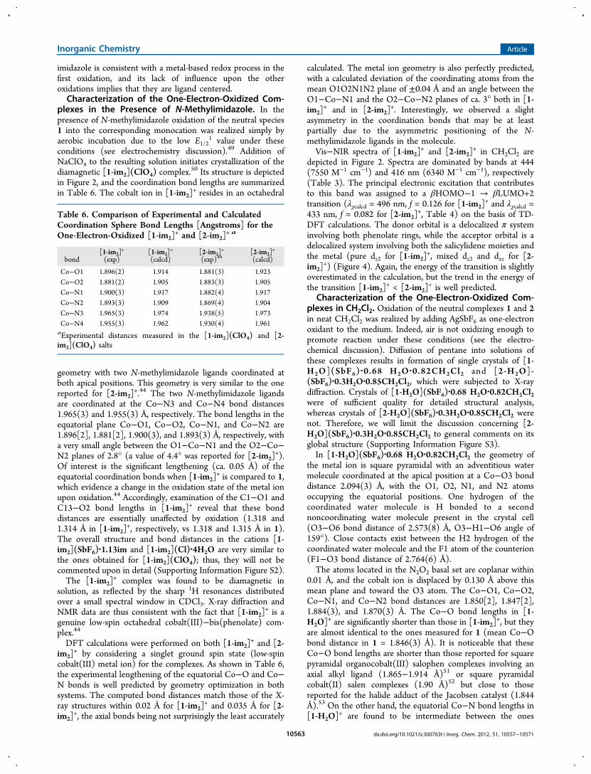
Figure 2. X-ray crystal structures of (a) 1, (b) [1-im2](ClO4), and (c)[1-H2O](SbF6) shown with 30% thermal ellipsoids. H atoms areomitted except those of the coordinated water molecule in [1-H2O]
+.
Table 2. Comparison of Experimental and CalculatedCoordination Sphere Bond Lengths (Angstroms) for 1 and 2
bond1 (exp),
molecule A1 (exp),
molecule B1
(calcd) 2 (exp)452
(calcd)
Co−O1 1.835(4) 1.848(4) 1.850 1.84(1) 1.851Co−O2 1.847(4) 1.855(4) 1.850 1.84(1) 1.853Co−N1 1.861(5) 1.851(5) 1.858 1.89(1) 1.860Co−N2 1.864(5) 1.852(5) 1.858 1.87(1) 1.861
Inorganic Chemistry Article
dx.doi.org/10.1021/ic300763t | Inorg. Chem. 2012, 51, 10557−1057110560

distances calculated by geometry optimization match those ofthe X-ray structures by 0.01 Å (Table 2) in the case of 1. Asexpected, the calculated Co−O and Co−N bond distances donot differ significantly on going from 1 and 2.The 10 K X-band EPR spectrum of 1 in CH2Cl2 is depicted
in Supporting Information Figure S1. It displays an anisotropic(S = 1/2) signal with hyperfine splitting into eight lines in thelow-field component. This pattern arises from the interaction ofthe electron spin with the cobalt nucleus (ICo = 7/2). Thespectrum could be simulated with the set of parameters gx =2.92, gy = 1.91, gz = 2.03 (giso = 2.29), and Ax(Co) = 210 MHz(the other hyperfine coupling constants could not bedetermined from simulation). It is noteworthy that the valuesdetermined for 1 are close to the ones reported for 2 (gx = 3.21,gy = 1.89, gz = 1.98, (giso = 2.36),10 thus confirming that theoverall geometry of the complexes in solution is very similar.These parameters point to a main dz2
1 orbital ground state.EPR data are thus consistent with a square planar geometry fora d7 metal ion and indicate that the cobalt(II) complex retainsits solid state structure in CH2Cl2 solution. DFT calculations onthe geometry-optimized complexes 1 and 2 confirm the metal-based character of the SOMO for both 1 (56% dz2, 40% dyz)and 2 (53% dz2, 43% dyz) as shown in Supporting InformationFigures S10 and S11.Visible spectra of 1 and 2 are characterized by intense
transitions at 423 (17 330 M−1 cm−1) and 419 nm (12 990 M−1
cm−1), respectively, with weaker d−d bands above 500 nm(Figure 3, Table 3). On the basis of its high intensity the
highest energy absorption is assigned to a charge transfer (CT)transition. The exact nature of the orbitals involved in the CTtransition was investigated by TD-DFT calculations. In both 1and 2 the principal calculated electronic excitation thatcontributes to the CT band is a βHOMO−3 → βLUMO+1transition (λcalcd = 470 nm, f = 0.146 for 1 and λcalcd = 467 nm, f= 0.197 for 2). In both cases, the donor orbital is a pure metalorbital (dxz) while the acceptor one is a delocalized π orbitalinvolving the β-iminoalkoxy moiety with a small dx2−y2contribution (Figure 4, Table 4). Although the computed
Figure 3. Vis−NIR spectra of solutions of the neutral and one-electronoxidized complexes in 0.05 mM CH2Cl2 solutions: (a) 1 (black), [1-im2]
+ (blue), and [1-H2O]+ (red); (b) 2 (black), [2-im2]
+ (blue), and[2-H2O]
+ (red). T = 298 K.
Table 3. Electronic Spectral Data of the Complexesa
complex λmax, nm (ε, M−1 cm−1)
1 390 (15850), 423 (17330), 509sh (3370), 557sh (1710)[1-im2]
+ 444 (7550), 584br (600)[1-H2O]+ 386 (9150), 1217br (7370)[1]2+ 408 (8120)b
2 359 (11940), 419 (12990), 499sh (3040)[2-im2]
+ 416 (6340)[2-H2O]+ 372 (9690), 452sh (4880), 1060br (5560)[2]2+ 402 (7720), 800br (1420)b
aIn CH2Cl2 solution, T = 298 K. Abbreviations used: sh, shoulder; br,broad. bIn CH2Cl2 + 0.01 M TBAP solution, T = 240 K.
Figure 4. TD-DFT assignment of the lowest energy transition in (a) 1and (b) [1-im2]
+.
Table 4. Lowest Energy Calculated Electronic Excitations ofthe Complexesa
complex transition (MO number) λ, nm ( f)
1 βHOMO−3→ βLUMO+1 470 (0.146)[1-im2]
+ βHOMO−1→ βLUMO+2 496 (0.126)[1-H2O]+b βHOMO−2→ βLUMO 1013 (0.110)[1-H2O]+c βHOMO → βLUMO+2 1183 (0.210)2 βHOMO−3→ βLUMO+1 467 (0.197)[2-im2]
+ βHOMO−1→ βLUMO+2 433 (0.082)[2-H2O]+b βHOMO−2→ βLUMO+1 952 (0.158)[2-H2O]+c βHOMO→ βLUMO+2 1154 (0.103)
aIn CH2Cl2.bTriplet state. cQuintet state.
Inorganic Chemistry Article
dx.doi.org/10.1021/ic300763t | Inorg. Chem. 2012, 51, 10557−1057110561

energy for the transition is slightly higher than the experimentalone, calculations correctly predict the absence of a significantshift on going from 1 to 2.Electrochemistry. The electrochemical behavior of the
square planar cobalt(II) complexes 1 and 2 has been studied bycyclic voltammetry (CV) in the noncoordinating solventCH2Cl2 (+0.1 M TBAP). All potentials are referenced versusthe Fc+/Fc redox couple. Both 1 and 2 are electroactive in thepotential range from −1.0 to +1.0 V. The CV curves of 1 and 2display three reversible one-electron redox waves in the anodicregion of potentials (Figure 5, Table 5). Oxidation potentialsare summarized in Table 5.
The redox processes are observed at E1/21 = 0.00 V, E1/2
2 =0.44 V, and E1/2
3 = 0.62 V for 1 and E1/21 = 0.01 V, E1/2
2 = 0.70V, and E1/2
3 = 0.74 V vs Fc+/Fc for 2. The position of the firstredox wave does not depend on the nature of the phenolatesubstituent. At first glance this oxidation process may beattributed to a metal-centered process; nevertheless, a non-negligible contribution of the ligand to the SOMO wasevidenced spectroscopically in the one-electron-oxidizedspecies (see below). The redox couple is therefore not purelymetal based. The second and third oxidation waves are stronglysubstituent dependent and correspond to successive formationof the Co3+−phenoxyl and Co3+−bis(phenoxyl) speciesaccording to eq 2. Interestingly, the electrochemical commu-nication between the noninnocent phenolate rings is muchsmaller in 2 than in 1, as reflected by the smaller ΔE = E1/2
3 −E1/2
2 in 2.
X Yoooo X Yoooo
X Yoooo
− − − −
−
+ + · + + ·
+ ··
Co L Co L or Co L Co L
Co L
E E
E
2 2 3 3
3
1/21
1/22
1/23
(2)
The CV of the genuine octahedral cobalt(III)−bis-(phenolate) complexes [1-im2]
+ and [2-im2]+ prepared by air
oxidation of 1 and 2, respectively, in the presence of N-methylimidazole (see below for full characterization) are shownin Figure 5. [1-im2]
+ and [2-im2]+ exhibit an irreversible
reduction wave at Epc,1 = −1.61 V and Ep
c,1 = −1.43 V,respectively. The reduction peak is associated with an oxidationone at Ep
a,1 = −0.23 V for [1-im2]+ (peak separation ΔEp1 of
1.38 V), while no oxidation signal could be detected for [2-im2]
+. The large peak separation ΔEp1 indicates that a
significant molecular rearrangement (ligand exchange, seebelow) occurs during the redox process. After addition of 10equiv of N-methylimidazole to the medium the ΔEp1 value of[1-im2]
+ decreases to 1.08 V (Epc,1 = −1.48 V; Ep
a,1 = −0.40 V)while the Ep
a,1 peak of [2-im2]+ (which was absent in neat
CH2Cl2) could be detected at Epa,1 = −0.40 V (Ep
c,1 = −1.48 V,giving a ΔEp1 value of 1.08 V). The large peak separation ΔEp1as well as the half-wave potentials E1/2
1 (−0.94 V, roughlyestimated as a midpoint potential between Ep
c,1 and Epa,1, the
redox couple being chemically irreversible) are thus identicalfor [1-im2]
+ and [2-im2]+ in a medium containing CH2Cl2 + 10
equiv of N-methylimidazole. This indicates that the redoxprocess is metal centered (Co(III)/Co(II) redox couple).47,48
The decrease in ΔEp1 resulting from addition of N-
methylimidazole to the medium is clear evidence that amolecular rearrangement occurs upon reduction of theoctahedral cobalt(III) complex.48 It is attributed to release ofN-methylimidazole in the medium upon reduction, whichaffords the square planar cobalt(II) complexes 1 and 2. It isnoteworthy that axial ligation by the N-methylimidazole basegreatly stabilizes the cobalt(III) complex, as reflected by thelower Ep
c,1 of [1-im2]+ and [2-im2]
+ when compared to 1 and2.47 Examination of the second oxidation process reveals adramatic difference between [1-im2]
+ and [2-im2]+. The peak
separation ΔEp2 is found to be abnormally high (ΔEp
2 > 0.5 V)and dependent on the concentration of N-methylimidazole inthe case of [1-im2]
+, whereas it is 0.10 V and not dependent onthe N-methylimidazole concentration in the case of [2-im2]
+.We interpret this behavior by either a release of the N-methylimidazole ligand of [1-im2]
+ upon oxidation or slowelectrode kinetics. In order to obtain insight on the origin of thephenomenon we synthesized a derivative of [1-im2]
+ that bearsan ethylene bridge instead of the cyclohexylidene one butmaintains the 2-tert-butyl-4-methoxyphenolate moieties.48 Forthis complex we observed an anodic signal at E1/2
2 = 0.33 V, i.e.,a value close to the one observed for 1, with a much smallerΔEp
2 value of 0.08 V. This supports the fact that the large ΔEp2of [1-im2]
+ arises from slow electrode kinetics, although wecannot definitely rule out changes in coordination uponoxidation. Interestingly, the half-wave potentials E1/2
2 (calcu-lated as the midpoint between the Ep
a,2 and the Epc,2 values) are
found to range between 0.42 and 0.55 V for all compounds.E1/2
2 values are almost identical in [1-H2O]+ and [1-im2]
+
(0.44 and 0.42 V, respectively), while they differ by 0.15 Vwhen [2-H2O]
+ is compared to [2-im2]+ (0.70 and 0.55 V,
respectively). E1/21 values are observed in a much larger
potential window (from −0.94 to 0.01 V for all compounds),the lower ones being observed for the N-methylimidazolecomplexes. The large stabilization of the monocation by
Figure 5. CV curves of 0.5 mM solutions of 1 and 2 in CH2Cl2 (+0.1M TBAP): (a) 1 in CH2Cl2; (b) [1-im2]
+ in CH2Cl2 + 10 equiv of N-methylimidazole; (c) 2 in CH2Cl2; (d) [2-im2]
+ in CH2Cl2 + 10 equivof N-methylimidazole. Potentials are given vs the Fc+/Fc reference.Scan rate: 0.1 V/s; T = 298 K.
Table 5. Electrochemical Properties of 1 and 2 in CH2Cl2Solutionsa
complex Epa,1 Ep
c,1 E1/21 Ep
a,2 Epc,2 E1/2
2 E1/23
1 0.04 −0.04 0.00 0.49 0.39 0.44 0.62[1-im2]
+ −0.23 −1.61 −0.92 0.70 0.14 0.42[1-im2]
+b −0.40 −1.48 −0.94 0.65 0.19 0.422 0.06 −0.04 0.01 0.65 0.75 0.70c 0.74c
[2-im2]+ −1.43 0.59 0.49 0.55
[2-im2]+b −0.40 −1.48 −0.94 0.59 0.49 0.55
aIn the presence of 0.1 M TBAP. Potential values given in V vs theFc+/Fc reference electrode, T = 298 K. bIn the presence of 10 equiv ofimidazole. cMeasured by DPV given the close proximity of E1/2.
Inorganic Chemistry Article
dx.doi.org/10.1021/ic300763t | Inorg. Chem. 2012, 51, 10557−1057110562
imidazole is consistent with a metal-based redox process in thefirst oxidation, and its lack of influence upon the otheroxidations implies that they are ligand centered.Characterization of the One-Electron-Oxidized Com-
plexes in the Presence of N-Methylimidazole. In thepresence of N-methylimidazole oxidation of the neutral species1 into the corresponding monocation was realized simply byaerobic incubation due to the low E1/2
1 value under theseconditions (see electrochemistry discussion).49 Addition ofNaClO4 to the resulting solution initiates crystallization of thediamagnetic [1-im2](ClO4) complex.50 Its structure is depictedin Figure 2, and the coordination bond lengths are summarizedin Table 6. The cobalt ion in [1-im2]
+ resides in an octahedral
geometry with two N-methylimidazole ligands coordinated atboth apical positions. This geometry is very similar to the onereported for [2-im2]
+.44 The two N-methylimidazole ligandsare coordinated at the Co−N3 and Co−N4 bond distances1.965(3) and 1.955(3) Å, respectively. The bond lengths in theequatorial plane Co−O1, Co−O2, Co−N1, and Co−N2 are1.896[2], 1.881[2], 1.900(3), and 1.893(3) Å, respectively, witha very small angle between the O1−Co−N1 and the O2−Co−N2 planes of 2.8° (a value of 4.4° was reported for [2-im2]
+).Of interest is the significant lengthening (ca. 0.05 Å) of theequatorial coordination bonds when [1-im2]
+ is compared to 1,which evidence a change in the oxidation state of the metal ionupon oxidation.44 Accordingly, examination of the C1−O1 andC13−O2 bond lengths in [1-im2]
+ reveal that these bonddistances are essentially unaffected by oxidation (1.318 and1.314 Å in [1-im2]
+, respectively, vs 1.318 and 1.315 Å in 1).The overall structure and bond distances in the cations [1-im2](SbF6)·1.13im and [1-im2](Cl)·4H2O are very similar tothe ones obtained for [1-im2](ClO4); thus, they will not becommented upon in detail (Supporting Information Figure S2).The [1-im2]
+ complex was found to be diamagnetic insolution, as reflected by the sharp 1H resonances distributedover a small spectral window in CDCl3. X-ray diffraction andNMR data are thus consistent with the fact that [1-im2]
+ is agenuine low-spin octahedral cobalt(III)−bis(phenolate) com-plex.44
DFT calculations were performed on both [1-im2]+ and [2-
im2]+ by considering a singlet ground spin state (low-spin
cobalt(III) metal ion) for the complexes. As shown in Table 6,the experimental lengthening of the equatorial Co−O and Co−N bonds is well predicted by geometry optimization in bothsystems. The computed bond distances match those of the X-ray structures within 0.02 Å for [1-im2]
+ and 0.035 Å for [2-im2]
+, the axial bonds being not surprisingly the least accurately
calculated. The metal ion geometry is also perfectly predicted,with a calculated deviation of the coordinating atoms from themean O1O2N1N2 plane of ±0.04 Å and an angle between theO1−Co−N1 and the O2−Co−N2 planes of ca. 3° both in [1-im2]
+ and in [2-im2]+. Interestingly, we observed a slight
asymmetry in the coordination bonds that may be at leastpartially due to the asymmetric positioning of the N-methylimidazole ligands in the molecule.Vis−NIR spectra of [1-im2]
+ and [2-im2]+ in CH2Cl2 are
depicted in Figure 2. Spectra are dominated by bands at 444(7550 M−1 cm−1) and 416 nm (6340 M−1 cm−1), respectively(Table 3). The principal electronic excitation that contributesto this band was assigned to a βHOMO−1 → βLUMO+2transition (λχcalcd = 496 nm, f = 0.126 for [1-im2]
+ and λχcalcd =433 nm, f = 0.082 for [2-im2]
+, Table 4) on the basis of TD-DFT calculations. The donor orbital is a delocalized π systeminvolving both phenolate rings, while the acceptor orbital is adelocalized system involving both the salicylidene moieties andthe metal (pure dz2 for [1-im2]
+, mixed dz2 and dxz for [2-im2]
+) (Figure 4). Again, the energy of the transition is slightlyoverestimated in the calculation, but the trend in the energy ofthe transition [1-im2]
+ < [2-im2]+ is well predicted.
Characterization of the One-Electron-Oxidized Com-plexes in CH2Cl2. Oxidation of the neutral complexes 1 and 2in neat CH2Cl2 was realized by adding AgSbF6 as one-electronoxidant to the medium. Indeed, air is not oxidizing enough topromote reaction under these conditions (see the electro-chemical discussion). Diffusion of pentane into solutions ofthese complexes results in formation of single crystals of [1-H2O](SbF6) ·0.68 H2O ·0.82CH2Cl2 and [2-H2O]-(SbF6)·0.3H2O·0.85CH2Cl2, which were subjected to X-raydiffraction. Crystals of [1-H2O](SbF6)·0.68 H2O·0.82CH2Cl2were of sufficient quality for detailed structural analysis,whereas crystals of [2-H2O](SbF6)·0.3H2O·0.85CH2Cl2 werenot. Therefore, we will limit the discussion concerning [2-H2O](SbF6)·0.3H2O·0.85CH2Cl2 to general comments on itsglobal structure (Supporting Information Figure S3).In [1-H2O](SbF6)·0.68 H2O·0.82CH2Cl2 the geometry of
the metal ion is square pyramidal with an adventitious watermolecule coordinated at the apical position at a Co−O3 bonddistance 2.094(3) Å, with the O1, O2, N1, and N2 atomsoccupying the equatorial positions. One hydrogen of thecoordinated water molecule is H bonded to a secondnoncoordinating water molecule present in the crystal cell(O3−O6 bond distance of 2.573(8) Å, O3−H1−O6 angle of159°). Close contacts exist between the H2 hydrogen of thecoordinated water molecule and the F1 atom of the counterion(F1−O3 bond distance of 2.764(6) Å).The atoms located in the N2O2 basal set are coplanar within
0.01 Å, and the cobalt ion is displaced by 0.130 Å above thismean plane and toward the O3 atom. The Co−O1, Co−O2,Co−N1, and Co−N2 bond distances are 1.850[2], 1.847[2],1.884(3), and 1.870(3) Å. The Co−O bond lengths in [1-H2O]
+ are significantly shorter than those in [1-im2]+, but they
are almost identical to the ones measured for 1 (mean Co−Obond distance in 1 = 1.846(3) Å). It is noticeable that theseCo−O bond lengths are shorter than those reported for squarepyramidal organocobalt(III) salophen complexes involving anaxial alkyl ligand (1.865−1.914 Å)51 or square pyramidalcobalt(II) salen complexes (1.90 Å)52 but close to thosereported for the halide adduct of the Jacobsen catalyst (1.844Å).53 On the other hand, the equatorial Co−N bond lengths in[1-H2O]
+ are found to be intermediate between the ones
Table 6. Comparison of Experimental and CalculatedCoordination Sphere Bond Lengths [Angstroms] for theOne-Electron-Oxidized [1-im2]
+ and [2-im2]+ a
bond[1-im2]
+
(exp)[1-im2]
+
(calcd)[2-im2]
+
(exp)44[2-im2]
+
(calcd)
Co−O1 1.896(2) 1.914 1.881(3) 1.923Co−O2 1.881(2) 1.905 1.883(3) 1.905Co−N1 1.900(3) 1.917 1.882(4) 1.917Co−N2 1.893(3) 1.909 1.869(4) 1.904Co−N3 1.965(3) 1.974 1.938(5) 1.973Co−N4 1.955(3) 1.962 1.930(4) 1.961aExperimental distances measured in the [1-im2](ClO4) and [2-im2](ClO4) salts
Inorganic Chemistry Article
dx.doi.org/10.1021/ic300763t | Inorg. Chem. 2012, 51, 10557−1057110563

observed for [1-im2]+ and 1. All data point to a different
electronic structure for the metal ion in the two cations [1-H2O]+ and [1-im2]
+. Accordingly, the cation [1-H2O]+ was
found to be paramagnetic in solution, as judged by the largespectral range in its 1H NMR spectrum.21
DFT calculations were undertaken on both [1-H2O]+ and
[2-H2O]+ by considering singlet (low-spin Co(III)−phenolatecomplex), triplet (low-spin Co(II)−phenoxyl complex), andquintet (high-spin Co(II)−phenoxyl complex) spin states. Asshown in Table 8 the bond distances calculated by geometry
optimization on the triplet form [1-H2O]+ match those of theX-ray structure within 0.01 Å. The bond distances calculated forthe singlet form diverge slightly, especially the Co−O ones,when compared to the experimental bond lengths (over-estimated by 0.025 Å). The bond distances calculated for thequintet form are largely overestimated (+0.06 and +0.15 Å forthe averaged Co−O and Co−N bonds, respectively). Inaddition, the triplet is found to be favored by 22.9 and 16.1kcal mol−1 (for [1-H2O]+ and [2-H2O]+, respectively) over thesinglet and 8.5 and 9.7 kcal mol−1 (for [1-H2O]+ and [2-
H2O]+, respectively) over the quintet, which makes the
diamagnetic form irrelevant. Therefore, on the basis ofcombined X-ray diffraction data and DFT calculations thecation is assigned as a paramagnetic radical species involving arather low-spin cobalt ion. The individual contributions of theatoms to the spin population in the triplets [1-H2O]
+ and [2-H2O]
+ are summarized in Table 7. We found that more than70% of the total spin density in the triplets [1-H2O]
+ and [2-H2O]
+ is localized on the metal, the remaining spin densitybeing equally distributed within the phenoxyl moieties.54
Regarding the quintets [1-H2O]+ and [2-H2O]+, 69% and
71%, respectively, of the total spin density is localized on themetal, the remaining spin density being mainly localized withinthe aromatic rings.The two localized SOMOs of the triplets [1-H2O]
+ and [2-H2O]
+ are shown in Figure 6. For [1-H2O]+ one SOMO has a
main metal character (82%), with small contributions of thecoordinating atom of the ligands due to the covalency of themetal−ligand bonds. The second SOMO has mainly phenoxylcharacter (66%), with a contribution of 33% of the metal. Thedetailed analysis of the SOMOs thus points to a strong orbitalmixing in [1-H2O]
+, which confers to the cation a non-negligible ligand radical character. Consequently, [1-H2O]
+ isbest described by the three canonical structures shown inScheme 2, with a larger weight for structure A.18 Regarding [2-H2O]
+, the composition of the first SOMO is again mainlymetallic (87%), whereas the second one has both ligand (54%)and metal (45%) character. As for [1-H2O]
+, DFT calculations
Table 7. Individual Contributions of the Atoms to the SpinDensity in [1-H2O]+, [2-H2O]+, [1-im2]
2+, and [2-im2]2+a
atom [1-H2O]+b [2-H2O]
+b [1-im2]2+ [2-im2]
2+
Co 1.50 2.77 1.60 2.83 0.01 0.02O1 0.09 0.19 0.10 0.24 0.00 0.01O2 0.09 0.26 0.10 0.27 0.24 0.27O3 0.04 0.05 0.05 0.03N1 0.01 0.08 0.02 0.11 0.00 0.00N2 0.01 0.08 0.02 0.11 0.04 0.08C1 0.00 0.00 0.00 0.00 0.00 0.00C2 0.03 0.04 0.02 0.04 0.00 0.00C4 0.06 0.04 0.04 0.06 0.00 0.00C6 0.03 0.04 0.02 0.04 0.00 0.00C13 0.00 0.03 0.00 0.02 0.10 0.06C14 0.03 0.09 0.02 0.06 0.17 0.16C16 0.06 0.11 0.04 0.09 0.22 0.26C18 0.03 0.09 0.02 0.06 0.08 0.13
aAtom numbering used.
bFirst column: triplet form, second column: quintet form.
Table 8. Comparison of Selected Experimental and Calculated Bond Lengths [Angstroms] for [1-H2O]+ and [2-H2O]
+
bond [1-H2O] (exp) [1-H2O]+ (calcd) [1-H2O]+ (calcd) [1-H2O]+ (calcd) [2-H2O]+ (calcd) [2-H2O]+ (calcd) [2-H2O]+ (calcd)
nature triplet tripletb singletb quintetb tripletc singletc quintetc
Co−O1 1.849(2) 1.856 1.867 1.888 1.856 1.850 1.868Co−O2 1.851(3) 1.845 1.872 1.925 1.856 1.857 1.882Co−N1 1.880(4) 1.881 1.872 2.047 1.882 1.893 2.055Co−N2 1.851(3) 1.881 1.880 2.060 1.888 1.911 2.055Co−O3 2.082(4) 2.143 2.194 2.107 2.137 1.975 2.097C1−O1 1.316(6) 1.312 1.302 1.308 1.316 1.319 1.310C13−O2 1.320(5) 1.316 1.302 1.293 1.318 1.325 1.305
aIn the crystals of [1-H2O](SbF6)·0.68 H2O·0.82CH2Cl2.bThe triplet state is favored by 22.9 kcal mol−1 over the singlet state and 8.5 kcal mol−1
over the quintet state. cThe triplet state is favored by 16.1 kcal mol−1 over the singlet state and 9.7 kcal mol−1 over the quintet state.
Figure 6. Localized SOMOs for the triplets (a) [1-H2O]+ and (b) [2-
H2O]+.
Inorganic Chemistry Article
dx.doi.org/10.1021/ic300763t | Inorg. Chem. 2012, 51, 10557−1057110564

thus evidence a non-negligible contribution of the Co(II)-radical valence isomer in the case of [2-H2O]+, Figure 6. Whencompared to [2-H2O]+ the SOMO of [1-H2O]+ features anincreased ligand (phenoxyl) contribution due to the enhancedelectron-donating ability of the methoxy substituent, asobserved in some nickel radical salen complexes.55 TheSOMOs of the quintets [1-H2O]+ and [2-H2O]+ are shownin Supporting Information Figures S14 and S15. For [1-H2O]
+
two of the four SOMOs have main metal character (87% and75%, which correspond mainly to dz2 and mixed dx2−y2−dxyorbitals, respectively). The two other ones have significantphenoxyl character (45% and 43%), in addition to the metalcharacter. For [2-H2O]+ three of the four SOMOs exhibit mainmetal character (83%, 80%, and 80%, which correspond to dz2,dx2−y2, and dxy orbitals, respectively) whereas the fourth one hasboth phenoxyl (62%) and metal (38%) character. As for thetriplets, DFT calculations thus highlight a significant con-tribution of the Co(II)-radical valence isomer in the electronicstructure of the quintets.Because the paramagnetic [1-H2O]+ (and [2-H2O]
+) infrozen solution was found to be difficult to detect at the X-bandfrequency both at 10 and at 100 K, we run Q-band EPRexperiments at 7 K on a powder sample. The 35 GHz spectrumdisplays a broad signal at g ≈ 5.3 as well as a radical signal at g≈ 2 (Supporting Information Figure S5). This spectrum wasfound difficult to interpret by considering a triplet on the basisof the calculated zero-field splitting (ZFS) parameters forcopper(II)−salen radical complexes.6 We alternatively interpretit by considering a high-spin cobalt ion (SCo= 3/2). For a high-spin Co(II) the ZFS parameters are much larger than themicrowave energy and the doublet separation is too high toobserve interdoublet transitions. Under this regime a signalcorresponding to the allowed transition within the |−1/2,+1/2⟩doublet is observed at an effective g value lying within the range5 < geff < 6,56 which matches quite well the experimental peak atg ≈ 5.8. EPR spectroscopy thus suggests that [1-H2O]+ consistsof a high-spin rather than low-spin Co(II) ion weaklyinteracting with a ligand radical. Additionally, a radical featureis observed on the spectrum with a strong signal centered at g =2.The discrepancy between EPR and crystallographic data,
which suggest a low-spin Co(II)−phenoxyl radical character for[1-H2O]+, likely results from packing forces that can be strongenough to maintain a low-spin configuration in the crystals aswell as the existence of a H-bond network, which may affect thenucleophilicity of the coordinating water molecule (a six-coordinate adduct may be also accessible). Noteworthy, bothlow- and high-spin configurations have been reported forsquare pyramidal cobalt(II) bis(phenolate) salen com-plexes.52,57−61 For example, the monohydrate N,N′-ethylenebis-
(3-methoxysalicylidene-iminato)cobalt(II) complex was foundto be high spin on the basis on magnetic measurements.58 X-raydiffraction showed that the N-methylimidazole imidazoleadduct of the N,N′-phenylenebis(salicylidene-iminato)cobalt-(II) complex was also high spin,59 but the pyridine adduct ofthe N,N′-ethylenebis(salicylidene-iminato)cobalt(II) complexwas low spin.52 In order to confirm that the cation [1-H2O]
+ isparamagnetic we measured the product of the magneticsusceptibility with temperature (χT) versus temperature on apowder sample of [1-H2O](SbF6)·0.68H2O·0.82CH2Cl2 (Supporting Information Figure S6). At room temperature χT is1.47 cm3 K mol−1 and then decreases almost linearly uponcooling down to 50 K, where the decrease becomes moreabrupt to reach almost zero at 2 K. The room-temperaturevalue of χT is lower than the value (2.25 cm3 K mol−1)expected for one high-spin cobalt(II) (S = 3/2, g ≈ 2, χT =1.875 cm3 K mol−1) plus one radical (S = 1/2, g = 2, χT = 0.375cm3 K mol−1) magnetically independent. It is however farabove the value (0.75 cm3 K mol−1) expected for a low-spincobalt(II) (S = 1/2, g ≈ 2, χT = 0.375 cm3 K mol−1) plus oneradical (S = 1/2; g = 2, χT = 0.375 cm3 K mol−1) without
Scheme 2. Canonical Structures Describing the One-Electron-Oxidized Co(II) Complexes
Figure 7. TD-DFT assignment of the lowest energy transition in (a)[1-H2O]
+ (triplet), (b) [2-H2O]+ (triplet), (c) [1-H2O]
+ (quintet),and (d) [2-H2O]
+ (quintet).
Inorganic Chemistry Article
dx.doi.org/10.1021/ic300763t | Inorg. Chem. 2012, 51, 10557−1057110565

considering any interaction. It is also too high if we consider alow-spin cobalt(II) ferromagnetically coupled with a radical atroom temperature (S = 1, χT = 1.0 cm3 K mol−1). Thus, thethermal variation of χT better suggests that the cobalt(II) ishigh spin but strongly antiferromagnetically coupled with thephenoxyl radical giving a ground state spin S = 1 (if we considera real spin S = 3/2 for the cobalt(II) ion). A survey of theliterature shows that most magnetic studies of Co(II)complexes are concerned with four-coordinated cobalt(II)ions that are unambiguously low spin in a square planargeometry and high spin in a tetrahedral one. Six-coordinatedcobalt(II) metal ions are always high spin.62 As stated abovethere are fewer studies of five-coordinated cobalt(II) ions,which may be either high or low spin.60,61 The trigonalbipyramidal environment results generally in a high-spin state,60
but for square planar pyramids both situations are found.61
Owing to the square pyramidal geometry of the cobalt ion in[1-H2O]+ the magnetic behavior is complicated by the lowsymmetry and spin−orbit coupling should be included tointerpret the low-temperature behavior.63 In order to estimatethe correctness of the model and have an estimation of thestrength of the interaction the magnetic susceptibility wastentatively simulated above 100 K by considering the spinHamiltonian H = −2JS1S2 (S1 = 1/2, S2 = 3/2) and using thefollowing equation
χβ
=+
+T
Ng J kTk J kT
[2 10 exp(4 / )][3 5 exp(4 / )]
2 2
A good fit of the experimental data was obtained in the range100−300 K with an antiferromagnetic coupling J = −110.3cm−1 and g = 2.11, without considering intermolecularinteraction. The whole set of data may be well fittedconsidering this model with J = −138.9 cm−1, g = 2.29, plusintermolecular interaction θ = −19 cm−1. However, this resultis only indicative since a more rigorous model including spin−orbit effect should be used here, as pointed out above.Noteworthy, attempts to fit the data by consideringintermolecular antiferromagnetic interactions in between a (S= 1) system corresponding to a low-spin cobalt(II)antiferromagnetically coupled with a phenoxyl radical at roomtemperature were unsuccessful.Therefore, our X-ray diffraction, EPR, and magnetic
susceptibility data are all consistent with a phenoxyl radicalcharacter for [1-H2O]+.Vis−NIR spectra of [1-H2O]+ and [2-H2O]+ are depicted in
Figure 3. Both cations absorb significantly over the entirevisible region, similarly to the analogous nickel radical salencomplexes8 and cobalt−iminosemiquinonate radical com-plexes.18−20 Most importantly, both [1-H2O]+ and [2-H2O]
+
exhibit a remarkable NIR feature at 1220 (7370 M−1 cm−1) and1060 nm (5560 M−1 cm−1), respectively. It is important to notethat neither 1 or [1-im2]
+ nor 2 or [2-im2]+ exhibits a band in
the NIR region (Table 3). On the basis of its high intensity, thisband cannot originate from a d−d transition; therefore, it isascribed to a CT transition. In addition, its low energy isindicative of a small energy gap between the ground and theexcited states, whose nature was investigated by time-dependent DFT calculations.The absence of intense NIR transitions for 1 and [1-im2]
+
(as well as 2 and [2-im2]+) is well predicted, whereas an
electronic excitation could be calculated in the NIR region forboth [1-H2O]+ and [2-H2O]+. Noteworthy, the NIR band is
predicted by considering both low-spin Co(II)−phenoxyl andhigh-spin Co(II)−phenoxyl complexes, but its origin differs. Byconsidering a low-spin Co(II)−phenoxyl configuration itcorresponds to a βHOMO−2→ βLUMO transition in thecase of [1-H2O]
+, while it is a βHOMO−2 → βLUMO+1transition in [2-H2O]
+. The predicted energies are 1013 ( f =0.110) and 952 nm ( f = 0.158) for [1-H2O]
+ and [2-H2O]+,
respectively, which is in fairly good agreement with theexperimental bands (1217 and 1060 nm, respectively). Thecomposition of the donor orbital is a delocalized π systeminvolving one aromatic ring and a dyz metal orbital in bothcases. The acceptor orbital is a delocalized π system involvingtwo aromatic rings and a metal orbital that mainly has dz2 (for[1-H2O]
+) or dyz character (for [2-H2O]+). By considering a
high-spin Co(II)−phenoxyl system the NIR band arises from aβHOMO → βLUMO+2 transition for both [1-H2O]
+ and [2-H2O]+. The transition is mainly a ligand-to-metal CTtransition: The donor orbital is a delocalized π system involvingthe aromatic rings (with a larger contribution of the phenolatering) and a dxz metal orbital, while the acceptor one has dz2character. The predicted energies are 1183 ( f = 0.210) and1154 nm ( f = 0.103) for [1-H2O]
+ and [2-H2O]+, respectively.
Although these values are in better agreement with theexperimental data, the difference in the calculated bands byconsidering low- and high-spin Co(II)-phenoxyl species isrelatively small and it is not possible to concludeunambiguously on the metal ion spin state in the radicals [1-H2O]
+ and [2-H2O]+ in solution.
Characterization of the Two-Electron-Oxidized Com-plexes in CH2Cl2. The two-electron-oxidized forms ofcomplexes 1 and 2 were generated electrochemically inCH2Cl2 at low temperature (240 K) due to their limitedstability. UV−vis spectra of the dications [1]2+ and [2]2+ inCH2Cl2 (+0.01 M TBAP) at 240 K are depicted in Figure 8.
They are dominated by bands at 408 (8120 M−1 cm−1) and 402nm (7720 M−1 cm−1) for [1]2+ and [2]2+, respectively. Anadditional broad band of weaker intensity could be observed at800 nm (ε = 1420 M−1 cm−1) in the spectrum of [2]2+. Thesespectral features match remarkably well those reported forphenoxyl−Co(III)11,64 and phenoxyl−Cu(II)65 complexesgenerated from Mannich bases. The dications thus comprisea phenoxyl radical moiety coordinated to a genuine Co(III)metal ion. Addition of N-methylimidazole (10 equiv) to the
Figure 8. UV−vis spectra of the electrochemically generated dicationsin 0.05 mM CH2Cl2 solutions (+0.01 M TBAP): (a) [1]2+ (blue) nd(b) [2]2+ (red). T = 240 K.
Inorganic Chemistry Article
dx.doi.org/10.1021/ic300763t | Inorg. Chem. 2012, 51, 10557−1057110566

CH2Cl2 solution of [1]2+ and [2]2+ results in a gradual decreaseof the intensity of the spectra, without significant shift of thebands. This indicates that the dications are poorly stable in thepresence of N-methylimidazole.The X-band EPR spectra of the dications [1]2+ and [2]2+ in
CH2Cl2 (+0.1 M TBAP) solution are shown in Figure 9. Both
spectra consist of an S = 1/2 signal centered at giso = 2.00. Thisbehavior is clear evidence for the presence of a phenoxyl radicalcoordinated to a low-spin Co(III) metal ion as similar g values(lower than the one of the free electron 2.0023) were reportedfor Co(III)−phenoxyl radical species.10,11,64 This assignment isconfirmed by the observation of a hyperfine interaction of theelectron spin with the cobalt nucleus which splits the EPRsignal. Because the hyperfine coupling constants are within ofrange of the line width the splitting could be mainly observed asshoulders in the S = 1/2 peak. It is worth noting that the signalis highly isotropic without any hyperfine coupling constanthigher than 100 MHz. Van Doorslaer et al. showed that thisbehavior is indicative of an octahedral geometry for the metalion, the calculated g values for pentacoordinated cobalt(III)radical complexes being less than 2.00 with stronger hyperfinecoupling constants.10 In addition, the signal is found to bedistributed over a larger spectral width when [2]2+ is comparedto [1]2+. That may suggest that the ligand radical spin interactsmore strongly with the Co(III) nuclear spin in the case of [2]2+.EPR spectra were recorded just after addition of 10 equiv of
N-methylimidazole to a CH2Cl2 solution of the electro-generated dication. A strong decrease of intensity was observed,consistent with fast decomposition of the dication. The EPRsignal resembles those of [1]2+ and [2]2+ in CH2Cl2, but theyare not superimposable. This confirms that N-methylimidazolecoordinates to the metal center before decomposition, affordingthe octahedral low-spin Co(III) complexes [1-im2]
2+ and [2-im2]
2+.10 The fact that only slight changes arise from additionof N-methylimidazole to the CH2Cl2 solution of the dicationsfurther confirms that the Co(III) ion likely already adopts anoctahedral geometry both in [1]2+ and in [2]2+.On the basis of our EPR results, DFT calculations were
undertaken on [1-im2]2+ and [2-im2]
2+ by considering a S =1/2 ground spin state and two axially bound N-methylimida-zoles. Geometry optimization reveals that the equatorialcoordinating atoms deviate by ±0.04 Å from the mean
O1,O2,N1,N2 plane. In addition, the angle between the twoO1−Co−N1 and O2−Co−N2 planes is ca. 4° both in [1-im2]
2+ and in [2-im2]2+. These geometrical features compare
quite well with those calculated for the monocations [1-im2]+
and [2-im2]+. Nevertheless, examination of the coordination
bond lengths reveals a strong asymmetry in the C−O bonddistances (Table 9): The Co−O1 bond length is 1.962 Å, while
the Co−O2 one is 1.893 Å. These values differ significantlyfrom the ones calculated for the monocation [1-im2]
2+ (1.914and 1.905 Å). Such asymmetry is indicative of phenoxyl radicallocalization on a single aromatic ring (the one bearing the O1atom).6 This assumption is confirmed by the quinonoiddistribution of bond length within one ring with C1−O1,C1−C2, C2−C3, C3−C4, C4−C5, C5−C6, and C1−C6 bonddistances of 1.270, 1.468, 1.376, 1.411, 1.409, 1.376, and 1.456Å, the corresponding bond distances in the other ring being1.316, 1.435, 1.391, 1.405, 1.375, 1.411, and 1.429 Å (atomnumbering is indicated in Table 7). Similar behavior is observedfor [2-im2]
2+. Not surprisingly, the SOMO is almost exclusivelydistributed on a single phenoxyl ring in both [1-im2]
2+ and [2-im2]
2+, Figure 10. Both [1-im2]2+ and [2-im2]
2+ are thuslocalized phenoxyl−Co(III) complexes.
Regarding [1]2+ and [2]2+, we performed DFT calculationsby considering a hexacoordinated cobalt ion, with two watermolecules occupying the axial positions in the metalcoordination sphere.66 From geometry optimization it appearsthat the overall geometry of the complex is hardly affected bythe nature of the axial ligands, the coordination sphere beingsomewhat slightly more contracted in [1]2+ and [2]2+ than in[1-im2]
2+ and [2-im2]2+ (Table 9). The composition of the
SOMO is again ligand centered and exclusively distributed on asingle phenoxyl ring (more than 90%).
Figure 9. X-band EPR spectra of the electrochemically generateddications (a) [1]2+ and (b) [2]2+ in 0.5 mM CH2Cl2 (+0.1 M TBAP)solution. Microwave frequency: 9.45 GHz. Power: 5.5 mW. Mod. amp.0.1 mT. Frequency: 100 kHz. T = 100 K.
Table 9. Calculated Bond Lengths [Angstroms] for theDications
bond [1]2+ [1-im2]2+ [2]2+ [2-im2]
2+
Co−O1 1.962 1.977 1.968 1.978Co−O2 1.893 1.911 1.893 1.912Co−N1 1.931 1.937 1.933 1.933Co−N2 1.898 1.907 1.896 1.903Co−Ax 1a 1.956 1.985 1.955 1.985Co−Ax 2a 1.959 2.002 1.955 2.001C1−O1 1.274 1.270 1.272 1.268C13−O2 1.323 1.316 1.322 1.311
aCoaxial bonds: Co−Ax 1 corresponds to Co−N3 and Co−Ax 2corresponds to Co−N4 for [1-im2]
2+ and [2-im2]2+. Co−Ax 1
corresponds to Co−O3 and Co−Ax 2 corresponds to Co−O4 for[1]2+ and [2]2+.
Figure 10. Localized SOMOs for (a) [1]2+ and (b) [1-im2]2+.
Inorganic Chemistry Article
dx.doi.org/10.1021/ic300763t | Inorg. Chem. 2012, 51, 10557−1057110567

DFT studies show that the dications are localized phenoxyl−Co(III) complexes irrespective of the phenol substituent andaxial ligand, in agreement with recent work published by VanDoorslaer et al.10 It is noteworthy that the small electroniccoupling between the two redox-active rings is also evidentfrom electrochemical studies. Indeed, the ΔE1/2 = E1/2
3 − E1/22
value is very low in both 1 and 2, consistent very poorelectrochemical communication between the phenoxyl and thephenolate moieties. The systematic ligand radical localization inthis series contrasts with the results obtained for the nickel(II)salen radical complexes,6,7e where the tert-butyl groups promoteligand radical delocalization (the SOMO is shared between thetwo rings) and more electron-donating groups like OMe induceligand radical localization. The behavior observed for the cobaltsalen complexes is more in line with the recent results obtainedby Fujii et al.67 on manganese salen radical complexes. Theyshowed that a nickel to manganese substitution drasticallylimits electron transfer between the two redox centers(phenolate and phenoxyl) and that manganese−salen radicalcomplexes exhibit a localized phenoxyl character. Modulation ofthe redox potential of the electron donor and acceptor relativeto the metal ion mediator was proposed to account for theresulting ligand radical localization. Noteworthy, the Co(III)metal ion exhibits a similar formal charge to the Mn(III) ionand an increase in the metal charge is expected to increase thephenoxyl/phenolate oxidation potential. This expectation isexperimentally verified, with formation of the Co(III)−phenoxyl species occurring at 0.49 and 0.65 V for [1]2+ and[2]2+, respectively, versus 0.22 and 0.37 V for thecorresponding Ni(II)−phenoxyl complex.A higher metal charge thus significantly alters the ligand
radical delocalization in this series, providing a localized radicalcharacter for the Co(III) salen complexes.
■ CONCLUSIONThe present study unambiguously shows that the electronicstructure of one-electron-oxidized square planar cobalt(II)−salen complexes strongly depends on the medium. Cations [1-H2O]+ and [2-H2O]+ were generated by silver oxidation in neatCH2Cl2 (air is not powerful enough under these conditions).Adventitious water coordinates to the metal ion in the absenceof a strong donor in solution. Cations [1-H2O]+ and [2-H2O]
+
are consequently not square planar like their neutral precursorbut square pyramidal with a water molecule coordinated at theapical position. These species may constitute crucialintermediates in the catalytic ring opening of epoxides. Itshould be underlined that the catalytic opening of epoxide isbelieved to occur by a cooperative bimetallic mechanisminvolving a square pyramidal Co(III) complex containing anaxially bound nucleophile, which attacks an epoxide substratecoordinated to a second octahedral Co(III) complex.13,21 Thebest catalytic system involves one-electron-oxidized 2 as anequimolar mixture of Cl− and SbF6
− adducts in the presence asmall amount of water. A key step in the reaction is attack ofthe nucleophile (hydroxyl group) on the epoxide. Formation ofthis nucleophile is proposed to result from binding of water onthe counterion adduct of 2 and deprotonation due to the Lewisacidity of the cobalt(III) ion. The high-resolution structure of[1-H2O]+ and the general arrangement of [2-H2O]+ providethe first evidence for the absence of ligation of the SbF6
− anion.Instead, the SbF6
− adduct of the Jacobsen catalyst [2-H2O]+
probably already contains the nucleophile precursor (watermolecule) coordinated to the square pyramidal metal ion,
which may explain the high rates obtained when this complex isused for ring opening of epoxides. Both [1-H2O]
+ and [2-H2O]
+ exhibit a paramagnetic (S = 1) ground spin state, withthe spin density being mainly localized on the metal.Importantly, the remaining spin density is distributed overthe aromatic rings, showing that the cations have a non-negligible phenoxyl character never evidenced previously. Both[1-H2O]
+ and [2-H2O]+ exhibit a remarkable NIR band at
1220 (7370 M−1 cm−1) and 1060 nm (5560 M−1 cm−1),respectively, assigned to CT transition. Preliminary DFTcalculations were undertaken on structures derived from [1-H2O]+ and [2-H2O]+ with the water molecule beingsubstituted by an acetate anion. They highlight a dramaticeffect of ligand charge upon electronic structure (paramagneticCo(III)−bis(phenolate) cation in this case) that will be furtherinvestigated.54 In the presence of N-methylimidazole, 1 and 2are easily oxidized by air into the genuine octahedral cobalt(III)bis(phenolate) complexes [1-im2]
+ and [2-im2]+. Neither [1-
im2]+ nor [2-im2]
+ exhibits a NIR band in its electronicspectrum. The NIR feature of [1-H2O]
+ and [2-H2O]+ could
be a spectroscopic marker for a ligand contribution to theelectronic structure of the cations. The number and nature ofthe axial ligands and thus the external medium therefore play acrucial role in the stabilization of the Co(III)−L vs spin-coupled Co(II)−L• valence isomer. The electrochemicallygenerated dications [1]2+ and [2]2+ were identified asparamagnetic (S = 1/2) Co(III)−phenoxyl species by acharacteristic absorption band at ca. 400 nm in the UV−visspectrum and a highly isotropic EPR signal centered at g = 2.00.DFT studies show that both [1]2+ and [2]2+ are localizedphenoxyl−Co(III) complexes, in agreement with recent workpublished by Van Doorslaer et al. on a derivative of [2]2+ .10
The present study shows that ligand radical localization occursirrespective of the phenol substituent. This trend contrasts withthe results obtained for the nickel(II) salen radical complexes inwhich tert-butyl groups promote ligand radical delocalization(the SOMO is shared between the two rings), whereas moreelectron-donating groups like OMe induce ligand radicallocalization. The behavior observed for phenoxyl−Co(III)complex resembles more that reported for phenoxyl−Mn(III)complexes by Fujii et al.67 They nicely showed that a nickel tomanganese substitution in radical salen complexes drasticallylimits electron transfer between the two redox centers(phenolate and phenoxyl), resulting in radical localization. Ashift of the redox potential of the electron donor and acceptorrelative to the metal ion mediator was proposed to account forthe resulting ligand radical localization. The metal chargeinfluences both the phenoxyl/phenolate oxidation potential andthe metal ion geometry. The potential values corresponding toformation of [1]2+ and [2]2+ are almost similar to thosereported for oxidation of the corresponding Mn(III)−phenolate complexes into Mn(III)−phenoxyl species, with anidentical speculated geometry for the metal ion. Thesehomologies may be responsible for the localized ligand radicalcharacter of the dications. Further studies are in progress in ourlaboratory to better understand the parameters involved instabilization of the high-spin Co(III)−L vs spin-coupledCo(II)−L• valence isomer.
■ ASSOCIATED CONTENT*S Supporting InformationEPR spectra of 1, [1-im2]
2+, [2-im2]2+, and the superoxo/N-
methylimidazole adducts of 1 and 2; localized SOMOs for 1, 2,
Inorganic Chemistry Article
dx.doi.org/10.1021/ic300763t | Inorg. Chem. 2012, 51, 10557−1057110568

[1]2+, and [1-im2]2+; spin density plots of [1-H2O]+, [2-H2O]
+,and their water-to-acetate-substituted analogs; metrical param-eters issued from geometry optimization for all complexes; TD-DFT assignment of the lowest energy transition in 2 and [2-im2]
+. This material is available free of charge via the Internet athttp://pubs.acs.org.
■ AUTHOR INFORMATIONCorresponding Author*E-mail: [email protected] authors declare no competing financial interest.
■ ACKNOWLEDGMENTSThis work was supported by a doctoral fellowship (A.K.) fromthe DCM.
■ REFERENCES(1) (a) Whittaker, J. W. In Metal Ions in Biological Systems; Sigel, H.,Sigel, A., Eds.; Marcel Dekker: New York, 1994; Vol. 30, p 315.(b) Borman, C. D.; Saysell, C. G.; Sokolowski, A.; Twitchett, M. B.;Wright, C.; Sykes, A. G. Coord. Chem. Rev. 1999, 190−192, 771.(c) McPherson, M. J.; Parsons, M. R.; Spooner, R. K.; Wilmot, C. M.In Handbook for metalloproteins; Messerschmidt, A., Huber, R., Poulos,T., Wieghardt, K., Eds.; John Wiley and Sons: New York, 2001; Vol. 2,p 1272. (d) Whittaker, J. W. In Advances in Protein Chemistry;Richards, F. M., Eisenberg, D. S., Kuriyan, J., Eds.; Academic Press,Elsevier: New York, 2002; Vol. 60, p 1. (e) Whittaker, J. W. Chem. Rev.2003, 103, 2347. (f) Rogers, M. S.; Dooley, D. M. Curr. Opin. Chem.Biol. 2003, 7, 189. (g) Firbank, S. J.; Rogers, M.; Hurtado-Guerrero,R.; Dooley, D. M.; Halcrow, M. A.; Phillips, S. E. V.; Knowles, P. F.;McPherson, M. J. Biochem. Soc. Trans. 2003, 31, 506.(2) Gordon, A.; Hamilton, G. A.; Adolf, P. K.; de Jersey, J.; DuBois,G. C.; Dyrkacz, G. R.; Libby, R. D. J. Am. Chem. Soc. 1978, 100, 1899.(3) (a) Whittaker, M. M.; Whittaker, J. W. J. Biol. Chem. 1988, 263,6074. (b) Babcock, G. T.; El-Deeb, M. K.; Sandusky, P. O.; Whittaker,M. M.; Whittaker, J. W. J. Am. Chem. Soc. 1992, 114, 3121.(c) McGlashen, M. L.; Eads, D. D.; Spiro, T. G.; Whittaker, J. W. J.Phys. Chem. 1995, 99, 4918. (d) Whittaker, M. M.; Ekberg, C. A.;Peterson, J.; Sendova, M. S.; Day, E. P.; Whittaker, J. W. J. Mol. Catal.B: Enzym. 2000, 8, 3.(4) (a) Jazdzewski, B. A.; Tolman, W. B. Coord. Chem. Rev. 2000,200−202, 633. (b) Kruger, H. J. Angew. Chem., Int. Ed. 1999, 38, 627.(c) Itoh, S.; Taki, M.; Fukuzumi, S. Coord. Chem. Rev. 2000, 198, 3.(d) Chaudhuri, P.; Wieghardt, K. Prog. Inorg. Chem. 2001, 50, 151.(e) Thomas, F. Eur. J. Inorg. Chem. 2007, 2379. (f) Thomas, F. InStable Radicals: Fundamentals and Applied Aspects of Odd-ElectronCompounds; Hicks, R. G., Ed.; John Wiley and Sons: Chichester,2010;pp 281.(5) (a) Pratt, R. C.; Stack, T. D. P. J. Am. Chem. Soc. 2003, 125, 8716.(b) Pratt, R. C.; Stack, T. D. P. Inorg. Chem. 2005, 44, 2367.(c) Thomas, F.; Jarjayes, O.; Duboc, C.; Philouze, C.; Saint-Aman, E.;Pierre, J.-L. Dalton Trans. 2004, 2662. (d) Storr, T.; Verma, P.; Pratt,R. C.; Wasinger, E. C.; Shimazaki, Y.; Stack, T. D. P. J. Am. Chem. Soc.2008, 130, 15448.(6) Orio, M.; Jarjayes, O.; Kanso, H.; Philouze, C.; Neese, F.;Thomas, F. Angew. Chem., Int. Ed. 2010, 49, 4989.(7) (a) Shimazaki, Y.; Tani, F.; Fukui, K.; Naruta, Y.; Yamauchi, O. J.Am. Chem. Soc. 2003, 125, 10512. (b) Rotthaus, O.; Jarjayes, O.;Thomas, F.; Philouze, C.; Del Valle, C. P.; Saint-Aman, E.; Pierre, J. L.Chem.−Eur. J 2006, 12, 2293. (c) Rotthaus, O.; Thomas, F.; Jarjayes,O.; Philouze, C.; Saint-Aman, E.; Pierre, J. L. Chem.−Eur. J 2006, 12,6953. (d) Shimazaki, Y.; Yajima, T.; Tani, F.; Karasawa, S.; Fukui, K.;Naruta, Y.; Yamauchi, O. J. Am. Chem. Soc. 2007, 129, 2559. (e) Storr,T.; Wasinger, E. C.; Pratt, R. C.; Stack, T. D. P. Angew. Chem., Int. Ed.2007, 46, 5198. (f) Benisvy, L.; Kannappan, R.; Song, Y. F.;Milikisyants, S.; Huber, M.; Mutikainen, I.; Turpeinen, U.; Garnez,
P.; Bernasconi, L.; Baerends, E. J.; Hartl, F.; Reedijk, J. Eur. J. Inorg.Chem. 2007, 631. (g) Rotthaus, O.; Labet, V.; Philouze, C.; Jarjayes,O.; Thomas, F. Eur. J. Inorg. Chem. 2008, 4215. (h) Rotthaus, O.;Jarjayes, O.; Philouze, C.; Del Valle, C. P.; Thomas, F. Dalton Trans.2009, 1792. (i) Shimazaki, Y.; Stack, T. D. P.; Storr, T. Inorg. Chem.2009, 48, 8383. (j) Storr, T.; Verma, P.; Shimazaki, Y.; Wasinger, E.C.; Stack, T. D. P. Chem.−Eur. J 2010, 16, 8980. (k) Kochem, A.; Orio,M.; Jarjayes, O.; Neese, F.; Thomas, F. Chem. Commun. 2010, 46,6765. (l) Dunn, T. J.; Ramogida, C. F.; Simmonds, C.; Paterson, A.;Wong, E. W. Y.; Chiang, L.; Shimazaki, Y.; Storr, T. Inorg. Chem. 2011,50, 6746. (m) Shimazaki, Y.; Arai, N.; Dunn, T. J.; Yajima, T.; Tani, F.;Ramogida, C. F.; Storr, T. Dalton Trans. 2011, 40, 2469. (n) Verma,P.; Pratt, R. C.; Storr, T.; Wasinger, E. C.; Stack, T. D. P. Proc. Natl.Acad. Sci. U.S.A. 2011, 108, 18600.(8) (a) Rotthaus, O.; Jarjayes, O.; Thomas, F.; Philouze, C.; Saint-Aman, E.; Pierre, J.-L. Dalton Trans. 2007, 889. (b) Orio, M.; Philouze,C.; Jarjayes, O.; Neese, F.; Thomas, F. Inorg. Chem. 2010, 49, 646.(9) (a) Neirn, A. K.; Bhalla, R.; Foxon, S. P.; Liu, X.; Yellowlees, L. J.;Gilbert, B. C.; Walton, P. H. J. Chem. Soc., Dalton Trans. 2002, 1253.(b) Imbert, C.; Hratchian, H. P.; Lanznaster, M.; Heeg, M. J.;Hryhorczuk; McGarvey, B. R.; Schlegel, H. B.; Verani, C. N. Inorg.Chem. 2005, 44, 7414. (c) Floquet, S.; Ottenwaelder, X.; Boillot, M.-L.Inorg. Chem. Commun. 2007, 10, 1549. (d) Takuya Kurahashi, T.;Kobayashi, Y.; Nagatomo, S.; Tosha, T.; Kitagawa, T.; Fujii, H. Inorg.Chem. 2008, 47, 6804.(10) (a) Vinck, E.; Murphy, D. M.; Fallis, I. A.; Van Doorslaer, S.Appl. Magn. Reson. 2010, 37, 289. (b) Vinck, E.; Murphy, D. M.; Fallis,I. A.; Strevens, R. R.; Van Doorslaer, S. Inorg. Chem. 2010, 49, 2083.(11) Thomas, F.; Arora, H.; Philouze, C.; Jarjayes, O. Inorg. Chim.Acta 2010, 363, 3122.(12) (a) Cozzi, P. G. Chem. Soc. Rev. 2004, 33, 410. (b) Zulauf, A.;Mellah, M.; Hong, X.; Schulz, E. Dalton Trans. 2010, 39, 6911.(13) (a) Furrow, M. E.; Schauss, S. E.; Jacobsen, E. N. J. Org. Chem.1998, 6776. (b) Schaus, S. E.; Brandes, B. D.; Larrow, J. F.; Tokunaga,M.; Hansen, K. B.; Goult, A. E.; Furrow, M. E.; Jacobsen, E. N. J. Am.Chem. Soc. 2002, 124, 1307. (c) White, D. E.; Jacobsen, E. N.Tetrahedron: Asymmetry 2003, 14, 3633. (d) Nielsen, L. P. C.;Stevenson, C. P.; Blackmond, D. G.; Jacobsen, E. N. J. Am. Chem. Soc.2004, 126, 1360. (e) Lu, X.-B.; Shi, L.; Wang, Y.-M.; Zhang, R.; Zhang,Y.-J.; Peng, X.-J.; Zhang, Z.-C.; Li, B. J. Am. Chem. Soc. 2006, 126,1664. (f) Belser, T.; Jacobsen, E. N. Adv. Synth. Catal. 2008, 350, 967.(g) Kim, Y.-S.; Guo, X.-F.; Kim, G.-J. Chem. Commun. 2009, 4296.(h) Zhu, X.; Venkatasubbaiah, K.; Weck, M.; Jones, C. W. J. Mol.Catal. A: Chem. 2010, 329, 1.(14) Bailey, C. L.; Drago, R. S. Coord. Chem. Rev. 1987, 79, 321.(15) Benisvy, L.; Bill, E.; Blake, A. J.; Collison, D.; Davies, E. S.;Garner, C. D.; Guindy, C. I.; McInnes, E. J. L.; McArdle, G.;McMaster, J.; Wilson, C.; Wolowska, J. Dalton Trans. 2004, 3647.(16) (a) Peng, P.-H.; Cheng, H.-Y.; Lin, C. C.; Peng, S. M. Inorg.Chim. Acta 1990, 169, 119. (b) Poddel’sky, A. I.; Cherkasov, V. K.;Fukin, G. K.; Bubnov, M. P.; Abakumova, L. G.; Abakumov, G. A.Inorg. Chim. Acta 2004, 357, 3632.(17) Octahedral cobalt(II) complexes involving two semiquinonateligands are often found to be in thermal equilibrium with theircobalt(III) valence tautomer involving a single semiquinonate and onecatecholate ligand. Both valence states are very close in energy, see:(a) Kessel, S. L.; Emberson, R. M.; Debrunner, P. G.; Hendrickson, D.N. Inorg. Chem. 1980, 19, 1170. (b) Adams, D. M.; Die, A.; Rheingold,A. L.; Hendrickson, D. N. J. Am. Chem. Soc. 1993, 115, 8221.(c) Adams, D. M.; Noodleman, L.; Hendrickson, D. N. Inorg. Chem.1997, 36, 3966. (d) Cador, O.; Chabre, F.; Dei, A.; Snagregorio, C.;Van Slageren, J.; Vaz, M. G. F. Inorg. Chem. 2003, 42, 6432. (e) Sato,O.; Cui, A.; Matsuda, R.; Tao, J.; Hayami, S. Acc. Chem. Res. 2007, 40,361. (f) Dei, A.; Feis, A.; Poneti, G.; Sorace, L. Inorg. Chim. Acta 2008,361, 3842. (g) Dapporto, P.; Die, A.; Poneti, G.; Sorace, L. Chem.−Eur. J 2008, 14, 10915. (h) Yoshida, Y.; Tanaka, H.; Saito, G.; Ouahab,L.; Yoshida, H.; Sato, N. Inorg. Chem. 2009, 48, 9989. (i) Evangelio, E.;Bonnet, M.-L.; Cabanas, M.; Nakano, M.; Sutter, J.-P.; Dei, A.; Robert,V.; Ruiz-Molina, D. Chem.−Eur. J. 2010, 16, 6666.
Inorganic Chemistry Article
dx.doi.org/10.1021/ic300763t | Inorg. Chem. 2012, 51, 10557−1057110569

(18) (a) Herebian, D.; Ghosh, P.; Chun, H.; Bothe, E.;Weyhermuller, T.; Wieghardt, K. Eur. J. Inorg. Chem. 2002, 1957.(b) Bill, E.; Both, E.; Chaudhuri, P.; Chlopek, C.; Herebian, D.;Kokatam, S.; Ray, K.; Weyhermuller, T.; Neese, F.; Wieghardt, K.Chem. −Eur. J. 2005, 11, 204.(19) (a) Smith, A. L.; Clapp, L. A.; Hardcastle, K. I.; Soper, J. D.Polyhedron 2010, 29, 164. (b) Smith, A. L.; Hardcastle, K. I.; Soper, J.D. J. Am. Chem. Soc. 2010, 132, 14358. (c) Dzik, W. I.; van der Vlugt, J.I.; Reek, J. N. H.; de Bruin, B. Angew. Chem., Int. Ed. 2011, 50, 3356.(20) (a) Ray, K.; Begum, A.; Weyhermuller, T.; Piligkos, S.; vanSlageren, J.; Neese, F.; Wieghardt, K. J. Am. Chem. Soc. 2005, 127,4403. (b) Ray, K.; Petrenko, T.; Wieghardt, K.; Neese, F. Dalton Trans.2007, 1552. (c) Sproules, S.; Kapre, R. R.; Roy, N.; Weyhermuller, T.;Wieghardt, K. Inorg. Chim. Acta 2010, 363, 2702.(21) Kemper, S.; Hrobarik, P.; Kaupp, M.; Schlorer, N. E. J. Am.Chem. Soc. 2009, 131, 4172.(22) TEXSAN, Crystal Structure Analysis Package; MolecularStructure, Corp.: The Woodlands, TX, 1992.(23) Dolomanov, O. V.; Bourhis, L. J.; Gildea, R. J.; Howard, J. A. K.;Puschmann, H. J. Appl. Crystallogr. 2009, 42, 339.(24) Kahn, O. Molecular Magnetism; VCH Publishers: New York,1993.(25) Neese, F. ORCA−an ab initio, Density Functional andSemiempirical Program Package, 2.8.0; Universitat Bonn: Bonn,Germany, 2010.(26) Neese, F. J. Biol. Inorg. Chem. 2006, 11, 702.(27) Perdew, J. P. Phys. Rev. B 1986, 33, 8822.(28) Perdew, J. P. Phys. Rev. B 1986, 34, 7406.(29) Becke, A. D. Phys. Rev. A 1988, 38, 3098.(30) Schafer, A.; Huber, C.; Ahlrichs, R. J. Chem. Phys. 1994, 100,5829.(31) Neese, F. J. Comput. Chem. 2003, 24, 1740.(32) Weigend, A. Phys. Chem. Chem. Phys. 2006, 8, 1057.(33) Klamt, A.; Schurmann, G. J. Chem. Soc., Perkin Trans. 2 1993,793.(34) Becke, A. D. J. Chem. Phys. 1993, 98, 5648.(35) Lee, C.; Yang, W.; Parr, R. G. Phys. Rev. B 1988, 37, 785.(36) Casida, M. E. In Recent Advances in Density Functional Methods;Chong, D. P., Ed.; World Scientific: Singapore, 1995.(37) Stratmann, R. E.; Scuseria, G. E.; Frisch, M. J. J. Chem. Phys.1998, 109, 8218.(38) Bauernschmitt, R.; Ahlrichs, R. Chem. Phys. Lett. 1996, 454.(39) Hirata, S.; Head-Gordon, M. Chem. Phys. Lett. 1999, 314, 291.(40) Hirata, S.; Head-Gordon, M. Chem. Phys. Lett. 1999, 302, 375.(41) Neese, F.; Olbrich, G. Chem. Phys. Lett. 2002, 170.(42) Neese, F. Inorg. Chim. Acta 2002, 337C, 181.(43) Barone, V. Recent Advances in Density Functional Methods, Part 1,Chong, D. P., Ed.; World Scientific: Singapore, 1996.(44) Zhang, Y.-L.; Ruan, W.-J.; Zhao, X.-J.; Wang, H.-G.; Zhu, Z.-A.Polyhedron 2003, 22, 1535.(45) Leung, W.-H.; Chan, E. Y. Y.; Chow, E. K. F.; Williams, I. D.;Peng, S.-M. J. Chem. Soc., Dalton Trans. 1996, 1229.(46) Avdeef, A.; Shaefer, W. P. J. Am. Chem. Soc. 1976, 98, 5153.(47) Schenk, K. J.; Meghdadi, S.; Amirnasr, M.; Habibi, M. H.; Amiri,A.; Salehi, M.; Kashi, A. Polyhedron 2007, 26, 5448.(48) Nishinaga, A.; Tajima, K.; Speiser, B.; Eichhorn, E/; Rieker, A.;Ohya-Nishiguchi, H.; Ishizu, K. Chem. Lett. 1991, 1403.(49) Oxidation of 1 and 2 by O2 is initiated by the binding of one N-methylimidazole ligand to the apical position of the complex. Anoctahedral Co(III)−O2
•− adduct is then formed ( Gall, R. S.; Rogers, J.F.; Schaefer, W. P.; Christoph, G. G. J. Am. Chem. Soc. 1976, 98, 5135) and slowly converted in [1-im2]
+ or [2-im2]+. The superoxo/ N-
methylimidazole adducts of 1 and 2 are paramagnetic and show similarEPR spectra (Supporting Information). The superoxo adducts of 1and 2 exhibit different rates for conversion into [1-im2]
+ or [2-im2]+.
After 1 h >95% of adduct is converted in the case of 1, whereas only50% is converted in the case of 2.(50) Selected metrical parameters for [1-im2](SbF6)·1.63 and [1-im2](Cl)·4H2O are given in the Supporting Information.
(51) Marzilli, L. G.; Summers, M. F.; Bresciani-Pahor, N.; Zangrando,E.; Charland, J.-P.; Randaccio, L. J. Am. Chem. Soc. 1985, 107, 6880.(52) Calligaris, M.; Minichelli, D.; Nardin, G.; Randaccio, L. J. Chem.Soc. A 1970, 2411.(53) Cohen, C. T.; Thomas, C. M.; Peretti, K. L.; Lobkovsky, E. B.;Coates, G. W. Dalton Trans. 2006, 237.(54) We performed calculations by replacing the water molecule byan acetate ligand in order to check the effect of the ligand charge onthe electronic structure of [1-H2O]
+ and [2-H2O]+. Both SOMOs are
mainly metal centered (>75%), showing that these complexes are bestdescribed as paramagnetic high-spin cobalt(III) species (see ref 21).The Co(III) oxidation state is therefore accessible in the context of acharge compensation mechanism. The nature and especially the chargeof the axial donor thus play a crucial role in stabilization of theCo(III)−L vs Co(II)−L• valence isomer.(55) Rotthaus, O.; Jarjayes, O.; Del Valle, C. P.; Philouze, C.;Thomas, F. Chem. Commun. 2007, 4462.(56) Pilbrow, J. R. J. Magn. Reson. 1978, 31, 479.(57) (a) Charpin, P.; Nierlich, M.; Vigner, D.; Lance, M.; Thuery, P.;Zarembowitch, J.; Yvoire, F. J. Crystallogr. Spectrosc. Res. 1988, 18, 71.(b) Thuery, P.; Zarembowitch, J.; Michalowicz, A.; Kahn, O. Inorg.Chem. 1987, 26, 851. (c) Goodwin, H. A. Top. Curr. Chem. 2004, 234,23.(58) Earnshaw, A.; Hewlett, P. C.; King, E. A.; Larkworthy, L. F. J.Chem. Soc. A 1968, 241.(59) Kennedy, B. J.; Fallon, G. D.; Gatehouse, B. M. K. C.; Murray,K. S. Inorg. Chem. 1984, 23, 580.(60) Das, U. K.; Bobak, J.; Fowler, C.; Hann, S. E.; Petten, C. F.;Dawe, L. N.; Decken, A.; Kerton, F. M.; Kozak, C. M. Dalton Trans.2010, 39, 5462.(61) (a) Furutachi, H.; Fujinami, S.; Suzuki, M.; Okawa, H. J. Chem.Soc., Dalton Trans. 2000, 2761. (b) Costes, J. P.; Dahan, F.; Garcia-Tojal, J. Chem.Eur. J. 2002, 8, 5430. (c) Kita, S.; Furutachi, H.;Okawa, H. Inorg. Chem. 1999, 38, 4038. (d) Makhankova, V. G.;Vassilyeva, O. Y.; Kokozay, V. N.; Skelton, B. W.; Reedijk, J.; VanAlbada, G. A.; Sorace, L.; Gatteschi, D. New J. Chem. 2001, 25, 685.(62) (a) Borta, A.; Gillon, B.; Gukasov, A.; Cousson, A.; Luneau, D.;Jeanneau, E.; Ciumacov, I.; Sakiyama, H.; Tone, K.; Mikuriya, M. Phys.Rev. B 2011, 83. (b) Blake, A. B.; Sinn, E.; Yavari, A.; Moubaraki, B.;Murray, K. S. Inorg. Chim. Acta 1995, 229, 281. (c) Kahn, O.; Tola, P.;Coudanne, H. Chem. Phys. 1979, 42, 355. (d) Dojer, B.; Pevec, A.;Jagodic, M.; Kristl, M.; Drofenik, M. Inorg. Chim. Acta 2012, 383, 98.(e) Schmidt, R. D.; Shultz, D. A.; Martin, J. D. Inorg. Chem. 2010, 49,3162. (f) Titis, J.; Boca, R. Inorg. Chem. 2011, 50, 11838. (g) Min, K.S.; DiPasquale, A. G.; Golen, J. A.; Rheingold, A. L.; Miller, J. S. J. Am.Chem. Soc. 2007, 129, 2360.(63) (a) Boudreaux, E. A.; Mulay, L. N. Theory and Application ofMolecular Paramagnetism; Wiley Interscience: New York, 1976.(b) Carlin, R. L. Magneto-chemistry; Springer: Berlin, 1986.(64) (a) Zats, G. M.; Arora, H.; Lavi, R.; Yufit, D.; Benisvy, L. DaltonTrans. 2011, 40, 10889. (b) Shimazaki, Y.; Kabe, R.; Huth, S.; Tani, F.;Naruta, Y.; Yamauchi, O. Inorg. Chem. 2007, 46, 6083. (c) Muller, J.;Kikuchi, A.; Bill, E.; Weyhermuller, T.; Hildebrandt, P.; Ould-Moussa,L.; Wieghardt, K. Inorg. Chim. Acta 2000, 297, 265. (d) Sokolowski, A.;Adam, B; Weyhermuller, T.; Kikuchi, A.; Hilden-brand, K.; Schnepf,R.; Hildebrandt, P.; Bill, E.; Wieghardt, K. Inorg. Chem. 1997, 36, 3702.(e) Kimura, S.; Bill, E.; Bothe, E.; Weyhermuller, T.; Wieghardt, K. J.Am. Chem. Soc. 2001, 123, 6025.(65) (a) Itoh, S.; Taki, M.; Takayama, S.; Nagatomo, S.; Kitagawa, T.;Sakurada, N.; Arakawa, R.; Fukuzumi, S. Angew. Chem., Int. Ed. 1999,38, 2774. (b) Itoh, S.; Taki, M.; Kumei, H.; Takayama, S.; Nagatomo,S.; Kitagawa, T.; Sakurada, N.; Arakawa, R.; Fukuzumi, S. Inorg. Chem.2000, 39, 3708. (c) Shimazaki, Y.; Huth, S.; Hirota, S.; Yamauchi, O.Bull. Chem. Soc. Jpn. 2000, 73, 1187. (d) Shimazaki, Y.; Huth, S.;Hirota, S.; Yamauchi, O. Inorg. Chim. Acta 2002, 331, 168. (e) Thomas,F.; Gellon, G.; Gautier-Luneau, I.; Saint-Aman, E.; Pierre, J.-L. Angew.Chem., Int. Ed. 2002, 41, 3047. (f) Shimazaki, Y.; Huth, S.; Hirota, S.;Yamauchi, O. Inorg. Chim. Acta 2002, 331, 168. (g) Philibert, A.;Thomas, F.; Philouze, C.; Hamman, S.; Saint-Aman, E.; Pierre, J.-L.
Inorganic Chemistry Article
dx.doi.org/10.1021/ic300763t | Inorg. Chem. 2012, 51, 10557−1057110570

Chem.−Eur. J 2003, 9, 3803. (h) Michel, F.; Thomas, F.; Hamman, S.;Saint-Aman, E.; Bucher, C.; Pierre, J.-L. Chem.−Eur. J. 2004, 10, 4115.(i) Taki, M.; Hattori, H.; Osako, T.; Nagatomo, S.; Shiro, M.;Kitagawa, T.; Itoh, S. Inorg. Chim. Acta 2004, 357, 3369. (j) Michel, F.;Torelli, S.; Thomas, F.; Duboc, C.; Philouze, C.; Belle, C.; Hamman,S.; Saint-Aman, E.; Pierre, J.-L. Angew. Chem., Int. Ed. 2005, 44, 438.(k) Michel, F.; Thomas, F.; Hamman, S.; Philouze, C.; Saint-Aman, E.;Pierre, J.-L. Eur. J. Inorg. Chem. 2006, 3684. (l) John, A.; Shaikh, M.M.; Ghosh, P. Dalton Trans. 2008, 2815. (m) Michel, F.; Hamman, S.;Philouze, C.; Perez Del Valle, C.; Saint-Aman, E.; Thomas, F. DaltonTrans. 2009, 832.(66) We investigated the electronic structure of [1]2+ by consideringa pentacoordinated cobalt ion with one coordinated water molecule ina geometry directly derived from [1-H2O]
+. A bis(phenoxyl) radicalcharacter was obtained for the dication, whose ground spin state isparamagnetic (S = 3/2).(67) Kurahashi, T.; Fujii, H. J. Am. Chem. Soc. 2011, 133, 8307.
Inorganic Chemistry Article
dx.doi.org/10.1021/ic300763t | Inorg. Chem. 2012, 51, 10557−1057110571