On cobalt-chrome frameworks in implant dentistry - CORE
84
On cobalt-chrome frameworks in implant dentistry Lars Hjalmarsson Department of Prosthetic Dentistry/Dental Materials Science Institute of Odontology Göteborg 2009
-
Upload
khangminh22 -
Category
Documents
-
view
0 -
download
0
Transcript of On cobalt-chrome frameworks in implant dentistry - CORE
On cobalt-chrome frameworks in implant dentistry
Lars Hjalmarsson
Department of Prosthetic Dentistry/Dental Materials Science Institute of Odontology
Göteborg 2009
On cobalt-chrome frameworks in implant dentistry
Lars Hjalmarsson
Department of Prosthetic Dentistry/Dental Materials Science Institute of Odontology
Göteborg 2009
© Lars Hjalmarsson 2009All rights reserved. No part of this publication may be reproduced ortransmitted, in any form or by any means, without written permission.
ISBN 978-91-628-7923-5ISSN 0348-6672; Swedish Dental Journal Supplement 201, 2009
Printed by Geson Hylte Tryck, Göteborg, Sweden 2009
Croyez ceux qui cherchent la vérité, doutez de ceux qui la trouventAndré Gide
Utan tvivel är man inte riktigt klokTage Danielsson
Pour E
3
Abstract Background: Cobalt-chrome (CoCr) alloys have been used in dentistry in decades but very little is known about their behavior and biological impact as framework materials in implant dentistry. Furthermore, few studies have evaluated and compared the clinical and radiological results of abutment and abutment-free implant treatment concepts. Aims: To investigate in vitro CoCr and commercially pure (CP) titanium frameworks regarding precision of fit, estimated material degradation and possible adverse cellular responses. In addition, to retrospectively evaluate the clinical and radiological five-year outcome of abutment-free porcelain-veneered CoCr prostheses compared to acrylic-veneered CP titanium prostheses, with or without abutments. Materials and methods: Paper I. Two groups of cast, sectioned and laser-welded frameworks were fabricated, either in a CoCr alloy or in CP titanium. A third group comprised computer numeric controlled (CNC) milled CP titanium frameworks. Measurements of fit were performed with a coordinate measuring machine. Paper II. Ion leakage from titanium implants, CoCr and CP titanium framework sections into artificial saliva was observed with mass spectrometry. Surface structures were registered with optical interferometry. Paper III. Viability of epithelial cells and fibroblasts cultured on CoCr and titanium specimens were evaluated with the Alamar Blue™ method. Specimen surface structures were registered with optical interferometry and cell morphology observed with SEM. Paper IV. A test group (n=40) comprised of patients treated with prostheses made at implant level in dental-porcelain veneered CoCr alloy (n=15) or acrylic-veneered CP titanium (n=25). A control group (n=40) was provided with prostheses made at abutment level, in acrylic-veneered CNC-milled CP titanium. Clinical and radiological five-year data were evaluated. Results: Paper I. The transversal width decreased in CoCr frameworks, but increased in both groups of titanium frameworks. Less vertical distortions were present in the CNC-milled frameworks compared to the two other groups. Paper II. Significantly more cobalt ion leaked than titanium and chrome ions. Both framework sections and implants roughened after saliva exposure. Paper III. Both cell groups were more viable on titanium than on CoCr surfaces. The CoCr surfaces had a lower height deviation but were denser than the CP titanium surfaces. No major deviations from normal cell morphology were present. Paper IV. No significant differences in implant cumulative survival rates were demonstrated between the test and control groups after five years in function (98.6% and 97.6%, respectively). No major differences in bone levels were demonstrated. Mucositis and veneer fracture were the most common complications in all groups. Conclusions: None of the frameworks presented a perfect, completely “passive fit”. There were indications of active corrosive processes for both implants and framework materials. Epithelial cells and fibroblasts preferred titanium to CoCr surfaces. The clinical outcomes of implant level prostheses made of porcelain-veneered CoCr or acrylic-veneered titanium seem comparable to acrylic-veneered titanium prostheses made at abutment level. Key-words: cobalt-chrome, titanium, implants, misfit, corrosion, viability, abutment-free Corr espondence: Dr Lars Hjalmarsson, Specialist Dental Clinic, The Mälar Hospital, SE-631 88 Eskilstuna, Sweden [email protected]
4
LIST OF PAPERS This thesis is based on the following papers, which will be referred to in the text by their Roman numerals (I-IV): I Hjalmarsson L, Örtorp A, Smedberg J-I, Jemt T. Precision of Fit to Implants: A
Comparison of Cresco™ and Procera® Implant Bridge Frameworks. Clinical Implant Dentistry and Related Research, in press.
II Hjalmarsson L, Smedberg J-I, Wennerberg A. Material degradation in implant-retained cobalt-chrome and titanium frameworks. Submitted for publication.
III Hjalmarsson L, Smedberg J-I, Aronsson G, Wennerberg A. Cellular responses to cobalt-chrome and CP titanium: an in vitro comparison of frameworks for implant-retained oral prostheses. Submitted for publication.
IV Hjalmarsson L, Smedberg J-I, Pettersson M, Jemt T. Implant level Cresco-prostheses in the edentulous upper jaw. A comparison with conventional abutment level prostheses
after 5 years in function. Submitted for publication. Paper I is reproduced with permission from Blackwell Publishing Inc.
5
CONTENTS INTRODUCTION …………………………………………………………….. 7 BACKGROUND……………………………………………………………… 7 MATERIALS FOR IMPLANT-SUPPORTED PROSTHESES ……………... 8 Cobalt-chrome alloys………………………………………………………… 8 Titanium……………………………………………………………………… 9 Gold-alloys…………………………………………………………………. 10 All-ceramics………………………………………………………………… 10 Fiber-reinforced frameworks……………………………………………….. 10 Occlusal materials…………………………………………………………... 10 FABRICATION OF IMPLANT-SUPPORTED FRAMEWORKS…………… 11 Casting………………………………………………………………………. 11 Joining metals……………………………………………………………….. 12 Laser welding ………………………………………………………………. 12 The Procera® concept……………………………………………………….. 13 The Cresco™ method……………………………………………………….. 13 Milling techniques…………………………………………………………... 13 Sinter technique……………………………………………………………... 14 PRECISION OF FIT …………………………………………………………. 15 Impact of misfit …………………………………………………………….. 15 Implant components ……………………………………………………….... 15 Impression materials and dental stone………………………………………. 15 Casting procedures…………………………………………………………... 15 Misfit measurements………………………………………………………… 16 Misfit comparisons…………………………………………………………... 16 Biological impact of misfit …………………………………………………. 16 Technical impact of misfit…………………………………………………... 17 CORROSION…………………………………………………………………. 17 Intra-oral corrosion………………………………………………………….. 17 Corrosion measurements……………………………………………………. 18 Implant frameworks and corrosion………………………………………….. 18 Clinical importance………………………………………………………….. 18 SURFACE STRUCTURE.................................................................................. 19 Surface examination........................................................................................ 19 Plaque retention .............................................................................................. 19 Impact on corrosion ………………………………………………………… 20 Surface roughness and cell preferences……………………………………... 20 Impact on preload …………………………………………………………… 20 BIOCOMPATIBILITY………………………………………………………... 20 Definition and principles…………………………………………………….. 21 Hypersensitivity……………………………………………………………… 21 Cytotoxicity………………………………………………………………….. 21 Carcinogenicity………………………………………………………………. 21 IMPLANT-LEVEL PROSTHESES…………………………………………… 22 EVALUATION OF TREATMENT RESULTS………………………………. 23 Biological complications……………………………………………………. 23 Technical complications ……………………………………………………. 23 Acceptance problems……………………………………………………….. 23 Clinical evaluation …………………………………………………………. 24 Radiological evaluation ……………………………………………………. 24
6
BACKGROUND TO THE PRESENT THESIS……………………………… 26 STRUCTURE OF THE THESIS……………………………………………… 27 AIMS …………………………………………………………………………… 28 MATERIALS AND METHODS ……………………………………………… 29 PART 1. IN VITRO STUDIES OF CAST, SECTIONED AND LASER- WELDED FRAMEWORKS IN COBALT-CHROME ALLOY AND CP TITANIUM (I-III)……………………………………………………………. 29 Study I ……………………………………………………………………… 29 Study II …………………………………………………………………….. 31 Study III ……………………………………………………………………. 33 PART 2. CLINICAL AND RADIOLOGICAL EVALUATION OF IMPLANT LEVEL PROSTHESES IN COMPARISON TO ABUTMENT LEVEL PROSTHESES (IV) …………………………………. 34 Study IV…………………………………………………………………….. 34 ERRORS OF METHODS, ACCURACY AND PRECISION OF MEASUREMENTS………………………………………………………. 37 STATISTICAL ANALYSES…………………………………………………. 38 RESULTS………………………………………………………………………. 39 PART 1. IN VITRO STUDIES OF CAST, SECTIONED AND LASER- WELDED FRAMEWORKS IN COBALT-CHROME ALLOY AND CP TITANIUM (I-III)……………………………………………………………. 39 Study I ……………………………………………………………………… 39 Study II …………………………………………………………………….. 43 Study III ……………………………………………………………………. 44 PART 2. CLINICAL AND RADIOLOGICAL EVALUATION OF IMPLANT LEVEL PROSTHESES IN COMPARISON TO ABUTMENT LEVEL PROSTHESES (IV) …………………………………. 47 Study IV…………………………………………………………………….. 47 DISCUSSION………………………………………………………………….. 50 PART 1. IN VITRO STUDIES OF CAST, SECTIONED AND LASER- WELDED FRAMEWORKS IN COBALT-CHROME ALLOY AND CP TITANIUM (I-III)…………………………………………………………… 50 The precision of fit ………………………………………………………… 50 Material degradation and cell responses……………………………………. 52 PART 2. CLINICAL AND RADIOLOGICAL EVALUATION OF IMPLANT LEVEL PROSTHESES IN COMPARISON TO ABUTMENT LEVEL PROSTHESES (IV)…………………………………. 55 FUTURE PERSPECTIVES …………………………………………………… 59 MAIN OBSERVATIONS AND CONCLUSIONS …………………………… 61 ACKNOWLEDGEMENTS ……………………………………………………. 62 REFERENCES…………………………………………………………………. 63 APPENDICES PAPER I-IV
7
INTRODUCTION BACK GROUND Modern oral implantology started in the late 1960s when Per-Ingvar Brånemark and his group presented positive results from animal studies with screw-shaped titanium implants.[1] At the time, implants were generally regarded with some suspicion, both by clinicians and the academic community. The clinical outcomes of early blade and subperiostal implants are debatable, but even today they have their supporters.[2-7] Different implant concepts were described during the 1970s, starting with titanium plasma spray implants proposed by André Shroeder and co-workers.[8] Schulte and colleagues later described alumina implants, followed some years later by titanium implants with a different shape from the Shroeder model.[8, 9] Several arguments in the 1970s claimed that direct contact between bone and an implant required the implant to be ceramic.[10] In fact, a 10 µm oxide layer immediately covers the surface of titanium when it comes into contact with air[11], making the titanium implant surface become ceramic.[12, 13]
In 1977, the Brånemark group demonstrated favorable data on their “osseointegrated implants” from ten-year treatment studies in humans.[14] In 1981, Albrektsson et al. proposed a set of criteria for successful treatment outcomes[12] and a year later, osseointegrated implants found international acceptance at the Toronto conference.[13] At that time, positive long-term results had then been presented, principally from totally edentulous patients.[15] Since then, numerous studies have reported various brands of implants and treatment concepts, both for the partially and totally edentulous jaw.[16-24] Even so, the criteria and requirements suggested in the 1980s[12, 25] have been challenged. Today, indications are that implants of hydroxyl apatite, titanium alloys, tantalum and niobium can integrate with the bone[26-28] and moderately rough implant surfaces have been shown to deliver better clinical outcomes compared to the earlier, common and smoother (Brånemark) or rougher (Shroeder) surfaces.[29-31] Surgical protocols have also changed, and several methods to compensate for sparse bone volumes have been presented.[32, 33] Among these are sinus lifts with bone grafts or synthetic bone substitutes as well as distraction techniques and nerve transpositions.[33-37] It has further been demonstrated that drilling followed by tapping, as recommended earlier, is not necessary in poor quality bone and in addition, self-tapping implants are available.[38-43] Over the years, patient selection criteria have changed and total edentulism can now be treated with implant-supported prostheses even at advanced ages.[44] At present, more than 200 companies market and promote dental implants, but many of these implants and related treatment protocols are poorly documented. Four leading implant companies (Nobel Biocare, Straumann®, Biomet-3i and Astra Tech) have recently released new products and concepts such as Nobel Active™, Nobel Speedy™, SLActive, Nanotite™, Certain® Prevail® and Osseo Speed™. These products reached the market with sparse long-term documentation. On the other hand, unlike the first decades of modern implant dentistry, implant components of today have a short life-time and when long-term follow-up studies are eventually published, the components may no longer be available on the market.[45] In this context, it must be remembered that success, or at least survival, in implant treatment stresses the entire chain from component fabrication to prosthesis delivery. Each step from initial planning and reliable surgical procedures, to a trustworthy prosthodontic protocol using biocompatible and durable materials will influence the long-term prognosis.

8
MATERIALS FOR IMPLANT-SUPPORTED PROSTHESES The most frequently used materials for implant-supported frameworks are metal alloys. Pure metals such as gold and platinum foil do exist in dentistry but alloys, i.e. mixtures of two or more metals, or one or more metals with a non-metal, are by far more common.[11, 46, 47] To reduce costs, a number of alternatives to gold have been presented, including high-noble as well as base-metal alloys and titanium, both commercially pure (CP) and titanium alloys.[48-51] The experiences of other areas of prosthetic dentistry are usually the sources for these suggestions. Base-metal alloys such as nickel-chrome for dental supported prosthetic frameworks have been used for decades in the United States.[52] In Scandinavia on the other hand, there is resistance against base-metal alloys in fixed prosthodontics, mainly because of the well documented risks for hypersensitivity, especially to nickel.[53-57] In fact, until 1999 fixed teeth-supported base-metal alloy prostheses were not allowed for permanent use in Sweden. Partly because of that, this part of the world has focused on titanium. Recently however, there appears to be renewed interest in cobalt-chrome alloys, mainly due to their favorable mechanical properties and positive esthetic outcome when the frameworks are covered with dental porcelain.[58] Cobalt-chrome alloys Cobalt-chrome alloys have many applications in medicine, e.g. in coronary stents, for intervertebral disc replacement and in knee and hip arthroplasty.[59-61] In dentistry, cobalt-chrome alloys have been used since 1929, mainly for frameworks in removable partial dentures but in the last decades also in resin-bonded partial prostheses.[62-64] Although the hard metals cobalt and chrome dominate, many other elements are added to the alloy in order to obtain desirable properties. A common cobalt-chrome alloy, Wirobond® C (BEGO Bremer Goldschlägerei Wilh. Herbst GmbH & Co, Bremen, Germany), has the following chemical composition, according to the manufacturer: Co 63.3, Cr 24.8, W 5.3, Mo 5.1, Si <1, Fe <1, Ce <1, C <0.02 (weight as a percentage).[65]
Table 1 Material properties for Wirobond C, commercially pure titanium grade 1-4 (CP Ti 1-4) and gold-alloy type IV. Adapted from reference [11, 65-67].
Wirobond C CP Ti 1 CP Ti 2 CP Ti 3 CP Ti 4 Gold-alloy type IV
Density [g/cm³] 8.5 ca. 4.5 ca. 4.5 ca. 4.5 ca. 4.5 >15*
Modulus of elasticity [GPa] ca. 210 ca. 117 ca. 117 ca. 117 ca. 117 ca. 100
Tensile strength [MPa] 720 240 340 450 540 750
Vickers Hardness HV 310 140 170 190 310 200
* Depending on chemical composition. The corrosion resistance is regarded as excellent, due to the adherent layer of chrome-based oxides on the surface that creates a passivating effect.[68, 69] Minor elements are generally added to improve the castability, handling and mechanical properties (Table 1).[64, 69] For
9
example, carbon effects ductility, hardness and strength, but too much carbon decreases the ductility and increases the brittleness and risk of fracture.[69] In addition, tungsten helps to increase the corrosion resistance.[70] Cobalt-chrome alloys have the highest melting ranges of all casting alloys apart from titanium alloys, and manipulation at the laboratory such as casting, adjustment and polishing is difficult and time-consuming.[47, 69] Cobalt-chrome alloys for implant-supported frameworks have been used for many years,[48]
yet studies on the clinical performance of these frameworks are rare.[14, 48, 71] In 1991, Hulterström and Nilsson demonstrated different methods of connecting gold cylinders to cobalt-chrome frameworks in order to compensate for casting distortions.[48] Helldén et al. reported on cobalt-chrome alloys in connection to the Cresco™ method.[71] Both studies discussed the methods rather than the material.[48, 71]
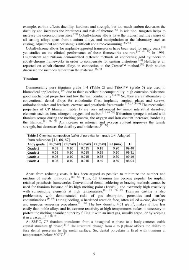
Tit anium Commercially pure titanium grade 1-4 (Table 2) and Ti6Al4V (grade 5) are used in biomedical applications, [66] due to their excellent biocompatibility, high corrosion resistance, good mechanical properties and low thermal conductivity.[72-76] So, they are an alternative to conventional dental alloys for endodontic files; implants; surgical plates and screws; orthodontic wires and brackets; crowns; and prosthetic frameworks.[14, 25, 76-80] The mechanical properties of CP titanium (Table 1) are very influenced by minor interstitial dissolved elements such as iron, nitrogen, oxygen and carbon.[11, 66, 74] If titanium sponge is mixed with titanium scraps during the melting process, the oxygen and iron content increases, hardening the titanium.[11, 66, 74] An increase in nitrogen and oxygen content improves the tensile strength, but decreases the ductility and brittleness.[11, 74]
Table 2 Chemical composition (wt%) of pure titanium grade 1-4. Adapted from references [11, 66, 67]. Alloy grade N (max) C (max) H (max) O (max) Fe (max) Ti Grade 1 0.03 0.10 0.015 0.18 0.20 99.48 Grade 2 0.03 0.10 0.015 0.25 0.30 99.31 Grade 3 0.05 0.10 0.015 0.35 0.30 99.19 Grade 4 0.05 0.10 0.015 0.40 0.50 98.94
Apart from reducing costs, it has been argued as positive to minimize the number and mixture of metals intra-orally.[81, 82] Thus, CP titanium has become popular for implant retained prosthesis frameworks. Conventional dental soldering or brazing methods cannot be used for titanium because of its high melting point (1668°C) and extremely high reactivity with surrounding elements at high temperatures.[11, 74, 75, 83] Titanium casting is also problematic, with demonstrated risks of gas absorption, porosities and surface contaminations.[84-86] During cooling, a hardened reaction face, often called α-case, develops and impedes veneering procedures.[11, 87] The low density, 4.51 g/cm3, makes it flow less easily than noble alloys and its extreme reactivity at high temperatures makes it necessary to protect the melting chamber either by filling it with an inert gas, usually argon, or by keeping it in a vacuum.[11, 88, 89] At 885°C, CP titanium transforms from a hexagonal α phase to a body-centered cubic crystal structure (β phase).[11] The structural change from α to β phase affects the ability to fuse dental porcelain to the metal surface. So, dental porcelain is fired with titanium at temperatures below 800°C.[11]

10
Gold alloys In 1985, the Brånemark group presented their definition of “state of the art for the totally edentulous jaw”: an acrylic-veneered gold framework with resin teeth.[25] In many ways, the long-established model of dental supported fixed prostheses was imitated.[90, 91] Among others, Adell et al. have presented favorable clinical results from this concept.[15] In dentistry, gold alloys type III or IV are commonly used for resin-veneered prostheses.[11] According to the ISO/DIS1562 standard, they can be described as: - Type III - High strength, for onlays, thin copings, pontics, crowns and saddles; - Type IV – Extra-high strength, for saddles, bars, clasps, thimbles, single units and partial denture frameworks (Table 1).[11] The original metal-ceramic alloys presented some 50 years ago contained about 90% gold with added platinum and palladium. Unfortunately, they were to soft for fixed dentures and there were reports of porcelain veneer detaching from the metal alloy framework.[11] By adding less than one percent of surface oxide-forming elements such as iron, tin or iridium to the alloy, a much stronger porcelain-metal bonding strength was achieved.[11, 92] All -ceramics As in teeth-supported prosthodontics, interest for all-ceramic solutions has recently increased in implant prosthodontics. In particular, oxidic ceramics such as alumina and zirconia have been in focus, mainly because of their mechanical strength.[92, 93] However, there is limited long-term documentation, especially for larger prostheses. Larsson et al. demonstrated a high frequency of porcelain superficial fractures (32%) after one year in all-ceramic two- to five-unit zirconia implant-supported reconstructions, but observed a significant difference between the two studied brands.[94] Vult von Steyern et al. compared loading on abutment-teeth and dental implants to support all-ceramic fixed partial dentures in vitro.[95] They suggested decreased strain and stress levels in the prosthesis when loaded on implants in comparison to natural teeth.[95] In a review of five-year survival for implant-supported single-crowns, Jung et al. found a 95.4%, survival rate for implant-supported metal-ceramic crowns, significantly better than the 91.2% survival rate of all-ceramic crowns.[96] A review of five-year survival, however, suggested that implant abutments performed comparably, whether or not they were made of ceramic or metal.[97] Fiber-reinforced frameworks Carbon/graphite fiber-reinforced poly-methyl methacrylate frameworks have been presented as a low-budget alternative to gold frameworks.[98, 99] Although Bergendal et al. demonstrated a high frequency of framework fractures[98], reports on enhanced in-vitro performance have recently been published.[100-103] Occlusal materials In the first animal studies, both gold and cobalt-chrome alloys were used for implant-retained frameworks[1, 104] and the first patient treatments with these materials together with veneers in acrylic and in porcelain were described in 1977.[14] One of the initial reasons for suggesting acrylic-veneers on gold frameworks was a perceived need for a shock-absorbing construction, thus not overloading the peri-implant bone by occlusal forces.[105] However, it was later demonstrated that the choice of occlusal material – acrylic, porcelain or gold, do not have an impact on the forces generated on the implants.[106, 107] Today, both acrylic- and
11
porcelain-veneers are used together with several alloys. Ceramic veneered high noble and base metal alloys such as nickel-chrome and cobalt-chrome alloys have successfully been used in dental supported fixed prosthodontics for decades.[64] Studies on titanium-ceramic restorative systems are few but in a recent review, Haag and Nilner reported early problems with porcelain chipping, even if this has become less of a problem as technical experience has increased.[87]
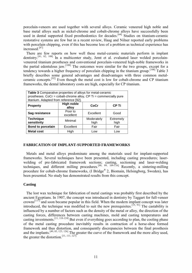
There are few reports on how well these metal-ceramic materials perform in implant dentistry.[81, 82, 108] In a multicenter study, Jemt et al. evaluated laser welded porcelain-veneered titanium prostheses and conventional porcelain-veneered high-noble frameworks in the partial edentulous jaw.[108] The outcomes were similar for the two groups, except for a tendency towards a higher frequency of porcelain chipping in the titanium group.[108] Table 3 briefly describes some general advantages and disadvantages with three common metal-ceramic concepts.[92] Even though the metal cost is low for cobalt-chrome and CP titanium frameworks, the dental laboratory costs are high, especially for CP titanium.
Table 3 Comparative properties of alloys for metal-ceramic prostheses. CoCr = cobalt-chrome alloy, CP Ti = commercially pure titanium. Adapted from reference [92].
Property High noble alloy CoCr CP Ti
Sag resistance Poor to excellent
Excellent Good
Technique sensitivity Minimal
Moderately high
Extremely high
Bond to porcelain Excellent Fair Fair
Metal cost High Low Low
FABRICATION OF IMPLANT-SUPPORTED FRAMEWORKS Metals and metal alloys predominate among the materials used for implant-supported frameworks. Several techniques have been presented, including casting procedures; laser-welding of pre-fabricated framework sections; casting, sectioning and laser-welding techniques, and different milling procedures.[88, 89, 109-116] Recently, a sintering-milling procedure for cobalt-chrome frameworks, (I Bridge® 2, Biomain, Helsingborg, Sweden), has been presented. No study has demonstrated results from this concept. Casting The lost wax technique for fabrication of metal castings was probably first described by the ancient Egyptians. In 1907, the concept was introduced in dentistry by Taggart for full-veneer crowns[117] and soon became popular in this field. When the modern implant concept was later introduced, the technique was modified to suit the new prerequisites.[15, 91] The castability is influenced by a number of factors such as the density of the metal or alloy, the direction of the casting forces, differences between casting machines, mold and casting temperatures and casting investments.[11, 118-124] But even if everything goes according to plan, the cooling phase of the metal casting procedure inevitably results in contraction of a horse-shoe formed framework and thus distortion, and consequently discrepancies between the final prosthesis and the implants.[49, 85, 125, 126] The greater the curve of the framework and the more alloy used, the greater the distortion.[11, 111, 127]
12
Joining metals
Joining metal sections by fusing is common in dentistry, and gas torches and furnaces have been used for many decades.[128] In recent years concentrated infrared radiation, electric arc and electromagnetically accelerated particles techniques, such as laser, have been developed as heat sources.[128, 129] Two of the more widespread methods used by dental laboratories are traditionally brazing and laser-welding.[130, 131] Brazing takes place when metallic sections are joined by fusing an intermediary alloy with a melting temperature above 450ºC but below the melting temperature of the parent metal or alloy.[11, 115, 132] The more common expression soldering involves joining metals or alloys by fusing an intermediary alloy at a melting temperature below 450ºC.[132] One disadvantage with soldering and brazing is the creation of a heat-affected zone close to the weld-joint.[11, 128]
Laser welding
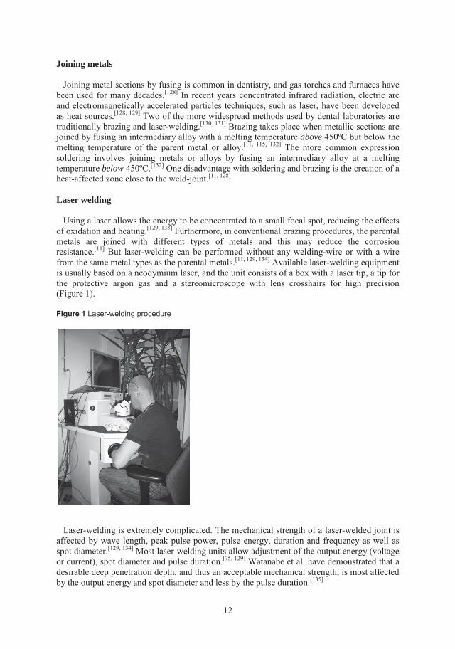
Using a laser allows the energy to be concentrated to a small focal spot, reducing the effects of oxidation and heating.[129, 133] Furthermore, in conventional brazing procedures, the parental metals are joined with different types of metals and this may reduce the corrosion resistance.[11] But laser-welding can be performed without any welding-wire or with a wire from the same metal types as the parental metals.[11, 129, 134] Available laser-welding equipment is usually based on a neodymium laser, and the unit consists of a box with a laser tip, a tip for the protective argon gas and a stereomicroscope with lens crosshairs for high precision (Figure 1). Figure 1 Laser-welding procedure
Laser-welding is extremely complicated. The mechanical strength of a laser-welded joint is affected by wave length, peak pulse power, pulse energy, duration and frequency as well as spot diameter.[129, 134] Most laser-welding units allow adjustment of the output energy (voltage or current), spot diameter and pulse duration.[75, 129] Watanabe et al. have demonstrated that a desirable deep penetration depth, and thus an acceptable mechanical strength, is most affected by the output energy and spot diameter and less by the pulse duration.[135]

13
The Procera® concept Since the introduction of the Procera concept (Nobel Biocare, Göteborg, Sweden) in the 1980s, four generations of titanium frameworks have been described.[110] Briefly, the first construction consisted of pre-fabricated cylinders mounted on a master cast. After customization, the cylinders were joined to a horizontal bar with holes for the cylinders, using a laser-welding technique.[109, 115] However, problems were reported with hygienic procedures due to a somewhat bulky design.[109, 110] The second generation consisted of pre-fabricated cylinders and components in different configurations mounted on a master cast ground to the same level.[109, 110] Next, a horizontal bar was laser-welded to these components and after adjustment, resin was wrapped to the framework and resin teeth put into place, as with the first generation.[110] Still, these prostheses also became bulky, and distortion problems have also been demonstrated.[109, 111] In the third generation of the Procera concept, individually shaped components, instead of the earlier pre-fabricated, were made for each abutment replica on the master cast and were laser-welded together after adjustments.[110, 136, 137] Resin or porcelain teeth were then baked or fused to the framework.[110, 138] The design became less bulky, but due to the excessive grinding procedures the technique was time-consuming.[66] In a ten-year follow-up study, Örtorp and Jemt demonstrated a higher incidence of porcelain chipping with this third generation of Procera titanium bridges in comparison to similar gold partial prostheses.[81] By means of a milling-technique (see below), the fourth generation of the Procera frameworks have been developed and seem to be successful.[110, 116, 139] The Cresco™ method The Cresco method (Astra Tech AB, Mölndal, Sweden) is based to a certain extent on the same principles as an earlier procedure for fabricating laser-welded titanium frameworks.[109-
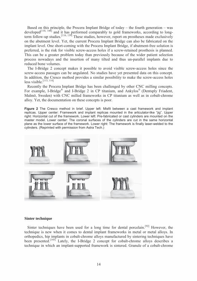
111] The method presents a way of fabricating a cast metal framework (originally made of titanium) for fixed implant-supported prostheses, to eliminate the unavoidable distortions created during framework casting (Figure 2). In this technique, the cast framework is first horizontally sectioned. Thereafter, new pre-machined or cast cylinders are mounted on a master cast and coronal surfaces of the cylinders are cut in the same horizontal plane as the lower surface of the framework. Finally, the framework is attached by a laser-welding technique to the cylinders.[113, 114] Several reports on the experimental and clinical outcomes of the Cresco method demonstrate good clinical performance, both with cobalt-chrome alloys and CP titanium frameworks.[71, 112-114, 140-142]
Milling techniques It is possible to use CAD/CAM systems for milling both in the dental laboratory (metals, alloys, polymers and ceramics) and chairside (polymers and ceramics).[66, 143] Indirect techniques involving scanning a plaster model are most common but direct techniques with optical intra-oral impressions for onlays, crowns, veneers and long-term provisional bridges exist.[143] When it comes to an entire implant framework with integrated cylinders, centers with industrial computer numeric control (CNC) milling machines are used. The dental technician fabricates a resin pattern and lets a laser scan it. A coordinate-measuring machine (CMM) collects information on the positions of the implant replicas in the master cast and a computer collects the data. Finally, a framework is milled in one piece. This technique can be used for metals and alloys, as well as for ceramics.[66]
14
Based on this principle, the Procera Implant Bridge of today – the fourth generation – was developed[110, 144] and it has performed comparably to gold frameworks, according to long-term follow-up studies.[116, 139] These studies, however, report on prostheses made exclusively on the abutment level. Yet, the current Procera Implant Bridge can also be fabricated on the implant level. One short-coming with the Procera Implant Bridge, if abutment-free solution is preferred, is the risk for visible screw-access holes if a screw-retained prosthesis is planned. This can be a greater problem today than previously because of the wider patient selection process nowadays and the insertion of many tilted and thus un-parallel implants due to reduced bone volumes. The I-Bridge 2 concept makes it possible to avoid visible screw-access holes since the screw-access passages can be angulated. No studies have yet presented data on this concept. In addition, the Cresco method provides a similar possibility to make the screw-access holes less visible.[113, 114]
Recently the Procera Implant Bridge has been challenged by other CNC milling concepts. For example, I-Bridge® and I-Bridge 2 in CP titanium, and Ankylos® (Dentsply Friadent, Malmö, Sweden) with CNC milled frameworks in CP titanium as well as in cobalt-chrome alloy. Yet, the documentation on these concepts is poor.
Figure 2 The Cresco method in brief. Upper left: Misfit between a cast framework and implant replicas. Upper center: Framework and implant replicas mounted in the articulator-like “jig”. Upper right: Horizontal cut of the framework. Lower left: Pre-fabricated or cast cylinders are mounted on the master model. Lower center: The coronal surfaces of the cylinders are cut in the same horizontal plane as the lower surface of the framework. Lower right: The framework is finally laser-welded to the cylinders. (Reprinted with permission from Astra Tech.)
Sinter technique
Sinter techniques have been used for a long time for dental porcelain.[92] However, the technique is new when it comes to dental implant frameworks in metal or metal alloys. In orthopedics, hip implants in cobalt-chrome alloys manufactured by sintering techniques have been presented.[145] Lately, the I-Bridge 2 concept for cobalt-chrome alloys describes a technique in which an implant-supported framework is sintered. Granule of a cobalt-chrome
15
alloy is sintered by the mean of a laser to a solid mass. Thereafter, the framework-to-be is adjusted through a milling and grinding process to the suitable dimensions. PRECISION OF FIT Impact of misfit The importance of precision of fit has been disputed for a long time and from many standpoints, and biological and technological consequences have been discussed. A perfect fit, according to Patterson’s definition with mating surfaces in 100% contact[146], does not seem realistic. Gaps up to 150 µm between framework and abutment/implant have been regarded as acceptable.[111, 147, 148] But even though guidelines for an acceptable degree of misfit are unavailable, it seems reasonable to try to achieve minimal misfit. Misfit introduces strain and tension to the prosthesis and the peri-implant marginal bone and may increase the risk for complications.[149-151] Taylor at al. suggested connections between misfit and mechanical complications,[152] although Wee et al. argued that although theoretically possible, scientific evidence was lacking for a connection between misfit and mechanical complications.[153] Furthermore, it is not known what constitutes acceptable misfit.[154, 155] There are many steps on fabrication before an implant-supported prosthesis can be connected to the implants, and every one of these affects the final fit.[49, 85, 125, 156-160] Implant components Machining tolerance among the different implant system components leads to unavoidable gaps.[159, 161] For example, the discrepancy between impression copings and implants or abutments can be as large as 100 µm.[159] As long as the implants are placed in parallel, horizontal displacements can be compensated to some degree by the machining tolerance of the implant components.[111] Today, when an increasing number of patients request implant rehabilitation, it is not always possible to fulfill the precondition of placing all implants in parallel, as previously recommended.[25] Impression materials and dental stone Distortion in the impression materials and expansion of dental stone during setting takes place.[11, 156-158, 160] In an in vitro study, a plaster impression material tended to expand the implant arch whereas a polyether material seemed to reduce the arch.[162] Other reports have come to similar conclusions.[157, 163] Dental stone can expand up to 0.5% and among other factors, the setting expansion is influenced by the water/powder ratio.[11] In an attempt to avoid the problems with impression distortions and dental stone expansion the photogrammetric technique has been introduced.[127, 147, 162, 164, 165] With the use of parallel mirrors 3-D registrations of the implant position are made possible. In comparison to the two mentioned impression methods, photogrammetric technique has been reported to give an equal precision.[162] Yet, a digital technique as photogrammetry requests a digital framework fabrication method such as CNC-milling.[162] Casting procedures Conventional casting procedures for alloy frameworks unavoidably result in misfits between the frameworks and the implants owing to distortion.[85, 125, 126] One way to handle this
16
problem is to cut a cast framework and solder/braze it together. But soldering per se may increase the misfit.[166, 167] Laser-welding of prefabricated titanium components, CAD/CAM procedures, and spark erosion or machine milling processes are among the alternative methods proposed.[168-170] However, because of solidification and thermal shrinkage, laser-welding can result in distortions as well.[171] Misfit measurements Different misfit measuring and evaluation concepts have been developed, including direct vision, finger pressure, tactile sensation, one screw test, screw resistance test and radiographic methods.[172, 173] Other methods include optical observation with microscopes, and evaluation of the thickness of light body impression materials syringed between the mating components before prosthesis placement.[49, 126, 174-176] Further, measurement of screw-joint loosening has been demonstrated[141, 177-179] and several computer-supported techniques have been developed, both with stylus, laser and photogrammetric techniques.[127, 147, 165, 180, 181] A multicenter-study of these latter methods regarded them as clinical valuable, although only photogrammetry can be used intraorally.[165] It has further been suggested that strain gauges should be utilized to objectively test misfit.[182] Yet, according to Smedberg et al. the strain-gauge technique is an indirect way to measure misfit since it registers stress and preload in a screw-joint area.[183] Misfit comparisons Örtorp et al. evaluated CNC-milled titanium frameworks, and concluded that these frameworks, milled from one piece of titanium, have a better fit than traditional, individually cast gold alloy frameworks.[184] However, these titanium frameworks were fabricated from one and the same replica, and variations in fit between frameworks made from different master models were not analyzed. In contrast, al-Fadda et al.[185] studied CNC-milled titanium frameworks, fabricated on individual models, in comparison to cast frameworks in a silver-palladium alloy.[185] The fit of the CNC-milled frameworks was better than for the cast frameworks, but not as good as the fit of the CNC-milled frameworks described by Örtorp and colleagues[184] Eliasson et al. reported on CNC-milled titanium frameworks made by two different methods, the Procera Implant Bridge and I-bridge[186] Signs of misfit were demonstrated in all evaluated frameworks but were regarded as clinically acceptable.[186] Schmitt et al. compared the screw-joint stability of bars for mandibular over-dentures.[187] They concluded that bars passivated according to the Cresco method did not show superiority compared to conventionally cast bars.[187] Yet, the comparison can be questioned since pre-fabricated components in the implant connection zone were used in the conventional bars but cast components were used in the Cresco-bars. Biological impact of misfit There is no consensus on the biological effects of misfit between framework and implant/abutment. Adverse tissue reactions such as bone loss and loss of integration have been suggested together with symptoms such as pain.[173] Another study reported bone remodeling when rabbit tibia implants were put under strain, but no signs of implant failure.[188] In an animal study, Hermann et al. reported an increase in bone resorption when the microgap caused by misfit between implant and framework was below the bone crest in comparison to more coronal levels.[189, 190] But these results are disputed, and in a clinical
17
study in 1996 Jemt and Book found no correlation between misfit and peri-implant marginal bone loss.[164] More recently, Heijdenrijk et al. reported that a microgap at the crestal level in two-piece implants did not appear to have an adverse effect on the amount of peri-implant bone loss.[191] It has been suggested that vertical discrepancies may lead to higher stress levels than those obtained by horizontal distortions[147, 172] Yet, the few animal studies available that focus on the biological impact of vertical misfit indicate that misfit preload seems to have more impact on bone response than the magnitude of the vertical gap[192-194] Animal models with static loading of implants did not show any adverse effects, but rather an adaptation to the load.[194-
196] Studies on dynamic loading, on the other hand, reveal differing conclusions.[197, 198] Szmukler-Moncler et al. have underlined the importance of avoiding micromotion during the healing phase, especially for immediate or early loading treatment protocols.[199] Technical impact of misfit Since preload can reduce when misfit exists in a screw-joint, there is a risk that setting screws will loosen.[149] Up to 90% of the applied torque may be needed to overcome the friction[200] and it has reported that the mating surfaces in a screw-joint are affected by plastic, that is, permanent, deformation.[201] al-Turki et al. investigated changes in screw stability and misfit between prostheses and abutments in vitro.[202] With a vertical misfit gap of 100 or 175 µm at the terminal abutment they reported the loosening torque of prosthetic retention screws in most locations being less then ten percent of the tightening torque.[202] When frameworks are connected directly to the implants with no intermediary abutments, the screw-driver torque is higher than when frameworks are connected to abutments. The Cresco method protocol recommends 35 Ncm as compared to the 10-15 Ncm recommended for abutment connected Astra Tech and Brånemark System treatments. Cheshire and Hobkirk demonstrated a reduction of misfit by using an increased torque.[86] As a consequence, higher stress levels in the screw-joint and the peri-implant tissues might take place. CORROSION Intra-oral corrosion Applied to dentistry, two major mechanisms of corrosion are interesting. Via saliva, different alloys in the mouth get income into temporary or permanent contact. In this way, two alloys may produce a galvanic cell, generating an electric current due to their potential difference.[69, 203] A reduction of oxygen takes place at the cathode in the electrolyte (saliva): O2 + 4e- + 2H20 → 4OH- O2 + 4H+ + 4e- → 2H20 At the same time, the anode metal dissolves into the saliva: Me → Men+ + ne-
The result is a decrease in oxygen and an increase in metal ions in solution. Several factors influence the galvanic reactions such as the electron potential, the cathode/anode surface area ratio, the distance between them, temperature and pH in the saliva.[203] Surface roughness and excessive bending through which cracks are formed can also effect corrosion as localized pit
18
or crevice corrosion.[69, 203] The increase in metal ions in solution and a diffusion of chloride ions give rise to an increase in acidity through two reactions: Men+ + nH20 → Me(OH)n + nH+
Men+ + Cl-n + nH20 → Me(OH)n + nHCl
With time the acidity will increase and the passive layer of the alloy can dissolve and thus accelerate the local corrosion.[203] Yet when the ion concentration in saliva increases this will per se decrease the tendency for the same element to dissolve.[11] But since the saliva is constantly exchanged, e.g. through eating and drinking, corrosion can continue. A heterogeneous surface composition with differences in electrode potentials between different surface zones further increases the risk.[11] This can be the case with an alloy, especially if it consists of two or more phases or includes impurities.[11] As a consequence, the relative amount of released metal ions does not reflect their relative volume or weight portion in a metal alloy.[204, 205] Corr osion measurements Measuring ion leakages is one way to estimate corrosive processes; electrochemical procedures another. al-Hity et al. reported a strong correlation between polarization resistance and low elemental release, by using and comparing electrochemical and immersion tests.[206] When galvanic corrosion was examined in one study, titanium was found to be more corrosive than cobalt-chrome alloys in frameworks connected to titanium implants.[207] On the other hand, nickel-chrome alloy frameworks have been reported to produce higher ion leakage than titanium frameworks.[208] Wataha et al. recommends studying ion leakage and not just galvanic corrosion when biological effects are of interest.[46] Yet, since corrosion involves leakage of anions and cations as well as electronic transport ion leakages measurements alone can be regarded as an estimation of corrosion. The same can of course be said about galvanic corrosion measurements alone. Implant frameworks and corrosion CP titanium and cobalt-chrome alloys are generally regarded as being resistant to corrosion.[11, 68, 206, 209] In fact, in contact with oxygen they both corrode immediately. However, the results are stable metallic oxides on the surfaces.[11] Yet, it is well known that in a highly corrosive environment such as the mouth, ion leakage from dental devices occurs through corrosive processes.[53, 57] It has been demonstrated that laser welding of cobalt-chrome alloys may be a problem from a corrosion standpoint since microcracks and porosities easily develop in the weld joints.[210,
211] In an in-vitro study, Reclaru and Meyer reported on corrosion between dental implants and different framework alloys.[203] They concluded that from an electrochemical point of view, titanium connected to a superstructure must have a weak anodic polarization, the galvanic cell current must also be weak, and the crevice potential must be much higher than the common potential.[203]
Clinical importance The patient’s dietary and oral cleaning habits can effect corrosion.[67] In a review article, Tschernitschek et al. reported that cast titanium is more susceptible to corrosion than machined titanium and that fluoride can dissolve the stabilizing oxide layer.[67] It was
19
suggested that the combination of fluoride concentration and pH is important but toothpastes with low fluoride concentration may be regarded as harmless at neutral pH.[67, 212] In conclusion, although leakage of titanium, cobalt and chrome ions from dental devices is small in relation to the daily dietary intake of these elements[213, 214], corrosion cannot be excluded when toxicity and hypersensitivity are discussed.[11, 57] Consequently the nature of the released elements, and the quantity and duration of the exposure are fundamental for the biological responses.[46] SURFACE STRUCTURE Surface examination Since surfaces are in 3-D structure, 2-D surface characterizations are insufficient.[31, 66] Several types of methods and equipment can be appropriate for surface examination and they all have their advantages and disadvantages, e.g. mechanical contact profilometers, non-contact laser profilometers, interference microscopy, confocal laser scanning-microscopy, atomic force microscopy and scanning tunnel microscopy.[215, 216] All the mentioned quantitative techniques have limitations in range and resolution and they are also scale-dependent, i.e. information on measurement scale and cutoff filters are needed when discussing measurement results.[216] In contrast, scanning electron microscopy (SEM) characterizes surface-topography qualitatively and has a high lateral resolution as well as a large depth of focus.[216] When two SEM micrographs are studied, e.g. stereo-SEM, quantitative assessments can also be performed.[216] Roughness, waviness and form characterize the topography of a surface and Wennerberg and Albrektsson proposed that parameters from the three groups height, spatial and hybrid parameters should be included in 3-D measurements.[31] Plaque retention Bacterial adhesion, and to some degree cell adhesion, to an intra-oral surface is initially influenced by a number of factors, e.g. surface roughness, surface-free energy, distance between bacteria and surface and ionic strength of the surrounding liquid medium (gingival fluid and saliva).[217, 218] The surface structure seems to be of greater importance in this respect than the chemical composition and it has been argued that no specific alloy or group of alloys stimulate plaque adhesion that is resistant to improved oral hygiene.[56] Still, it has been demonstrated in vitro that alloys releasing copper and silver can be more antibacterially active than base-metal alloys.[219] Recently, Bürgers et al. demonstrated in vitro the antibacterial and anti-adherence capacity of particulate silver additives in composite resin materials.[220] In vivo conditions are however different and since a pellicle is instantly formed on an intra-oral surface, differences between materials are probably reduced.[221] The microbiota colonization preferably starts at sheltered localizations, far away from oral hygiene measures and natural removal forces.[222, 223] The positive relationship between increasing surface roughness and the rate of supragingival bacterial colonization has been demonstrated in several in vivo studies.[224-227] In addition, a more pathogenic flora could be observed on rougher surfaces.[217] It has further been reported that rough surfaces (Ra = 0.8 µm) can accumulate 25 times more subgingival plaque than smoother surfaces.[228] Quirynen et al. and Bollen and co-workers have, using titanium abutments with different surface roughness in patients, demonstrated the existence of a threshold roughness (Ra = 0.2 µm)
20
below which no further gain in resistance to bacterial adhesion can be expected.[229] Yet, despite different surface roughness on titanium surfaces, differences in soft-tissue inflammation grade were not revealed in two other studies.[230, 231] In addition, titanium has been suggested to form a bacteriostatic gel in contact with gingival fluid and to have an inhibitory effect on Streptococcus mutans.[232, 233] Impact on corrosion Surface roughness can be interesting from a corrosive point of view as well since a greater cathode and/or anode surface area promotes a greater element release.[203] Wennerberg at al. studied titanium release from implants with different surface roughness.[234] At a level relevant for commercial dental implants they found no correlation between increasing surface roughness and ion release, neither in vitro nor in vivo.[234] Corrosion can change both the chemical composition and the surface structure in an implant component. These factors have been discussed in conjunction with reactions in peri-implant soft tissues.[235, 236] In alloys, polished surfaces have been suggested to be more biocompatible in the transmucosal area than “as-cast” surfaces.[236-239] Surface roughness and cell preferences Different cells prefer different surface roughness and in vitro studies indicate that epithelial cells prefer smooth surfaces and fibroblasts rougher surfaces[236, 238, 240-243] Yet, this is an issue where no consensus exists. Consequently, Niederauer et al. demonstrated in vitro a higher degree of cell attachment for gingival epithelial cells on a rough osteoceramic surface, whereas gingival fibroblasts preferred smoother surfaces.[244] Further, no general definition of “smooth” and “rough” exists; everything is relative. In a comparison study, Jang et al. examined partial denture frameworks fabricated in CP titanium or a cobalt-chromium alloy and found that the titanium surfaces were slightly smoother, although no statistical differences in surface roughness were detected.[245] However, this insignificance can have been affected by the limited number of dentures, ten in each group.[245] Yet, today when nano-structures of surfaces are discussed[29, 30, 246], the distinction between chemical surface composition and surface roughness seems to become less obvious. Impact on preload As mentioned above, up to 90% of the applied torque can be needed to overcome the friction in a screw joint.[200] As a consequence, the degree of surface roughness is important in screw-joint evaluations as well, e.g. between framework and abutment or implant.[200] Örtorp et al. reported on surface roughness and preload in screw-joints in an in vitro study.[201] They demonstrated that unloaded milled titanium screw sites had rougher surfaces than loaded, and loaded gold screws had rougher surfaces than unloaded.[201] BIOCOMPATIBILITY Definition and principles Biocompatibility has been defined as the ability of a material to function in a specific application in the presence of an appropriate host response.[247] Both corrosion and surface roughness can affect the biocompatibility of a dental device. During corrosive processes, the
21
nature and quantity of the released elements and the duration of the exposure are fundamental for the biological responses.[248] Other major factors are the composition of the alloy, whether it has one or multiple phases and the conditions in the surrounding environment.[249-252] Cell culture cytotoxicity tests, animal models and clinical human studies are among the techniques used to evaluate the biocompatibility of a dental material.[11] But it must be remembered that elements released from dental devices (implants excluded) are not per se inside the body.[57] In addition, the biological effects depend on the route into the body, how the released elements are distributed and eliminated, and each such process is unique to the specific element.[57] Biological responses to material degradation have been associated with local and systemic toxicity, hypersensitivity, allergy as well as mutagenicity and carcinogenicity.[57, 253-255] Hypersensitivity Every metal used in dental devices can be associated with hypersensitivity.[55, 67] Studies indicate that 8-15% of the general population is sensitive to nickel, chrome or cobalt with the highest frequency for nickel.[57] Case reports on hypersensitivity reactions to titanium have been presented, but Sicilia et al. recently reported an estimated prevalence of 0.6% for titanium hypersensitivity among patients who received titanium dental implants.[256] It was not obvious, though whether the implants were made in CP titanium or in an alloy (Ti6Al4V?). Yet, it is not known why some metal ions can cause allergic reactions with various clinical symptoms while other ions do not. Cytotoxicity The cytotoxicity of cobalt to fibroblast cultures and the ability of cobalt-chrome particles to induce the release of inflammatory mediators from macrophages have been described.[253, 257] Further, Berstein et al. have demonstrated inhibited cell growth when gingival epithelial cells and lymphoma cells were exposed to cobalt alloys.[73] Evans reported on fibroblast cell damage in vitro after contact with powders of titanium, Ti6Al4V and a cobalt-chrome-molybdenum alloy and suggested that the damage to the fibroblasts was independent of the chemical composition of the powders.[254] However, when a microporous membrane was used, only the finest Co-Cr-Mo-alloy particles caused cell damage.[254] In a dog model, it was demonstrated that a normal soft-tissue interface forms around titanium, zirconia, alumina and hydroxyl apatite abutments, but that no such soft-tissue adhesion takes place around gold or dental porcelain abutments.[258] Welander et al. also used a dog model and confirmed the previously reported results: titanium or zirconia abutments promoted proper soft tissue integration while gold-alloy abutments failed to reach the same condition.[259] However, there have been no similar animal studies on cobalt-chrome alloys. Several clinical studies of the soft tissue around abutments with either titanium or ceramic surfaces have found no significant differences between these two materials.[260-264] Yet, there are no clinical reports on the histology of the peri-implant soft tissue when different metals or alloys have been used for the transmucosal components. Carcinogenicity Until recently, there was no evidence that dental alloys could contribute to cancer in humans. However, a review study in 2000 suggested that alloys containing cadmium, cobalt or beryllium should be avoided because of the carcinogenicity risks.[57] It must be underlined
22
though, that a precondition for carcinogenicity or mutagenicity is that an alloy releaseselements.[57]
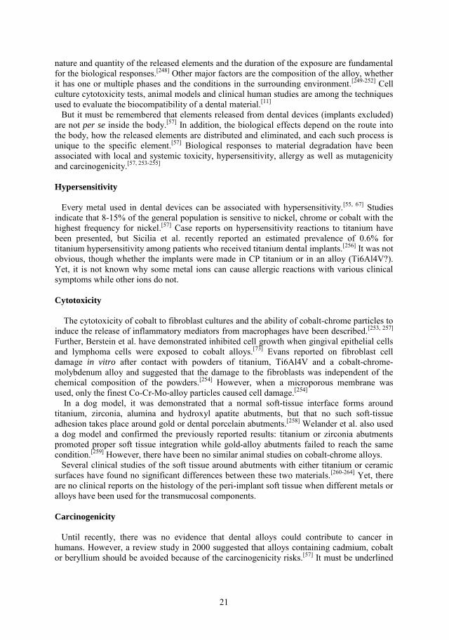
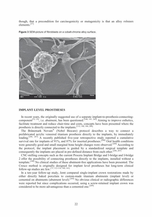
Figure 3 SEM-picture of fibroblasts on a cobalt-chrome alloy surface.
IMPLANT-LEVEL PROSTHESES
In recent years, the originally suggested use of a separate implant-to-prosthesis-connecting-component[12, 25], i.e. abutment, has been questioned.[140, 141, 265] Aiming to improve esthetics, facilitate treatment and reduce chair-time and costs, concepts have been presented where the prosthesis is directly connected to the implants.[112, 140, 186, 266] The Brånemark Novum® (Nobel Biocare) protocol describes a way to connect a prefabricated acrylic veneered titanium prosthesis directly to the implants, by immediately loading.[266, 267] A recently published five-year retrospective study reported a cumulative survival rate for implants of 91%, and 87% for inserted prostheses.[268] Oral health conditions were generally good and small marginal bone height changes were observed.[268] According to the protocol, the implant placement is guided by a standardized surgical template and consequently the implants are placed in pre-defined distance from each other.[266, 267]
CNC-milling concepts such as the current Procera Implant Bridge and I-bridge and I-bridge 2 offer the possibility of connecting prostheses directly to the implants, installed without a template.[186] No clinical studies of these abutment-free applications have been presented. The Cresco method is originally designed for implant level prostheses but long-term clinical follow-up studies are few.[71, 112-114, 140, 141]
In a ten-year follow-up study, Jemt compared single-implant crown restorations made by either directly baked porcelain to custom-made titanium abutments (implant level) or cemented on abutments (abutment level).[269] No obvious clinical or radiographic differences were reported but since complications occurred, using a screw-retained implant crown was considered to be more advantageous than a cemented one.[269]
23
EVALUATION OF TREATMENT RESULTS Several studies have presented favorable long-term results for implant treatment in the edentulous jaw.[15-19, 270-273] More than 95% percent of the loaded implants can be osseointegrated after 15 years.[270] Because marginal bone loss and implant loss tend to cluster in patients it seems important to report on patient/prosthesis performance as well.[274-276] Thus, the implants in an edentulous jaw can not be looked upon as independent of each other. Biological complications Biological complications concern the host’s ability to create and maintain osseointegration and healthy tissues in the peri-implant area. Both systemic and local factors can influence this ability. Yet, Wood et al. found no absolute contraindication to implant treatment at all, neither among habits nor among systemic or local factors.[277] The most important factors, they concluded, are the quantity and quality of available bone.[277] Jemt and Häger studied early failures in edentulous maxillae and demonstrated that bone quantity had an impact on implant failure.[275] Herrmann et al. found an increasing risk for implant failures in patients with earlier implant failures.[278] But no consensus exists, and diabetes, periodontitis, and smoking are all disputed risk factors for successful treatment results.[279-285] General health, osteoporosis and age are other factors discussed but no evidence for their potentially negative influence on treatment outcomes have been presented.[279-285] Adell et al. stated that implant treatment in the edentulous maxilla required a greater technical skill than treatment in the mandible.[15] It has been suggested that the performance in the maxilla would be less reliable than in the mandible.[286, 287] Other reports have demonstrated more bone loss at lower jaw implants.[288, 289] There is further no general agreement on how to regard reactions in the surrounding tissues. In 2008, Jemt and Albrektsson, and Albrektsson et al. argued that a certain degree of bone remodeling is quiet normal, whereas Fransson et al. in a previous study suggested that 28% of 662 included patients followed for at least five years with fixed complete or partial prosthesis or single-tooth replacements had progressive peri-implant bone-loss, indicating pathology.[290-292] Technical complications Among the technical complications described are fractures of frameworks, veneers, implant components and problems with loose connecting screws.[116, 141, 149, 172, 293-297] Misfit and prosthetic design, overloading and material fatigue has been suggested as possible causes for mechanical complications.[272] Even though veneer fractures seem to be most frequent among the mechanical complications reported, a decrease over the years can be noticed.[19] Improved technical skill and patient selection are plausible explanations. Acceptance problems Patient related acceptance problems have also been described. Hjalmarsson and Smedberg suggested that esthetic complaints were more common among edentulous patients with high expectations of the treatment.[141] Other studies have demonstrated that adaptation depends to a large degree on how well the patient’s needs and wishes have been met by the given treatment.[298, 299] Köndell et al.[300] found phonetic problems to be the most frequent complication in implant-treated edentulous patient but others have suggested that phonetic problems vanish within a
24
few weeks after prosthesis delivery.[141] Anatomical limits, implant placement and prosthesis design are probably crucial in this respect.[301-303]
Clinical evaluation
Clinical evaluation of dental implants requires the removal of the prosthesis. Clinical inspection where absence of mobility is noticed or tapping on the implant and hearing a metallic sound are two of the suggested methods for evaluating osseointegration.[304, 305] Other techniques include the Periotest (Siemens, Bensheim, Germany) measurement of implant mobility, originally a device for quantifying tooth mobility.[306] The method has been described as operator sensitive and its clinical value has been questioned.[307] A technique using resonance frequency analysis has also been presented.[308-311] Yet, despite initial signs of implant stability the predictable value for the long-term implant prognosis of such measurements has been questioned.[312]
It was suggested that the clinical examination of peri-implant tissues should include registration of bleeding on probing, pocket depths and suppuration.[307] Due to the differences in tooth and implant surrounding tissues[313], probing around implants is more operator sensitive and a potential association between probing depth and marginal bone loss has been questioned.[21, 290, 314, 315]
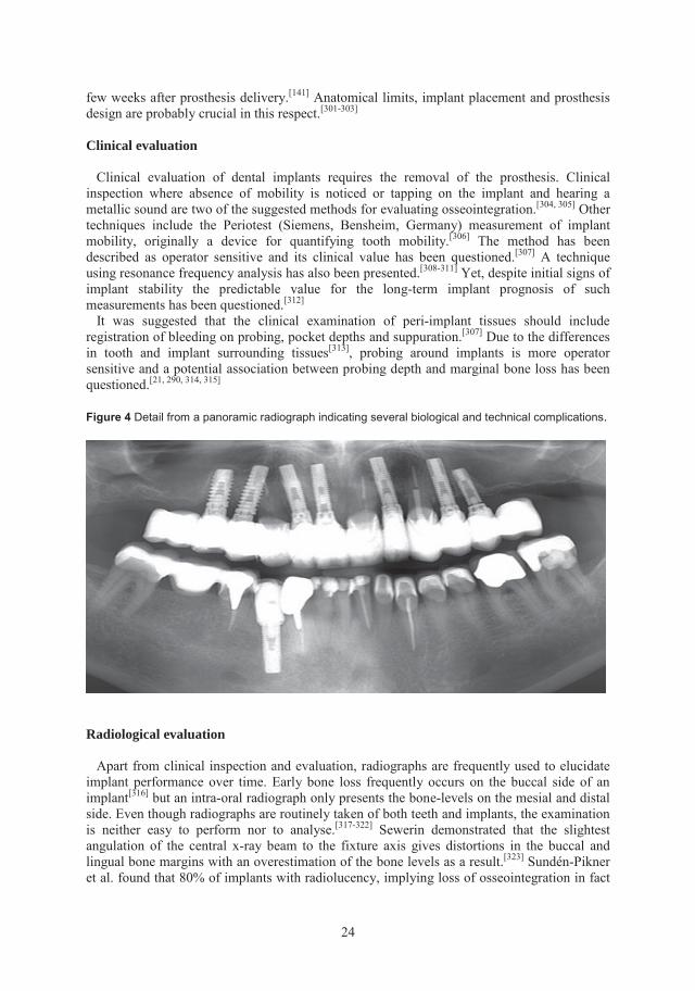
Figure 4 Detail from a panoramic radiograph indicating several biological and technical complications.
Radiological evaluation
Apart from clinical inspection and evaluation, radiographs are frequently used to elucidate implant performance over time. Early bone loss frequently occurs on the buccal side of an implant[316] but an intra-oral radiograph only presents the bone-levels on the mesial and distal side. Even though radiographs are routinely taken of both teeth and implants, the examination is neither easy to perform nor to analyse.[317-322] Sewerin demonstrated that the slightest angulation of the central x-ray beam to the fixture axis gives distortions in the buccal and lingual bone margins with an overestimation of the bone levels as a result.[323] Sundén-Pikner et al. found that 80% of implants with radiolucency, implying loss of osseointegration in fact

25
was clinically stable.[319] Consequently as many as 80% of the clinically instable implants would remain undetected on radiographs.[319] In an attempt to reduce the radiation burden, it was proposed that radiological examinations of Brånemark System® implants wait until five years after prosthesis delivery.[324] It was also suggested that one randomly chosen implant could be representative for the edentulous patient’s peri-implant bone status.[324] Lang et al. recommended that indications for radiographs ought to include bleeding on probing and pocket depths exceeding 5 mm.[325] But general recommendations cannot exclude the judgment of the clinician and it seems reasonable to use the same criteria as for patients with teeth. Subsequently, radiological examinations ought to be performed when it is indicated by the individual patient’s symptoms and dental health status. Therefore, the radiographic analysis must also take into account the clinical signs.
26
BACKGROUND TO THE PRESENT THESIS
• Cobalt-chrome alloys have been used in dentistry for 80 years, but very little is known about their behavior and biological impact as framework materials in implant-supported prostheses.
• Cast frameworks for implant-supported prostheses are associated with misfit problems
due to unavoidable casting distortions. The Cresco method promotes a technique to achieve a “perfect fit” (according to the manufacturer) regardless of metal alloy used. No documented evidence supports this.
• Cobalt-chrome alloys and commercially pure (CP) titanium are generally regarded as
non-corrosive. Yet, knowledge of their degradation when used in implant-supported prostheses is sparse.
• The biocompatibility of cobalt-chrome alloys has been questioned whereas CP
titanium is generally regarded as being highly biocompatible. However, little is known about the effects on peri-implant tissue cells from cobalt-chrome alloy frameworks compared to CP titanium frameworks.
• Metal-ceramic solutions have been used in dental supported prostheses for decades.
They have also become popular in implant dentistry due to favorable esthetic outcomes. Early use of high-noble alloys has been challenged by the introduction of base-metal alloys such as cobalt-chrome for frameworks in implant applications. Yet, no study has presented the clinical outcome of porcelain-veneered implant-supported cobalt-chrome frameworks.
• A handful of concepts describe abutment-free procedures for implant dentistry. Yet,
only a few comparative studies have evaluated the clinical results of abutment and abutment-free techniques. Further, very little is known about the advantages and disadvantages of abutment and abutment-free complete prostheses in this context.
27
STRUCTURE OF THE THESIS This thesis is in two parts. Both discuss the investigation of cobalt-chrome alloy frameworks compared to CP titanium frameworks for implant-supported prostheses. Part 1. In vitro studies of cast, sectioned and laser-welded frameworks in cobalt-chrome alloy and CP titanium (I-III). Study I evaluated and compared the precision of fit of three groups of frameworks: cast, sectioned and laser-welded frameworks fabricated in a cobalt-chrome alloy or in CP titanium, and a group of individually scanned CNC-milled CP titanium frameworks. Study II investigated material degradation in titanium implants and frameworks made of cobalt-chrome or CP titanium according to the Cresco method, before and after exposure to artificial saliva. Study III assessed possible adverse cellular responses to frameworks by comparing the viability and morphology of epithelial cells and fibroblasts cultivated on cobalt-chrome and titanium specimens. Part 2. Clinical and radiological evaluation of implant level prostheses in comparison to abutment level prostheses (IV). Study IV was a five-year clinical and radiological retrospective investigation. The clinical function of implant-supported prostheses made on implant level and on abutment level in the edentulous maxilla was evaluated and compared. Three different types of prostheses were made: one in porcelain-veneered cobalt-chrome alloy, and two in acrylic-veneered CP titanium.
28
AIMS General aim
• To evaluate whether cobalt-chrome alloys are generally suitable for implant-supported prostheses.
Specific aims
• To investigate the precision of fit of implant-supported cast, sectioned and laser-welded frameworks fabricated in a cobalt-chrome alloy or in CP titanium. Further, to compare these frameworks to individually scanned CNC-milled CP titanium frameworks.
• To investigate and estimate material degradation in frameworks made of cobalt-
chrome or CP titanium according to the Cresco method, and in titanium implants before and after exposure to artificial saliva.
• To assess possible adverse cellular responses to cobalt-chrome frameworks for
implant-retained, intra-oral prostheses by comparing the viability and morphology of epithelial cells and fibroblasts cultivated on cobalt-chrome and titanium specimens.
• To evaluate and compare the clinical function between implant-supported prostheses
in the edentulous maxilla made on implant level and prostheses fabricated on abutment level. Further, to compare porcelain-veneered cobalt-chrome alloy prostheses with two groups of prostheses made in acrylic-veneered CP titanium.
29
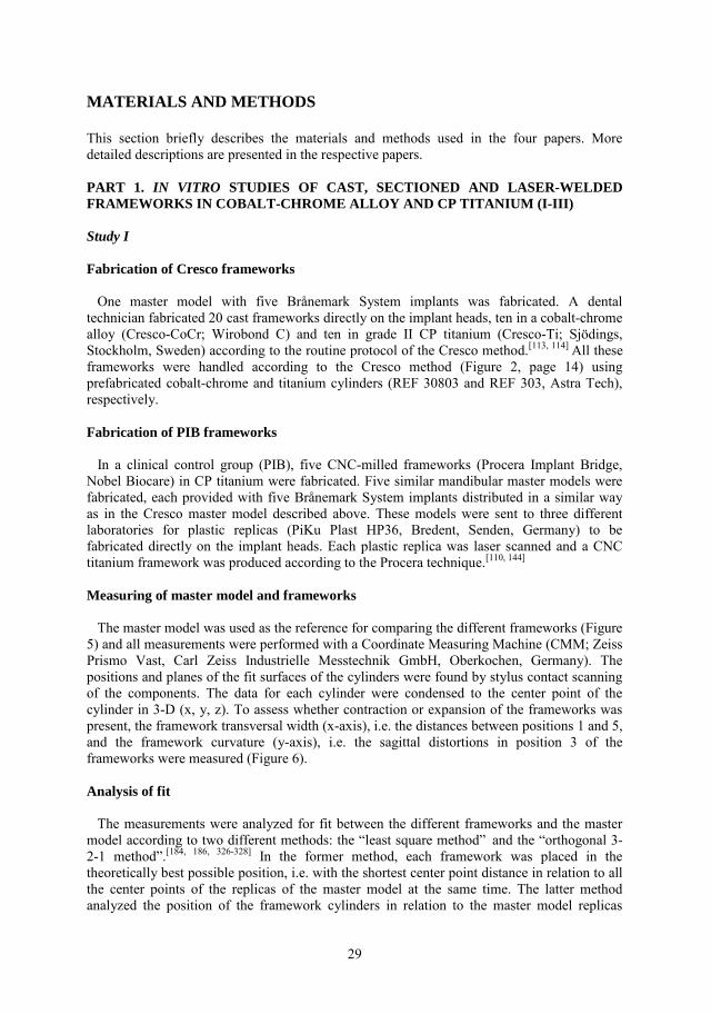
MATERIALS AND METHODS This section briefly describes the materials and methods used in the four papers. More detailed descriptions are presented in the respective papers. PART 1. IN VITRO STUDIES OF CAST, SECTIONED AND LASER-WELDED FRAMEWORKS IN COBALT-CHROME ALLOY AND CP TITANIUM (I-III) Study I Fabrication of Cresco frameworks One master model with five Brånemark System implants was fabricated. A dental technician fabricated 20 cast frameworks directly on the implant heads, ten in a cobalt-chrome alloy (Cresco-CoCr; Wirobond C) and ten in grade II CP titanium (Cresco-Ti; Sjödings, Stockholm, Sweden) according to the routine protocol of the Cresco method.[113, 114] All these frameworks were handled according to the Cresco method (Figure 2, page 14) using prefabricated cobalt-chrome and titanium cylinders (REF 30803 and REF 303, Astra Tech), respectively. Fabrication of PIB frameworks In a clinical control group (PIB), five CNC-milled frameworks (Procera Implant Bridge, Nobel Biocare) in CP titanium were fabricated. Five similar mandibular master models were fabricated, each provided with five Brånemark System implants distributed in a similar way as in the Cresco master model described above. These models were sent to three different laboratories for plastic replicas (PiKu Plast HP36, Bredent, Senden, Germany) to be fabricated directly on the implant heads. Each plastic replica was laser scanned and a CNC titanium framework was produced according to the Procera technique.[110, 144] Measuring of master model and frameworks The master model was used as the reference for comparing the different frameworks (Figure 5) and all measurements were performed with a Coordinate Measuring Machine (CMM; Zeiss Prismo Vast, Carl Zeiss Industrielle Messtechnik GmbH, Oberkochen, Germany). The positions and planes of the fit surfaces of the cylinders were found by stylus contact scanning of the components. The data for each cylinder were condensed to the center point of the cylinder in 3-D (x, y, z). To assess whether contraction or expansion of the frameworks was present, the framework transversal width (x-axis), i.e. the distances between positions 1 and 5, and the framework curvature (y-axis), i.e. the sagittal distortions in position 3 of the frameworks were measured (Figure 6). Analysis of fit The measurements were analyzed for fit between the different frameworks and the master model according to two different methods: the “least square method” and the “orthogonal 3-2-1 method”.[184, 186, 326-328] In the former method, each framework was placed in the theoretically best possible position, i.e. with the shortest center point distance in relation to all the center points of the replicas of the master model at the same time. The latter method analyzed the position of the framework cylinders in relation to the master model replicas
30
when a software program placed the framework center points in a coordinate system. Thus, the center point of framework cylinder 5 was placed at the origin of the corresponding master replica cylinder for all three coordinates (x, y, z). Cylinder 3 was placed in the x-y plane and cylinder 1 on the x-axis. Framework distortions were presented in relation to the center points of the remaining cylinders and axes. The distance between the center points of the frameworks and the master model in three dimensions was also calculated for each individual cylinder (3-D=√x2+y2+z2). The distortions were presented in µm using absolute and real values. Figure 5 Framework mounted in the mold before measuring in the CMM. Orientation of the coordinates in the x- and y-axes. The vertical axis is oriented with negative values (-z) toward the master model.
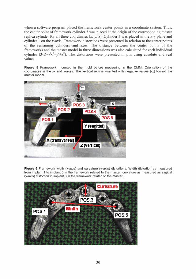
Figure 6 Framework width (x-axis) and curvature (y-axis) distortions. Width distortion as measured from implant 1 to implant 5 in the framework related to the master, curvature as measured as sagittal (y-axis) distortion in implant 3 in the framework related to the master.
31
Apart from the orthogonal 3-2-1 method and the least square method, a third theoretical investigation method was executed. The angular gap distortion was measured in the same way as previously described.[184] Disappointingly, the accuracy in the measurements was inferior to the demands and the analysis could nor be performed. Additionally, the framework distortions for the two groups of Cresco-frameworks were compared to the results for gold alloy frameworks and CP titanium PIB-frameworks earlier presented by Örtorp et al.[184], although this comparisons were not presented in Study I.
Study II
Test specimens
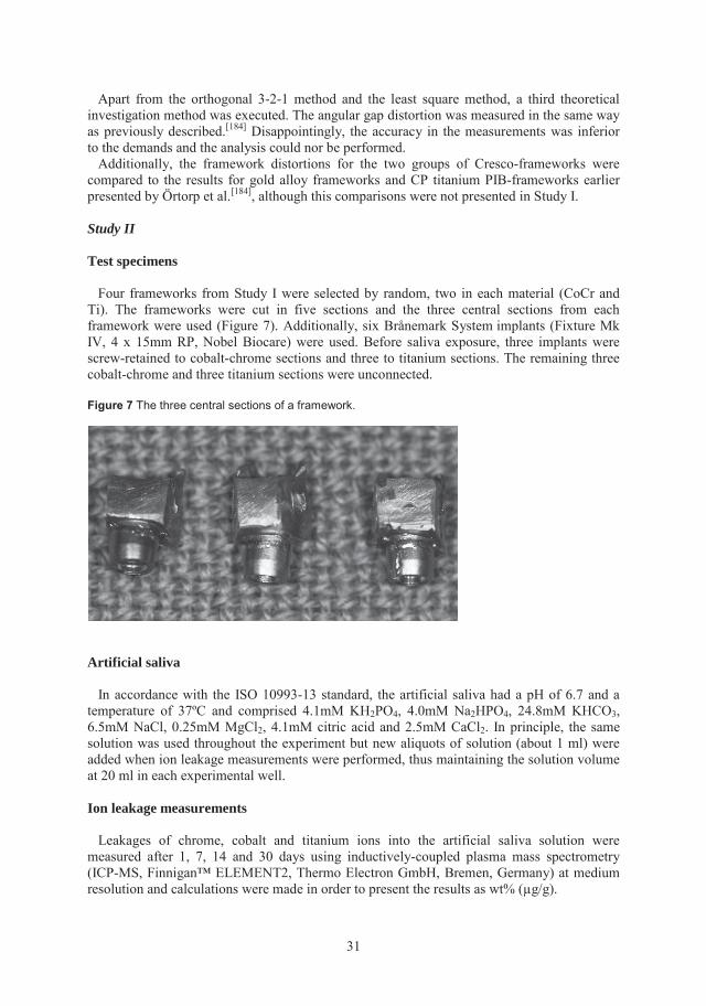
Four frameworks from Study I were selected by random, two in each material (CoCr and Ti). The frameworks were cut in five sections and the three central sections from each framework were used (Figure 7). Additionally, six Brånemark System implants (Fixture Mk IV, 4 x 15mm RP, Nobel Biocare) were used. Before saliva exposure, three implants were screw-retained to cobalt-chrome sections and three to titanium sections. The remaining three cobalt-chrome and three titanium sections were unconnected.
Figure 7 The three central sections of a framework.
Artificial saliva
In accordance with the ISO 10993-13 standard, the artificial saliva had a pH of 6.7 and a temperature of 37ºC and comprised 4.1mM KH2PO4, 4.0mM Na2HPO4, 24.8mM KHCO3, 6.5mM NaCl, 0.25mM MgCl2, 4.1mM citric acid and 2.5mM CaCl2. In principle, the same solution was used throughout the experiment but new aliquots of solution (about 1 ml) were added when ion leakage measurements were performed, thus maintaining the solution volume at 20 ml in each experimental well.
Ion leakage measurements
Leakages of chrome, cobalt and titanium ions into the artificial saliva solution were measured after 1, 7, 14 and 30 days using inductively-coupled plasma mass spectrometry (ICP-MS, Finnigan™ ELEMENT2, Thermo Electron GmbH, Bremen, Germany) at medium resolution and calculations were made in order to present the results as wt% (µg/g).
32
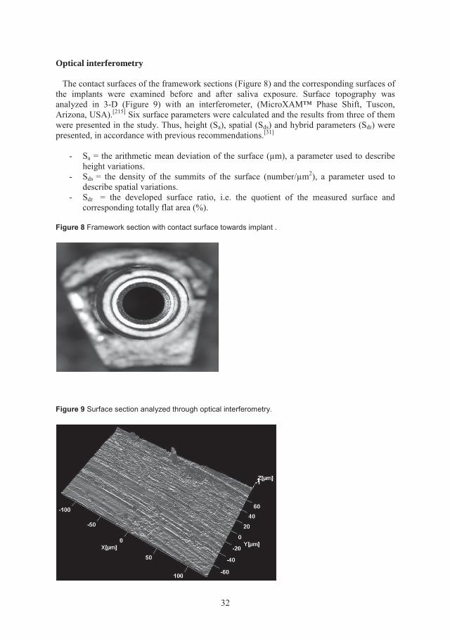
Optical interferometry
The contact surfaces of the framework sections (Figure 8) and the corresponding surfaces of the implants were examined before and after saliva exposure. Surface topography was analyzed in 3-D (Figure 9) with an interferometer, (MicroXAM™ Phase Shift, Tuscon, Arizona, USA).[215] Six surface parameters were calculated and the results from three of them were presented in the study. Thus, height (Sa), spatial (Sds) and hybrid parameters (Sdr) were presented, in accordance with previous recommendations.[31]
- Sa = the arithmetic mean deviation of the surface (µm), a parameter used to describe height variations.
- Sds = the density of the summits of the surface (number/µm2), a parameter used to describe spatial variations.
- Sdr = the developed surface ratio, i.e. the quotient of the measured surface and corresponding totally flat area (%).
Figure 8 Framework section with contact surface towards implant .
Figure 9 Surface section analyzed through optical interferometry.
33
Study III
Test specimens
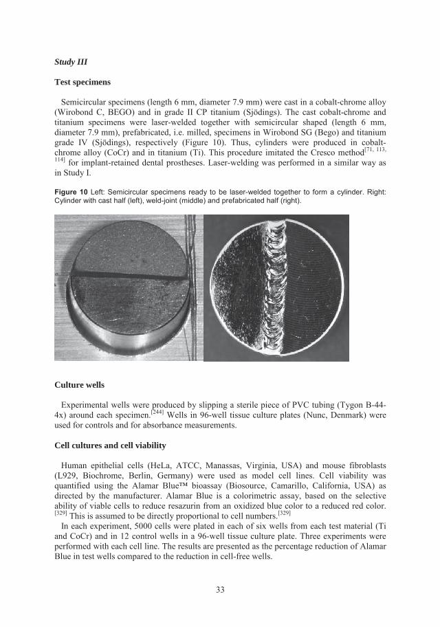
Semicircular specimens (length 6 mm, diameter 7.9 mm) were cast in a cobalt-chrome alloy (Wirobond C, BEGO) and in grade II CP titanium (Sjödings). The cast cobalt-chrome and titanium specimens were laser-welded together with semicircular shaped (length 6 mm, diameter 7.9 mm), prefabricated, i.e. milled, specimens in Wirobond SG (Bego) and titanium grade IV (Sjödings), respectively (Figure 10). Thus, cylinders were produced in cobalt-chrome alloy (CoCr) and in titanium (Ti). This procedure imitated the Cresco method[71, 113,
114] for implant-retained dental prostheses. Laser-welding was performed in a similar way as in Study I.
Figure 10 Left: Semicircular specimens ready to be laser-welded together to form a cylinder. Right: Cylinder with cast half (left), weld-joint (middle) and prefabricated half (right).
Culture wells
Experimental wells were produced by slipping a sterile piece of PVC tubing (Tygon B-44-4x) around each specimen.[244] Wells in 96-well tissue culture plates (Nunc, Denmark) were used for controls and for absorbance measurements.
Cell cultures and cell viability
Human epithelial cells (HeLa, ATCC, Manassas, Virginia, USA) and mouse fibroblasts (L929, Biochrome, Berlin, Germany) were used as model cell lines. Cell viability was quantified using the Alamar Blue™ bioassay (Biosource, Camarillo, California, USA) as directed by the manufacturer. Alamar Blue is a colorimetric assay, based on the selective ability of viable cells to reduce resazurin from an oxidized blue color to a reduced red color. [329] This is assumed to be directly proportional to cell numbers.[329]
In each experiment, 5000 cells were plated in each of six wells from each test material (Ti and CoCr) and in 12 control wells in a 96-well tissue culture plate. Three experiments were performed with each cell line. The results are presented as the percentage reduction of Alamar Blue in test wells compared to the reduction in cell-free wells.
34
Optical interferometry Optical interferometry was used in a similar way as in Study II. The surfaces of four test specimens, two in cobalt-chrome and two in titanium, were examined at 15 locations each: at the cast part, at the weld-joint and at the prefabricated part (Figure 10). Surface topography was analyzed in the same way as in Study II and six surface parameters were calculated. Scanning electron microscopy (SEM) For morphology studies, one specimen of each material was prepared with cells (5000 cells/specimen) as in the viability assay. All specimens were investigated using a Zeiss DSM 982 Gemini (Zeiss, Oberkochen, Germany) SEM and micrographs were recorded at several defined areas of the specimens and at different magnifications. PART 2. CLINICAL AND RADIOLOGICAL EVALUATION OF IMPLANT LEVEL PROSTHESES IN COMPARISON TO ABUTMENT LEVEL PROSTHESES (IV) Study IV Two groups of patients were consecutively treated with fixed prostheses supported by implants in the edentulous maxilla. The first group (test group) was treated with screw-retained fixed prostheses at the implant level. The second group of patients (control group) was provided with abutment level fixed prostheses. Test groups A total of 123 patients were treated. Out of these, 65 patients (58.2%) were provided with fixed prostheses on the implant level, designed with either cobalt-chrome alloy or CP titanium frameworks supporting porcelain or resin veneers, respectively. Forty of these patients (61.5%) were followed-up for five years and formed the test groups. The test groups comprised of 18 males and 22 females. Ages ranged from 36 to 88 years at implant placement surgery. Eleven (27.5%) of the patients reported no general health problems or use of medication, and eight patients (20 %) reported smoking habits.
Table 4 Distributions of implants with regard to systems and groups of patients. Number of prostheses is given within brackets. Cresco-CoCr Cresco-Ti PIB Total Astra Tech 82(13) 131*(22) 0 213(35)
Straumann 6(1) 12(2) 0 18(3) 3i 6(1) 6(1) 0 12(2) Brånemark System 0 3*(1) 249**(40) 252(41)
Total 94(15) 152(25) 249(40) 495(80) * One patient had 3 Astra Tech and 3 Brånemark System implants placed in one jaw in the test group ** 148 implants with turned and 101 implants with TiUnite surfaces. In total, 246 implants were placed in the 40 edentulous maxillae, using four different implant systems, none of which was provided with a turned surface (Table 4). All but three of the patients provided with Straumann implants ([n=18 implants] Straumann AB, Göteborg,
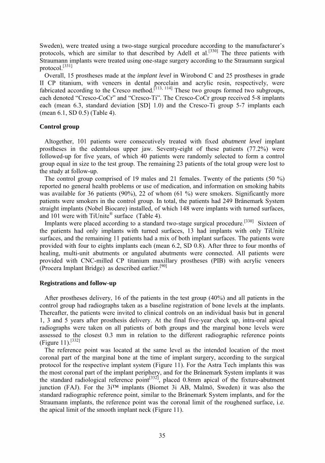
35
Sweden), were treated using a two-stage surgical procedure according to the manufacturer’s protocols, which are similar to that described by Adell et al.[330] The three patients with Straumann implants were treated using one-stage surgery according to the Straumann surgical protocol.[331] Overall, 15 prostheses made at the implant level in Wirobond C and 25 prostheses in grade II CP titanium, with veneers in dental porcelain and acrylic resin, respectively, were fabricated according to the Cresco method.[113, 114] These two groups formed two subgroups, each denoted “Cresco-CoCr” and “Cresco-Ti”. The Cresco-CoCr group received 5-8 implants each (mean 6.3, standard deviation [SD] 1.0) and the Cresco-Ti group 5-7 implants each (mean 6.1, SD 0.5) (Table 4). Control group Altogether, 101 patients were consecutively treated with fixed abutment level implant prostheses in the edentulous upper jaw. Seventy-eight of these patients (77.2%) were followed-up for five years, of which 40 patients were randomly selected to form a control group equal in size to the test group. The remaining 23 patients of the total group were lost to the study at follow-up. The control group comprised of 19 males and 21 females. Twenty of the patients (50 %) reported no general health problems or use of medication, and information on smoking habits was available for 36 patients (90%), 22 of whom (61 %) were smokers. Significantly more patients were smokers in the control group. In total, the patients had 249 Brånemark System straight implants (Nobel Biocare) installed, of which 148 were implants with turned surfaces, and 101 were with TiUnite® surface (Table 4). Implants were placed according to a standard two-stage surgical procedure.[330] Sixteen of the patients had only implants with turned surfaces, 13 had implants with only TiUnite surfaces, and the remaining 11 patients had a mix of both implant surfaces. The patients were provided with four to eights implants each (mean 6.2, SD 0.8). After three to four months of healing, multi-unit abutments or angulated abutments were connected. All patients were provided with CNC-milled CP titanium maxillary prostheses (PIB) with acrylic veneers (Procera Implant Bridge) as described earlier.[90] Registrations and follow-up After prostheses delivery, 16 of the patients in the test group (40%) and all patients in the control group had radiographs taken as a baseline registration of bone levels at the implants. Thereafter, the patients were invited to clinical controls on an individual basis but in general 1, 3 and 5 years after prosthesis delivery. At the final five-year check up, intra-oral apical radiographs were taken on all patients of both groups and the marginal bone levels were assessed to the closest 0.3 mm in relation to the different radiographic reference points (Figure 11).[332] The reference point was located at the same level as the intended location of the most coronal part of the marginal bone at the time of implant surgery, according to the surgical protocol for the respective implant system (Figure 11). For the Astra Tech implants this was the most coronal part of the implant periphery, and for the Brånemark System implants it was the standard radiological reference point[332], placed 0.8mm apical of the fixture-abutment junction (FAJ). For the 3i™ implants (Biomet 3i AB, Malmö, Sweden) it was also the standard radiographic reference point, similar to the Brånemark System implants, and for the Straumann implants, the reference point was the coronal limit of the roughened surface, i.e. the apical limit of the smooth implant neck (Figure 11).
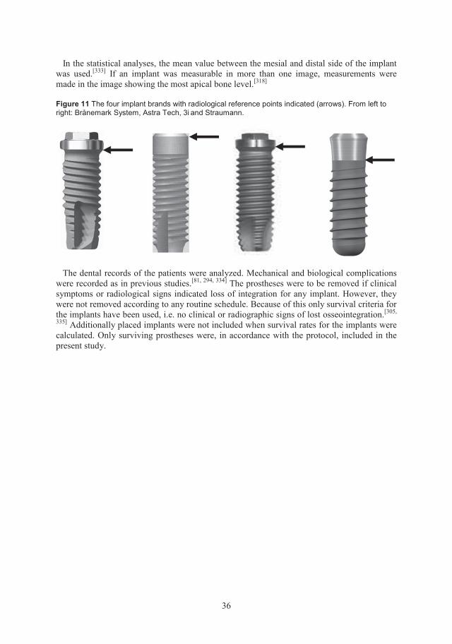
36
In the statistical analyses, the mean value between the mesial and distal side of the implant was used.[333] If an implant was measurable in more than one image, measurements were made in the image showing the most apical bone level.[318]
Figure 11 The four implant brands with radiological reference points indicated (arrows). From left to right: Brånemark System, Astra Tech, 3i and Straumann.
The dental records of the patients were analyzed. Mechanical and biological complications were recorded as in previous studies.[81, 294, 334] The prostheses were to be removed if clinical symptoms or radiological signs indicated loss of integration for any implant. However, they were not removed according to any routine schedule. Because of this only survival criteria for the implants have been used, i.e. no clinical or radiographic signs of lost osseointegration.[305,
335] Additionally placed implants were not included when survival rates for the implants were calculated. Only surviving prostheses were, in accordance with the protocol, included in the present study.
37
ERRORS OF METHODS, ACCURACY AND PRECISION OF MEASUREMENTS Selected techniques for estimating or controlling precision in the primary methods used in the four studies (I-IV) are presented below. More information can be obtained from the respective papers (I-IV). Study I According to the manufacturer, the precision of the CMM is less than 1 µm when measuring in small volumes. All five components in the master model and in one framework were measured five times. The standard deviation was within ±3 µm for these measurements for all five positions. Study II and III In the ion leakage measurements, inductively-coupled plasma mass spectrometry was used. The accuracy of the measurements was estimated to ±10%. When the optical interferometry was performed, the maximal vertical range was 5 mm, the horizontal resolution was 0.3 µm and the vertical resolution was 0.1 nm. Before analyses, a 50 x 50 µm Gaussian digital filter was used to distinguish errors in form and waviness from roughness. In the viability tests three types of control wells were used, according to recommendations. In each experiment, 12 control wells were used. Six of the control wells were used as negative controls (untreated cells) and six as positive controls (azide treated). In the positive control wells, the culture medium was supplemented with 20 mM sodium azide in the final 24 hour-period. Additionally, reference wells without cells were used for calculating the reduction of Alamar Blue. Study IV One month after the first radiograph measurements, one implant from each patient in the test group was randomly selected and a second measurement was performed on these implants in the same way as the first to assess intra-examiner variability.[318] Mean bone level variation in the 40 measured implants was 0.2 mm (SD 0.3).
38
STATISTICAL ANALYSIS The statistical methods used in the thesis are described by Altman and Good.[336, 337] The level of statistical significance in all studies (I-IV) was set at 5% (p<0.05). The following statistical methods were used:
Study I Conventional descriptive statistics were used to present the distortion of the frameworks. Analysis of variance and Tukey’s post hoc test were used to identify and study differences between the groups. Because normal distribution could not be verified and the observations were rather few, all significant differences were also validated with Fisher’s nonparametric permutation test. Contraction in each framework group was examined with Fisher’s nonparametric permutation test for paired observations. Comparisons between the least squares method and the orthogonal 3-2-1 method were performed by Wilcoxon’s rank sum test. Study II Conventional descriptive statistics were used to present the mass spectrometry and the interferometry. One-way ANOVA and Dunnett’s T3 post-hoc test were used to identify and study differences between the groups. The Student’s t-test was used to highlight changes within the groups. Study III Cell viability. Conventional descriptive statistics were used to present the percentage of reduction in the Alamar Blue tests. For detecting differences between the four groups, ANOVA was applied. Due to differences in variance, both the Dunnett T3-test and the Bonferroni method were used for detecting groups that differed. The Student’s t-test was used when the differences between the two test groups were compared. Optical interferometry. Conventional descriptive statistics were used to present the surface roughness values for the two groups. The Student’s t-test was used for detecting differences between the groups regarding the parameters Sa, Sds, Ssk and Sci, and Mann Whitney’s U-test was used for studying differences between the groups for the parameters St and Sdr. Study IV Conventional descriptive statistics were used. The chi-square test was applied when analyses of differences in smoking habits, single complication frequencies and bone levels categories were performed. Student’s t-test was used for the analyses of the mean bone levels and Fisher’s exact test for the survival rates.

39
RESULTS This section summarizes the major results from the four papers. More detailed descriptions are presented in the respective papers. PART 1. IN VITRO STUDIES OF CAST, SECTIONED AND LASER-WELDED FRAMEWORKS IN COBALT-CHROME ALLOY AND CP TITANIUM (I-III) Study I Transversal width and sagittal curvature Statistically significant expansion of framework width compared to the master model was found in the Cresco-Ti group (Table 5). The same tendency was found in the PIB, although this was insignificant. On the other hand, the Cresco-CoCr presented a contraction (x-axis), although it was insignificant. Statistically significant differences between the groups could be observed for the transversal width (x-axis), but not for the sagittal curvature (y-axis) of the frameworks (Figure 6, page 30).
Table 5 Horizontal Distortions. Width Differences: Difference in Mean Distance (Standard Deviation) in µm between Positions #1 and #5 of the Frameworks Compared to the Distance between the Same Positions of the Master Model. Curvature Differences: Mean Sagittal Difference, y-value, (Standard Deviation) in µm between Position #3 of the Frameworks Compared to the Same Position of the Master Model. Directions of Distortions are Related to Figure 5, page 30. Width
Differences Related to
Master
Number of Frameworks
with Expansion of Width
Number of Frameworks
with Contraction
of Width
Curvature Differences Related to
Master
Number of Frameworks
with Curvature Expansion
Number of Frameworks
with Curvature
Contraction
Cresco-CoCr (n=10) -12 (19) 2 8 4 (59) 5 5
Cresco- Ti (n=10) 38 (34) 9 1 -10 (62) 5 5
PIB (n=5) 71 (44) 5 0 -1 (27) 1 4
Distortions in real and absolute figures Horizontal distortions (x- and y-axes) were greater than vertical distortions (z-axis) for all groups (Table 6). The maximal values in absolute figures (Table 7, Figures 12-14) for the PIB group revealed a significantly greater range in the x-axis compared to Cresco-CoCr, but a significantly smaller range in the z-axis in relation to the two Cresco groups. Analysis of distortions in absolute figures revealed significant transversal (x-axis) differences between Cresco-CoCr and PIB, and differences in vertical dimensions (z-axis) between Cresco-CoCr and PIB, as well as between Cresco-Ti and PIB (Table 7, Figures 12-14).
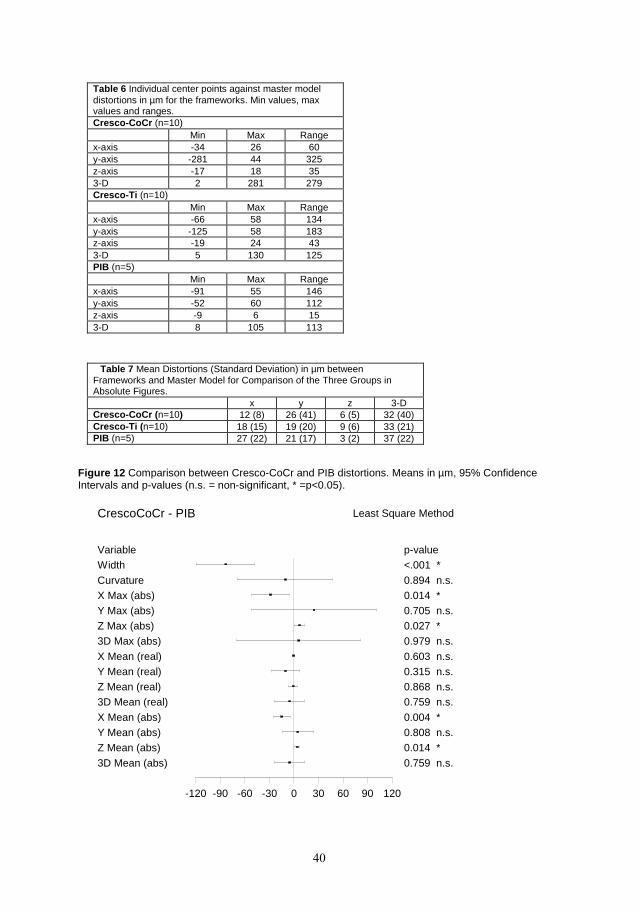
40
Table 6 Individual center points against master model distortions in µm for the frameworks. Min values, max values and ranges. Cresco-CoCr (n=10) Min Max Range x-axis -34 26 60 y-axis -281 44 325 z-axis -17 18 35 3-D 2 281 279 Cresco-Ti (n=10) Min Max Range x-axis -66 58 134 y-axis -125 58 183 z-axis -19 24 43 3-D 5 130 125 PIB (n=5) Min Max Range x-axis -91 55 146 y-axis -52 60 112 z-axis -9 6 15 3-D 8 105 113
Table 7 Mean Distortions (Standard Deviation) in µm between Frameworks and Master Model for Comparison of the Three Groups in Absolute Figures. x y z 3-D Cresco-CoCr ( n=10) 12 (8) 26 (41) 6 (5) 32 (40) Cresco-Ti ( n=10) 18 (15) 19 (20) 9 (6) 33 (21) PIB (n=5) 27 (22) 21 (17) 3 (2) 37 (22)
Figure 12 Comparison between Cresco-CoCr and PIB distortions. Means in µm, 95% Confidence Intervals and p-values (n.s. = non-significant, * =p<0.05).
CrescoCoCr - PIB
Variable p-value
Least Square Method
-120 -90 -60 -30 0 30 60 90 120
Width <.001 *
Curvature 0.894 n.s.X Max (abs) 0.014 *Y Max (abs) 0.705 n.s.Z Max (abs) 0.027 *3D Max (abs) 0.979 n.s.
X Mean (real) 0.603 n.s.Y Mean (real) 0.315 n.s.Z Mean (real) 0.868 n.s.3D Mean (real) 0.759 n.s.
X Mean (abs) 0.004 *Y Mean (abs) 0.808 n.s.Z Mean (abs) 0.014 *
3D Mean (abs) 0.759 n.s.
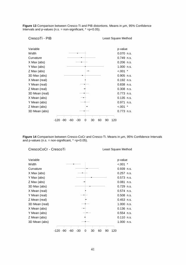
41
Figure 13 Comparison between Cresco-Ti and PIB distortions. Means in µm, 95% Confidence Intervals and p-values (n.s. = non-significant, * =p<0.05).
CrescoTi - PIB
Variable p-value
Least Square Method
-120 -90 -60 -30 0 30 60 90 120
Width 0.070 n.s.Curvature 0.749 n.s.X Max (abs) 0.206 n.s.Y Max (abs) 1.000 n.s.Z Max (abs) <.001 *3D Max (abs) 0.905 n.s.X Mean (real) 0.192 n.s.Y Mean (real) 0.838 n.s.Z Mean (real) 0.308 n.s.3D Mean (real) 0.773 n.s.X Mean (abs) 0.135 n.s.Y Mean (abs) 0.971 n.s.Z Mean (abs) <.001 *3D Mean (abs) 0.773 n.s.
Figure 14 Comparison between Cresco-CoCr and Cresco-Ti. Means in µm, 95% Confidence Intervals and p-values (n.s. = non-significant, * =p<0.05).
CrescoCoCr - CrescoTi
Variable p-value
Least Square Method
-120 -90 -60 -30 0 30 60 90 120
Width <.001 *
Curvature 0.939 n.s.X Max (abs) 0.257 n.s.Y Max (abs) 0.573 n.s.Z Max (abs) 0.081 n.s.3D Max (abs) 0.729 n.s.
X Mean (real) 0.574 n.s.Y Mean (real) 0.508 n.s.Z Mean (real) 0.453 n.s.3D Mean (real) 1.000 n.s.
X Mean (abs) 0.136 n.s.Y Mean (abs) 0.554 n.s.Z Mean (abs) 0.110 n.s.
3D Mean (abs) 1.000 n.s.
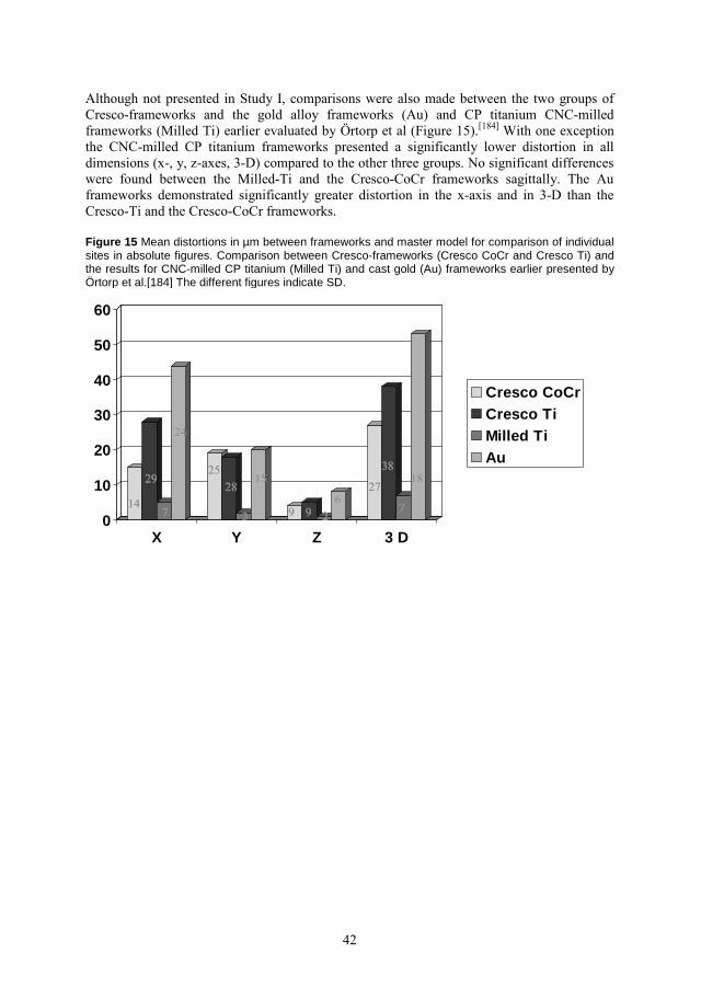
42
Although not presented in Study I, comparisons were also made between the two groups of Cresco-frameworks and the gold alloy frameworks (Au) and CP titanium CNC-milled frameworks (Milled Ti) earlier evaluated by Örtorp et al (Figure 15).[184] With one exception the CNC-milled CP titanium frameworks presented a significantly lower distortion in all dimensions (x-, y, z-axes, 3-D) compared to the other three groups. No significant differences were found between the Milled-Ti and the Cresco-CoCr frameworks sagittally. The Au frameworks demonstrated significantly greater distortion in the x-axis and in 3-D than the Cresco-Ti and the Cresco-CoCr frameworks. Figure 15 Mean distortions in µm between frameworks and master model for comparison of individual sites in absolute figures. Comparison between Cresco-frameworks (Cresco CoCr and Cresco Ti) and the results for CNC-milled CP titanium (Milled Ti) and cast gold (Au) frameworks earlier presented by Örtorp et al.[184] The different figures indicate SD.
0
10
20
30
40
50
60
X Y Z 3 D
Cresco CoCrCresco TiMilled TiAu
1414
2929
77
2424
33
28282525
662727
3838
77
1818
99
1515
2299
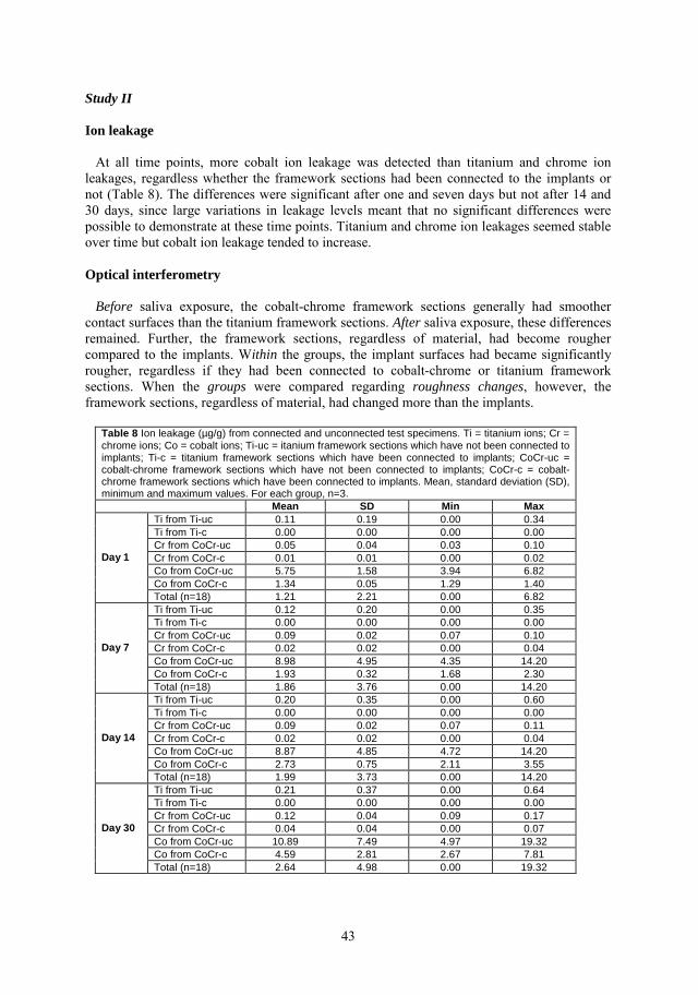
43
Study II Ion leakage At all time points, more cobalt ion leakage was detected than titanium and chrome ion leakages, regardless whether the framework sections had been connected to the implants or not (Table 8). The differences were significant after one and seven days but not after 14 and 30 days, since large variations in leakage levels meant that no significant differences were possible to demonstrate at these time points. Titanium and chrome ion leakages seemed stable over time but cobalt ion leakage tended to increase. Optical interferometry Before saliva exposure, the cobalt-chrome framework sections generally had smoother contact surfaces than the titanium framework sections. After saliva exposure, these differences remained. Further, the framework sections, regardless of material, had become rougher compared to the implants. Within the groups, the implant surfaces had became significantly rougher, regardless if they had been connected to cobalt-chrome or titanium framework sections. When the groups were compared regarding roughness changes, however, the framework sections, regardless of material, had changed more than the implants.
Table 8 Ion leakage (µg/g) from connected and unconnected test specimens. Ti = titanium ions; Cr = chrome ions; Co = cobalt ions; Ti-uc = itanium framework sections which have not been connected to implants; Ti-c = titanium framework sections which have been connected to implants; CoCr-uc = cobalt-chrome framework sections which have not been connected to implants; CoCr-c = cobalt-chrome framework sections which have been connected to implants. Mean, standard deviation (SD), minimum and maximum values. For each group, n=3. Mean SD Min Max
Ti from Ti-uc 0.11 0.19 0.00 0.34 Ti from Ti-c 0.00 0.00 0.00 0.00 Cr from CoCr-uc 0.05 0.04 0.03 0.10 Cr from CoCr-c 0.01 0.01 0.00 0.02 Co from CoCr-uc 5.75 1.58 3.94 6.82 Co from CoCr-c 1.34 0.05 1.29 1.40
Day 1
Total (n=18) 1.21 2.21 0.00 6.82 Ti from Ti-uc 0.12 0.20 0.00 0.35 Ti from Ti-c 0.00 0.00 0.00 0.00 Cr from CoCr-uc 0.09 0.02 0.07 0.10 Cr from CoCr-c 0.02 0.02 0.00 0.04 Co from CoCr-uc 8.98 4.95 4.35 14.20 Co from CoCr-c 1.93 0.32 1.68 2.30
Day 7
Total (n=18) 1.86 3.76 0.00 14.20 Ti from Ti-uc 0.20 0.35 0.00 0.60 Ti from Ti-c 0.00 0.00 0.00 0.00 Cr from CoCr-uc 0.09 0.02 0.07 0.11 Cr from CoCr-c 0.02 0.02 0.00 0.04 Co from CoCr-uc 8.87 4.85 4.72 14.20 Co from CoCr-c 2.73 0.75 2.11 3.55
Day 14
Total (n=18) 1.99 3.73 0.00 14.20 Ti from Ti-uc 0.21 0.37 0.00 0.64 Ti from Ti-c 0.00 0.00 0.00 0.00 Cr from CoCr-uc 0.12 0.04 0.09 0.17 Cr from CoCr-c 0.04 0.04 0.00 0.07 Co from CoCr-uc 10.89 7.49 4.97 19.32 Co from CoCr-c 4.59 2.81 2.67 7.81
Day 30
Total (n=18) 2.64 4.98 0.00 19.32
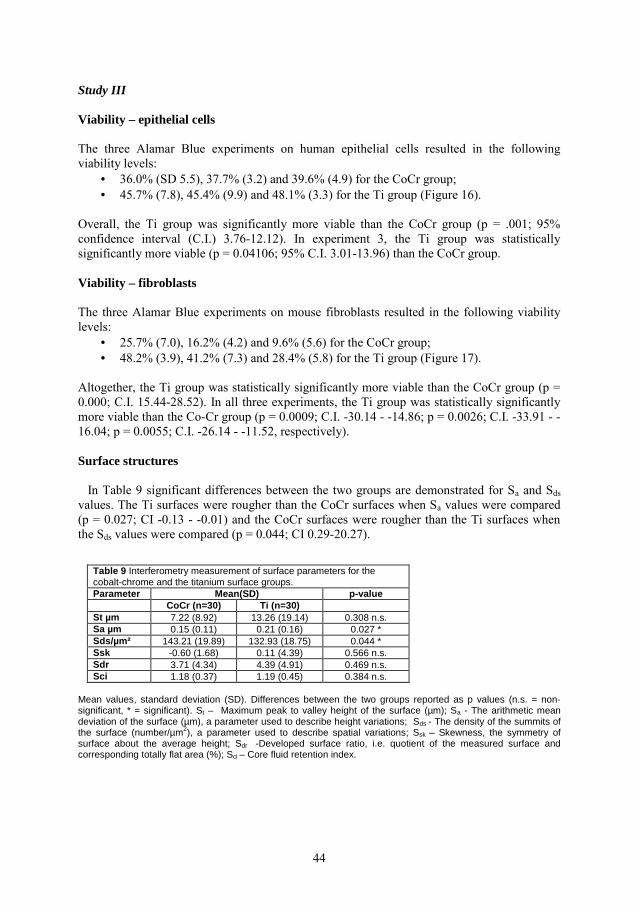
44
Study III Viability – epithelial cells The three Alamar Blue experiments on human epithelial cells resulted in the following viability levels:
• 36.0% (SD 5.5), 37.7% (3.2) and 39.6% (4.9) for the CoCr group; • 45.7% (7.8), 45.4% (9.9) and 48.1% (3.3) for the Ti group (Figure 16).
Overall, the Ti group was significantly more viable than the CoCr group (p = .001; 95% confidence interval (C.I.) 3.76-12.12). In experiment 3, the Ti group was statistically significantly more viable (p = 0.04106; 95% C.I. 3.01-13.96) than the CoCr group. Viability – fibroblasts The three Alamar Blue experiments on mouse fibroblasts resulted in the following viability levels:
• 25.7% (7.0), 16.2% (4.2) and 9.6% (5.6) for the CoCr group; • 48.2% (3.9), 41.2% (7.3) and 28.4% (5.8) for the Ti group (Figure 17).
Altogether, the Ti group was statistically significantly more viable than the CoCr group (p = 0.000; C.I. 15.44-28.52). In all three experiments, the Ti group was statistically significantly more viable than the Co-Cr group (p = 0.0009; C.I. -30.14 - -14.86; p = 0.0026; C.I. -33.91 - -16.04; p = 0.0055; C.I. -26.14 - -11.52, respectively). Surface structures In Table 9 significant differences between the two groups are demonstrated for Sa and Sds values. The Ti surfaces were rougher than the CoCr surfaces when Sa values were compared (p = 0.027; CI -0.13 - -0.01) and the CoCr surfaces were rougher than the Ti surfaces when the Sds values were compared (p = 0.044; CI 0.29-20.27).
Table 9 Interferometry measurement of surface parameters for the cobalt-chrome and the titanium surface groups. Parameter Mean(SD) p-value CoCr (n=30) Ti (n=30) St µm 7.22 (8.92) 13.26 (19.14) 0.308 n.s. Sa µm 0.15 (0.11) 0.21 (0.16) 0.027 * Sds/µm² 143.21 (19.89) 132.93 (18.75) 0.044 * Ssk -0.60 (1.68) 0.11 (4.39) 0.566 n.s. Sdr 3.71 (4.34) 4.39 (4.91) 0.469 n.s. Sci 1.18 (0.37) 1.19 (0.45) 0.384 n.s.
Mean values, standard deviation (SD). Differences between the two groups reported as p values (n.s. = non-significant, * = significant). St – Maximum peak to valley height of the surface (µm); Sa - The arithmetic mean deviation of the surface (µm), a parameter used to describe height variations; Sds - The density of the summits of the surface (number/µm2), a parameter used to describe spatial variations; Ssk – Skewness, the symmetry of surface about the average height; Sdr -Developed surface ratio, i.e. quotient of the measured surface and corresponding totally flat area (%); Sci – Core fluid retention index.
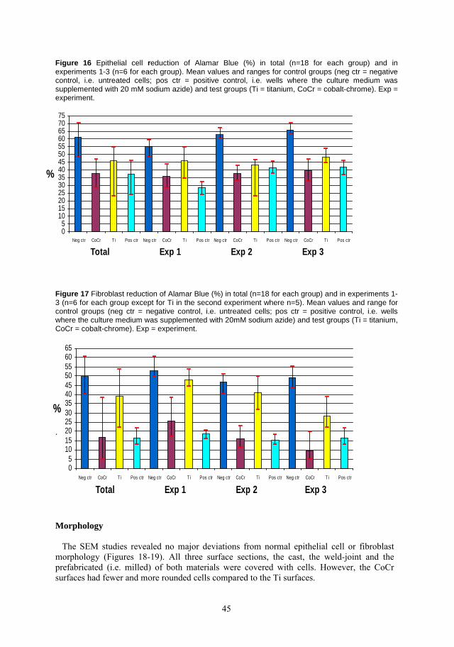
45
Figure 16 Epithelial cell reduction of Alamar Blue (%) in total (n=18 for each group) and in experiments 1-3 (n=6 for each group). Mean values and ranges for control groups (neg ctr = negative control, i.e. untreated cells; pos ctr = positive control, i.e. wells where the culture medium was supplemented with 20 mM sodium azide) and test groups (Ti = titanium, CoCr = cobalt-chrome). Exp = experiment.
Figure 17 Fibroblast reduction of Alamar Blue (%) in total (n=18 for each group) and in experiments 1-3 (n=6 for each group except for Ti in the second experiment where n=5). Mean values and range for control groups (neg ctr = negative control, i.e. untreated cells; pos ctr = positive control, i.e. wells where the culture medium was supplemented with 20mM sodium azide) and test groups (Ti = titanium, CoCr = cobalt-chrome). Exp = experiment.
. Mor phology The SEM studies revealed no major deviations from normal epithelial cell or fibroblast morphology (Figures 18-19). All three surface sections, the cast, the weld-joint and the prefabricated (i.e. milled) of both materials were covered with cells. However, the CoCr surfaces had fewer and more rounded cells compared to the Ti surfaces.
05
1015202530354045505560657075
Neg ctr CoCr Ti Pos ctr Neg ctr CoCr T i Pos ctr Neg ctr CoCr Ti Pos ctr Neg ctr CoCr Ti Pos ctr
Total Exp 1 Exp 2 Exp 3
%
05
101520253035404550556065
Neg ctr CoCr T i Pos ctr Neg ctr CoCr Ti Pos ctr Neg ctr CoCr Ti Pos ctr Neg ctr CoCr T i Pos ctr
Total Exp 1 Exp 2 Exp 3
%

46
Figure 18 Cobalt-chrome specimen surface (left) and titanium specimen surface specimen (right) with epithelial cells.
Figure 19 Cobalt-chrome specimen surface (left) and titanium specimen surface specimen (right) with fibroblasts. The two upper photographs show fibroblasts on and close to the weld joints. Note the irregularities in the weld joints.
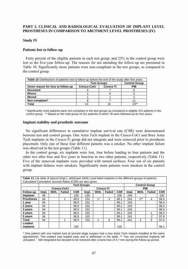
47
PART 2. CLINICAL AND RADIOLOGICAL EVALUATION OF IMPLANT LEVEL PROSTHESES IN COMPARISON TO ABUTMENT LEVEL PROSTHESES (IV) Study IV Patients lost to follow-up Forty percent of the eligible patients in each test group, and 23% in the control group were lost to the five-year follow-up. The reasons for not attending the follow-up are presented in Table 10. Significantly more patients were non-compliant in the test groups, as compared to the control group.
Table 10 Distributions of patients lost to follow-up before the end of the study after five years. Test Groups Control Group Given reason for loss to follow-up Cresco-CoCr Cresco-Ti PIB Deceased 0 7 6 Illness 1 0 3 Moved 1 1 3 Non-compliant* 8 7 11 Total 10 15 23**
* Significantly more patients were non-compliant in the test groups as compared to eligible 101 patients in the control group. ** Based on the total group of 101 patients of which 78 were followed-up for five years. Implant stability and prosthetic outcome No significant differences in cumulative implant survival rate (CSR) were demonstrated between test and control groups. One Astra Tech implant in the Cresco-CoCr and three Astra Tech implants in the Cresco-Ti group did not integrate and were removed prior to prosthesis placement. Only one of these four different patients was a smoker. No other implant failure was observed in the test groups (Table 11). In the control group, six implants were lost, four before loading in four patients and the other two after four and five years in function in two other patients, respectively (Table 11). Five of the removed implants were provided with turned surfaces. Four out of six patients with implant failures were smokers. Significantly more patients were smokers in the control group.
* One patient with one implant loss at second stage surgery had a new Astra Tech implant installed at the same appointment. This implant was loaded early and is withdrawn in the table. ** Two not connected implants left unloaded. † Still integrated but decided to be removed after a bone loss of 4.7 mm during the follow-up period.
Table 11 Life table of placed (Impl.), withdrawn (With.) and failed implants in the different groups of patients. Calculated Cumulative Survival Rates (CSR) are also given. Test Groups Control Group Cresco-CoCr Cresco-Ti PIB Follow-up Impl. With. Failed CSR Impl. With. Failed CSR Impl. With. Failed CSR Implants 95 100 155 100 249 100 Prosthesis 94 1 98.9 152 1* 3 98.1 243 2** 4 98.4 1 year 94 98.9 152 98.1 243 98.4 2 years 94 98.9 152 98.1 243 98.4 3 years 94 98.9 152 98.1 243 98.4 4 years 94 98.9 152 98.1 242 1 98.0 5 years 94 98.9 152 98.1 241 1† 97.6 Total 94 1 98.9 152 1 3 98.1 241 2 6 97.6 Loaded implants 100 100 99.2
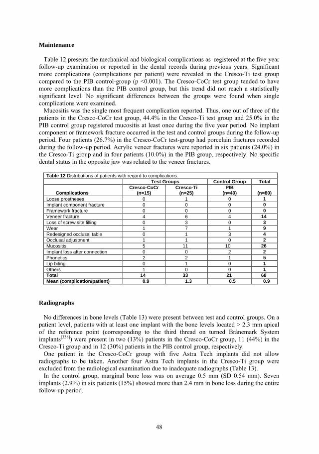
48
Maintenance Table 12 presents the mechanical and biological complications as registered at the five-year follow-up examination or reported in the dental records during previous years. Significant more complications (complications per patient) were revealed in the Cresco-Ti test group compared to the PIB control-group (p <0.001). The Cresco-CoCr test group tended to have more complications than the PIB control group, but this trend did not reach a statistically significant level. No significant differences between the groups were found when single complications were examined. Mucositis was the single most frequent complication reported. Thus, one out of three of the patients in the Cresco-CoCr test group, 44.4% in the Cresco-Ti test group and 25.0% in the PIB control group registered mucositis at least once during the five year period. No implant component or framework fracture occurred in the test and control groups during the follow-up period. Four patients (26.7%) in the Cresco-CoCr test-group had porcelain fractures recorded during the follow-up period. Acrylic veneer fractures were reported in six patients (24.0%) in the Cresco-Ti group and in four patients (10.0%) in the PIB group, respectively. No specific dental status in the opposite jaw was related to the veneer fractures.
Table 12 Distributions of patients with regard to complications. Test Groups Control Group Total
Complications Cresco-CoCr
(n=15) Cresco-Ti
(n=25) PIB
(n=40) (n=80) Loose prostheses 0 1 0 1 Implant component fracture 0 0 0 0 Framework fracture 0 0 0 0 Veneer fracture 4 6 4 14 Loss of screw site filling 0 3 0 3 Wear 1 7 1 9 Redesigned occlusal table 0 1 3 4 Occlusal adjustment 1 1 0 2 Mucositis 5 11 10 26 Implant loss after connection 0 0 2 2 Phonetics 2 2 1 5 Lip biting 0 1 0 1 Others 1 0 0 1 Total 14 33 21 68 Mean (complication/patient) 0.9 1.3 0.5 0.9
Radiographs No differences in bone levels (Table 13) were present between test and control groups. On a patient level, patients with at least one implant with the bone levels located > 2.3 mm apical of the reference point (corresponding to the third thread on turned Brånemark System implants[338]) were present in two (13%) patients in the Cresco-CoCr group, 11 (44%) in the Cresco-Ti group and in 12 (30%) patients in the PIB control group, respectively. One patient in the Cresco-CoCr group with five Astra Tech implants did not allow radiographs to be taken. Another four Astra Tech implants in the Cresco-Ti group were excluded from the radiological examination due to inadequate radiographs (Table 13). In the control group, marginal bone loss was on average 0.5 mm (SD 0.54 mm). Seven implants (2.9%) in six patients (15%) showed more than 2.4 mm in bone loss during the entire follow-up period.
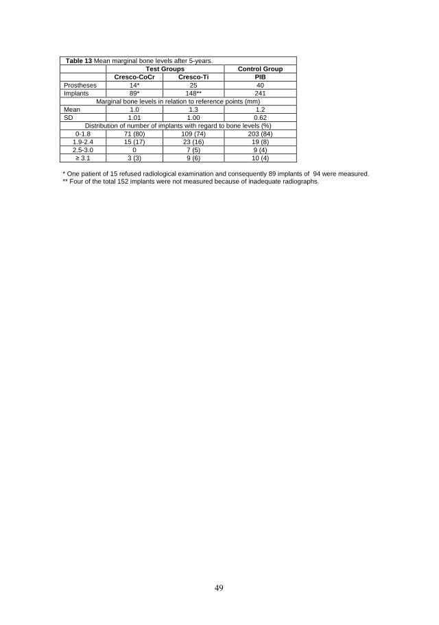
49
Table 13 Mean marginal bone levels after 5-years. Test Groups Control Group Cresco-CoCr Cresco-Ti PIB
Prostheses 14* 25 40 Implants 89* 148** 241
Marginal bone levels in relation to reference points (mm) Mean 1.0 1.3 1.2 SD 1.01 1.00 0.62
Distribution of number of implants with regard to bone levels (%) 0-1.8 71 (80) 109 (74) 203 (84)
1.9-2.4 15 (17) 23 (16) 19 (8) 2.5-3.0 0 7 (5) 9 (4) ≥ 3.1 3 (3) 9 (6) 10 (4)
* One patient of 15 refused radiological examination and consequently 89 implants of 94 were measured. ** Four of the total 152 implants were not measured because of inadequate radiographs.

50
DISCUSSION For this thesis, new techniques and ways to use well-known materials in the manufacture of implant-supported, complete prostheses were examined. Cobalt-chrome (CoCr) alloy frameworks were compared with CP titanium frameworks fabricated using techniques such as cast, sectioning and laser-welding as well as CNC-milling. The studies were conducted in vitro and in vivo. Further, abutment and abutment-free techniques were investigated, and porcelain-veneered and acrylic-veneered frameworks were compared. Even though they have been in use for some time, many of these techniques and materials are poorly documented, at least in the implant dentistry context. And in areas where documentation exists, consensus is generally absent. Biostatisticians were consulted before, during and after the studies were conducted, and the study results were analyzed with their guidance. Because Studies I-III involved several comparisons, robust statistical methods were chosen to control multiple testing. PART 1. IN VITRO STUDIES OF CAST, SECTIONED AND LASER-WELDED FRAMEWORKS IN COBALT-CHROME ALLOY AND CP TITANIUM (I-III) Precision of fit There were two aims here: 1) to investigate the precision of fit of implant-supported cast, sectioned and laser-welded frameworks fabricated in a CoCr alloy or in CP titanium; and 2) to compare these frameworks to individually scanned CNC-milled CP titanium frameworks. Study I identified no framework that was a perfect, completely passive fit with the master model. Patterson in 1995 proposed the following criteria:
• A perfect fit exists if the mating surfaces are in contact at all points without applying external pressure.
• A passive fit is less than perfect, but applying external pressure to produce a perfect fit has a negligible effect on the prosthesis.
• An active fit exists when applying external pressure can produce a perfect fit, but the applied pressure is harmful to the performance of the prosthesis.
• A poor fit exists if external pressure cannot produce a perfect fit.[146]
The geometry of the surfaces of the Cresco replicas facing the frameworks and their corresponding surfaces on the frameworks have a less distinct profile than the abutment replicas and corresponding framework cylinders used earlier by Örtorp et al.[184] Because of this, it was more difficult to register the surface geometry in Study I. As a result, it was not possible to analyze angular gap distortions as initially intended. These measurement problems may be reflected in the ranges and means of distortions; the distortions were also analyzed with the least square and the orthogonal 3-2-1 method. According to the manufacturer, the precision of the CMM is less than 1 µm when measuring small volumes. As a control for the method, we had the laboratory measure all five components in the master model and in one Cresco framework, five times. For all five positions, the standard deviation for each measurement was within ±3 µm. These findings support the assumption of an influence on the obtained distortion values caused by the less distinct profile of the Cresco frameworks cylinders.
51
The least square and the orthogonal 3-2-1 methods are commonly used techniques.[165] Slightly lower levels of distortion for the least square method were observed in Study I, thus confirming the reports from Eliasson et al.[186] These results indicate that precision of fit may be related to the mathematical model used. The direction of distortions varied in Study I but the horizontal distortions were of similar magnitudes. Comparable results have been presented for CNC-milled and cast frameworks, measured in the same way.[185, 186] In addition, Study I demonstrated a precision of fit that was higher for the Cresco groups compared to the gold alloy frameworks but lower compared to the PIB frameworks, which had been previously evaluated.[184] The lower levels of distortions for the CNC-milled frameworks in the report by Örtorp and colleagues can be explained by the fact that they measured 20 frameworks fabricated from one single scanning procedure, i.e. duplicates, whereas in Study I and other studies the plastic patterns were individually scanned for each CNC-framework.[184-186] The PIB technicians in Study I were not informed of the ongoing study. The Cresco framework technicians, on the other hand, knew of Study I. This might have an impact on the results, which is supported to a certain extent by Eliasson et al. who demonstrated the value of blinding when they evaluated I-bridge frameworks fabricated in two ways.[186] The laboratory was informed that the first group of frameworks would be evaluated for precision of fit. The second group of frameworks was produced to mimic the clinical situation, e.g. the frameworks were sent to the manufacturer on different days. The magnitude of distortion was lower for the non-blinded group.[186]
The PIB framework production may be regarded as more “high-tech” and controlled than the Cresco method, the latter demanding a high degree of manual work and technical skill in every step of the production. Some support for this assumption was found in previous reports where CNC-milled frameworks were compared to conventional cast frameworks and a better fit for the CNC-milled frameworks was demonstrated.[184, 185] Even so, Study I demonstrated only minor differences between the two investigated fabrication methods; the clinical importance of this is not known. Study I also found that the CoCr alloy frameworks contracted, whereas both groups of titanium frameworks, regardless of fabrication techniques, seemed to expand. Previous studies found either similar or contradictory results.[184-187] Even though the fabrication of the two groups of Cresco frameworks was comparable, small differences existed. For example, the laser-welding procedures were not exactly the same. A higher energy was achieved within the CoCr alloy frameworks due to a longer duration of the laser pulse. Taking into account the superior thermal conductivity of the CoCr alloy compared to titanium[11, 65], both these factors may have influenced the different distortion patterns. The frameworks in Study I had a greater maximum distortion range of individual points in the horizontal plane compared to the vertical plane, in accordance with observations in previous studies.[184-186] In addition, Study I revealed a lower degree of vertical misfit in the PIB frameworks compared to both groups of Cresco frameworks. These findings are interesting since it has been suggested that a vertical misfit influences complications to a higher degree than horizontal distortions.[147, 172] Further, both methods, “orthogonal 3-2-1” and “least square”, underestimate the vertical distortions since they work in a virtual world and ignore the physical limits of reality by permitting negative vertical values. As a consequence, the real vertical distortions for all three groups of frameworks were probably underestimated, both in Study I and in previous reports.[184-186]
It has been suggested that misfit preload can have a greater impact on bone response than the magnitude of the vertical gap.[192-194] Thus, it may be more crucial to reduce vertical gaps in frameworks connected directly to the implants, where higher preload forces could be expected. It is possible to reduce vertical misfit by increasing the tightening force[86] and
52
consequently introduce higher stress in the screw joint and the peri-implant tissues. Earlier more commonly used materials such as gold alloys are more flexible than titanium or Co-Cr alloys for prosthesis-retention screws and frameworks. Therefore, even higher stress levels might be introduced with these new materials. What is an “acceptable vertical misfit” is in dispute and varies between 10 and 150 µm.[111,
147, 173, 339] In the clinical situation, a misfit of 50 µm can be detected using magnification lenses at x 2 magnification, at a distance of 25 cm.[125, 141] Study I found mean vertical misfits of less than 10 µm (absolute figures) for all three groups. The maximum range observed between minimum and maximum real values was 43 µm. Consequently, the magnitude of misfit in Study I can be regarded as acceptable and comparable to previous reports. But it seems obvious that the accuracy of a single framework is hard to predict and that the technique for measuring the precision of fit influences the distortion figures obtained. As a result, one study might be difficult to compare to another. Although misfit seems to have an impact on mechanical complications such as screw loosening and retention screw fractures, whether, and to what extent, it might influence the peri-implant tissues remains a point of dispute. Material degradation and cell responses The aims here were: 1) to investigate and estimate material degradation in titanium implants and frameworks made of CoCr alloy or CP titanium according to the Cresco method, before and after exposure to artificial saliva, and 2) to assess possible adverse cellular responses to CoCr alloy frameworks by comparing the viability and morphology of epithelial cells and fibroblasts cultivated on CoCr alloy and titanium specimens. Study II demonstrated material degradation after saliva exposure both in titanium implants and in frameworks made from CoCr alloy and CP titanium. The material degradation was reported as ion leakage and as changed surface structure. Significantly more cobalt ions leaked than titanium and chrome ions. Both framework sections and implants became rougher after saliva exposure. Since only ion leakage and not galvanic corrosion was measured, the material degradation in Study II can be regarded as an estimation of corrosion. Ion leakage was registered by an inductively-coupled plasma mass spectrometer connected to a computer, a common set up for chemical analyses.[340] In a clinical situation, the saliva is normally continually exchanged due to secretion and swallowing. In contrast, in Study II only 1 ml of the saliva solution (total volume 20 ml) in each experimental well was exchanged at four different time points. It has been suggested that when the ion concentration of an element in saliva increases this will per se decrease the tendency for the same element to dissolve.[11]
In accordance with this assumption, Study II demonstrated stable leakages of chrome and titanium ions. Quite the opposite happened to the cobalt ion leakage, which tended to increase over time. This might be a sign of an active corrosion process. The findings are supported by a previous report on removable partial dentures describing a minor decrease over time in cobalt ion leakage compared to a more obvious decrease in chrome ion leakage.[214]
Ion leakages from unconnected framework sections were higher than from sections connected to implants, at all time points and from both materials. One possible explanation might be a larger exposed framework section surface in unconnected frameworks. The differences remained, regardless if the data was presented as absolute values (weight, µg) or relatively (µg/g, weight percent, wt%). Even though ion leakage in several studies is expressed as weight per surface unit, it was decided to express the results in Study II as wt%. A main reason was that the surfaces of the framework sections differed in size and there was concern that measuring the area would introduce errors which could influence the final results
53
to a high degree. When describing the chemical composition of an alloy, Wataha recommends the use of number of atoms of an element (at%) instead of weight percent (wt%).[57] Since the difference in at% between cobalt and chrome is only about 10% (Co 58.9332 u, Cr 51.996 u) this further strengthens the decision to regard wt% as appropriate in Study II. The artificial saliva solution used in Study II was blended according to an ISO standard. The mix is commonly used for polymers but can be regarded as appropriate for general use as well. An almost neutral pH was chosen for the saliva solution, to mimic a clinically “normal” situation. Yet, it is well known that the pH value in saliva is not constant and different protein solutions may alter the pH.[341, 342] Thus, plasma proteins such as albumin and fibrinogen might affect the corrosion of metals and alloys.[343] Milleding et al. studied protein adhesion to ceramic surfaces and reported a higher affinity for plasma than for saliva proteins.[344] This could partly explain why these materials are generally well tolerated close to the peri-implant mucosa and in the transmucosal zone.[344] Further, the interaction between dental device surface, the bio-film created immediately on every intra-oral surface, and the surrounding tissues will influence the biological response to a new dental device. These topics, however, were not examined in the present thesis. Optical instruments have been recommended for the study of surfaces in relatively soft metals such as titanium[215] and optical interferometry has been used recently in surface studies similar to Study II and III .[201, 345, 346] The equipment can be regarded as a development of the earlier, more common confocal laser scanning profilometer but the interferometer is faster and has a larger maximum vertical range as well as maximum measuring area. Since so many subgroups were defined in Study II, three surface parameters were decided to be sufficient and were subsequently analyzed as previous recommended by Wennerberg and Albrektsson[31]: one height, one space and one hybrid parameter. This was performed at four defined locations on each contact surface of the implants and the framework sections, before and after saliva exposure. One of the 144 measurements (0.7%) was impossible to evaluate because manufacturing defects in the surface exceeded the measuring range. In a similar evaluation of screw-joint surfaces, ten of 431 measurements (2.3%) were impossible to perform due to fabrication defects.[201] Study II displayed rougher contact surfaces on the titanium framework sections than on the CoCr alloy frameworks, both before and after saliva exposure. Craig and Hanks demonstrated that a polished surface (smaller area) may release less elements than an “as-cast” (greater area).[347] Yet, even though a greater surface area was probably exposed to the saliva, the titanium ion leakage was lower than the cobalt leakage in Study II. The differences in material properties may explain some of the differences in roughness. Since a CoCr alloy is harder than titanium, a titanium surface is more difficult to polish and create a smooth surface. Further, the contact surfaces of the implants became rougher after saliva exposure. This may be caused by the saliva exposure per se. Yet, it seems reasonable that contact with the framework sections also influenced the implant surfaces. The CoCr alloy framework sections might have roughened the implant surfaces through friction more than the titanium framework sections would. As a consequence, the implants connected to the CoCr alloy framework sections showed the highest roughness values. This increase in surface roughness, i.e. surface area, might increase the risk of future corrosion. The change in contact surface roughness between frameworks and implants during the saliva exposure might change the friction – and thus the preload – in the screw joints as well. Consequently, the risk for mechanical complications such as loosening of prosthetic retention screws due to corrosion must be considered when framework material is decided. In Study III, the same three surface parameters as in Study II were used, with the addition of three more. This was possible since the number of specimens studied was very limited. In Study III, registrations were made from two specimen of each material, CoCr alloy and
54
titanium, respectively. To compensate, in some way, for the small number of observations more surface data was registered and analyzed. This was also done to thoroughly investigate whether or not differences in surface roughness could explain any variability in cell reactions between the two examined framework materials. Significant differences in surface roughness between CoCr alloy and titanium were identified in Study III. The CoCr alloy specimens had a smaller height variation (Sa = the arithmetic mean deviation of the surface) but were denser (Sds = the density of the summits of the surface) than the titanium. Yet, the differences between the two materials were small. No significant differences were demonstrated regarding the four additional surface parameters. Study III demonstrated that both epithelial cells and fibroblasts had a better reduction capacity according to the Alamar Blue method, when in contact with titanium compared to CoCr alloy surfaces. The Alamar Blue method was presented as an alternative to more expensive, common and complicated radioactive assays to monitor and determine lymphocyte proliferation.[329] The test allows cells to be monitored without compromising the sterility of the cultures and permits further analyses by other methods.[329] Only small differences in surface roughness were described, indicating the presence of other properties that might influence the cell reactions. Previous studies have presented contradictory results regarding surface roughness. In a study using dogs, Abrahamsson et al. demonstrated no influence on soft tissue adhesion regardless of whether titanium abutments were smooth or rough (Sa 0.22 µm or 0.45 µm).[348] Glauser et al. supported these findings in a human study, reporting an equal height of the soft tissue seal regardless of surface roughness.[349] Even so, they found a higher epithelium layer on smooth surfaces and a higher connective tissue layer on rougher surfaces.[349] Other studies have demonstrated that epithelial cells prefer smoother metal surfaces but rougher osteoceramic surfaces in contrast to fibroblasts, who preferred rougher metal surfaces but smother osteoceramic surfaces. [236,
238, 240-244] Therefore, it seems reasonable to believe that apart from physical properties such as surface free-energy interfering with the adhesion capacity, the material, CoCr alloy or titanium, per se had an impact on the cell viability. This argument seems even more reasonable taking into account the differences reported in Study II where cobalt ion leakage was significantly greater than ion leakage of chromium and titanium, and the toxic capability of cobalt to cell cultures described in previous reports.[253, 257] Cell culture studies can give information about mechanisms behind cell and material interactions. But even if several studies have focused on the in vitro behavior of fibroblasts rather than on epithelial cells, it was suggested that the in vitro environment for fibroblasts is generally less representative of the normal in vivo situation than the corresponding conditions for epithelial cells.[350, 351] Because both epithelial cells and fibroblasts are in contact with the transmucosal part of an implant system, it was regarded as interesting to examine both cell types in Study III. This assumption is in accordance with a previous study.[244] Solid metal alloy specimens were used in Study III. The specimens were fabricated to mimic the Cresco method and while in direct contact with the examined cells, in accordance with previous recommendations.[237, 352] Cell culture tests have been regarded as appropriate when local material effects, such as in the transmucosal peri-prosthetis zone, are studied.[353, 354] Arvidson et al. used “normal” fibroblasts (primary cells) from human gingiva while Study III used permanent cell lines: “immortalized” human epithelial cells and mouse fibroblasts.[253] They proposed that “immortalized” cells may have developed not only a different morphology but also an altered metabolism.[253] Experiments with primary cells can be regarded closer to a normal, clinical situation compared to tests with permanent cell lines.[246] As Arvidson et al. faintly outlined, primary cells can probably maintain normal
55
functions, such as to differentiate and adhere. On the other hand, because of the variations between donors, results from studies with primary cells are more likely to vary. [246] Thus, permanent cell line reports such as Study III are more reproducible. Cell reactions to metallic materials from primary cells (fibroblast) and permanent cell lines (lymphoma cells) have been demonstrated.[73] It was suggested that primary cells are more sensitive than permanent cell lines.[73] With this background, the Alamar Blue results from Study III become even more interesting since a significant difference in cell reactions to the two examined materials was present, despite the use of suggested less sensitive permanent cell lines. In the SEM analysis (Study III), only minor differences in cell morphology were revealed. Still, the cells on the CoCr alloy surfaces were regarded as fewer and more rounded, the latter indicating a somewhat hostile environment. Yet, one shortcoming with the SEM analysis was that the specimen material (CoCr alloy and titanium) was not blinded for the examiners. Titanium is a well-documented material for implants as well as for transmucosal components and prosthetic frameworks. Alternative materials such as CoCr alloys have been introduced and may be interesting due to low costs and good mechanical properties. Nevertheless, the results from Study II and III raise doubts about their biocompatibility as components in an oral implant system. PART 2. CLINICAL AND RADIOLOGICAL EVALUATION OF IMPLANT LEVEL PROSTHESES IN COMPARISON TO ABUTMENT LEVEL PROSTHESES (IV) The aims here were: 1) to evaluate and compare the clinical function between implant-supported prostheses in the edentulous maxilla made on implant level and prostheses fabricated on abutment level, and 2) to investigate porcelain-veneered CoCr alloy prostheses in comparison to two groups of prosthesis made in acrylic-veneered CP titanium. Many patients in Study IV were lost to follow-up but comparable levels of lost patients have been reported in earlier five-year follow-up studies.[16, 116, 138, 355-358] A significant difference in non-compliance between the test and control groups was observed, most likely indicating a different follow-up protocol between the two clinics. It has to be emphasized that only surviving prostheses were included in the test group, with equal numbers of similar patients in the control group. The reason was that the major aim of the study was to compare the biological and mechanical impact of prostheses after continuous function over five years, rather than to study survival rates of individual prostheses of different designs. Since there are indications that patients lost to follow-up demonstrate a higher frequency of implant loss and other complications than followed-up compliant patients[19, 287], reported data involving high levels of non-compliant patients must be interpreted with caution. Thus, a 9 to 14-year follow-up study of partially and complete prostheses reported 4.5% implant loss among patients lost to follow-up 1-5 years after implant insertion but only 0.3% in the participating patients over the same period.[287] A 15-year follow-up study on implant treatment in the edentulous maxillae presented a trend, although insignificant, of higher implant failures in patients lost to follow-up.[19] Whether the results from Study IV are reproducible or not can be discussed since all patients were selected and referred to two specialist clinics and all treatments were performed by experienced prosthodontists. Yet, similar study designs have been used previously in implant dentistry research.[44, 109, 138, 294, 355] A further limitation of the study was that the control group tended to have less health problems but had significantly more smokers. However, the
56
correlation between systemic diseases and implant loss has been regarded as low[359], but a meta-analysis reported an increased risk for implant failure among smokers.[285] A high implant survival rate was seen in the Cresco groups as well as in the PIB group, in accordance with previous studies on Astra Tech and Brånemark System implants.[15-19, 270, 271,
273, 360] The ten implant losses took place in ten different patients. Thus, no signs of previous reported “cluster-effects” were found.[274-276, 278] Consistent with previous reports, smokers were over-represented among the patients with implant loss: five of the ten patients were smokers in Study IV. [284, 285, 361-364] Mucositis was the most frequent complication presented in Study IV. The prevalence was 33% in Cresco-CoCr, 44% in Cresco-Ti and 25% of the patients in the PIB control group, respectively. Thus, the frameworks connected directly to the implants, without intermediary abutments, had a higher frequency of mucositis, although this was insignificant. A previous three-year retrospective study on acrylic-veneered titanium Cresco maxillary and mandibular prostheses revealed bleeding on probing in 8% of patients with Astra Tech implants and 24% of patients with Brånemark System implants.[141] In a prospective study soft-tissue inflammation was reported in one of 23 patients with abutment-connected maxillary Procera prostheses during a five year period.[116, 141] Yet, comparisons are difficult since the study protocols and definitions varied in the three mentioned studies. In addition, in Study IV it is highly likely that protocols differ between the clinics and definitions vary among the clinicians. Since the differences in mucositis in Study IV were most pronounced (although in-significant) between Cresco-Ti and PIB, the framework materials used in Study IV do not appear to have influenced the results. It seems interesting to consider whether the fact that the prostheses were connected to abutments or directly to implants had an impact on the mucositis frequencies reported in Study IV, especially taking into account that Study I presented a lower degree of vertical misfit for the PIB-group compared to the Cresco groups. Not surprisingly, there is no general consensus on the definition of mucositis. Even though it was defined in a consensus report 1994 in a similar way to gingivitis as a reversible inflammatory process in the peri-implant soft tissues[365], later studies have presented different definitions. Weibrich et al. considered a probing depth ≥4 mm a sign of peri-implant infection without any presented support for their standpoint[366] while Roos-Jansåker et al. proposed pocket depths ≥4 mm combined with bleeding on probing.[338] But pocket probing around implants can be complicated due to the design of the prostheses.[339] In fact, a proper investigation demands removal of the prosthesis. This was performed in the previous mentioned Cresco study but it is debatable whether the mucosal problems reported would have been the same with the prostheses kept in place through the examination.[141] Considering the evident difference in tissue-anchorage of teeth compared to implants[25], the current similarity in evaluation methods for teeth with gingivitis and implants with mucositis and their association to bone loss is still an issue for debate.[290, 314] Even though minor differences in the prevalence of mucositis could be detected between implant level and abutment level prostheses, mucositis per se does not seem to endanger implant survival. Changes in peri-implant bone levels might be more important for implant survival, while the prevalence of bone loss at individual implant sites is more valuable for prosthetic protocol and implant system comparisons. No major alterations in bone levels were demonstrated between the Cresco and CNC groups, either with regard to mean levels or to prevalence of implants with apparent apical bone-levels. The test group radiographs were evaluated by one of the authors, while the measurements in the control group were performed by specialists in oral radiology but validated by the research team. This may have had an impact on the results. The intra-examiner variation for the test groups in Study IV was 0.2 mm (SD 0.3). A previous study on turned Brånemark System
57
implants evaluated by oral radiologists, found a mean intra-examiner variation of 0.08 mm whereas the inter-examiner variation was 0.14 mm.[318] Due to different radiological protocols between the two clinics, bone loss comparisons were not possible to perform. Considering the fact that the Cresco groups mainly had Astra Tech implants and the control group consisted of only Brånemark System implants, comparing bone levels is questionable with regard to implant/abutment level comparisons. But even though it seems valuable to study bone loss, it has been difficult to use historical data on bone levels or bone loss to predict potential implant failure.[367, 368] Thus, bone levels can never be more than surrogate measurements for the true endpoint – implant loss. In addition, Lang and Salvi argued that standard radiological examinations are confirmatory of clinical findings rather than exploratory as such.[317, 367] Further, the low radiological sensitivity for pathological and bone remodeling changes and the limited value of the measurements of peri-implant bone-levels have been highlighted, unless more sophisticated methods are used.[322,
369] The use of e.g. panoramic radiographs for bone loss evaluations can consequently be questioned but it has been used even in scientific studies.[366] Criteria for clinical as well as radiological success have been presented, with suggestions for acceptable bone loss after a certain time.[305, 370-372] Recently, Sundén-Pikner proposed new criteria with regard to age and the remaining implant bone support, in line with previous suppositions for periodontal decision-making.[373, 374] None the less, radiographs can be valuable in other respects, e.g. for indicating mechanical complications such as fractures of prosthetic retention screws.[324] However, no such fractures were found in Study IV, nor were any framework fractures identified. The latter was formerly a frequently reported complication with gold-alloy and earlier generations of Procera frameworks[81, 109, 294, 334, 357, 358] but it has also been reported for Cresco and CNC-milled Procera prostheses.[139, 141] However, a general trend towards a decrease seems credible and can probably be explained by increasing technical skill with CP titanium, and the use of CoCr alloys. A low frequency of loss of screw site fillings was presented in Study IV in accordance with a recent suggestion that deeper screw access holes might reduce the risk for loss of fillings.[139] No fracture of implant components and just one loose prosthesis were reported in Study IV (one Cresco-Ti). But unless the prostheses, or at least the screw site fillings, are removed fractured implant components can be difficult to detect. Thus, 1/3 of the prostheses with cast gold frameworks on Brånemark System implants had problems with loose retention screws or component fractures in a three-year follow-up study in which the prostheses were removed.[141] None of the Cresco prostheses on Brånemark System implants in the same study demonstrated similar problems.[141] This was explained by different loading conditions in the patients, preloads in the screw joints and differences in accuracy of fit between prostheses and abutments or implants.[141] Loading conditions may also be a major factor for veneer fractures.[116, 141, 149, 172, 293-297] Veneer fracture was the most common mechanical problem in Study IV, in keeping with previous reports.[116, 294, 358, 375] Apart from previous studies,[358, 376-378] no specific dental status in the mandible was related to the veneer fractures reported in Study IV. The porcelain-veneered CoCr alloy prostheses had the highest frequency of veneer fractures in Study IV. It might be suspected that porcelain veneers and a cobalt-chrome framework were chosen for patients where the risk for acrylic veneer fractures was anticipated. Yet, the inclusion criterion for a specific treatment was unclear and not revealed by the dental records. Despite a relatively high frequency of veneer chipping (almost 27% of the prostheses), redesign of the occlusal table was not performed in any of the patients with porcelain-veneered prostheses. Most patients took no notice of the minor fractures but some of the chippings were adjusted with the prostheses still in place. However, restoring a fracture in a
58
porcelain veneer is generally far more complicated and expensive than the same procedure on acrylic-veneered prostheses. Thus, Study IV demonstrated that prostheses made with porcelain-veneered CoCr alloy frameworks show a similar clinical performance to comparable prostheses in titanium with acrylic veneers. On condition that the prosthesis is screw-retained, Study IV indicates that a metal-ceramic design has no major mechanical advantage over a metal-acrylic alternative. Since they are generally more expensive than their titanium counterparts, it can be concluded from a cost-benefit point of view based on Study I-IV that cast, sectioned and laser-welded frameworks in a CoCr alloy with porcelain veneers do not seem to have any major advantages compared to acrylic-veneered CP titanium frameworks, fabricated in the same way or CNC-milled.
59
FUTURE PERSPECTIVES In a time when youth is worshipped and ageing has become a disease, it is not surprising that patient expectations of implant treatment are increasing, both in terms of function and esthetics. This trend will probably continue. To some degree, what was impossible to achieve some decades ago is now possible due to early intensive research and innovations. Virtual planning and tissue engineering will probably develop even further and help us to meet future demanding expectations. But at the same time as we are meeting an accelerating technical complexity in the treatments and expectations from patients, partly as a result of pressure from the implant industry, we are expanding the inclusion criteria for treatment due to improved treatment concepts. More and more implants are installed. Together with an increasing number of active clinicians in implant dentistry this might create a climate where implant treatment success will be taken for granted. The view on osseointegration becomes simplified “…as if implant placement were as easy as turning a screw into a piece of wood.” [379] With the development of photogrammetry and similar digital techniques, impression materials and dental stone will probably be left behind by implant prosthontists. We will probably see a decrease in the number of cast frameworks and an increase in the number of CNC-milled and sintered frameworks. These frameworks will be offered in different metals, alloys and ceramics due to reduced costs for the techniques and the developments of the materials. As a consequence, the precision of fit for the implant-supported complete prosthesis will increase. Whether there is need or not for an improved fit will continue to be discussed. The techniques for combining titanium frameworks with porcelain veneer will no doubt improve. All-ceramic solutions will also develop and it is very doubtful that cobalt-chrome alloy frameworks can survive in this struggle considering their suggested biocompatibility short-comings and possible health risks, not least for the dental technician grinding the frameworks. Further, they will have to compete with low-budget alternatives as fiber-reinforced acrylic prostheses. There will probably be a market for abutment as well as abutment-free solutions. Ceramics and CP titanium will probably dominate as materials for the transmucosal part of implant-supported prostheses. New implant components with sparse documentation will be offered to clinicians and the implants might be individually shaped for the desired position in the patient’s jaw, due to advanced virtual planning techniques. Before long, stem cells will be used to cultivate additional bone crests or later maybe even a third dentition for the partial or total edentulous patient. Yet, the skill and experience of the clinician confronted with the edentulous patient will continue to be of the utmost importance and just as essential as the available materials and techniques for treatment success. It would be good if the clinician had something more than pure empiricism and colorful advertisements to look to for guidance. Kiene proposed that the gold standard of evidence-based medicine is determined by four major paradigms:
1. Paradigm of the experiment, Francis Bacon, Novum Organum, 1620 2. Paradigm of repeated observation, David Hume, An Enquiry Concerning Human
Understanding, 1843 3. Paradigm of control by comparison, John Stuart Mill, A System of Logic, 1843 4. Paradigm of randomization, Ronald Fisher, The Design of Experiments, 1935[380]
60
However, it does not seem realistic to believe in a wider scale of randomized, double-blinded, prospective clinical trials for implant prosthodontics, even though they no doubt can improve planning, decision-making and treatment. The main reason is costs but ethical considerations must also be reflected on. Taylor suggested a more pragmatic research strategy: large, retrospective evaluation of treatment results in homogeneous populations as an addition.[152] This view was supported by Walther arguing that each prosthodontic patient is unique, and randomizing patients to defined treatment protocols may be difficult, not least for ethical reasons. As a consequence, it was suggested that evaluation of a chosen therapy can only be performed by observing the treated cohort itself.[381] This may not be the ideal scientific solution but perhaps the only option available? Guidance for the un-experienced clinician seems very difficult to give. Jokstad pointed out the unfeasible task for students to absorb the rapidly increasing load of theoretical knowledge.[382] Instead, he argued for a life-long learning approach and highlighted the importance of identifying and critically assessing the scientific literature.[382] The same approach is probably also relevant for the more experienced clinician. The reason why the expert clinician is more successful than the beginner is most likely related to the ability to recognize patterns and judge the probability of diagnoses and outcomes.[383] Consequently, the learning process probably relies more on the art of clinical judgment than on evidence based knowledge.[384] Where humanism evaluates and judges behavior and beliefs, science explains the physical structures of nature through observations and experiments. If we are going to be successful as clinicians and/or as researchers in implant dentistry we must undoubtedly seek guidance in both these traditions.
61
MAIN OBSERVATIONS AND CONCLUSIONS A general aim of this thesis was to evaluate whether cobalt-chrome alloys are generally suitable for implant-supported prostheses. It was observed that Cresco cast, sectioned and laser-welded frameworks in a cobalt-chrome alloy do not seem to have any major advantages compared to CP titanium frameworks, fabricated in the same way or CNC-milled. The following main conclusions were made from the four papers, I-IV.
• None of the investigated frameworks presented a perfect, completely passive fit to the master model. Although the direction of distortions varied, the horizontal distortions were of similar magnitudes and in accordance with previous studies. However, the CNC-milled frameworks had statistically significant less vertical distortions compared to the Cresco groups.
• Cobalt ion leakage was higher than leakage of titanium or chrome ions and tended to
increase over time. The contact surfaces of the implants as well as of the framework sections (regardless of material) became rougher after saliva exposure. The contact surfaces of the titanium framework sections were rougher than the cobalt-chrome surfaces both before and after saliva exposure. These findings indicate active material degradation processes for both implants and framework materials.
• The viability of epithelial cells and fibroblasts was better on titanium surfaces than on
cobalt-chrome alloy surfaces. These differences were explained by the material (cobalt-chrome or titanium) per se and not by the small differences in surface structure.
• After five years, the clinical outcomes of implant level prostheses made of porcelain-
veneered cobalt-chrome alloy or acrylic-veneered titanium seem comparable to acrylic-veneered titanium prostheses made at standard abutment level. Mucositis and veneer fractures were the most common complications recorded. A trend of implants with mucositis was more frequently observed in implant level prostheses. A trend of higher prevalence of veneer fractures in the porcelain-veneered cobalt-chrome prostheses as compared to the acrylic-veneered titanium prostheses was observed.
62
ACKNOWLEDGEMENTS
I would like to express my sincere gratitude to everyone with whom I have had the pleasure of collaborating during the work of this thesis. In particular, I would like to thank: Jan-Ivan Smedberg, my primary supervisor and co-author for sharing his knowledge, constant support, guidance, never failing enthusiasm, believing in me and encouraging me to start with research. Torsten Jemt, my secondary supervisor and co-author for sharing his knowledge in scientific work, always having time, fruitful discussions, valuable input and advices, and encouraging me to continue with research. Ann Wennerberg, my secondary supervisor and co-author for sharing her knowledge in scientific methods and work, always having time, never failing enthusiasm and introducing me to the post-graduate research world. Gunilla Aronsson, my co-author for sharing her great knowledge, for patience, valuable collaboration and inspiring discussions. Mattias Pettersson, my co-author for valuable discussions, collaboration and sharing ideas of future projects. Anders Örtorp , my co-author for sharing his knowledge, fruitful and inspiring discussions, valuable collaboration, for patience, advice and support. Benny Lyvén, for sharing his knowledge of inductively-coupled plasma mass spectrometry. Ulf Nannmark , for introducing me to the world of scanning electronic microscopy. Gunilla Johansson and Marianne Spångberg, for their excellent assistance. Dan Gustafsson, Fredrik Granström, Mattias Molin, Oscar Räntfors and Lotta Sahlqvist for guidance and expert help on statistical issues. Mary-Rose Hoja, for fruitful discussions and linguistic revisions of the thesis. The dental technicians at the Specialist Dental Clinic, S:t Erik Hospital, Stockholm; Graham’s Dental, Eskilstuna and Titanbron, Kristianstad for useful help and information. The staff at the Medical Library, The Mälar Hospital, Eskilstuna for excellent assistance with the bibliography. The staff at the R&D Center/Center for Clinical Research, Eskilstuna for assistance and support. Lars Sundell, my colleague, friend and first mentor who once upon a time introduced me to the fascinating world of implant dentistry. Marie Fennö, my colleague and friend for her enthusiasm, support, and stimulating and inspiring discussions through the years. All colleagues and friends at the Specialist Dental Clinic, The Mälar Hospital, Eskilstuna for their never ending support, and creating a happy and open-minded atmosphere. All the staff at the Special Dental Clinic, St Erik Hospital, Stockholm for valuable collaboration. Eva for your love and patience, and for making my life enjoyable even in hard times. This work has been financially supported by Sörmland County Council, by the Public Dental Health Service, Sörmland County Council and by Astra Tech AB, Mölndal, Sweden.
63
REFERENCES 1. Brånemark PI, Adell R, Breine U, Hansson BO, Lindström J, Ohlsson A. Intra-
osseous anchorage of dental prostheses. I. Experimental studies. Scand J Plast Reconstr Surg 1969; 3: 81-100.
2. Linkow LI. The blade vent--a new dimension in endosseous implantology. Dent Concepts 1968; 11: 3-12.
3. Linkow LI, Giauque F, Ghalili R, Ghalili M. Levels of osseointegration of blade-/plate-form implants. J Oral Implantol 1995; 21: 23-34.
4. Sconzo J. The complete mandibular subperiosteal implant: an overview of its evolution. J Oral Implantol 1998; 24: 14-15.
5. Bodine RL, Jr., Kotch RL. Experimental subperiosteal dental implants. U S Armed Forces Med J 1953; 4: 440-451.
6. Bodine RL, Yanase RT, Bodine A. Forty years of experience with subperiosteal implant dentures in 41 edentulous patients. J Prosthet Dent 1996; 75: 33-44.
7. Weiss CM. Fibro-osteal and osteal integration. Int J Oral Implantol 1988; 5: 75-79. 8. Schroeder A, Pohler O, Sutter F. [Tissue reaction to an implant of a titanium hollow
cylinder with a titanium surface spray layer]. SSO Schweiz Monatsschr Zahnheilkd 1976; 86: 713-727.
9. Schulte W, Heimke G. [The Tubinger immediate implant]. Quintessenz 1976; 27: 17 - 23.
10. Muster D, Champy M. [Problem of bone-biomaterial interface]. Actual Odontostomatol (Paris) 1978: 109-124.
11. Anusavice KJ, Gascone P. Dental casting and soldering alloys. In: Anusavice, KJ, ed. Phillip's science of dental materials. St Louis, Missouri, Elsevier 2003.
12. Albrektsson T, Brånemark PI, Hansson HA, Lindström J. Osseointegrated titanium implants. Requirements for ensuring a long-lasting, direct bone-to-implant anchorage in man. Acta Orthop Scand 1981; 52: 155-170.
13. Zarb GA, Brånemark PI, Kasemo B, Parsegian A, Skalak R, Eriksson AR, et al. Introduction to osseointegration in clinical dentistry. Proceedings of the Toronto conference on osseointegration in clinical dentistry. J Prosthet Dent 1983: 49 (46) and 50 (41-43).
14. Brånemark PI, Hansson BO, Adell R, Breine U, Lindström J, Hallén O, et al. Osseointegrated implants in the treatment of the edentulous jaw. Experience from a 10-year period. Scand J Plast Reconstr Surg Suppl 1977; 16: 1-132.
15. Adell R, Lekholm U, Rockler B, Brånemark PI. A 15-year study of osseointegrated implants in the treatment of the edentulous jaw. Int J Oral Surg 1981; 10: 387-416.
16. Arvidson K, Bystedt H, Frykholm A, von Konow L, Lothigius E. Five-year prospective follow-up report of the Astra Tech Dental Implant System in the treatment of edentulous mandibles. Clin Oral Implants Res 1998; 9: 225-234.
17. Ekelund JA, Lindquist LW, Carlsson GE, Jemt T. Implant treatment in the edentulous mandible: a prospective study on Brånemark system implants over more than 20 years. Int J Prosthodont 2003; 16: 602-608.
18. Engquist B, Åstrand P, Dahlgren S, Engquist E, Feldmann H, Gröndahl K. Marginal bone reaction to oral implants: a prospective comparative study of Astra Tech and Brånemark System implants. Clin Oral Implants Res 2002; 13: 30-37.
19. Jemt T, Johansson J. Implant treatment in the edentulous maxillae: a 15-year follow-up study on 76 consecutive patients provided with fixed prostheses. Clin Implant Dent Relat Res 2006; 8: 61-69.
64
20. Buser D, von Arx T. Surgical procedures in partially edentulous patients with ITI implants. Clin Oral Implants Res 2000; 11 Suppl 1: 83-100.
21. Lekholm U, Gröndahl K, Jemt T. Outcome of oral implant treatment in partially edentulous jaws followed 20 years in clinical function. Clin Implant Dent Relat Res 2006; 8: 178-186.
22. Naert I, Koutsikakis G, Duyck J, Quirynen M, Jacobs R, van Steenberghe D. Biologic outcome of single-implant restorations as tooth replacements: a long-term follow-up study. Clin Implant Dent Relat Res 2000; 2: 209-218.
23. Naert I, Koutsikakis G, Duyck J, Quirynen M, Jacobs R, van Steenberghe D. Biologic outcome of implant-supported restorations in the treatment of partial edentulism. part I: a longitudinal clinical evaluation. Clin Oral Implants Res 2002; 13: 381-389.
24. Naert I, Koutsikakis G, Quirynen M, Duyck J, van Steenberghe D, Jacobs R. Biologic outcome of implant-supported restorations in the treatment of partial edentulism. Part 2: a longitudinal radiographic study. Clin Oral Implants Res 2002; 13: 390-395.
25. Brånemark P-I, Zarb G, Albrektsson T. Tissue-integrated prostheses: osseointegration in clinical dentistry. Chicago, Illinois, Quintessence 1985.
26. Gottlander M, Johansson CB, Albrektsson T. Short- and long-term animal studies with a plasma-sprayed calcium phosphate-coated implant. Clin Oral Implants Res 1997; 8: 345-351.
27. Jeffcoat MK, McGlumphy EA, Reddy MS, Geurs NC, Proskin HM. A comparison of hydroxyapatite (HA) -coated threaded, HA-coated cylindric, and titanium threaded endosseous dental implants. Int J Oral Maxillofac Implants 2003; 18: 406-410.
28. Johansson CB, Hansson HA, Albrektsson T. Qualitative interfacial study between bone and tantalum, niobium or commercially pure titanium. Biomaterials 1990; 11: 277-280.
29. Albrektsson T, Wennerberg A. Oral implant surfaces: Part 2--review focusing on clinical knowledge of different surfaces. Int J Prosthodont 2004; 17: 544-564.
30. Albrektsson T, Wennerberg A. Oral implant surfaces: Part 1--review focusing on topographic and chemical properties of different surfaces and in vivo responses to them. Int J Prosthodont 2004; 17: 536-543.
31. Wennerberg A, Albrektsson T. Suggested guidelines for the topographic evaluation of implant surfaces. Int J Oral Maxillofac Implants 2000; 15: 331-344.
32. Esposito M, Grusovin MG, Coulthard P, Worthington HV. The efficacy of various bone augmentation procedures for dental implants: a Cochrane systematic review of randomized controlled clinical trials. Int J Oral Maxillofac Implants 2006; 21: 696-710.
33. Pjétursson BE, Tan WC, Zwahlen M, Lang NP. A systematic review of the success of sinus floor elevation and survival of implants inserted in combination with sinus floor elevation. J Clin Periodontol 2008; 35: 216-240.
34. Friberg B, Ivanoff CJ, Lekholm U. Inferior alveolar nerve transposition in combination with Brånemark implant treatment. Int J Periodontics Restorative Dent 1992; 12: 440-449.
35. Hallman M, Thor A. Bone substitutes and growth factors as an alternative/complement to autogenous bone for grafting in implant dentistry. Periodontol 2000 2008; 47: 172-192.
36. Lekholm U. New surgical procedures of the osseointegration technique A.M. Brånemark. Aust Prosthodont J 1993; 7 Suppl: 25-32.
37. Rocchietta I, Fontana F, Simion M. Clinical outcomes of vertical bone augmentation to enable dental implant placement: a systematic review. J Clin Periodontol 2008; 35: 203-215.
65
38. Bahat O. Brånemark system implants in the posterior maxilla: clinical study of 660 implants followed for 5 to 12 years. Int J Oral Maxillofac Implants 2000; 15: 646-653.
39. Friberg B, Sennerby L, Gröndahl K, Bergström C, Bäck T, Lekholm U. On cutting torque measurements during implant placement: a 3-year clinical prospective study. Clin Implant Dent Relat Res 1999; 1: 75-83.
40. Arvidson K, Esselin O, Felle-Persson E, Jonsson G, Smedberg JI, Söderström U. Early loading of mandibular full-arch bridges screw retained after 1 week to four to five Monotype implants: 3-year results from a prospective multicentre study. Clin Oral Implants Res 2008; 19: 693-703.
41. Widmark G, Friberg B, Johansson B, Sindet-Pedersen S, Taylor A. Mk III: a third generation of the self-tapping Brånemark System implant, including the new Stargrip internal grip design. A 1-year prospective four-center study. Clin Implant Dent Relat Res 2003; 5: 273-279.
42. Wennström JL, Ekestubbe A, Gröndahl K, Karlsson S, Lindhe J. Oral rehabilitation with implant-supported fixed partial dentures in periodontitis-susceptible subjects. A 5-year prospective study. J Clin Periodontol 2004; 31: 713-724.
43. Wennström JL, Ekestubbe A, Gröndahl K, Karlsson S, Lindhe J. Implant-supported single-tooth restorations: a 5-year prospective study. J Clin Periodontol 2005; 32: 567-574.
44. Engfors I, Örtorp A, Jemt T. Fixed implant-supported prostheses in elderly patients: a 5-year retrospective study of 133 edentulous patients older than 79 years. Clin Implant Dent Relat Res 2004; 6: 190-198.
45. Sennerby L. Dental implants: matters of course and controversies. Periodontol 2000 2008; 47: 9-14.
46. Wataha JC. Alloys for prosthodontic restorations. J Prosthet Dent 2002; 87: 351-363. 47. Wataha JC, Messer RL. Casting alloys. Dent Clin North Am 2004; 48: vii-viii, 499-
512. 48. Hulterström M, Nilsson U. Cobalt-chromium as a framework material in implant-
supported fixed prostheses: a preliminary report. Int J Oral Maxillofac Implants 1991; 6: 475-480.
49. de Torres EM, Rodrigues RC, de Mattos Mda G, Ribeiro RF. The effect of commercially pure titanium and alternative dental alloys on the marginal fit of one-piece cast implant frameworks. J Dent 2007; 35: 800-805.
50. Bessing C, Lundqvist P, Tillström B. A 3-year clinical study of alternatives to high noble dental casting alloys type 3. Acta Odontol Scand 1990; 48: 319-325.
51. Murphy WM, Absi EG, Gregory MC, Williams KR. A prospective 5-year study of two cast framework alloys for fixed implant-supported mandibular prostheses. Int J Prosthodont 2002; 15: 133-138.
52. Kelly JR, Rose TC. Nonprecious alloys for use in fixed prosthodontics: a literature review. J Prosthet Dent 1983; 49: 363-370.
53. Geurtsen W. Biocompatibility of dental casting alloys. Crit Rev Oral Biol Med 2002; 13: 71-84.
54. Grimaudo NJ. Biocompatibility of nickel and cobalt dental alloys. Gen Dent 2001; 49: 498-503; quiz 504-495.
55. Namikoshi T, Yoshimatsu T, Suga K, Fujii H, Yasuda K. The prevalence of sensitivity to constituents of dental alloys. J Oral Rehabil 1990; 17: 377-381.
56. Schmalz G, Garhammer P. Biological interactions of dental cast alloys with oral tissues. Dent Mater 2002; 18: 396-406.
57. Wataha JC. Biocompatibility of dental casting alloys: a review. J Prosthet Dent 2000; 83: 223-234.
66
58. Eliasson A, Arnelund CF, Johansson A. A clinical evaluation of cobalt-chromium metal-ceramic fixed partial dentures and crowns: A three- to seven-year retrospective study. J Prosthet Dent 2007; 98: 6-16.
59. Bocksch W, Pomar F, Dziarmaga M, Tresukosol D, Ismail O, Janek B, et al. Clinical safety and efficacy of a novel thin-strut cobalt-chromium coronary stent system: Results of the real world Coroflex Blue Registry. Catheter Cardiovasc Interv 2009: E-published ahead of print.
60. Taksali S, Grauer JN, Vaccaro AR. Material considerations for intervertebral disc replacement implants. Spine J 2004; 4: 231S-238S.
61. Sheth NP, Lementowski P, Hunter G, Garino JP. Clinical applications of oxidized zirconium. J Surg Orthop Adv 2008; 17: 17-26.
62. Pjétursson BE, Tan WC, Tan K, Brägger U, Zwahlen M, Lang NP. A systematic review of the survival and complication rates of resin-bonded bridges after an observation period of at least 5 years. Clin Oral Implants Res 2008; 19: 131-141.
63. Bergman B, Hugoson A, Olsson CO. A 25 year longitudinal study of patients treated with removable partial dentures. J Oral Rehabil 1995; 22: 595-599.
64. Report on base metal alloys for crown and bridge applications: benefits and risks. Council on Dental Materials, Instruments, and Equipment. J Am Dent Assoc 1985; 111: 479-483.
65. www.bego.com. 66. Örtorp A. On titanium Frameworks and Alternative Impression techniques in Implant
Dentistry. Thesis. The Sahlgrenska Academy, Institution of Odontology, University of Gothenburg, Göteborg 2005.
67. Tschernitschek H, Borchers L, Geurtsen W. Nonalloyed titanium as a bioinert metal--a review. Quintessence Int 2005; 36: 523-530.
68. Zupancic R, Legat A, Funduk N. Tensile strength and corrosion resistance of brazed and laser-welded cobalt-chromium alloy joints. J Prosthet Dent 2006; 96: 273-282.
69. McCabe JF. Applied dental materials. London, Blackwell 1990. 70. Viennot S, Dalard F, Lissac M, Grosgogeat B. Corrosion resistance of cobalt-
chromium and palladium-silver alloys used in fixed prosthetic restorations. Eur J Oral Sci 2005; 113: 90-95.
71. Helldén LB, Ericson G, Olsson CO. The Cresco Bridge and implant concept: presentation of a technology for fabrication of abutment-free, passively fitting superstructures. Int J Periodontics Restorative Dent 2005; 25: 89-94.
72. Wang RR, Li Y. In vitro evaluation of biocompatibility of experimental titanium alloys for dental restorations. J Prosthet Dent 1998; 80: 495-500.
73. Berstein A, Bernauer I, Marx R, Geurtsen W. Human cell culture studies with dental metallic materials. Biomaterials 1992; 13: 98-100.
74. Chai T, Chou CK. Mechanical properties of laser-welded cast titanium joints under different conditions. J Prosthet Dent 1998; 79: 477-483.
75. Liu J, Watanabe I, Yoshida K, Atsuta M. Joint strength of laser-welded titanium. Dent Mater 2002; 18: 143-148.
76. Geurtsen W, Marx R. [Use of titanium in conservative dentistry]. Zwr 1990; 99: 977-980.
77. Glickman GN, Koch KA. 21st-century endodontics. J Am Dent Assoc 2000; 131 Suppl: 39S-46S.
78. Tomakidi P, Koke U, Kern R, Erdinger L, Kruger H, Kohl A, et al. Assessment of acute cyto- and genotoxicity of corrosion eluates obtained from orthodontic materials using monolayer cultures of immortalized human gingival keratinocytes. J Orofac Orthop 2000; 61: 2-19.
67
79. Andersson M, Bergman B, Bessing C, Ericson G, Lundquist P, Nilson H. Clinical results with titanium crowns fabricated with machine duplication and spark erosion. Acta Odontol Scand 1989; 47: 279-286.
80. Bergman B, Bessing C, Ericson G, Lundquist P, Nilson H, Andersson M. A 2-year follow-up study of titanium crowns. Acta Odontol Scand 1990; 48: 113-117.
81. Örtorp A, Jemt T. Laser-welded titanium frameworks supported by implants in the partially edentulous mandible: a 10-year comparative follow-up study. Clin Implant Dent Relat Res 2008; 10: 128-139.
82. Nilson H, Bergman B, Bessing C, Lundqvist P, Andersson M. Titanium copings veneered with Procera ceramics: a longitudinal clinical study. Int J Prosthodont 1994; 7: 115-119.
83. Oda Y, Okabe T. Effect of corrosion on the strength of soldered titanium and Ti-6Al-4V alloy. Dent Mater 1996; 12: 167-172.
84. Gebelein M, Richter G, Range U, Reitemeier B. Dimensional changes of one-piece frameworks cast from titanium, base metal, or noble metal alloys and supported on telescopic crowns. J Prosthet Dent 2003; 89: 193-200.
85. Carr AB, Stewart RB. Full-arch implant framework casting accuracy: preliminary in vitro observation for in vivo testing. J Prosthodont 1993; 2: 2-8.
86. Cheshire PD, Hobkirk JA. An in vivo quantitative analysis of the fit of Nobel Biocare implant superstructures. J Oral Rehabil 1996; 23: 782-789.
87. Haag P, Nilner K. Questions and answers on titanium-ceramic dental restorative systems: a literature study. Quintessence Int 2007; 38: e5-13.
88. Bessing C, Bergman M. The castability of unalloyed titanium in three different casting machines. Swed Dent J 1992; 16: 109-113.
89. Zinelis S. Effect of pressure of helium, argon, krypton, and xenon on the porosity, microstructure, and mechanical properties of commercially pure titanium castings. J Prosthet Dent 2000; 84: 575-582.
90. Zarb GA, Jansson T. Prosthetic procedures. In: Brånemark P-I, Zarb G.A., Albrektsson T, eds. Tissue-integrated prostheses: osseointegration in clinical dentistry.
Chicago, Illinois, Quintessence 1985. 91. Zarb GA, Jansson T. Laboratory procedure and protocol. In: Brånemark P-I, Zarb
G.A., Albrektsson T, eds. Tissue-integrated prostheses: osseointegration in clinical dentistry. Chicago, Illinois, Quintessence 1985.
92. Anusavice KJ. Dental ceramics. In: Anusavice, KJ, ed. Phillip's science of dental materials. St Louis, Missouri, Elsevier 2003.
93. Hegenbarth EA, Holst S. Esthetic alumina and zirconia rehabilitation: a team approach to treatment planning and material selection. Eur J Esthet Dent 2007; 2: 370-388.
94. Larsson C, Vult von Steyern P, Sunzel B, Nilner K. All-ceramic two- to five-unit implant-supported reconstructions. A randomized, prospective clinical trial. Swed Dent J 2006; 30: 45-53.
95. Vult von Steyern P, Kokubo Y, Nilner K. Use of abutment-teeth vs. dental implants to support all-ceramic fixed partial dentures: an in-vitro study on fracture strength. Swed Dent J 2005; 29: 53-60.
96. Jung RE, Pjétursson BE, Glauser R, Zembic A, Zwahlen M, Lang NP. A systematic review of the 5-year survival and complication rates of implant-supported single crowns. Clin Oral Implants Res 2008; 19: 119-130.
97. Sailer I, Philipp A, Zembic A, Pjétursson BE, Hämmerle CH, Zwahlen M. A systematic review of the performance of ceramic and metal implant abutments supporting fixed implant reconstructions. Clin Oral Implants Res 2009; 20 Suppl 4: 4-31.
68
98. Bergendal T, Ekstrand K, Karlsson U. Evaluation of implant-supported carbon/graphite fiber-reinforced poly (methyl methacrylate) prostheses. A longitudinal multicenter study. Clin Oral Implants Res 1995; 6: 246-253.
99. Björk N, Ekstrand K, Ruyter E. Carbon/graphite fiber reinforced polymer implant bridge prostheses. Swed Dent J Suppl 1985; 28: 77-84.
100. Segerström S, Meric G, Knarvang T, Ruyter IE. Evaluation of two matrix materials intended for fiber-reinforced polymers. Eur J Oral Sci 2005; 113: 422-428.
101. Segerström S, Ruyter IE. Mechanical and physical properties of carbon-graphite fiber-reinforced polymers intended for implant suprastructures. Dent Mater 2007; 23: 1150-1156.
102. Segerström S, Ruyter IE. Adhesion properties in systems of laminated pigmented polymers, carbon-graphite fiber composite framework and titanium surfaces in implant suprastructures. Dent Mater 2009; 25: 1169-1177.
103. Segerström S, Ruyter IE. Effect of thermal cycling on flexural properties of carbon-graphite fiber-reinforced polymers. Dent Mater 2009; 25: 845-851.
104. Schroeder A, van der Zypen E, Stich H, Sutter F. The reactions of bone, connective tissue, and epithelium to endosteal implants with titanium-sprayed surfaces. J Maxillofac Surg 1981; 9: 15-25.
105. Skalak R. Aspects of biomechanical considerations. In: Brånemnark P-I, Zarb G.A., Albrektsson T, eds. Tissue-integrated prostheses: osseointegration in clinical. dentistry. Volume 1985:117–128. Chicago, Illinois, Quintessence 1985.
106. Cibirka RM, Razzoog ME, Lang BR, Stohler CS. Determining the force absorption quotient for restorative materials used in implant occlusal surfaces. J Prosthet Dent 1992; 67: 361-364.
107. Bassit R, Lindström H, Rangert B. In vivo registration of force development with ceramic and acrylic resin occlusal materials on implant-supported prostheses. Int J Oral Maxillofac Implants 2002; 17: 17-23.
108. Jemt T, Henry P, Lindén B, Naert I, Weber H, Wendelhag I. Implant-supported laser-welded titanium and conventional cast frameworks in the partially edentulous law: a 5-year prospective multicenter study. Int J Prosthodont 2003; 16: 415-421.
109. Jemt T, Lindén B. Fixed implant-supported prostheses with welded titanium frameworks. Int J Periodontics Restorative Dent 1992; 12: 177-184.
110. Örtorp A, Jemt, T. Développement des armatures en titane pour la prothèse implantaire. Implant 2001; 7: 169-175.
111. Jemt T. Three-dimensional distortion of gold alloy castings and welded titanium frameworks. Measurements of the precision of fit between completed implant prostheses and the master casts in routine edentulous situations. J Oral Rehabil 1995; 22: 557-564.
112. Hedkvist L, Mattsson T, Helldén LB. Clinical performance of a method for the fabrication of implant-supported precisely fitting titanium frameworks: a retrospective 5- to 8-year clinical follow-up study. Clin Implant Dent Relat Res 2004; 6: 174-180.
113. Helldén LB, Dérand T. Description and evaluation of a simplified method to achieve passive fit between cast titanium frameworks and implants. Int J Oral Maxillofac Implants 1998; 13: 190-196.
114. Helldén LB, Dérand T, Johansson S, Lindberg A. The CrescoTi Precision method: description of a simplified method to fabricate titanium superstructures with passive fit to osseointegrated implants. J Prosthet Dent 1999; 82: 487-491.
115. Sjögren G, Andersson M, Bergman M. Laser welding of titanium in dentistry. Acta Odontol Scand 1988; 46: 247-253.
69
116. Örtorp A, Jemt T. Clinical experiences of computer numeric control-milled titanium frameworks supported by implants in the edentulous jaw: a 5-year prospective study. Clin Implant Dent Relat Res 2004; 6: 199-209.
117. Taggart WH. A new and accurate method of making gold inlays. Dent Cosmos 1907; 49: 1117-1120.
118. Henning G. The casting of precious metal alloys in dentistry: a rational approach. Br Dent J 1972; 133: 428-435.
119. Howard WS, Newman SM, Nunez LJ. Castability of low gold content alloys. J Dent Res 1980; 59: 824-830.
120. Vincent PF, Stevens L, Basford KE. A comparison of the casting ability of precious and nonprecious alloys for porcelain veneering. J Prosthet Dent 1977; 37: 527-536.
121. Eames WB, MacNamara JF. Evaluation of casting machines for ability to cast sharp margins. Oper Dent 1978; 3: 137-141.
122. Barreto MT, Goldberg AJ, Nitkin DA, Mumford G. Effect of investment on casting high-fusing alloys. J Prosthet Dent 1980; 44: 504-507.
123. Thomson DH. A study of the effect of an increased mold temperature on the casting ability of some nonprecious alloys for porcelain veneers. J Prosthet Dent 1982; 48: 52-58.
124. Myers GW, Cruickshanks-Boyd DW. Mechanical properties and casting characteristics of a silver-palladium bonding alloy. Br Dent J 1982; 153: 323-326.
125. Riedy SJ, Lang BR, Lang BE. Fit of implant frameworks fabricated by different techniques. J Prosthet Dent 1997; 78: 596-604.
126. Mitha T, Owen CP, Howes DG. The three-dimensional casting distortion of five implant-supported frameworks. Int J Prosthodont 2009; 22: 248-250.
127. Lie A, Jemt T. Photogrammetric measurements of implant positions. Description of a technique to determine the fit between implants and superstructures. Clin Oral Implants Res 1994; 5: 30-36.
128. Wiskott HW, Doumas MT, Scherrer SS, Susz C, Belser UC. Microstructures of brazings and welds using grade 2 commercially pure titanium. Int J Prosthodont 2001; 14: 40-47.
129. Steen WM. Laser Material Processing. London, Springer 2005. 130. Watanabe I, Liu J, Baba N, Atsuta M, Okabe T. Optimizing mechanical properties of
laser-welded gold alloy through heat treatment. Dent Mater 2004; 20: 630-634. 131. Berg E, Wagnere WC, Davik G, Dootz ER. Mechanical properties of laser-welded
cast and wrought titanium. J Prosthet Dent 1995; 74: 250-257. 132. Craig RG, Powers JM, Wataha JC. Dental materials: properties and manipulation. St
Louis, Missouri, Mosby 2004. 133. Yamagishi T, Ito M, Fujimura Y. Mechanical properties of laser welds of titanium in
dentistry by pulsed Nd:YAG laser apparatus. J Prosthet Dent 1993; 70: 264-273. 134. Baba N, Watanabe I, Liu J, Atsuta M. Mechanical strength of laser-welded cobalt-
chromium alloy. J Biomed Mater Res B Appl Biomater 2004; 69: 121-124. 135. Watanabe I, Baba N, Chang J, Chiu Y. Nd:YAG laser penetration into cast titanium
and gold alloy with different surface preparations. J Oral Rehabil 2006; 33: 443-446. 136. Jemt T, Bergendal B, Arvidson K, Bergendal T, Karlsson LD, Lindén B, et al.
Implant-supported welded titanium frameworks in the edentulous maxilla: a 5-year prospective multicenter study. Int J Prosthodont 2002; 15: 544-548.
137. Jemt T, Bergendal B, Arvidsson K, Bergendal T, Karlsson U, Lindén B, et al. Laser-welded titanium frameworks supported by implants in the edentulous maxilla: a 2-year prospective multicenter study. Int J Prosthodont 1998; 11: 551-557.
70
138. Örtorp A, Jemt T. Clinical experiences of implant-supported prostheses with laser-welded titanium frameworks in the partially edentulous jaw: a 5-year follow-up study. Clin Implant Dent Relat Res 1999; 1: 84-91.
139. Örtorp A, Jemt T. CNC-Milled Titanium Frameworks Supported by Implants in the Edentulous Jaw: A 10-Year Comparative Clinical Study. Clin Implant Dent Relat Res 2009. E-published ahead of print
140. Helldén L, Ericson G, Elliot A, Fornell J, Holmgren K, Nilner K, et al. A prospective 5-year multicenter study of the Cresco implantology concept. Int J Prosthodont 2003; 16: 554-562.
141. Hjalmarsson L, Smedberg JI. A 3-year retrospective study of Cresco frameworks: preload and complications. Clin Implant Dent Relat Res 2005; 7: 189-199.
142. Uysal H, Kurtoglu C, Gurbuz R, Tutuncu N. Structure and mechanical properties of Cresco-Ti laser-welded joints and stress analyses using finite element models of fixed distal extension and fixed partial prosthetic designs. J Prosthet Dent 2005; 93: 235-244.
143. Gedosev M. The perfect companion: Cerec 3D software upgrade V3.40. Int J Comput Dent 2009; 12: 59-70.
144. Jemt T, Bäck T, Petersson A. Precision of CNC-milled titanium frameworks for implant treatment in the edentulous jaw. Int J Prosthodont 1999; 12: 209-215.
145. Davey JR, Harris WH. Loosening of cobalt chrome beads from a porous-coated acetabular component. A report of ten cases. Clin Orthop Relat Res 1988: 97-102.
146. Patterson EA. Passivity: its meaning, significance and assessment in relation to implant supported prostheses. In: Naert IE, ed. Passive fit of implant supported superstructures: fiction or reality? Volume 1995:17–28. Leuven, Belgium, Leuven University Press 1995.
147. Jemt T, Lie A. Accuracy of implant-supported prostheses in the edentulous jaw: analysis of precision of fit between cast gold-alloy frameworks and master casts by means of a three-dimensional photogrammetric technique. Clin Oral Implants Res 1995; 6: 172-180.
148. Jemt T, Carlsson L, Boss A, Jörneus L. In vivo load measurements on osseointegrated implants supporting fixed or removable prostheses: a comparative pilot study. Int J Oral Maxillofac Implants 1991; 6: 413-417.
149. Kallus T, Bessing C. Loose gold screws frequently occur in full-arch fixed prostheses supported by osseointegrated implants after 5 years. Int J Oral Maxillofac Implants 1994; 9: 169-178.
150. Hobkirk JA. Fit-related stress in the implant-host complex. In: Naert IE. Passive fit of implant supported superstructures. . Leuven, Leuven University Press 1995. 1995.
151. Jemt T, Lekholm U, Johansson CB. Bone response to implant-supported frameworks with differing degrees of misfit preload: in vivo study in rabbits. Clin Implant Dent Relat Res 2000; 2: 129-137.
152. Taylor TD, Agar JR, Vogiatzi T. Implant prosthodontics: current perspective and future directions. Int J Oral Maxillofac Implants 2000; 15: 66-75.
153. Wee AG, Aquilino SA, Schneider RL. Strategies to achieve fit in implant prosthodontics: a review of the literature. Int J Prosthodont 1999; 12: 167-178.
154. Jemt T. In vivo measurements of precision of fit involving implant-supported prostheses in the edentulous jaw. Int J Oral Maxillofac Implants 1996; 11: 151-158.
155. Sahin S, Cehreli MC. The significance of passive framework fit in implant prosthodontics: current status. Implant Dent 2001; 10: 85-92.
156. Assif D, Marshak B, Schmidt A. Accuracy of implant impression techniques. Int J Oral Maxillofac Implants 1996; 11: 216-222.
71
157. Carr AB. Comparison of impression techniques for a five-implant mandibular model. Int J Oral Maxillofac Implants 1991; 6: 448-455.
158. Vigolo P, Millstein PL. Evaluation of master cast techniques for multiple abutment implant prostheses. Int J Oral Maxillofac Implants 1993; 8: 439-446.
159. Ma T, Nicholls JI, Rubenstein JE. Tolerance measurements of various implant components. Int J Oral Maxillofac Implants 1997; 12: 371-375.
160. Phillips KM, Nicholls JI, Ma T. The accuracy of three implant impression techniques. Int J Oral Maxillofac Implants 1994; 9: 533-540.
161. Rubenstein JE, Ma T. Comparison of interface relationships between implant components for laser-welded titanium frameworks and standard cast frameworks. Int J Oral Maxillofac Implants 1999; 14: 491-495.
162. Örtorp A, Jemt T, Bäck T. Photogrammetry and conventional impressions for recording implant positions: a comparative laboratory study. Clin Implant Dent Relat Res 2005; 7: 43-50.
163. Piwowarczyk A, Ottl P, Buchler A, Lauer HC, Hoffmann A. In vitro study on the dimensional accuracy of selected materials for monophase elastic impression making. Int J Prosthodont 2002; 15: 168-174.
164. Jemt T, Book K. Prosthesis misfit and marginal bone loss in edentulous implant patients. Int J Oral Maxillofac Implants 1996; 11: 620-625.
165. Jemt T, Rubenstein JE, Carlsson L, Lang BR. Measuring fit at the implant prosthodontic interface. J Prosthet Dent 1996; 75: 314-325.
166. Huling JS, Clark RE. Compratative distortion in three-unit fixed prostheses joined by laser welding, conventioanl soldering, or casting in one piece. J Dent Res 1977; 56: 128-134.
167. Zoidis PC, Winkler S, Karellos ND. The effect of soldering, electrowelding, and cast-to procedures on the accuracy of fit of cast implant bars. Implant Dent 1996; 5: 163-168.
168. Andersson M, Carlsson L, Persson M, Bergman B. Accuracy of machine milling and spark erosion with a CAD/CAM system. J Prosthet Dent 1996; 76: 187-193.
169. Clelland NL, Carr AB, Gilat A. Comparison of strains transferred to a bone simulant between as-cast and postsoldered implant frameworks for a five-implant-supported fixed prosthesis. J Prosthodont 1996; 5: 193-200.
170. Van Roekel NB. Prosthesis fabrication using electrical discharge machining. Int J Oral Maxillofac Implants 1992; 7: 56-61.
171. Iwasaki K, Ohkawa S, Rosca ID, Uo M, Akasaka T, Watari F. Distortion of laser welded titanium plates. Dent Mater J 2004; 23: 593-599.
172. Jemt T. Failures and complications in 391 consecutively inserted fixed prostheses supported by Brånemark implants in edentulous jaws: a study of treatment from the time of prosthesis placement to the first annual checkup. Int J Oral Maxillofac Implants 1991; 6: 270-276.
173. Kan JY, Rungcharassaeng K, Bohsali K, Goodacre CJ, Lang BR. Clinical methods for evaluating implant framework fit. J Prosthet Dent 1999; 81: 7-13.
174. Koke U, Wolf A, Lenz P, Gilde H. In vitro investigation of marginal accuracy of implant-supported screw-retained partial dentures. J Oral Rehabil 2004; 31: 477-482.
175. Takahashi T, Gunne J. Fit of implant frameworks: an in vitro comparison between two fabrication techniques. J Prosthet Dent 2003; 89: 256-260.
176. Binon PP, Weir D, Watanabe l, Walker L. Implant component compatibility. In: Laney WR, Tolman, DE (eds.). Tissue integration in oral, orthopedic, and maxillofacial reconstructions. Chicago, Illinois, Quintessense 1990.
72
177. Binon PP. Evaluation of machining accuracy and consistency of selected implants, standard abutments, and laboratory analogs. Int J Prosthodont 1995; 8: 162-178.
178. Wicks RA, deRijk WG, Windeler AS. An evaluation of fit in osseointegrated implant components using torque/turn analysis. J Prosthodont 1994; 3: 206-212.
179. Hanses G, Smedberg JI, Nilner K. Analysis of a device for assessment of abutment and prosthesis screw loosening in oral implants. Clin Oral Implants Res 2002; 13: 666-670.
180. Mulcahy C, Sherriff M, Walter JD, Fenlon MR. Measurement of misfit at the implant-prosthesis interface: an experimental method using a coordinate measuring machine. Int J Oral Maxillofac Implants 2000; 15: 111-118.
181. Tan PL, Gratton DG, Diaz-Arnold AM, Holmes DC. An In Vitro Comparison of Vertical Marginal Gaps of CAD/CAM Titanium and Conventional Cast Restorations. J Prosthodont 2008: E-published ahead of print.
182. Karl M, Winter W, Taylor TD, Heckmann SM. In vitro study on passive fit in implant-supported 5-unit fixed partial dentures. Int J Oral Maxillofac Implants 2004; 19: 30-37.
183. Smedberg JI, Nilner K, Rangert B, Svensson SA, Glantz SA. On the influence of superstructure connection on implant preload: a methodological and clinical study. Clin Oral Implants Res 1996; 7: 55-63.
184. Örtorp A, Jemt T, Bäck T, Jälevik T. Comparisons of precision of fit between cast and CNC-milled titanium implant frameworks for the edentulous mandible. Int J Prosthodont 2003; 16: 194-200.
185. al-Fadda S, Zarb, GA, Finer, Y. A Comparison of the Accuracy of Fit of 2 Methods for Fabricating Implant-Prosthodontic Frameworks. Int J Prosthodont 2007; 20: 125-131.
186. Eliasson A, Wennerberg A, Johansson A, Örtorp A, Jemt T. The Precision of Fit of Milled Titanium Implant Frameworks (I-Bridge®) in the Edentulous Jaw. Clin Implant Dent Relat Res 2008: E-published ahead of print.
187. Schmitt J, Holst S, Eitner S, Schlegel A, Wichmann M, Hamel J. Prosthetic screw detorque values in implants retained as cast bar superstructures or bars modified by the Cresco Ti Precision technique--a comparative in vivo study. Int J Prosthodont 2009; 22: 193-200.
188. Michaels GC, Carr AB, Larsen PE. Effect of prosthetic superstructure accuracy on the osteointegrated implant bone interface. Oral Surg Oral Med Oral Pathol Oral Radiol Endod 1997; 83: 198-205.
189. Hermann JS, Buser D, Schenk RK, Cochran DL. Crestal bone changes around titanium implants. A histometric evaluation of unloaded non-submerged and submerged implants in the canine mandible. J Periodontol 2000; 71: 1412-1424.
190. Hermann JS, Cochran DL, Nummikoski PV, Buser D. Crestal bone changes around titanium implants. A radiographic evaluation of unloaded nonsubmerged and submerged implants in the canine mandible. J Periodontol 1997; 68: 1117-1130.
191. Heijdenrijk K, Raghoebar GM, Meijer HJ, Stegenga B, van der Reijden WA. Feasibility and influence of the microgap of two implants placed in a non-submerged procedure: a five-year follow-up clinical trial. J Periodontol 2006; 77: 1051-1060.
192. Duyck J, Ronold HJ, Van Oosterwyck H, Naert I, Vander Sloten J, Ellingsen JE. The influence of static and dynamic loading on marginal bone reactions around osseointegrated implants: an animal experimental study. Clin Oral Implants Res 2001; 12: 207-218.
73
193. Duyck J, Vrielinck L, Lambrichts I, Abe Y, Schepers S, Politis C, et al. Biologic response of immediately versus delayed loaded implants supporting ill-fitting prostheses: an animal study. Clin Implant Dent Relat Res 2005; 7: 150-158.
194. Gotfredsen K, Berglundh T, Lindhe J. Bone reactions adjacent to titanium implants subjected to static load of different duration. A study in the dog (III). Clin Oral Implants Res 2001; 12: 552-558.
195. Gotfredsen K, Berglundh T, Lindhe J. Bone reactions adjacent to titanium implants with different surface characteristics subjected to static load. A study in the dog (II). Clin Oral Implants Res 2001; 12: 196-201.
196. Gotfredsen K, Berglundh T, Lindhe J. Bone reactions at implants subjected to experimental peri-implantitis and static load. A study in the dog. J Clin Periodontol 2002; 29: 144-151.
197. Isidor F. Loss of osseointegration caused by occlusal load of oral implants. A clinical and radiographic study in monkeys. Clin Oral Implants Res 1996; 7: 143-152.
198. Berglundh T, Abrahamsson I, Lindhe J. Bone reactions to longstanding functional load at implants: an experimental study in dogs. J Clin Periodontol 2005; 32: 925-932.
199. Szmukler-Moncler S, Piattelli A, Favero GA, Dubruille JH. Considerations preliminary to the application of early and immediate loading protocols in dental implantology. Clin Oral Implants Res 2000; 11: 12-25.
200. Carr AB, Brunski JB, Hurley E. Effects of fabrication, finishing, and polishing procedures on preload in prostheses using conventional "gold' and plastic cylinders. Int J Oral Maxillofac Implants 1996; 11: 589-598.
201. Örtorp A, Jemt T, Wennerberg A, Berggren C, Brycke M. Screw preloads and measurements of surface roughness in screw joints: an in vitro study on implant frameworks. Clin Implant Dent Relat Res 2005; 7: 141-149.
202. al-Turki LE, Chai J, Lautenschlager EP, Hutten MC. Changes in prosthetic screw stability because of misfit of implant-supported prostheses. Int J Prosthodont 2002; 15: 38-42.
203. Reclaru L, Meyer JM. Study of corrosion between a titanium implant and dental alloys. J Dent 1994; 22: 159-168.
204. Garhammer P, Hiller KA, Reitinger T, Schmalz G. Metal content of saliva of patients with and without metal restorations. Clin Oral Investig 2004; 8: 238-242.
205. Garhammer P, Schmalz G, Hiller KA, Reitinger T. Metal content of biopsies adjacent to dental cast alloys. Clin Oral Investig 2003; 7: 92-97.
206. al-Hity RR, Kappert HF, Viennot S, Dalard F, Grosgogeat B. Corrosion resistance measurements of dental alloys, are they correlated? Dent Mater 2007; 23: 679-687.
207. Taher NM, Al Jabab AS. Galvanic corrosion behavior of implant suprastructure dental alloys. Dent Mater 2003; 19: 54-59.
208. Cortada M, Giner L, Costa S, Gil FJ, Rodriguez D, Planell JA. Metallic ion release in artificial saliva of titanium oral implants coupled with different metal superstructures. Biomed Mater Eng 1997; 7: 213-220.
209. Grosgogeat B, Reclaru L, Lissac M, Dalard F. Measurement and evaluation of galvanic corrosion between titanium/Ti6A14V implants and dental alloys by electrochemical techniques and auger spectrometry. Biomaterials 1999; 20: 933-941.
210. Srimaneepong V, Yoneyama T, Kobayashi E, Doi H, Hanawa T. Mechanical strength and microstructure of laser-welded Ti-6Al-7Nb alloy castings. Dent Mater J 2005; 24: 541-549.
211. Srimaneepong V, Yoneyama T, Kobayashi E, Doi H, Hanawa T. Comparative study on torsional strength, ductility and fracture characteristics of laser-welded alpha+beta
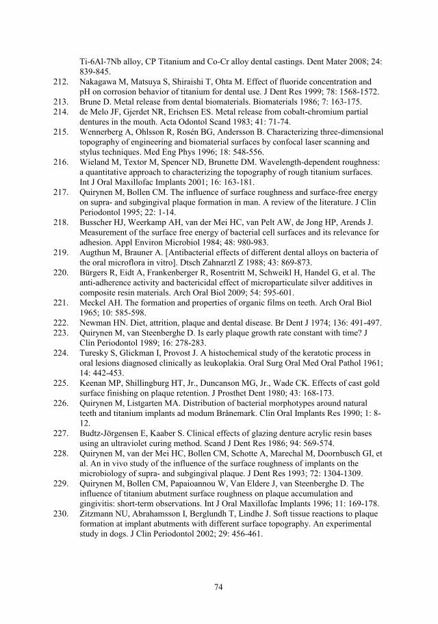
74
Ti-6Al-7Nb alloy, CP Titanium and Co-Cr alloy dental castings. Dent Mater 2008; 24: 839-845.
212. Nakagawa M, Matsuya S, Shiraishi T, Ohta M. Effect of fluoride concentration and pH on corrosion behavior of titanium for dental use. J Dent Res 1999; 78: 1568-1572.
213. Brune D. Metal release from dental biomaterials. Biomaterials 1986; 7: 163-175. 214. de Melo JF, Gjerdet NR, Erichsen ES. Metal release from cobalt-chromium partial
dentures in the mouth. Acta Odontol Scand 1983; 41: 71-74. 215. Wennerberg A, Ohlsson R, Rosén BG, Andersson B. Characterizing three-dimensional
topography of engineering and biomaterial surfaces by confocal laser scanning and stylus techniques. Med Eng Phys 1996; 18: 548-556.
216. Wieland M, Textor M, Spencer ND, Brunette DM. Wavelength-dependent roughness: a quantitative approach to characterizing the topography of rough titanium surfaces. Int J Oral Maxillofac Implants 2001; 16: 163-181.
217. Quirynen M, Bollen CM. The influence of surface roughness and surface-free energy on supra- and subgingival plaque formation in man. A review of the literature. J Clin Periodontol 1995; 22: 1-14.
218. Busscher HJ, Weerkamp AH, van der Mei HC, van Pelt AW, de Jong HP, Arends J. Measurement of the surface free energy of bacterial cell surfaces and its relevance for adhesion. Appl Environ Microbiol 1984; 48: 980-983.
219. Augthun M, Brauner A. [Antibacterial effects of different dental alloys on bacteria of the oral microflora in vitro]. Dtsch Zahnarztl Z 1988; 43: 869-873.
220. Bürgers R, Eidt A, Frankenberger R, Rosentritt M, Schweikl H, Handel G, et al. The anti-adherence activity and bactericidal effect of microparticulate silver additives in composite resin materials. Arch Oral Biol 2009; 54: 595-601.
221. Meckel AH. The formation and properties of organic films on teeth. Arch Oral Biol 1965; 10: 585-598.
222. Newman HN. Diet, attrition, plaque and dental disease. Br Dent J 1974; 136: 491-497. 223. Quirynen M, van Steenberghe D. Is early plaque growth rate constant with time? J
Clin Periodontol 1989; 16: 278-283. 224. Turesky S, Glickman I, Provost J. A histochemical study of the keratotic process in
oral lesions diagnosed clinically as leukoplakia. Oral Surg Oral Med Oral Pathol 1961; 14: 442-453.
225. Keenan MP, Shillingburg HT, Jr., Duncanson MG, Jr., Wade CK. Effects of cast gold surface finishing on plaque retention. J Prosthet Dent 1980; 43: 168-173.
226. Quirynen M, Listgarten MA. Distribution of bacterial morphotypes around natural teeth and titanium implants ad modum Brånemark. Clin Oral Implants Res 1990; 1: 8-12.
227. Budtz-Jörgensen E, Kaaber S. Clinical effects of glazing denture acrylic resin bases using an ultraviolet curing method. Scand J Dent Res 1986; 94: 569-574.
228. Quirynen M, van der Mei HC, Bollen CM, Schotte A, Marechal M, Doornbusch GI, et al. An in vivo study of the influence of the surface roughness of implants on the microbiology of supra- and subgingival plaque. J Dent Res 1993; 72: 1304-1309.
229. Quirynen M, Bollen CM, Papaioannou W, Van Eldere J, van Steenberghe D. The influence of titanium abutment surface roughness on plaque accumulation and gingivitis: short-term observations. Int J Oral Maxillofac Implants 1996; 11: 169-178.
230. Zitzmann NU, Abrahamsson I, Berglundh T, Lindhe J. Soft tissue reactions to plaque formation at implant abutments with different surface topography. An experimental study in dogs. J Clin Periodontol 2002; 29: 456-461.
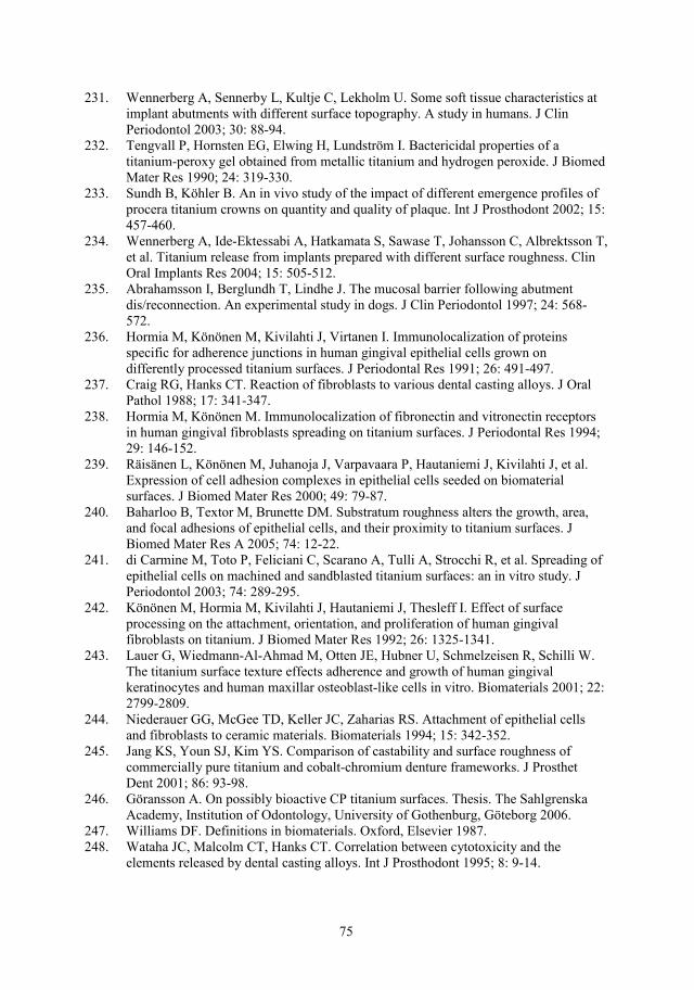
75
231. Wennerberg A, Sennerby L, Kultje C, Lekholm U. Some soft tissue characteristics at implant abutments with different surface topography. A study in humans. J Clin Periodontol 2003; 30: 88-94.
232. Tengvall P, Hornsten EG, Elwing H, Lundström I. Bactericidal properties of a titanium-peroxy gel obtained from metallic titanium and hydrogen peroxide. J Biomed Mater Res 1990; 24: 319-330.
233. Sundh B, Köhler B. An in vivo study of the impact of different emergence profiles of procera titanium crowns on quantity and quality of plaque. Int J Prosthodont 2002; 15: 457-460.
234. Wennerberg A, Ide-Ektessabi A, Hatkamata S, Sawase T, Johansson C, Albrektsson T, et al. Titanium release from implants prepared with different surface roughness. Clin Oral Implants Res 2004; 15: 505-512.
235. Abrahamsson I, Berglundh T, Lindhe J. The mucosal barrier following abutment dis/reconnection. An experimental study in dogs. J Clin Periodontol 1997; 24: 568-572.
236. Hormia M, Könönen M, Kivilahti J, Virtanen I. Immunolocalization of proteins specific for adherence junctions in human gingival epithelial cells grown on differently processed titanium surfaces. J Periodontal Res 1991; 26: 491-497.
237. Craig RG, Hanks CT. Reaction of fibroblasts to various dental casting alloys. J Oral Pathol 1988; 17: 341-347.
238. Hormia M, Könönen M. Immunolocalization of fibronectin and vitronectin receptors in human gingival fibroblasts spreading on titanium surfaces. J Periodontal Res 1994; 29: 146-152.
239. Räisänen L, Könönen M, Juhanoja J, Varpavaara P, Hautaniemi J, Kivilahti J, et al. Expression of cell adhesion complexes in epithelial cells seeded on biomaterial surfaces. J Biomed Mater Res 2000; 49: 79-87.
240. Baharloo B, Textor M, Brunette DM. Substratum roughness alters the growth, area, and focal adhesions of epithelial cells, and their proximity to titanium surfaces. J Biomed Mater Res A 2005; 74: 12-22.
241. di Carmine M, Toto P, Feliciani C, Scarano A, Tulli A, Strocchi R, et al. Spreading of epithelial cells on machined and sandblasted titanium surfaces: an in vitro study. J Periodontol 2003; 74: 289-295.
242. Könönen M, Hormia M, Kivilahti J, Hautaniemi J, Thesleff I. Effect of surface processing on the attachment, orientation, and proliferation of human gingival fibroblasts on titanium. J Biomed Mater Res 1992; 26: 1325-1341.
243. Lauer G, Wiedmann-Al-Ahmad M, Otten JE, Hubner U, Schmelzeisen R, Schilli W. The titanium surface texture effects adherence and growth of human gingival keratinocytes and human maxillar osteoblast-like cells in vitro. Biomaterials 2001; 22: 2799-2809.
244. Niederauer GG, McGee TD, Keller JC, Zaharias RS. Attachment of epithelial cells and fibroblasts to ceramic materials. Biomaterials 1994; 15: 342-352.
245. Jang KS, Youn SJ, Kim YS. Comparison of castability and surface roughness of commercially pure titanium and cobalt-chromium denture frameworks. J Prosthet Dent 2001; 86: 93-98.
246. Göransson A. On possibly bioactive CP titanium surfaces. Thesis. The Sahlgrenska Academy, Institution of Odontology, University of Gothenburg, Göteborg 2006.
247. Williams DF. Definitions in biomaterials. Oxford, Elsevier 1987. 248. Wataha JC, Malcolm CT, Hanks CT. Correlation between cytotoxicity and the
elements released by dental casting alloys. Int J Prosthodont 1995; 8: 9-14.
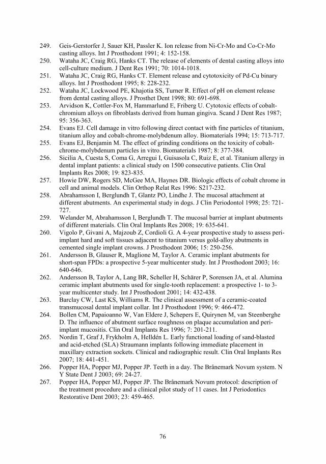
76
249. Geis-Gerstorfer J, Sauer KH, Passler K. Ion release from Ni-Cr-Mo and Co-Cr-Mo casting alloys. Int J Prosthodont 1991; 4: 152-158.
250. Wataha JC, Craig RG, Hanks CT. The release of elements of dental casting alloys into cell-culture medium. J Dent Res 1991; 70: 1014-1018.
251. Wataha JC, Craig RG, Hanks CT. Element release and cytotoxicity of Pd-Cu binary alloys. Int J Prosthodont 1995; 8: 228-232.
252. Wataha JC, Lockwood PE, Khajotia SS, Turner R. Effect of pH on element release from dental casting alloys. J Prosthet Dent 1998; 80: 691-698.
253. Arvidson K, Cottler-Fox M, Hammarlund E, Friberg U. Cytotoxic effects of cobalt-chromium alloys on fibroblasts derived from human gingiva. Scand J Dent Res 1987; 95: 356-363.
254. Evans EJ. Cell damage in vitro following direct contact with fine particles of titanium, titanium alloy and cobalt-chrome-molybdenum alloy. Biomaterials 1994; 15: 713-717.
255. Evans EJ, Benjamin M. The effect of grinding conditions on the toxicity of cobalt-chrome-molybdenum particles in vitro. Biomaterials 1987; 8: 377-384.
256. Sicilia A, Cuesta S, Coma G, Arregui I, Guisasola C, Ruiz E, et al. Titanium allergy in dental implant patients: a clinical study on 1500 consecutive patients. Clin Oral Implants Res 2008; 19: 823-835.
257. Howie DW, Rogers SD, McGee MA, Haynes DR. Biologic effects of cobalt chrome in cell and animal models. Clin Orthop Relat Res 1996: S217-232.
258. Abrahamsson I, Berglundh T, Glantz PO, Lindhe J. The mucosal attachment at different abutments. An experimental study in dogs. J Clin Periodontol 1998; 25: 721-727.
259. Welander M, Abrahamsson I, Berglundh T. The mucosal barrier at implant abutments of different materials. Clin Oral Implants Res 2008; 19: 635-641.
260. Vigolo P, Givani A, Majzoub Z, Cordioli G. A 4-year prospective study to assess peri-implant hard and soft tissues adjacent to titanium versus gold-alloy abutments in cemented single implant crowns. J Prosthodont 2006; 15: 250-256.
261. Andersson B, Glauser R, Maglione M, Taylor A. Ceramic implant abutments for short-span FPDs: a prospective 5-year multicenter study. Int J Prosthodont 2003; 16: 640-646.
262. Andersson B, Taylor A, Lang BR, Scheller H, Schärer P, Sorensen JA, et al. Alumina ceramic implant abutments used for single-tooth replacement: a prospective 1- to 3-year multicenter study. Int J Prosthodont 2001; 14: 432-438.
263. Barclay CW, Last KS, Williams R. The clinical assessment of a ceramic-coated transmucosal dental implant collar. Int J Prosthodont 1996; 9: 466-472.
264. Bollen CM, Papaioanno W, Van Eldere J, Schepers E, Quirynen M, van Steenberghe D. The influence of abutment surface roughness on plaque accumulation and peri-implant mucositis. Clin Oral Implants Res 1996; 7: 201-211.
265. Nordin T, Graf J, Frykholm A, Helldén L. Early functional loading of sand-blasted and acid-etched (SLA) Straumann implants following immediate placement in maxillary extraction sockets. Clinical and radiographic result. Clin Oral Implants Res 2007; 18: 441-451.
266. Popper HA, Popper MJ, Popper JP. Teeth in a day. The Brånemark Novum system. N Y State Dent J 2003; 69: 24-27.
267. Popper HA, Popper MJ, Popper JP. The Brånemark Novum protocol: description of the treatment procedure and a clinical pilot study of 11 cases. Int J Periodontics Restorative Dent 2003; 23: 459-465.
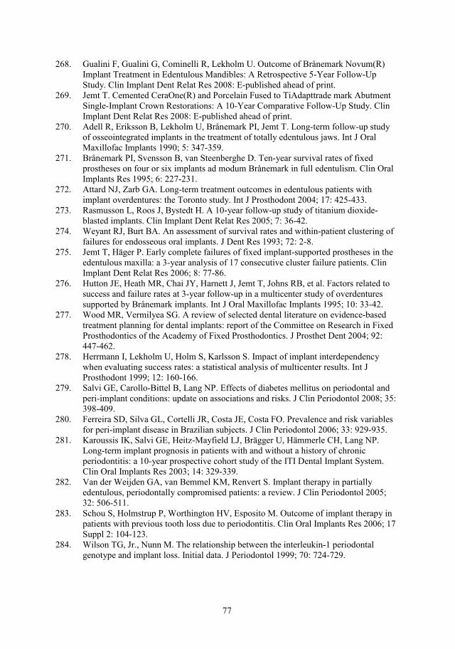
77
268. Gualini F, Gualini G, Cominelli R, Lekholm U. Outcome of Brånemark Novum(R) Implant Treatment in Edentulous Mandibles: A Retrospective 5-Year Follow-Up Study. Clin Implant Dent Relat Res 2008: E-published ahead of print.
269. Jemt T. Cemented CeraOne(R) and Porcelain Fused to TiAdapttrade mark Abutment Single-Implant Crown Restorations: A 10-Year Comparative Follow-Up Study. Clin Implant Dent Relat Res 2008: E-published ahead of print.
270. Adell R, Eriksson B, Lekholm U, Brånemark PI, Jemt T. Long-term follow-up study of osseointegrated implants in the treatment of totally edentulous jaws. Int J Oral Maxillofac Implants 1990; 5: 347-359.
271. Brånemark PI, Svensson B, van Steenberghe D. Ten-year survival rates of fixed prostheses on four or six implants ad modum Brånemark in full edentulism. Clin Oral Implants Res 1995; 6: 227-231.
272. Attard NJ, Zarb GA. Long-term treatment outcomes in edentulous patients with implant overdentures: the Toronto study. Int J Prosthodont 2004; 17: 425-433.
273. Rasmusson L, Roos J, Bystedt H. A 10-year follow-up study of titanium dioxide-blasted implants. Clin Implant Dent Relat Res 2005; 7: 36-42.
274. Weyant RJ, Burt BA. An assessment of survival rates and within-patient clustering of failures for endosseous oral implants. J Dent Res 1993; 72: 2-8.
275. Jemt T, Häger P. Early complete failures of fixed implant-supported prostheses in the edentulous maxilla: a 3-year analysis of 17 consecutive cluster failure patients. Clin Implant Dent Relat Res 2006; 8: 77-86.
276. Hutton JE, Heath MR, Chai JY, Harnett J, Jemt T, Johns RB, et al. Factors related to success and failure rates at 3-year follow-up in a multicenter study of overdentures supported by Brånemark implants. Int J Oral Maxillofac Implants 1995; 10: 33-42.
277. Wood MR, Vermilyea SG. A review of selected dental literature on evidence-based treatment planning for dental implants: report of the Committee on Research in Fixed Prosthodontics of the Academy of Fixed Prosthodontics. J Prosthet Dent 2004; 92: 447-462.
278. Herrmann I, Lekholm U, Holm S, Karlsson S. Impact of implant interdependency when evaluating success rates: a statistical analysis of multicenter results. Int J Prosthodont 1999; 12: 160-166.
279. Salvi GE, Carollo-Bittel B, Lang NP. Effects of diabetes mellitus on periodontal and peri-implant conditions: update on associations and risks. J Clin Periodontol 2008; 35: 398-409.
280. Ferreira SD, Silva GL, Cortelli JR, Costa JE, Costa FO. Prevalence and risk variables for peri-implant disease in Brazilian subjects. J Clin Periodontol 2006; 33: 929-935.
281. Karoussis IK, Salvi GE, Heitz-Mayfield LJ, Brägger U, Hämmerle CH, Lang NP. Long-term implant prognosis in patients with and without a history of chronic periodontitis: a 10-year prospective cohort study of the ITI Dental Implant System. Clin Oral Implants Res 2003; 14: 329-339.
282. Van der Weijden GA, van Bemmel KM, Renvert S. Implant therapy in partially edentulous, periodontally compromised patients: a review. J Clin Periodontol 2005; 32: 506-511.
283. Schou S, Holmstrup P, Worthington HV, Esposito M. Outcome of implant therapy in patients with previous tooth loss due to periodontitis. Clin Oral Implants Res 2006; 17 Suppl 2: 104-123.
284. Wilson TG, Jr., Nunn M. The relationship between the interleukin-1 periodontal genotype and implant loss. Initial data. J Periodontol 1999; 70: 724-729.
78
285. Hinode D, Tanabe S, Yokoyama M, Fujisawa K, Yamauchi E, Miyamoto Y. Influence of smoking on osseointegrated implant failure: a meta-analysis. Clin Oral Implants Res 2006; 17: 473-478.
286. Snauwaert K, Duyck J, van Steenberghe D, Quirynen M, Naert I. Time dependent failure rate and marginal bone loss of implant supported prostheses: a 15-year follow-up study. Clin Oral Investig 2000; 4: 13-20.
287. Roos-Jansåker AM, Lindahl C, Renvert H, Renvert S. Nine- to fourteen-year follow-up of implant treatment. Part I: implant loss and associations to various factors. J Clin Periodontol 2006; 33: 283-289.
288. Pikner SS, Gröndahl K, Jemt T, Friberg B. Marginal bone loss at implants: a retrospective, long-term follow-up of turned Brånemark System implants. Clin Implant Dent Relat Res 2009; 11: 11-23.
289. Strietzel FP, Lange KP, Svegar M, Hartmann HJ, Kuchler I. Retrospective evaluation of the success of oral rehabilitation using the Frialit-2 implant system. Part 1: Influence of topographic and surgical parameters. Int J Prosthodont 2004; 17: 187-194.
290. Jemt T, Albrektsson T. Do long-term followed-up Brånemark implants commonly show evidence of pathological bone breakdown? A review based on recently published data. Periodontol 2000 2008; 47: 133-142.
291. Fransson C, Lekholm U, Jemt T, Berglundh T. Prevalence of subjects with progressive bone loss at implants. Clin Oral Implants Res 2005; 16: 440-446.
292. Albrektsson T, Sennerby L, Wennerberg A. State of the art of oral implants. Periodontol 2000 2008; 47: 15-26.
293. Göthberg C, Bergendal T, Magnusson T. Complications after treatment with implant-supported fixed prostheses: a retrospective study. Int J Prosthodont 2003; 16: 201-207.
294. Örtorp A, Lindén B, Jemt T. Clinical experiences with laser-welded titanium frameworks supported by implants in the edentulous mandible: a 5-year follow-up study. Int J Prosthodont 1999; 12: 65-72.
295. Lindquist LW, Carlsson GE, Jemt T. A prospective 15-year follow-up study of mandibular fixed prostheses supported by osseointegrated implants. Clinical results and marginal bone loss. Clin Oral Implants Res 1996; 7: 329-336.
296. Goodacre CJ, Kan JY, Rungcharassaeng K. Clinical complications of osseointegrated implants. J Prosthet Dent 1999; 81: 537-552.
297. Zarb GA, Schmitt A. The longitudinal clinical effectiveness of osseointegrated dental implants: the Toronto study. Part III: Problems and complications encountered. J Prosthet Dent 1990; 64: 185-194.
298. de Grandmont P, Feine JS, Tache R, Boudrias P, Donohue WB, Tanguay R, et al. Within-subject comparisons of implant-supported mandibular prostheses: psychometric evaluation. J Dent Res 1994; 73: 1096-1104.
299. Feine JS, de Grandmont P, Boudrias P, Brien N, LaMarche C, Tache R, et al. Within-subject comparisons of implant-supported mandibular prostheses: choice of prosthesis. J Dent Res 1994; 73: 1105-1111.
300. Köndell PÅ, Landt H, Nordenram Å, Carlsson B, Danielsson K. The tissue-integrated prosthesis in the treatment of edentulous patients. A follow-up study. Swed Dent J 1988; 12: 11-16.
301. Lundqvist S. Speech and other oral functions. Clinical and experimental studies with special reference to maxillary rehabilitation on osseointegrated implants. Swed Dent J Suppl 1993; 91: 1-39.
79
302. Lundqvist S, Haraldson T. Oral function in patients wearing fixed prosthesis on osseointegrated implants in the maxilla: 3-year follow-up study. Scand J Dent Res 1992; 100: 279-283.
303. Lundqvist S, Lohmander-Agerskov A, Haraldson T. Speech before and after treatment with bridges on osseointegrated implants in the edentulous upper jaw. Clin Oral Implants Res 1992; 3: 57-62.
304. Adell R, Lekholm U, Brånemark PI, Lindhe J, Rockler B, Eriksson B, et al. Marginal tissue reactions at osseointegrated titanium fixtures. Swed Dent J Suppl 1985; 28: 175-181.
305. Albrektsson T, Zarb G, Worthington P, Eriksson AR. The long-term efficacy of currently used dental implants: a review and proposed criteria of success. Int J Oral Maxillofac Implants 1986; 1: 11-25.
306. Schulte W, d'Hoedt B, Lukas D, Muhlbradt L, Scholz F, Bretschi J, et al. [Periotest--a new measurement process for periodontal function]. Zahnarztl Mitt 1983; 73: 1229-1230, 1233-1226, 1239-1240.
307. Lang NP, Berglundh T, Heitz-Mayfield LJ, Pjétursson BE, Salvi GE, Sanz M. Consensus statements and recommended clinical procedures regarding implant survival and complications. Int J Oral Maxillofac Implants 2004; 19 Suppl: 150-154.
308. Friberg B, Sennerby L, Meredith N, Lekholm U. A comparison between cutting torque and resonance frequency measurements of maxillary implants. A 20-month clinical study. Int J Oral Maxillofac Surg 1999; 28: 297-303.
309. Glauser R, Sennerby L, Meredith N, Ree A, Lundgren A, Gottlow J, et al. Resonance frequency analysis of implants subjected to immediate or early functional occlusal loading. Successful vs. failing implants. Clin Oral Implants Res 2004; 15: 428-434.
310. Östman PO, Hellman M, Sennerby L. Direct implant loading in the edentulous maxilla using a bone density-adapted surgical protocol and primary implant stability criteria for inclusion. Clin Implant Dent Relat Res 2005; 7 Suppl 1: S60-69.
311. Östman PO, Hellman M, Wendelhag I, Sennerby L. Resonance frequency analysis measurements of implants at placement surgery. Int J Prosthodont 2006; 19: 77-83; discussion 84.
312. Huwiler MA, Pjétursson BE, Bosshardt DD, Salvi GE, Lang NP. Resonance frequency analysis in relation to jawbone characteristics and during early healing of implant installation. Clin Oral Implants Res 2007; 18: 275-280.
313. Listgarten MA, Lang NP, Schroeder HE, Schroeder A. Periodontal tissues and their counterparts around endosseous implants [corrected and republished with original paging, article orginally printed in Clin Oral Implants Res 1991 Jan-Mar;2(1):1-19]. Clin Oral Implants Res 1991; 2: 1-19.
314. Albrektsson T, Brunski J, Wennerberg A. 'A requiem for the periodontal ligament' revisited. Int J Prosthodont 2009; 22: 120-122.
315. Mombelli A, Mühle T, Brägger U, Lang NP, Bürgin WB. Comparison of periodontal and peri-implant probing by depth-force pattern analysis. Clin Oral Implants Res 1997; 8: 448-454.
316. Cardaropoli G, Lekholm U, Wennström JL. Tissue alterations at implant-supported single-tooth replacements: a 1-year prospective clinical study. Clin Oral Implants Res 2006; 17: 165-171.
317. Lang NP, Hill RW. Radiographs in periodontics. J Clin Periodontol 1977; 4: 16-28. 318. Gröndahl K, Sundén S, Gröndahl HG. Inter- and intraobserver variability in
radiographic bone level assessment at Branemark fixtures. Clin Oral Implants Res 1998; 9: 243-250.
80
319. Sundén S, Gröndahl K, Gröndahl HG. Accuracy and precision in the radiographic diagnosis of clinical instability in Branemark dental implants. Clin Oral Implants Res 1995; 6: 220-226.
320. Goldman M, Pearson AH, Darzenta N. Endodontic success--who's reading the radiograph? Oral Surg Oral Med Oral Pathol 1972; 33: 432-437.
321. Goldman M, Pearson AH, Darzenta N. Reliability of radiographic interpretations. Oral Surg Oral Med Oral Pathol 1974; 38: 287-293.
322. Brägger U. Radiographic parameters for the evaluation of peri-implant tissues. Periodontol 2000 1994; 4: 87-97.
323. Sewerin IP. Errors in radiographic assessment of marginal bone height around osseointegrated implants. Scand J Dent Res 1990; 98: 428-433.
324. Pikner SS, Gröndahl K. Radiographic analyses of "advanced" marginal bone loss around Brånemark dental implants. Clin Implant Dent Relat Res 2009; 11: 120-133.
325. Lang NP, Pjétursson BE, Tan K, Brägger U, Egger M, Zwahlen M. A systematic review of the survival and complication rates of fixed partial dentures (FPDs) after an observation period of at least 5 years. II. Combined tooth--implant-supported FPDs. Clin Oral Implants Res 2004; 15: 643-653.
326. Bühler WK. The method of least squares. In: Gauss: A Biographical Study. Volume 1981: 138-141. Berlin, Springer 1981.
327. Eliasson A. On the role of number of fixtures, surgical technique and timing of loading. Thesis. The Sahlgrenska Academy, Institution of Odontology, University of Gothenburg, Göteborg 2008.
328. Söderberg R, Lindkvist L, Carlson JS. Managing physical dependencies through location system design J Eng Design 2006; 17: 325-346.
329. Ansar Ahmed S, Gogal RM, Jr., Walsh JE. A new rapid and simple non-radioactive assay to monitor and determine the proliferation of lymphocytes: an alternative to [3H]thymidine incorporation assay. J Immunol Methods 1994; 170: 211-224.
330. Adell R, Lekholm U, Brånemark P. Surgical procedures. In: Brånemark P-I, Zarb G.A., Albrektsson T, eds. Tissue-integrated prostheses: osseointegration in clinical dentistry. Chicago, Illinois, Quintessence 1985.
331. Buser D, Weber HP, Brägger U. The treatment of partially edentulous patients with ITI hollow-screw implants: presurgical evaluation and surgical procedures. Int J Oral Maxillofac Implants 1990; 5: 165-175.
332. Hollender L, Rockler B. Radiographic evaluation of osseointegrated implants of the jaws. Experimental study of the influence of radiographic techniques on the measurement of the relation between the implant and bone. Dentomaxillofac Radiol 1980; 9: 91-95.
333. Strid KC. Radiographic results and radiographic procedures. In: Brånemnark P-I, Zarb G.A., Albrektsson T, eds. Tissue-integrated prostheses: osseointegration in clinical dentistry. Volume 187-198, 317-327. Chicago, Quintessence 1985.
334. Örtorp A, Jemt T. Clinical experiences with laser-welded titanium frameworks supported by implants in the edentulous mandible: a 10-year follow-up study. Clin Implant Dent Relat Res 2006; 8: 198-209.
335. Esposito M, Hirsch JM, Lekholm U, Thomsen P. Biological factors contributing to failures of osseointegrated oral implants. (I). Success criteria and epidemiology. Eur J Oral Sci 1998; 106: 527-551.
336. Altman GA. Practical Statistics for Medical Research. London, Chapman & Hall 1999.
337. Good P. Permutation Tests. A Practical Guide to Resampling Methods for Testing Hypotheses. New York, Springer 2000.
81
338. Roos-Jansåker AM, Lindahl C, Renvert H, Renvert S. Nine- to fourteen-year follow-up of implant treatment. Part II: presence of peri-implant lesions. J Clin Periodontol 2006; 33: 290-295.
339. Klineberg IJ, Murray GM. Design of superstructures for osseointegrated fixtures. Swed Dent J Suppl 1985; 28: 63-69.
340. Becker JS, Jakubowski N. The synergy of elemental and biomolecular mass spectrometry: new analytical strategies in life sciences. Chem Soc Rev 2009; 38: 1969-1983.
341. Khan MA, Williams RL, Williams DF. The corrosion behaviour of Ti-6Al-4V, Ti-6Al-7Nb and Ti-13Nb-13Zr in protein solutions. Biomaterials 1999; 20: 631-637.
342. Schmalz G, Langer H, Schweikl H. Cytotoxicity of dental alloy extracts and corresponding metal salt solutions. J Dent Res 1998; 77: 1772-1778.
343. Clark GC, Williams DF. The effects of proteins on metallic corrosion. J Biomed Mater Res 1982; 16: 125-134.
344. Milleding P, Carlén A, Wennerberg A, Karlsson S. Protein characterisation of salivary and plasma biofilms formed in vitro on non-corroded and corroded dental ceramic materials. Biomaterials 2001; 22: 2545-2555.
345. Göransson A, Gretzer C, Johansson A, Sul YT, Wennerberg A. Inflammatory response to a titanium surface with potential bioactive properties: an in vitro study. Clin Implant Dent Relat Res 2006; 8: 210-217.
346. Göransson A, Gretzer C, Tengvall P, Wennerberg A. Inflammatory response to titanium surfaces with fibrinogen and catalase coatings: an in vitro study. J Biomed Mater Res A 2007; 80: 693-699.
347. Craig RG, Hanks CT. Cytotoxicity of experimental casting alloys evaluated by cell culture tests. J Dent Res 1990; 69: 1539-1542.
348. Abrahamsson I, Zitzmann NU, Berglundh T, Linder E, Wennerberg A, Lindhe J. The mucosal attachment to titanium implants with different surface characteristics: an experimental study in dogs. J Clin Periodontol 2002; 29: 448-455.
349. Glauser R, Schupbach P, Gottlow J, Hämmerle CH. Periimplant soft tissue barrier at experimental one-piece mini-implants with different surface topography in humans: A light-microscopic overview and histometric analysis. Clin Implant Dent Relat Res 2005; 7 Suppl 1: S44-51.
350. Klinge B, Meyle J. Soft-tissue integration of implants. Consensus report of Working Group 2. Clin Oral Implants Res 2006; 17 Suppl 2: 93-96.
351. Rompen E, Domken O, Degidi M, Pontes AE, Piattelli A. The effect of material characteristics, of surface topography and of implant components and connections on soft tissue integration: a literature review. Clin Oral Implants Res 2006; 17 Suppl 2: 55-67.
352. Messer RL, Lucas LC. Cytotoxicity of nickel-chromium alloys: bulk alloys compared to multiple ion salt solutions. Dent Mater 2000; 16: 207-212.
353. Browne RM, Tyas MJ. Biological testing of dental restorative materials in vitro--a review. J Oral Rehabil 1979; 6: 365-374.
354. Pizzoferrato A, Vespucci A, Ciapetti G, Stea S. Biocompatibility testing of prosthetic implant materials by cell cultures. Biomaterials 1985; 6: 346-351.
355. Jemt T. Fixed implant-supported prostheses in the edentulous maxilla. A five-year follow-up report. Clin Oral Implants Res 1994; 5: 142-147.
356. Roos J, Sennerby L, Lekholm U, Jemt T, Gröndahl K, Albrektsson T. A qualitative and quantitative method for evaluating implant success: a 5-year retrospective analysis of the Brånemark implant. Int J Oral Maxillofac Implants 1997; 12: 504-514.
82
357. Bergendal B, Palmqvist S. Laser-welded titanium frameworks for implant-supported fixed prostheses: a 5-year report. Int J Oral Maxillofac Implants 1999; 14: 69-71.
358. Eliasson A, Palmqvist S, Svenson B, Sondell K. Five-year results with fixed complete-arch mandibular prostheses supported by 4 implants. Int J Oral Maxillofac Implants 2000; 15: 505-510.
359. Mombelli A, Cionca N. Systemic diseases affecting osseointegration therapy. Clin Oral Implants Res 2006; 17 Suppl 2: 97-103.
360. Attard NJ, Zarb GA. Long-term treatment outcomes in edentulous patients with implant-fixed prostheses: the Toronto study. Int J Prosthodont 2004; 17: 417-424.
361. Bain CA. Smoking and implant failure--benefits of a smoking cessation protocol. Int J Oral Maxillofac Implants 1996; 11: 756-759.
362. Bain CA, Moy PK. The association between the failure of dental implants and cigarette smoking. Int J Oral Maxillofac Implants 1993; 8: 609-615.
363. Lambert PM, Morris HF, Ochi S. The influence of smoking on 3-year clinical success of osseointegrated dental implants. Ann Periodontol 2000; 5: 79-89.
364. Wallace RH. The relationship between cigarette smoking and dental implant failure. Eur J Prosthodont Restor Dent 2000; 8: 103-106.
365. Albrektsson T, Isidor F. Consensus report of session IV. In: Lang NP, Karring T, eds. Proceedings of the 1st European workshop on periodontology. London, Quintessence 1994.
366. Weibrich G, Buch RS, Wegener J, Wagner W. Five-year prospective follow-up report of the Astra tech standard dental implant in clinical treatment. Int J Oral Maxillofac Implants 2001; 16: 557-562.
367. Salvi GE, Lang NP. Diagnostic parameters for monitoring peri-implant conditions. Int J Oral Maxillofac Implants 2004; 19 Suppl: 116-127.
368. Lindh T. Should we extract teeth to avoid tooth-implant combinations? J Oral Rehabil 2008; 35 Suppl 1: 44-54.
369. Brägger U, Pasquali L, Rylander H, Carnes D, Kornman KS. Computer-assisted densitometric image analysis in periodontal radiography. A methodological study. J Clin Periodontol 1988; 15: 27-37.
370. Smith DE, Zarb GA. Criteria for success of osseointegrated endosseous implants. J Prosthet Dent 1989; 62: 567-572.
371. Östman PO, Hellman M, Sennerby L. Immediate occlusal loading of implants in the partially edentate mandible: a prospective 1-year radiographic and 4-year clinical study. Int J Oral Maxillofac Implants 2008; 23: 315-322.
372. Schnitman PA, Shulman LB. Recommendations of the consensus development conference on dental implants. J Am Dent Assoc 1979; 98: 373-377.
373. Pikner SS. Radiographic follow-up analysis of Brånemark dental implants. Thesis. The Sahlgrenska Academy, Institution of Odontology, University of Gothenburg, Göteborg 2008.
374. Wennström JL, Papapanou PN, Gröndahl K. A model for decision making regarding periodontal treatment needs. J Clin Periodontol 1990; 17: 217-222.
375. Eliasson A, Eriksson T, Johansson A, Wennerberg A. Fixed partial prostheses supported by 2 or 3 implants: a retrospective study up to 18 years. Int J Oral Maxillofac Implants 2006; 21: 567-574.
376. Eliasson A, Blomqvist F, Wennerberg A, Johansson A. A retrospective analysis of early and delayed loading of full-arch mandibular prostheses using three different implant systems: clinical results with up to 5 years of loading. Clin Implant Dent Relat Res 2009; 11: 134-148.

83
377. Carlson B, Carlsson GE. Prosthodontic complications in osseointegrated dental implant treatment. Int J Oral Maxillofac Implants 1994; 9: 90-94.
378. Davis DM, Packer ME, Watson RM. Maintenance requirements of implant-supported fixed prostheses opposed by implant-supported fixed prostheses, natural teeth, or complete dentures: a 5-year retrospective study. Int J Prosthodont 2003; 16: 521-523.
379. Watzek G. Oral implants--quo vadis? Int J Oral Maxillofac Implants 2006; 21: 831-832.
380. Kiene H, von Schon-Angerer T. Single-case causality assessment as a basis for clinical judgment. Altern Ther Health Med 1998; 4: 41-47.
381. Walther W. On diverse approaches to prosthodontic research: the case series approach to clinical research. Int J Prosthodont 2007; 20: 374-375.
382. Jokstad A. The evidence-based approach to prosthodontic practice and research. Int J Prosthodont 2007; 20: 376-377.
383. Norman G. Research in clinical reasoning: past history and current trends. Med Educ 2005; 39: 418-427.
384. MacEntee MI. Where science fails prosthodontics. Int J Prosthodont 2007; 20: 377-381.