Academy of General Dentistry
132
GERIATRIC DENTISTRY n IMPLANTS DIAGNOSIS & TREATMENT PLANNING ORAL DIAGNOSIS n OPERATIVE DENTISTRY ORAL AND MAXILLOFACIAL SURGERY DIGITAL RADIOGRAPHY n WWW.AGD.ORG March/April 2010 ~ Volume 58 Number 2 Peer-Reviewed Journal of the Academy of General Dentistry GENERAL DENTISTRY
-
Upload
khangminh22 -
Category
Documents
-
view
3 -
download
0
Transcript of Academy of General Dentistry

Geriatric Dentistry n implantsDiaGnosis & treatment planninGoral DiaGnosis n operative Dentistryoral anD maxillofacial surGeryDiGital raDioGraphy n www.agd.org
march/april 2010 ~ volume 58 number 2
peer-reviewed Journal of the academy of General Dentistry
GENERALDENTISTRY

SAVOR THE
AGD 2010 Annual Meeting & Exhibits July 6 to 8, 2010: aGD house of DelegatesJuly 8 to 11, 2010: aGD annual meeting & exhibits
visit the aGD Web site for more information at www.agd.org/neworleans.
For your convenience, registration is available at www.agd.org/neworleans. Participants can register online on our web site or by downloading the registration brochure and faxing the registration form with their credit card information to 312.440.0513 or by mail to academy of general dentistry, annual Meeting, 38943 Eagle way, Chicago, Illinois 60678-1389. on-site registration will be available as well. See you in New orleans!
Register Today! register now for the academy of general dentistry (agd) 2010 annual Meeting & Exhibits, July 8 to 11, and the agd House of delegates, July 6 to 8, and prepare to savor the flavors of New orleans! Visit www.agd.org/neworleans and click on “register Now” to sign up today. The registration brochure is available for download exclusively on the agd 2010 annual Meeting & Exhibits web site. This registration program is simply too hot to mail!

Departments
86 EditorialLife’s challenges
87 PharmacologyCardioprotective aspirin—Update on three previous special alerts
91 RestorativeDentistryIntracoronal cast gold restorations
94 DentalMaterialsImpression materials—Are there any really new ones?
150 OralDiagnosisPersistent tongue ulcer and Diffuse oral mucosal pigmentations
152 Self-Assessment166Differential diagnosis of orofacial pain
153 AnswersOral Diagnosis, Self-Assessment quiz No. 166, and Self-Instruction exercises No. 231, 232, and 233
157 2009Reviewers
Clinical articles
97 DigitalRadiographyAsymptomatic carotid artery calcifications discovered on panoramic radiographymohammad ali Dolatabadi, DDs mohammad hosein Kalantar motamedi, DDs eshagh lassemi, DDs yousef Janbaz, DDs
100 GeriatricDentistryMedication use in geriatric populations: Dental implications of frequently prescribed medicationsmary a. aubertin, DmD carlton horbelt, DDs Waletha Wasson, DDs marjorie Woods, DDs
110 Diagnosis&TreatmentPlanningMethods for analyzing saliva proteins for systemic disease detectiontara luther, Ba, ms carlos f. carrion, Bs, ms nicholas cobb, Bs Giao le, Bs cynthia edwards, Bs stephen schwartz, DDs, ms charles streckfus, DDs, ma
114 AnesthesiaandPainControlDentistry’s wonder drugs: Local anesthetics and vasoconstrictorsmichael J. Wahl, DDs ronald s. Brown, DDs, ms
126 OralDiagnosisA novel, minimally invasive approach to managing mild epithelial dysplasiaKevin D. huff, DDs, maGD Kurt c. Garren, mD marlene s. huff, rn, msn, phD
www.agd.org General Dentistry March/April 2010 81
Contents

130 OperativeDentistryResistance of composite and amalgam core foundations retained with and without pins and bonding agents Terence A. Imbery, DDS Ryan Swigert Brian Richman Vincent Sawicki, DDS, PhD Lauren Pace Peter C. Moon, PhD
140 Implants Immediate provisional restoration fabrication or immediate implant loading using a modified technique: A clinical reportM. Erhan Comlekoglu, DDS, PhD A. Yucel Parlar, DDS Bulent Gokce, DDS,PhD Mine Dundar, DDS, PhD Erdem Kaya, DDS Tayfun Gunbay, DDS, PhD
144 OralandMaxillofacialSurgeryCriteria for performing extraction in the treatment of certain malocclusionsFarhad Moshiri, DMD, MS
General Dentistry onlineMembers and subscribers can read the full text of these articles online. Visit us at www.agd.org/publications/gd
e52 DentalMaterialsEvaluation of shear bond strength between self-etching adhesive systems and dentin and analysis of the resin-dentin interface Cleonice Silveira Teixeira, DDS, MSc, PhD Marcelo Carvalho Chain, DDS, MSc, PhD
e62 AnesthesiaandPainControlElusive dental painStanley Markman, DDS, FAGD Junad Khan, BDS Jennifer Howard, RDH
e68 PeriodonticsPhotodynamic therapy in periodontal therapy: Microbiological observations from a private practiceGeorgios E. Romanos, DDS, Dr.med.dent, PhD Birgit Brink, DMD, Dr.med.dent
e74 EstheticsChanges in surface morphology and mineralization level of human enamel following in-office bleaching with 35% hydrogen peroxide and light irradiation Sandrine Bittencourt Berger, DDS, MSc Vanessa Cavalli, DDS, MSc, PhD Glaucia Maria Bovi Ambrosano, PhD Marcelo Giannini, DDS, MSc, PhD
82 March/April 2010 General Dentistry www.agd.org

Continuing Dental Education OpportunitiesEarn two hours of CDE credit by signing up and completing these exercises based on various subjects.
108 Self-InstructionExerciseNo.255Geriatric Dentistry
124 Self-InstructionExerciseNo.256Anesthesia & Pain Control
138 Self-InstructionExerciseNo.257Operative Dentistry
CDE2 HOURSCREDIT
self instructione80 EndodonticsClinical importance of the presence of lateral canals in endodontics carina folgearini silveira, Ds Josue martos, DDs, phD Joao Batista cesar neto, DDs, mDs, phD carmen maria ferrer-luque, DDs, mDs, phD luiz fernando m. silveira, DDs, mDs, phD
e84 OperativeDentistryInfluence of cavity design and restorative material on the fracture resistance of maxillary premolars Gloria Beatriz de azevedo cubas, DDs Guilherme Briao camacho, DDs, msc, phD tatiana pereira-cenci, DDs, msc, phD tomio nonaka, DDs, msc, phD eduardo luiz Barbin, DDs, msc, phD
e89 OcclusalAdjustment/TherapyProsthetic rehabilitation of hemimandibular hyperplasia: Five-year follow-up Bulent Gokce, DDs, phD Birgul ozpınar, DDs, phD, prof.Dr. levent ozgur, DDs m. erhan comlekoglu, DDs, phD akın aladag, DDs, phD Gulcan coskun akar, DDs, phD
e94 DentalMaterialsFactors influencing the microhardness of a microhybrid compositemarcos Britto correa, DDs, ms sandrina henn, DDs, ms Jose laurindo machado marimon, DDs sinval adalberto rodrigues Jr., DDs, ms, phD flavio fernando Demarco, DDs, phD
www.agd.org General Dentistry March/April 2010 83
Instructions for AuthorsFor an electronic copy of General Dentistry’s Instructions for Authors, please visit the journal’s Web site at www.agd.org/publications/GD/AuthorInfo
Coming Next IssueIn the May/June issue of General Dentistry• A special section on
pediatric dentistry• Implications of drug
dependence on dental patient management
• Enamel hardness after exposure to acidic drinks and brushing
In the April issue of AGD Impact• Online reputation
management• Sports dentistry • AGD candidate
biographies

General Dentistrye-mail: [email protected]: 312.335.3442
BackIssuesandChangeofAddressmembers, call 888.aGD.Dent (toll-free) and ask for a member services representative. nonmembers, call salithia Graham (ext. 4097).
Reprintsto order reprints of any article in General Dentistry, contact morene stark at foster printing service (866.879.9144, ext. 119) or e-mail your request to [email protected].
all materials subject to copying and appearing in General Dentistry may be photocopied for the noncommercial purposes of scientific or educational advancement. reproduction of any portion of General Dentistry for commercial purposes is strictly prohibited unless the publisher’s written permission is obtained.
Mediakitandeditorialcalendarcall megan timmons (ext. 4355) or fax your request to 312.335.3441.
Disclaimerthe aGD does not necessarily endorse opinions or statements contained in essays or editorials published in General Dentistry. the publication of advertisements in General Dentistry does not indicate endorsement for products and services. aGD approval for continuing education courses or course sponsors will be clearly stated.
General Dentistry (issn 0363-6771) is published bimonthly (every other month) by the aGD, 211 e. chicago ave., suite 900, chicago, il 60611-1999. aGD members receive General Dentistry as part of membership.
periodicals postage paid at chicago, il and additional mailing office. postmaster: send address changes to General Dentistry, 211 e. chicago ave., suite 900, chicago, il 60611-1999. e-mail: [email protected].
canadian mailing information: ipm agreement number 40047941. change of address or undeliverable copies should be sent to: station a, p.o. Box 54, Windsor, ontario, n9a 6J5, canada. e-mail: [email protected].
the nonmember individual subscription rate for General Dentistry is $100 for the print version, $100 for the online version, and $175 for print and online versions; the nonmember institution rate is $300 (add $20 for canada and $50 for outside the u.s. and canada). single copies of General Dentistry are available to nonmember individuals for $15 and nonmember institutions for $18 (add $3 for orders outside the u.s.).
© copyright 2010 by the academy of General Dentistry. all rights reserved.
Academy of General Dentistryadvancing the value and excellence of general dentistry211 east chicago avenue, suite 900, chicago, il 60611-1999, usa
monday through friday, 8:30 am to 4:30 pm, central timetelephone 888.aGD.Dent (toll-free) or 312.440.4300www.agd.org
printed in u.s.a.
Editorroger D. Winland, DDs, ms, maGD
AssociateEditorpeter G. sturm, DDs, maGD
ExecutiveEditorcathy mcnamara fitzgerald
ManagingEditorchris Zayner
EditorialAssociatesteve Darnall
AcquisitionsEditorryan Boyle
Self-InstructionCoordinatorcassandra Bannon
CirculationCoordinatorsalithia Graham
Manager,Production/Designtimothy J. henney
AssociateDesignerJason thomas
PublicationsReviewCouncilnorman D. magnuson, DDs, faGD, chairWilliam e. chesser, DmD, maGDJon l. hardinger, DDs, maGD
Advertisingm.J. mrvica associates2 West taunton ave.Berlin, nJ [email protected]
84 March/April 2010 General Dentistry www.agd.org
Academy of General Dentistry Corporate Sponsors

www.agd.org General Dentistry March/April 2010 85
Advisory BoardDental materialsSteveCarstensen,DDS,FAGD, is in general practice in Bellevue, Washington. he is a visiting faculty member of the pankey institute and the pride institute and is a Diplomate of the american Board of Dental sleep medicine.
Dental public healthLarryWilliams,DDS,ABGD,MAGD, is a captain in the united states navy Dental corps, currently stationed at the Great lakes naval training center as a member of the Great lakes naval health clinic. capt Williams is the public health emergency officer for the 16 states of the navy region midWest and for the naval health clinic. he also serves as the co-chair of the navy’s tobacco cessation action team and as a member of the Department of Defense alcohol and tobacco advisory council. he also teaches for the Dental hygiene program at the college of lake county and as an assistant clinical professor for the rosalind franklin university for medicine and science.
esthetic DentistryWynnH.Okuda,DMD, is past national president (2002–03) and a board-accredited member of the american academy of cosmetic Dentistry (aacD). he also is on the advisory Board of Best Dentists in america and on the executive council of the international federation of esthetic Dentistry (ifeD). he practices cosmetic, implant, and restorative dentistry at the Dental Day spa of hawaii in honolulu.
endodonticsStephenCohen,DDS, is one of the foremost endodontic clinicians in the country and lectures worldwide on endodontics. a board-certified endodontist, Dr. cohen specializes exclusively in the diagnosis and treatment of endodontic infections.
Geriatric DentistryLeaErickson,DDS,MSPH, is chief, Dental service, at va salt lake city health care system and clinical assistant professor, university of utah in salt lake city.
implantologyWesleyBlakeslee,DMD,FAGD, is a general dentist who practices in new Jersey. he is a Diplomate of both the american Board of oral implantology/implant Dentistry and the international congress of oral implantologists, and an associate fellow of the american academy of implant Dentistry.
oral and maxillofacial pathologyJohnSvirsky,DDS,MEd, is a board-certified oral and maxillofacial pathologist at virginia commonwealth university in richmond. he currently is a professor of oral and maxillofacial pathology and maintains a private practice in oral medicine and oral pathology.
oral and maxillofacial radiologyKavasThunthy,BDS,MS,MEd, has been a professor of oral and maxillofacial radiology in the Department of oral Diagnosis, medicine, radiology, at the louisiana state university school of Dentistry in new orleans since 1975. he was named a fellow in the american academy of oral and maxillofacial radiology in 1978 and was board certified by the american Board of oral and maxillofacial radiology in 1981.
oral and maxillofacial surgeryKarlKoerner,DDS,FAGD, is a general dentist in utah who performs oral surgery exclusively. he lectures extensively on oral surgery in general practice and has made articles, books, and video presentations available to general practitioners.
oral medicineSook-BinWoo,DMD,MMSc, is assistant professor of oral medicine, infection and immunity at harvard school of Dental medicine. she is board certified in oral and maxillofacial pathology and oral medicine and practices both specialities in the Boston/cambridge area in massachusetts.
orthodonticsYoshJefferson,DMD,FAGD, is past president, international association for orthodontics; a fellow of the american academy of craniofacial pain; and a member of the american academy of Dental sleep medicine. he maintains a general practice in mt. holly, new Jersey.
pain managementHenryA.Gremillion,DDS,MAGD, is dean of the louisiana state university school of Dentistry and a professor in the Department of orthodontics at lsusD.
pediatricsJaneSoxman,DDS, is a Diplomate of the american Board of pediatric Dentistry, has authored numerous articles in the dental literature, and has been recognized as a leader in continuing education. she maintains a private practice in allison park, pennsylvania.
periodonticsSebastianCiancio,DDS, is Distinguished service professor and chair, Department of periodontics and endodontics, adjunct professor of pharmacology, and Director of the center for Dental studies at the university at Buffalo, state university of new york.
pharmacologyDanielE.Myers,DDS,MS, is a member of the oral Diagnosis Department, Dental associates of Wisconsin, ltd. in Wauwatosa.
prosthodonticsJosephMassad,DDS, is currently the Director of removable prosthodontics at the scottsdale center for Dentistry in arizona. he is adjunct faculty at tufts university school of Dental medicine in Boston and the university of texas Dental school at san antonio.
JackPiermatti,DMD, is a Diplomate of the american Board of prosthodontics, the american Board of oral implantology, and the international congress of oral implantologists. he is a board-certified prosthodontist in private practice in voorhees, new Jersey.

most of life’s lessons are learned the hard way, and I
make this statement from personal experience. All of us seek to combine happi-ness with self-esteem, and our life experiences shape that pursuit every day. The key to being happy is balancing the internal scale that measures positive and negative feedback and reactions. Every dentist wants to lead a full life, complete with loving relationships, satisfying work, and an overall sense of happiness. With our world in an increasingly violent, sad, stressful, and fast-paced state, happiness is often hard-won and can be even harder to hold onto. We need to find a way to deal with the triumphs, tragedies, stress, and changes in our lives.
The most important internal coping skill we can develop is our own self-esteem. We are all born with-out judgment; however, as we grow, the world tends to shuttle us into categories, grouping us based on what we look like and where we come from, and telling us what we can and cannot do. With a solid, positive sense of self-esteem, we can accept who we are and what we do, and not waste energy wondering why we are not someone else or feeling sorry for ourselves for being what we are. We can boost our self-esteem by using the most powerful tool we have—our thoughts. To say this aloud might sound like boasting, but the fact is that there is no one like you and no one who can do what you can do. It’s no crime to know that—or to remind yourself of it when you need to.
While your work is clearly valued by patients and colleagues, none of us are completely free from criticism. Allowing those barbs to get under our skin, however, is
another matter, as I learned from two experiences that took place during my time in the military.
During my internship, an oral surgeon berated those around him to make his points. He used criticism as a teaching tool. Conversely, at my first duty station, the colonel (an oral surgeon to whom I directly reported) would tell me to try a surgical procedure; if I ran into difficulty, he would be there to help. Now whose name do you think I remember some 40 years later? Which one do you think I still respect and admire? All of us know that we are better than those whose only way to elevate themselves is to put others down. Those people are more to be pitied than censured.
We can build up an immunity to the criticisms of others by silencing the critic within ourselves. We should keep track of our accomplishments to keep self-criticism to a minimum. We must remember all of the dental procedures that went well, rather than dwelling on the one case that turned out less than perfect. Ours is not the only profession that fails to score a triumph every single time.
Years ago, Franklin D. Roosevelt proclaimed to a frightened people that “the only thing we have to fear is fear itself.” Our fears are usually rooted in low self-esteem—the fear that we will fail at what must be done, and thus shouldn’t even attempt it. To overcome these fears, I recommend trying new things—by seeking new experiences and facing new challenges, you’ll develop a wide range of skills, talents, and qualities. What’s more, you’ll demystify the monster known as Failure. Confi-dence comes not from always being right, but from not fearing to be wrong.
Roger D. Winland, DDS, MS, MAGDEditor
Life’schallenges
editorial
86 March/April 2010 General Dentistry www.agd.org

through its antiplatelet action, low-dose aspirin can prevent arterial thrombosis in both high-risk patients with known occlusive vascular disease and
in low-risk healthy patients with no known history of vascular disease.1
Among patients with a 4–8% annual risk of serious vascular events, aspirin prevents at least 10–20 fatal and nonfatal vascular events for every 1,000 patients who take the drug for one year.1 In addition, it is estimated that aspirin (and possibly other platelet-inhibiting drugs as well) reduces the risk of nonfatal myocardial infarction (MI), non-fatal stroke, or death from vascular causes by approximately 25%.2 Studies suggest that daily doses of aspirin (75–100 mg) are optimal for the long-term prevention of serious vascular events in high-risk patients.2,3
Among every 100 patients at a lower annual risk of vascular events (<4%), aspirin reduces the risk of MI by about 30%.4 However, it probably has no significant effect on the risk of stroke, as the literature reported a similar number of strokes among those using aspirin and those who did not.4
This column will discuss three special alerts of clinical importance that relate to aspirin patients.
Sudden aspirin discontinuation may elevate the risk of MIIt was reported in 2004 that patients with acute coronary syndrome (ACS) who discontinued aspirin therapy had worse short-term outcomes than individuals not previ-ously on aspirin therapy.5 That same year, Fischer et al reported similar findings and suggested that daily aspirin users who discontinue aspirin use may increase the risk of MI.6 In 2005, a Harvard Health Letter stated that quitting aspirin “cold turkey” could be dangerous and
that aspirin withdrawal has been linked to heart attacks.7 According to Fischer et al, patients who stopped
taking NSAIDs (including aspirin) were at greater risk for acute myocardial infarction (AMI) over a 29-day period compared to non-users. The risk of AMI was highest in subjects with rheumatoid arthritis or systemic lupus erythematosus. Current or past NSAID use (past meaning discontinued therapy 60 days or more prior to evaluation) was not associated with any increased risk of AMI. The authors concluded that the risk of
AMI increases during the first several weeks after cessation of NSAID or aspirin therapy.6
Collett et al reported that temporary with-drawal of aspirin is common and an acute rebound effect with coronary thrombosis may result. This 2004 study examined a cohort of
1,358 patients admitted for suspected ACS: 930 nonus-ers, 355 prior users, and 73 recent withdrawers. Nonusers were defined as patients who had not taken any oral antiplatelet agents for the six months prior to admission and had no history of vascular disease. Prior users were patients who took either aspirin (97%) or another oral antiplatelet agent as chronic therapy to prevent acute vascular events without cessation during the three weeks prior to admission. Recent withdrawers were patients who had stopped taking oral antiplatelet agents during the three weeks before admission.5
At 30 days, there was no statistical difference between nonusers and prior users in terms of the incidence of death or MI (10.3% for nonusers compared to 12.4% for users). The withdrawers had higher 30-day rates of death or MI (21.9% vs. 12.4%) and bleedings (13.7% vs. 5.9%) than prior users. Five percent of the 73 patients admitted with ACS had withdrawn oral anti-platelet agents during the three weeks before admission.
Cardioprotective aspirin— Update on three previous special alerts richard L. wynn, Phd
Pharmacology
www.agd.org General Dentistry March/April 2010 87
Studiessuggestthatdailydosesofaspirin(75–100mg)areoptimalforthelong-termpreventionofseriousvasculareventsinhigh-riskpatients.

Oral antiplatelet agents were found to be an independent predictor of both mortality and bleedings at 30 days. It was concluded that prior users of oral antiplatelet agents and patients who had recently interrupted oral antiplatelet agent use displayed worse clinical outcomes than nonusers.5
Update Warnings against the premature discontinuation of aspi-rin remain valid. A 2009 literature review updated the risks associated with discontinuing aspirin antiplatelet therapy and the bleeding risks associated with continu-ing aspirin during surgical procedures.8 The authors confirmed the possibility of a pharmacological rebound phenomenon that could lead to adverse ischemic events, and supported previously issued warnings against prema-ture discontinuation of aspirin.5,6,8
In an analysis of data obtained from 50,279 patients, Biondi-Zoccai et al reported that the patients who with-drew or did not adhere to aspirin therapy had a threefold risk of major adverse cardiac events compared to those who used aspirin. The risk was amplified by a factor of 89 among patients who had undergone stenting.9
A 2005 study by Burger et al reported that as many as 10.2% of ACS cases follow interruption of aspirin ther-apy by a mean delay of 8.5 days; this delay is consistent with rebound platelet activity. The delay was longer for a cerebrovascular event (approximately 14.3 days) and for peripheral arterial syndromes (approximately 25.8 days). The authors also reported that acute thrombotic compli-cations are not immediate and usually follow interrup-tion of aspirin therapy after a mean delay of 8–25 days, a time lapse consistent with normal platelet turnover required to replace the platelet pool in circulation and one that suggests a rebound phenomenon.10
Ibuprofen may interfere with aspirin’s cardioprotectionIn a statement released on September 8, 2006, the FDA notified consumers and health care professionals that administering ibuprofen as a pain reliever may interfere with aspirin’s cardiovascular benefits. The report stated that ibuprofen can interfere with the antiplatelet effect of low-dose aspirin (81 mg daily), which could diminish the effectiveness of aspirin used for cardioprotection and stroke prevention. The FDA added that although ibu-profen and aspirin can be taken together, patients should talk with their health care providers for additional infor-mation concerning the effectiveness of such a regimen.11
In addressing situations where these drugs would be used concomitantly, the FDA indicated that patients
who use immediate-release aspirin (non-enteric-coated aspirin) and take a single 400 mg dose of ibuprofen should wait at least 30 minutes after taking aspirin before taking ibuprofen, or take the ibuprofen more than eight hours before aspirin ingestion to avoid attenuating the effect of aspirin.11
Although available data did not allow the FDA to issue recommendations about the timing of a 400 mg dose of ibuprofen for patients taking enteric-coated low-dose aspirin, one study showed that the antiplatelet effect of enteric-coated low-dose aspirin was attenuated when ibuprofen 400 mg was taken 2, 7, or 12 hours after aspi-rin.12 With occasional use of ibuprofen, there was likely to be minimal risk from any attenuation of the antiplate-let effect of low-dose aspirin, due to aspirin’s long-lasting effect on platelets.12
At present, there are no clear data regarding if or how the antiplatelet effect of aspirin would be affected by chronic ibuprofen dosing of more than 400 mg; how-ever, according to Catella-Lawson et al, acetaminophen does not appear to interfere with the antiplatelet effect of low-dose aspirin.12
Other OTC NSAIDs (that is, naproxen sodium) should be considered capable of interfering with the antiplatelet effect of low-dose aspirin until proven otherwise. A 2005 study by Capone et al suggested that naproxen may interfere with aspirin’s antiplatelet activity when the two are co-administered; however, 500 mg of naproxen admin-istered two hours before or after 100 mg of aspirin did not interfere with aspirin’s antiplatelet effect.13
UpdateAccording to recent studies, other NSAIDs may be involved in blunting the antiplatelet effects of aspirin. A 2008 study by Gladding et al compared the ex vivo antiplatelet effects of six NSAIDs (300 mg tiaprofenic acid, 400 mg ibuprofen, 25 mg indomethacin, 550 mg naproxen, 200 mg sulindac, and 200 mg celecoxib) to determine whether these agents antagonize the effects of aspirin. Platelet function was measured 12 hours after the administration of each NSAID. The NSAID was administered again two hours before aspirin (300 mg) and platelet function was reassessed 24 hours after aspirin.14 Platelet function was assessed by Platelet Func-tion Analyzer 100 closure time in normal subjects in a randomized, blinded, multiple crossover study.
The Platelet Function Analyzer 100 closure time is an in vitro test that simulates the conditions of platelet aggregation at a vascular wall injured site. Whole blood is aspirated from a reservoir through a capillary and a biologically active membrane. As blood flows through
88 March/April 2010 General Dentistry www.agd.org

the aperture, platelets begin to adhere and aggregate; the closure time refers to the time required before the platelet thrombus occludes the aperture completely; as the length of closure time increases, so does the antiplatelet effect.
Closure time was significantly prolonged 12 hours after the administration of naproxen, while the other NSAIDs did not cause significant prolongations. Compared with placebo plus aspirin, closure time was significantly reduced when ibuprofen, indomethacin, naproxen, or tiaprofenic acid were given before aspirin. The authors concluded that ibuprofen, indomethacin, and naproxen all block the anti-platelet effect of aspirin. Sulindac and celecoxib did not demonstrate any significant antiplatelet effect or reduce aspirin’s antiplaletlet actions. Based on these results, it was suggested that sulindac and celecoxib may be the NSAIDs of choice for patients who must take aspirin and NSAIDs concomitantly.14
A 2008 study by Gengo et al measured the magnitude and duration of inhibition of platelet aggregation in a group of healthy volunteers following doses of aspirin or ibuprofen taken alone or in combination.15 Ten subjects underwent three randomized treatment sessions: aspirin (325 mg) alone, ibuprofen (400 mg) alone, and finally ibuprofen (400 mg) followed two hours later by aspirin (325 mg). Ibuprofen given prior to aspirin resulted in a significant reduction in both the magnitude and the dura-tion of aspirin’s inhibitory effect on platelet aggregation.15
The same authors performed a confirmatory study over 27 months, as patients treated with aspirin (325 mg daily) for secondary stroke prophylaxis while taking an NSAID were identified.15 None of the 18 patients who were taking either ibuprofen (200–800 mg per dose) or naproxen (220–500 mg per dose) with aspirin demonstrated inhibited platelet aggregation; however, all 18 showed such inhibition after discontinuing the NSAID and 13 experienced a recurrent ischemic episode while taking an NSAID and aspirin concomitantly. The authors concluded that ibuprofen and naproxen prevent aspirin’s irreversible inhibition of platelet aggregation, which is needed for secondary stroke prophylaxis. This interaction can have clinical consequences for patients taking aspirin.15
A strong advisory warning against the discontinuation of dual aspirin/clopidogrel antiplatelet therapy in patients with coronary artery stents For coronary patients, aspirin and clopidogrel (Plavix, Bristol-Myers Squibb) in combination is the primary prevention strategy against stent thrombosis after the placement of a drug-eluting metal stent.16 According
to a 2007 advisory issued by the American Heart Asso-ciation (AHA), discontinuing this drug combination prematurely increases the risk of a catastrophic event of stent thrombosis, which can lead to MI and/or death.16 To prevent thrombosis at the site of a drug-eluting stent, the advisory stresses a 12-month combination therapy of aspirin and clopidogrel after placement and recommends educating both the patient and the health care provider about the hazards of premature antiplatelet-drug dis-continuation. Any elective surgery should be postponed for one year after stent implantation. If surgery must be performed on high-risk patients with drug-eluting stents, the practitioner should consider continuing the antiplatelet therapy during the perioperative period.16
The advisory panel was concerned that antiplatelet therapy sometimes is prematurely discontinued within a year after stent implantation, either by the patient or by a health care provider who may not realize the conse-quences of discontinuing the antiplatelet combination. According to the panel, the leading adverse event result-ing from discontinuation is stent thrombosis, which can result in AMI or death.16
UpdateA 2008 report by Chhatriwalla and Bhatt recommended extending dual antiplatelet therapy with aspirin and clopidogrel for more than one year (perhaps indefinitely) in all patients receiving drug-eluting stents. This recom-mendation was based on a current body of randomized and observational evidence which indicated that extend-ing antiplatelet therapy improved cardiovascular out-comes for patients with ACS, a prior history of ischemic events, or percutaneous coronary intervention with bare metal stents or drug-eluting stents.17
More recently, a literature review by Eisenberg et al sought to examine the safety of short-term discontinu-ation of antiplatelet therapy. Of 161 cases of late stent thrombosis found in the literature, 19 occurred in patients who were receiving dual antiplatelet therapy (aspirin and Plavix) at the time of the event. If patients stopped both drugs, the median time to late stent thrombosis was seven days. Among patients who stopped Plavix with no ill effect and subsequently stopped aspi-rin, the median time to event was seven days from the time of aspirin cessation. By comparison, the median time to event was 122 days when Plavix was stopped but aspirin was maintained.18
Among the 48 patients who stopped both agents, 36 cases of late stent thrombosis (75%) occurred within 10 days. By comparison, of the 95 patients who discon-tinued Plavix but continued aspirin, only six cases (6%)
www.agd.org General Dentistry March/April 2010 89

occurred within 10 days. The authors concluded that short-term discontinuation of Plavix may be relatively safe in patients with drug-eluting stents, provided that aspirin therapy is maintained.18
Author informationDr. Wynn is a professor of pharmacology, Department of Oral Craniofacial Biological Sciences, Dental School, University of Maryland at Baltimore.
References 1. patrono c, Garcia rodriquez la, landolfi r, Baigent c. low-dose aspirin for
the prevention of atherothrombosis. n engl J med 2005;353(22):2373-2383. 2. antithrombotic trialists’ collaboration. collaborative meta-analysis of random-
ized trials of antiplatelet therapy for prevention of death, myocardial infarc-tion, and stroke in high-risk patients. BmJ 2002;324(7329):71-86.
3. patrono c, ciabattoni G, patrignani p, pugliese f, filabozzi p, catella f, Davì G, forni l. clinical pharmacology of platelet cyclo-oxygenase inhibition. circula-tion 1985;72(6):1177-1184.
4. sanmuganathan ps, Ghahramani p, Jackson pr, Wallis eJ, ramsay le. aspirin for primary prevention of coronary heart disease: safety and absolute benefit related to coronary risk derived from meta-analysis of randomized trials. heart 2001;85(3):265-271.
5. collet Jp, montalescot G, Blanchet B, tanguy ml, Golmard Jl, choussat r, Beygui f, payot l, vignolles n, metzger Jp, thomas D. impact of prior use or re-cent withdrawal of oral antiplatelet agents on acute coronary syndromes. cir-culation 2004;110(16):2361-2367.
6. fischer lm, schlienger rG, matter cm, Jick h, meier cr. Discontinuation of nonsteroidal anti-inflammation drug therapy and risk of acute myocardial in-farction. arch intern med 2004;164(22):2472-2476.
7. aspirin: Quitting cold turkey could be dangerous. studies have linked aspirin withdrawal to heart attacks. harv health lett 2005;30(12):6.
8. lordkipanidze m, Diodati JG, pharand c. possibility of a rebound phenomenon following antiplatelet therapy withdrawal: a look at the clinical and pharmaco-logical evidence. pharmacol ther 2009;123(2):178-186.
9. Biondi-Zoccai GG, lotrionte m, agostoni p, abbate a, fusaro m, Burzotta f, testa l, sheiban i, sangiorgi G. a systematic review and meta-analysis on the hazards of discontinuing or not adhering to aspirin among 50,279 patients at risk for coronary artery disease. eur heart J 2006;27(22):2667-2674.
10. Burger W, chemnitius Jm, Kneissl GD, rucker G. low-dose aspirin for second-ary cardiovascular prevention—cardiovascular risks after its perioperative withdrawal versus bleeding risks with its continuation—review and meta-analysis. J intern med 2005;257(5):399-414.
11. ibuprofen and aspirin taken together. available at: http://www.fda.gov/ safety/medWatch/safetyinformation/safetyalertsforhumanmedicalproducts/ucm150611.htm. accessed october 8, 2009.
12. catella-lawson f, reilly mp, Kapoor sc, cucchiara aJ, Demarco s, tournier B, vyas sn, fitzGerald Ga. cyclooxygenase inhibitors and the antiplatelet effects of aspirin. n engl J med 2001;345(25):1809-1817.
13. capone ml, sciulli mG, tacconelli s, Grana m, ricciotti e, renda G, Di Grego-rio p, merciaro G, patrignani p. pharmacodynamic interaction of naproxen with low-dose aspirin in healthy subjects. J am coll cardiol 2005;45(8):1295-1301.
14. Gladding pa, Webster mW, farrell hB, Zeng is, park r, ruijne n. the anti-platelet effect of six non-steroidal anti-inflammatory drugs and their phar-macodynamic interaction with aspirin in healthy volunteers. am J cardiol 2008;101(7):1060-1063.
15. Gengo fm, rubin l, robson m, rainka m, Gengo mf, mager De, Bates v. ef-fects of ibuprofen on the magnitude and duration of aspirin’s inhibition of platelet aggregation: clinical consequences in stroke prophylaxis. J clin phar-macol 2008;48(1):117-122.
16. Grines cl, Bonow ro, casey De Jr, Gardner tJ, lockhart pB, moliterno DJ, o’Gara p, Whitlow p; american heart association; american college of car-diology; society for cardiovascular angiography and interventions; american college of surgeons; american Dental association; american college of phy-sicians. prevention of premature discontinuation of dual antiplatelet therapy in patients with coronary artery stents: a science advisory from the american heart association, american college of cardiology, society for cardiovascular angiography and interventions, american college of surgeons, and american Dental association, with representation from the american college of physi-cians. circulation 2007;115(6):813-818.
17. chhatriwalla aK, Bhatt Dl. should dual antiplatelet therapy after drug-elut-ing stents be continued for more than 1 year? Dual antiplatelet therapy after drug-eluting stents should be continued for more than one year and preferably indefinitely. circ cardiovasc interv 2008;1:217-225.
18. eisenberg mJ, richard pr, libersan D, filion KB. safety of short-term discontin-uation of antiplatelet therapy in patients with drug-eluting stents. circulation 2009;119(12):1634-1642.
ManufacturersBristol-myers squibb, new york, ny 800.332.2056, www.bms.com
90 March/April 2010 General Dentistry www.agd.org

cast gold has been used as a dental restorative material for more than 100 years. Taggart is gener-ally credited with being the first to perform the
dental gold casting technique.1 Cast gold that is cast and finished properly displays wear very similar to that of natural teeth.2 A 2004 article by Donovan et al reported that intra- and extracoronal cast gold restorations had an overall survival rate of 95.4% after 52 years.3 In many cases, intracoronal cast gold restorations can be con-structed for both functional and esthetic purposes (Fig. 1–3)—which begs the question: Why do so relatively few restorative dentists place intracoronal cast gold res-torations? This column attempts to answer that question while also describing the technique for the preparation of a Class II cast gold inlay.
Intracoronal gold—Why not?The technique for casting intracoronal cast gold res-torations does not get sufficient attention from most dental schools, especially now that the construction of a cast restoration is no longer included in licensing examinations within the U.S. Since the demand for cast gold restorations has diminished, qualified cast gold technicians have retired and new ones are not being trained. Consequently, many teeth that could be restored using a conservative intracoronal restoration are being prepared for ceramic inlays or full coverage of some kind.
In the 1970s, porcelain-fused-to-metal (PFM) crowns were introduced, as were direct and indirect tooth-colored restorations. All three contributed to the shift from cast gold to materials with a shorter lifespan.
In addition to the paucity of opportunities to learn about cast gold restorations, some dental insurance com-panies have declined to cover such procedures, which affects a dentist’s treatment plan options when a tooth needs to be restored.
Class II inlay technique Most intracoronal cast gold restorations begin with a Class II inlay. The following paragraphs will describe the clinical procedure for a Class II inlay.
Removal of existing restoration and cariesAfter anesthesia is administered and a rubber dam is placed, the old restoration and/or caries is removed (Fig. 4). If the caries is on a virgin tooth and this is the initial entry into that tooth, one might consider a direct gold foil or other type of restoration. Do not remove any healthy tooth structure at this time, even if the excava-tion creates an undercut. The tooth should be examined very closely for any fracture lines or wear facets, particu-larly internal cracks seen on the pulpal floor. If there are fracture lines on the pulpal floor but no clinical signs of pain or discomfort, it may be possible to complete the preparation and place a long-lasting inlay. The patient
Intracoronal cast gold restorationsBruce w. Small, dMd, Magd
www.agd.org General Dentistry March/April 2010 91
Restorative Dentistry
fig. 1. anterior retracted view of a patient with
eight cast gold restorations and three direct
gold restorations.
fig. 2. a maxillary occlusal view of the patient
in figure 1.
fig. 3. a mandibular occlusal view of the
patient in figure 1.

should be informed as to the prognosis and an onlay or other restoration that will help to hold the tooth together should be considered.
Placement of blockout and preparationAfter removing the old restoration and any caries, place a blockout or a build-up material in the cavity. The blockout is utilized to fill in any undercuts, thus allowing the operator to create an ideal preparation with the proper depth, draw, and flare of the proximal walls while conserv-ing as much tooth structure as possible. The blockout should have a draw of approximately 6 degrees on each wall (including the axial wall), with the proximal walls flared enough to break contact, thus allowing the dentist to finish the margins with sandpaper disks (Fig. 5). The occlusal portion of the preparation should be at least 2 mm deep, with an axial depth of approximately 1.5 mm.
The preparation (particularly the cavosurface margins) can be refined using hand instruments and a 7404 finishing bur with a pear-shaped head. Using hand instru-ments, place an external bevel of 60 degrees on the cavo-surface of any proximal box and add a 30 degree internal acuteness to two surface restorations on premolars without a definitive keyway. The internal acuteness aids in seating and draws the casting close to the axial wall.
Impression and laboratory constructionAfter the final refinement of the preparation, a very precise impression should be taken. The rubber dam septum should be cut and a retraction cord placed. Fol-lowing the appropriate amount of time (usually three to five minutes), remove the rubber dam and place a cotton roll holder (if operating on the mandibular arch) and possibly a dri-angle (if working on the maxillary arch). Controlling moisture is mandatory.
The impression should be poured as soon as possible to prevent any dimensional change in the impression mate-rial. The wax-up is completed and the inlay cast is finished and polished to the operator’s specifications. A Type 2 gold is recommended for constructing the casting.
Seating, finishing, and polishingAfter placing the rubber dam, try-in the inlay and adjust the contact, if necessary. If the preparation was designed to provide sufficient retention and it fits properly, zinc phos-phate is the cement of choice, as it allows the operator to adjust the working time of the cement mix. Other, more adhesive cements are available, but most will harden too quickly, making it difficult to remove excess material.
Finish the inlay using paper-backed sandpaper disks. Rotating carefully from gold to tooth, the disks are used to level the gold with the tooth in three planes. Three grits of sandpaper disks are recommended for finishing: medium garnet, fine sand, and fine cuttle used on a straight mandrel at slow speeds.
92 March/April 2010 General Dentistry www.agd.org
fig. 4. an ill-fitting all-ceramic restoration prior to removal. fig. 5. preparation of maxillary premolar for a disto-occlusal cast gold
inlay.
fig. 6. an occlusal view of a
completed disto-occlusal cast
gold inlay.
fig. 7. the lingual view of
completed disto-occlusal cast
gold inlay.
Finally, any scratches made on the inlay by the disks should be removed and the polishing procedure com-pleted. This step is accomplished by using three pow-ders (a wet No. 4 flour of pumice, a wet 15 µ aluminum oxide powder, and a dry 1 µ aluminum oxide powder) in webbed rubber prophy cups. The end result should be highly polished and have no reflective margins (Fig. 6 and 7).
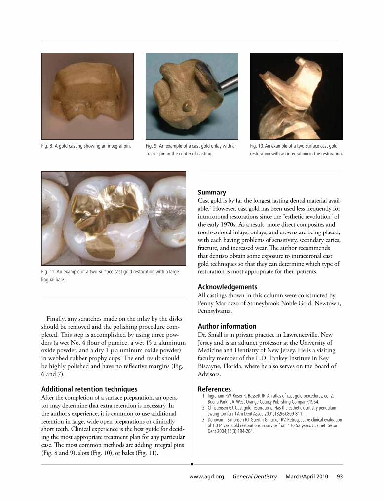
Additional retention techniquesAfter the completion of a surface preparation, an opera-tor may determine that extra retention is necessary. In the author’s experience, it is common to use additional retention in large, wide open preparations or clinically short teeth. Clinical experience is the best guide for decid-ing the most appropriate treatment plan for any particular case. The most common methods are adding integral pins (Fig. 8 and 9), slots (Fig. 10), or bales (Fig. 11).
SummaryCast gold is by far the longest lasting dental material avail-able.3 However, cast gold has been used less frequently for intracoronal restorations since the “esthetic revolution” of the early 1970s. As a result, more direct composites and tooth-colored inlays, onlays, and crowns are being placed, with each having problems of sensitivity, secondary caries, fracture, and increased wear. The author recommends that dentists obtain some exposure to intracoronal cast gold techniques so that they can determine which type of restoration is most appropriate for their patients.
AcknowledgementsAll castings shown in this column were constructed by Penny Marrazzo of Stoneybrook Noble Gold, Newtown, Pennsylvania.
Author informationDr. Small is in private practice in Lawrenceville, New Jersey and is an adjunct professor at the University of Medicine and Dentistry of New Jersey. He is a visiting faculty member of the L.D. Pankey Institute in Key Biscayne, Florida, where he also serves on the Board of Advisors.
References 1. ingraham rW, Koser r, Bassett Jr. an atlas of cast gold procedures, ed. 2.
Buena park, ca: West orange county publishing company;1964. 2. christensen GJ. cast gold restorations. has the esthetic dentistry pendulum
swung too far? J am Dent assoc 2001;132(6):809-811. 3. Donovan t, simonsen rJ, Guertin G, tucker rv. retrospective clinical evaluation
of 1,314 cast gold restorations in service from 1 to 52 years. J esthet restor Dent 2004;16(3):194-204.
www.agd.org General Dentistry March/April 2010 93
fig. 8. a gold casting showing an integral pin. fig. 10. an example of a two-surface cast gold
restoration with an integral pin in the restoration.
fig. 11. an example of a two-surface cast gold restoration with a large
lingual bale.
fig. 9. an example of a cast gold onlay with a
tucker pin in the center of casting.

polyethers (PEs) and vinyl polysiloxane (VPS) materials are the most popular classifications of impression materials for precision restorations
such as inlays, onlays, crowns, and bridges. But you might be amazed to know that PEs were first intro-duced by ESPE (before the company was purchased by 3M) in 1965—yes, Imp-regum (3M ESPE) has been around that long! Dentsply Caulk led the way with VPS materials by bringing Reprosil to the market back in 1982. A quick check shows that there have been no other major category advance-ments in the material side of impression-taking for 28 years!
So what has changed—and which of these changes really affect your chances of taking the perfect impression the first time?
Hydrophilicity One of the main advantages PEs have over VPS prod-ucts is their inherent hydrophilicity. Hydrocolloid, which still has a very small segment of the impression material market, is the epitome of this type of material. It is generally considered that the greater a material’s hydrophilicity, the less likely that fluid in the sulcus or really anywhere else on the preparation will distort the impression; the hydrophilic material will merely absorb the fluid and continue with its mission of registering an accurate and detailed impression. This property also goes hand-in-hand with the ability of the impression material to “wet out” on the preparation and capture better detail. This latter property has enhanced my own experience taking impressions over the years with PEs, especially Permadyne (3M ESPE), which has long been one of my favorite materials.
Dentsply Caulk trumped the market again with the
first “hydrophilic” VPS material (Aquasil) in 1997. Since that time, there has been a race among manufacturers to be the first to create VPS materials with as much hydro-philicity as PE materials. Note that hydrophilic proper-ties in VPS products need to be additives, since, unlike PEs, these materials are not inherently hydrophilic. This
race escalated recently when several manufactur-ers released marketing videos to illustrate what happens when you place a drop of water on a set or even an unset mix of impression material. Pre-sumably, the material is not hydrophilic if it beads up like water on a freshly waxed car; however, if it flattens out, it will do the same on a preparation in
the mouth, showing that it has enhanced hydrophilicity and wetting out ability.
The REALITY Research Lab (RRL) has developed a more clinically relevant (albeit more labor-intensive) test, in which an acrylic model with prepped and intact extracted teeth is impressed with different materials after the teeth have been dried, coated with a glisten-ing layer of water, or coated with a rather thick film of freshly captured saliva. Not only are the impressions and models examined closely, but full-cast crowns are fabricated and marginal gaps are measured under a stereomicroscope at 50x. A recent RRL product com-parison showed virtually no differences between two popular materials, Flexitime (Heraeus Kulzer, Inc.) and Aquasil Ultra (Dentsply Caulk).
On the other hand, one VPS material bucking the hydrophilicity trend is Precision (Discus Dental), which is marketed as “hydrokinetic” (which simply means “moving water”). Well, you can’t move water if you also have an affinity for it, which is the essence of the meaning of “hydrophilic.” Therefore, another way of
Impression materials— Are there any really new ones?Michael B. Miller, ddS, Fagd
Dental Materials
94 March/April 2010 General Dentistry www.agd.org
Aquickcheckshowsthattherehavebeennoother
majorcategoryadvancementsinthematerialsideof
impression-takingfor28years!

describing “hydrokinetic” would be “hydrophobic.” In other words, this product essentially returns to the early days when all VPS materials were hydrophobic. The RRL also tested this product, but the manufacturer did not specify another product as a control. This makes it more difficult to interpret the data, although there were virtually no differences between the experimental groups, indicating that this product will perform as the manufac-turer claims it will.
Does any of this matter when you are trying to take an accurate impression? Well, if the sulcus is filled with fluid (including blood), thus obscuring your margin, it could definitely make a difference. If you are using a supremely hydrophilic material, you hope that the product will literally soak up the fluid similar to a sponge and, at the same time, register the impression.
On the other hand, if the material is hydrokinetic, the aim is to move the fluid out of the sulcus first and then capture the margin. Is this a better strategy? The answer is probably yes, since there is less chance that the fluid will distort the material, which could happen if the fluid is absorbed.
But if this strategy is preferred, why have virtually all manufacturers opted for the hydrophilic route? One reason could be the mob mentality: If it works for one company, then other companies will produce the same item with some slight tweaks. Another reason is that the hydrokinetic concept flies in the face of the trend. Hydrophilic is the in concept, from bonding agents to cements to sealants. Why should impression materials be any different? Hydrophilic PEs followed in the successful footprints of hydrophilic hydrocolloid. Finally, only one company thought of using the hydrokinetic approach.
So should you switch to a hydrokinetic impression material? Not necessarily. There are numerous other fac-tors to consider, such as working and setting times, flow, availability in different delivery systems, and so forth. All of these criteria may be as important or even more so than hydrophilicity.
Of course, none of this matters at all if you use proper soft tissue management before you even lay a diamond on the tooth. I obsess over tissue management, so I believe that preventing a bloody sulcus is much more effective than having to deal with it after the fact. As admirable as this goal may be, though, it doesn’t always happen. Therefore, an impression material that will work in less than optimal conditions has significant value, which is why PEs continue to garner kudos from their devotees: These products tend to be less sensitive to moisture and have a terrific ability to wet out the prepa-ration under adverse conditions.
Viscosity and flow This issue depends on how you prefer to take an impres-sion. Personally, I prefer a very light body/heavy body combination: I look for a light body material that syringes easily and flows well without being too runny, and a heavy body tray material that will push the syringe material firmly against the preparation and, at the same time, not allow it to run down the patient’s throat, which materials with very low viscosity have a tendency to do. Less popular is a monophase material for both the syringe and tray.
The combination of very low viscosity syringe materi-als and heavy body tray materials is not new, although the RRL tests on flow using the Shark Fin device devel-oped by 3M ESPE have found more recently introduced materials with high flow. So, if you’re like me, you no longer have to stick with one or two brands to get better flow in your syringe material.
Hardness/stiffness With the increasing popularity of closed mouth impres-sions (especially with sideless trays), a more rigid or stiff material should work better by providing lateral support, although to my knowledge, this theory has never been proven in a clinical comparison. Neverthe-less, using a digital durometer, the RRL has found a few materials that are, indeed, stiffer than the rest. Just don’t be tempted to use a very rigid material for a full-arch impression, especially if you are using a well-fitting custom tray—you may need a “knee-on-chest” maneuver to remove it from the patient’s mouth!
Dispensing options Another area that has undergone some significant changes is the mixing/dispensing of materials. The hand-mixing required for tube-based products has been largely replaced with cartridge-based products that are mixed and dispensed using a ubiquitous automix gun. How-ever, these guns are not exactly cutting-edge any longer; they look like you bought them in a home improve-ment store, and can make filling a full-arch tray a real challenge for an auxiliary due to the hand and forearm strength required for heavy body materials.
To overcome the disadvantages of guns, ESPE intro-duced the first electronic mixer in 1995. There have been tweaks and speed improvements in these machines, which have been cloned by a handful of competitors over the ensuing 15 years, but the overall design is largely the same as that of the original version.
For syringe materials, at least two VPS products have unidose versions. While I like unidose packaging, it
www.agd.org General Dentistry March/April 2010 95

doesn’t seem to have caught on with impression materials and has not been a significant factor in product selection.
Intraoral working time Our thirst for speed has resulted in the availability of a number of very fast-setting materials, which can be a real time-saver when you impress one or two teeth. However, when you try to stretch the use of fast-set materials for more than the aforementioned one or two teeth, the intraoral working time of these materials becomes a major issue.
Unfortunately, the working times provided by manu-facturers are typically determined at room temperature. While this provides some comparison between products, it doesn’t really give you much indication about how much time you have between starting to syringe the material around your preparation and when you need to seat the tray. For example, if you are taking a 10-unit impression, how much time do you have from when you syringe material around the first preparation and when you need to seat the tray? This is critical to know because the material syringed around the first of the 10 prepara-tions is already starting to set before you’ve reached the last preparation; this setting is accelerated by the heat and moisture of the mouth. If it sets too fast, the tray material will not bond adequately to the syringe material and you’ll most likely end up with wrinkles or other types of distortion.
To my knowledge, there are only two extended work-ing time VPS materials on the market—Aquasil Ultra Xtra (Dentsply Caulk) and Multi-Prep (Clinician’s Choice)—both of which have been introduced in recent years. For large cases, it would be prudent to consider using one of them.
Tear strength If you have ever removed an impression from a patient’s mouth and found that it has torn on a critical marginal area, you know how important this property is. I recently took an impression for 10 veneers in a patient who had open gingival embrasures. Normally, I would block out these embrasures from the lingual to prevent the impression material from locking into them and tear-ing when it is removed from the mouth. But I was using an “improved” formula of a well-known material (Take 1 Advanced, Kerr Dental) that had claims of high tear strength. Therefore, on this case, I decided to go for it and dispense with the block out procedure. Sure enough,
the impression tore. I took a second impression and it also tore.
The guru of tear strength testing, in my opinion, is Dr. Alan Boghosian, a member of the REALITY Edito-rial Team. Dr. Boghosian and his colleague recently completed a test of eight impression materials for the RRL. The material I used that tore in the mouth scored in the middle of the pack, meaning it did not quite match the strength forecast by the manufacturer. To be fair, even though the impressions I took did indeed tear, the margins were still captured and the veneers seated beautifully.
Nevertheless, since a torn impression can ruin an otherwise perfect effort, it would be wise not to tempt fate. Block out areas that could cause tears, such as the aforementioned open embrasures—assuming, of course, that these areas don’t need to be captured.
What to use? Many aspects of taking an impression are personal. For example, you get to select the material that meets your flow and set time requirements. Beyond that, don’t get too caught up with pseudo-categories like “vinyl poly-ether silicone” or marketing slogans such as “polyeasier.” There are still only two real classes of impression mate-rial, the same as there have been for the past 28 years. And remember, no impression material can do it all. To get the best of all worlds, you probably need to stock two or three different types of material to cover all clinical situations as efficiently and productively as possible.
Author informationDr. Miller is the president of REALITY Publishing Company and Editor-in-Chief of its publications. He also maintains a general practice in Houston, Texas.
Manufacturersclinician’s choice, new milford, ct 800.265.3444, www.clinicianschoice.comDentsply caulk, milford, De 800.532.2855, www.caulk.comDiscus Dental, culver city, ca 800.422.9448, www.discusdental.comheraeus Kulzer, inc., armonk, ny 800.431.1785, www.heraeus-kulzer-us.comKerr Dental, orange, ca 800.537.7123, www.kerrdental.com3m espe, st. paul, mn 888.364.3577, www.3mespe.com
96 March/April 2010 General Dentistry www.agd.org

Asymptomatic carotid artery calcifications discovered on panoramic radiographyMohammad ali dolatabadi, ddS n Mohammad Hosein Kalantar Motamedi, ddS n Eshagh Lassemi, ddSYousef Janbaz, ddS
the most common manifesta-tions of atherosclerosis are cor-onary artery disease, peripheral
vascular disease, and cerebrovascular accidents (strokes).1 A 2009 article reported that carotid artery calcifica-tion is responsible for an estimated 5% of ischemic strokes.2 Stroke survivors face lifelong disabilities such as loss of mobility, aphasia, and depression.3 Atheroma-related formations of thrombi and emboli
in the carotid artery are the most frequent causes of stroke.2 Early detection of carotid atherosclerosis not only can save lives but also may reduce medical expenditures.
Friedlander and Lande reported that panoramic radiology could aid in detecting patients at risk of stroke.4 Calcified atherosclerotic lesions in the carotid bifurcation can be detected in the lower corners of the panoramic radiograph, adjacent
to the cervical spine and hyoid bone; such lesions may appear as a nodular radiopaque mass or as double radiopaque vertical lines within the neck. These calcifications are found on panoramic radiographs inferior-posterior to the angle of the mandible, at the lower margin of the third cervical vertebra and the entirety of the fourth cervical verte-bra; such lesions are approximately 1.5–4.5 mm in size (Fig. 1).5
this article presents the case of a 50-year-old asymptomatic man whose panoramic radiograph revealed calcium deposits within the left internal carotid bifurcation region. subsequent duplex ultrasonic examination indicated unilateral low-grade carotid arterial stenosis, a condition associated with a significant risk of stroke, which had not been identified previously. the findings on the panoramic
radiograph prompted appropriate and potentially lifesaving treat-ment. Dentists who are well-versed in diagnosing calcified plaques on panoramic radiographs can play a major role in the early referral and treatment of undiagnosed asymptomatic patients.
received: november 21, 2008accepted: february 6, 2009
fig. 1. a schematic illustration of the relationship of the common carotid artery, the internal carotid artery, the external carotid artery, and the structures
usually seen on a panoramic radiograph. note the process of embolization of atherosclerotic debris (black arrow) at the carotid bifurcation (white arrow).
Digital Radiography
www.agd.org General Dentistry March/April 2010 97
C2
C3
C4
C5
Thyroid
Common carotid artery
Internal carotid artery
External carotid artery
Hyoid bone

Case reportA 50-year-old man came to the dental office for comprehensive dental care. His medical history revealed non-insulin-dependent dia-betes mellitus, hyperlipidemia (con-trolled with oral medication), and tobacco use (which he had ceased one year earlier). The patient had no history of medical problems, and his primary care physician had not diagnosed any other illness during his last examination. A panoramic radiograph taken during his dental treatment revealed the presence of single, irregular, non-homogenous radiopacities lying over the left carotid bifurcations. The calcifica-tions were located inferior to the angle of the mandible and the tip of the hyoid bone and superior to the tip of the thyroid cartilage and the C3, C4, and C5 vertebrae (Fig. 2).
Carotid duplex ultrasonography revealed a left unilateral carotid ste-nosis (Fig. 3). Small calcified plaque (4 x 2 mm) was seen in the left internal carotid artery but did not display any hemodynamic symp-toms. The patient was referred to his specialist for further management.
DiscussionAccording to Khosropanah et al, panoramic radiographs have a sensi-tivity of 66.6% and a positive predic-tive value of 45%, indicating that they cannot be considered accurate or reliable for detecting carotid artery calcifications.2 However, dentists who review oral panoramic radio-graphs should look for incidental calcifications lying over the carotid bifurcation region. The patient in the present case had no signs or symptoms of carotid artery disease and may not have been evaluated or screened for atherosclerotic disease had these calcified carotid plaques not appeared on the panoramic radiograph. He had unilateral low-grade stenosis and he needed follow-up for changes in plaque size and form, which could cause symptoms requiring surgical removal.
DiagnosisStenosis is best determined by using duplex ultrasonography, which is inexpensive, easily available, accu-rate, and noninvasive. Duplex ultra-sonography measures the increase in blood velocity produced by a
focal stenosis (a process known as the Bernoulli Effect), thus indirectly yielding information concerning the severity of stenosis. Similar calcifica-tions are found in the coronary arteries of individuals with ischemic heart disease.6
Differential diagnosisAtherosclerosis is not the only cause of soft tissue calcifications seen anterior to the cervical ver-tebrae on panoramic radiographs; in fact, carotid calcifications must be differentiated from calcified triticeous/thyroid cartilage, calci-fied lymph nodes, and non-carotid phleboliths (sclerosing hemangio-mata).7 When an anterior-posterior radiograph of the neck uses soft tissue exposure settings, calcifica-tions within the carotid arteries will appear lateral to the spine; by contrast, calcifications in the thyroid gland, thyroid cartilage, triticeous cartilage, or epiglottis will appear in the midline, super-imposed over the spine. Phleboliths (sclerosing hemangiomata) and calcified acne or lymph nodes are other calcifications that may be
fig. 2. the calcifications (arrow) appeared as heterogeneous radiopacities
overlying the carotid bifurcation near the tip of the greater horn of the
hyoid bone, approximately 2.5 cm posterior and 2.5 cm inferior to the
cortical rim of the midpoint of the mandibular angle.
fig. 3. a color-flow duplex sonography image of the left internal carotid
artery confirming the presence of atheroma.
Digital Radiography Asymptomatic carotid artery calcifications discovered on panoramic radiography
98 March/April 2010 General Dentistry www.agd.org

superimposed over the same part of the panoramic film. By contrast, the stylohyoid and calcified stylo-mandibular ligaments are situated posterior to the mandibular ramus.
TreatmentCarotid endarterectomy, which consists of using a variety of techniques for local removal of the atherosclerotic plaque, has been conclusively shown to reduce the risk of stroke among symptomatic and asymptomatic patients who have significant plaque lesions (that is, a stenosis of 60% or more).8,9 Duplex ultrasonography—the most accurate screening method short of angiography—is noninvasive and relatively inexpensive; however, screening all patients is impractical and not cost-effective. High-risk groups for whom ultrasonic screen-ing might be cost-effective include those with bruits or atherosclerosis in other parts of the body.
Audible cervical bruits may be caused by turbulent blood flow, tortuousity, high flow rates through otherwise normal vessels, a cardiac problem, or carotid artery stenosis. Although the presence of a bruit does not necessarily indicate carotid artery stenosis, most physicians believe that their presence increases the patient’s risk of developing carotid artery stenosis and that they are an indication for ultrasonic evaluation.10,11 In addition, because atherosclerosis tends to be a sys-temic problem, 10–12% of patients with lower extremity and coronary atherosclerosis also have significant carotid artery stenosis.12,13 Dentists
should refer these patients to a phy-sician for a cardiovascular evalua-tion to receive proper and timely medical treatment.
Occlusive disease in either loca-tion (extremity or coronary) has become a de facto indication for carotid ultrasonography. To date, however, there are no universally accepted screening criteria, and the decision to refer a patient for ultra-sonic evaluation remains that of the individual physician.
SummaryAlthough panoramic radiographs do not have a 100% sensitivity or positive predictive value for detect-ing carotid artery calcifications, dentists should make a point of examining them for incidental calcifications over the carotid bifur-cation region. When that is the case, dentists can refer their patients to a physician for a cardiovascular evaluation to receive proper and timely medical treatment.
Author informationDr. Dolatabadi is an assistant professor, Department of Oral and Maxillofacial Surgery, Faculty of Dentistry, Azad University of Medical Sciences, Dental College, Tehran, Iran, where Dr. Lassemi is an associate professor and Dr. Mota-medi is a professor, BMSU Trauma Research Center. Dr. Janbaz is in private practice in Tehran.
References 1. tegos tJ, Kalodiki e, sabetai mm, nicolaides an.
the genesis of atherosclerosis and risk factors: a review. angiology 2001;52(2):89-98.
2. Khosropanah sh, shahidi sh, Bronoosh p, rasekhi a. evaluation of carotid calcification detected using panoramic radiography and ca-rotid Doppler sonography in patients with and without coronary artery disease. Br Dent J 2009; 207(4):e8.
3. romano-sousa cm, Krejci l, medeiros fm, Graciosa-filho rG, martins mf, Guedes vn, fenyo-pereira m. Diagnostic agreement be-tween panoramic radiographs and color Dop-pler images of carotid atheroma. J appl oral sci 2009;17(1):45-48.
4. friedlander ah, lande a. panoramic radiograph-ic identification of carotid arterial plaques. oral surg oral med oral pathol 1981;52(1):102-104.
5. friedlander ah, august m. the role of panoram-ic radiography in determining an increased risk of cervical atheromas in patients treated with therapeutic irradiation. oral surg oral med oral pathol oral radiol endod 1998;85(3):339-344.
6. Baron mG. significance of coronary artery calci-fication. radiology 1994;192(3):613-614.
7. carter lc. Discrimination between calcified triti-ceous cartilage and calcified carotid atheroma on panoramic radiography. oral surg oral med oral pathol oral radiol endod 2000;90(1):108-110.
8. endarterectomy for asymptomatic carotid artery stenosis. executive committee for the asymp-tomatic carotid atherosclerosis study. Jama 1995;273(18):1421-1428.
9. hertzer nr, o’hara pJ, mascha eJ, Krajewski lp, sullivan tm, Beven eG. early outcome assess-ment for 2228 consecutive carotid endarterec-tomy procedures: the cleveland clinic experience from 1989 to 1995. J vasc surg 1997;26(1):1-10.
10. taylor lm, porter Jm. carotid endarterectomy. in: porter Jm, taylor lm, eds. Basic data underly-ing clinical decision making in vascular surgery. st. louis: Quality medical publishing;1994:182- 185.
11. roederer Go, langlois ye, Jager Ka, primozich Jf, Beach KW, phillips DJ, strandness De Jr. the natural history of carotid arterial disease in asymptomatic patients with cervical bruits. stroke 1984;15(4):605-613.
12. Gentile at, taylor lm, moneta Gl, porter Jm. prevalence of asymptomatic carotid stenosis in patients undergoing infrainguinal bypass sur-gery. arch surg 1995;130(8):900-904.
13. Berens e, Kouchoukos n, murphy s, Wareing t. preoperative carotid artery screening in elderly patients undergoing cardiac surgery. J vasc surg 1992;15(2):313-323.
www.agd.org General Dentistry March/April 2010 99

Medication use in geriatric populations: Dental implications of frequently prescribed medicationsMary a. aubertin, dMd n Carlton Horbelt, ddS n waletha wasson, ddS n Marjorie woods, ddS
the demographic changes antici-pated in the next 20 years are expected to lead to an increased
interest in the health concerns of older adults.1 The prevalence of medical conditions is high among geriatric patients, and the con-comitant incidence of medication use increases with age.2-6 Geriatric patients may be at high risk medi-cally due to cumulative illnesses, and their treatments require specialized dental skills, medical monitoring, and careful pharmacological man-agement.4 The medications com-monly used to treat disease states associated with aging may have nota-ble side effects and drug interactions; these effects may be exacerbated in geriatric patients. In a recent study of dental school patients, Miller et al reported that approximately 57% of all drugs taken had the potential to affect dental care adversely and to create life-threatening drug interactions.4 Today’s dentists must be able to assess complex medical conditions, multiple and complex medication regimens, and how each of these affects the patient’s oral and overall health and the provision of dental care.
Common medical disorders among geriatric patients include cardiovascular disease, hyperten-sion, dislipidemia, diabetes mellitus and other endocrine disorders, respiratory diseases, musculoskeletal disorders, neurological disorders, and chronic pain.2-4,7 Cardiovascular drugs are the most common group of drugs used by geriatric patients and elicit dentally significant adverse effects.2,3,6-8 More often than not, however, geriatric patients are taking multiple medications. The most fre-quently prescribed medications that may impact dental management of older patients include cardiovascular drugs, NSAIDs, gastrointestinal agents, psychotropic agents, and endocrine agents.8
Not only do dentists need to be familiar with their patients’ medical disorders, they also must carefully analyze their patients’ medications.2,9 Increased knowledge of pharmacol-ogy as it pertains to geriatric patients and advanced dental management of this special patient population are essential.4 When patients provide incomplete or vague medical or medication histories, medical con-sultation, pharmacist consultation,
or family member clarification should be performed before any dental treatment begins. Many of these chronic complex medical conditions and their drug therapies, combined with the frequent use of OTC drugs, place the patient at risk for adverse outcomes during dental therapies.4,5 To compound matters, at least 40% of geriatric patients receive drugs from two or more phy-sicians and 12% of older patients either take medications prescribed for someone else or take their own medications incorrectly.6
Geriatric patients often have chronic and sometimes complex health problems and consume more medication than any other age group.2,5 The use of multiple drugs (from multiple drug categories) increases with age.2 Independent elders take at least one to four medications daily, while elders in long-term care take an average of 5–14 medications daily.1-4,6,9 Polypharmacy, multiple co-morbid medical conditions, and physiologi-cal changes in terms of the absorp-tion, distribution, metabolism, and excretion of drugs affect geriatric patients’ responses to medications,
anticipated demographic changes in the u.s. during the next 20 years will bring increasing numbers of geriatric patients into dental practices. it is expected that these patients will have multiple co-morbid medical conditions and will have to take multiple medications as a result. Dental practitioners must stay informed concerning newly marketed drugs and those commonly prescribed
to geriatric patients, and the potential dental implications of those drugs. specialized training in geriatric dentistry, continuing educa-tion, and consultation with medical and pharmacy practitioners can provide valuable tools for managing this special patient population.
received: may 20, 2009accepted: august 18, 2009
Geriatric Dentistry
100 March/April 2010 General Dentistry www.agd.org
CDE2 HOURSCREDIT

placing them at a higher risk of side effects and adverse effects.6,7,9,10
Physiological changes that result during normal aging are in large part due to increases in total body fat, circulatory changes, decreased organ function, and decreases in total body water and lean body muscle mass.6,9 As a result, lower doses of medications are often necessary to reach therapeutic concentrations in older patients, and these doses may remain in body tissues for longer periods of time. Poor nutritional status and drug interactions also enhance adverse or toxic reactions.11
Adverse effects are considered to be unwanted, unintended, preventable, or toxic injuries caused by a drug; they may appear as physical or oral manifestations.2 Important adverse reactions include side effects, drug allergy, and toxic reactions.1 Known risk factors for adverse drug interac-tions include administration to geriatric patients, administration to medically compromised patients, use of drugs with small margins of safety, and chronic drug therapies which utilize drugs that are excreted slowly.5,11 Adverse drug effects are common in geriatric patients, affecting approximately 25% of older patients and accounting for 10–17% of hospitalizations of geri-atric patients.6,9
As patients take increasing numbers of prescription and non-prescription drugs, several factors can increase the likelihood of adverse reactions and potential mortality in geriatric patients, including drug interactions, errors in taking medications, prescriptions from multiple physicians, and the physiologic states produced by each drug. The mortality rate associated with adverse drug reactions and the average hospital stay due to drug reaction increases exponentially with the number of drugs taken.6
Adverse drug reactions occur most commonly in connection with drugs used to treat congestive heart failure, arthritis, hypertension, diabetes, respiratory tract infections, and prostatic hypertrophy.6 In addi-tion, the drugs dentists administer and prescribe to geriatric patients may interact adversely with medica-tions prescribed by their physicians. As the number of geriatric patients increases, dentists must be familiar with currently prescribed medica-tions taken by geriatric patients, the diseases for which they are taking these medications, self-administered OTC drugs, potential interactions with dentally prescribed drugs, and adverse drug effects.6
This article lists medications commonly prescribed to geriatric patients and adverse effects associ-ated with those drugs.12-16 Dentists should have a working knowledge of how these drugs act, potential drug interactions, dosing restrictions, and how the body handles these drugs. Because new drugs and new drug combinations are prescribed frequently, unreported drug interac-tions should be considered.5
Adverse interactions with drugs prescribed in dentistryACE inhibitorsNo dentally significant effects or complications have been reported with quinapril (Accupril, Pfizer Inc.) or benazepril (Lotensin, Novartis Pharmaceuticals). Lisinopril (Prini-vil, Merck & Co.) is associated with orthostatic hypotension. Enalapril (Vasotec, Merck & Co.) is associ-ated with orthostatic hypotension and abnormal taste.
In patients with compromised renal function, prescribing NSAIDs may result in further deterioration of renal function. Quinapril may reduce the absorption of quino-lone and tetracycline antibiotics.
High-dose aspirin, NSAIDs, and salicylates may reduce the thera-peutic effects of ACE inhibitors. Lisinopril may increase the toxicity of adverse events from azathioprine, cyclosporine, or NSAIDs.13
Aldosterone antagonistsNo dentally significant effects or complications of using this drug group have been reported. Prescribing salicylates and NSAIDs (indomethacin) may decrease the natriuretic effect of spironolactone (Aldactone, Pfizer Inc.).13
VasodilatorsNo dentally significant effects or complications have been reported with hydralazine (Apresoline, Novar-tis Pharmaceuticals). Nitroglycerin is associated with xerostomia, although normal salivary flow returns once the drug is discontinued.13 Prescrib-ing NSAIDs may decrease hydrala-zine’s hemodynamic effects.
Calcium channel blockersDiltiazem (Cardizem, Abbott Laboratories) has been reported to cause a greater than 10% incidence of gingival hyperplasia, which usually disappears once the drug is discontinued.13 There have been few reports of gingival hyperplasia with amlodipine (Norvasc, Pfizer Inc.).
The blood levels/physiologic effects of diltiazem and amlodipine may be increased by systemic azole antifungals, clarithromycin, diclofe-nac, doxycycline, and erythromycin. Diltiazem may increase the levels/effects of selected benzodiazepines and cyclosporine.13
Anti-arrhythmicsWhen patients are taking medica-tions that prolong the QT interval (specifically amiodarone), dentists should consult with the patient’s physician prior to administering a
www.agd.org General Dentistry March/April 2010 101

vasoconstrictor. Use epinephrine and levonordefrin with caution. Amiodarone is associated with abnormal salivation and taste.13
Azithromycin (Zithromax, Pfizer Inc.) may prolong the effect of amiodarone. Cimetidine (Tagamet, GlaxoSmithKline) may decrease the metabolism of amiodarone, which may in turn diminish the therapeutic effect of codeine and decrease the metabolism of cyclo-sporine and lidocaine.13
Anti-adrenergic agents/Beta adrenergic blockersCarvedilol (Coreg, GlaxoSmith-Kline) has been associated with pos-tural hypotension and periodontitis. Using NSAIDs (such as ibuprofen and indomethacin) for three weeks or longer may reduce the hypoten-sive effect of beta blockers; no special precautions are needed for short-term use.13 Propranolol and nadolol (nonselective beta blockers) enhance the pressor response to epinephrine, resulting in hypertension and bradycardia. Local anesthetic with vasoconstrictor can be used safely on patients taking atenolol (Tenormin, AstraZeneca) or metoprolol.13,17
Barbiturates may decrease the serum concentration of beta block-ers. Cimetidine may decrease the metabolism of carvedilol. Carvedilol may increase the serum concentra-tion of cyclosporine. Beta blockers may decrease the metabolism of lidocaine. Propoxyphene may decrease the metabolism of beta blockers. Ampicillin may decrease the bioavailability of atenolol. NSAIDs may diminish the antihy-pertensive effect of beta blockers.13
Anti-arrhythmics/Nonselective beta adrenergic blockersEpinephrine has interacted with nonselective beta blockers such as sotalol to cause an initial
hypertensive episode followed by bradycardia.13 Sotalol is one of the drugs that is known to prolong the QT interval and could result in torsade de pointes, a ventricular tachycardia in which the heart rate can range from 150–250 beats per minute.18 It is not known what effect vasoconstrictors in local anesthetic will have on patients with a known history of congenital pro-longed QT interval or on patients taking drugs that prolong the QT interval. Until more information is obtained and reported in the medical literature, it is suggested that dentists consult with the patient’s physician prior to the use of a vasoconstrictor and that the vasoconstrictor be used with caution due to the potential for hypertensive episodes.13
Angiotensin II inhibitors with or without diureticOrthostatic hypotension has been reported with angiotensin II inhibi-tors.13 Systemic antifungal azole derivatives may decrease the metab-olism of losartan (Cozaar, Merck & Co.). NSAIDs may diminish the therapeutic effect of angiotensin II receptor blockers (ARBs), and NSAIDs used in combination with ARBs may significantly decrease glomerular filtration and renal function.13
Loop diureticsXerostomia and oral irritations have been reported with loop diuretics such as furosemide (Lasix, Sanofi-Aventis).16 Systemic corticosteroids may enhance the hypokalemic effect of loop diuretics, while NSAIDs may diminish the diuretic effect.13
Thiazide diureticsXerostomia, orthostatic hypoten-sion, and hypotension have been reported with thiazide diuretics such as hydrochlorothiazide (Microzide,
Watson Pharmaceuticals). Systemic corticosteroids may enhance the hypokalemic effect of thiazide diuretics, while NSAIDs may diminish the diuretic and antihyper-tensive effects.13,16
Anti-anginal agentsIsosorbide dinitrate may cause xerostomia and affect salivation. Normal salivary flow resumes once the drug is discontinued. No signifi-cant effects have been noted with isosorbide mononitrate (Imdur, AstraZeneca). There are no reported drug interactions with dentally prescribed drugs.
Potassium replacementsNo dentally significant effects, complications, or drug interactions between potassium chloride and dentally prescribed drugs have been reported.
Cardiac glycosidesVasoconstrictors should be used with caution due to risk of cardiac arrhythmias with digoxin. Digoxin has been reported to cause a sensi-tive gag reflex, which could affect taking a dental impression.13
Systemic antifungal azole deriva-tives may increase the serum con-centration of cardiac glycosides, with the exception of miconazole. Cyclo-sporine may decrease the metabolism of cardiac glycosides, while kaolin may decrease their absorption.13
Osteoporosis treatmentsOsteonecrosis of the jaw (ONJ)—a condition generally associated with local infection and/or tooth extrac-tion and delayed healing—has been reported in patients taking bisphos-phonates.13 Symptoms include a nonhealing extraction socket or an exposed jawbone. Most reported cases of bisphosphonate-associated osteonecrosis have been in cancer
Geriatric Dentistry Dental implications of frequently prescribed medications
102 March/April 2010 General Dentistry www.agd.org
patients treated with intravenous bisphosphonates; however, it has also been reported in patients taking oral bisphosphonates for postmeno-pausal osteoporosis.13 For patients requiring dental procedures, there are no data available to suggest whether discontinuation of bisphos-phonate treatment reduces the risk of ONJ. NSAIDs may enhance the adverse/toxic effect of bisphospho-nate derivatives.19
Antidiabetic agentsPatients with Type 2 diabetes who are taking glimepiride (Amaryl, Sanofi-Aventis), glyburide, rosiglitazone (Avandia, GlaxoSmithKline), met-formin (Glucophage, Bristol-Myers Squibb), or glipizide (Glucotrol, Pfizer Inc.) should undergo dental treatment in the morning to mini-mize the chance of stress-induced hypoglycemia. Metformin has been associated with taste disorder.13
Systemic corticosteroids may diminish the hypoglycemic effect of antidiabetic agents. Adrenal suppression may lead to acute adrenal crisis, which may manifest as enhanced hypoglycemia. Sulfo-nylureas may increase the serum concentration of cyclosporine. Fluconazole may increase the serum concentration of sulfonylureas. Sulfonamide derivatives and trim-ethoprim may enhance the hypo-glycemic effect of sulfonylureas.13
Anxiolytics, sedatives, hypnoticsZolpidem (Ambien, Sanofi-Aventis), lorazepam, and alprazolam are associated with xerostomia. Normal salivary flow resumes when the drug is discontinued.13
Azole derivative antifungal agents (with the exception of miconazole) may decrease the metabolism of zol-pidem and decrease the metabolism of benzodiazepines. Zithromax also
may decrease the metabolism of benzodiazepines.13
Anti-anxiety medications/antihistaminesHydroxyzine is associated with xero-stomia, although normal salivary flow resumes when the drug is dis-continued.13 No drug interactions have been reported with dentally prescribed drugs.
Anti-dizziness medicationsSlight to moderate drowsiness, thickening of bronchial secretions, and significant xerostomia have been reported with meclizine; normal salivary flow resumes when the drug is discontinued.13 No drug interactions with dentally prescribed drugs have been reported.
Anti-Alzheimer’s medicationsNo dentally significant effects, complications, or drug interactions with dentally prescribed drugs have been reported with drugs such as donepezil (Aricept, Pfizer Inc.).
Respiratory agentsXerostomia and changes in salivation and dry mucous membranes are associated with ipratropium (Atro-vent and Atrovent HFA, Boehringer Ingelheim Pharmaceuticals). Taking ipratropium and albuterol in com-bination may enhance the adverse/toxic effects of other anticholinergic and sympathomimetic drugs.
Anti-inflammatory medications/analgesicsCelecoxib (Celebrex, Pfizer Inc.) is associated with stomatitis, abnormal taste, xerostomia (normal salivary flow resumes when the drug is dis-continued), and unspecified tooth disorder.13 Nonselective NSAIDs are known to reversibly decrease platelet aggregation via mechanisms different than those observed
with aspirin.20 Celebrex taken at single doses of up to 800 mg or in multiple doses (600 mg twice daily) have no reported effect on platelet aggregation or bleeding time. Meloxicam (Mobic, Boehringer Ingelheim Pharmaceuticals) has been associated with abnormal taste, ulcerative stomatitis, and xerostomia (normal salivary flow resumes when the drug is discontinued).13
Systemic corticosteroids may enhance the adverse/toxic effects of COX-2 inhibitors. NSAIDs may enhance the nephrotoxic effect of cyclosporine and increase its serum concentration; they may also enhance the adverse/toxic effects of other NSAIDs and may decrease the excretion of vancomycin.13
AntidepressantsNo interactions have been reported between vasoconstrictors and citalopram (Celexa, Forest Phar-maceuticals), paroxetine, trazadone (Desyrel, Bristol-Myers Squibb), or sertraline (Zoloft, Pfizer Inc.). However, citalopram and sertraline have been associated with xerostomia and citalopram has been associated with abnormal taste.13 Xerostomia and changes in salivation, postural hypotension, and abnormal taste have been reported with paroxetine.13 Problems such as bruxism have been reported with selective serotonin reuptake inhibitors (SSRIs) and may preclude their use.13 Clinicians attempting to evaluate any patient with bruxism or involuntary muscle movement who is simultaneously being treated with an SSRI should be aware of the potential association. Prolonged use of antidepressants may decrease or inhibit salivary flow.13
For patients taking drugs that block the uptake of norepineph-rine, vasoconstrictors should be used in limited amounts due to the potential for exacerbation of
www.agd.org General Dentistry March/April 2010 103

hypertension.13 Dentists should monitor the vital signs of patients who are taking antidepressants that affect norepinephrine, espe-cially venlafaxine (Effexor, Wyeth Pharmaceuticals) and mirtazapine (Remeron, Schering-Plough), which may produce a sustained increase in diastolic blood pressure and heart rate.13 Significant xerostomia with venlafaxine, mirtazapine, and trazadone may contribute to oral discomfort, especially in the elderly; these drugs are also associated with abnormal taste.13 No interac-tions have been reported between vasoconstrictors and escitalopram (Lexapro, Forest Pharmaceuticals), although the drug is associated with xerostomia and toothache.13
Amitriptyline prolongs the QT interval and can put a patient at risk for torsade de pointes.13 Dentists should consult with the patient’s physician prior to administering a vasoconstrictor to a patient taking amitriptyline. Amitriptyline has been associated with xerostomia and changes in salivation, ortho-static hypotension, stomatitis, peculiar taste, and black tongue. Long-term treatment with tricyclic antidepressants (TCAs) (such as amitriptyline) increases the risk of caries by reducing salivation and the salivary buffer capacity.13
SSRIs have several other sig-nificant drug interactions of note relevant to dentistry. Opioid analge-sics and tramadol (Ultram, Ortho-McNeil Pharmaceutical, Inc.) may enhance the serotonergic effect of SSRIs, resulting in serotonin syndrome. Conversely, SSRIs may enhance tramadol’s neuroexcitatory effect and/or potential to cause sei-zures.13 Macrolide antibiotics (with the exception of azithromycin) may decrease the metabolism of SSRIs.
SSRIs also interact with common medications to alter bleeding
hemostasis. SSRIs may enhance the antiplatelet effects of aspirin and COX-2 inhibitors, while SSRIs taken with TCAs may enhance the antiplatelet effect of nonselective NSAIDs.
Significant interaction between antidepressants and other drugs may affect their toxicity, metabolism, and potency. Paroxetine may enhance the adverse/toxic effect of other cen-tral nervous system (CNS) depres-sants. Barbiturates may increase the metabolism of TCAs. Propoxyphene may enhance the CNS depressant effect of TCAs, while SSRIs may decrease their metabolism. Traza-done may diminish the therapeutic effect of codeine and tramadol.13
Non-sedating antihistaminesXerostomia and stomatitis in chil-dren have been reported in connec-tion with loratadine; however, no drug interactions with dentally pre-scribed drugs have been reported.13
Anticoagulants/antithromboticsSystemic corticosteroids and tet-racycline derivatives may enhance the anticoagulant effect of warfarin. Mouth ulcers and abnormal taste have been reported with warfarin; bleeding gingival tissue has been associated with the first signs of warfarin overdose.13 If temporary reduction or discontinuation of the medication is warranted prior to sur-gery, consultation with the patient’s prescribing physician is advisable.
Patients who take more than 1.3 g of acetaminophen daily for more than one week are likely to enhance the anticoagulant effect of couma-rin derivatives.13 Systemic azole antifungal derivatives (including fluconazole), macrolide antibiotics, metronidazole, and propoxyphene may decrease the metabolism of coumarin derivatives, while barbitu-rates may increase their metabolism.
Azathioprine may diminish the anti-coagulant effect of coumarin deriva-tives, while NSAIDs and tricyclic antidepressants may enhance it. Cephalosporins (with the exception of cephalexin) and fluorouracil may enhance the anticoagulant effect of coumarin derivatives.13
Anti-platelet medicationsPremature discontinuation of aspirin and clopidogrel antiplatelet therapy strongly increases the risk of a cata-strophic event of stent thrombosis, leading to myocardial infarction and/or death.13 Any elective surgery should be postponed for one year after stent implantation; if surgery must be performed, dentists should consider continuing the antiplatelet therapy during the perioperative period in high-risk patients.
Macrolide antibiotics (except for azithromycin) may diminish the therapeutic effect of clopidogrel (Plavix, Bristol-Myers Squibb). NSAIDs may enhance the adverse/toxic effect of antiplatelet agents and increase the risk of bleeding.13
Analgesics/narcoticsNo significant effects or complica-tions have been reported. Hepato-toxicity caused by acetaminophen is potentiated by chronic ethanol consumption. Patients who use acetominophen (even in therapeutic doses) and consume ethanol at the same time are at risk of developing hepatotoxicity. Xerostomia, nausea, sedation, and constipation have been reported with oxycodone and acetaminophen use.13 Barbiturates may increase the metabolism of acetaminophen, diminishing its effect and increasing the risk of liver damage.
Anticonvulsant drugsValproic acid (and its derivatives) is associated with periodontal abscess,
Geriatric Dentistry Dental implications of frequently prescribed medications
104 March/April 2010 General Dentistry www.agd.org
taste perversion, stomatitis, and xerostomia. Gingival hyperplasia is common during the first six months of phenytoin therapy.13 To minimize the severity and growth of gingival tissue, the patient should begin a program of professional cleaning and at-home plaque con-trol within 10 days of the start of anticonvulsant therapy.
Some adverse effects have been reported in connection with anti-convulsant drugs: Xerostomia, dry throat, and dental abnormalities have been reported with gabapentin (Neurontin, Pfizer Inc.).13 Valproic acid may decrease the metabolism of barbiturates and lorazepam (barbiturates, in turn, may decrease the serum concentration of valproic acid) and may increase the serum concentration of TCAs. Anticonvul-sants may increase the metabolism of acetaminophen, thus diminishing its effect and increasing the risk of liver damage. Phenytoin may increase the metabolism of systemic azole derivative antifungal agents, doxycycline, and cyclosporine. Ben-zodiazepines may increase the serum concentration of phenytoin. Fluco-nazole may decrease the metabolism of phenytoin.13
Peripherally acting anti-adrenergic blockersOrthostatic hypotension and tooth disorder have been reported with tamsulosin (Flomax, Boehringer Ingelheim Pharmaceuticals). No drug interactions with dentally pre-scribed drugs have been reported.
Anti-psychotic drugsZiprasidone (Geodon, Pfizer Inc.) and quetiapine (Seroquel, AstraZen-eca) prolong the QT interval, which could lead to torsade de pointes.13 Hypotension and tachycardia may result among patients who take vasoconstrictors with anti-psychotic
drugs.16 Local anesthetics that contain vasoconstrictors should be used with caution and the patient’s physician should be consulted to determine the risk.13
Xerostomia and changes in salivation, orthostatic hypotension, tongue edema, dysphagia, and unspecified tooth disorder have been reported with ziprasidone. Signifi-cant xerostomia and toothache have been reported with risperidone.13 No significant effects or complications have been reported with olanzapine (Zyprexa, Eli Lilly and Company).
Anti-estrogen and estrogen replacement drugsNo significant effects, complica-tions, or drug interactions between raloxifene (Evista, Eli Lilly and Company) or conjugated estrogen and dentally prescribed drugs have been reported in the literature.
Lipid-lowering agentsFew reports of gingival hyperplasia with atorvastatin (Lipitor, Pfizer Inc.) have been reported in the literature.13 In the event of hyperplasia, consulta-tion with the patient’s physician is suggested. No significant effects or complications have been reported in connection with lovastatin, pravastatin (Pravachol, Bristol-Myers Squibb), simvistatin (Zocor, Merck & Co.), or ezetimibe with simvistatin (Vytorin, Merck & Co.).
Systemic azole derivative anti-fungal agents may decrease the metabolism of atorvastatin, lovas-tatin, pravastatin, and simvistatin, while cyclosporine may increase the serum concentration of atorvastatin, pravastatin, and simvistatin. Macro-lide antibiotics (with the exception of azithromycin) may decrease the metabolism of atorvastatin, prava-statin, and simvistatin. Atorvastatin may increase the serum concentra-tion of midazolam.13
Quinolone antibioticsBefore giving a vasoconstrictor to patients taking levofloxacin, dentists should consult with the patient’s physician, as this drug prolongs the QT interval and puts patients at risk for torsade de pointes. However, the risk of drug-induced torsade de pointes is extremely low when a single QT interval-prolonging drug is prescribed. Quinolone antibiotics may enhance the adverse/toxic effect of systemic corticosteroids and the risk of tendon-related side effects, including tendonitis and rupture. NSAIDs may enhance quinolone antibiotics’ potential for neuroexcit-atory effects and/or seizures.
Sulfonamide antibiotic combinationsSulfonamide derivatives may enhance the nephrotoxic effect of cyclosporine and decrease its serum concentration. Glossitis and stomatitis have been reported with trimethoprim/sulfamethoxazole.13
Macrolide antibioticsNo significant effects, complications, or drug interactions have been reported between dentally pre-scribed drugs and azithromycin.
Antidiarrheal drugsSignificant xerostomia has been reported with diphenoxylate and atropine.13 No drug interactions between these antidiarrheal drugs and dentally prescribed drugs have been reported.
Gastrointestinal agentsNo significant effects or complica-tions from lansoprazole (Prevacid, Takeda Pharmaceuticals) or pantoprazole (Protonix, Wyeth Pharmaceuticals) have been reported; however, taste perversion, dry mouth, esophageal candidiasis, and mucosal atrophy of the tongue
www.agd.org General Dentistry March/April 2010 105

have been reported in connec-tion with omeprazole (Prilosec, AstraZeneca). Xerostomia has been reported in connection with meto-clopramide use.13
Proton pump inhibitors may decrease the absorption of systemic azole derivative antifungal agents (with the exception of miconazole). Omeprazole may increase the serum concentration of benzodiazepines. No dental drug interactions have been reported with metoclopramide.
Anti-Parkinson’s drugsAnti-Parkinson’s agents have been associated with orthostatic hypotension; however, no drug interactions between carbidopa-levodopa (Sinemet, Merck & Co.) and dentally prescribed drugs have been reported.13
Glucocorticoids/steroidsNo dentally significant effects or complications have been reported with systemic steroid use.
Systemic corticosteroids may enhance the hypokalemic effect of amphotericin B and the adverse/toxic effects of both COX-2 inhibi-tors and nonselective NSAIDs. Systemic azole derivative antifun-gal agents (such as fluconazole) and macrolide antibiotics (with the exception of azithromycin) may decrease the metabolism of corticosteroids. Barbiturates may increase the metabolism of systemic corticosteroids; in turn, systemic corticosteroids may increase the serum concentration of cyclosporine.13
Synthetic thyroid agentsNo significant effects or complica-tions have been reported. No precautions with vasoconstrictor are necessary if a patient’s thyroid disease is well-controlled with levothyroxine.13
Glaucoma treatmentsNo dentally significant adverse effects or drug interactions have been reported between latanoprost (Xalatan, Pfizer Inc.) and dentally prescribed drugs.
Anti-gout medicationsNo dentally significant effects or complications or drug interactions have been reported between allopu-rinol and dentally prescribed drugs.
Impact on patient careTreating more elderly, medically complex dental patients will chal-lenge dentists’ technical and cogni-tive skills. These patients will require a greater knowledge of medicine and more thorough evaluation.3 As newer drugs are prescribed on a frequent basis to treat complex medical problems, dentists should keep abreast of new regimens their patients may be taking (including OTC medications) and update the medical history as needed at each patient visit. Each new drug brings with it the possibility of a side effect or adverse effect that could impact the patient’s dental treatment.
When evaluating a patient’s medication regimen, dentists should recognize the drug category or class and each drug’s potential for complicating dental treatment, compromising treatment outcomes, and producing oral side effects.5
Dentists should be ready to advise patients or change dental manage-ment plans based on the latest drug information; consultation with the patient’s physician may be advis-able as well. Before prescribing or administering any dental drug, den-tists should evaluate the potential for adverse drug interactions with the patient’s existing medication(s) or medical conditions, the effi-ciency with which the dental drugs are metabolized or eliminated,
the possibility of emergency situa-tions, and how the patient’s health dictates what is considered a safe dosage.5 Continuing education via literature, courses, or specialized training in geriatric dentistry can provide updated and current drug information concerning this special patient population.
SummaryIncreasing numbers of geriatric patients in dental practices challenge dental professionals to continually update their knowledge of common medical conditions, the new medications used to treat those conditions, and the unique medi-cal concerns of geriatric patients. Complex medical conditions that require complex medication regimens increase the risk when pro-viding dental care to these patients. Many of the commonly prescribed drugs have significant drug interac-tions with dentally administered or prescribed drugs. Thorough collection and evaluation of each patient’s medical and medication history, with physician consultation when prudent, will help to ensure appropriate dental management. Advanced education in geriatric dentistry as well as continuing education in oral medicine and pharmacology offer immeasurable aid in the comprehensive care of these patients.
Author informationDr. Aubertin is an associate profes-sor, Department of Biologic and Diagnostic Sciences, University of Tennessee College of Dentistry in Memphis, where Dr. Wasson is an associate professor, Department of Endodontics and Operative Dentistry; Dr. Woods is a professor, Department of Biologic and Diag-nostic Sciences; and Dr. Horbelt was a professor, Department of
Geriatric Dentistry Dental implications of frequently prescribed medications
106 March/April 2010 General Dentistry www.agd.org

Pediatric Dentistry and Community Oral Health, prior to his death in November 2009.
References 1. heft mW, mariotti aJ. Geriatric pharmacology.
Dent clin north am 2002;46(4):869-885, xii. 2. Jainkittivong a, aneksuk v, langlais rp. medical
health and medication use in elderly dental pa-tients. J cont Dent pract 2004;5(1):31-41.
3. radfar l, suresh l. medical profile of a dental school patient population. J Dent educ 2007; 71(5):682-686.
4. miller cs, epstein JB, hall eh, sirois D. changing oral care needs in the united states: the con-tinuing need for oral medicine. oral surg oral med oral pathol oral radiol endod 2001;91(1): 34-44.
5. moore pa, Gage tW, hersh ev, yagiela Ja, haas Da. adverse drug interactions in dental practice. professional and educational implications. J am Dent assoc 1999;130(1):47-54.
6. picozzi a, neidle ea. Geriatric pharmacology for the dentist. Dent clin north am 1984;28(3): 581-593.
7. straand J, rokstad Ks. elderly patients in gener-al practice: Diagnoses, drugs, and inappropriate prescriptions. a report from the more & roms-dal prescription study. fam pract 1999;16(4): 380-388.
8. Williams Br, Kim J. medication use and pre-scribing considerations for elderly patients. Dent clin north am 2005;49(2):411-427.
9. Wright rm, Warpula rW. Geriatric pharmacolo-gy: safer prescribing for the elderly patient. J am podiatr med assoc 2004;94(2):90-97.
10. Gloth fm 3rd. pain management in older adults: prevention and treatment. J am Geriatr soc 2001;49(2):188-199.
11. Jacobsen pl, chavez em. clinical management of the dental patient taking multiple drugs. J contemp Dent pract 2005;6(4):144-151.
12. steinmetz Kl, coley Kc, pollock BG. assessment of geriatric information on the drug label for commonly prescribed drugs in older people. J am Geri soc 2005;53(5):891-894.
13. Wynn rl, meiller tf, crossley hl, eds. Drug in-formation handbook for dentistry, ed. 14. hud-son, oh: lexi-comp, inc.;2008.
14. Drugs commonly prescribed to the elderly. avail-able at: http://www.homemeds.org/images/ medialibrary/0620a6a37f028f33feDc05fc1272963D.pdf. accessed may 2009.
15. no bargain: medicare drug plans deliver high prices. (January 2007). families usa publication no. 07-101. families usa foundation.
16. ciancio sG, ed. aDa guide to dental therapeu-tics, ed. 3. chicago: american Dental associa-tion;2003:313-333.
17. Wynn rl. epinephrine interactions with beta-blockers. Gen Dent 1994;42(1):16-18.
18. Bessette mJ, Jacobson s. torsade de pointes. available at: http://emedicine.medscape.com/article/760667-print. accessed october 2009.
19. marx re, sawatari y, fortin m, Broumand v. Bisphosphonate-induced exposed bone (osteo-necrosis/osteopetrosis) of the jaws: risk factors, recognition, prevention, and treatment. J oral maxillofac surg 2005;63(11):1567-1575.
20. schafer ai. effects of nonsteroidal anti-inflam-matory therapy on platelets. am J med 1999; 106(5B):25s-36s.
Manufacturersabbott laboratories, abbott park, il 800.255.5162, www.abbott.comastraZeneca, Westborough, ma 800.236.9933, www.astrazeneca.comBoehringer ingelheim pharmaceuticals, ridgefield, ct 800.243.0127, www.boehringer-ingelheim.comBristol-myers squibb, new york, ny 800.332.2056, www.bms.comeli lilly and company, indianapolis, in 800.545.5979, www.lilly.comforest pharmaceuticals, st. louis, mo 800.678.1605, www.forestpharm.comGlaxosmithKline, research triangle park, nc 888.825.5249, www.gsk.commerck & co., Whitehouse station, nJ 800.922.1557, www.merck.comnovartis pharmaceuticals, east hanover, nJ 800.257.3273, www.novartis.comortho-mcneil pharmaceutical, inc., raritan, nJ 800.682.6532, www.ortho-mcneil.compfizer inc., new york, ny 800.223.0182, www.pfizer.comsanofi-aventis, Bridgewater, nJ 800.981.2491, www.sanofi-aventis.usschering-plough, Kenilworth, nJ 800.252.7484, www.merck.comtakeda pharmaceuticals, lincolnshire, il 877.825.3327, www.tpna.comWatson pharmaceuticals, morristown, nJ 800.272.5525, www.watson.comWyeth pharmaceuticals, madison, nJ 800.882.3845, www.wyeth.com
www.agd.org General Dentistry March/April 2010 107

Subjectcode:752The 15 questions for this exercise are based on the article: “Medication use in geriatric populations: Dental implications of frequently prescribed medications” on pages 100-107. This exercise was developed by Daniel S. Geare, DMD, in association with the General Dentistry Self-Instruction Committee.
Reading this article and successfully completing the exercises will enable you to understand:• medical conditions that might afflict elderly patients in
dental practices;• which medications are used to treat medical conditions
in elderly patients;• the implications of the medical pharmaceuticals in
dental practice; and• the interactions between medications and how they may
impact dental treatment.
1. Which of the following frequently prescribed medications affects dental management of patients?
A. Cardiovascular drugs and NSAIDs
B. Topical corticosteroids
C. Estrogen replacement medications
D. Erectile dysfunction medications
2. Why are therapeutic concentrations of medications in the elderly important?
A. Lower doses can produce the desired effects.
B. Elders often forget how much medication
they have taken.
C. Higher dosing is required due to lower
metabolic activity.
D. Less-active elderly patients take more
medications.
3. Adverse drug reactions are least likely to occur with drugs that treat
A. congestive heart failure.
B. diabetes.
C. high cholesterol.
D. respiratory tract conditions.
4. Risk factors for adverse drug interactions in elderly patients include all but which of the following?
A. Medically compromised patients
B. Drugs with a small margin of safety
C. Chronic drug therapies with slowly excreted
drugs
D. Drugs with a short half-life
5. Why is deteriorated renal function a concern for dental practices?
A. High doses of anesthetics can deteriorate the
renal function further.
B. NSAIDs can contribute to further
deterioration of renal function.
C. Heavy metals in restorations can damage
kidney function.
D. Diltiazem can damage a compromised renal
system.
6. Why should anesthetics with epinephrine be used with caution?
A. Epinephrine can cause hypertension in
patients taking beta blockers.
B. Epinephrine can interact with NSAIDs to
cause hypertension.
C. Epinephrine can interact with anti-anginals to
cause hypertension.
D. Epinephrine can cause myocardial infarction.
7. Thiazide diuretics have been associated with all but which of the following?
A. Orthostatic hypotension
B. Xerostomia
C. Hypotension
D. Hyperkalemic effects
8. Bisphosphonate treatment for osteoporosis is important in dentistry for all but which of the following reasons?
A. Healing after dental surgical procedures can
be disrupted.
B. Implant placement can be compromised in
patients taking bisphophanates.
C. Bisphosphonates can cause xerostomia.
D. Long-term bisphosphonate use can
accelerate bone loss in periodontal patients.
Self-Instruction 255
GeriatricDentistry
108 March/April 2010 General Dentistry www.agd.org
CDE2 HOURSCREDIT
self instruction

9. Which of the following is true about torsades de pointes?
A. It is a ventricular tachycardia.
B. It causes a prolonged QT interval.
C. It can be caused by vasoconstrictors.
D. It is an arrhythmia caused by medications
decreasing the QT interval.
10. What is the significance of anti-anxiety agents to dental treatment?
A. Caution should be used with lorazapam
because of induced bruxism.
B. Alprazolam has been associated with
xerostomia.
C. Trazadone is an effective anti-anxiety
medication.
D. Local anesthetics are more effective with
patients taking anti-anxiety medications.
11. Amitriptyline is associated with all but which of the following?
A. Xerostomia
B. Orthostatic hypotension
C. Stomatitis
D. Tardive dyskinesia
12. Anticoagulant coumarin derivatives areA. less effective with cephalosporins.
B. reduced in effect with acetominohen
products.
C. decreased in metabolism with propoxyphene.
D. are less effective with NSAIDs and
antidepressants.
13. Adverse drug effects occur in what percentage of elderly patients?
A. 15
B. 25
C. 50
D. 65
14. Which of the following is true concerning adverse drug reactions in elderly dental patients?
A. OTC pain medications can increase the
therapeutic effects of ACE inhibitors.
B. Bisphosphonates increase the risk of success
for dental procedures.
C. Hypoglycemia is difficult to control in a
diabetic patient.
D. Medications containing digoxin increase
the risk of arrhythmias when used with
vasoconstrictors.
15. Medications that affect dental treatment include all but which of the following?
A. Cardiovascular drugs
B. Diabetes medications
C. Alzheimer’s medications
D. OTC anti-inflammatories
www.agd.org General Dentistry March/April 2010 109
Answer form and Instructions are on pages 159-160. Answers for this exercise must be received by February 28, 2011.

Methods for analyzing saliva proteins for systemic disease detectionTara Luther, Ba, MS n Carlos F. Carrion, BS, MS n Nicholas Cobb, BS n giao Le, BS n Cynthia Edwards, BSStephen Schwartz, ddS, MS n Charles Streckfus, ddS, Ma
early detection of cancer is vital to providing timely treatment and producing a favorable out-
come. An inexpensive, minimally invasive, convenient, and accurate method for detecting cancer (one that can be employed in a clinical/private practice environment) would be enormously beneficial to both dentists and patients. This article reviews the literature con-cerning the current biotechnologies that can be used to analyze saliva and identify specific proteins that would indicate cancer.1,2
Salivary diagnostic methods Any technology that is to be used for saliva screenings must be inex-pensive, accurate (that is, with min-imal false positives and negatives), self-contained, easy to use, and capable of handling a low volume sample.3 Saliva has a measured concentration of 10-12 picograms of protein, which is considered a low concentration compared to other fluids in the body. Any measuring device should be able to analyze multiple varieties of proteins and compare the ratio of one protein to another.
Using the aforementioned criteria, this article evaluated the strengths and weaknesses of three different technologies.
Lateral flow immunochromatographic testThe most easily recognizable example of this technology is the
OTC pregnancy test. This type of rapid diagnostic test contains a base membrane (typically consist-ing of nitrocellulose and detector reagents) (Fig. 1). The detector reagent complex (that is, an antigen/antibody indicator specific to the protein analyte that is to be ana-lyzed) is inserted at one end of the
new technological developments, coupled with the limitations of existing methodologies for the detection of disease, are propel-ling the field of salivary diagnostics forward at unprecedented rates. advancements in proteomics and nanotechnology are paving the way for diagnostic tests that will be capable of rapid multi-analyte detection in both laboratory and nonlaboratory settings. technological advancements have also benefited
biomarker research to the point where saliva is now recognized as an excellent diagnostic medium that can be collected simply and noninvasively. this article reviews the varying nanotechnological platforms and how they will utilize saliva as the diagnostic medium.
received: november 25, 2008accepted: february 20, 2009
Sample pad
Plastic backing
Conjugate pad
Migration membrane
Test line absorbent pad
Control line
Diagnosis & treatment Planning
110 March/April 2010 General Dentistry www.agd.org
fig. 1. the lateral flow immunochromatography platform.

membrane, and a special reagent used to capture the analyte is coated on the test region of the membrane. When the analyte is added to the test pad, the fluid flows quickly through the membrane and binds to the detector antigen/antibody when the substance of interest is present. As the specimen moves down the test lane, it reaches the capture reagent and immobilizes the detector-reagent complex; at that point, a band proportional to the amount of analyte present in the sample develops when dyed reagent in the test lane binds with the tested molecules in the analyte.4 This band serves as a visual signal to the test reader to indicate whether the tested analyte was detected.
The lateral flow immunochro-matographic test detects the pres-ence of entities that normally are not found in healthy individuals and makes it possible to quanti-tatively determine the presence of multiple analytes.5-7 The testing strip would be lightweight and just as small as a pregnancy test in order to be transported easily.
One problem foreseen when utilizing such technologies for saliva analysis is the extremely small amount of protein in the saliva sample. This test is not as
accurate as an in-house laboratory analysis test; however, the lateral flow immunochromatographic test platform has proven to be a rapid and useful test for diagnosing spe-cific viruses and can be extrapolated to detect the presence of specific proteins in saliva.8 It is unclear as to whether the saliva will have to be manipulated (centrifuged) to enhance its accuracy. Neverthe-less, from a field use standpoint, minimal pre-test manipulation is expected, making it acceptable to use this test in the field.
Protein microarrayHistorically, two types of technolo-gies have been used for scanning microarrays: laser confocal excita-tion and charged coupled device (CCD) imaging.9 Laser confocal
excitation uses a photo multiplier tube. The analytic process for the photo multiplier tube begins with the aggregation of the tested particles (Fig. 2), which allows the particles to be characterized by scattered light and fluorescence. The biological sample is introduced and the proteins bind to the polyclonal antibodies to form two-body quantum dot agglomerates (Fig. 3). The quantum dots (Fig. 4) are bound with polyclonal antibodies in response to a variety of proteins in the laser confocal excitation process. The conjugates and groups of par-ticles are then placed into a sample well (Fig. 5) and tested using flow cytometry (Fig. 6). This method offers high resolution and sensitivity, but is very time-consuming, as each point must be scanned individually.
www.agd.org General Dentistry March/April 2010 111
fig. 2. the particles agglomerate and can
be characterized by light scattering and
fluorescence.
fig. 3. an illustration of the introduction of the
biological sample.
fig. 4. Quantum dots are surface-
functionalized with polyclonal antibodies.
fig. 5. the conjugates are placed into a sample
well.
fig. 6. flow cytometry is used to detect the
agglomerates.

CCD imaging analyzes the entire array at once by illuminating the sample uniformly. While this method reduces the required read-ing time, it also results in reduced sensitivity due to background inter-ference, such as instrument noise.9
Research concerning microarray analysis has focused primarily on identifying the target proteins and reducing the complexity of the measuring device. A 2008 article reported on a method for target-ing the proteins by conjugating commercial quantum dots (with emission wavelengths of 525, 585, and 705 nm), using a streptavidin-biotin interaction.10 This attach-ment with the antibodies leads to self-agglomeration around the target proteins; at that point, the results are obtained with flow cytometry using a microplate reader. A device that analyzes light scatter was used to determine if the particles detected were free quantum dots or agglom-erated to the target proteins. This method made it possible to detect protein concentrations as small as 0.5 picamoles.10
Researchers at Massachusetts Institute of Technology have ana-lyzed microarrays for various pro-teins simultaneously, thus reducing the analysis time.11 This procedure allows for increased sensitivity
because the probe becomes porous, allowing the target molecules to diffuse into the material. The probes are created from two monomers (hydrogel polymer and polyethyl-ene glycol) that flow side-by-side through a microfluidic device and are exposed to ultraviolet light, which initiates a chemical reaction that causes the fluids to change into a solid. A filter is placed over the ultraviolet light to determine the shape and size of the created probe. The probe consists of a fluorescent chemical group (chromofore) attached to a molecule (protein) that is matched specifically to the target protein and fluoresces when it detects a target (Fig. 7). The unique barcode allows for the use of mul-tiple probes with a single sample, thus allowing the quantification of many different proteins.11
The microarray is analyzed by using flow cytometry while the fluo-rescent bar codes are scanned with a standard microscope. The probes can be created inexpensively due to the simplicity of the manufacturing process and analyzed using relatively inexpensive (but typically nonport-able) scanning devices.11 Studies that have used this method identified DNA oligomers in concentrations as low as 500 attomoles.12
The array itself is inexpensive due to the private companies that can custom-load microarrays; however, the technology required to scan the microarray is not as affordable. Advances in these technologies are helping to reduce the prices. The equipment required to analyze a microarray requires special training. Newer technologies are attempting to make the analysis easier.
Lab on a chipThe lab-on-a-chip (LOC) is based on microtechnology, which itself is derived from lithography-based
processing steps on semiconduc-tor substrate. LOC actually is a compilation of miniature reservoirs, pumps, valves, and channels that handle the flow of sample and reagents in volumes as minute as nanoliters; LOC also employs advances in nanotechnology based on the miniaturization of mechani-cal systems through precision engineering.13 For convenience, the reagents can be manufactured onto the chips and used in the field with-out any additional support.
LOC is an example of efficient biotechnology, as it can consolidate several laboratory steps on a single device. Some of the upstream sample preparation steps that LOC can perform include cracking cells to extract intracellular proteins and DNA; separating proteins and DNA by chromatography, gel electropho-resis, and capillary electrophoresis; and real-time polymerase chain reac-tion to amplify the DNA content of a sample. LOC can be utilized to perform assay antigen-antibody reactions on multiple samples and multiple reagent combinations in a single step.14,15
Researchers are still working to combine all of the functions that can possibly be performed on the LOC; however, many problems have to be solved before a one-chip test can be utilized to treat any com-plex medical condition. Research is underway to generate an LOC test for HIV to replace the traditional flow cytometry method of counting CD4 cells.13
Because the LOC would replace individual laboratory methods with a mass-produced device, the cost is not a significant factor compared to doing these steps individually; in addition, its compact size would allow it to be transported easily. The degree of sensitivity is currently being improved; however, it can handle
Diagnosis & treatment Planning Analyzing saliva proteins for systemic disease detection
112 March/April 2010 General Dentistry www.agd.org
fig. 7. an example of fluorescent microarray
probes.

specimens in extremely small vol-umes. The accuracy depends on the methods used with this technology.
SummaryEach of the technologies discussed in this article should be critically analyzed and tested in terms of its specific ability to detect cancers. Researchers also should conduct more specific cost-versus-benefit investigations for each of these technologies in medical settings such as clinics and outreach pro-grams, where medical professionals can diagnose cancer properly. More research needs to be performed to determine how commercially feasible and cost-effective the newer microarray technologies are or must become.
AcknowledgementsThis work was supported in part by the Komen Foundation Grant No. KG080928, the Avon Breast Cancer Foundation Grant No. 07-2007-071, and the Gillison-Longenbaugh Foundation. The authors would like to thank Sue Luther for providing the original versions of Figures 2–6.
Author informationMs. Luther is a research technician, University of Texas Dental Branch at Houston, where Mr. Carrion, Mr. Cobb, and Mr. Le are dental students, Ms. Edwards is a research coordinator, Dr. Schwartz is a pro-fessor, Department of Endodontics, and Dr. Streckfus is a professor, Department of Diagnostic Sciences, CCTS Salivary Proteomics Center.
References 1. paige sZ, streckfus cf. salivary analysis in the
diagnosis and treatment of breast cancer: a role for the general dentist. Gen Dent 2007;55(2): 156-157.
2. streckfus cf, Bigler lr. saliva as a diagnostic fluid. oral Dis 2002;8(2):69-76.
3. Debattista J, Bryson G, roudenko n, Dwyer J, Kelly m, hogan p, patten J. pilot of non-invasive (oral fluid) testing for hiv within a clinical set-ting. sex health 2007;4(2):105-109.
4. Bagh a, cruz a, Goa a. perspectives on mem-brane-based rapid diagnostic tests & detection of hcG using these tests. tech notes 2001;2.
5. Wu Jc, yang hc. Development of lateral-flow immunochromatography detection of avian in-fluenza. available at: http://aiche.confex.com/aiche/2008/techprogram/p121473.htm. ac-cessed november 18, 2008.
6. al-yousif y, anderson J, chard-Bergstrom c, Kapil s. Development, evaluation, and applica-tion of lateral-flow immunoassay (immunochro-matography) for detection of rotavirus in bovine fecal samples. clin Diag lab immunol 2002; 9(3):723-725.
7. Jeong D, choi e. simultaneous quantitative de-termination of multiple analytes with fluores-cence-tagged probes by immunochromato- graphy. Korean J Bio sci 2003;7:89-92.
8. Kikuta h, sakata c, Gamo r, ishizaka a, Koga y, Konno m, ogasawara y, sawada h, taguchi y, takahashi y, yasuda K, ishiguro n, hayashi a, ishiko h, Kobayashi K. comparison of a lateral-flow immunochromatography assay with real-time reverse transcription-pcr for detection of human metapneumovirus. J clin microbiol 2008;46(3):928-932.
9. Wei p, huang p, chen y. evanescent planar wave system for reading Dna microarrays on thin glass slides. in: the second asian and pacific rim symposium on Biophotonics. los alamitos, ca: ieee publishing;2004:236-237.
10. Greenwood m. early disease detection through quantum dots. Biophotonics international 2008; 15:24.
11. trafton a. hydrogel microparticles pave way for bedside diagnostics. available at: http://web.mit.edu/newsoffice/2007/techtalk51-20.pdf. accessed november 23, 2008.
12. pregibon D, toner m, Doyle p. multifunctional encoded particles for high-throughput biomole-cule analysis. science 2007;315:1393-1396.
13. alyassin ma, moon s, Keles ho, manzur f, lin rl, hæggstrom e, Kuritzkes Dr, Demirci u. rap-id automated cell quantification on hiv micro-fluidic devices. lab chip 2009;9(23):3364-3369.
14. christodoulides n, mohanty s, miller cs, langub mc, floriano pn, Dharshan p, ali mf, Bernard B, romanovicz D, anslyn e, fox pc, mcDevitt Jt. application of microchip assay system for the measurement of c-reactive protein in human saliva. lab chip 2005;5(3):261-269.
15. lo c, throckmorton D, singh a, herr a. photopo-lymerized diffusion-defined polyacrylamide gra-dient gels for on-chip protein sizing. lab chip 2008;8(8):1273-1279.
www.agd.org General Dentistry March/April 2010 113

Dentistry’s wonder drugs: Local anesthetics and vasoconstrictorsMichael J. wahl, ddS n ronald S. Brown, ddS, MS
it is essential that dentists under-stand all aspects of local anesthet-ics and vasoconstrictors. This
article outlines the properties of and techniques used with local anesthet-ics and vasoconstrictors, including injection pain, efficacy, toxicity, maximum dosages, duration, and drug interactions.
Injection painThe paradox of using local anes-thetics in dentistry is that while they lead to decreased pain for dental procedures, they usually are administered via injection. These injections can cause pain, due to the necessity of a needle puncture, the pressure of the solution entering the area to be anesthetized, and/or the pH of the anesthetic solution. Den-tists have used various techniques to reduce the pain of anesthetic injection. This article examines the factors involved in the injection of anesthetics and explores which injection techniques affect pain.
Prilocaine vs. lidocaineSome practicing dentists consider prilocaine 4% plain to be less painful on injection than other anesthetics.1 Some dentists use a two-injection technique to achieve
a painless or near-painless injec-tion. This technique begins by injecting only enough prilocaine to anesthetize the soft tissue, followed with the normal dose of lidocaine 2% with epinephrine for the full anesthetic effect. Studies have shown that injections with prilo-caine 4% plain may be perceived as less painful than injections with lidocaine with epinephrine, but only slightly. In a 2001 study, 334 injections were administered to 310 patients and the average pain response was recorded using a six-point scale (with 0 = no pain and 5 = severe pain). The average pain response was 0.63 for prilocaine injections and 0.71 for lidocaine injections, a difference that was not statistically significant.2
By contrast, other studies have reported a statistically significant decrease in pain perception with prilocaine compared to lidocaine.3,4 In a 2006 study involving 1,391 patients, articaine with 1:100,000 epinephrine, lidocaine with 1:100,000 epinephrine, and mepi-vacaine plain were perceived to be approximately equal in terms of injection pain; however, each pro-duced significantly more injection pain than prilocaine plain.4 Patients
estimated injection pain using a 10-point scale, with 1 being no pain and 10 being severe pain. There was a decrease in perceived pain rating from lidocaine (3.24 on average) to prilocaine (2.63 on average). But since the vast majority of injections were rated as no pain or very mild pain, the slight difference in per-ceived pain between lidocaine with epinephrine and prilocaine plain may not be clinically significant for the majority of patients.2,4
Bupivacaine 0.5% with 1:200,000 epinephrine is perceived by patients to be significantly more painful than prilocaine 4% plain. In a 2002 study by Wahl et al, 153 patients receiving nonpalatal injections of bupivacaine 0.5% with 1:200,000 epinephrine reported a mean pain rating of 1.63 (using the same six-point scale cited earlier) compared to a mean pain rating of 0.64 for 139 patients receiving nonpalatal injections of prilocaine plain, a decrease that was statisti-cally significant.2,5 Out of a total of 300 patients receiving bupivacaine injections, only 53.3% reported no pain or mild pain (as opposed to moderate pain or worse), compared to 85.9% of 291 patients receiv-ing lidocaine with epinephrine.5
this article reviews recent developments concerning local anesthetics, including the amount of pain resulting from injection, which drugs achieve anesthesia most effectively, proper dosing for anesthetizing children and adults, the maximum recommended doses of lidocaine 2% with epinephrine for cardiac patients, and which drugs can be used for patients taking monoamine
oxidase (mao) inhibitors, tricyclic antidepressants, or nonselective beta blockers. Dentists should be familiar with all aspects of local anesthetics, especially anesthetic toxicity and maximum recommended doses.
received: July 7, 2009accepted: september 21, 2009
Anesthesia and Pain Control
114 March/April 2010 General Dentistry www.agd.org
CDE2 HOURSCREDIT

This difference between bupi-vacaine with epinephrine and prilocaine plain may be clinically important. A 2002 study reported that when a dentist injected 100 patients with bupivacaine with 1:200,000 epinephrine, 46 patients experienced a moderately painful or worse injection compared to 14 of 100 patients injected with lidocaine with 1:100,000 epinephrine. In other words, more than three times as many patients would experience a moderately painful or worse injection with bupivacaine than with lidocaine.5
Prilocaine’s relative painlessness may or may not be related to its pH, which is relatively neutral (6.0–7.0) when compared with other anesthetics (see Table 1).6-10 Typically, manufacturers of local anesthetic solutions with vasocon-strictors make the pH more acidic to prevent oxidation of the vaso-constrictor; sodium metabisulfite is an antioxidant preservative that is added to anesthetic solutions containing vasoconstrictors, thus lowering pH levels of the solution even more.11 Clinical pH measure-ments of solutions have been shown to be lower than those taken by manufacturers, possibly because of degradation of the ingredients of the solutions over time.11
There are several potential prob-lems concerning the two-injection technique, which utilizes both a plain anesthetic (for example, prilocaine 4% plain) and lidocaine with epinephrine. There may be slightly more postoperative pain at the site of both injections, even if there is less intraoperative pain with the conventional one-injection technique. There are no known studies concerning postopera-tive pain from the two-injection technique. Four percent solutions like prilocaine plain are more likely
to be associated with postinjection paresthesia. Although paresthesia is rare, the benefit of slightly reduced injection pain may not be worth the corresponding risk.
If an entire cartridge of anesthetic is used, the patient may lose the ability to detect nerve impingement during the second injection, remov-ing a possible clue for the dentist not to inject local anesthetic there. Furthermore, since local anesthetic toxicity is additive, doubling the dose of local anesthetic administered leads to a significant increase in the amount administered, which could be problematic for patients who receive large volumes of anesthetic for full-mouth treatment.
Needle gaugeTo decrease injection pain, dentists have also utilized smaller gauge needles for injections, based on the idea that the these needles produce less pain than larger gauge needles. Although it makes intuitive sense that smaller mucosal punctures would produce less pain, clinical studies have shown repeatedly that there is no difference in perceived pain between smaller gauge (27- and 30-gauge) and larger gauge needles (25-gauge).12-16 In other words, the old adage “size doesn’t matter” is true when it comes to needle gauge and injection pain; as
a result, dentists may wish to choose larger gauge needles for injections, as they are less likely to break during injection. By contrast, smaller gauge needles may be more likely to deflect during injection, resulting in an inaccurate injection.17
Larger gauge needles also may pro-duce more accurate aspiration than smaller gauge needles, although the literature has noted that this hypoth-esis has not been confirmed.18,19 Malamed points out that more pressure is required for aspiration with smaller gauge needles than with larger gauge needles, meaning that the harpoon is more likely to dislodge during aspiration.17
On the other hand, smaller gauge needles require more pressure to inject than larger gauge needles, which could force the operator to inject more slowly. Slower injections cause less pain than faster injections, since the anesthetic solution can dissipate from the area without causing excess pressure in the tissues (provided the injection is performed slowly enough).20 Injecting with smaller gauge needles creates a fast-moving stream of anesthetic, which might cause some hydrostatic damage when an injection is per-formed rapidly and could result in more pain than would result from a larger gauge needle injecting a greater volume of anesthetic.
Table 1. pH measurements of local anesthetics.4,6-10
Clinical measurement
Manufacturers’ measurement
articaine with 1:100,000 epinephrine 3.5–4.5 5.0
Bupivacaine with 1:200,000 epinephrine n/a 3.4–4.5
lidocaine with 1:100,000 epinephrine 4.0–4.5 5.0
mepivacaine plain 4.5–5.5 4.5–6.8
prilocaine plain 5.5–6.5 6.0–7.0
www.agd.org General Dentistry March/April 2010 115

Because of its stiffness and strength, the larger gauge needle may cause more pain if the peri-osteum is struck tangentially (for example, when the lingual is hit during an inferior alveolar nerve block injection). If the dentist applies heavy pressure during the injection, the larger gauge needle may be stiff enough to penetrate and tear the periosteum, whereas a smaller gauge needle may simply bend in the soft tissues. The authors are unaware of any studies concern-ing this phenomenon.
Anesthetic efficacyArticaine vs. lidocaine Many dentists use articaine 4% with epinephrine, presumably because they believe it is more effective than lidocaine 2% with 1:100,000 epinephrine. In fact, articaine 4% has been shown to be more effective for anesthesia than lidocaine 2% when used at similar volumes; however, the difference has been slight, and in most studies was not statistically significant.21-27
A prospective, randomized, double-blind clinical study of 1,129 patients in 2007 showed that when similar volumes of solution were used, articaine 4% with epineph-rine was slightly more effective
than lidocaine 2% with epineph-rine for first-dose anesthesia during 128 crown preparations, 360 extractions or implant placements, 574 fillings, and/or 67 root canal treatments; overall, the difference was statistically significant (but not for every parameter, such as tooth location and the operator conducting the injection).28 Over-all, articaine’s first-dose efficacy was 68.7% compared to 60.1% for lidocaine; in other words, slightly more than six of 10 injections with lidocaine 2% with 1:100,000 epi-nephrine produced adequate local anesthesia, compared to slightly less than seven of 10 injections that used articaine 4% with 1:100,000 epinephrine.28
A 2008 study of 73 patients showed that inferior alveolar nerve anesthesia was significantly more successful with articaine than with lidocaine.29 In other studies using similar volumes of solution, articaine 4% with epinephrine has been shown to be more effective than lidocaine 2% with epinephrine for buccal infiltration injections (to achieve mandibular molar anesthesia) and for maxillary lateral incisor injections.30,31 For maxil-lary molar injections, articaine 4% with epinephrine was slightly more
effective than lidocaine 2% with epinephrine, although the difference was not statistically significant.31
Anesthetic toxicityAnesthetic toxicity is probably the most important factor to consider in choosing a local anesthetic. Unfortunately there are reported cases of dental patients who have died after receiving large doses of local anesthetics, especially children, who weigh less than adults and thus can receive fewer cartridges of anes-thetic before reaching the maximum recommended dose.32-36 Articaine’s slightly greater efficacy compared to lidocaine must be weighed against certain disadvantages.
Articaine and lidocaine have the same maximum recommended dose (3.2 mg/lb; up to 500 mg maximum).37,38 Articaine is avail-able in a 4% solution (compared to lidocaine’s 2% solution); as a result, a 154-lb adult may receive only 7.4 cartridges (1.7 mL) of articaine 4% with 1:100,000 epinephrine com-pared to 13.9 cartridges (1.8 mL) of lidocaine 2% with 1:100,000 epinephrine before reaching the maximum recommended dose. Similarly, one may use significantly fewer cartridges of mepivacaine 3% plain and prilocaine 4% plain
Table 2. Maximum recommended doses of local anesthetics for dental infiltration/block injections.73
anesthetic Maximum dose dose/cartridgeMaximum cartridges
for 154-lb adultMaximum cartridges
for 50-lb child
articaine 4% with 1:100,000 or 1:200,000 epinephrine
500 mg (3.2 mg/lb) 68 mg/1.7 ml cartridge 7.4 2.4
lidocaine 2% with 1:100,000 epinephrine
500 mg (3.2 mg/lb) 36 mg/1.8 ml cartridge 13.9 4.4
mepivacaine 3% plain 400 mg (3.0 mg/lb) 54 mg/1.8 ml cartridge 7.4 2.8
prilocaine 4% plain 600 mg (8.0 mg/lb for adults; 7.0 mg/lb for children, not to exceed 150 mg)
72 mg/1.8 ml cartridge 8.3 2.1
Anesthesia and Pain Control Local anesthetics and vasoconstrictors
116 March/April 2010 General Dentistry www.agd.org

before reaching the maximum recommended dose.10,39 Lidocaine 2% with epinephrine has the widest margin of safety per cartridge of all commercially available injectable local anesthetic formulations, since more lidocaine 2% cartridges can be administered before reaching the maximum recommended doses (see Table 2). In addition, each cartridge of lidocaine 2% with epinephrine is approximately half as toxic as mepivacine 3% plain.
Although articaine’s 4% solution allows dentists to safely admin-ister approximately half as many cartridges as can be administered when using lidocaine 2%, articaine’s shorter elimination half-life (~44 minutes for articaine vs. 1.5–2.0 hours for lidocaine) may permit dentists to administer additional cartridges of articaine during a long procedure.38,40,41 In addition, articaine with epinephrine and prilocaine plain (both 4% solu-tions) have been associated with a somewhat higher rate of paresthesia after anesthesia compared with other anesthetics.42 Gaffen and Haas reported that over a two-year period, prilocaine (1:332,000) and articaine (1:410,000) demonstrated higher incidences of paresthesia compared to lidocaine (1:2,580,000) and mepivicaine (1:839,000).43
In a 1995 study, Haas and Lennon studied 143 reported cases of paresthesia after the administra-tion of local anesthetic (not associ-ated with surgery) over a 21-year period. Of the 102 cases where the anesthetic agent was known, 50 (49.0%) received articaine, while 43 (42.2%) received prilocaine, 5 (4.9%) received lidocaine, and 4 (3.9%) received mepivacaine, with no reports of bupivacaine use.44 Based on the total of all cartridges of local anesthetic administered in Ontario in 1993, the probability of
paresthesia occurring from a local anesthetic injection for a nonsurgi-cal dental procedure was estimated at 2.27 per million injections of articaine and 1.7 per million injec-tions of prilocaine, compared to an overall probability of approximately 1:785,000.44
Paresthesia is associated almost exclusively with inferior alveolar block injections and is clearly very rare, regardless of the anesthetic used. It was estimated that more than 11 million local anesthetic injections were performed in Ontario in 1993 alone. Over the 21-year period, therefore, there prob-ably were more than 100 million local anesthetic injections but only 143 cases of paresthesia after non-surgical procedures.44 In any case, dentists must determine whether articaine’s slightly improved efficacy outweighs its dosing limitations and the relatively rare reports of paresthe-sia associated with this drug.
Plain anesthetics and lip mutilation in childrenIt is believed that anesthetics con-taining vasoconstrictors prolong the anesthetic effect; as a result, many dentists prefer to use plain anesthet-ics for young children instead of lidocaine 2% with epinephrine. A plain anesthetic, it is alleged, is less likely to be associated with prolonged lip anesthesia; therefore, young children would be less likely to bite their lips after the procedure.
Unfortunately, plain anesthetics have higher drug concentrations than lidocaine 2% with epinephrine and significantly fewer cartridges can be administered before the maximum recommended dose is reached. For example, a maximum of 13.9 cartridges of lidocaine 2% with epinephrine can be administered to a 154-lb adult, compared to only 7.4 cartridges
of mepivacaine 3% plain or 8.3 cartridges of prilocaine 4% plain. For a 50-lb child, a maximum of 4.4 cartridges of lidocaine 2% can be administered, compared to only 2.8 cartridges of mepivacaine 3% plain or 2.1 cartridges of prilocaine 4% plain.
Although adding vasoconstrictors to anesthetics may produce more profound and longer-lasting pulpal anesthesia, lidocaine 2% with epinephrine and mepivacaine 3% plain produce a similar duration of lip anesthesia; as a result, there is no less likelihood of lip mutilation when one of these anesthetics is chosen over the other.45
Cases involving small children who have died or gone into convul-sions after overdoses of mepivacaine 3% plain or prilocaine 4% plain have been reported.32-36 Some of these children may have survived had they received a similar number of cartridges containing lidocaine 2% with epinephrine instead. A 1992 article by Moore reported the case of a 50-lb, 8-year-old girl who had been sedated for multiple extractions and died after receiving six cartridges of mepivicaine 3% plain, even though the maximum number of cartridges permitted was only 2.8.32 By contrast, up to 4.4 cartridges of lidocaine 2% with epinephrine can be administered (although the six cartridges admin-istered in this case would still exceed the maximum number of cartridges allowed); for this reason, lidocaine 2% with epinephrine should be the preferred anesthetic in children (although at no time should the maximum recommended dosages for any local anesthetic be exceeded).
It is imperative for clinicians to stay within maximum recom-mended dosages for local anesthet-ics, especially in small children, and to maintain the patient airway if
www.agd.org General Dentistry March/April 2010 117
signs of overdose reactions occur. Exceeding maximum recommended dosages with both local anesthetics and sedatives and failing to main-tain a patient airway when symp-toms of overdosage occur have been cited as major causes of mishaps among child patients.34
Recent studies have documented that submucosal injections of the alpha-1 blocking agent phentol-amine mesylate significantly reduce the duration of soft tissue anesthesia from combined local anesthesia and vasoconstrictor injections.46,47 This pharmacologic agent can decrease the negative effects of continued soft tissue anesthesia for both adults and children after dental procedures with local anesthesia and vasocon-striction have been completed.
In 2008, Hersh et al reported that when phentolamine was injected, the median recovery time of the lower lip was reduced from 155 minutes to 85 minutes, suggest-ing that phentolamine is able to accelerate the systemic absorption of the local anesthetic from the oral tissues into the systemic circula-tion.46 Unfortunately, phentolamine is not recommended for children under the age of six years—the very patients who are most likely to suffer lip mutilation while numb.48
Intraosseous injectionsIntraosseous injections have gained popularity, especially as a supple-mental injection when conventional injections have failed. Intraosseous injections can be more effective and have a faster onset than conven-tional infiltration/block injections. According to the manufacturer of the Stabident intraosseous injection system (Fairfax Dental), dentists should never administer more than two cartridges of any anesthetic per visit.49 According to Jastak et al, den-tists can intravenously administer
two to three times the amount of lidocaine in a single anesthetic cartridge in a two-minute period for most patients without incident; however, injecting the drug more rapidly may cause convulsions.50
These recommendations may be overly cautious. The lidocaine manu-facturers’ maximum recommended dose for intravenous injection is 4 mg/kg (1.8 mg/lb), as opposed to 7 mg/kg (3.2 mg/lb) for infiltra-tion/block injections.8 Therefore, a maximum recommended dose of up to 7.7 cartridges of lidocaine 2% can be administered intravenously in a 154-lb adult. In terms of speed and the quantity of anesthetic that enters the bloodstream, intraosseous injections are probably somewhere between infiltration/block injections and intravenous injections; therefore, the maximum recommended dose of lidocaine 2% for intraosseous injections is probably somewhere between 7.7 cartridges for intrave-nous injections and 13.9 cartridges for infiltration/block injections.
Still, it would be prudent for den-tists not to exceed 7.7 cartridges of lidocaine 2% for intraosseous injec-tions, as these maximum recom-mended doses are additive between infiltration/block injections and intraosseous injections. Fortunately, since intraosseous injections are usually very effective in very small doses, clinical situations in which a dentist would need to administer anything close to 7.7 anesthetic car-tridges intraosseously are unlikely.
Vasoconstrictors in local anestheticsIn 1955, the New York Heart Asso-ciation recommended no more than 0.2 mg (200 µg) of epinephrine in one session for patients with heart disease.51 This amount of epineph-rine would require slightly more than 11 cartridges of lidocaine 2%
with 1:100,000 epinephrine—an amount close to the maximum recommended dose of 13.9 car-tridges of lidocaine 2% for 154-lb patients. Since epinephrine has a half-life of approximately one to two minutes, virtually all exogenous epinephrine would be eliminated within 30 minutes.52-57 It is unlikely that more than 11 cartridges of anesthetic would be administered in a 30-minute period in a typical clini-cal setting. As long as the cartridges are administered over a period longer than 30 minutes, the maximum recommended dose of lidocaine 2% (500 mg or 3.2 mg/lb) is clinically more significant than the maximum recommended dose of the epineph-rine within the cartridges.
Due to the relatively short half-life of epinephrine, toxicity issues are limited. With a 30-second half-life, 50% of the drug is metabolized by monoamine oxidase (MAO) and/or catechol O-methyltransferase (COMT) within one minute and 75% of the drug is metabolized at two minutes. Using two minutes as the half-life, half of the drug is metabolized in two minutes, and three-fourths is metabolized in four minutes. Utilizing Malamed’s atrau-matic local anesthesia slow injection technique, an injection of a single 1.8 mL local anesthetic cartridge should be administered in approxi-mately two minutes.58 Malamed noted that many clinicians inject anesthetic much faster than the suggested two-minute approach.58 However, since secondary injections require unloading and reloading anesthetic cartridges and relocating the injection site, the two-minute time frame is probably reasonable.
A 1:50,000 epinephrine local anesthesia formulation contains 36 µg for each 1.8 mL anesthetic cartridge. Utilizing the model of a two-minute half-life, the seventh
Anesthesia and Pain Control Local anesthetics and vasoconstrictors
118 March/April 2010 General Dentistry www.agd.org

injection with a 1:50,000 epineph-rine local anesthetic solution would add less than 36 µg of epinephrine total, for a total of less than 72 µg of epinephrine—well below the recommended dosage of 200 µg for patients with heart disease (see Table 3). Furthermore, additional injections would not result in any significant increase of serum epinephrine. Even if 14 cartridges of lidocaine with 1:50,000 epineph-rine were administered (slightly more than the maximum of 13.9 cartridges of lidocaine 2% permit-ted for a 154-lb adult), the patient would still receive less than 72 µg of serum epinephrine, assuming a two-minute injection time per cartridge.
When 1:100,000 epinephrine is used with this protocol, the total amount of serum epinephrine from 14 cartridges would be less than 36 µg. Of course, if the cartridges are administered faster than the recom-mended two minutes each, sig-nificantly more serum epinephrine would accumulate. Although the biotransformation of epinephrine occurs once it enters the serum, epinephrine as a vasoconstrictor injected intramuscularly for a dental procedure gradually leaches out of the injection area and into the serum, resulting in a rate of epi-nephrine biotransformation that is slower than what would be expected from half-life dynamics alone.
Certainly, epinephrine toxicity from overdose is and should be a serious consideration, but it does not appear that vasoconstriction utilization within dentistry presents any viable epinephrine toxicity risk. The same cannot be said for other vasoconstrictors like levonordefrin, which have a poor β1:β2 ratio and thus tend to increase blood pres-sure reactivity. Conversely, the β1 and β2 activity is approximately equal for epinephrine.59-63
A 1987 study by Troullos et al reported that the mean arterial pressure (MAP) increased in 10 subjects who received eight local anesthetic cartridges of 2% lido-caine with 1:100,000 epinephrine, compared to five subjects who each received six anesthetic cartridges of mepivacaine without vasoconstric-tor.64 Compared to the group that received mepivicaine without vaso-constrictor, the systolic blood pres-sure for those receiving lidocaine with 1:100,000 epinephrine was approximately 20 mmHg higher (to approximately 150 mmHg) while the diastolic blood pressure essen-tially stayed unchanged or dropped only slightly. It was not noted how quickly the anesthetic cartridges were administered.64
Two years later, Troullos et al compared 15 patients who each received seven cartridges of 2% lidocaine with 1:100,000
epinephrine with 15 patients who each received six cartridges of mepivacaine without vasoconstrictor and noted an increase in systolic blood pressure of only 10 mmHg (to approximately 130 mmHg) in the group receiving 2% lidocaine.65 Again, it was not noted how quickly the anesthetic cartridges were admin-istered, and it is possible that the rise in blood pressure in both of these studies could be attributed to the clinicians injecting the epinephrine-containing cartridges one after the other in quick succession. Future clinical studies are necessary to evaluate the cardiovascular dynamics of increased local anesthesia vaso-constriction dosages and to establish the dose-response curve.
In a 2007 study involving patients with cardiovascular disease undergo-ing restorative dentistry, one group was administered either one or two cartridges of lidocaine 2% with
Table 3. Serum epinephrine levels after consecutive injections of lidocaine
with 1:50,000 epinephrine every two minutes.
Cartridges Minutes µg of epinephrine for each cartridge Total µg
1 2 36 36
2 4 18 54
3 6 9 63
4 8 4.5 67.5
5 10 2.25 69.75
6 12 1.125 70.875
7 14 0.5625 71.4375
8 16 0.28125 71.71875
9 18 0.140625 71.859375
10 20 0.0703125 71.9296875
11 22 0.03515625 71.9648438
12 24 0.01757813 71.9824219
13 26 0.00878906 71.9912109
14 28 0.00439453 71.9956054
total after 14 cartridges <72 µg
www.agd.org General Dentistry March/April 2010 119

1:100,000 epinephrine (0.018 mg or 0.036 mg of epinephrine), while another group received one or two cartridges of lidocaine 2% without epinephrine. There was no differ-ence in blood pressure or heart rate between the two groups, leading to the conclusion that epinephrine was safe to use among these patients.66
In a similar study, 24 patients received up to two cartridges of mepivacaine 2% with 1:100,000 epinephrine (≤0.036 mg of epineph-rine), while three patients received more than two cartridges (>0.036 mg of epinephrine) and 27 patients received mepivacaine 3% cartridges without epinephrine. There were no additional ischemic risks among coronary patients undergoing dental extraction with local anesthetic, regardless of whether it contained epinephrine.67 These results confirm earlier studies which stated that local anesthetics with epinephrine are relatively safe in cardiac patients and in hypertensive patients.59,68 Dental patients with hypertension and cardiovascular disease appear to tolerate two to three local anesthetic cartridges of 2% lidocaine with 1:100,000 epinephrine.60,69
Drug interactions and misconceptions concerning local anesthetics and vasoconstrictorsAdding vasoconstrictors to local anesthetic solutions generally increases the depth and duration of anesthesia and may indirectly decrease anesthetic toxicity, as less anesthetic is necessary to achieve the same anesthetic effect as anesthetic used alone.60,61 It has been alleged that vasoconstrictors added to local anesthetics may interact with MAO inhibitors, tricyclic antidepressants (TCAs), nonselective beta block-ers, and cocaine, and that they can potentially cause hypertensive
crises.70 A hypertensive crisis may result from stress, a single drug effect, or the interactions of two or more drugs, causing an increase in blood pressure that may produce a cardiovascular and/or cerebral vas-cular accident.63,71-73 Some of these allegations are still published on product inserts of local anesthetics that contain vasoconstrictors.
MAO inhibitors and vasoconstrictorsBoth animal and human studies have failed to show an interaction between local anesthetics containing vasoconstrictors and MAO inhibi-tors.74-76 All anesthetics containing vasoconstrictors can be used with-out special reservation for patients taking MAO inhibitors.77,78
Drug interactions with epinephrine and other vasoconstrictorsThere are significant differences between the actions of epinephrine and other vasoconstrictors. The fail-ure to appreciate these differences has led to misconceptions regard-ing epinephrine vasoconstrictor pharmacology and drug interactions with TCAs, nonselective beta block-ers, and cocaine. Brown and Rhodus noted that there are very few if any articles showing any interaction at all between the epinephrine vaso-constrictor within local anesthetics and other drugs.60
Epinephrine has relatively equal β1 and β2 adrenergic agonist actions, while norepinephrine and levonordefrin possess consider-ably greater β1 receptor potency compared to β2 receptor potency. Since β1 activity increases blood pressure and β2 activity decreases it, using norepinephrine and levo-nordefrin as vasoconstrictors tends to increase MAP; by contrast, using epinephrine as a vasoconstrictor
does not have a significant effect on MAP.59,61,62
It appears that norepinephrine and levnordefrin’s unfavorable β1:β2 ratio increases the risk of hypertensive crisis. Hyperten-sion and negative cardiac effects secondary to the utilization of nor-epinephrine and levonordefrin vaso-constriction have been documented in the literature.63,71,72 By contrast, epinephrine vasoconstriction does not appear to be a causative agent of hypertension. Many dentists have believed that the supposed hemodynamic action of epinephrine vasoconstriction is due to epineph-rine’s alpha adrenergic potency.60 However, alpha adrenergic vasocon-striction is equalized because dental anesthetic formulations with less adrenergic potency are provided in increased concentrations; for exam-ple, levonordefrin is provided in a 1:20,000 formulation compared to the more potent epinephrine, which typically is provided in 1:100,000 formulations.60
TCAs and vasoconstrictorsThe possibility of a problematic interaction between TCAs and epi-nephrine vasoconstriction has been a source of controversy.60,70,73-75,79-83 In three studies that alleged such an interaction, the authors failed to explain the mechanism of the pharmacologic antidepressant drug action.74,75,79 These authors were privy to the initial catecholamine reuptake blockade within the neurotransmit-ter synapse, the initial action of the antidepressant drug which would be expected to initially increase the agonist activity of catecholamines. However, the long-term neurotrans-mitter/drug action is downregula-tion of both alpha-1 receptors and serotonin (5HT) receptors; receptor downregulation takes approximately two to three weeks.84-88
Anesthesia and Pain Control Local anesthetics and vasoconstrictors
120 March/April 2010 General Dentistry www.agd.org

These studies used either naive human subjects or animal models (and none of these subjects received the drugs for the two weeks neces-sary for alpha-1 downregulation); as a result, these studies reported the initial increase in blood pres-sure reactivity (due to the initial drug action of re-uptake synaptic blockade) and failed to evaluate the reality of the clinical utilization of these drugs in terms of long-term alpha-1 downregulation.60,74,75,79 The authors have not found a single reported case of such an interaction (concerning drugs that have been utilized for millions of patients) in the medical or dental literature, suggesting that there is no clinical interaction between epinephrine vasoconstriction and TCAs within clinical practice.60
Nonselective beta blockers and vasoconstrictorsBrown and Rhodus made a similar conclusion about the alleged inter-action between epinephrine in local anesthetics and nonselective beta blockers that produces hyperten-sion.60 The authors could find no reported cases of local anesthetic and epinephrine formulations uti-lized in dental treatment and nonse-lective beta blocker drug interaction in the literature.
Interaction between beta blockers and a local anesthetic with epineph-rine vasoconstrictor may be possible, but it is extremely unlikely and may be related to a vasoconstrictor other than epinephrine, as the only reported dental clinical case report actually involved the vasoconstric-tor levonordefrin.89 There have been reports of hypertensive crises secondary to the vasoconstrictor norepinephrine.63,71,90,91 As with levonordefrin, norepinephrine has an unfavorable β1:β2 ratio.72 Norepinephrine is no longer used
in the U.S.; however, levonordefrin is added to some formulations of mepivacaine. As a result, although there is no interaction between local anesthetics with levonordefrin and MAO inhibitors or TCAs, it may be prudent for patients taking nonselec-tive beta blockers to avoid local anesthetics with levonordefrin vason-strictors and instead use an alternate local anesthetic formulation.
Cocaine and vasoconstrictorsAlthough an interaction between epinephrine vasoconstriction and cocaine has been alleged, the real problem is additive, not interac-tive—in other words, there is a potential additive toxicity between cocaine and the dental local anes-thetic used.70,80
Local anesthetics (including cocaine, mepivicaine, and lido-caine) all have cardiac toxicity potential. There is a danger that a dental patient using cocaine will be exposed to quantities of a dental local anesthetic that could add significantly to the total local anesthetic level of cardiotoxicity. However, the fact that cocaine is the only local anesthetic with significant vasoconstrictive properties suggests that adding a vasoconstrictor local anesthesia formulation (such as epi-nephrine) would also create another additive toxicity situation.60 The vasoconstrictor tends to limit local anesthetics into the systemic circula-tion and thus may help to decrease local anesthesia toxicity.60
Malignant hyperthermia and local anestheticsIn the past, amide local anesthetics (for example, articaine, bupivacaine, etidocaine, lidocaine, mepivacaine, and prilocaine) were alleged to cause malignant hyperthermic reactions in patients susceptible to malignant hyperthermia; ester local anesthet-
ics (for example, procaine) were advised for such patients.1 However, according to the literature, animals that were susceptible to malignant hyperthermia showed no reaction to large doses of amide local anesthet-ics.92,93 In addition, amide local anesthetics have been used safely in humans who were susceptible to malignant hyperthermia.94,95 All local anesthetics are considered safe for these patients.96-98
SummaryThere are no more important drugs in dentistry than local anesthetics and vasoconstrictors. Dentists should have an intimate knowledge of the properties of and techniques used with local anesthetics and vasoconstrictors, including injection pain, efficacy, toxicity, maximum dosages, duration, and drug interactions. Used properly, local anesthetics and vasoconstrictors are extremely safe and effective.
Author informationDr. Wahl is in private practice in Wilmington, Delaware. Dr. Brown is a professor, Department of Oral Diagnostic Services, Howard University College of Dentistry in Washington, DC, and a clinical associate professor, Department of Otolaryngology, Georgetown University Medical Center in Wash-ington, DC.
References 1. strupp W. a clinical technique for giving pain-
less injections. Dent today 1998;17(12):34-37. 2. Wahl mJ, overton D, howell J, siegel e, schmitt
mm, muldoon m. pain on injection of prilocaine plain vs. lidocaine with epinephrine. a prospec-tive double-blind study. J am Dent assoc 2001; 132(10):1396-1401.
3. Kramp lf, eleazer pD, scheetz Jp. evaluation of prilocaine for the reduction of pain associated with transmucosal anesthetic administration. anesth prog 1999;46(2):52-55.
4. Wahl mJ, schmitt mm, overton Da. injection pain of prilocaine plain, mepivacaine plain,
www.agd.org General Dentistry March/April 2010 121

articaine with epinephrine, and lidocaine with epinephrine. Gen Dent 2006;54(3):168-171.
5. Wahl mJ, schmitt mm, overton Da, Gordon mK. injection pain of bupivacaine with epinephrine versus prilocaine plain. J am Dent assoc 2002; 133(12):1652-1656.
6. septocaine. available at: http://www.drugs.com/pro/septocaine.html. accessed february 14, 2009.
7. marcaine (bupivacaine hydrochloride). available at: http://dailymed.nlm.nih.gov/dailymed/druginfo.cfm?id=5970. accessed January 24, 2009.
8. xylocaine (lidocaine hydrochloride). available at: http://dailymed.nlm.nih.gov/dailymed/druginfo.cfm?id=5764. accessed february 25, 2009.
9. carbocaine. available at: http://www.drugs.com/pro/carbocaine.html. accessed october 11, 2009.
10. citanest plain (prilocaine hydrochloride). avail-able at: http://nccs-dailymed-2.nlm.nih.gov/dailymed/fda/fdaDrugxsl.cfm?id=4648&type= display. accessed December 6, 2008.
11. hondrum so, ezell Jh. the relationship between ph and concentrations of antioxidants and va-soconstrictors in local anesthetic solutions. anesth prog 1996;43(3):85-91.
12. Brownbill JW, Walker po, Bourcy BD, Keenan Km. comparison of inferior dental nerve block injections in child patients using 30-gauge and 25-gauge short needles. anesth prog 1987; 34(6):215-219.
13. lehtinen r. penetration of 27- and 30-gauge needles. int J oral surg 1983;12(6):444-445.
14. fuller np, menke ra, meyers WJ. perception of pain to three different intraoral penetrations of needles. J am Dent assoc 1979;99(5):822-824.
15. mollen aJ, ficara aJ, provant Dr. needles—25 gauge versus 27 gauge—can patients really tell? Gen Dent 1981;29(5):417-418.
16. flanagan t, Wahl mJ, schmitt mm, Wahl Ja. size doesn’t matter: needle gauge and injection pain. Gen Dent 2007;55(3):216-217.
17. malamed sf. the needle. in: handbook of local anesthesia, ed. 4. st. louis: mosby;1997:85-90.
18. Delgado-molina e, Bueno-lafuente s, Berini-ay-tes l, Gay-escoda c. comparative study of dif-ferent syringes in positive aspiration during inferior alveolar nerve block. oral surg oral med oral pathol oral radiol endod 1999;88(5):557-560.
19. Delgado-molina e, tamarit-Borras m, Berini-ay-tes l, Gay-escoda c. evaluation and comparison of 2 needle models in terms of blood aspiration during truncal block of the inferior alveolar nerve. J oral maxillofac surg 2003;61(9):1011-1015.
20. malamed sf. Basic injection technique. in: handbook of local anesthesia, ed. 4. st. louis: mosby;1997:132-143.
21. malamed sf, Gagnon s, leblanc D. efficacy of articaine: a new amide local anesthetic. J am Dent assoc 2000;131(5):635-642.
22. claffey e, reader a, nusstein J, Beck m, Weaver J. anesthetic efficacy of articaine for inferior al-veolar nerve blocks in patients with irreversible pulpitis. J endod 2004;30(8):568-571.
23. mikesell p, nusstein J, reader a, Beck m, Weav-er J. a comparison of articaine and lidocaine for inferior alveolar nerve blocks. J endod 2005; 31(4):265-270.
24. Berlin J, nusstein J, reader a, Beck m, Weaver J. efficacy of articaine and lidocaine in a primary intraligamentary injection administered with a computer-controlled local anesthetic delivery system. oral surg oral med oral pathol oral radiol endod 2005;99:361-366.
25. Kanaa mD, Whitworth mJ, corbett ip, meechan JG. articaine and lidocaine mandibular infiltra-tion anesthesia: a prospective randomized dou-ble-blind cross-over study. J endod 2006;32(4): 296-298.
26. vahatalo K, antila h, lehtinen r. articaine and lidocaine for maxillary infiltration anesthesia. anesth prog 1993;40(4):114-116.
27. ram D, amir e. comparison of articaine 4% and lidocaine 2% in paediatric dental patients. int J paediatr Dent 2006;16(4):252-256.
28. Wahl mJ, flanagan t, schmitt mm, Wahl Ja, Ganjavian s. articaine versus lidocaine: a pro-spective, double-blind, randomized study of an-esthetic efficacy. ind J maxillofac oral surg 2007;6(4):7-10.
29. haase a, reader a, nusstein J, Beck m, Drum m. comparing anesthetic efficacy of articaine ver-sus lidocaine as a supplemental buccal infiltra-tion of the mandibular first molar after an inferior alveolar nerve block. J am Dent assoc 2008;139(9):1228-1235.
30. robertson D, nusstein J, reader a, Beck m, mc-cartney m. the anesthetic efficacy of articaine in buccal infiltration of mandibular posterior teeth. J am Dent assoc 2007;138(4):1104-1112.
31. evans G, nusstein J, Drum m, reader a, Beck m. a prospective, randomized, double-blind com-parison of articaine and lidocaine for maxillary infiltrations. J endod 2008;34(4):389-393.
32. moore pa. preventing local anesthesia toxicity. J am Dent assoc 1992;123(9):60-64.
33. hersh ev, helpin ml, evans oB. local anesthetic mortality: report of case. asDc J Dent child 1991;58(6):489-491.
34. Goodson Jm, moore pa. life-threatening reac-tions after pedodontic sedation: an assessment of narcotic, local anesthetic, and antiemetic drug interaction. J am Dent assoc 1983;107(2): 239-245.
35. virts Be. local anesthesia toxicity review. pedi-atr Dent 1999;21(6):375.
36. moore pa, hersh ev. local anesthesia toxicity review revisited. pediatr Dent 2000;22(1):7-8.
37. septocaine with epinephrine 1:100,000 [package insert]. available at: http://www.septodontusa.com/home/portals/_septodontus/documents/septocaine100_200pi.pdf. accessed December 6, 2008.
38. xylocaine dental insert [package insert]. avail-able at: http://www.dentsplypharma.com/pdf/xyolcaine%20updated%20pil.pdf. accessed December 6, 2008.
39. scandonest plain (mepivacaine hydrochloride). http://dailymed.nlm.nih.gov/dailymed/druginfo.cfm?id=699. accessed January 24, 2009.
40. hersh ev, Giannakopoulos h, levin lm, secreto s, moore pa, peterson c, hutcheson m, Bouha-jib m, mosenkis a, townsend rr. the pharma-cokinetics and cardiovascular effects of high-dose articaine with 1:100,000 and 1:200,000 epinephrine. J am Dent assoc 2006; 137(11):1562-1571.
41. malamed sf. author’s response [to letter]. J am Dent assoc 2000;131:1243.
42. haas Da. articaine and paresthesia: epidemio-logical studies. J am coll Dent 2006;73(3):5-10.
43. Gaffen as, haas Da. retrospective review of voluntary reports of nonsurgical paresthesia in dentistry. J can Dent assoc 2009;75(8):579.
44. haas Da, lennon D. a 21 year retrospective study of reports of paresthesia following local anesthetic administration. J can Dent assoc 1995;61(4):319-330.
45. hersh ev, hermann DG, lamp cJ, Johnson pD, macafee Ka. assessing the duration of mandib-ular soft tissue anesthesia. J am Dent assoc 1995;126(11):1531-1536.
46. hersh ev, moore pa, papas as, Goodson Jm, navalta la, rogy s, rutherford B, yagiela Ja; soft tissue anesthesia recovery Group. reversal of soft tissue local anesthesia with phentol-amine mesylate in adolescents and adults. J am Dent assoc 2008;139(8):1080-1093.
47. moore pa, hersh ev, papas as, Goodson Jm, yagiela Ja, rutherford B, rogy s, navalta l. pharmacokinetics of lidocaine with epinephrine following local anesthesia reversal with phen-tolamine mesylate. anesth prog 2008;55(2):40-48.
48. oraverse [package insert]. available at: http://www.novalar.com/assets/pdf/package_inset_jan09.pdf. accessed october 6, 2009.
49. Dosage to use. available at: http://www. stabident.com/related-topics-dosage.html. accessed December 24, 2008.
50. Jastak Jt, yagiela Ja, Donaldson D. local anes-thesia of the oral cavity. philadelphia: WB saun-ders;1995:294.
51. use of epinephrine in connection with procaine in dental procedures. J am med assoc 1955; 157:854.
52. axelrod Jh, Weil-malherbe h, tomchick r. the physiological disposition of h3-epinephrine and its metabolite metanephrine. J pharmacol exptl therap 1959;127:251-256.
53. Wurtman rJ, irwin KJ, horst D, fischer Je. epi-nephrine and organ blood flow: effects of hy-perthyroidism, cocaine, a denervation. amer J physiol 1964;207:(6):1247-1250.
54. Deguchi t, axelrod J. control of circadian change of serotonin n-acetyltransferace activity in the pineal organ by beta-adrenergic receptor. proc natl acad sci u s a 1972;69(9):2547-2550.
55. martin-Gomez Ji, ruiz J, Barrondo s, callado lf, meana JJ. opposite change in imidazoline 12 receptors and alpha 2-adrenoceptors density in rat front cortex after induced gliosis. life sci 2005;78(2):205-209.
Anesthesia and Pain Control Local anesthetics and vasoconstrictors
122 March/April 2010 General Dentistry www.agd.org

56. Wurtman rJ, Kopin iJ, axelrod J. thyroid function and the cardiac disposition of catecholamines. endocrinology 1963;73:63-74.
57. lipp m, Dick W, Daublander m, fuder h, stan-ton-hicks m. exogenous and endogenous plas-ma levels of epinephrine during dental treatment under local anesthesia. reg anesth 1993;18(1):6-12.
58. malamed sf. handbook of local anesthesia, ed. 5. philadelphia: elsevier;2004:11.
59. meyer fu. hemodynamic changes of local den-tal anesthesia in normotensive and hypertensive subjects. int J clin pharmacol ther toxicol 1986; 24(9):477-481.
60. Brown rs, rhodus nl. epinephrine and local anesthesia revisited. oral surg oral med oral pathol oral radiol endod 2005;100(4):401-408.
61. Knoll-Kohler e, Knoller m, Brandt K, Becker J. cardiohemodynamic and serum catecholamine response to surgical removal of impacted man-dibular third molars under local anesthesia: a randomized double-blind parallel group and crossover study. J oral maxillofac surg 1991; 49(9):957-962.
62. meyer fu. haemodynamic changes under emo-tional stress following a minor surgical proce-dure under local anaesthesia. int J oral maxillofac surg 1987;16(6):688-694.
63. niwa h, hirota y, sibutani t, idohji y, hori t, sug-iyama K, Joh s, Kuji a, matsuura h. the effects of epinephrine and norepinephrine administered during local anesthesia on left ventricular dia-stolic function. anesth prog 1991;38(6):221-226.
64. troullos es, Goldstein Ds, hargreaves Km, Di-onne ra. plasma epinephrine levels and cardio-vascular response to high administered doses of epinephrine contained in local anesthesia. anes-th prog 1987;34(1):10-13.
65. troullos es, hargreaves Km, Goldstein Ds, stull r, Dionne ra. epinephrine suppresses stress-in-duced increases in plasma immunoreactive be-ta-endorphin in humans. J clin endocrinol metab 1989;69(3):546-551.
66. neves rs, neves il, Giorgi Dm, Grupi cJ, cesar la, hueb W, Grinberg m. effects of epinephrine in local dental anesthesia in patients with coro-nary artery disease. arq Bras cardiol 2007; 88(5):545-551.
67. conrado vc, de andrade J, de angelis Ga, de andrade ac, timerman l, andrade mm, moreira Dr, sousa aG, sousa Je, piegas ls. cardiovascu-lar effects of local anesthesia with vasoconstric-tor during dental extraction in coronary patients. arq Bras cardiol 2007;88(5):507-513.
68. niwa h, sugimura m, satoh y, tanimoto a. car-diovascular response to epinephrine-containing local anesthesia in patients with cardiovascular disease. oral surg oral med oral pathol radiol endod 2001;92(6):610-616.
69. rhodus nl, little JW. Dental management of the patient with cardiac arrhythmias: an update. oral surg oral med oral pathol oral radiol en-dod 2003;96(6):659-668.
70. yagiela Ja. adverse drug interactions in dental practice: interactions associated with vasocon-strictors. part v of a series. J am Dent assoc 1999;130(5):701-709.
71. verrill pJ. adverse reactions to local anesthetics and vasoconstrictor drugs. practitioner 1975; 214(1281):380-387.
72. robertson vJ, taylor se, Gage tW. Quantitative analysis of the pressor effects of levonordefrin. J cardiovasc pharmacol 1984;6(5):929-935.
73. Boakes aJ, laurence Dr, lovel KW, o’neil r, verrill pJ. adverse reactions to local anaesthetic-vasoconstrictor preparations. a study of the car-diovascular responses to xylestesin and hostacain-with-noradrenaline. Br Dent J 1972; 133(4):137-140.
74. yagiela Ja, Duffin sr, hunt lm. Drug interac-tions and vasoconstrictors used in local anes-thetic solutions. oral surg oral med oral pathol 1985;59(6):565-571.
75. Boakes aJ, laurence Dr, teoh pc, Barar fs, Benedikter lt, prichard Bn. interactions between sympathomimetic amines and antidepressant agents in man. Br med J 1973;1(5849):311-315.
76. elis J, laurence Dr, mattie h, prichard Bn. mod-ification by monoamine oxidase inhibitors of the effect of some sympathomimetics on blood pressure. Br med J 1967;2(5544):75-78.
77. hersh ev. local anesthetics in dentistry: clinical considerations, drug interactions, and novel for-mulations. compendium 1993;14(8):1020-1028.
78. Jastak Jt, yagiela Ja, Donaldson D. local anes-thesia of the oral cavity. philadelphia: WB saun-ders;1995:133.
79. svedmyr n. the influence of a tricyclic antide-pressive agent (protriptyline) on some of the circulatory effects of noradrenaline and adrena-line in man. life sci 1968;7(1):77-84.
80. Goulet Jp, perusse r, turcotte Jy. contraindica-tions to vasoconstrictors in dentistry: part iii. pharmacologic interactions. oral surg oral med oral pathol 1992;74(5):692-697.
81. Brown rs. more on drug interactions. J am Dent assoc 1999;130(9):1272-1274.
82. Brown rs, lewis vh. more on the contraindica-tions to vasoconstrictors in dentistry. oral surg oral med oral pathol 1993;76(1):2-5.
83. scully c, clawson ra. cardiovascular disease. in: clawson ra, scully c, ed. medical problems in dentistry. london: Wright psG;1982.
84. Gurguis Gn, Blakeley Je, antai-otong D, vo sp, orsulak pJ, petty f, rush aJ. adrenergic receptor function in panic disorder. ii. neutrophil beta 2 receptors: Gs protein coupling, effects of imipramine treatment and relationship to
treatment outcome. J psychiatr res 1999;33(4): 309-322.
85. stahl sm. neuroendocrine markers of serotonin responsivity in depression. prog neuropsycho-pharmacol Biol psychiatry 1992;16(5):655-659.
86. stockmeier ca, Kellar KJ. electroconvulsive shock decreases beta-adrenoreceptors despite serotonin lesions. eur J pharmacol 1988;153(1): 135-139.
87. Kellar KJ, stockmeier ca. effects of electrocon-vulsive shock and serotonin axon lesions on beta-adrenergic and serotonin-2 receptors in rat brain. ann n y acad sci 1986;462:76-90.
88. charney Ds, heninger Gr, sternberg De. sero-tonin function and mechanism of action of anti-depressant treatment. arch Gen psychiatry 1984;41(4):359-365.
89. mito rs, yagiela Ja. hypertensive response to levonordefrin in a patient receiving propranolol: report of case. J am Dent assoc 1988;116(1): 55-57.
90. van der Bijl p, victor am. adverse reactions as-sociated with norepinephrine in dental local anesthesia. anesth prog 1992;39(3):87-89.
91. okada y, suzuki h, ishiqama i. fatal subarach-noid haemorrhage associated with dental local anaesthesia. aust Dent J 1989;34(4):323-325.
92. Wingard DW, Bobko s. failure of lidocaine to trigger porcine malignant hyperthermia. anesth analg 1979;58(2):99-103.
93. harrison GG, morrell Df. response of mhs swine to i.v. infusion of lignocaine and bupivacaine. Br J anaesth 1980;52(4):385-387.
94. steelman r, holmes D. outpatient dental treat-ment of pediatric patients with malignant hy-perthermia: report of three cases. J Dent child 1992;59(1):62-65.
95. Gielen m, viering W. 3-in-1 lumbar plexus block for muscle biopsy in malignant hyperthermia patients. amide local anaesthetics may be used safely. acta anaesthesiol scand 1986;30(7): 581-583.
96. anesthetic agent choice for the mh-susceptible patient. available at: http://patients.mhaus.org/index.cfm/fuseaction/content.Display/pagepK/anestheticlist.cfm. accessed January 24, 2009.
97. Dershwitz m, ryan Jf, Guralnick W. safety of amide local anesthetics in patients susceptible to malignant hyperthermia. J am Dent assoc 1989;118(3):276-280.
98. minasian a, yagiela Ja. the use of amide local anesthetics in patients susceptible to malignant hyperthermia. oral surg oral med oral pathol 1988;66(4):405-415.
Manufacturersfairfax Dental, inc., miami, fl 305.476.7180, www.stabident.com
www.agd.org General Dentistry March/April 2010 123

Exercise No. 256
Anesthesia&PainControl
CDE2 HOURSCREDIT
self instruction
Subjectcode132The 15 questions for this exercise are based on the article, “Dentistry’s wonder drugs: Local anesthetics and vasoconstrictors” on pages 114-123. This exercise was developed by Thomas C. Johnson, DMD, MAGD, in association with the General Dentistry Self-Instruction Committee.
Reading the article and successfully completing the exercise will enable you to:• evaluate the factors that affect the pain from an
anesthetic injection;• understand the significance of the relative efficacy of
lidocaine and articaine;• understand the factors affecting the toxicity of local
anesthetics and epinephrine; and• review potential drug interactions with local anesthetics
and vasoconstrictors.
1. Which of the following is true regarding the different anesthetics used?
A. Bupivacaine is more painful than prilocaine.
B. Prilocaine is more painful than lidocaine.
C. Lidocaine is more painful than mepivacaine.
D. Mepivacaine is more painful than articaine.
2. What is the most important factor in choosing a local anesthetic?
A. Efficacy
B. Concentration of epinephrine
C. Toxicity
D. pH
3. Compared to lidocaine 2% with epinephrine, which of the following is true about anesthetic without a vasoconstrictor?
A. Less likelihood of lip mutilation
B. A shorter duration of pulpal anesthesia
C. A lower concentration of anesthetic
D. Both have the same maximum recommended
dose
4. What is the maximum recommended dose of lidocaine (in mg) for a 154-lb adult?
A. 300
B. 400
C. 500
D. 600
5. What is the maximum number of 1.8 cc cartridges of mepivacaine that should be used for a 50-lb child?
A. 2.1
B. 2.8
C. 2.4
D. 3.0
6. Which of the following is true about the administration of phentolamine mesylate?
A. It reduces the chances of lip mutilation for
children of all ages.
B. It accelerates the systemic absorption of the
local anesthetic from the injection site.
C. It blocks the beta-one receptor, thus
complementing the action of the
vasoconstrictor.
D. It is recommended when signs of anesthetic
overdose are observed.
7. Which of the following is true about intraosseous injections?
A. They commonly elicit convulsions.
B. They require a larger volume of anesthetic
than conventional injections.
C. They demonstrate slower absorption into the
bloodstream than infiltration/block injections.
D. They can be more effective and have a faster
onset than conventional injections.
8. What is the half-life of epinephrine?A. Less than one minute
B. One to two minutes
C. Three to four minutes
D. More than four minutes
124 March/April 2010 General Dentistry www.agd.org

Answer form and Instructions are on pages 159-160. Answers for this exercise must be received by February 28, 2011.
9. Which of the following is not true regarding the biotransformation or metabolism of epinephrine?
A. Biotransformation is increased indirectly by its
vasoconstrictor effects.
B. Biotransformation occurs at a predictable rate
once it enters the serum.
C. Methylation is catalyzed by Catechol
O-methyltransferase (COMT).
D. Oxidation is catalyzed by monoamine oxidase
(MAO).
10. What is the maximum dose (in mg) of epinephrine recommended for patients with heart disease?
A. 0.2
B. 0.4
C. 0.6
D. 0.8
11. How many mg of epinephrine are in an anesthetic cartridge containing 1.8 cc of lidocaine 2% with 1:50,000 epinephrine?
A. 0.018
B. 0.028
C. 0.036
D. 0.050
12. The maximum recommended dose of lidocaine 2% is clinically more significant than the maximum recommended dose of epinephrine. The utilization of two to three local anesthetic cartridges of 2% lidocaine with 1:100,000 epinephrine appears to be well-tolerated in dental patients with hypertension and cardiovascular disease.
A. Both statements are true.
B. The first statement is true;
the second statement is false.
C. The first statement is false;
the second statement is true.
D. Both statements are false.
13. Which of the following is true regarding anesthetic selection and dosage for a 50-lb child?
A. An anesthetic without epinephrine provides a
wider margin of safety to avoid overdose.
B. Dentists can administer 6.5 cartridges of
lidocaine with epinephrine before reaching
the maximum recommended dose.
C. Dentists can administer 2.4 cartridges of
articaine with epinephrine before reaching
the maximum recommended dose.
D. The recommended dose for intraosseous
injections usually is the same as that for
infiltration/block injections.
14. What is the approximate pH of prilocaine plain?A. 3.5
B. 4.5
C. 5.5
D. 6.5
15. An atraumatic injection technique should take approximately two minutes per cartridge. If the maximum number of cartridges is administered more quickly than two minutes each, significant serum epinephrine can accumulate.
A. Both statements are true.
B. The first statement is true;
the second statement is false.
C. The first statement is false;
the second statement is true.
D. Both statements are false.
www.agd.org General Dentistry March/April 2010 125

A novel, minimally invasive approach to managing mild epithelial dysplasiaKevin d. Huff, ddS, Magd n Kurt C. garren, Md n Marlene S. Huff, rN, MSN, Phd
classically, epithelial dysplasia appears as subtle tissue changes that may be erythroplakic,
leukoplakic, or erythroleukoplakic.1 However, pre-neoplastic lesions may be undetectable when conventional oral cancer screening is performed under white lighting. It has been reported that adding direct tissue fluorescence visualization technology (VELscope, LED Dental Inc.) to a conventional oral cancer screening protocol is useful in identifying lesions that had not been detected on the same patients by conventional screening alone. In a 2009 study, 83% of those lesions detected with adjunctive technology were dysplas-tic, although they were occult.2
Twelve percent of dysplasias will become carcinoma in situ within five years, and 73% of those will likely progress to metastatic carci-noma. Mild dysplasia may take 58 months to convert to carcinoma, while severe dysplasias can become cancer within one year.3 Unfor-tunately, there is no documented correlation between the clinical appearance and the grade of dys-plasia; for such cases, a surgical biopsy is required for a definitive
diagnosis.4 Clearly, overall survival and patient morbidity is improved following early diagnosis and appro-priate intervention and treatment.5
There is controversy as to whether mild epithelial dysplasia should be treated or monitored. The argument for observation without surgical intervention is based on the fact that the majority of dysplasias do not become cancer and that surgical intervention may cause unnecessary tissue injury and potential dysfunc-tion.3 The argument for surgical intervention may be that high-risk lesions should be radically excised to minimize the risk of carcinogen-esis. As with all aspects of health care, there are multiple approaches to the management of any given situation, and the choice of care should be driven by a professional code of ethics.6
Early dysplasia in cervical tissues has been treated conservatively by using cryotherapy with liquid nitrogen.7 Since the histological compositions of cervical tissues and oral mucosa are similar, it is plaus-able that cryotherapy may be useful for conservatively managing early dysplasias or pre-neoplastic lesions
intraorally. The following case report illustrates how a case of mild epithelial dysplasia in a high-risk site was managed via cryotherapy.1
Case reportA 67-year-old man sought treatment for a broken mandibular right first molar. He was healthy and ambula-tory with no significant medical history; specifically, he had no his-tory of intraoral or extraoral cancer. He denied the use of alcohol, but he reported that he previously had a long-term habit (approximately 40 years) of chewing long tobacco. He claimed to have stopped chewing tobacco several years earlier.
As part of a comprehensive oral evaluation, a conventional oral cancer screening examination was conducted according to standard technique.8 In addition, direct tissue fluorescence visualization imaging with the VELscope was employed as an adjunctive visual screening tool. An expansive loss of fluorescence did not blanch when blunt pres-sure was applied with the side of a periodontal probe, which indicated increased metabolic activity of epithelial cells (Fig. 1 and 2). The
conventional oral cancer screening examinations can be enhanced by direct tissue fluorescence visualization. early dysplastic lesions detected during screening examinations often are monitored for progression or changes in appearance. aggressive surgical intervention usually is contraindicated for mild epithelial dysplasia. as epithelial dysplasia progresses from mild to severe, the likelihood of it developing into carcinoma increases. minimally invasive tissue management procedures should be considered as
a possible method of early intervention to reduce the occurrence of oral cancer. this case report describes a novel approach to managing mild epithelial dysplasia when therapy is indicated (due to a high risk for oral cancer) but aggressive surgical management is contraindicated (due to a potential loss of function and increased morbidity).
received: march 2, 2009accepted: may 5, 2009
oral Diagnosis
126 March/April 2010 General Dentistry www.agd.org

patient was informed that a suspi-cious lesion had been discovered that required re-evaluation in two weeks, and the fractured tooth was restored to eliminate the obvious source of potential trauma.
After two weeks, the lesion was still present. Liquid-based cytology was utilized as a secondary screen-ing measure to confirm that the questionable area discovered during the initial examination was, in fact, abnormal tissue. A tissue sample was collected using a brushing technique and the entire sample (including the brush) was placed into SurePath
solution (BD Diagnostics) and processed according to SurePath protocol. A board-certified oral pathologist reported that the sample was “suspicious for mild epithelial dysplasia” and recommended conducting a surgical biopsy of any persistent lesion.
Immediately following receipt of the positive cytology report, an inci-sional biopsy was performed. Follow-ing adequate local anesthesia using lidocaine with 1:100,000 epineph-rine, a tissue sample was collected surgically from the center of the ques-tionable area (Fig. 3) identified by
the VELscope according to accepted protocol.4,9 The biopsy specimen was placed in formalin and submitted for processing and diagnosis by a board-certified oral pathologist, who reported a diagnosis of mild epithelial dysplasia and recommended excising any persistent lesion.
The VELscope was used to help identify the margins of the lesion as described by Poh et al.9 Liquid nitrogen was applied to the lesion and approximately 5 mm beyond the margin using the dip-stick applicator method described by Orengo and Salasche (Fig. 4).10
fig. 1. a conventional oral cancer examination indicates trauma from the
fractured mandibular molar to the buccal mucosa.
fig. 2. Direct tissue fluorescence imaging reveals an expansive loss of
fluorescence distal to the mandibular molar and extending distobuccally
and distolingually around the retromolar pad.
fig. 3. an incisional biopsy was taken at the center of the lesion, which is
outlined in indelible ink.
fig. 4. liquid nitrogen was applied to the lesion using the dip-stick
application method.
www.agd.org General Dentistry March/April 2010 127

The patient returned for follow-up appointments and re-evaluation (consisting of oral cancer screening examination and direct tissue fluorescence visualization with the VELscope) every three months for one year (Fig. 5 and 6); during that time, tissue healing occurred uneventfully. At one year, direct tissue fluorescence imaging indi-cated no loss of fluorescence, sug-gesting that the tissues were healthy (Fig. 7). Throughout the course of therapy, the patient reported no pain, paresthesia, or morbidity.
DiscussionThis case represents an example of utilizing minimally invasive abla-tion for the management of mild epithelial dysplasia. Conventional radical excision in the retromylohy-oid region carries an elevated risk of injury to the lingual nerve that may result in permanent paresthesia and loss of taste. Scar tissue forma-tion may lower the quality of life by complicating the swallowing and agglutination functions of the tongue. Therefore, avoiding surgical insult was desirable and
in the patient’s best interest. Laser ablation was considered; however, the authors anticipated a higher degree of postoperative discomfort following laser ablation. The patient reported no postoperative pain fol-lowing cryotherapy, although he did complain that the tissues felt “leath-ery” for approximately one week.
Cryotherapy has not been documented for intraoral use as of this writing, and the patient was advised that this therapy was unconventional. He agreed to
follow-up visits on a three-month basis. The tissues appeared to be normal after one year of close obser-vation; at that time, the patient opted for semi-annual re-evaluation. Cytology was not repeated because a surgical biopsy was the only way to definitively confirm the presence of healthy or dysplastic tissues at fol-low-up visits. The authors and the patient felt that additional biopsies would be an unnecessary surgical insult to a site that appears to have responded favorably to treatment.
fig. 5. the patient three months post-
treatment, demonstrating a generalized loss
of fluorescence that blanches with diascopic
pressure, which is indicative of inflammation
and tissue maturation.
fig. 7. normal fluorescence indicates that the dysplastic tissue in the right retromolar and
retromylohyoid areas has resolved completely.
fig. 6. the tissues appear healthy one year after treatment.
oral Diagnosis Managing mild epithelial dysplasia
128 March/April 2010 General Dentistry www.agd.org

However, the patient has been faith-ful with regular re-evaluation since the initial submission of this report, with no apparent change in the healthy appearance of the treated tissues.
SummarySince survival rates for oral cancer patients have not changed sig-nificantly over the past 30 years, proactive measures are indicated to improve the prognosis of oral cancer.3 Minimally invasive measures that can manage early, potentially premalignant oral lesions should be seriously considered. Based on the results of the present case, cryotherapy is a novel and effective approach to appropriately managing mild epithelial dysplasia.
Author informationDr. Kevin Huff is a clinical instructor, Case School of Dental Medicine, Cleveland, Ohio, and
director of Oral Mucosal Screen-ing for the Mercy Medical Center General Dental Practice Residency in Canton, OH. Dr. Garren is an otolaryngologist practicing in New Philadelphia, OH. Dr. Marlene Huff is an associate professor, Uni-versity of Akron College of Nursing in Ohio.
References 1. huff K. early screening and diagnostic sampling
techniques for oral mucosal lesions. available at: http://www.adinet.org/individual.aspx. ac-cessed november 24, 2009.
2. huff K, stark pc, solomon lW. sensitivity of di-rect tissue fluorescence visualization in screen-ing for oral premalignant lesions in general practice. Gen Dent 2009;57(1):34-38.
3. premalignant lesions. available at: http://www.oralcancerfoundation.org/cdc/cdc_chapter4.htm. accessed november 24, 2009.
4. melrose rJ, handlers Jp, Kerpel s, summerlin DJ, tomich cJ; american academy of oral and max-illofacial pathology. the use of biopsy in general practice. the position of the american academy of oral and maxillofacial pathology. Gen Dent 2007;55(5):457-461.
5. choong n, vokes e. expanding role of the medi-cal oncologist in the management of head and neck cancer. ca cancer J clin 2008;58(1):32-53.
6. huff K, huff m, farah c. ethical decision-making for multiple prescription dentistry. Gen Dent 2008;56(6):538-547.
7. Kuwahara rt. cryotherapy. available at: http://emedicine.medscape.com/article/1125851-overview. accessed february 25, 2009.
8. huff K. magnification enhances the oral cancer screening examination (video). available at: http://www.dentistrytoday.net/me2/dirmod.asp?sid=&nm=&type=publishing&mod= publications: article&mid=8f3a7027421841978f18Be895f87f791&tier=4&id=046acc71ccfc40D989aB879BD1871787. accessed no-vember 24, 2009.
9. poh cf, ng sp, Williams pm, Zhang l, laronde Dm, lane p, macaulay c, rosin mp. Direct fluores-cence visualization of clinically occult high-risk oral premalignant disease using a simple hand-held device. head neck 2007;29(1):71-76.
10. orengo i, salasche sJ. surgical pearl: the cot-ton-tipped applicator—the ever-ready, multi-purpose superstar. J am acad Dermatol 1994;31(4):658-660.
ManufacturersBD Diagnostics, Burlington, nc 866.874.7284, www.bd.com leD Dental inc., White rock, Bc, canada 888.541.4614, www.leddental.com
www.agd.org General Dentistry March/April 2010 129

Resistance of composite and amalgam core foundations retained with and without pins and bonding agents Terence a. Imbery, ddS n ryan Swigert n Brian richman n Vincent Sawicki, ddS, Phd n Lauren PacePeter C. Moon, Phd
a well-retained core foundation is a prerequisite for cast gold, porcelain-fused-to-metal
(PFM), and ceramic crowns. Both amalgam alloy and composite resin may be used for core foundations.1 Amalgam has the advantage of having proven its effectiveness for nearly 100 years. Historically, amalgam core foundations have been retained with pins; however, pin-retained amalgam core foundations present several disadvantages, including crazing of dentin, pulpal inflamma-tion, decreased physical strength of the amalgam, and pin placement errors that may lead to periodontal or pulpal perforation.2-5 In addition, unless a fast-setting, high-strength amalgam is used, the majority of amalgam core foundations and crown preparations must be completed and placed at separate appointments.1
Today, dentin bonding agents are able to bond amalgam to tooth struc-ture. Several in vitro studies have demonstrated that amalgam core foundations retained with only an amalgam bonding agent are as reten-
tive as foundations retained by pins.6-8 An additive effect is obtained when pins are combined with amalgam bonding agents.9 A six-year clinical study by Summitt et al reported that pin retention and amalgam bonding were equally effective for retaining extension amalgam restorations.10 A recent in vitro study demonstrated that Scotchbond Multi-Purpose Plus (3M ESPE) provided more retention and resistance for flat, nonretentive preparations than four TMS Link Plus Regular pins (Coltene/Whale-dent, Inc.), and that PQ Amalgam (Ultradent Products, Inc.) was the most cost-effective (with the fewest procedural steps) and was as effective as four TMS Regular pins.8
Composite resins are the most widely used core foundation mate-rial in private practice.1 The main advantage of resins is that they allow dentists to prepare crowns at the same appointment. In addition, composites can match tooth shades very closely, allowing for a better shade match when an all-ceramic crown is planned. However, this
advantage becomes a disadvantage if the shade match makes it difficult to discern the composite-tooth junction during crown preparation. Another disadvantage is that self-etch and total etch bonding agents containing acidic primers may cause incomplete polymerization; the basic tertiary amine activator in the dual-cured resin becomes inactive (through an acid-base reaction) with acidic monomers contained in the self-etch and total etch bonding agents.11-15 To compensate for possible incomplete polymerization of the dual-cured resin, a bond enhancer or coupling agent must be applied to the cured dentin bonding agent. The use of a bond enhancer such as BondLink (Den-Mat Corporation) has been shown to improve the bonding of dual-cured resins to dentin bonding agents utilizing self-etching primers.16
CompCore AF (Premier Dental Products) and FluoroCore 2 (Dentsply Caulk) are dual-cured, fluoride-releasing resin composites with a viscosity that allows them to be stacked, while Tytin (Kerr
to compare the resistance of different amalgam and composite core foundations retained by pins, bonding agents, or both, 100 molars were mounted in acrylic resin and their occlusal surfaces were reduced to expose dentin. pins were inserted at the four line angles of the teeth and matrices were placed. Bonding agents were applied according to the manufacturers’ instructions. amalgam was hand-condensed and composite was incrementally added and photocured. restorations were adjusted to produce specimens (n = 10) 5 mm
in height with a 1 mm bevel at the axial-occlusal surface. after immersion in deionized water for 24 hours, specimens were loaded at a 45 degree angle on their beveled surfaces in a universal testing machine at a crosshead speed of 0.02 in./minute. anova and tukey’s tests indicated that fluorocore 2 (with or without pins) was statistically stronger than all other combinations (p < 0.05).
received: may 15, 2009accepted: July 24, 2009
operative Dentistry
130 March/April 2010 General Dentistry www.agd.org
CDE2 HOURSCREDIT

Dental) and Valiant Ph.D. (Ivoclar Vivadent) are two high-copper amalgam alloys. Tytin is a spheri-cal, fast-setting alloy and Valiant Ph.D. is an admixed, slower-setting alloy. Adequate proximal contacts are easier to obtain via admixed alloys such as Valiant Ph.D.17
When Haller et al compared the retention of pin-retained amalgam and composite cores, the composite cores demonstrated only 56% the strength of amalgam cores.18 Con-versely, Tjan et al found composite cores to be more retentive than amalgam cores.19 The present study sought to compare the retention of Valiant Ph.D., Tytin, CompCore AF, and FluoroCore 2 foundations when using pins, bonding agents, or a combination of the two. This study also evaluated how pin size, type of amalgam bonding agent, and type of core material affected retention. The chemical composi-tion of each material used in the study is listed in Table 1.
Materials and methodsOne hundred caries-free third molars of a similar size were cleaned of debris and disinfected for 30 minutes in a 0.5% solution of sodium hypochlorite and sterile water. The teeth were embedded
in Orthodontic Resin (Dentsply Caulk), 2 mm apical to their cementoenamel junction (CEJ) (Fig. 1). The teeth were assigned ran-domly to one of 10 groups (n = 10): Tytin retained by four TMS Regular pins (Group A); Tytin retained by Scotchbond Multi-Purpose Plus
(Group B); Tytin retained by four TMS Minim pins and Scotchbond Multi-Purpose Plus (Group C); Valiant Ph.D. retained by four TMS Minim pins (Group D); Valiant Ph.D. retained by PQ Amalgam (Group E); Valiant Ph.D. retained by PQ Amalgam and four TMS
fig. 1. a specimen mounted in acrylic resin.
www.agd.org General Dentistry March/April 2010 131
Table 1. The specific components of the materials used in the present
study.
Scotchbond Multi-Purpose Plus
primer 1.5 (water, 2-hydroxyethyl methacrylate (hema), copolymers of acrylic and itaconic acids)
activator 2.0 (ethyl alcohol, sodium benzene sulfinate)
activator 3.0 (bisphenol a diglycidyl ether dimethacrylate (Dma), hema, blend of animes)
catalyst 3.5 (bisphenol a digylcidyl ether Dma, hema, benzoyl peroxide)
optibond Solo Plus Bisphenol a glycidyl methacrylate (Bis-Gma)
hema
Glycerol dimethacrylate (GDm)
Glycerol phosphate dimethacrylate (GpDm)
ethanol
silicon oxide
Barium borosilicate
PQ amalgam Bis-Gma
hema
camphorquinone
Benzoyl peroxide
phosphate methacrylates
fumed silica
BondLink 2 propanone
Benzene sulfinic acid
CompCore aF catalyst (Bis-Gma and triethylene glycol dimethacrylates (teGma), benzoyl peroxide, barium silicate, fumed silica)
Base (Bis-Gma and teGma, co-initiator, photoinitiator, barium silicate, fumed silica)
FluoroCore 2 catalyst (urethane dimethacrylate (uDma), aluminum oxide (al2o3), benzoyl peroxide, barium boron fluoro alumino silicate glass)
Base (uDma, barium boron fluoro alumino silicate glass, camphoroquinone, photoinitiators, and accelerators)
Valiant Ph.d. admixed high copper alloy (52.5% silver, 29.7% tin, 17.5% copper, 0.3% palladium, 47% mercury)
Tytin spherical high copper alloy (59% silver, 28% tin, 13% copper, 42% mercury)

Regular pins (Group F); CompCore AF retained by Optibond Solo Plus and BondLink (Group G); CompCore AF retained by four TMS Minim pins, Optibond Solo Plus, and BondLink (Group H); FluoroCore 2 retained by four TMS Regular pins, Optibond Solo Plus, and BondLink (Group I); and Fluo-roCore 2 retained by only Optibond Solo Plus and BondLink (Group J).
The occulsal surface of each tooth was reduced to within 2 mm of its CEJ junction by using an Isomet saw (Buehler Ltd.) to essentially produce a flat, nonretentive surface. Using a 2 mm self-limiting twist drill (Coltene/Whaledent, Inc.) in a slow-speed handpiece (A-Dec), the teeth
receiving four TMS Link Plus Regu-lar or Minim pins had pin channels prepared at the four line angles of the tooth, within 1 mm of their dentin-enamel junctions. The pins were placed manually until the shoulder of each pin contacted dentin (Fig. 2).
Copper band matrices (Moyco Technologies) were adapted to the prepared teeth and supported with Impression Compound (Kerr Dental) (Fig. 3). All specimens restored with CompCore AF or FluoroCore 2 (Groups G–J) were etched for 15 seconds with an etchant (Scotchbond Etchant, 3M ESPE) containing 35% phosphoric acid, rinsed for 20 seconds with an air/water aerosol, and blotted dry. Optibond Solo Plus was applied by rubbing it gently onto the dentin surface for 15 seconds and gently air-thinning for five seconds to evaporate the ethanol solvent. This step was repeated and the bond-ing agent was photocured for 20 seconds (L.E.Demetron II, Kerr Dental). BondLink was applied by gently rubbing for 15 seconds and air-drying gently for five seconds. CompCore AF and FluoroCore 2 were applied in 2 mm increments, both of which were photocured for 60 seconds (Fig. 4).
The dentin surfaces of the Scotch-bond Multi-Purpose Plus samples (Groups B and C) were treated with Scotchbond Etchant for 15 seconds and rinsed with an air/water aerosol for 30 seconds. The dentin surface was dried carefully to remove excess water while remaining slightly moist. Scotchbond Multi-Purpose Plus was applied according to manufacturer’s instructions.
The dentin surfaces for the PQ Amalgam specimens (Groups E and F) were etched with Ultra-Etch (Ultradent) for 15 seconds and rinsed with an air/water aerosol for 30 seconds. A uniform layer of PQ Amalgam was applied according to manufacturer’s instructions.
For specimens restored with amalgam, Tytin or Valiant Ph.D. was triturated in a Touch Pad amalgam-ator (Henry Schein, Inc.) for eight seconds and hand-condensed. For all groups, the copper band matrices were removed 10 minutes after the restorations were completed. Speci-mens were adjusted using a high-speed handpiece (A-Dec) to produce restorations 5 mm in height with a 1 mm bevel at the axial-occulsal sur-face (Fig. 5). All groups were stored in deionized water for 24 hours at 37°C; at that point, specimens were
fig. 5. a tytin specimen with a 1 mm bevel at
the axial-occlusal line angle.
fig. 4. a specimen from Group J, following the
application of fluorocore 2.
fig. 3. a copper band matrix reinforced with
dental impression compound.
fig. 2. a specimen with four tms link plus
regular pins.
operative Dentistry Resistance of composite and amalgam core foundations
132 March/April 2010 General Dentistry www.agd.org

placed in a universal testing machine (TTC, Instron Corp.) at a 45 degree angle and loaded in compression on their beveled surface until failure (Fig. 6). The load required for failure was recorded in Newtons. The para-metric data were analyzed with an ANOVA and significant differences among the means were determined by Tukey’s test at a confidence level of p = 0.05. In addition, linear contrasts were used to ascertain the effect of pin size, bonding agent, and core material.
ResultsThe mean fracture resistance, stan-dard deviations (SD), and number of nonrestorable tooth fractures are listed in Table 2. The means ranged from a low of 1,345 N for Group A to a high of 3,035 N for Group J. Amalgam alloys and both resin cores retained by only the bonding agents (Groups B, E, G, and J) were statistically equivalent to their pin-retained counterparts. FluoroCore 2 specimens with and without pins (Groups I and J) were statistically
stronger than all other groups. Of the 20 FluoroCore 2 specimens, 15 had nonrestorable tooth fractures; three of these were the result of the roots fracturing within the acrylic base without the core fracturing (Fig. 7). In general, as the resistance to fracture increased, so did the number of nonrestorable tooth frac-tures. The effect of pin size was not statistically significant (p = 0.3172). Similarly, the load required for fail-ure in specimens retained by pins or an amalgam bonding agent was not statistically significant (p = 0.0596).
There was no statistical difference between the Scotchbond Multi-Purpose Plus used in Groups B
and C and the PQ Amalgam used in Groups E and F (p = 0.5299), nor was there any difference between Valiant Ph.D. and Tytin (p = 0.9559). However, there was a significant difference among the four restorative materials, with FluoroCore 2 proving significantly stronger than Tytin, Valiant Ph.D., and CompCore AF (p = 0.00001). Together, the composite core materials were stronger than the amalgam alloys (p = 0.0001), with FluoroCore 2 being stronger than CompCore AF (p = 0.0001). Table 3 illustrates how pins, bonding agents, and restorative materials affected retention.
fig. 7. root fracture of a fluorocore 2 specimen
retained by pins and optibond solo plus with
Bondlink. the core foundation remained intact.
fig. 6. a fluorocore 2 specimen in the
universal testing machine.
www.agd.org General Dentistry March/April 2010 133
Table 2. Mean fracture
resistance. Groups with the
same superscript letters are
statistically similar (p < 0.05).
group Mean (Sd) Fractures
a 1,345 n (204)a 1
B 1,990 n (374)a,b,c 4
c 2,060 n (574)a,b,c 4
D 1,645 n (601)a,b 0
e 1,880 n (564)a,b,c 2
f 1,900 n (567)a,b,c 3
G 1,930 n (562)a,b,c 5
h 2,170 n (690)b,c 3
i 2,560 n (534)c,d 7
J 3,035 n (605)d 8
Table 3. Main effect of pin size, bonding agent, and core material
(p < 0.05 was not statistically significant).
Main effect p value
minim vs. regular pin size 0.3172
valiant ph.D. vs. tytin 0.9559
fluorocore ii vs. compcore af 0.0001
scotchbond multipurpose plus vs. pQ amalgam 0.5299
amalgam vs. composite 0.00001
pins vs. amalgam bonding agent 0.0596

DiscussionThe results from Group A (1,345 N) are nearly identical to prior studies that used Tytin with four TMS Link Plus Regular pins (1,325 N) and Valiant Ph.D. with four TMS Link Plus Regular pins (1,340 N).8,20 Smaller pins are recom-mended whenever possible, primar-ily because the smaller Minim pin has proven to be clinically successful while causing less dentin crazing.1,2 Furthermore, if a Minim pin fails to insert adequately, it can be removed and the pin channel prepared for the larger Regular pin.
It was once suggested that the composite-to-dentin bond alone does not provide the required bond strength to resist crown rotation and dislodgement.21 However, in the present study, CompCore AF retained by only Optibond Solo Plus (Group G) had the weakest composite core (1,930 N), which was statistically equivalent to all amalgam core foundations that used pins or a combination of pins and amalgam bonding agents. This result suggests that the strength of the composite-to-dentin bond can resist intraoral forces as well as pin-retained or bonded amalgam restorations.
Using an amalgam bonding agent as the only source of retention and resistance is not recommended.1,17,22,23 However, Summitt et al used specimens similar to those used in the present study and demonstrated that bonded amalgam restorations were as successful as pin-retained restorations in terms of retention, pulpal vitality, recurrent caries, and additional tooth fracture.10 As in the present study, several of the specimens restored by Summitt et al were essentially flat without any additional retention or resistance features such as grooves or pins, but they did not de-bond; any failures in that study were due to root fractures and recurrent caries rather than a loss of retention.10
A dentin bonding agent is not recommended as the only method of retention and resistance for com-posite core foundations.1 Yet Group J samples (FluoroCore 2 retained by only Optibond Solo Plus with BondLink) demonstrated more than twice the strength of Group A samples (Tytin retained by pins only) (3,035 N to 1,345 N).
Fracture resistance of amalgam foundations increased when pins and bonding agents were utilized
together, in keeping with previous laboratory studies that evaluated amalgam bonding in conjunction with pin retention.7,9,24,25 Generally, filled resins provide greater bond strength than unfilled resins and spherical alloys provide greater bond strength compared to admixed alloys.26 Mixing and matching the different types of alloys with amal-gam bonding agents produced vary-ing results. The bond between the spherical alloy and an unfilled resin (Group B) was slightly stronger (1,990 N) than the bond between an admixed alloy and a filled bond-ing agent (Group E) (1,880 N).
Four common failure patterns were evident. The first involved shearing off the entire core founda-tion at the dentin/core interface (Fig. 8), which occurred most often when amalgam was used with bonding agents only, suggesting that the bond between amalgam and dentin can be improved.
Failures also occurred within the core material, especially when pins were used without bonding agents (Fig. 9). This failure occurred more commonly in amalgam specimens than composite core specimens and always occurred at the pin/core interface, confirming that the
fig. 8. Debonding of the entire amalgam core. fig. 10. a partial fracture within a compcore
af specimen without pins.
fig. 9. a fracture within the amalgam at the
pin/amalgam interface.
operative Dentistry Resistance of composite and amalgam core foundations
134 March/April 2010 General Dentistry www.agd.org

presence of pins weakens the physi-cal properties of amalgam.
The third type of failure was a par-tial loss of the core material, in which a portion of the core remained intact on the tooth (Fig. 10); this phe-nomenon occurred most commonly with composite core materials and suggests that the bonding configura-tion to dentin was nearly equal to the physical strength of the core material.
The fourth type of failure was a catastrophic root fracture, in which the roots embedded in the acrylic base fractured without the core foundation failing; this failure was observed in three FluoroCore 2 specimens and one CompCore AF specimen.
As the mean fracture resistance of the core foundations increased, so did the number of nonrestor-able tooth fractures. Teeth were considered nonrestorable if failure resulted in a pulpal exposure or occurred 2 mm or more beyond the CEJ (Fig. 11 and 12). Most of the FluoroCore 2 specimens were nonre-storable, while amalgam foundations exhibited the fewest nonrestorable failures. This should not be a clinical concern because the reported maximum biting force in the molar region is approximately 800 N, far
less than the mean fracture resistance measurement of all core foundations in the present study.27,28
It was noted that pins in the composite core materials tended to bend prior to the core’s failure; however, pins in the amalgam cores remained upright or were directly sheared off at the level of the dentin, which could result from the stiff elastic nature of amalgam compared to the more viscoelastic nature of composite. The pins in the compos-ite cores tended to bend away from the applied force, whereas pins in amalgam simply fractured (Fig. 13).
Historically, pin-retained amal-gam restorations have been accepted as the clinical standard for a core foundation. Eight experimental groups in this study exceeded this standard, with some displaying more than twice the resistance strength, indicating that the restora-tion that had been considered the standard is actually the weakest among the groups tested.
One disadvantage of a pin-retained foundation is that the pin must be confined within the core after crown preparation. A PFM crown requires at least 1.5 mm of occlusal and axial reduction for max-imum strength and esthetics; as a
result, it may be necessary to extend a core preparation axially toward the pulp to ensure that pins are confined within the final crown preparation. This extension is especially difficult for premolars, which have smaller mesial-distal and buccal-lingual dimensions than molars.
The results of the present study indicate that it may be possible to rely on bonding a composite core in lieu of placing pins and risking pulpal exposure. There is a general decrease in the fracture resistance of cores following crown preparation; this decrease is significantly greater for amalgam than for composite.29
Compared to amalgam, compos-ite core foundations can result in an increase in water absorption, less dimensional stability, weaker physi-cal properties, and less caries inhibi-tion.1 The delayed water absorption may cause the crown to bind along the axial walls of the preparation, resulting in incomplete seating; however, this condition may be alleviated by sufficient application of die spacer.30,31
Another disadvantage of a dual-cured composite core is the potential incompatibility between the enamel/dental adhesive and the core material. A clinician may use
fig. 11. a nonrestorable specimen with four tms
link plus pins, after fracture and pulp exposure.
fig. 13. a specimen of compcore af, with pins
bending away from the applied force.
fig. 12. a nonrestorable partial fracture within
a specimen of compcore af without pins.
www.agd.org General Dentistry March/April 2010 135

an enamel/dental adhesive from one manufacturer for routine operative procedures and a dual-cured com-posite core material from a different manufacturer, only to learn later that they are incompatible. Possible incompatibility between the bonding agent and dual-cured composite core materials can be prevented by using a bond enhancer such as BondLink.16 The manufacturer of a core material may list compatible enamel/dental adhesives and bond enhancers.
An optimal composite-to-dentin bond cannot be formed if the smear layer is not completely removed. The conditioners/primers of many self-etching enamel/dentin adhe-sives do not contain a low enough pH to remove the smear layer completely. For that reason, the authors used Optibond Solo Plus, a two-step total-etch system that removes the smear layer completely and etches uncut enamel.
When choosing a core mate-rial, the core’s physical properties are important. The compressive strengths of CompCore AF (40,600 psi) and FluoroCore 2 (44,092 psi) reported by the products’ manufac-turers are considerably lower than those of their amalgam counterparts, Tytin (65,000 psi) and Valiant Ph.D. (76,900 psi). However, the present study reported that the fracture resis-tance of the foundations of Groups H–J were approximately twice that of amalgam cores (Groups A and D), indicating that the effectiveness of the core’s bonding configuration may be just as important as the core’s physical properties. The pres-ence of a ferrule may be even more important than physical properties, retention, and resistance form. In vitro studies have demonstrated that when crown preparations are located on sound tooth structure and extend at least 2 mm apically beyond the core foundation, there is
no statistical difference among the different types of core materials (or even in the absence of a core mate-rial) in terms of the amount of force necessary to dislodge the crown.32,33
ConclusionAmalgam and dual-cured resins both provided adequate core foun-dations. Pins and bonding agents were equally effective at providing retention form. Clinicians must understand the functional require-ments of the core, as well as the advantages and disadvantages of various core materials and bond-ing agents and their compatibility with dual-cured resins. Dentists also must understand principles of retention and resistance and know the risks of pin placement and how it affects the physical properties of the core material. The ultimate choice of core material and retentive features may come down to personal preference, based on factors such as handling characteristics.
AcknowledgementsThe authors would like to thank Kerr Dental, Dentsply Caulk, Ultradent Products, Inc., Premier Dental Products, Ivoclar Vivadent, 3M ESPE, Coltene/Whaledent, Inc., and Den-Mat Corporation for supplying the dental materials used in this study. In addition, the A.D. Williams Research Fellowship Foundation at Virginia Commonwealth University provided financial support to the undergraduate dental students.
DisclaimerThe authors have no financial or consulting relationship with any of the manufacturers cited in this study.
Author informationDrs. Imbery and Sawicki are assistant professors, Department of General Practice, Virginia
Commonwealth University School of Dentistry in Richmond, where Dr. Moon is an associate professor, Mr. Swigert and Mr. Richman are third-year dental students, and Ms. Pace is an undergraduate student.
References 1. robbins JW. restoration of endodontically treat-
ed teeth. in: summitt JB, robbins JW, hilton tJ, schwartz rs, eds. fundamentals of operative dentistry: a contemporary approach, ed. 3. chica-go: Quintessence publishing co.;2006:570-590.
2. Welk Da, Dilts We. influence of pins on the compressive strength and transverse strength of dental amalgam and retention pins in amalgam. J am Dent assoc 1969;78(1):101-104.
3. Going re, moffa Jp, nostrant GW, Johnson Be. the strength of dental amalgam as influenced by pins. J am Dent assoc 1968;77(6):1331-1334.
4. schuchard a, reed om. pulpal response to pin placement. J prosthet Dent 1973;29(3):292-300.
5. Dilts We, Welk Da, lasell hr, George l. crazing of tooth structure associated with placement of pins for amalgam restorations. J am Dent assoc 1970;81(2):387-391.
6. imbery ta, hilton tJ, reagan se. retention of complex amalgam restorations using self-threading pins, amalgapins, and amalgambond. am J Dent 1995;8(3):117-121.
7. imbery ta, Burgess Jo, Batzer rc. comparing the resistance of dentin bonding agents and pins in amalgam restorations. J am Dent assoc 1995;126(6):753-759.
8. imbery ta, coudron J, moon pc. fracture resis-tance of extensive amalgam restorations retained by pins, amalgapins and amalgam bonding agents. oper Dent 2008;33(6):666-674.
9. Burgess Jo, alvarez a, summitt JB. fracture re-sistance of complex amalgams. oper Dent 1997;22(3):128-132.
10. summitt JB, Burgess Jo, Berry tG, robbins JW, osborne JW, haveman cW. six-year clinical evaluation of bonded and pin-retained complex amalgam restorations. oper Dent 2004;29(3): 261-268.
11. cheong c, King nm, pashley Dh, ferrari m, toledano m, tay fr. incompatibility of self-etch adhesives with chemical/dual-cured composites: two-step vs. one-step systems. oper Dent 2003;28(6):747-755.
12. tay fr, pashley Dh, yiu cK, sanares am, Wei sh. factors contributing to the incompatibility between simplified-step adhesives and chemi-cally-cured composites. part 1. single-step self-etching adhesive. J adhes Dent 2003;5(1): 27-40.
13. tay fr, King nm, suh Bi, pashley Dh. effect of delayed activation of light-cured resin compos-ites on bonding of all-in-one adhesives. J adhes Dent 2001;3(3):207-225.
14. tay fr, suh Bi, pashley Dh, prati c, chuang sf, li f. factors contributing to the incompatibility between simplified-step adhesives and self-cured or dual-cured composites. part ii.
operative Dentistry Resistance of composite and amalgam core foundations
136 March/April 2010 General Dentistry www.agd.org

single-bottle, total-etch adhesive. J adhes Dent 2003;5(2):91-105.
15. Bolhuis pB, de Gee aJ, Kleverlaan cJ, el Zohairy aa, feilzer aJ. contraction stress and bond strength to dentin for compatible and incom-patible combinations of bonding systems and chemical and light-cured core build-up resin composites. Dent mater 2006;22(3):223-233.
16. Weaver J, moon pc. Dual-cure core bond com-patibility to vlc DBa with self-activators. J Dent res 2006;89[special issue]:abstract no. 1645.
17. overton JD, summitt JB, osborne JW. amalgam restorations. in: summitt JB, robbins JW, hilton tJ, schwartz rs, eds. fundamentals of operative dentistry: a contemporary approach, ed. 3. chica-go: Quintessence publishing co.;2006:340-393.
18. haller B, Gotze W, Weiss G. parapulpal pins and their effects on the fracture resistance of pin-re-tained cores. J oral rehabil 1991;18(5):459-469.
19. tjan ah, Dunn Jr,Grant Be. fracture resistance of composite and amalgam cores retained by pins coated with a new adhesive. J prosthet Dent 1992;67(6):752-776.
20. summitt JB, rindler ea, robbins JW, Burgess Jo. effect of distribution of resistance features in complex amalgam restorations. oper Dent 1994;19(2):53-58.
21. powers Jm, sakaguchi rl. craig’s restorative dental materials, ed. 12. st. louis: mosby elsevi-er inc.;2006:189-212.
22. Bayne sc, thompson Jt. Biomaterials. in: rober-son tm, heymann ho, swift eJ Jr, eds. stur-devant’s art and science of operative dentistry, ed. 5. st. louis: mosby-elsevier co.;2006:133-242.
23. perdiagao J, swift eJ Jr. fundamental concepts of enamel and dentin adhesion. in: roberson
tm, heymann ho, swift eJ Jr, eds. sturdevant’s art and science of operative dentistry, ed. 5. st. louis: mosby-elsevier co.;2006:243-279.
24. sen D, nayir e, cetiner f. shear bond strength of amalgam reinforced with a bonding agent and/or dentin pins. J prosthet Dent 2002;87(4):446-450.
25. ianzano Ja, mastrodomenico J, Gwinnett aJ. strength of amalgam restorations bonded with amalgambond. am J Dent 1993;6(1):10-12.
26. Diefenderfer Ke, reinhardt JW. shear bond strengths of 10 adhesive resin/amalgam combi-nations. oper Dent 1997;22(2):50-56.
27. ringqvist m. isometric bite force and its relation to dimensions of the facial skeleton. acta odon-tal scand 1973;31(1):35-42.
28. helkimo e, ingervall B. Bite force and functional state of the masticatory system in young men. swed Dent J 1978;2(5):167-175.
29. Burke fJ, shaglouf aG, combe ec, Wilson nh. fracture resistance of five pin-retained core build-up materials on teeth with and without extracoronal preparation. oper Dent 2000;25 (5):388-394.
30. oliva ra, lowe Ja. Dimensional stability of composite used as a core material. J prosthet Dent 1986;56(5):554-561.
31. martin n, Jedynakiewicz n. measurement of water sorption in dental composites. Biomateri-als 1998;19(1-3):77-83.
32. Bolhuis hpB, De Gee aJ, feilzer aJ, Davidson cl. fracture strength of different core build-up de-signs. am J Dent 2001;14(5):286-290.
33. cormier cJ, Burns Dr, moon p. in vitro compari-son of the fracture resistance and failure mode of fiber, ceramic, and conventional post systems at various stages of restoration. J prosthdont 2001;10(1):26-36.
Manufacturersa-Dec, newberg, or 800.547.1883, www.a-dec.comBuehler ltd., lake Bluff, il 800.283.4537, www.buehler.comcoltene/Whaledent, inc., cuyahoga falls, oh 800.221.3046, www.coltenewhaledent.comDen-mat corporation, santa maria, ca 800.445.0345, www.denmat.comDentsply caulk, milford, De 800.532.2855, www.caulk.comhenry schein, inc., melville, ny 800.472.4346, www.henryschein.cominstron corp., canton, ma 800.564.8378, www.instron.comivoclar vivadent, inc., amherst, ny 800.533.6825, www.ivoclarvivadent.usKerr Dental, orange, ca 800.537.7123, www.kerrdental.commoyco technologies, montgomeryville, pa 215.855.4300, www.moycotech.compremier Dental products, plymouth meeting, pa 888.670.6100, www.premusa.comultradent products, inc., south Jordan, ut 800.496.8337, www.ultradent.com3m espe, st. paul, mn 888.364.3577, www.3mespe.com
www.agd.org General Dentistry March/April 2010 137

Subjectcode250The 15 questions for this exercise are based on the article “Resistance of composite and amalgam core foundations retained with and without pins and bonding agents,” on pages 130-137. This exercise was developed by William U. Wax, DDS, FAGD, in association with the General Dentistry Self-Instruction Committee.
Reading the article and successfully completing the exercises will enable you to: • determine the best method for placing cores; • understand the testing procedures involved in
determining core resistance to displacement;• determine the best material to use in core formation;
and• be aware of problems that can arise when using
different types of cores.
1. Which of the following is not a disadvantage of a pin-retained amalgam core?
A. Pulpal inflammation
B. Dentin crazing
C. Need for bonding agent
D. Periodontal perforation
2. Amalgam bonding is equal to the use of pins to retain a core restoration. Combining pins with a bonding agent has a deleterious effect on core retention.
A. Both statements are true.
B. The first statement is true;
the second statement is false.
C. The first statement is false;
the second statement is true.
D. Both statements are false.
3. The groups containing which of the following were statistically stronger than all of the others?
A. Valiant
B. CompCore AF
C. FluoroCore 2
D. Multi-Purpose Plus
4. The strength of the FluoroCore 2 samples was _______ times that of the Tytin group.
A. two
B. three
C. four
D. five
5. Amalgam’s physical properties are weakened by which of the following?
A. Bonding agents
B. Smooth particle alloy
C. Using an admixed alloy
D. Retentive pins
6. Which of the following was not a common failure pattern seen in this study?
A. Complete core loss
B. Intracore fracture
C. Pin evulsion
D. Root fracture
7. The stronger the core foundation, the greater the possibility of unrestorable tooth fracture. In the present study, maximum molar biting force was greater than the mean fracture resistance.
A. Both statements are true.
B. The first statement is true;
the second statement is false.
C. The first statement is false;
the second statement is true.
D. Both statements are false.
8. Which of the following conclusions can be drawn from this study?
A. Pinned amalgam cores may be the ideal
restoration foundation.
B. Bonded composite cores may be the ideal
restoration foundation.
C. There is no risk in using pins in a premolar.
D. Fracture resistance increases following crown
preparation.
Exercise No. 257
OperativeDentistry
CDE2 HOURSCREDIT
self instruction
138 March/April 2010 General Dentistry www.agd.org

9. Which core material had the greatest compressive strength?
A. CompCore AF
B. Tytin
C. FluoroCore 2
D. Valiant Ph.D.
10. Which of the following contributes most to the prevention of crown dislodgement?
A. A 2 mm ferrule
B. The core material’s physical properties
C. The core material’s adhesive properties
D. Absence of a smear layer
11. What type of cores are used most widely in private practice?
A. Bonded amalgam
B. Pinned amalgam
C. Composite resin
D. Pinned composite
12. When inserted properly, what do Minim pins minimize?
A. Amalgam expansion
B. Dentin crazing
C. Composite failure
D. Oral fluid leakage
13. The composite-to-dentin bond is strong enough to resist intraoral forces. Recurrent caries may be a factor in core loss.
A. Both statements are true.
B. The first statement is true;
the second statement is false.
C. The first statement is false;
the second statement is true.
D. Both statements are false.
14. Historically, which of the following have been accepted as the standard for cores?
A. Pin-retained composites
B. Bonded composites
C. Bonded amalgams
D. Pin-retained amalgams
15. Cured composite cores resist water absorption. Should core expansion occur due to water absorption, the problem can be solved by using a die spacer.
A. Both statements are true.
B. The first statement is true;
the second statement is false.
C. The first statement is false;
the second statement is true.
D. Both statements are false.
Answer form and Instructions are on pages 159-160. Answers for this exercise must be received by February 28, 2011.
www.agd.org General Dentistry March/April 2010 139

Immediate provisional restoration fabrication for immediate implant loading using a modified technique: A clinical reportM. Erhan Comlekoglu, ddS, Phd n a. Yucel Parlar, ddS n Bulent gokce, ddS, Phd n Mine dundar, ddS, PhdErdem Kaya, ddS n Tayfun gunbay, ddS, Phd
implant treatment can produce considerable anxiety, discom-fort, and inconvenience for
patients. Immediate loading of an implant refers to placing an implant-supported restoration into occlusal loading within 48 hours after implant placement.1 Implants placed using the standard loading protocol include a healing period of three to six months, followed by an osseointegration period of another three to six months, resulting in up to one year of stress and discom-fort.2 Immediate placement and loading of implants has been uti-lized to reduce these inconveniences, especially through the introduction of objective implant primary (ini-tial) stability measurements.3-5
To maintain the primary stability of implants that are intended to be loaded immediately, stabilizing splinting (using rigid, precisely fitted superstructures) has been recommended to decrease the risk of overloading the implants and to place them in favorable cross-arch positions.6,7 Splinting the implants distributes the occlusal forces over a greater surface area, offering a bio-mechanical advantage.6,7 Regardless
of the implant design, splinted pros-theses have demonstrated a higher success rate (94.7%) than implants restored with single crowns (88.4%).8
Temporary metal-supported acrylic prostheses are used most commonly for splinting.9 However, making an impression immediately after surgery can cause more discomfort for an already tired patient. Placing the impression caps on the abutments at the fresh surgical site (that is, the site of the finished surgical procedure) requires a meticulous procedure and sutures should be maintained without the risk of disturbing the impression material.9-11 This article describes a simple method for taking an immediate impression for immedi-ate loading of mandibular posterior solid-screw implants.
Case reportA 55-year-old man was considering implant treatment due to severe periodontal problems. Radiographic and clinical examinations revealed a loss of alveolar height and gingival tissue at the mandibular implant sites. His opposing maxillary dentition consisted of an implant-
supported full-mouth metal-ceramic fixed partial denture (FPD), which had been placed one year earlier by two of the authors (TG and EK) without any advanced surgical tech-niques or grafting. The patient had worn a mandibular removable par-tial denture (RPD) until maxillary treatment was completed. Since his maxillary implants (SLActive, Strau-mann) were immediately loaded at that time, and his definitive FPDs were fabricated six months after sur-gery, he was acquainted with imme-diate loading treatment procedures. The patient was offered treatment options (including a mandibular RPD or implant-supported crowns) but accepted implant therapy because he did not want to wear a removable denture.
To determine the patient’s eligibility for immediate loading, the quality and quantity of the hard and soft tissue in the implant site were examined; in addition, radiographic and computed tomog-raphy (CT) examinations were performed. The patient had slightly decreased levels of hard and soft tissues at the site of the mandibular left implant but did not have any
this article describes the immediate fabrication and placement of a provisional restoration, using a modified method for impression-making. an impression was made before surgery and provisional acrylic temporary restorations with composite resin frameworks were prepared on the solid-screw implant abutments. this article
demonstrates this simple method and discusses the benefits of immediate provisionalization after surgery.
received: December 30, 2008accepted: march 20, 2009
Implants
140 March/April 2010 General Dentistry www.agd.org

parafunctional habits and was will-ing to receive immediately loaded provisional cemented crowns. The patient had no systemic disorders to contraindicate implant surgery.
One week before the surgery, preliminary impressions of both arches were made with an irreversible hydrocolloid (CA37, Cavex Dental) and immediately poured with a Type IV dental stone (Glastone, Dentsply Trubyte). Composite resin caps for solid-screw implants (Straumann) were prepared on solid-screw implant analogues to serve as cores (frameworks) for the acrylic crown superstructures. An acrylic surgical template was prepared (Fig. 1). The distal extensions of the template were removed, leaving the bilateral buccal acrylic template supports (flanges) to
connect the resin core caps with the template. The template was tried in the mouth for adaptation and stabil-ity before surgery.
The surgery was performed under local anesthesia and the 4.1 mm solid-screw implants were placed as guided by the acrylic surgical tem-plate (Fig. 2). Implants were placed to provide at least 1.5 mm from the adjacent dentition. Primary stability was optimized by selecting the 12 mm implants and by the result-ing resonance frequency analysis measurements, using the Osstell Mentor (Osstell) with a minimum Implant Stability Quotient (ISQ) of 65. The final insertion torque was 45 Ncm. The prepared composite resin frameworks were placed on the abut-ments. The disinfected template was
placed in the mouth; at that point, using a mini-brush, an autopolymer-izing acrylic resin (Pattern Resin, GC America Inc.) was applied meticu-lously to the composite cores and the buccal supports of the template (Fig. 3 and 4). After the resin set, the template-composite core assembly was removed from the mouth (Fig. 5) and the flap was closed.
At that point, the implant sites on the master cast were carved and the solid-screw implant analogues were mounted on the template-composite core assembly (Fig. 6). Type IV dental stone was poured into the cre-ated cavities on the master cast (Fig. 7) and ceramic polymer provisional crowns (Solidex, Shofu Dental Corporation) were fabricated on the composite resin cores (Fig. 8).
fig. 1. an acrylic surgical template is used to
place the implants.
fig. 2. the solid-screw implants are placed. fig. 3. composite resin cores are placed on the
implants.
fig. 4. an autopolymerizing acrylic pattern
resin was applied to the buccal flanges of the
modified template to connect the composite
cores to the template.
fig. 5. composite resin cores are attached to
the acrylic template.
fig. 6. the template is placed on the master
cast.
www.agd.org General Dentistry March/April 2010 141

On the day of surgery, the crowns were cemented temporarily (Temp-Bond Cement, Kerr Dental) (Fig. 9). Occlusion was checked, poste-rior disclusion with anterior group functioning was maintained, and the occluding surfaces were brought into contact from a single point. Since excess cement could lead to peri-implantary tissue infection over time, periapical radiographs were taken to observe the excess cement and/or any adaptation problem.
After surgery, the patient was given ibuprofen (1.2 g per day for five days) and instructed to rinse with 0.2% chlorhexidine gluconate for at least one minute twice a day for two weeks. During the immediate loading period, the patient was given instructions for a specific soft diet. Restorative
composite resin (Filtek Z250, 3M ESPE) was applied to the occlusal surfaces incrementally for progres-sive loading. The resin was applied monthly onto the occlusal surfaces to gradually bring the surfaces into full contact. Definitive crown fabrication was performed four months after surgery (Fig. 10) after resonance frequency analysis results reported an ISQ value of approximately 82.
The patient was scheduled for follow-up evaluations at 1, 3, 6, and 12 months postsurgery and annually thereafter. At each follow-up visit, periapical radiographs were taken using a paralleling technique to evaluate peri-implant crestal bone-level changes over time. No complaints were reported for two years postsurgery.
DiscussionImmediate loading of dental implants requires a nonsubmerged, one-stage surgery technique, in addition to loading the recently placed fixtures with a provisional or definitive prosthetic restoration. Immediate implant restoration with functional loading provides better patient comfort and allows quick chewing function and esthetics (when the implants are placed in the anterior region) while eliminating the need for additional surgery to place transepithelial abutments.7,10
Immediate implant restoration often leads to early soft tissue healing and early stabilization of the peri-implant mucosa, ensuring a higher implant survival rate.7
The immediate loading performed in the present case eliminated the need to take any painful postsurgi-cal impressions. Metal-reinforced acrylic is the recommended FPD design for immediate loading protocols; however, since this case required a crown rather than an FPD, it was decided to prepare the crowns by taking an impression before surgery. Although esthetics was not a major concern in the present case, the acrylic/restorative composite resin materials provided good esthetics, indicating that this method could be easily applied for
fig. 7. type iv dental stone is poured into the
cavities.
fig. 8. acrylic crowns are prepared on the
composite cores on the cast and finished.
fig. 9. the patient after composite resin was
progressively added to the occlusal surfaces.
fig. 10. a panoramic radiograph of the patient one week after surgery.
Implants Immediate provisional restoration fabrication
142 March/April 2010 General Dentistry www.agd.org

immediate implant-crown fabrica-tion in the anterior, where esthetics is very important.
Since the implants were placed in the posterior region, one-point contact—followed by progressive loading with anterior group func-tioning occlusion—was applied to improve mastication. Restorative composite resin was applied incre-mentally on the occlusal surfaces of the restorations to gradually bring the crowns into full contact with-out the need for crown removal and restoration fabrication proce-dures in the dental laboratory.
Stereolithography is a technique that uses computer-generated templates with a rapid prototyping technology. This method transfers the implant position intraopera-tively from a three-dimensional computer model to one-stage surgery.12 However, this technique requires individual surgical tem-plate fabrication, which costs more and is used only during the surgical stage of implant treatment.
In the present case, an acrylic surgical template was used for implant placement. Although an acrylic template does not pro-vide three-dimensional implant placement, the surgeons were experienced enough to position the implant correctly once the loca-tion of the implants was indicated by the holes in the template. The abutments in the present case were solid and not angulated, suggest-ing that this method could not be applied for angulated abutments, since composite resin cores can be fabricated easily on solid analogues. Since fewer implants were placed, implant-supported provisional
crowns were fabricated with composite cores. When multi-unit implant-supported FPDs are made for full-mouth reconstructions, frameworks should be prepared using metal instead of resin to guar-antee the mechanical strength of the restorations.7,8
SummaryImmediate implant placement and loading provides many advan-tages for the patient. However, impression-making after surgery and dental laboratory procedures present difficulties for both the clinician and the patient. This case report defined a simple and cost-effective method of immediate loading by using composite resin cores and acrylic without the need for making an impression after surgery.
Author informationDrs. Comlekoglu, Gokce, and Dundar are assistant professors, Department of Prosthodontics, School of Dentistry, University of Ege, Izmir, Turkey, where Dr. Parlar is a doctoral student, Depart-ment of Prosthodontics; Dr. Kaya is a doctoral student, Department of Oral Surgery; and Dr. Gunbay is a professor, Department of Oral Surgery.
References 1. Wang hl, ormianer Z, palti a, perel ml, trisi p,
sammartino G. consensus conference on imme-diate loading: the single tooth and partial eden-tulous areas. implant Dent 2006;15(4):324-333.
2. albrektsson t. a multicenter report on osseoin-tegrated oral implants. J prosthet Dent 1988;60 (1):75-84.
3. misch ce, Wang hl, misch cm, sharawy m, lemons J, Judy KW. rationale for the applica-tion of immediate load in implant dentistry: part i. implant Dent 2004;13(3):207-217.
4. Gapski r, Wang hl, mascarenhas p, lang np. critical review of immediate implant loading. clin oral implants res 2003;14(5):515-527.
5. chang WJ, lee sy, Wu cc, lin ct, abiko y, yama-michi n, huang hm. a newly designed reso-nance frequency analysis device for dental implant stability detection. Dent mater J 2007; 26(5):665-671.
6. misch c, scortecci Gm. immediate load applica-tions in implant dentistry. in: misch c, ed. Den-tal implant prosthetics. st. louis: mosby;2005: 531-567.
7. avila G, Galindo p, rios h, Wang hl. immediate implant loading: current status from available literature. implant Dent 2007;16(3):235-245.
8. Kim y, oh tJ, misch ce, Wang hl. occlusal con-siderations in implant therapy: clinical guide-lines with biomechanical rationale. clin oral implants res 2005;16(1):26-35.
9. rocci a, massimiliano m, Gottlow J. immediate loading of Branemark systems tiunite and ma-chined-surface implants in the posterior mandi-ble: a randomized open-ended clinical trial. clin implant Dent relat res 2003;5 suppl 1:57-63.
10. sadan a, Blatz mB, salinas tJ, Block ms. single-implant restorations: a contemporary approach for achieving a predictable outcome. J oral maxillofac surg 2004;62 (9 suppl 2):73-81.
11. nordin t, Graf J, frykholm a, hellden l. early functional loading of sand-blasted and acid-etched (sla) straumann implants following im-mediate placement in maxillary extraction sockets. clinical and radiographic result. clin oral impl res 2007;18(4):441-451.
12. lal K, White Gs, morea Dn, Wright rf. use of stereolithographic templates for surgical and prosthodontic ımplant planning and placement. part 1. the concept. J prosthodont 2006;15(1): 51-58.
Manufacturerscavex Dental, haarlem, holland 31.0.23.530.77.00, www.cavex.nlDentsply trubyte, york, pa 800.877.0020, trubyte.dentsply.comGc america inc., alsip, il 800.323.7063, www.gcamerica.comKerr Dental, orange, ca 800.537.7123, www.kerrdental.comosstell, Gothenburg, sweden 877.296.6177, www.osstell.comshofu Dental corporation, san marcos, ca 800.827.4638, www.shofu.comstraumann, andover, ma 800.448.8168, www.straumann.us3m epse, st. paul, mn 888.364.3577, www.3mespe.com
www.agd.org General Dentistry March/April 2010 143

Criteria for performing extraction in the treatment of certain malocclusionsFarhad Moshiri, DMD, MS
In the past, orthodontists have vacillated radically about the need to extract. Many cephalometric
analyses have relied heavily on the angulation of the mandibular incisors.1-4 Consequently, many practitioners would accept only an ideal relationship of the mandibular incisors to bone, discounting the wide range of variability existing in normal, stable dentition.
Today, as a result of the efforts of many orthodontic researchers and clinicians, there is information about the importance of soft tissue changes (secondary to skeletal mod-ifications) and dental movement.5-11 There also is research regarding soft tissue remodeling as it relates to aging and gender differences.6,7 This information has provided an impor-tant context for treatment decisions and has strengthened practitioners’ skill in projecting the long-term stability of treatment outcomes.
Increasingly, dentists have employed a combination of hard and soft tissue parameters, early interceptive treatment, reducing enamel from the interproximal areas of the dentition in borderline cases, orthognathic surgery, and temporary anchorage devices (TADs); as a result, many have been able to treat their patients without performing extractions.7,8,10,12-14 In fact, to some
extent, the number of extractions has decreased. As reflected in the literature, this trend has arisen in part in response to concerns about whether extraction contributes to a more retrusive, or dished-in, profile.7 Numerous studies have examined the alleged differences produced by the two types of treatment, and most conclude that, if a thorough diagnos-tic assessment has occurred—with particular attention to the extent of pretreatment crowding—there is no detrimental effect on the face as a function of extraction and, indeed, the stability of treatment outcomes may be improved.11,13,15
In any case, it is clear that the extraction of teeth should be regarded as the treatment method of choice for certain types of maloc-clusion. The question of which teeth to extract may be addressed by con-sidering the patient’s profile or facial balance; the amount of arch length discrepancy/crowding; the inclina-tion of the mandibular incisors; the discrepancy in tooth size; the man-dibular plane angle (paying particular attention to projected growth); the health of the patient’s gingiva, bone, root structure, and temporo-mandibular joints; the patient’s age and gender; and the etiology of the malocclusion (that is, whether it is skeletal and/or dental in origin).
The following classification system offers criteria for performing what are probably the most common types of extraction currently used by most orthodontists in the United States.11,13,15-22 Cases illustrating each method are included.
Extraction of the maxillary and mandibular first premolarsThis type of extraction is utilized for patients with bimaxillary protrusion (that is, dentoalveolar protrusion that causes the teeth to thrust for-ward beyond the normal position on the basal bone), anterior crowding of 5 mm or more, and Class I or Class II occlusion (with the potential for additional growth). Prior to such an extraction, the dentist should deter-mine the need to preserve anchorage (that is, keeping the molars close to their original position).
Figure 1 shows a 12-year-old girl with facial convexity, a lack of chin prominence, a constricted maxilla, and crowded dental arches. Extraction of her four first premolars helped to correct her arch forms and midlines, provide a detailed occlusion, and improve facial balance (Fig. 2).
Figure 3 shows the pretreatment views of a 12-year-old girl who had a functional occlusion but also had bimaxillary protrusion,
This article identifies variables that should be assessed in patients with certain dental malocclusions that may require extraction. In addition, by presenting criteria for performing the more popular types of extraction, the article facilitates treatment decision-making. Pre- and
post-treatment illustrations demonstrate the desired patient outcomes.Received: November 18, 2008Final revisions: April 30, 2009
Accepted: May 15, 2009
Orthodontics
144 March/April 2010 General Dentistry www.agd.org

a constricted maxilla, and a lack of chin prominence. Despite a lack of crowding, evaluation of hard and soft tissue parameters dictated extraction of the four first premolars. This course of treat-ment resulted in improved facial balance and fine tuning of the occlusion (Fig. 4).
Extraction of the maxillary and mandibular second premolarsExtraction of the maxillary and mandibular second premolars should be considered for patients who have a balanced facial profile with moderate crowding and good axial inclination of the mandibular
incisors. Preserving anchorage is not critical for these patients, so molars can move mesially. This type of extraction is warranted for a patient whose first premolars are already in good alignment, but who would require extensive orthodontic move-ment to bring the second premolars into a satisfactory position. These
www.agd.org General Dentistry March/April 2010 145
Fig. 1. Extra- and intraoral pretreatment views of a 12-year-old girl with
facial convexity.
Fig. 2. Extra- and intraoral post-treatment views of the patient in Figure 1.
Fig. 3. Extra- and intraoral pretreatment views of a 12-year-old girl with
bimaxillary protrusion and a constricted maxilla.
Fig. 4. Extra- and intraoral post-treatment views of the patient in Fig. 3.

patients may also present with evidence of caries or other unsound dental conditions (that is, compro-mised occlusion or the loss of one or more second premolars).
Figure 5 shows the pretreatment views of a 12-year-old girl who had a reasonably balanced face, a Class I occlusion, and two missing
mandibular second premolars. Two maxillary second premolars were extracted, which helped to improve facial balance, correct arch forms, and provide a detailed occlusion. This method of treat-ment also avoided the need for any future prosthodontic reha-bilitation (Fig. 6).
Extraction of the maxillary first and mandibular second premolarsThis type of extraction is appro-priate for patients who exhibit Class II occlusion with increased overjet, and should be performed when facial growth is completed or nearly complete. Additionally,
Orthodontics Criteria for performing extraction to treat certain malocclusions
146 March/April 2010 General Dentistry www.agd.org
Fig. 5. Extra- and intraoral pretreatment views of a 12-year-old girl with
Class I occlusion and two missing mandibular second premolars.
Fig. 6. Extra- and intraoral post-treatment views of the patient in Fig. 5.
Fig. 7. Extra- and intraoral pretreatment views of a 9-year-old boy with
severe facial convexity.
Fig. 8. Extra- and intraoral post-treatment views of the patient in Fig. 7.
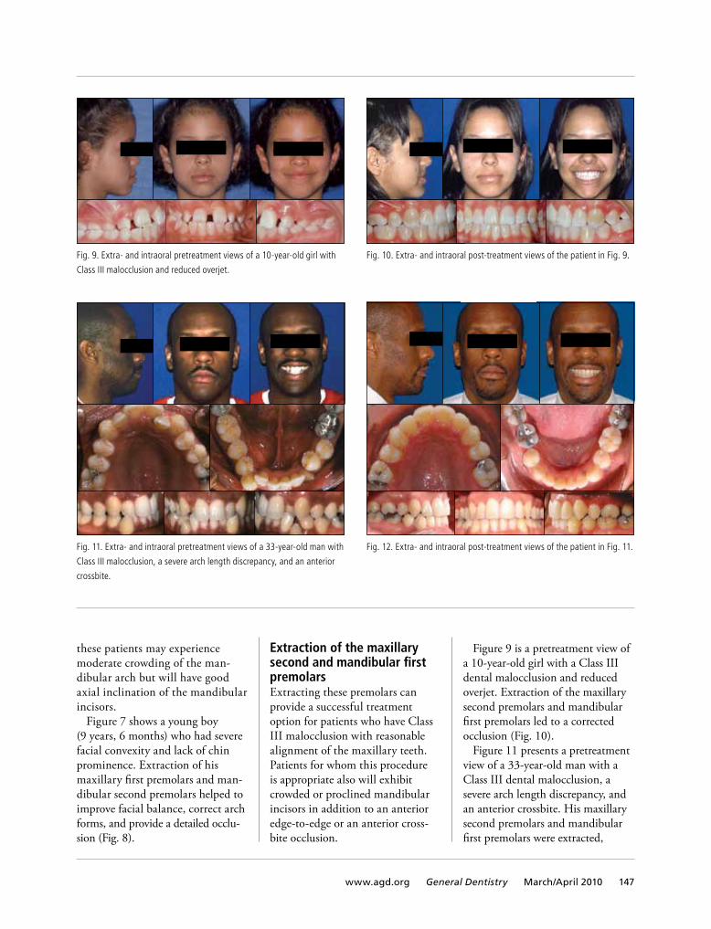
these patients may experience moderate crowding of the man-dibular arch but will have good axial inclination of the mandibular incisors.
Figure 7 shows a young boy (9 years, 6 months) who had severe facial convexity and lack of chin prominence. Extraction of his maxillary first premolars and man-dibular second premolars helped to improve facial balance, correct arch forms, and provide a detailed occlu-sion (Fig. 8).
Extraction of the maxillary second and mandibular first premolarsExtracting these premolars can provide a successful treatment option for patients who have Class III malocclusion with reasonable alignment of the maxillary teeth. Patients for whom this procedure is appropriate also will exhibit crowded or proclined mandibular incisors in addition to an anterior edge-to-edge or an anterior cross-bite occlusion.
Figure 9 is a pretreatment view of a 10-year-old girl with a Class III dental malocclusion and reduced overjet. Extraction of the maxillary second premolars and mandibular first premolars led to a corrected occlusion (Fig. 10).
Figure 11 presents a pretreatment view of a 33-year-old man with a Class III dental malocclusion, a severe arch length discrepancy, and an anterior crossbite. His maxillary second premolars and mandibular first premolars were extracted,
www.agd.org General Dentistry March/April 2010 147
Fig. 9. Extra- and intraoral pretreatment views of a 10-year-old girl with
Class III malocclusion and reduced overjet.
Fig. 10. Extra- and intraoral post-treatment views of the patient in Fig. 9.
Fig. 11. Extra- and intraoral pretreatment views of a 33-year-old man with
Class III malocclusion, a severe arch length discrepancy, and an anterior
crossbite.
Fig. 12. Extra- and intraoral post-treatment views of the patient in Fig. 11.

which led to a corrected dental relationship, showing that extrac-tion can be performed easily despite completion of growth (Fig. 12).
Extraction of maxillary first premolars onlyAccording to the literature, extrac-tion of maxillary first premolars is indicated for patients who have minimum growth potential, maxil-lary protrusion (with the mandibu-lar teeth in good alignment), good buccal occlusion with a Class II relationship (or a half-cusp Class II finishing to a solid Class II), and a slight open bite tendency.19 In the author’s experience, some patients experience orofacial pain as a result of this type of extraction.
Extraction of one mandibular incisorThis procedure can help patients who have an anterior edge-to-edge occlusion with minimal man-dibular crowding (that is, 5 mm or less—approximately the width of a mandibular central incisor).
This procedure is appropriate for patients with complete exclusion of the incisor from the dental arch, the presence of tooth size discrepancy, and good buccal occlusion.18
Figure 13 shows a 52-year-old woman who expressed concern about the misalignment of her mandibular incisors. Extraction of one mandibular incisor resulted in corrected arch forms and a fine-tuned occlusion (Fig. 14). Based on the literature and the author’s clinical experience, this method of treatment is effective for many adult patients.18
Bilaterally dissimilar extraction of premolarsGuided by the above criteria regard-ing appropriate candidates for this treatment, dentists can use bilaterally dissimilar extraction of premolars to correct midline discrepancies or asymmetrical classification of dento-skeletal conditions (for example, one side Class II and the other side Class I). Figure 15 shows the pretreatment views of a 14-year-old boy who had
a reasonably balanced face but also had moderate crowding of his maxil-lary and mandibular arches and a midline discrepancy. After extracting the maxillary right second premolar, the maxillary left first premolar, and the mandibular right and left second premolars, the patient’s arch forms and midlines were corrected; he also demonstrated detailing of his occlu-sion (Fig. 16).
SummaryThe debate over whether to extract teeth has existed in orthodontics for decades and is likely to continue ad infinitum. While many patients have been successfully treated without extraction, failure to extract and an over-reliance on alternative methods of space management could lead to a compromised pro-file, long-term instability, and the risk of periodontal problems.4,5,8,9,12
This article does not seek to compare the efficacy of extraction versus nonextraction; rather, it emphasizes the complexity of the treatment planning process and the
Orthodontics Criteria for performing extraction to treat certain malocclusions
148 March/April 2010 General Dentistry www.agd.org
Fig. 13. Extra- and intraoral pretreatment views of a 52-year-old woman
with misaligned mandibular molars.
Fig. 14. Extra- and intraoral post-treatment views of the patient in Figure 13.

need to arrive at treatment decisions that are informed by thorough assessment and careful diagnosis. By applying the criteria provided, together with detailed clinical examination and proper hard and soft tissue analyses, dentists can feel confident in choosing extraction as a treatment option and performing it in a manner that ensures the best outcome for each patient.
Author informationDr. Moshiri is a clinical professor, Department of Orthodontics, Uni-versity of Louisville, Kentucky, and is in private practice in St. Louis and Springfield, Missouri.
References 1. Tweed CH. Frankfort mandibular incisor angle in
orthodontic diagnosis, treatment planning and prognosis. Angle Orthod 1954;24:121-169.
2. Begg PR. Begg orthodontic theory and tech-nique, ed. 3. St. Louis: WB Saunders Co.;1977.
3. Steiner CC. Cephalometrics in clinical practice. Angle Orthod 1959;29:8-29.
4. Ricketts RM, Roth RH, Chaconas SJ, Schulhof RH, Engel GA. Orthodontic diagnosis and plan-ning. Denver: Rocky Mountain Orthodontics; 1982.
5. Kasai K. Soft tissue adaptability to hard tissue in facial profiles. Am J Orthod Dentofacial Orthop 1998;113(6):674-684.
6. Nanda RS. Growth changes in skeletal facial profile and their significance in orthodontic di-agnosis. Am J Orthod 1971;59(5):501-513.
7. Bowman SJ. More than lip service: Facial esthet-ics in orthodontics. J Am Dent Assoc 1999; 130(8):1173-1181.
8. Connor AM, Moshiri F. Orthognathic surgery norms for American blacks. Am J Orthod 1985; 87:119-134.
9. Formby WA, Nanda RS, Currier GF. Longitudinal changes in the adult facial profile. Am J Orthod Dentofac Orthop 1994;105(5):464-476.
10. Moshiri F, Jung S, Sclaroff A, Marsh J, Gay WD. Surgical diagnosis and treatment planning: A visual approach. J Clin Orthod 1982;16(1):37-59.
11. Boley JC, Pontier JP, Smith S, Fulbright M. Facial changes in extraction and nonextraction pa-tients. Angle Orthod 1998;69(6):539-546.
12. Moshiri F. Evidence based diagnosis and treat-ment planning. St. Louis Dent 2004;75(4):32-37.
13. Bishara S, Cummons D, Zaher A. Treatment and posttreatment changes in patients with Class II,
Division I malocclusion, after extraction and nonextraction treatment. Am J Orthod Dentofa-cial Orthop 1997;111(1):18-27.
14. Ngan P, Alkire R, Fields H. Management of space problems in the primary and mixed denti-tions. J Am Dent Assoc 1999;130(9):1330-1339.
15. Basciftci FA, Usumez S. Effects of extraction and nonextraction treatment on Class I and Class II subjects. Angle Orthod 2003;73(1):36-42.
16. Logan LR. Second premolar extraction in Class I and Class II. Am J Orthod 1973;63(2):115-147.
17. DeCastro N. Second premolar extraction in clini-cal practice. Am J Orthod 1974; 65 (2):115-137.
18. Bahreman AA. Lower incisor extraction in ortho-dontic treatment. Am J Orthod 1977;72(5):560-567.
19. Stalpers MJ, Booji JW, Bronkhorst EM, Kuijpers-Jagtman AM, Katsaros C. Extraction of maxillary first premolars in patients with Class II Division 1 malocclusion. Am J Orthod Dentofacial Orthop 2007;132(3):316-323.
20. Ozaki T, Ozaki S, Kuroda K. Premolar and addi-tional first molar extraction effects on soft tis-sue. Effects on high Angle Class II Division 1 patients. Angle Orthod 2007;77(2):244-253.
21. Uribe F, Nanda R. Treatment of Class II, Division 2 malocclusion in adults: Biomechanical consid-erations. J Clin Orthod 2003;37(11):599-606.
22. Fukui T, Tsurata M. Invisible treatment of a Class III female adult patient with severe crowding and cross-bite. J Orthod 2002;29(4):267-275.
www.agd.org General Dentistry March/April 2010 149
Fig. 15. Extra- and intraoral pretreatment views of a 14-year-old boy
with midline discrepancy and moderate crowding of his maxillary and
mandibular arches.
Fig. 16. Extra- and intraoral post-treatment views of the patient in Figure 15.

Persistent tongue ulcerNikolaos G. Nikitakis, DDS, PhD John K. Brooks, DDS
A 60-year-old man was referred by his general dentist for evaluation of a tongue ulcer of a few weeks’ duration. The patient did not recall biting or burning the area; however, for approximately a year, he had noticed the frequent occurrence of white-to-yellow lesions on the lat-eral border of the tongue in the area of the current ulcer. The patient indicated that the ulcer was painful during eating or when it rubbed against his teeth. The patient’s medical history was significant for chronic obstructive pulmonary disease (for which he was receiving oxygen therapy and theophylline, as well as fluticasone, salbuta-mol, budesonide, and formoterol inhalers); in addition, he was receiving antihypertensive diuretics (amiloride and hydrochlorothiazide) and omeprazole for gastric ulcer protection. He had smoked four packs of cigarettes a day for 40 years (a habit he discontinued four years earlier) and was a social drinker.
Clinical examination revealed a 1 cm x 1 cm ulcer
on the left lateral border of the tongue (Fig. 1), which was making contact with a sharp-edged retained root of the mandibular left first molar. The root fragment was extracted; however, a two-week follow-up assess-ment revealed no clinical signs of improvement in the ulceration. An incisional biopsy was undertaken for histopathologic assessment (Fig. 2).
Which of the following is the most appropriate diagnosis?
A. Squamouscellcarcinoma
B. Atypicalhistiocyticgranuloma
C. Chancre
D. Mucormycosis
E. Eosinophiliculceration
Diagnosisisonpage153.
Oral Diagnosis
150 March/April 2010 General Dentistry www.agd.org
Fig.1.Ulcersurroundedbyawhitehaloontheleftlateralborderofthe
tongue.
Fig.2.Mucosalulcerationcoveredbyafibrinopurulentmembrane;the
ulcerbedconsistsofgranulationtissuediffuselyinfiltratedbychronic
inflammatorycellswithnumerouseosinophils(H&Estain,magnification
100x).

A 54-year-old woman was evaluated for numerous brownish lesions that were affecting her mouth. She had discovered the lesions incidentally one month earlier, although she was unsure when they first appeared. The lesions were asymptomatic and had not changed perceptibly in appearance since their onset. No similar hyperpigmentations were apparent on the skin or other mucosal surfaces. The patient had smoked 5–10 cigarettes per day for approximately 30 years. The medical history was significant only
for a nodular goiter, which had been regulated by levothyroxine for the last 10 years. In addition, the patient had experienced an eruption of urticaria that had been managed with levocetirizine for approxi-mately three years; however, this medication had been discontinued for at least six months.
Clinical examination revealed diffuse brown macules with irregular borders, affecting the buccal mucosa bilaterally and the upper and lower labial mucosa (Fig. 1). An incisional biopsy was performed (Fig. 2). After
the patient discontinued her smok-ing habit, the lesions demonstrated a slow progressive reduction in terms of size and color intensity.
Which of the following is the most
appropriate diagnosis?
A. Malignantmelanoma
B. Intramucosalnevus
C. Smoker’smelanosis
D. Amalgamtattoo
E. Addison’sdisease
Diagnosisisonpage153.
Diffuse oral mucosal pigmentationsNikolaos G. Nikitakis, DDS, PhD John K. Brooks, DDS
www.agd.org General Dentistry March/April 2010 151
Fig.1.Diffusebrownpigmentedlesionsontherightbuccalmucosa.
Author informationDr. Nikitakis is an assistant professor, Department of Oral Pathology and Oral Medicine,
School of Dentistry, National and Kapodistrian University of Athens, Athens, Greece.
Dr. Brooks is a clinical professor, Department of Oncology and Diagnostic Sciences,
Dental School, University of Maryland, Baltimore.
Fig.2.Diffusemelaninpigmentation,freeandwithinmelanophages,in
thesuperficialconnectivetissueandoccasionallywithintheepithelial
basalcelllayer(H&Estain,magnification100x).

1. Which of the following central nervous system (CNS) structures is involved in the trigeminal nociceptive mechanism?A. Subnucleus interpolaris
B. Subnucleus oralis
C. Main sensory nucleus
D. Subnucleus caudalis
2. Patients sometimes have difficulty localizing pulpal pain due to convergence of neurons in theA. thalamus.
B. cerebral cortex.
C. trigeminal spinal tract nucleus.
D. dorsal horn of the spinal cord.
3. Nociceptive signals along the trigeminal pain pathway are perceived as pain when the signals reach theA. subnucleus caudalis.
B. posterior parietal cortex.
C. thalamus.
D. motor cortex.
4. Which of the following is not caused by central sensitization?A. Secondary hyperalgesia
B. Primary hyperalgesia
C. Referred pain
D. Muscle co-contraction and myofascial trigger
points
5. Which of the following represents deep somatic pain?A. Mucogingival pain
B. Traumatic neuralgia
C. Musculoskeletal pain
D. Atypical odontalgia
6. Which of the following is an example of neuropathic pain?A. Myofascial pain
B. Fibromyalgia
C. Postherpetic neuralgia
D. Cervical pain
7. Which of the following statements is true of psychogenic pain?A. It is caused by noxious stimuli.
B. It is caused by an abnormality of the nervous system.
C. It has a predictable pattern of pain location.
D. There is no relationship between symptoms and an
organic basis for pain.
8. Which of the following should be considered first when making a differential diagnosis of orofacial pain?A. Heterotropic pain
B. Intracranial pain disorder
C. Migraine
D. Myofascial trigger point pain
9. Which of the following causes a reproducible pattern of referred pain?A. Myofascial pain
B. Pain of pulpal origin
C. Pain of periodontal origin
D. Cluster headache
10. Which of the following neurovascular pain disorders is referred to as a suicide headache?A. Classic migraine
B. Tension-type headache
C. Cluster headache
D. Chronic daily headache
This quiz was written by Chikka M. Raju, DMD, MAGD,
ABGD, in cooperation with the other Self-Assessment
Committee members.
Answers are on pages 154–156.
Quiz No. 166
Differentialdiagnosisoforofacialpain
self-Assessment
152 March/April 2010 General Dentistry www.agd.org

Persistent tongue ulcerDiagnosis:E. Eosinophilic ulcerationTraumatic ulcerations of the oral mucosa are commonly encountered. They are caused by a variety of stimuli, including accidental biting, sharp teeth or restorations, prosthetic appliances, aggressive tooth brushing or flossing, and foodstuffs. Traumatic mucosal ulcers can be classified as acute or chronic, depending on their duration. Chronic ulcers may result from persistent injury (for example, a fractured tooth that continuously irritates the mucosa), although they may remain even after the traumatic etiol-ogy is removed. Eosinophilic ulceration (traumatic ulcer-ative granuloma with stromal eosinophilia) is a special subtype of chronic traumatic ulceration, characterized by a more protracted course and the microscopic presence of eosinophils. Similar lesions on the ventral and (less often) dorsal surface of the tongue in infants are attributed to nursing-associated trauma from anterior natal or neonatal teeth, referred to as Riga-Fede disease.
Clinically, chronic traumatic ulcers (including eosino-philic ulcerations) appear as well-defined lesions, usually covered by a yellowish pseudomembrane and surrounded by a white raised border. Induration of the margins may be discerned on palpation. Although lesions may occur anywhere in the oral mucosa, they typically occur at sites that are easily exposed to injury, such as the buccal mucosa, lips, and tongue. Lesions are often minimally symptomatic. Eosinophilic ulcerations sometimes appear raised, due to granulation tissue overproduction.
On microscopic examination, chronic traumatic ulcer-ations are covered by a fibrinopurulent membrane and exhibit granulation tissue with a mixed inflammatory infiltrate. The surrounding epithelium may show regen-erative changes and appear hyperplastic. In addition, eosinophilic ulceration features deeper extension of the inflammatory response into the connective tissue, with a conspicuous population of eosinophils and sometimes an exuberant granulation tissue reaction. This lesion needs to be distinguished from atypical eosinophilic ulceration (a monoclonal process characterized by cellular prolifera-tion of large cells of T-lymphocytic lineage), which shows a potential for frequent recurrence and dissemination.
Lesions that are clinically compatible with chronic traumatic ulcers or eosinophilic ulcerations that do not
heal within two weeks after removal of the purported traumatic stimulus must be biopsied to rule out other possibilities, most notably squamous cell carcinoma or ulcers of infectious etiology (tuberculosis, syphilis, deep mycoses). Incisional biopsies frequently promote healing. In persistent cases, topical or intralesional application of corticosteroids, in combination with mucosal barriers, may induce or accelerate healing.
Bibliography 1. segura s, pujol rm. eosinophilic ulcer of the oral mucosa: a distinct
entity or a non-specific reactive pattern? oral Dis 2008;14(4):287-295.
2. physical and chemical injuries. in: neville BW, Damm DD, allen cW, Bouquot Je. oral and maxillofacial pathology, ed. 3. st. louis: saunders/elsevier;2009:287-289.
Diffuse oral mucosal pigmentationsDiagnosis:C. Smoker’s melanosisOral pigmentation due to smoking is a relatively common finding and is associated with the degree and duration of usage. Melanin production increases in response to smoke substances and has been suggested to serve a protective role against cellular damage. Women are affected more frequently than men, presumably because of the synergis-tic effect of the female sex hormones.
The most commonly affected site associated with smoker’s melanosis is the anterior facial gingiva; however, any oral mucosal surface may be involved. Smoker’s mela-nosis may be seen in the gingiva of children, ostensibly due to secondhand smoke exposure. Lesions may be quite diffuse and appear brown, with varying intensity of color. They must be distinguished from racial pigmentation and other diffuse pigmented lesions of various causes, includ-ing drug-induced pigmentations, post-traumatic pigmen-tation, heavy metal poisoning, and systemic diseases such as Addison’s disease, neurofibromatosis, hemochromatosis, McCune-Albright syndrome, and Peutz-Jeghers syndrome. Malignant melanoma, which is rare in the oral mucosa, also may appear as diffuse pigmentation.
A biopsy of a suspected smoker’s melanosis lesion is indicated when there are atypical clinical findings, such as irregular borders, heterogenous pigmenta-tion, surface elevation of texture, pigmentary changes, and abrupt onset. Microscopic examination reveals increased melanin deposition in the basal cell layer of the
Oral Diagnosis
Answers
www.agd.org General Dentistry March/April 2010 153

epithelium and/or dispersed free or within melanophages in the superficial connective tissue. Cessation of smoking usually promotes a slow, progressive improvement in the clinical appearance of the lesions.
Bibliography 1. physical and chemical injuries. in: neville BW, Damm DD, allen cW,
Bouquot Je. oral and maxillofacial pathology, ed. 3. st. louis: saunders/elsevier co.;2009:316-317.
2. hedin ca, axell t. oral melanin pigmentation in 467 thai and malaysian people with special emphasis on smoker’s melanosis. J oral pathol med 1991;20(1):8-12.
3. hanioka t, tanaka K, ojima m, yuuki K. association of melanin pigmentation in the gingiva of children with parents who smoke. pediatrics 2005;116(2):e186-e190.
1. Answer: DThe pain sensation is carried to the central nervous system (CNS) by a complex arrangement of neurons, interneurons, and synaptic connec-tions that together are referred to as the trigeminal system. Primary afferent neurons carry sensory infor-mation from the face and mouth (except proprioception) to synapse with second order neurons in the trigeminal brain stem complex. This complex can be separated into the trigeminal main sensory nucleus and the trigeminal spinal tract nucleus. The trigeminal spinal tract nucleus is divided into three separate nuclei. From a superior (rostral) to inferior (caudal) direction, they are called the subnucleus oralis, the subnucleus interpolaris, and the subnucleus cau-dalis (the latter of which is involved in the nociceptive mechanism of the trigeminal nerve). 1. conti pc, pertes ra, heir Ga, nasri c,
cohen hv, araujo c. orofacial pain: Basic mechanisms and implication for successful management. J appl oral sci 2003;11(1):1-7.
2. okeson Jp. Bell’s orofacial pains, ed. 5. chicago: Quintessence publishing co., inc.;1995:13-42.
2. Answer: CThe trigeminal spinal tract nucleus
primarily receives afferent input from the trigeminal nerve and also from the facial, glossopharyngeal, vagus, and upper cervical (C2, C3) nerves. This convergence of neurons from different areas may synapse on another neuron, resulting in the brain being unable to interpret the exact location of the original pain source. For example, in early pulpal pain, the brain may appreciate that there is a toothache somewhere but cannot localize it. 1. conti pc, pertes ra, heir Ga, nasri c,
cohen hv, araujo c. orofacial pain: Basic mechanisms and implication for successful management. J appl oral sci 2003;11(1):1-7.
2. okeson Jp. Bell’s orofacial pains, ed. 5. chicago: Quintessence publishing co., inc.;1995:13-42.
3. Answer: BAfter first order neurons synapse with second order neurons in the sub-nucleus caudalis, nociceptive signals are carried to the thalamus. From the thalamus, signals reach different parts of the somatosensory cortex through third order neurons. Incoming noci-ceptive signals from the subnucleus caudalis and ascending nociceptive signals on their way to the thalamus can be modified by descending nerve fibers from higher levels of the
CNS or through various drugs. This process is called pain modulation, which is the inherent ability of the nervous system to alter the intensity of nociceptive signals and reduce pain intensity. After transmission and modulation, the nociceptive signals are perceived as pain in the posterior parietal cortex of the brain. 1. conti pc, pertes ra, heir Ga, nasri c,
cohen hv, araujo c. orofacial pain: Basic mechanisms and implication for successful management. J appl oral sci 2003;11(1):1-7.
2. okeson Jp. Bell’s orofacial pains, ed. 5. chicago: Quintessence publishing co., inc.;1995:13-42.
4. Answer: BFollowing tissue injury, a con-tinuous barrage of noxious afferent input results in the spontaneous firing of afferent fibers and the sensi-tization of nociceptors at the site of injury (primary hyperalgesia). When a second order neuron receives these prolonged nociceptive inputs, it may also become sensitized, causing hyperexcitability of CNS interneurons. This phenomenon is referred to as central sensitiza-tion. The central excitatory effects produce changes in afferent sensory neurons (secondary hyperalgesia and referred pain), efferent motor
154 March/April 2010 General Dentistry www.agd.org
Self-Assessment Quiz No. 166

neurons (muscle co-contraction and myofascial trigger points) and the autonomic nervous system (injec-tion of the conjunctiva, lacrimation, nasal secretion, nasal congestion). 1. okeson Jp. Bell’s orofacial pains, ed. 5.
chicago: Quintessence publishing co., inc.;1995:64-73.
2. cohen s, Burns r. pathways of the pulp, ed. 9. st. louis: mosby;2006:62-64.
5. Answer: CSomatic pain is caused by the noxious stimulation of normal neural structures of the affected area. When the external surface of the body is involved, the pain is called superficial somatic pain (for example, cutaneous pain and mucogingival pain) and is char-acterized by a bright, stimulating quality. The pain can be localized, is anatomically accurate, and is proportionate to direct stimulation. Topical anesthetic at the site will temporarily arrest the pain.
In contrast, if the pain originates from a deep body structure, it is called deep somatic pain (for example, musculoskeletal pain and visceral pain); it is characterized by a dull, depressing quality. The pain may not be localized, is anatomi-cally less accurate, and may not be proportional to the stimulus. Appli-cation of a topical anesthetic usually does not arrest the pain except in the visceral mucosa. Deep somatic pain also frequently generates effects secondary to CNS sensitization. These effects may include referred pain, localized autonomic effects, secondary hyperalgesia, and muscle co-contraction. Atypical odontolgia is a continuous neuropathic pain disorder, characterized by the absence of dental pathology. 1. pertes ra. Differential diagnosis of orofacial
pain. mt sinai J med 1998;65(5-6):348-354.
2. okeson Jp. Bell’s orofacial pains, ed. 5. chicago: Quintessence publishing co., inc.;1995:103-133.
3. Balasubramaniam r, Klasser GD. orofacial pain and dysfunction. oral maxillofac surg clin north am 2008;20(2):ix-x.
6. Answer: CNeuropathic pain results from a structural abnormality of the ner-vous system itself, characterized by the absence of any obvious noxious stimulation. It is out of proportion to the degree of stimulation (that is, light touch can cause intense pain), may present paresthesia along the nerve distribution, and is relatively unresponsive to low doses of narcotic analgesics. Neuropathic pain is usually described as being bright, intense, stimulating, and burning, and can be divided into episodic (for example, trigeminal, glossopharyngeal, superior laryn-geal, and geniculate and interme-dius neuralgias) and continuous disorders (for example, peripheral neuritis, herpes zoster, postherpetic neuralgia, traumatic neuralgia, and atypical odontolgia). 1. pertes ra. Differential diagnosis of
orofacial pain. mt sinai J med 1998;65(5-6):348-354.
2. okeson Jp. Bell’s orofacial pains, ed. 5. chicago: Quintessence publishing co., inc.;1995:103-133.
3. Balasubramaniam r, Klasser GD. orofacial pain and dysfunction. oral maxillofac surg clin north am 2008;20(2):ix-x.
7. Answer: DPsychogenic pain is not caused by noxious stimuli or any abnormal-ity of the nervous system; it is characterized by the history of definite emotional or personality disorders, and the absence of any anatomical relationship between the site and source of pain. There is no relationship between physical symptoms and any organic basis for the pain. Psychogenic pain may be felt in many areas and locations. The degree of pain is often exag-gerated and the treatment outcome usually is inconsistent. Examples of
psychogenic pain include depressive disorders, bipolar disorders, and post-traumatic stress disorders. 1. pertes ra. Differential diagnosis of
orofacial pain. mt sinai J med 1998;65(5-6):348-354.
2. okeson Jp. Bell’s orofacial pains, ed. 5. chicago: Quintessence publishing co., inc.;1995:103-133.
3. Balasubramaniam r, Klasser GD. orofacial pain and dysfunction. oral maxillofac surg clin north am 2008;20(2):ix-x.
8. Answer: BIntracranial pain disorders (for example, neoplasm, aneurism, abscess, hematoma, and edema) must be considered first in the differential diagnosis because of life-threatening consequences. The characteristics of pain from an intracranial source include an abrupt onset, progressively more severe pain, interruption of sleep, weight loss, ataxia, weakness, fever, neuro-logic signs, and the pain precipitated by exertion or positional change (for example, coughing and sneezing). 1. okeson Jp, ed. orofacial pain: Guidelines
for assessment, diagnosis and manage-ment, ed. 4. carol stream, il: Quintessence publishing co.;2008:55.
2. pertes ra. Differential diagnosis of orofacial pain. mt sinai J med 1998;65(5-6):348-354.
9. Answer: AMyofascial pain is the most common form of musculoskeletal pain, affecting the head, neck, and face. It is characterized by a localized area of tight, palpable, tender bands of muscle called myofascial trigger points (TPs). When provoked or palpated, TPs refer pain in repro-ducible patterns to areas distant from the site of TPs (known as the zone of reference). The key to diag-nosing myofacial pain is to identify TPs and “a reproducible pattern of referred pain.” 1. conti pc, pertes ra, heir Ga, nasri c,
cohen hv, araujo c. orofacial pain: Basic
www.agd.org General Dentistry March/April 2010 155
mechanisms and implication for successful management. J appl oral sci 2003;11(1):1-7.
2. pertes ra. Differential diagnosis of orofacial pain. mt sinai J med 1998;65(5-6):348-354.
10. Answer: CNeurovascular pain disorders (also known as primary headache disorders) are a vascular response to a neurologic mechanism. The cluster headache (CH) is also referred to as a suicide headache and is one of the most severe forms of headache and facial pain. It is relatively uncommon. CH manifests as sharp, throbbing, or boring pain of severe intensity that
can be associated with lacrimation, conjunctival injection, rhinorrhea, nasal congestion, miosis, ptosis, or eyelid edema and facial sweating. These headaches are unilateral, usually side-fixed throughout the lifetime of the patient, and may last from 15 minutes to three hours.
Migraine headache usually mani-fests as unilateral pain, has a pulsat-ing quality, is moderate to severe in intensity, is aggravated by physical activity, and is accompanied by nausea, vomiting, photophobia, or phonophobia. The attack usually lasts 4–72 hours.
Tension-type headache often
manifests as bilateral dull pain, is non-pulsating, has a mild to moder-ate intensity, is not aggravated by physical activity, and is not accom-panied by nausea or vomiting. The attack may last from 30 minutes to seven days.
Chronic daily headache has a similar presentation to tension-type headache with superimposed episodes of migraine. 1. okeson Jp, ed. orofacial pain: Guidelines
for assessment, diagnosis and manage-ment, ed. 4. carol stream, il: Quintessence publishing co.;2008:55-78.
2. pertes ra. Differential diagnosis of orofacial pain. mt sinai J med 1998;65 (5-6):348-354.
156 March/April 2010 General Dentistry www.agd.org
Correctionin the september-october 2009 issue, the abstract for the october case study, “chondrosarcoma of the mandible: case report and literature review” (p. 467) should have read as follows:
chondrosarcoma is a malignant cartilaginous tumor that rarely occurs in the maxillofacial bones. a 44-year-old woman complained about swelling and mild pain during mastication in the right parasymphysis region. clinical and radiographic examinations revealed characteristics of osteosarcoma. a microscopic examination revealed an abundant proliferation of malignant neoplastic cartilage cells of varying sizes arranged as immature tissue and the absence of an osteoid matrix. this article presents a case of chondrosarcoma of the jaw and discusses the differences between osteosarcoma and chondrosarcoma.
Self-Instruction
Exercise No. 231March/April 2009, p. 127 1. a 2. C 3. d 4. C 5. a 6. B 7. C 8. d 9. a 10. a 11. C 12. d 13. d 14. d 15. B
Exercise No. 232March/April 2009, p. 144 1. C 2. C 3. d 4. a 5. d 6. B 7. B 8. a 9. B 10. C 11. a 12. d 13. B 14. a 15. C
Exercise No. 233March/April 2009, p. 157 1. d 2. B 3. B 4. C 5. a 6. C 7. B 8. B 9. B 10. B 11. a 12. a 13. d 14. C 15. d
Correctionin the november/December 2009 issue of General Dentistry, the Dental materials column (pp. 550-551) included an incorrectly cited reference. the paragraph in question should read:
With self-etch products being introduced on almost a weekly basis, you would think that the gold standard had shifted away from total-etch. But even the owner of one of the most prolific manufactur-ers of both types of adhesives was quoted recently as stating that he still considers “fourth generation, total-etch adhesives” as the pinnacle.2 in addition, the only bonding agent to receive a five-star rating (the highest possible rating based on clinical and laboratory testing) in the 2009 annual edition of reality happens to be a fourth generation, total-etch product. furthermore, the bonding agent that created the so-called seventh generation (that is, an all-in-one self-etch material) has recently undergone brand extension with the introduction of a total-etch sibling.

abdullaibrahim abdulwaheed, dMd, Magd, aBgd Quincy, Ma andrew achord, dMd Flowood, MS Sidney L. adler, ddS, Magd Bronx, NY Seyed Hamzeh aghaie, ddS, Fagd Thornhill, oNMert N. aksu, ddS ann arbor, MI a. Lichelle aldana, ddS, Fagd Salem, Ma Charles d. aldridge, ddS, Magd Bend, or Saad abdullah M. al-Harbi, BdS Jeddah, Saudi arabiaPaul o. aliker, ddS, Fagd Kampala, UgandaFiras al-Khalili, ddS, Fagd ottawa, oNFrancis w. allen, dMd, Magd Salem, or wael M. al-omari, BdS, MdSC, Phd Irbid, JordanThomas L. anderson, ddS Prairie Village, KS Paul a. andrews, ddS Lake Mary, FL John r. antonelli, ddS davie, FL James r. arneson, ddS Kodiak, aK andrew J. avillo, ddS, Fagd, aBgd San diego, Ca Michael L. Babinski, dMd, Magd Colchester, CT william r. Baez, ddS, Fagd, aBgd aldie, Va ritu Bahl, dMd Edmonds, wa Saul Bahn, dMd, MScd New York, NY Hisham Barakat, ddS, Magd Virginia Beach, Va Peter g. Bastian, ddS, Magd Huntsville, oNwillie K. Beasley, ddS, Magd Mandeville, La ryle a. Bell, ddS washington, dC Joseph a. Belsito, ddS, Fagd windsor, oNSteven d. Bender, ddS Plano, TX Marvin H. Berman, ddS Bannockburn, IL Charles E. Berner, ddS, Magd Cleveland, oH Beatriz Bezerra, ddS, MSc Piracicaba, SP, Brazilwesley S. Blakeslee, dMd, Fagd Manasquan, NJ warren E. Boardman, dMd, Fagd ridgewood, NJ rebecca M. Bockow, ddS Seattle, wa richard Bohay, dMd, MSc, MrCd(C) London, oN
Nicholas J. Borrello, ddS, Magd Bethel, CT Barry L. Bowden, ddS, Magd austin, TX william Bowen, BdS, Phd rochester, NY Carl H. Boykin, ddS, Magd Jackson, MS Terry w. Bradigan, ddS, Fagd, aBgd Hanover, NH Craig r. Brandon, ddS, Magd San diego, Ca John K. Brooks, ddS owings Mills, Md ronald S. Brown, ddS washington, dC Joseph B. Burley, ddS, Fagd Hagerstown, Md Carroll r. Butler, ddS, Fagd Kerrville, TX Michael J. Calabrese, dMd, Magd Feeding Hills, Ma Jefferson r. Call, dMd, Fagd Hillsboro, or Thomas H. Callahan, ddS, Magd atlanta, ga Kim L. Capehart, ddS Simpsonville, SC Keefe E. Carbone, ddS Lawton, oK Jean J. Carlson, ddS, Fagd Cambridge, Md John F. Carpenter, dMd, Magd New windsor, NY James L. Carroll, ddS, Magd auburn, aL Stephen w. Carstensen, ddS, Fagd Bellevue, wa ambrose Chan, BdS (hons) dCd, MSd, FraCdS Sydney, australia James w. Chancellor, ddS, Fagd San antonio, TX richard M. Chapin, ddS Kinston, NC william r. Chase, ddS, Fagd Cathedral City, Ca Leslie C. Chew, ddS richmond, BSVenu M. Chimmiri, ddS, Fagd Manchester, CT randolph a. Coffey, dMd, Magd, aBgd Bradenton, FL Harvey S. Cohen, ddS, Magd Baltimore, Md Kaianne M. Conibear, ddS orlando, FL Mullen o. Coover, ddS, Magd, aBgd Mount Pleasant, SC Steven a. Corben, dMd, Fagd danvers, Ma adam P. Cormier, ddS Bossier City, La Tadeasha E. Culbreath, dMd washington, dC Bruce E. Cunningham, dMd, Magd Jacksonville, aL Joseph L. dautremont III, ddS, Magd woodland Hills, Ca
T. Bob davis, dMd, Fagd dallas, TX Paul dawood, ddS, Fagd anaheim, Ca Scott S. derossi, dMd augusta, ga Joseph M. diacovo, dMd, Magd Kingston, NY Shihab M. diais, ddS, Fagd odessa, TX Mark donaldson, Pharmd Kalispell, MT Valerie a. drake, ddS, Fagd Harlingen, TX Scott M. dubowsky, dMd, Fagd Bayonne, NJ dwight d. duckworth, ddS, Magd Springdale, ar alicia L. dwyer, ddS, Magd Irving, TX James S. Eaves, ddS, Fagd woodbridge, Va E. Mac Edington, ddS, Magd, aBgd Clarksville, TN Michael d. Edwards, dMd, Magd wedowee, aL denise Estafan, ddS, MS New York, NY Michael a. Forman, ddS Vaughn, oNadam J. Freeman, ddS, Magd westport, CT Jeffrey J. gardner, dMd, Fagd Mount Pleasant, SC Jeffrey B. geno, ddS, Magd League City, TX Craig B. gimbel, ddS, Fagd denville, NJ Michael L. glass, ddS, Fagd Lancaster, Ca Jason H. goodchild, dMd Berwyn, Pa Tim L. goodheart, ddS, Fagd raytown, Mo John J. graeber, dMd, Magd East Hanover, NJ Scott E. gray, ddS, aBgd, Fagd Englewood, oH Janiene F. gresla, ddS, Fagd Burlington, Ma Paul a. guidi, dMd, Fagd Clearwater, FL Jeffery w. Hadley, ddS, Fagd Henderson, NV dan Hagi, ddS, Fagd Thornhill, oNJosh M. Halderman, ddS Columbus, oH Thomas d. Hamilton, ddS, Fagd granbury, TX guy M. Hanson, ddS, Magd Boise, Id royce a. Hatch, ddS, MS denver, Co Tina Heil, dMd, Fagd Suwanee, ga gerald N. Hino, ddS, Magd Yakima, wa
william L. Hoch, dMd, Magd Beaver, Pa Isaac a. House, ddS, Fagd Haughton, La James H. Howard, ddS, Fagd omaha, NE Kimberly Quan Hubenette, ddS, Fagd Santa rosa, Ca Kevin d. Huff, ddS, Magd dover, oH Eric M. Hurst, ddS, Fagd Friendswood, TX Fahmida Hussain, dMd, Fagd Philadelphia, Pa Emily Hwang Cambridge, MaLanny Jacob, dMd winnipeg, MBThomas C. Jagor, ddS, Magd atlanta, ga Kevin a. Jasinski, dMd, Fagd McColl, SC Mark E. Jensen, ddS, Fagd, aBgd diamondhead, MS gregory K. Johnson, ddS, Fagd Kansas City, Mo greggery E. Jones, dMd, Magd redmond, or Bhushan S. Joshi, dMd, Magd, aBgd Tinton Falls, NJ gary J. Kaplowitz, ddS, aBgd Pikesville, Md ashim Kapur, ddS westford, Ma Kristopher J. Kelly, ddS, Fagd Clear Lake, Ia John H. Kilian, dMd, Fagd Troutdale, or dimitrios Kilimitzoglou, ddS, Magd Smithtown, NY Eugene Y. Kim, ddS San diego, CaJoseph S. Kim, ddS, Fagd Yorkville, IL william r. Kisker, dMd, Fagd Lake Villa, IL Bruce g. Kleeberger, ddS Langley, BCLisa M. Klemes, dMd, Fagd Naples, FL James S. Knight, ddS, Magd, aBgd Mount Pleasant, SC robert J. Koolkin, ddS, Fagd Turners Falls, Ma Harish Koratkar, BdS Hopkins, MN Lawrence Kotlow, ddS albany, NY gregori M. Kurtzman, ddS, Magd Silver Spring, Md william C. Kuttler, ddS, Fagd dubuque, Ia Frederick C. Lally, ddS, Magd Tunkhannock, Pa Edward E. Lancaster III, dMd, Fagd williamston, SC Brent a. Larson, ddS, Fagd Salt Lake City, UT daniel P. Lavin, dMd, Magd, aBgd Mililani, HI
2009 Reviewers
www.agd.org General Dentistry March/April 2010 157
We express our gratitude here to the following individuals who reviewed materials for General Dentistry from January 1 to December 31, 2009.

Kenneth J. Lawlor, ddS, Magd Markham, oNandrew S. Lazaris, ddS, Fagd Plano, TX Marden K. Leavitt, ddS Saint george, UT Hyewon Lee Cambridge, MaNathan B. Lee, dMd Easley, SC Sonny w. Lee, ddS, Fagd Thornhill, oNHarry M. Lehrer, dMd, MS Hollywood, FL Steven M. Lepowsky, ddS, Fagd Farmington, CT d. Lewis, ddS, Fagd Seattle, wa Stuart Lieblich, dMd avon, CT adriano Fonseca Lima, ddS, MS Piracicaba, SP, BrazilJames S. Lindemuth, ddS, aBgd, Magd Benicia, Ca Edward Lowe, dMd Vancouver, BCFrederick Magaziner, ddS, Magd randallstown, Md wayne w. Maibaum, dMd Poughquag, NY Mickel a. Malek, ddS, Fagd Tucson, aZ Moody w. Malek, ddS, Fagd Salinas, Ca John Mamoun, dMd green Village, NJ Michael d. Marcus, dMd, Fagd Silver Spring, Md daniel K. Marinic, ddS, Fagd Evanston, IL Stanley Markovits, ddS, Magd white Plains, NY anthony a. Martin, dMd, Fagd Knoxville, TN Jurga d. Martini, dMd, Fagd Seattle, wa James Martyniak, ddS, Fagd white Plains, NY Loren K. Masuoka, dMd Ewa Beach, HI robert Steven Matthews, ddS, Magd Clinton, TN Venu Maturi, ddS, MS, Fagd Champaign, IL Michael T. McClure, dMd, Fagd orange Park, FL david g. McMillan, ddS Layton, UT Vivek S. Mehta, dMd, Fagd Littleton, Ma gigi Meinecke, dMd, Fagd Potomac, Md Mark Meraner, ddS Philadelphia, Pa Kenneth a. Mertz Jr., dMd, Magd Fernandina Beach, FL gregory T. Messina, ddS, Fagd rockford, IL John r. Miller, ddS, Magd, aBgd Chesterfield, Mo Travis T. Miller, ddS, Magd orange, TX
david Mock, ddS Toronto, oNBijan Modjtahedi, ddS, Fagd Fountain Valley, CaJoseph d. Molinaro, dMd, Magd, aBgd San diego, Ca Kevin J. Mosmen, dMd Brick, NJ robert E. Murphy, ddS, Fagd El Segundo, Ca Ellis J. Neiburger, ddS waukegan, IL Clarisse C. Ng, ddS, Fagd Singapore, SingaporeLinda C. Niessen, dMd dallas, TX Michael F. Nolan, ddS, Magd Monroe, La Se-Lim oh, dMd Baltimore, Md wynn H. okuda, dMd Honolulu, HI Erica r. oliveira, ddS, Fagd San antonio, TX Judy olshin, ddS woodmere, NY Larry J. o’Neill, dMd, Magd, aBgd Parker, Co Howard E. ong, ddS, Fagd Seal Beach, Ca Trent L. outhouse, ddS, Fagd, aBgd Norfolk, Va Barry M. owens, ddS Memphis, TN Charles J. Palenik, Phd, MS Indianapolis, IN Leena Palomo, ddS, MSd Cleveland, oH dianne d. Pannes, ddS, Magd, aBgd Kapolei, HI dana r. Parker, dMd Easley, SC Steven Parker, BdS, LdS, rCS Harrogate, EnglandJoseph w. Parkinson, ddS Kansas City, Mo Nathan C. Parrish, ddS, aBgd Fort Knox, KY amit P. Patel, dMd Lutz, FL Ivy d. Peltz, ddS, Magd New York, NY Jefferson ricardo Pereira, ddS, MSc, Phd Mar grosso Laguna, SC, BrazilChristopher J. Perry, dMd, Fagd San antonio, TX Kirk J. Petersen, dMd, Fagd Hemet, Ca Jack Piermatti, dMd, Fagd Voorhees, NJ gregory Polyzois, ddS, dr.dent, MScd athens, greeceBart F. Presti, ddS, Magd McKinney, TX Thad K. Putnam, ddS, Fagd georgetown, TX danish Qadri, ddS Fort Lee, NJ diana L. Quilici, ddS, Fagd Mather, Ca richard rapoport, ddS, Magd westmount, QC
david E. reid, ddS, Magd, aBgd augusta, ga george d. reskakis, ddS, Fagd New York, NY Elio reyes, ddS, MSd alton, IL Pat riley, ddS, Fagd Mcallen, TX Tanya M. robinson, dMd, Fagd New orleans, La Cliff E. rogge, ddS, Fagd Longmont, Co gerald r. ross, ddS Tottenham, oNwilliam F. ross III, dMd, Fagd rock Hill, SC Ezedin Sadeghi, ddS, MS Milwaukee, wI Saman Saghafi, ddS, Fagd woodinville, wa Fouad S. Salama, BdS, MS omaha, NE remedios J. Santos, dMd, Magd Jacksonville, FL Pooja Saroha, ddS Burlington, Ma Corrine r. Scalzitti, dMd, Magd austin, TX Lawrence E. Scheitler, ddS, Magd, aBgd Seminole, FL Sanford N. Schwartz, ddS, Fagd Brandon, FL Kevin d. Scott, ddS, Fagd Bulverde, TX george d. Segel, dMd, Fagd Newtown, Pa gretchen E. Seibert, ddS, Fagd Frostburg, Md andrea Leigh Shah Brighton, Ma Paresh Shah, dMd, Fagd winnipeg, MBwesley E. Shankland II, ddS Columbus, oH Eric Z. Shapira, ddS Half Moon Bay, Ca Eric N. Shelly, dMd, Magd west Chester, Pa Kishore Shetty, ddS, Magd denver, Co Kim C. Skaggs, ddS, Fagd Blue Springs, Mo John E. Smith, dMd, Magd Helena, MT gregory J. Solof, ddS, Fagd avon, CT Jane a. Soxman, ddS allison Park, Pa gregory K. Spackman, ddS San antonio, TX Felipe F. Sperandio, ddS, MS Sao Paulo, SP, Brazilruth d. Spieker, ddS, Fagd Iowa City, Ia Thomas J. Spranley, ddS, Fagd Mandeville, La wayne w. St. Hill, ddS, Magd Stone ridge, NY Jonathan M. Stahl, ddS, Magd, aBgd San Francisco, Ca robert E. Steele, ddS, Fagd denison, TX
Charles d. Stetler, ddS, Fagd wylie, TX Jerry M. Strauss, dMd, Magd Fairfield, NJ Lawrence J. Sutton, ddS, Magd ocala, FL Jon B. Suzuki, ddS, Phd, MBa Philadelphia, Pa John Svirsky, ddS, MEd richmond, Va Mark J. Szarejko, ddS, Fagd Palm Harbor, FL Charles d. Tatlock, ddS, Magd albuquerque, NM John d. Thomas Jr., ddS, Magd Cordova, TN Harry P. Thompson, dMd, Fagd Elizabethtown, KY Shreedevi Thulasidas, ddS, Fagd Milltown, NJ richard P. Timm, ddS, Magd oakdale, MN Jon M. Trabosh, ddS, Magd Elkton, Va Linda g. Trotter, dMd, Fagd Jacksonville, FL James H. Tyack, dMd, Magd Clatskanie, or Joseph Valenzi, dMd, Magd Port orange, FL Valerie a. Venterina, ddS Brooklyn, NY Sharon M. Verdinelli, dMd, Fagd Mantua, NJ gary Vander Vliet, ddS, Magd Hackettstown, NJ Carl B. Vorhies, ddS, Magd Portland, or Charles wakefield Jr., ddS, Magd dallas, TX Christopher J. walinski, ddS Fall river, Ma Judson B. wall, ddS, Fagd Bountiful, UT rudy wassenaar, dMd, Magd williams Lake, BC Michelle Sabater weddle, ddS, Fagd Millington, NJ randy S. weiner, dMd, Fagd Millis, Ma Barclay K. weisberg, ddS, Fagd Chesapeake, Va Larry N. williams Jr., ddS, Magd grayslake, IL Jon w. williamson, ddS, Magd Cedar Hill, TX Joseph a. wineman, dMd, aBgd Henderson, NV Joseph B. wommack, ddS, Fagd Parsons, KS Paul r. wonsavage, ddS, Magd Hanover, NH Peter Yaman, ddS, MS ann arbor, MI Martin H. Zase, dMd, Magd Colchester, CT
158 March/April 2010 General Dentistry www.agd.org

www.agd.org General Dentistry March/April 2010 159
Now it’s easier than ever to record and submit exercises and earn CE credit! An exercise-specific answer form for submitting your Self-Instruction exercises is located on the back side of this page. Please use this form if you submit your Self-Instruction exercises via regular mail or fax.
The form is easy to use: Simply tear it out or make a copy of it, record your answers for the Self-Instruction exercises in this issue, and fax or mail the form to us. For AGD members, credit will be entered on your transcript immediately once the form is graded.
For participants, the Self-Instruction program offers convenience, simplicity, and faster accreditation—what could be better? If you have any questions regarding this answer form, call toll-free 888.243.3368, ext. 4353.
How do I register?Simply submit the form below. A letter confirming your enrollment will be sent within two weeks of registration. Exercises may be submitted upon receipt of registration.
How long do I have to submit my exercises?All exercises must be submitted within 12 months of enrollment.
How do I submit answers?Fill out the exercise-specific answer form on the back side of this page and mail or fax it as indicated on the form. Answer forms must be completed as directed in the instructions; otherwise, they will not be processed.
When are answers due?Answers for the exercises in this issue (No. 255–257) must be received by February 28, 2011. Credit will not be awarded for exercises postmarked after the deadline.
How much credit is earned?Participants must answer at least 12 of the 15 questions correctly (80%) to earn two CE credits.
How is credit registered?AGD membersThe results are entered directly into your CDE record, which is maintained by the AGD. You may request a printout of your CDE record at any time during the year, or you can view it on the AGD’s Web site at www.agd.org/education/transcript.
NonmembersYou will receive a letter acknowledging successful completion of an exercise. These letters may be submitted to the appropriate state board or agency for licensure purposes.
What if I don’t pass?A letter will be sent to all participants who score below 80%.
Please direct all other questions to Cassandra Bannon, either via phone (toll-free) at 888.AGD.DENT (888.243.3368), ext. 4353 or via e-mail at [email protected].
The program
FAQs
Sign up today!6 exercises (12 credits)
q $100 (AGD member rate)
q $150 (nonmember rate)
12 exercises (24 credits)
q $150 (AGD member rate)
q $225 (nonmember rate)
q I have enclosed a check or money order in U.S. funds made payable to AGD.
q I prefer to charge my credit card: q Visa q MasterCard
ImportantIf you are not a member of the AGD, you must enroll by phone. Call toll-free at 888.243.3368, ext. 4353 to enroll today!
Fax: 312.440.4261
Mail with payment:Academy of General Dentistry-DE28148 Network PlaceChicago, IL 60673-1281
Name:
AGD Number:
Card Number:
Expiration Date:
self instructionCDECREDIT
For more than 15 years, the Self-Instruction program has offered in-formative articles, challenging exercises, and low-priced, high-quality continuing dental education. If you have yet to take advantage of the opportunity to participate in this exciting PACE-approved program, sign up below or visit us on the Web at www.agd.org/publications/gd/selfinstruct to find out what you’ve been missing.
Whether you’re looking for continuing education credits for state license renewal, your AGD membership maintenance requirement, or the AGD Fellowship or Mastership award, or if you’re simply seeking to expand your knowledge, the Self-Instruction program has exactly what you need.
CE credits from a source you can trust

2 5 5 2 5 6 2 5 7
2 5 5 2 5 6 2 5 7
7164108412

Have you visited the AGD’s consumer Web portal, www.KnowYourTeeth.com? The AGD has launched this new Web portal to meet a growing need for accurate, accessible information in a user-friendly format for you and your patients. Use the site to…
•AddContenttoYourWebSite– Consider www.KnowYourTeeth.com an extension of your existing Web site. There are Web stickers and flyers available in the Marketing Toolkit (Members Only).
•ProvidePatientswithValuableInformation–You and your patients can search through more than 400 articles about popular oral health topics, such as oral hygiene and dentures.
•IncreaseReferralstoYourOffice–All active AGD members are entered into the “Find an AGD Dentist” referral service. Consumers who visit the site can find you just by entering their ZIP code.
Visit and share the site today!
www.KnowYourTeeth.comAn AGD Resource for You and Your Patients to Smile About

Evaluation of shear bond strength between self-etching adhesive systems and dentin and analysis of the resin-dentin interface Cleonice Silveira Teixeira, ddS, MSc, Phd n Marcelo Carvalho Chain, ddS, MSc, Phd
esthetic dentistry has developed considerably in recent decades. The majority of modern adhe-
sive systems utilize a preliminary acid-etching step, which is applied over the enamel and dentin. Dentin etching is necessary to remove the smear layer and to expose the col-lagen fibrils.1
Problems have been associated with the adhesive systems that use acid preconditioning, related to the adhesive’s capacity for infiltrating into the demineralized collagen. Some articles have stressed that the adhesive does not always penetrate into the dentin as deeply as the acid does during the preconditioning, leaving an unprotected basal portion of collagen that is not embedded in the resin.2-4 Collagen fibrils that are unprotected and incompletely coated with resin are more easily affected by hydrolysis, enzymatic attack, and functional and thermal stress, eventually resulting in the
degradation of the bond.5 This weakened zone would be susceptible to hydrolysis from oral and dentinal fluids, jeopardize the bonding strength and durability, and cause microleakage and its consequences.5
Systems that act as a conditioner and primer at the same time may be used to avoid this type of problem, since the dentin will be demineral-ized and infiltrated simultaneously by the monomers.6,7 At present, self-etching adhesives are used widely, mainly because of their ease of use, their low technique-sensitivity, and their adequate performance in Class V clinical trials.8-10 One-step self-etching systems mix hydrophobic and hydrophilic monomers together with a large amount of solvent. This adhesive strategy is based on the dissolution and fixation of the smear layer, through the infiltration of mildly acidic hydrophilic mono-mers.6,7 Most self-etching systems are methacrylate-based and contain
highly acidic monomers, with a pH of approximately 1.5–2.5.6,7,11
Various studies have questioned the capacity of these adhesive systems to demineralize the dentin sufficiently to form the hybrid layer.10 This layer may have a limited thickness or may be absent in some areas of the dentin/restoration interface, resulting in a decreased bond strength and loss of adhe-sion.4,10 Some studies have indicated that adhesives which use the total etching technique result in a better performance.2,11 Other studies have reported better performance from self-etching adhesives, indicating a need for additional research to resolve this question and verify the bond strength of current self-etching primer systems.8,9
This study sought to determine the in vitro shear bond strength of four self-etching adhesive systems: Clearfil SE Bond (J. Morita USA, Inc.) (Group 1), Optibond Solo
this study evaluated the shear bond strength between dentin and four self-etching adhesive systems: clearfil se Bond (Group 1), optibond solo plus se (Group 2), adper prompt se (Group 3), and tyrian spe (Group 4). a single-bottle adhesive system (optibond solo plus) was used as the control (Group 5). the resin-dentin interface was analyzed by using scanning electron microscopy (sem). the facial and lingual surfaces of 40 human molars were wet-ground flat; the teeth then were assigned randomly to one of five groups. each adhesive system was applied to the dentin and the respective resin was applied using a teflon mold. after 24 hours, the specimens were sheared at a crosshead speed of 1 mm/minute. five additional teeth were prepared for sem.
mean scores (±sD) in mpa were highest for Group 1 (33.23 ± 12.67), followed by Group 2 (32.41 ± 9.90), Group 5 (30.68 ± 4.08), Group 4 (21.37 ± 5.87), and Group 3 (17.50 ± 4.24). the statistical analysis by Kruskal-Wallis and Wilcoxon rank-sum tests revealed no significant difference (p > 0.05) between Groups 1, 2, and 5. Groups 3 and 4 were different from the others and from each other (p < 0.05). the fracture modes were mostly interfacial/adhesive and cohesive in the resin. sem analysis of the resin-dentin interface showed a homogeneous gap-free hybrid layer for all groups.
received: march 9, 2008accepted: may 9, 2008
Dental Materials
e52 March/April 2010 General Dentistry www.agd.org

Plus SE (Kerr Dental) (Group 2), Adper Prompt SE (3M ESPE) (Group 3), and Tyrian SPE com-bined with One Step Plus (Bisco Inc.) (Group 4). A one-step total-etching adhesive system (Optibond Solo Plus, Kerr Dental) (Group 5)was used as a control. In addition, scanning electron microscopy (SEM) was used to verify the formation of the hybrid layer on the tooth/restoration interface that resulted from the use of each adhesive system. The null hypothesis
tested was that there are no differ-ences between the different types of self-etching systems (either one- or two-step) regarding bond strength and formation of the resin-dentin interface.
Materials and methodsThis study utilized 45 extracted caries-free maxillary and mandibular human molars. The teeth were stored in 0.1% thymol and 0.9% saline solution (pH = 7) for three to six months; 40 teeth were used for
the shear bond testing and five were used for SEM analysis.
Shear bond testUsing double-faced flexible diamond discs (KG 7020, KG Sorensen Ind. Com. Ltda.) at low speed, the teeth were cut in a mesiodistal direction to enable the use of the buccal and lingual surfaces. The selected faces were ground with diamond points (No. 1956, KG Sorensen Ind. Com. Ltda.) at high speed under irrigation until a smooth dentin surface was
Table 1. Materials, batch numbers, composition, and application procedures as used in the present study.
Material (batch number) Composition application steps
clearfil se Bond (00151 B) primer: mDp, hema, cQ, n,n-diethanol p-toluidine, hydrophilic dimethacrylate, and water (ph 2)
a (20 seconds), b (3 seconds)
Bond: mDp, Bis-Gma, hema, hydrophobic dimethacrylate, cQ, n,n-diethanol p-toluidine, and silanated colloidal silica
c, b, d (10 seconds each)
clearfil ap-x (00384B) silanated barium glass; silanated silica; silanated colloidal silica; bisphenol a diglycidylmethacrylate; triethyleneglycol dimethacrylate; d,1-camphorquinone.
e, d (40 seconds)
optibond solo plus se (205187) ethyl alcohol; alkyl dimethacrylate resins; stabilizers and activators; water
a (15 seconds), b
optibond solo plus (206171) ethyl alcohol; alkyl dimethacrylate resins; barium alumino borosilicate glass; fumed silica (silicon dioxide); sodium hexafluorosilicate
c (15 seconds), c (15 seconds again), b, d (20 seconds)
point 4 (212194) uncured methacrylate ester monomers; inert mineral fillers; activators; stabilizers
e, d (40 seconds)
adper prompt (liquid a: 131438; liquid B: 127613)
liquid a (red blister): methacrylated phosphoric esters, Bis-Gma, initiators based on camphorquinone, stabilizers; liquid B (yellow blister): Water, hema, polyalkenoic acid, stabilizers
mix liquid a and liquid B (5 seconds), a (15 seconds), b, d (10 seconds)
filtek Z250 (2 tt) uncured methacrylate ester monomers; inert mineral fillers; activators; stabilizers
e and d (40 seconds)
tyrian spe (0200002694) 2-acrylamido-2-methyl propanesulfonic acid; bis (2-(methacryloyloxy)ethyl) phosphate; ethanol
activation (10 seconds), a (30 seconds), f
one step plus (0200003755) Bi-phenyl dimethacrylate; hema; acetone; glass filler c, b, d (20 seconds)
pyramid (0200003588) Bisphenol a diglycidylmethacrylate; ethoxylated bisphenol a dimethacrylate; triethyleneglycol dimethacrylate; glass filler
e, d (40 seconds)
magic acid (00302) (vigodent ltd.) 37.0% phosphoric acid; water; silica gel; dye colorant g (15 seconds), h (30 seconds), f
optibond solo plus (206171) ethyl alcohol; alkyl dimethacrylate resins; barium alumino borosilicate glass; fumed silica (silicon dioxide); sodium hexafluorosilicate
c (15 seconds), c (15 seconds), b, d (20 seconds)
a — application of self-etching primer
b — air spray
c — adhesive application
d — photocuring
e — composite resin application (1.5 mm thickness increments)
f — removal of excess with cotton pellet
g — acid application
h — rinsing in tap water
www.agd.org General Dentistry March/April 2010 e53

exposed. After the roots had been sectioned at the cemento-enamel junction, each specimen was embed-ded in self-curing acrylic resin (Dencrilon, Dencril) inside plastic cylinders (2.0 cm tall and 0.75 in. diameter) with the abraded surface facing upward and parallel to the sur-face of the cylinder. Each specimen was smoothed under irrigation, with wet silicon carbide paper of decreas-ing abrasiveness (up to 1200 grit), to create a homogeneous smear layer.
All 80 specimens were rinsed in tap water, dried with oil-free compressed air, and randomly divided into five groups, with 15 specimens in each group; the remaining five specimens were discarded. Each group was treated with one adhesive system, following the manufacturer’s recom-mendations. The information regard-ing the materials, manufacturers, composition, and application steps for each group is listed in Table 1.
After the adhesive was cured with
a photocuring unit (L.E.Demetron I, Kerr Dental) for the recom-mended time, a bonding jig (Ultradent Products, Inc.) with a cylindrical Teflon mold (2.2 mm in diameter) was placed securely on the adhesive-covered dentin surface. Using a condenser (Tactile Tone, Thompson Dental Mfg.), the respec-tive composite resin for each group was placed in two increments of 1.5 mm each. Each increment was pho-tocured for 40 seconds. The curing
fig. 1. left: a micrograph of the adhesive interface from a slice of occlusal dentin treated with the clearfil se Bond adhesive system and restored with
clearfil apx composite resin (magnification 1,000x). right: a micrograph of the central portion of this specimen (magnification 4,000x). ad = adhesive;
hl = hybrid layer; D = dentin; r = resin; t = tag.
fig. 2. left: a micrograph of the adhesive interface from a specimen treated with the optibond solo plus se system and restored with point 4 composite
resin (magnification 1,000x). right: a micrograph of the central portion of this specimen (magnification 4,000x) reveals the anatomy of the extremely
irregular, short tags. ad = adhesive; hl = hybrid layer; D = dentin; r = resin; t = tag.
Dental Materials Evaluation of shear bond strength
e54 March/April 2010 General Dentistry www.agd.org
R
D
T
T
R
Ad
HL
Ad
HL
R
D
Ad
HLTT
T
D

power was checked initially using a curing radiometer (Kerr-Demetron) and again after every four irradia-tions to ensure that the light output exceeded 400 mW/cm2.
After storage in distilled water for 24 hours at 37°C, the specimens were subjected to the shear strength test, using a Universal Testing Machine (Instron 4444, Instron Corp.) with a cross-head speed of 1.0 mm/minute. A notched blade
was used flush against the dentin surface to contour the specimen. The shear bond strength was cal-culated by dividing the failure load by the area and the resulting values were expressed in MPa. After this test, the specimens were analyzed by stereomicroscopy (10x magnifica-tion) to determine type of failure at the resin-dentin interface.
All specimens were categorized into one of three types of failure:
when more than 5% of the dentin surface was covered by composite resin, the failure was considered to be cohesive in resin; when the dentin fractured, the failure was determined to be cohesive in dentin; and when there were almost no resin remnants on the dentin but only adhesive or exposed dentin, the failure was labeled interfacial or adhesive. Based on these criteria, cohesive failure in
fig. 3. left: a micrograph of the adhesive interface of occlusal dentin from a specimen treated with the adper prompt adhesive system and restored with filtek
Z250 composite resin in a dentin slice of the occlusal surface (magnification 1,000x). the dentinal tubules can be seen perpendicular to the restoration. right:
the central area of the sample (magnification 4,000x) indicates short, irregular tags can be observed. hl = hybrid layer; t = tag; ad = adhesive; D = dentin.
fig. 4. left: a micrograph of the adhesive interface of occlusal dentin from a specimen treated with tyrian spe and one step plus adhesives and restored with
pyramid composite resin (Bse mode, magnification 1,000x). the dentinal tubules can be seen positioned slightly oblique to the restoration. right: the same
image in the se mode (magnification 4,000x) reveals the separation between the two adhesives (arrows) and the predominance of tyrian in the structure of
the hybrid layer and the resin tags. ad = adhesive; hl = hybrid layer.
www.agd.org General Dentistry March/April 2010 e55
Ad
D
HL
Ad
HL
T
Ad-One Step Plus
HL-Tyrian

the adhesive was also considered interfacial/adhesive.
The results were analyzed using ANOVA. The results revealed a normal distribution but a lack of homogeneity of variance (as shown by the Levene test). Non-parametric Kruskal-Wallis and Wilcoxon Rank-Sum tests were applied (α = 0.05).
Preparation for SEMThe five remaining teeth were pre-pared for observation of the dentin-resin interfaces under SEM. A slice of the dentin was selected from the occlusal surface of each adhesive. These specimens received applica-tions of the adhesive systems as described for each group (see Table 1) and were restored with a layer
of composite resin (1.5 mm thick) from the same manufacturer.
Each dentin disk was cut in half in a mesiodistal orientation, fixed in 2.5% glutaraldehyde solution, and dehydrated in ascending grades of ethanol (25%, 50%, 75%, and 95%) for 20 minutes per solution, fol-lowed by one minute in 100% alco-hol. At that point, all samples were transferred to hexamethyldisilizane (HMDS, Ted Pella, Inc.) for 10 min-utes and embedded in epoxy resin (Epo-Thin, Buehler Ltd.) to expose the adhesive interfaces at the center of the tooth surface. The exposed interfaces were polished with wet silicon carbide paper of decreas-ing abrasiveness (up to 1200 grit) and with 1.0 and 0.3 µm alumina
polishing pastes. The specimens were sonicated with 100% alcohol for five minutes, subjected to demineraliza-tion using 6 mol/L hydrochloric acid (for 30 seconds), and deproteinized with 2% sodium hypochlorite solu-tion for 10 minutes.
All of the specimens were dried, mounted in aluminum stubs (Ted Pella, Inc.), placed in a vacuum chamber, and sputter-coated with a 300 Å gold layer (Bal-Tec SCD 005, Bal-Tec Co.). The analysis was performed with a scanning electron microscope (SEM XL30, FEI Company) operating between 15 kV and 20 kV.
To illustrate the results from the tested adhesive systems, photomi-crographs were taken of areas that represented interaction between the adhesive systems and the con-ditioned dentin and formation of the hybrid layer at the dentin-resin interface (Fig. 1–5).
Results Shear bond strength testTable 2 shows the results of the dentin shear bond strength test for the experimental groups. A
fig. 5. left: a micrograph of the adhesive interface of occlusal dentin from a specimen treated with the optibond solo plus adhesive system and restored
with point 4 composite resin (magnification 1,000x). right: the central area of this specimen (magnification 4,000x). hl = hybrid layer; t = tag.
Table 2. Mean values for shear bond strength for the different
experimental groups. The same superscript letter indicates statistical
similarity (Kruskal-Wallis and Wilcoxon Rank-Sum tests, α = 0.05, p > 0.05).
group adhesive system Shear bond strength (in MPa) (±Sd) n
1 clearfil se Bond 33.23 (12.67) a 15
2 optibond se solo plus 32.41 (9.90) a 15
3 adper prompt 17.50 (4.24) c 15
4 tyrian se + one step plus 21.37 (5.87) B 15
5 optibond solo plus 30.68 (4.08) a 15
HL
T
Dental Materials Evaluation of shear bond strength
e56 March/April 2010 General Dentistry www.agd.org

statistical analysis of the shear bond strength data was performed using SPSS 14.0 for Windows (SPSS, Inc.). The confidence level was set at 95%. The analysis of data (Kruskal-Wallis and Wilcoxon Rank-Sum tests) showed that Groups 1 and 2 demonstrated statistically similar results (p > 0.05) to the control group (see Table 3). Conversely, Groups 3 and 4 differed from the other groups and from each other (p < 0.05); in addition, Group 3 specimens demonstrated a lower mean bond strength (see Table 2).
Each group was examined with stereoscopic magnification glasses (10x) to determine the types of fail-ures that occurred (see Table 4). The experimental groups with the lowest mean shear bond strength (Groups 3 and 4) exhibited predominantly interfacial/adhesive failure, while the predominant failure for the remain-ing samples was cohesive in resin.
SEM analysis of the adhesive interfaceUsing SEM, the adhesive interface was analyzed for the following char-acteristics: formation and uniformity of the hybrid layer, adhesive layer and hybrid layer thickness, mor-phology of the resin tags and their relationship with the intratubular dentin, and resin/adhesive interface.
In general, the SEM analysis revealed that the hybrid layer forma-tion was uniform for all self-etching adhesive systems, despite the fact that self-etching primer systems typi-cally produce thin hybrid layers (that is, a width of less than 1 µm). Group 1 specimens displayed regular resin tags, ranging in size from 5–15 µm); tags were also observed along the entire extension of the adhesive layer. A thin but uniform hybrid layer also was observed (Fig. 1). Group 2 specimens displayed short, irregular tags and a thin, uniform hybrid layer
(1.0–1.35 µm). The adhesive layer was relatively thick (12.3–12.9 µm) and appeared to be well-bonded to both the composite resin and the hybrid layer (Fig. 2). Group 3 specimens displayed a uniform hybrid layer (mean thickness of 2–4 µm) and new resin tags (Fig. 3). A micrograph of Group 4 specimens revealed formation of a continuous and uniform adhesive layer, with a large number of tags (Fig. 4). In specimens from Group 5, the adhe-sive interfaces revealed a hybrid layer approximately 5–7 µm thick (Fig. 5).
Discussion The main objective of this in vitro study was to verify the shear bond strength between self-etching adhe-sive systems and dentin.
The dentin surface was exposed by grinding the buccal and lingual surfaces of each tooth, making it possible to use the two faces. According to Konishi et al, there are no differences between the shear bond strengths of these sites.12 The grinding was carried out using cylindrical diamond points to approximately 0.5–1.0 mm below the dentin-enamel junction. At this depth, the dentin is apparently more resistant to shear tensions than it is at deeper sites.12 In addition, a larger amount of intertubular dentin is
present, the amount and diameter of the tubules are smaller, and the dentin is less moist.13 These factors may have contributed to the higher values for shear bond strength, com-pared with the adhesion observed in dentin closer to the pulp.
In the present study, the area of adhesion of the adhesive resin/composite system was precisely delimited using a bonding jig (2.2 mm in diameter), ensuring a total area of contact with the dentin of approximately 3.8 mm2. This area is relatively small compared with those reported in most shear bond strength studies.14-16 In addition, the notched blade of the bonding jig that was used to apply the com-pressive force adapts flush with the specimen and acts more uniformly, distributing the force over a large area and reducing the concentration of stresses adjacent to the interface. This may have led to higher shear bond strength values than might be achieved with a knife-edged blade, which applies a higher stress concentration over a small area, decreasing the stress required for bond failure.16
This study tested four self-etching adhesive systems (Clearfil SE Bond, Optibond Solo Plus SE, Adper Prompt, and Tyrian SPE) and compared the results to those of a
Table 3. Statistical analysis of shear bond strength values using the
Wilcoxon Rank-Sum test for the different groups (n = 15). The same
superscript letter indicates statistical similarity (p > 0.05).
Expected under Sd < null group Sum of scores null hypothesis hypothesis Mean score
1 743.00 570.00 75.49 49.53a
2 747.00 570.00 75.49 49.80a
3 218.00 570.00 75.49 14.53c
4 390.00 570.00 75.49 26.03B
5 751.50 570.00 75.49 50.10a
www.agd.org General Dentistry March/April 2010 e57

total-etch fifth-generation adhesive system (Optibond Solo Plus).
The Kruskal-Wallis test showed a statistical difference between the groups (P < 0.0001). According to the Wilcoxon Rank-Sum test, the paired comparison of the groups showed that Clearfil SE Bond, Optibond Solo Plus SE, and Opti-bond Solo Plus obtained similar results, all of which were superior to the Tyrian SPE and Adper Prompt systems. The latter two systems were also significantly dif-ferent from each other, with Adper Prompt exhibiting the lowest bond strength results.
Self-etching adhesives are used in clinical practice with increasing frequency, as they have shown less technique sensitivity than adhesive systems that require a separate etching step.17-19 Schulze et al tested the shear bond strength of the Clearfil SE system bonding to dry, over-wet, and moist dentin and discovered no significant differences in bonding among these three dentin conditions.17 The dispersion of the mean bond strength values was approximately twice as much for over-wet and moist dentin com-pared to dry dentin, although dry dentin displayed higher shear bond strength values. The Clearfil SE system was considered less sensitive to the technique used compared to the conventional one-bottle system Single Bond, for which the bond strength results depend on the moisture condition.17
Clearfil SE Bond can be classified as a two-step adhesive because the primer is applied separately from the adhesive resin. Similarly, Opti-bond Solo Plus SE is used prior to the application of Optibond Solo Plus and acts as a primer. In the present study, the Optibond Solo Plus SE system produced very good bond strength results compared
with both Clearfil SE Bond and Optibond Solo Plus. Optibond Solo Plus SE is relatively easy to use, although it is more time-con-suming to apply (see Table 1). The two-step application may have con-tributed to better bond strengths from these systems. The one-step self-etching adhesive systems were shown to be less effective than the multiple-step systems.20-22
In the present study, Adper Prompt demonstrated the lowest shear bond strength values (see Table 2). Adper Prompt can actually be considered a one-step adhesive system, because it carries out conditioning, prim-ing, and bonding in a single step. Tay et al considered the one-step adhesive systems as semi-permeable membranes that leave the moisture of the underlying dentin intact to diffuse through the composite resin interface.23 This type of moisture dif-fusion results in blisters and droplets (also known as water trees) forming at the adhesive interface when the composite resin is placed on the pre-viously activated adhesive system and not cured immediately. Although the formation of water trees was not verified in the present study, this condition can result in a weakening of the adhesion.23
For all-in-one self-etching adhesive systems, high amounts of solvents (water, acetone, or ethanol) or hydrophilic monomers like 2-hydroxyethyl methacrylate (HEMA) are added to their formu-lations to facilitate diffusion of the resins inside the dental substrates. Water is an essential component because it provides hydrogen ions that are necessary for demineraliza-tion. However, these components raise the hydrophilicity of the adhesive, which in turn may increase the risk of water uptake into the matrix and decrease the resins’ polymerization rate.7,22,24
Tyrian SPE is a self-etching primer that is available as two separate bottles of solutions that must be mixed correctly. In the present study, the active application of Tyrian SPE was followed by removing the excess with a cotton pellet. The solution changed in color from purple to transparent, indicating that the acidic solution was buffered by the dentin substrate. According to the literature, the effect of the self-etching primers is self-limiting due to the release of calcium and phosphate ions during the demineralization process.6,25 Increasing the concentration of these ions tends to limit the depth of dissolution of the underlying intact dentin by buffering the acid present in the primer.6 Applying One-Step Plus after Tyrian SPE is intended to provide a bond strength similar to that of the conventional systems, due to the additional layer covering the Tyrian adhesive.
The Optibond Solo Plus system was selected as the control because its efficiency has been proven in earlier experiments and because it was possible to compare the product with its self-etching version (Opti-bond Solo Plus SE). The results obtained were as expected and demonstrated the system’s ability to adhere to the dentin substrate.
To observe the type of failure, each specimen was individually examined under a magnification stereoscopic glass (magnification 10x) without knowing the strength value that had been obtained in the shear bond strength test.
The majority of the experiments produced adhesive and mixed bonding failures.26,27 In the present study, it was decided to determine criteria to facilitate the observation and categorization of the failure that occurred. It was preferable to classify adhesive failures and cohesive failures in the adhesive as interfacial/adhesive
Dental Materials Evaluation of shear bond strength
e58 March/April 2010 General Dentistry www.agd.org

failures, since the tested adhesive systems reach high bond strength values, form hybrid layers, and do not debond completely from the dentin. With adhesive failures, the failure usually occurs in the middle or at the top of the hybrid layer and partly in the body of the adhesive, which is a cohesive failure of the adhesive. Representative specimens of all groups were selected and observed under SEM.
The experimental groups that produced the lowest mean shear bond strength (Adper Prompt and Tyrian SPE) had primarily interfa-cial/adhesive failures, while the rest of the failures were predominantly cohesive in resin (see Table 4).
Among the self-etching systems, cohesive failure in dentin was observed in only four of the speci-mens in Group 2. Cohesive failure in resin or in dentin occurred only in the control group (Optibond Solo Plus). As Optibond Solo Plus is a total-etching adhesive, it demineralizes the underlying dentin more deeply than the self-etching systems; as a result, the level of adhesive infiltration may not reach the deepest limit of the demineral-ization, which results in a weakened area of surface dentin that is more susceptible to fracture.26
The resin/dentin interface of the occlusal dentin was easily observed under SEM, making it possible to analyze and measure the hybrid and adhesive layers and note the forma-tion of the resin tags and other important features. The Clearfil SE Bond adhesive system made it possible to observe the formation of a uniform gap-free adhesive layer; while the layer was thin, it appeared to be well-integrated with the com-posite resin and the hybrid layer. A large number of funnel-shaped, short, wide resin tags were formed. The shape and shortness of the tags
can be explained by the chemical composition of Clearfil SE Bond; its primer is not considered to be very aggressive (pH = 2.0) and it has a low ionic dissociation potential, with a relatively low demineraliza-tion potential that is rapidly buff-ered by the dental substrate.
The apparent hybridization of the resin tags may be due to the infiltra-tion capacity of Clearfil SE Bond, because of the presence of HEMA in both the primer and the bonding agent. HEMA is found in most adhesive resins because of its wetta-bility and affinity for dentin, which makes the dentin acid-resistant after impregnation.6,28 When used in combination with dentin adhesives, HEMA optimizes the wettability and hydrophilicity, which in turn increases the adhesive resin’s bond strength to the tooth.12 The hybrid layer was only 1.0–1.5 µm thick; by contrast, the layer usually obtained from conventional adhesive systems has a thickness of up to 5 µm.20 The Clearfil SE Bond system obtained the highest mean shear bond strength value in this study.
According to the literature, there appears to be no correlation between the thickness of the hybrid layer and bond strength values, which sug-gests that the bond strength is more closely related to the presence and quality of the impregnated dentin
layer than to its thickness.29-33
The use of the self-etching primer Optibond Solo Plus SE resulted in the formation of a thin hybrid layer that was well-integrated with the adhesive layer and the underly-ing dentin. Although the tags that formed were short and irregular, they were well-bonded to the hybrid layer. This system resulted in shear bond strength values equal to those of Clearfil SE Bond and the control Optibond Solo Plus.
A thick adhesive layer was observed when Adper Prompt was used, probably because two layers of the adhesive were applied to most of the specimens. However, despite the formation of a uniform hybrid layer, the tags that formed were short, irregular, thin, and sparse.
Conversely, the Tyrian SPE adhesive resulted in the formation of a thick adhesive layer and a great quantity of long, thin resin tags. This system has a pH of less than 1.0, with a higher demineralization capacity than the other self-etching systems tested in the present study, which may explain why Tyrian SPE dissolves the smear layer well and opens the dentinal tubules. In addi-tion, the presence of P-phenyl in Tyrian SPE and HEMA in One-Step Plus may have increased the wettabil-ity capacity and the diffusion of the monomers in the tubules.6,28
Table 4. Type of failure observed after shear tests for each of the
experimental groups.
Type of failure (by %)
group interfacial/adhesive cohesive/resin cohesive/dentin
1 20 80 0
2 20 53 27
3 80 20 0
4 53 47 0
5 0 67 33
www.agd.org General Dentistry March/April 2010 e59

For Group 5 samples, conven-tional Optibond Solo Plus resulted in the formation of a uniform hybrid layer and funnel-shaped, wide-based resin tags. In this system, as with One-Step Plus, the manufacturers included nanopar-ticles in the composition to reduce polymerization shrinkage and improve the mechanical proper-ties. Studies have shown that such particles can occupy the microscopic spaces in the hybrid layer, penetrate the dentinal tubules, and contribute to the formation of the tags.34 How-ever, it was not possible to confirm these findings in the present study.
The analysis of the interface for all of the adhesive systems used in the present study contributed to under-standing their relationship with the dentin substrate and the restorative material. It does not appear that the thickness of the hybrid layer influenced the shear bond strength results. Although the Optibond Solo Plus system resulted in a thicker hybrid layer (between 5 µm and 7 µm), its bond strength values were equivalent to those of the Clearfil SE Bond and Optibond Solo Plus SE systems, which resulted in hybrid layers of approximately 1–1.5 µm mean thickness.
Since different bonding systems have different chemical composi-tions, the results obtained in the present study cannot be related to other materials. Following this rationale, these results cannot be extrapolated directly into daily practice, although they suggest great promise in terms of the durability of the tooth resin restorations, especially for Clearfil SE Bond and Optibond Solo Plus SE.
ConclusionWithin the limitations of this in vitro study, the results meant that the null hypothesis could not be accepted
completely. There were significant differences between the in vitro dentin shear bond strengths of the self-etching adhesive materials tested, with Clearfil SE Bond and Optibond Solo Plus SE demonstrating the highest bond strength values. The adhesive failure was predominant in the experimental groups with the lower mean shear bond strengths (Adper Prompt and Tyrian SPE); among the other groups, cohesive failure in resin predominated. Furthermore, for all of the adhesive systems analyzed, it was possible to observe the formation of a uniform gap-free hybrid layer, despite the dif-ferences in their thickness.
Author informationDr. Teixeira is a professor, Department of Endodontics, School of Dentistry, University of Santa Catarina, Florianopolis, SC, Brazil, where Dr. Chain is a professor, Department of Dental Materials.
References 1. swift eJ Jr. Dentin/enamel adhesives: review of
the literature. pediatr Dent 2002;24(5):456-461. 2. De munck J, van landuyt K, peumans m,
poitevin a, lambrechts p, Braem m, van meer-beek B. a critical review of the durability of ad-hesion to tooth tissue: methods and results. J Dent res 2005;84(2):118-132.
3. lopes Gc, Baratieri ln, de andrada ma, vieira lc. Dental adhesion: present state of the art and future perspectives. Quintessence int 2002;33(3):213-224.
4. abdalla ai, feilzer aJ. morphological character-ization of the interface between self-etching adhesives and vital dentin. am J Dent 2007;20 (5):305-308.
5. pashley Dh, tay fr, yiu c, hashimoto m, Breschi l, carvalho rm, ito s. collagen degradation by host-derived enzymes during aging. J Dent res 2004;83(3):216-221.
6. Gordan vv, vargas ma, cobb Ds, Denehy Ge. evaluation of adhesive systems using acidic primers. am J Dent 1997;10(5):219-223.
7. moszner n, salz u, Zimmermann J. chemical aspects of self etching enamel-dentin adhesives: a systematic review. Dent mater 2005;21:895-910.
8. peumans m, Kanumilli p, De munck J, van lan-duyt K, lambrechts p, van meerbeek B. clinical effectiveness of contemporary adhesives: a
systematic review of current clinical trials. Dent mater 2005;21(9):864-881.
9. van meerbeek B, Kanumilli p, De munck J, van landuyt K, lambrechts p, peumans m. a ran-domized controlled study evaluating the effec-tiveness of a two-step self-etch adhesive with and without selective phosphoric-acid etching of enamel. Dent mater 2005;21(4):375-383.
10. perdigao J, lambrechts p, van meerbeek B, Braem m, yildiz e, yucel t, vanherle G. the inter-action of adhesive systems with human dentin. am J Dent 1996;9(4):167-173.
11. van meerbeek B, perdigao J, lambrechts p, van-herle G. the clinical performance of adhesives. J Dent 1998;26(1):1-20.
12. Konishi n, Watanabe lG, hilton Jf, marshall GW, staninec m. Dentin shear strength: effect of dis-tance from the pulp. Dent mater 2002;18(7): 516-520.
13. Garberoglio r, Brannstrom m. scanning elec-tron microscopic investigation of human dentin-al tubules. arch oral Biol 1976;21(6):355-358.
14. toledano m, osorio r, leonardi G, rosales-leal J, ceballos l, cabrerizo-vilchez ma. influence of self-etching primer on the resin adhesion to enamel and dentin. am J Dent 2001;14(4):205-210.
15. hagge ms, lindemuth Js. shear bond strength of an autopolymerizing core buildup composite bonded to dentin with 9 dentin adhesive sys-tems. J prosthet Dent 2001;86(6):620-623.
16. pecora n, yaman p, Dennison J, herrero a. com-parison of shear bond strength relative to two testing devices. J prosthet Dent 2002;88(5): 511-515.
17. schulze Ka, oliveira ssa, marshall GW, Gansky sa, marshall sJ. technique sensitivity of a self-etching versus an acid etching system. J Dent res 2003;81:abstract no. 947.
18. leinfelder Kf, Kurdziolek sm. self-etching bond-ing agents. compend contin educ Dent 2003; 24(6):447-454.
19. Knobloch la, Gailey D, azer s, Johnston Wm, clelland n, Kerby re. Bond strengths of one- and two-step self-etch adhesive systems. J pros-thet Dent 2007;97(4):216-222.
20. Bouillaguet s, Gysi p, Wataha Jc, ciuchi B, cat-tani m, Godin c, meyer Jm. Bond strength of composite to dentin using conventional, one-step, and self-etching adhesive systems. J Dent 2001;29(1):55-61.
21. proenca Jp, polido m, osorio e, erhardt mc, aguilera fs, García-Godoy f, osorio r, toledano m. Dentin regional bond strength of self-etch and total-etch adhesive systems. Dent mater 2007;23(12):1542-1548.
22. Bortolotto t, onisor i, Krejci i, ferrari m, tay fr, Bouillaguet s. effect of cyclic loading under en-zymatic activity on resin-dentin interfaces of two self-etching adhesives. Dent mater 2008; 24(2):178-184.
23. tay fr, pashley Dh, suh Bi, carvalho rm, it-thagarun a. single-step adhesives are perme-able membranes. J Dent 2002;30(7-8):371-382.
24. tay fr, pashley Dh, yoshiyama m. two modes of nanoleakage expression in single-step adhe-sives. J Dent res 2002;81(7):472-476.
Dental Materials Evaluation of shear bond strength
e60 March/April 2010 General Dentistry www.agd.org

25. nasr K, sharrock p, Gregoire G. release of aque-ous calcium and phosphate from human dental enamel following administration of self-etching adhesives. J Biomater sci polym ed 2005;16(6): 745-759.
26. retief h, mandras r, russell cm. shear bond strength required to prevent microleakage at the dentin/restoration interface. am J Dent 1994;7(1):44-46.
27. Koshiro K, inoue s, sano h, De munck J, van meerbeek B. in vivo degradation of resin-dentin bonds produced by a self-etch and an etch-and-rinse adhesive. eur J oral sci 2005;113(4):341-348.
28. nakabayashi n, takarada K. effect of hema on bonding to dentin. Dent mat 1992;8(2):125-130.
29. Burrow mf, takakura h, nakajima m, inai n, tagami J, takatsu t. the influence of age and depth of dentin on bonding. Dent mater 1994; 10(4):241-246.
30. yoshiyama m, matsuo t, ebisu s, pashley Dh. regional bond strengths of self-etching/self-priming adhesive systems. J Dent 1998;26(8): 609-616.
31. prati c, chersoni s, mongiorgi r, pashley Dh. resin-infiltraded dentin layer formation of new bonding systems. oper Dent 1998;23(4):185-194.
32. nakajima m, ogata m, okuda m, tagami J, sa-no h, pashley Dh. Bonding to caries-affected dentin using self-etching primers. am J Dent 1999;12(6):309-314.
33. yamazaki pc, Bedran-russo aK, pereira pn. im-portance of the hybrid layer on the bond strength of restorations subjected to cyclic load-ing. J Biomed mater res B appl Biomater 2008; 84(1):291-297.
34. Gregoire Gl, akon Ba, millas a. interfacial mi-cromorphological differences in hybrid layer for-mation between water- and solvent-based dentin bonding systems. J prosthet Dent 2002; 87(6):633-641.
ManufacturersBal-tec, Balzers, switzerland 41.75.388.12.12, www.bal-tec.comBisco inc., schaumburg, il 800.247.3368, www.bisco.comBuehler ltd., lake Bluff, il 800.283.4537. www.buehler.comDencril, caieiras, sp, Brazil 55.11.4441.8492, www.dencril.com.brfei company, hillsboro, or 866.693.3426, www.fei.cominstron corp., canton, ma 800.564.8378, www.instron.comJ. morita usa, inc., irvine ca 888.566.7482, www.jmoritausa.comKerr Dental/Kerr-Demetron, orange, ca 800.537.7123, www.kerrdental.comKG sorensen ind. com. ltda., Barueri, sp, Brazil 55.7295.7544, www.kgsorensen.com.brspss, inc., chicago, il 800.543.2185, www.spss.comted pella, inc., redding, ca 800.237.3526, www.tedpella.comthompson Dental mfg., missoula, mt 406.728.0000ultradent products, inc., south Jordan, ut 800.496.8337, www.ultradent.comvigodent ltd., rio de Janeiro, Brazil 55.21.2560.7422, www.vigodent. com.br3m espe, st. paul, mn 888.364.3577, www.3mespe.com
www.agd.org General Dentistry March/April 2010 e61

Elusive dental painStanley Markman, ddS, Fagd n Junad Khan, BdS n Jennifer Howard, rdH
Dentists sometimes find it difficult to determine which tooth is causing a patient’s
pain. Too often, the patient can identify the side but not whether the pain is coming from a maxillary or mandibular tooth. Periodically, a patient will report dental pain but the clinical and diagnostic findings do not provide a specific dental-related reason for the pain. As a result, well-intentioned dentists sometimes overtreat.
What is dental pain? For that matter, what is pain? The Interna-tional Association for the Study of Pain defines pain as “an unpleasant sensory emotional experience associ-ated with actual or potential tissue damage or described in terms of such damage.”1 According to Sessle, pain in the face, head, mouth, and throat areas are the most common pains in the body.2 Noxious, mechanical, heat, cold, chemical, and inflam-matory stimuli can activate sensory pathways and lead to responses that are interpreted as pain.3
Nocioceptors are preferentially selective to noxious stimuli or to a stimulus that would be noxious if prolonged. Nocioceptors are free nerve endings that are activated by different noxious stimuli accord-ing to their functional properties.4 Mechanical nocioceptors are activated by mechanical stimuli;
thermal and mechanical-thermal receptors are activated by stimuli that cause slow, burning pain; and thermal receptors are activated by temperature.5
Pain in dentistry creates the need for both tooth removal and endodontic therapy. After root canal therapy is completed, restorative dentistry is required to avoid frac-turing the remaining coronal tooth structure and to re-establish form and function.
Periodically, a patient has tooth pain with a more elusive point of origin; for example, maxillary molar or premolar pain due to a sinus infection. An elusive variety
of dental pain may occur when a patient has a pain in a maxillary tooth. When a radiographic review provides no confirmation of pathol-ogy, the general dentist is likely to refer the patient to an endodontist who generally is more familiar with referred pain. The endodontist reports to the referring dentist that the pain is not coming from a tooth in the maxilla but rather from a tooth in the mandible. How does that happen?
NeuroanatomyFigure 1 shows a schematic of the side view of the head, with particular emphasis on the brain
the dental profession is devoted to treating and preventing dental pain. such pain can be referred from teeth in one jaw to teeth in the opposing jaw, and the origin of the pain a patient describes may not be the same as the source of that pain. as a result, dental procedures often produce no relief for the patient. this article
discusses the neural mechanisms involved in referred pain from one tooth to another and from muscles to teeth.
received: november 18, 2008accepted: february 6, 2009
fig. 1. a schematic illustration of the side view of the head.
ophthalmic division
Maxillary division
Mandibular division
Trigeminal ganglion
Brainstem
Anesthesia and Pain Control
e62 March/April 2010 General Dentistry www.agd.org

stem (the midbrain, the pons, and the medulla). The area of interest to dentists is the trigeminal nucleus located within the brain stem (Fig. 2). The trigeminal nucleus is the pathway for sensory and motor functions of the trigeminal nerve. The trigeminal nerve enters the brain stem at the level of the pons.
In terms of pain sensation, the direction of pain impulses come from the branches of the fifth cra-nial nerve and are directed into the brain stem. A part of the trigeminal nucleus is the spinal nucleus of V, the structure into which sensory and nocioceptive (primary) neurons enter. A synapse with a second-order neuron takes place and the nerve impulse begins its ascent to the thalamus.6,7 Neurons from the fifth, seventh, ninth, and tenth cranial nerves (including neurons from adjacent ascending spinal nerves) enter there also (Fig. 3).
Structurally, the spinal nucleus of V is further divided into function-ally separate segments (subnucleus oralis, subnucleus interpolaris, and subnucleus caudalis), although there is some overlapping in terms of the segments’ functions.8 Figure 3 presents a schematic rendition of these nuclei.
The neuroanatomy of referred painThe subnucleus caudalis is the struc-tural area of the spinal nucleus of V that needs to be understood relative to referred pain. The caudalis has been described as the gateway for the distribution of head, face, and neck pain.9 It is considered the main brain stem site responsible for processing and relaying nocioceptive inputs from the craniofacial area.10
A 1990 study confirmed that the caudalis of rats has a unique lamellar structure, similar to those found in cats and monkeys. By stimulating
a specific neuron, researchers have located the response of that stimulus from a specific segment of the caudalis; that is, when the pulp of a rat’s tooth was stimulated, the terminus of that stimulation was found in a specific lamellar area of the caudalis.11,12
The structure of the caudalis is similar to that of the spinal cord in that both are segmented. The caudalis has fewer segments than the spinal cord; however, the former is sometimes referred to as the medul-lary dorsal horn because its termina-tion melds into the spinal cord. There is no intrinsic anatomical architecture that demarcates where the spinal nucleus ends and the gray matter of the spinal cord begins.13
Neuronal convergenceWhen a pain impulse arises from one of the three sensory divisions of the trigeminal nerve, it enters into the brain stem, as it follows the neuron (whose cell body is located in the trigeminal ganglion) and turns down caudally via a bundle of common neurons called the spinal
tract of V (Fig. 3).14 The axonal por-tion of the neuron enters into the caudalis, where it synapses with a second-order neuron. The quantity of first-order neurons exceeds that of second-order neurons. It is in the structure of the caudalis that neuronal convergence can affect the brain’s perception or location of pain, thus confusing the patient and resulting in referred pain.
Dostrovsky reported that it is dif-ficult to localize the origin of pain to a single tooth, which is consistent with clinical studies which have reported that human patients also find localization difficult, a fact that is common knowledge among dentists.15 The convergence of deep inputs (also known as visceral sen-sory neurons) upon the majority of cutaneous nocioceptive neurons has led to the suggestion that this is why people often have poor discrimina-tory ability when it comes to locat-ing pain precisely.16 For instance, when patients with abdominal pain can’t determine if the pain is coming from the gall bladder or the pancreas, it’s because synapses with
fig. 2. a schematic illustration of a trigeminal
nucleus.
fig. 3. a schematic illustration of the spinal
nucleus of v.
Trigeminal ganglion
V1
V2V3
Mesencephalic nucleus
Main sensory nucleus
Medulla
Spinalnucleus
of V
Pons
Midbrain
Medulla
Pons
Midbrain
Trigeminal ganglion
V1
V2 V3
Spinal tract of V
Medullary dorsal horn
oralis
Interpolaris
Spinothal- amic tract
Caudalis
Spinalnucleus of V
www.agd.org General Dentistry March/April 2010 e63

a second-order neuron allow the neuron to connect to one of two visceral afferents. The second-order neuron cannot distinguish between the two afferents.
Typically, a number of primary nocioceptor neurons make a synap-tic transmission with a secondary neuron; this convergence reduces the acuity of stimulus location.17 The extensive convergent afferent input patterns that are characteristic of temporomandibular joint (TMJ)- or myofascial-activated neurons in the subnucleus caudalis may explain the poor localization of deep pain, as well as contribute to the spread and referral of pain typical of deep pain involving the TMJ and associ-ated musculature.
Pain of unremitting intensity can induce temporal summation. When temporal summation occurs in the caudalis, excitatory neurons produce excessive excitatory neurotransmit-ters, which spread into adjacent seg-ments of the caudalis, exciting more
neurons. As more segments of the caudalis become involved, the sensa-tion of pain spreads to other areas. If this process continues, the pain that initiated elsewhere is imprinted in the brain stem where the caudalis is located (a process known as central sensitization). Eventually, the central sensitization becomes chronic pain.18 The localization and frequent occurrence of pain referral in most toothaches and headaches can also be explained by analogous convergent patterns from tooth pulps and cervicovascular afferents.
Once the synapse occurs, the pain impulse flows to a second-order neuron. Subsequently, third-order neurons (whose cell bodies are in the thalamus) synapse with second-order neurons. Another synapse occurs in the thalamus, moving the pain impulse to the parietal lobe of the cerebellum, where it is perceived as pain. Until the pain impulse reaches that area, there is no pain perception.
Referred painReferred pain describes pain that is felt from a different location than its initial point of origin. Understanding referred pain also requires understanding the mecha-nism of convergence.19,20 Neural convergence occurs when multiple primary neurons compete to con-nect to a secondary neuron.21 At the same time, primary pain afferents compete to find a secondary con-necting neuron. As early as 1984, animal researchers reported that fewer secondary neurons receive pulp afferents exclusively from one tooth.15 Extensive convergence is characteristic of pulp-activated neurons. Spatial discrimination of tooth pulp stimuli occurs with dif-ficulty in the caudalis because of the discrepancy between the number of secondary neurons and the number of primary neurons. The occur-rences listed above, particularly the convergence of closely located primary nocioceptors with a smaller number of secondary neurons, are known to occur in animals and are likely to occur in humans, which could account for a patient’s inabil-ity to localize pain. In addition, muscle pain also can be diffuse, referred, and difficult to localize.
The concept of segmentationWhen discussing the pain referral mechanism, one must consider the importance of the microanatomy of the trigeminal nerve nucleus, which has been the subject of animal studies but has rarely been studied in humans. It is known that the caudalis is divided into at least five segments or lamellar layers; each one corresponds to a segment of the face. These caudalis segments have been referred to as the onion skin effect.22 Figure 4 shows the division of the dermatomes as they correspond to the divisions of
fig. 4. Divisions of the nucleus caudalis. the numbered icon on the right indicates the facial
segmentations that are likely to correspond to the lamellar layers of the caudalis.
Mesencephalic nucleus
Sensory nucleus
Subnucleus oralis
Seventh cranial nerve
Ninth cranial nerve
Eleventh cranial nerve
Medullary dorsal horn
Cervical neurons
Subnucleus caudalis
Subnucleus interpolaris
Spinal tract of V
Anesthesia and Pain control Elusive dental pain
e64 March/April 2010 General Dentistry www.agd.org

the trigeminal nerve and the phe-nomenon known as segmentation. According to the literature, as many as 10 segmented lamellar layers can be found in the dorsal horn of the spinal cord.23
Figure 5 demonstrates segmenta-tion as it relates to tooth nociocep-tors. Segment sharing and neuronal convergence explain why a patient may report pain in the maxillary premolar when the source of the pain is actually in the mandibular premolar, because the maxillary and mandibular premolars share the same segment within the caudalis. It is likely that nocioceptive neurons from the TMJ and the mandibular third molar share a synaptic com-municating neuron with a second-order neuron, resulting in perceived molar pain and perceived TMJ pain. Based on the authors’ experience, patients often explain TMJ pain as ear pain; also, a patient with pain in
a mandibular molar may also report pain in the TMJ on the same side.
Myofascial painIn certain instances, a patient may complain of tooth pain that cannot be corroborated clinically or radio-graphically. When examination rules out tooth-to-tooth referred pain, the dentist might consider an alternative diagnosis of myofascial pain.
Myofascial pain refers to pain emanating from skeletal muscle whose internal structure harbors one or more tiny bundles of taut tissue that induce an unpleasant or painful, obviously identifiable sensation by the trigger point’s (TP) location (Fig. 6). When a TP is present, the perception of pain is not likely to occur unless some kind of muscular contraction takes place. When a tooth is involved, the locations of the muscle TP and the pain are different. This is
an example of heterotrophic pain, where the source of the pain and the location where it is felt are not the same. Movement precipitates pain. According to Imamura et al, 10% of the U.S. population has single, multiple acute, or chronic dysfunctioning muscles.24
Myofascial pain is a disorder affecting one or more muscle groups, accompanied by local and referred pain, decreased range of motion, weakness, and (frequently) autonomic phenomena. The causative TPs of myofascial pain are excessively sensitive areas of muscle tissue.25 A TP is defined as a focus of hyperirritability in tissues that is locally tender to percussion. If the TP is sufficiently hypersensitive, referred pain results.26 Palpated TPs may evoke a twitch response at the site and cause the patient to report pain at the site or elsewhere—possibly
fig. 5. a schematic illustration of facial
segmentation as it relates to teeth. fig. 6. a schematic illustration of temporal muscles and pain referral patterns.
www.agd.org General Dentistry March/April 2010 e65

a significant distance from the source of the pain.27 When the source of the pain is determined to be a muscle TP, the pain could be alleviated by injecting an anesthetic (without epinephrine) into the TP.28 Referred pain can also be confirmed by palpating the TP with firm pressure, which will induce the tooth pain. Figures 6 and 7 demonstrate how TPs in the temporalis and masseter muscles can lead to referred pain in teeth.
Myofascial tooth pain is nonpul-satile and aches more constantly than pain of pulpal origin. It is variable over time (that is, it can shut down when muscle activity slows or stops) and may recur over a period of months or years.29 Pulpal tooth pain is persistent at all times, while myofascial pain can be intermittent and not as acute. Tooth pain increases with vigorous or extended use of the TP muscles,
due to the neuronal convergence and segment sharing. Referred pain felt in the teeth may originate from any structure that provides sensory convergence within the caudalis of the trigeminal spinal tract nucleus, including any structure innervated by the trigeminal nerve and the upper cervical nerves.30
Pain also can be referred from a more remote location, such as the trapezius or the sternocleidomastoid. The nocioceptor, which innervates the sternocleidomastoid, is from the fourth cervical nerve. This nerve enters the dorsal horn of the cervical spinal cord and, via an interneuron connector, finds its way into a shared segment of the caudalis, which also contains a tooth nocioceptor.
Differential diagnosisDetermining that dental pain is coming from a TP within a muscle involves a step-by-step diagnostic
process. A history of dental pain that is not relieved with regular care is an important clue. Such pain often is present for many months, and while the patient may identify a tooth in pain, the surrounding gingiva and alveolar process could be involved.31
In one scenario, a patient reports tooth pain but the radiographs and endodontic evaluation are negative. Clearly, referred pain is ruled out. Local anesthesia around the tooth will not relieve the pain. An inter-ligamentary injection may be used to develop a differential diagnosis. The pain is made worse with the patient’s jaw (muscular) movements. If a TP can be found, a spray-and-stretch procedure will eliminate the pain, as would a local anesthetic (without epinephrine) injected into the muscle TP. Remember to review the patient’s history; most toothaches usually do not stop or disappear for long periods and then return, as TP-induced tooth pain does. A sys-tematic examination of the closing and supporting muscles should be included in the diagnostic process.
SummaryIt is not always easy to diagnose dental pain appropriately. There are rational explanations for referred pain to teeth; it may be the result of a muscle problem, and it might be necessary to refrain from treatment when the dental examination and radiographs do not confirm the pain that the patient is experiencing.
Author informationDr. Markman is an instructor, Departments of Oral Diagnosis and Oral Facial Pain, University of Medicine and Dentistry of New Jersey in Newark, where Dr. Khan is a PhD student. Ms. Howard is a dental hygienist in private practice in Fort Lee, NJ.
fig. 7. a schematic illustration of masseter muscles and pain referral patterns.
Anesthesia and Pain control Elusive dental pain
e66 March/April 2010 General Dentistry www.agd.org

References 1. iasp pain terminology. available at: http://
http://www.iasp-pain.org/am/template.cfm?section=pain_Definitions&template=/cm/htmlDisplay.cfm&contentiD=1728#pain. ac-cessed october 2009.
2. sessle B. neural mechanisms of oral and facial pain. otolaryngol clin north am 1989;22(6): 1059-1072.
3. pappagallo m. the neurological basis of pain. new york: mcGraw hill medical publishing Divi-sion;2004.
4. mccaffery m, pasero c. pain: clinical manual. st. louis: mosby;1996:16.
5. shariv y, rafael B. orofacial pain and headache. st. louis: mosby-elsevier;2008:19-21.
6. mcmahon s, Kolzenburg m. Wall and melzack's textbook of pain, ed. 5. st. louis: churchill liv-ingstone;2005.
7. sessle BJ, Greenwood lf. inputs to trigeminal brain stem neurones from facial, oral, tooth, pulp and pharyngolaryngeal tissues: i. respons-es to innocuous and noxious stimuli. Brain res 1976;117(2):211-226.
8. sessle BJ. neural mechanisms and pathways in craniofacial pain. can J neurol sci 1999;26(3): s7-s11.
9. sessle BJ, hu JW, amano n, Zhong G. conver-gence of cutaneous, tooth pulp, visceral, neck and muscle afferents onto nociceptive and non-nociceptive neurones in trigeminal subnucleus caudalis (medullary dorsal horn) and its implica-tions for referred pain. pain 1986;27(2):219-235.
10. amano n, hu JW, sessle BJ. responses of neu-rons in feline trigeminal subnucleus caudalis (medullary dorsal horn) to cutaneous, intraoral, and muscle afferent stimuli. J neurophysiol 1986;55(2):227-243.
11. hu JW. response properties of nociceptive and non-nociceptive neurons in the rat’s trigeminal subnucleus caudalis (medullary dorsal horn) related to cutaneous and deep craniofacial af-ferent stimulation and modulation by diffuse noxious inhibitory controls. pain 1990;41(3): 331-345.
12. from Gh, sessle B, eds. trigeminal neuralgia: current concepts regarding pathogenesis and treatment. Boston: Butterworth-heinemann; 1991.
13. Bogduk n. the anatomical basis for cervicogenic headache. J manipulative physiol ther 1992;15 (1):67-70.
14. sessle BJ. the neurobiology of facial and dental pain: present knowledge, future directions. J Dent res 1987;66(5):962-981.
15. Dostrovsky Do. an electrophysiological study of canine, premolar and molar tooth pulp afferents and their convergence on medullary trigeminal neurons. pain 1984;19(1):1-12.
16. sessle BJ, hu JW. mechanisms of pain arising from articular tissues. can J physiol pharmacy 1991;69(5):617-626.
17. Barker ra, Barasi sB, neal mJ. neuroscience at a glance, ed. 1. oxford: Blackwell sciences ltd.; 1999:31.
18. Dostrovsky Jo, carr D, Kohlzenburg m. proceed-ings of the 10th World congress on pain (prog-ress in pain research and management, vol. 24). seattle: iasp press;2003.
19. Woda a. mechanism of neuropathic pain. in: lund Jp, lavigne Gp, Dubner r, sessle BJ, eds. orofacial pain: from basic science to clinical management: the transfer of knowledge in pain research to education. chicago: Quintessence publishing co., inc.;2001:68-77.
20. De leeuw r. orofacial pain guidelines for as-sessment, diagnosis, and management, ed. 4.
carol stream, il: Quintessence publishing co., inc.;2008:13-14.
21. sessle BJ. recent insights into brainstem mecha-nisms underlying craniofacial pain. J Dent educ 2002;66(1):108-112.
22. sessle BJ. acute and chronic craniofacial pain: Brainstem mechanisms of nocicpetive transmis-sion and neuroplasticity, and their clinical corre-lates. crit rev oral Biol med 2000;11(1):57-91.
23. okeson Jp. Bell’s orofacial pains: the clinical management of orofacial pain, ed. 6. carol stream, il: Quintessence publishing co., inc.; 2005.
24. imamura st, fischer aa, imamura m, teixeira mJ, tchia yeng lin, Kaziyama hs, et al. pain management using myofascial approach when other treatment failed. phys med rehabil clin north am 1997;8:179-196.
25. hong cZ, hsueh tc. Difference in pain relief after trigger point injections in myofascial pain patients with and without fibromyalgia. arch phys med rehabil 1996;77(11):1161-1166.
26. farella m, michelotti a, Gargano a, cimino r, ramaglia l. myofascial pain syndrome misdiag-nosed as odontogenic pain: a case report. cra-nio 2002;20(4):307-311.
27. sola ae, Bonica J. myofascial pain syndromes. in: loeser JD, Butler sh, chapman cr, turk Dc, eds. Bonica’s management of pain, ed. 3. Balti-more: lippincott Williams & Wilkins;2001:534.
28. murphy e, merrill rl. non-odontogenic tooth-ache. J ir Dent assoc 2001;47(2):46-58.
29. okeson Jp. non-odontogenic toothache. north-west Dent 2000;79(5):37-44.
30. okeson Jp, falace Dp. nonodontogenic tooth-ache. Dent clin north am 1997;41(2):367-383.
31. Kim s. myofascial pain and toothaches. aust endod J 2005;31(3):106-110.
www.agd.org General Dentistry March/April 2010 e67

Photodynamic therapy in periodontal therapy: Microbiological observations from a private practicegeorgios E. romanos, ddS, dr.med.dent, Phd n Birgit Brink, dMd, dr.med.dent
various clinical guidelines for the treatment of periodontitis have been used successfully
in daily practice, and the different methods of therapy and the various recall intervals are well-documented in the literature.1,2 However, antibiotic therapy is also necessary for subjects who are refractory for treatment or have an aggressive type of periodontitis. Concerns about resistance to and the side effects from antibiotic therapy indicate the need for alternative methods of treatment.
Recent years have seen an increased focus on using laser systems as an adjunct in periodontal therapy.3-5 Clinicians and researchers have different opinions regarding the results of laser-tissue interactions and the laser wavelengths that are used. Photodynamic therapy (PDT) uses a laser in combination with a dye, thus utilizing the power of light and its resulting antibacterial properties.6-8 PDT is used primarily as an alterna-tive to chemotherapy or radiotherapy for the treatment of cancer on a rou-tine basis; in addition, PDT has been
used effectively to reduce bacteria or viruses in the fields of dermatology, cardiology, ophthalmology, and gastroenterology.9
In periodontal disease, the inflamed junctional epithelium at the bottom of the sulcus migrates apically, thus establishing the envi-ronment of the periodontal pocket. This migration is caused directly by microorganisms and indirectly by the potentially harmful side effects of the inflammatory response to the accumulation of plaque.10,11 The inflammatory response to plaque is a fundamental defense mechanism of the organism against microbial infec-tions.12 However, this defense reac-tion simultaneously leads to tissue destruction, and the cytokines and prostaglandins can stimulate bone resorption. It appears that changes in the microflora can be very different from person to person as well as from site to site in the same person. In pocket formation, there are periods of disease activity followed by clinical findings, such as bleeding and suppuration.11 A disease can be
prevented not only by a specific kind of treatment against periodonto-pathogenic microorganisms, but also by influencing the environmental factors that promote changes to equi-librium in the microflora.12
One must understand the mecha-nisms of inflammation and the therapeutic options for controlling the growth of the anaerobic micro-organisms. Etiological cofactors like stress, occlusal trauma, and smoking can have a negative effect on tissue response. Changes in the immune system that result from systemic factors (that is, diabetes mellitus and thromboembolic, cardiovascular, and allergic and rheumatic diseases) have been shown to affect the perio-dontal tissues.13
Typical periodontal treatment begins with professional prophy-laxis, followed by a manual or mechanical debridement of the diseased root surfaces. In certain situations, surgery may be neces-sary to access the root surface and thus reduce bacteria and establish periodontal health. Bacteria or their
in recent years, the combination of laser light and photosensitizer known as photodynamic therapy (pDt) has been used in periodontal therapy. however, there are not enough clinical studies to fully evaluate the effects of pDt on the periodontal tissues. this microbiological study examined the effects of pDt on the periodontal bacteria in combination with scaling and root planing (srp) in the same group of patients by randomly selecting pDt or srp for use in different quadrants of the mouth.
for the present study, pDt was compared with a diode laser (980 nm) and an nd:yaG laser (1,064 nm). microbiological samples were examined and evaluated over a period of three months. significant bacterial reduction has been observed in all cases. the diode laser with srp presented long-term positive results, while pDt showed a significant bacteria reduction during the entire observation period.
received: september 29, 2008accepted: november 10, 2008
Periodontics
e68 March/April 2010 General Dentistry www.agd.org

endotoxins may remain even after complete treatment, and recoloniza-tion may lead to further loss of attachment.14
Laser-assisted therapy, which should lead to a greater reduction in bacteria, is a controversial sub-ject.3,15,16 The literature has discussed the risk of thermal damage to the surrounding tissues and tooth structures.17,18 Antimicrobial agents (such as chlorhexidine) or local and systematic antibiotic administra-tion (tetracycline, amoxicillin, or metronidazole) generally are recom-mended for periodontal therapy.19 However, the current increase in resistance to antibiotics must be considered and antibiotic therapy should follow bacterial analysis.13 Possible allergic reactions to mouth-rinses must be considered as well.20
PDT might also be used to control specific pathogenic microor-ganisms. According to the literature, periodontopathogenic germs (particularly Prevotolla intermedia, Porphyromonas gingivalis, Fusobacte-rium nucleatum, and Actinobacillus actinomycetemcomitans) can be significantly reduced by low-energy
laser light if the cells are marked beforehand with photosensitive dyes.21-26 This study sought to compare the antimicrobial effects of PDT with those of other laser wavelengths during periodontal therapy in a group of patients with periodontitis.
Materials and methodsTen patients (between 40 and 50 years of age) with active periodontal sites (in a total of 253 teeth) were treated with scaling and root plan-ing (SRP). None of the patients were smokers, had implants, had any significant findings in their medical history, or had received antibiotic therapy within the last six months prior to the start of treat-ment. At a re-evaluation six weeks after SRP, different laser systems were assigned randomly to the four tooth quadrants.
For Group 1, 62 sites received SRP and irradiation from an Nd:YAG laser (with a wavelength of 1,064 nm). For Group 2, 63 sites received SRP and irradiation from a diode laser (with a wave-length of 980 nm). For Group 3,
63 sites received SRP and PDT using a wavelength of 670 nm. The last quadrant (Group 4; 64 sites) received only SRP and served as the control group.
For Groups 1 and 2, the sulcus was first widened with the laser (using a 400 µm fiber), then scaled and irradiated (at a laser setting of 2 W) for 20 seconds in the periodontal pocket. The sites in Group 3 assigned to receive PDT were scaled before the application of a photo-sensitizer (Helbo Blue, HELBO Photodynamic Systems) (Fig. 1). At that point, the pocket was irradiated with a low-intensity laser (Minilaser 2075 dent, HELBO Photodynamic Systems) for 20 seconds. The pho-tosensitizer was left in the sulcus for 60 seconds before the residual dye was washed out using saline solu-tion; at that point, the pocket was irradiated again for 20 seconds (Fig. 2). In total, 325 microbiological samples were taken from sites (one or two sites per quadrant), all of which had a pocket depth of more than 5 mm (Fig. 3) six weeks after initial treatment (baseline); samples were collected at three days, seven
fig. 3. a sample taken six weeks after the start
of therapy.
fig. 2. the pocket is irradiated for a second
time using pDt.
fig. 1. a photosensitizer is applied to a sample
receiving pDt.
www.agd.org General Dentistry March/April 2010 e69

days, one month, and three months after the initial therapy.
The microbiological sample analysis was performed to evaluate the presence of the seven marker germs: A. actinomycetemcomitans, Porphyromonas gingivalis, Prevotolla intermedia, Treponema forsythensis, Peptostreptococcus micros, F. nuclea-tum, and T. denticola.
ResultsThe table indicates the percent-age of sites in each quadrant that eliminated bacteria as a result of treatment. Charts 1–4 indicate the bacterial reduction of the individual marker germs at the various sam-pling times.
Based on the results of the present study, Group 3 achieved the greatest bacterial reduction among all exam-ined individual germs. Three days after treatment, Group 2 showed a 67.72% reduction compared to baseline (p < 0.05), Group 4 showed a 64.11% reduction (p = 0.05), and Group 3 reported a reduction of 87.57% (p < 0.05). Group 1 achieved less reduction compared to baseline (55.31%; p < 0.05).
The total overall results for all groups improved at seven days.
After one month of treatment, Group 2 reported a 62.20% reduction in bacteria (p < 0.05) compared to baseline, compared to 42.7% for Group 1 (p < 0.05) and 54.43% for Group 4 (p < 0.05). Group 3 produced the greatest reduction in bacteria (p > 0.05) after one month (80.11%) and after three months (91.37%). Three months after the start of therapy, Group 1 reported a 48.14% reduction in bacteria (p < 0.05), while Group 2 reported a 71.65% reduction and Group 4 reported a 54.22% reduction (p < 0.05).
DiscussionThe present study showed that PDT was particularly efficient at reduc-ing pocket bacteria compared with the other laser-assisted treatment groups.23,26 However, the literature indicated that PDT led to a greater reduction of F. nucleatum than could be confirmed in the present study.23,25 In the present study, different therapies each resulted in significant reductions of bacteria.
It is possible that the so-called biostimulation using a low-intensity laser will become more popular than the traditional method of treatment.27,28 The long-term success observed in the present study sug-gests not only that the mechanism of cell destruction has clinical sig-nificance, but also that stimulation of wound-healing mechanisms and alterations in the intra- and extracel-lular cell areas may play a significant role in healing. Further multicenter and controlled studies (using longer periods of observation) are necessary to uncover the mechanisms of the cellular biological processes after laser irradiation.
Complete elimination of the examined bacteria was not observed in all cases. It should be considered that, in general, antibiotic therapy is not always clinically effective when the pathogenic germs (A. actino-mycetemcomitans, Porphyromonas gingivalis, and Prevotolla interme-dia) are present, particularly in cases of aggressive forms of periodontitis. To reduce the risk of resistance, a microbiological sampling should be conducted to determine which germs are present and thus select the correct antibiotic therapy.
Porphyromonas gingivalis is not found in the normal flora of periodontally healthy individuals.4 Porphyromonas gingivalis has a high pathogenic potential.19 Although Porphyromonas gingivalis is an anaer-obic germ, most patients can elimi-nate it from the oral cavity through normal periodontal therapeutic methods. A. actinomycetemcomitans is a key germ found in aggressive forms of periodontitis that appears only occasionally in healthy indi-viduals; it is considered responsible for many cases of advanced attach-ment loss in adults, even among those who have received generalized mechanical debridement.5 Clinical
Table. Percentage of examined sites that demonstrated complete
reduction of bacteria (in %) at examined sites.
groups
1 2 3 4
a. actinomycetemcomitans reduction not complete
reduction not complete
reduction not complete
reduction not complete
porphyromonas gingivalis 22.22 10.00 27.27 16.67
prevotolla intermedia 22.22 11.11 25.00 25.00
t. forsythensis 9.09 13.33 14.29 18.75
peptostroptococeus micros 7.14 11.11 33.33 18.75
f. nucleatum reduction not complete
5.56 reduction not complete
5.88
t. denticola reduction not complete
11.76 40.99 16.67
Periodontics Photodynamic therapy in periodontal therapy
e70 March/April 2010 General Dentistry www.agd.org

Chart 1. Reduction of each type of germ (%)
three days after the start of testing.
100
80
60
40
20
0
Chart 2. Reduction of each type of germ (%)
seven days after the start of testing.
Chart 4. Reduction of each type of germ (%)
three months after the start of testing.
Chart 3. Reduction of each type of germ (%)
one month after the start of testing.
studies have shown a direct associa-tion between the complete elimina-tion of A. actinomycetemcomitans and the success of therapy in susceptible individuals.11 Prevotolla intermedia is a typical opportunistic inhabitant
in the oral cavity; it appears com-monly in the general population (even occurring in periodontally healthy individuals) and especially among periodontitis patients.11 Due to its widespread distribution, it is
unrealistic to consider eliminating Prevotolla intermedia completely; rather, the goal of treatment should be to reduce this species of bacteria to an acceptable level in the periodontal tissues. Because
100
80
60
40
20
0
100
80
60
40
20
0
a.a. p.g. p.i. B.f. p.m. f.n. t.d.marker germs
a.a. p.g. p.i. B.f. p.m. f.n. t.d.marker germs
a.a. p.g. p.i. B.f. p.m. f.n. t.d.marker germs
a.a. p.g. p.i. B.f. p.m. f.n. t.d.marker germs
%
100
80
60
40
20
0%
% %
n nd: yaG + srp n Diode + srp n pDt + srp n srp
www.agd.org General Dentistry March/April 2010 e71

these pathogenic species have an additional systemic effect (due to their transmission and ability to penetrate into the bloodstream and the gastrointestinal tract), PDT could be utilized to significantly reduce bacteria and thus improve the clinical outcome.
Unlike other laser wavelengths, PDT does not require dentists to use local anaesthesia; as a result, PDT can be applied during the ini-tial phase of periodontal treatment (as an adjunct to SRP).
PDT may be especially relevant for pregnant women because a high prevalence of Prevotolla intermedia is associated within the second trimester, in pregnancy-associated periodontitis, or in patients with a compromised medical history. Based on the authors’ experience, antibiotic administration appears to be unnecessary when PDT is used. PDT’s bactericidal effects on perio-dontopathogenic bacteria mean that it also can be used as an adjunct treatment to SRP. A 2007 study by Andersen et al used PDT in com-bination with conventional SRP and reported a significant reduction of pocket depth after 6–12 weeks, which increased the effectiveness of PDT in the treatment of chronic periodontitis.29
In general, the photosensitizer dye absorbs photon energy and forms singlet oxygen (O2), which is capable of reacting with biological systems and destroying them.9 Spe-cifically, O2 exerts strong cytotoxic effects, destroying cellular con-stituents and microorganisms, such as viruses, bacteria, protozoa, and fungi. A higher level of energy may result in the formation of hydroxyl radicals reacting with organic mol-ecules in redox reactions; oxidative destructions of the membrane lipids and enzymes may cause cell destruction. This biochemical
effect occurs frequently in the unsaturated fatty acids of the bac-terial membranes and infrequently in the membranes of healthy cells, which have a defense mechanism against radicals.6,30
Even though PDT has no routine use in daily practice, there are potential benefits for this therapy beyond mechanical debridement. The amount of cementum that must be removed is reduced sig-nificantly, which allows for better tissue regeneration without an increased risk of hypersensitivity. Furthermore, PDT’s antibacterial effects are advantageous for patients with systemic diseases (such as cardiovascular diseases, diabetes, and immunosuppresion) and for those who display high resistance to antibiotic therapy.31
PDT cannot perform the various applications of other lasers during the surgical stage of periodontal therapy (that is, incision, excision, or carbonization), but it may improve both the wound healing mechanisms and the regenera-tive potential of cells. Additional research is necessary to examine these possibilities.
ConclusionThis study compared PDT’s abil-ity to reduce bacteria with that of diode and Nd:YAG lasers. During an observation period of three months, the examined periodontal sites showed significant bacterial reduction when PDT was used as an adjunctive therapy to SRP.
AcknowledgementsThe authors would like to thank Dr. M. Yunker for his support in the preparation of this manuscript.
Author informationDr. Romanos is a professor of clinical dentistry, Divisions of
Periodontology and General Dentisty, Eastman Institute for Oral Health; director of the Unit of Laser Dentistry, University of Rochester, New York; and an asso-ciate professor, Department of Oral Surgery and Implant Dentistry, University of Frankfurt, Germany. Dr. Brink is in private practice in Warburg, Germany.
References 1. Wilson tG, Kornman Ks. retreatment for pa-
tients with inflammatory periodontal disease. periodontology 2000;1996;12:119-121.
2. carranza fa, takei hh. the treatment plan. in: newman mG, takei hh, Klokkevold pr, carran-za fa. carranza’s clinical periodontology, ed. 10. st. louis: elsevier saunders;2006:626-629.
3. cobb cm. lasers in periodontics: a review of the literature. J periodontol 2006;77(4):545-564.
4. ishikawa i, sculean a. laser dentistry in perio-dontics. in: Gutknecht n, ed. evidence-based laser dentistry. Berlin: Quintessence publish-ing;2007:115-128.
5. moritz a, schoop u, Goharkhay K, schauer p, Doertbudak o, Wernisch J, sperr W. treatment of periodontal pockets with a diode laser. lasers surg med 1998;22(5):302-311.
6. Wainwright m. photodynamic antimiocrobial chemotherapy (pact). J antimicrob chemother 1998;42(1):13-28.
7. soukos ns, mulholland se, socransky ss, Dou-kas aG. photodestruction of human dental plaque bacteria: enhancement of the photody-namic effect by photomechanical waves in an oral biofolm model. lasers surg med 2003;33 (3):161-168.
8. Dortbudak o, haas r, Bernhart t, mailath-pokorny G. lethal photosensitization for decontamination of implant surfaces in the treatment of peri-implantitis. clin oral im-plants res 2001;12 (2):104-108.
9. henderson BW. photodynamic therapy: Basic principles and clinical applications. new york: marcel Dekker;1992.
10. Kornman Ks, page rc, tonetti ms. the host re-sponse to the microbial challenge in periodonti-tis: assembling the players. periodontol 2000 1997;14:33-53.
11. haffajee aD, socransky ss. microbial etiological agents of destructive periodontal diseases. perio-dontol 2000 1994;5:78-111.
12. Kornman Ks, crane a, Wang hy, di Giovine fs, newman mG, pirk fW, Wilson tG Jr, higginbot-tom fl, Duff GW. the interleukin-1 genotype as a severity factor in adult periodontal diseases. J clin periodontol 1997;24(1):72-77.
13. Genco r. current view of risk factors for perio-dontal diseases. J periodontol 1996;67(10 sup-pl):1041-1049.
14. meyer Dh, fives-taylor pm. the role of acti-nobacillus actinomycetemcomitans in the
Periodontics Photodynamic therapy in periodontal therapy
e72 March/April 2010 General Dentistry www.agd.org

pathogenesis of periodontal disease. trends mi-crobiol 1997;5(6):224-228.
15. trylovich DJ, cobb cm, pippin DJ, spencer p, Killoy WJ. the effects of the nd:yaG laser on in vitro fibroblast attachment to endotoxin-treated root surfaces. J periodontol 1992;63(7):626-632.
16. romanos Ge. re: lasers in periodontics: a re-view of the literature. cobb cm (2006;77:545-564). J periodontol 2007;78(4):595-597.
17. Ben hatit y, Blum r, severin c, maquin m, Jabro mh. the effects of pulsed nd:yaG laser on sub-gingival bacterial flora and on cementum: an in vivo study. J clin laser med surg 1996;14(3): 137-143.
18. Kreisler m, al haj h, Daublander m, Gotz h, Duschner h, Willershausen B, D’hoedt B. effect of diode laser irradiation on root surfaces in vitro. J clin laser med surg 2002 20(2):63-69.
19. van Winkelhoff aJ. microbial specificity in perio-dontal disease. in: shapiro s, Guggenheim B, eds. oral biology at the turn of the century. Ba-sel, switzerland: s. Karger;1999.
20. moritz a, Gutknecht n, Doertbudak o, Go-harkhay K, schoop u, schauer p, sperr W. Bacte-rial reduction in periodontal pockets through
irradiation with a diode laser: a pilot study. J clin laser surg 1997;15(1):33-37.
21. lamont rJ, Jenkinson hf. life below the gum line: pathogenic mechanisms of porphyromonas gingivalis. microbiol molec Biol revs 1998;62 (4):1244-1263.
22. Komerik n, nakanishi h, macrobert aJ, hender-son B, speight p, Wilson m. in vivo killing of por-phyromonas gingivalis by toluidine blue-mediated photosensitization in animal model. antimicrob agents chemother 2003;47 (3):932-940.
23. pfitzner a, sigusch BW, albrecht v, Glockmann e. Killing of periodontopathogenic bacteria by photodynamic therapy. J periodontol 2004;75 (10):1343-1349.
24. sigusch BW, pfitzner a, albrecht v, Glockmann e. efficacy of photodynamic therapy on inflam-matory signs and two selected periodonto-pathogenetic species in a beagle dog model. J periodontol 2005;76(7):1100-1105.
25. Doertbudak-Kneissl e, Dortbudak o, Bernhart r. Die photodynamische therapie zur keimreduk-tion bei parodontalen erkrankungen. Z stomatol 2000;1:1-4.
26. Wilson m. Bactericidal effect of laser light and its potential use in the treatment of plaque-re-lated diseases. int Dent J 1994;44(2):181-189.
27. Karu ti. molecular mechanism of the therapeu-tic effect of low-intensity laser irradiation. la-sers life sci 1988;2:53-74.
28. Karu ti. photobiology of low-power laser ef-fects. health phys 1989;56(5):691-704.
29. andersen r, loebel n, hammond D, Wilson m. treatment of periodontal disease by photodisin-fection compared to scaling and root planing. J clin Dent 2007;18(2):1-5.
30. malik Z, hanania J, nitzan y. Bactericidal effects of photoactivated porphyrins—an alternative approach to antimicrobial drugs. J photochem photobiol B 1990;5(3-4):281-293.
31. meisel p, Kocher t. photodynamic therapy for periodontal diseases: state of the art. J photo-chem photobiol 2005;79(2):159-170.
ManufacturershelBo photodynamic systems, Grieskirchen, austria43.07248.654.12.0, www.helbo.at
www.agd.org General Dentistry March/April 2010 e73

Changes in surface morphology and mineralization level of human enamel following in-office bleaching with 35% hydrogen peroxide and light irradiation Sandrine Bittencourt Berger, ddS, MSc n Vanessa Cavalli, ddS, MSc, Phd n glaucia Maria Bovi ambrosano, PhdMarcelo giannini, ddS, MSc, Phd
in-office bleaching procedures gen-erally use 35% hydrogen peroxide as a bleaching agent. This agent
can be further activated by light to accelerate reduction-oxidation (redox) reactions during the bleach-ing process. Light that is applied to a whitening product absorbs a small fraction of bleach; this absorption is considered the primary mechanism of action for all light-activated bleaching procedures.1 A variety of light sources have been developed for use during bleaching procedures; these include halogen curing lights, lasers, light-emitting diodes (LEDs), and plasma arc lamps, which are available in a range of wavelengths and spectral power.2-7
In-office bleaching agents contain high concentrations of peroxide
(25–38%). The changes on enamel morphology and demineralization seem to be more intense from 35% hydrogen peroxide than with home-applied bleaching, in which patients use a low concentration of peroxide daily for at least two weeks.8-11
According to several studies, using 35% hydrogen peroxide for vital bleaching has led to alterations on enamel.10 There is insufficient information concerning how light-activated bleaching agents affect enamel microhardness and surface alterations. This study used scanning electron microscopy (SEM) to eval-uate how light irradiation affected the morphology of human enamel that had been exposed to one of three in-office bleaching agents, each containing 35% hydrogen
peroxide. Surface and cross-sectional enamel mineral loss were evalu-ated through a microhardness test. Demineralization depth promoted by bleaching was evaluated by polarized light microscopy (PLM). The null hypothesis tested was that enamel surface changes and demin-eralization are not influenced by irradiating the bleaching agents.
Materials and methods Experimental design and specimen preparationSixty-five extracted human third molars were used for this study. The teeth were pumiced and stored in 0.1% thymol solution at 4°C for 30 days. Enamel blocks (4 mm long x 4 mm wide x 3 mm thick) were taken from the buccal and lingual
the objective of this study was to evaluate the alterations on surface morphology and mineral loss of human enamel following in-office bleaching with 35% hydrogen peroxide and light irradiation. Dental enamel samples were obtained from human third molars and randomly divided into 10 groups (n = 10). the control group remained untreated. Bleached groups were treated with one of three whitening products. Bleaching was performed in a single session, during which bleaching gel was applied to the enamel surface three times for 10 minutes each time. During treatment, the bleaching agents were either irradiated by a halogen light or an leD/diode laser or were not irradiated at all. microhardness testing was performed with a Knoop indentor and the surface morphologic observations were carried out by scanning electron microscopy (sem). cross-sectional
microhardness (csmh) and polarized light microscopy (plm) were used to measure the depth of demineralization.
the results revealed a significant decrease in surface microhardness values and changes to the enamel morphology after bleaching. csmh and plm showed that bleached enamel presented lower volume percentage of mineral up to 40 µm from the enamel surface and demineralization areas located in the sub-superficial region of enamel, respectively. it was concluded that 35% hydrogen peroxide can alter the surface morphology and the mineralization level of the dental enamel surface and sub-surface regardless of what type of bleaching light is used.
received: february 2, 2009accepted: may 5, 2009
esthetics
e74 March/April 2010 General Dentistry www.agd.org

surfaces. All surfaces of all samples were flattened using 600-grit Al2O3 abrasive paper, polished with 1000 and 1200 grit aluminum oxide abrasive papers, and polished (in sequential order) with 6, 3, 0.5, and 0.25 µm grit diamond pastes.
The baseline surface microhard-ness was determined and enamel blocks with a mean surface hard-ness of 303.8 (±30.4 SD) Knoop Hardness Number (KHN) units were selected. One hundred speci-mens of enamel blocks were divided randomly into 10 groups (n = 10), consisting of one control group (with unbleached samples placed in 100% humidity at 37°C) and nine experimental bleaching groups.
Experimental groups and bleaching proceduresIn a single session, one of three commercial in-office 35% hydrogen peroxide-based bleaching agents were used: Whiteness HP Maxx (FGM), Pola Office (SDI North America), and Opalescence Xtra (Ultradent Products, Inc.) (see Table 1). The bleaching agents were prepared according to the manufacturers’ instructions. Each bleaching agent was applied to 30 samples, as a layer 1 mm thick (±0.05 g) was applied to the enamel surface three times for 10 minutes each time. Each bleaching agent was either irradiated with a halogen curing light (XL 2500, 3M ESPE), irradiated with an LED/diode laser (Ultrablue Laser System, DMC Equipment), or not irradiated at all.
When specimens were irradiated with halogen curing lights (640 mW/cm2), the bleaching gel was left undisturbed for two minutes and each specimen was irradiated for 30 seconds; this process was repeated three times. For specimens irradiated with an LED (250 to 350 mW/cm2) and diode laser
(wavelength of 810 to 830 nm, with 20 to 30 W of power), the bleaching gel was left undisturbed for one minute and each specimen was irradiated for two minutes; this procedure was repeated three times. After bleaching, the specimens were thoroughly rinsed with deionized water for 10 seconds and stored in 100% humidity.
Microhardness, SEM observations, and PLM analysis Using a microhardness tester with a Knoop indentor (under a 50 g load for five seconds), surface microhard-ness (SMH) was determined in the enamel blocks before (baseline) and after bleaching. The Knoop inden-tor made five indentations, spaced 100 µm from each other and from the baseline. Data were analyzed by split-plot two-way ANOVA and Dunnett’s and Tukey’s tests at a 5% level of significance.
After SMH, all blocks were longitudinally sectioned with a diamond saw (Isomet 1000, Buehler Ltd.) through the center. Half of each block was sputter-coated with gold and representative areas of treated enamel surfaces were photographed using SEM (magnification 5,000x) (JSM-5600, Jeol USA, Inc.) to evaluate the treated enamel.
For the other half of each sample, cross-sectional microhardness (CSMH) was used to determine mineral content; these halves were subsequently subjected to PLM. These surfaces were polished with 1 µm and 0.25 µm-grit diamond pastes (APL-4). Using the same microhardness tester, indentations were made at 20, 40, 60, 80, 100, 120, 140, 160, 180, and 200 µm from the outer enamel surface. CSMH values were converted to volume percentage of mineral.12 A Knoop indenter (with a 50 g load) was used for five seconds. The values of volume percentage of mineral were analyzed by split-plot ANOVA statistical design, followed by Tukey’s test, with bleaching agents, irradiation mode, and depth of microhardness measure-ments as factors. SAS software (SAS Institute Inc.) was used for statistical analysis, with the signifi-cance limit set at 5%.
After CSMH was determined, the specimens were sectioned (100 ± 10 µm thickness), embedded in distilled and deionized water, and mounted on glass slides; at that point, the demineralization depth was analyzed by PLM as previously described. Photomicrographs of a representative area of the enamel were taken (magnification 20x).
Table 1. Bleaching agent compositions.
Material Manufacturer (batch no.) Composition
Whiteness hp maxx
fGm (02262005) 35% hydrogen peroxide, distilled water, carbopol, glycol, potassium ions
pola office sDi north america (0567652)
liquid: 35% hydrogen peroxide, distilled water, stabilizers; powder: thickener, catalyst, pigments, desensitizers
opalescence xtra
ultradent products, inc. (h103)
35% hydrogen peroxide, 1.5% carbopol, glycerin, flavoring
www.agd.org General Dentistry March/April 2010 e75

ResultsThe mean SMH values for enamel before (baseline) and after bleaching are displayed in Table 2. Tukey’s test showed that the initial SMH (baseline) was similar for all groups (p > 0.05); however, specimens
submitted to bleaching regimens had significantly lower SMH (p < 0.05) compared to the control group, but with no differences among the bleached groups. After bleaching, Dunnett’s test showed that all groups exhibited lower
SMH than the untreated control group. The exception to this was Whiteness HP Maxx irradiated with a halogen light, which had values similar to those of the control group (p < 0.05).
A representative photomicrograph of an unbleached enamel surface (control group) is shown in Figure 1. No significant morphologic altera-tions were detected on unbleached surfaces. Bleached groups showed altered surface smoothness, with similar levels of surface changes and dissolution of some of the enamel superficial areas (Fig. 2–4). The light irradiation did not exacerbate the mineral loss or the morphologic alterations on bleached surfaces.
Chart 1 lists the mean mineral volume (%) at each depth of enamel for all groups. According to the CSMH, all bleached enamel had lower mineral volume percentages at 20 µm and 40 from enamel surface; however, the CSMH did not change significantly at depths of 60–200 µm. PLM analysis did not identify any alterations of mineral content (Fig. 5) in unbleached enamel, while demineralization areas were observed in the superficial and subsuperficial regions of enamel (Fig. 6–8).
Table 2. Mean KHN (± SD) of the enamel surface for each group.
Experimental groups Baseline after bleaching
Whiteness hp maxx with no irradiation 301.7 ± 14.1 a a 284.1 ± 13.5 B a*
Whiteness hp maxx and halogen light 304.0 ± 11.1 a a 291.7 ± 16.2 B a
Whiteness hp maxx and leD/diode laser 304.0 ± 17.4 a a 268.7 ± 25.2 B a*
pola office with no irradiation 298.6 ± 16.2 a a 268.8 ± 23.9 B a*
pola office and halogen light 300.1 ± 17.0 a a 279.1 ± 23.8 B a*
pola office and leD/diode laser 317.9 ± 9.6 a a 283.2 ± 22.8 B a*
opalescence xtra with no irradiation 297.5 ± 16.3 a a 260.9 ± 17.2 B a*
opalescence xtra and halogen light 307.5 ± 12.0 a a 276.3 ± 22.2 B a*
opalescence xtra and leD/diode laser 299.9 ± 8.5 a a 264.9 ± 16.6 B a*
control group 307.7 ± 20.1
* significant differences from the control group (untreated enamel) by Dunnett´s test (p < 0.05).
uppercase letters compare Khn means of enamel before and after bleaching. lowercase letters compare means among treatments between types of treatment and bleaching agents (p > 0.05).
Chart 1. Volume of mineral (in %) as a function of depth from
the enamel surface, according to the experimental groups.
90
85
80
75
70
65
60
55
50
45 20 40 60 80 100 120 140 160 180 200
Depth from enamel surface (µm)
min
eral
vol
ume
(%)
esthetics Changes in surface morphology and enamel mineralization level
e76 March/April 2010 General Dentistry www.agd.org
fig. 1. a micrograph (magnification 5,000x)
of enamel surface morphology from an
unbleached sample. note the smooth and
unchanged surface is noted.
control GroupWhiteness hp maxx (no irradiation)Whiteness hp maxx and halogen lightWhiteness hp maxx and leD/diode laser
pola office (no irradiation)pola office and halogen lightpola office and leD/diode laser
opalescence xtra (no irradiation)opalescence xtra and halogen lightopalescence xtra and leD/diode laser

DiscussionStudies have indicated that 30–35% hydrogen peroxide can promote superficial enamel alterations and reduce the calcium:phosphorus ratio.8-11,13-15 However, because 35% hydrogen peroxide is a strong oxidizing agent, it is indicated for professional use only in the dental
office.16,17 SEM images revealed that all bleaching treatments used in the previous study (regardless of whether light irradiation was used) reduced Knoop enamel microhard-ness and surface morphological alterations, with a lack of enamel smoothness. The changes on the enamel surface produced by the
www.agd.org General Dentistry March/April 2010 e77
fig. 2. specimens bleached with Whiteness hp
maxx. top: a representative photomicrograph of
a bleached enamel surface without light irradia-
tion. center: a representative photomicrograph
of a sample cured with a halogen curing light.
Bottom: a representative photomicrograph of a
sample irradiated with an leD/diode laser. note
the altered surface smoothness, similar levels of
surface changes, and dissolution of some enamel
superficial areas in all cases (asterisks indicate
alterations to the enamel surface after bleaching).
fig. 3. specimens bleached with pola office
(magnification 20x). top: a representative
photomicrograph of a bleached enamel surface
without light irradiation. center: a representa-
tive photomicrograph of a sample cured with a
halogen curing light. Bottom: a representative
photomicrograph of a sample irradiated with
an leD/diode laser. note the altered surface
smoothness, similar levels of surface changes,
and dissolution of some enamel superficial areas
(asterisks indicate enamel demineralization).
fig. 4. specimens bleached with opalescence ex-
tra (magnification 5,000x). top: a representative
photomicrograph of a bleached enamel surface
without light irradiation. center: a representative
photomicrograph of a sample cured with a
halogen curing light. Bottom: a representative
photomicrograph of a sample irradiated with
an leD/diode laser. note the altered surface
smoothness, similar levels of surface changes,
and dissolution of some enamel superficial areas
(asterisks indicate enamel demineralization).
fig. 5. a plm of an unbleached sample
(magnification 20x) reveals no areas of
demineralization.

oxidizing process of bleaching are related to demineralization on some areas of the surface (Fig. 2–4). The decrease in SMH is associated with the loss of the enamel mineral content and the mineral content’s organic matrix.
Although SEM micrographs of the bleached enamel surfaces showed pits, waviness, erosions, and surface roughness, other areas showed no alterations. PLM images showed that demineralization was not uniform along the enamel sur-face and subsurface. The bleaching agents were applied consecutively three times on the enamel (for 10 minutes each time), which cor-responds to one clinical session.
Based on these findings, this mode of bleaching product application did not attack or alter the morphology of the entire enamel surface avail-able for analysis.
According to CSMH analysis and PLM images, the bleaching agents produced a demineralization depth of up to 40 µm from the enamel surface (Chart 1). Mineral volume (in %) was reduced at 20 µm and 40 µm from the enamel surface. The enamel was considered sound at depths of 60–200 µm, showing no effect from the bleaching treatment.
Peroxide diffuses through enamel toward the enamel-dentin junc-tion; however, the literature has demonstrated that the effects of
peroxides are only superficial and do not involve the entire thickness of human enamel.18-20 In a 2005 study, Efeoglu et al used comput-erized tomography to examine human enamel specimens that had been treated for 15 days (eight hours a day) with 10% carbamide peroxide and reported significant demineralization in the upper 50 µm.19 Bizhang et al also evaluated bovine enamel after treatments of 10% carbamide peroxide (eight hours a day for two weeks) or 5.3% hydrogen peroxide (one hour a day for two weeks) and found median lesion depths of 4.85 µm and 1.65 µm, respectively.20 A 2005 study by Attin et al showed that the reduc-tion in hardness was confined to the superficial layers.18
For products that contain perox-ide, the concentration of peroxide and the duration of the products’ application are important factors in determining the products’ whiten-ing efficacy and adverse effects.10,21 The bleaching agents in the present study had different compositions and colors, although all of them contained 35% hydrogen peroxide as their main ingredient (see Table 2); as a result, SEM, PLM, and microhardness testing did not reveal any differences among the products. Some colored products used in light-activated bleaching contain pigments (such as carotene, manganese sulphate, and Brazilian urucum) that are said to aid the energy transfer from the light to the peroxide gel.6,7,22,23
Because irradiating the bleaching agents did not affect enamel surface changes and demineralization, the null hypothesis tested in this study was accepted. The light source was used to activate peroxide degrada-tion and accelerate the chemical redox reactions of the bleaching process, which could exacerbate
esthetics Changes in surface morphology and enamel mineralization level
e78 March/April 2010 General Dentistry www.agd.org
fig. 6. plm analysis of the Whiteness hp maxx specimens seen in figure 2. asterisks note
demineralization areas located at superficial and sub-superficial regions of enamel.
fig. 7. plm analysis of the pola office samples seen in figure 3. asterisks note demineralization
areas at the superficial and sub-superficial regions of enamel.
fig. 8. plm analysis of the samples in figure 4. asterisks note demineralization areas located at
superficial and sub-superficial regions of enamel.

the adverse effects of bleaching agents.2,3,7 However, such an exacerbation was not observed in the present study. Although some studies have demonstrated the efficacy of light-activated peroxide tooth bleaching systems, the dental literature is controversial and limited regarding the evidence from clinical and in vitro studies about the true influence of light irradiation on tooth bleaching.4,5,6,21 The light sources may be important during bleaching for increasing the peroxide chemical reaction rate and energizing the tooth stain to acceler-ate the bleaching process; however, the irradiation had no influence on the results in the present study.21,24
ConclusionAll bleaching procedures tested in this study reduced enamel micro-hardness, altered surface morphol-ogy, and caused mineral loss for the enamel surface and subsurface. The light irradiation during the bleach-ing did not exacerbate the effects of 35% hydrogen peroxide.
AcknowledgementsThe authors wish to thank the Oral Biochemistry Laboratory at Pirici-caba Dental School for the use of the PLM. This study was supported by Sao Paulo State Research Fund-ing (FAPESP) (No. 05/60696-2) and Coordination for the Improve-ment of High Education Personnel (CAPES), Brazil.
Author informationDr. Berger is a graduate student, Department of Restorative Den-tistry, Piracicaba Dental School, State University of Campinas,
Piracicaba, Sao Paulo, Brazil, where Dr. Ambrosano is an assistant professor, Department of Social Dentistry, and Dr. Giannini is an associate professor, Department of Restorative Dentistry. Dr. Cavalli is an associate professor, Department of Restorative Dentistry, University of Taubate, Taubate, SP, Brazil.
References 1. Buchala W, attin t. external bleaching therapy
with activation by heat, light or laser—a sys-tematic review. Dent mat 2007;23(5):586-596.
2. reyto r. laser tooth whitening. Dent clin north am 1998;42:755-762, ix.
3. sun G. the role of laser in cosmetic dentistry. Dent clin north am 2000;44(4):831-850.
4. tavares m, stultz J, newman m, smith v, Kent r, carpino e, Goodson Jm. light augments tooth whitening with peroxide. J am Dent assoc 2003;134(2):167-175.
5. Dostalova t, Jelinkova h, housova D, sulc J, nemec m, miyagi m, Brugnera Junior a, Zanin f. Diode laser-activated bleaching. Braz Dent J 2004;15 spec no:si3-si8.
6. luk K, tam l, hubert m. effect of light energy on peroxide tooth bleaching. J am Dent assoc 2004;135(2):194-201.
7. Wetter nu, Barroso mc, pelino Je. Dental bleaching efficacy with diode laser and leD irra-diation: an in vitro study. lasers surg med 2004;35(4):254-258.
8. titley K, torneck cD, smith D. the effect of con-centrated hydrogen peroxide solutions on the surface morphology of human tooth enamel. J endod 1998;14(2):69-74.
9. mcGuckin rs, Babin msc, meyer B. alterations in human enamel surface morphology following vital bleaching. J prosthet Dent 1992;68(5):754-760.
10. pinto cf, oliveira r, cavalli v, Giannini m. perox-ide bleaching agents effects on enamel surface microhardness, roughness and morphology. Braz oral res 2004;18(4):306-311.
11. da silva ap, de oliveira r, cavalli v, arrais ca, Giannini m, de carvalho rm. effect of peroxide-based bleaching agents on enamel ultimate ten-sile strength. oper Dent 2005;30(3):318-324.
12. featherstone JD, ten cate Jm, shariati m, ar-ends J. comparison of artificial caries-like le-sions by quantitative microradiography and microhardness profiles. caries res 1983;17(5): 385-391.
13. lee cQ, cobb cm, Zargartalebi f, hu n. effect of bleaching on microhardness, morphology, and color of enamel. Gen Dent 1995;43(2):158-160.
14. rotstein i, Dankner e, Goldman a, heling i, stabholz a, Zalkind m. histochemical analysis of dental hard tissues following bleaching. J endod 1996;22(1):23-25.
15. lee Kh, Kim hi, Kim Kh, Kwon yh. mineral loss from bovine enamel by a 30% hydrogen perox-ide solution. J oral rehabil 2006;33(3):229-233.
16. li y. Biological properties of peroxide-containing tooth whiteners. food chem toxicol 1996;34(9): 887-904.
17. Joiner a. review of the effects of peroxide on enamel and dentine properties. J Dent 2007;35 (12):889-896.
18. attin t, vollmer D, Wiegand a, attin r, Betke h. subsurface microhardness of enamel and dentin after different external bleaching procedures. am J Dent 2005;18(1):8-12.
19. efeoglu n, Wood D, efeoglu c. microcomputer-ised tomography evaluation of 10% carbamide peroxide applied to enamel. J Dent 2005;33(7): 561-567.
20. Bizhang m, seeman r, Duve G, romhild G, al-tenburger mJ, Jahn Kr, Zimmer s. Demineraliza-tion effects of 2 bleaching procedures on enamel surfaces with and without post-treat-ment fluoride application. oper Dent 2006;31 (6):705-709.
21. Joiner a. the bleaching of teeth: a review of the literature. J Dent 2006;34(7):412-419.
22. lu ac, margiotta a, nathoo sa. in-office tooth whitening: current procedures. compend con-tin educ Dent 2001;22(9):798-800.
23. hein DK, ploeger BJ, hartup JK, Wagstaff rs, palmer tm, hansen lD. in-office vital tooth bleaching. compend contin educ Dent 2003;24(4a):340-352.
24. smigel i. laser tooth whitening. Dent today 1996;15(8):32-36.
ManufacturersBuehler ltd., lake Bluff, il 800.283.4537, www.buehler.comDmc equipment, sao carlos, Brazil 11.4432.0232fGm, Joenville, Brazil 0800.644.6100, www.fgd.ind.brJeol usa, inc., peabody, mma 978.535.5900, www.jeol.comsas institute inc., cary, nc 800.727.0025, www.sas.comsDi north america, Bensenville, il 800.228.5166, www.sdi.com.auultradent products, inc., south Jordan, ut 888.230.1420, www.ultradent.com3m espe, st. paul, mn 888.364.3577, www.3mespe.com
www.agd.org General Dentistry March/April 2010 e79

Clinical importance of the presence of lateral canals in endodonticsCarina Folgearini Silveira, dS n Josue Martos, ddS, Phd n Joao Batista Cesar Neto, ddS, MdS, PhdCarmen Maria Ferrer-Luque, ddS, MdS, Phd n Luiz Fernando M. Silveira, ddS, MdS, Phd
the anatomical relationship between the pulp and the periodontal structures plays a
major role in the etiopathogenesis of the pulp or pulp-periodontal lesions.1,2 Dentinal tubules, ramifi-cations, lateral canals, or deltas may contribute to the persistence of peri-apical lesions, even after endodontic treatment is completed.3,4
Cleaning the lateral canals mechanically is always a challenge and may favor the perpetuation of a predominantly anaerobic poly-microbial ecosystem that is able to sustain a periradicular lesion.5,6 Ana-tomically, a lateral canal is defined as a structure that extends from the main canal to the periodontal ligament. Previous studies have used different methods to evaluate the presence of lateral canals and reported such canals in 8.3–19% of the populations evaluated.4,7,8 Lateral canals are rarely diagnosed; however, this does not mean that they are infrequent.
Ramifications from the main root canal are rarely treated during endodontic preparation and instrumentation; however, they have been discovered during root canal restoration, especially when hybrid-filling techniques are
used.9-12 According to Weine and Buchanan, the presence of lateral canals is not indicative of endodon-tic failure; however, the presence of bacteria inside these canals can initiate and/or maintain periapical inflammation.10 Decalcifying solu-tions, such as citric acid and ethyl-enediaminetetracetic acid (EDTA), are used to remove the smear layer during canal preparation and to decrease the virulence of bacterial microflora.13
Filling a lateral canal denotes that at least part of the bacterial biofilm was chemically affected and that the septic/necrotic content has been partially removed. Finding the right irrigating solution and irrigation technique for such a situation may make the root canal system biocom-patible, allowing periradicular heal-ing. In the following case report, these concepts were taken into consideration for the treatment of a tooth that had an extensive lesion and a lateral canal.
Case reportA 26-year-old man reported experi-encing moderate pain in the right maxillary region (exacerbated in the dorsal decubitus) during the previous three months, with
swelling in the area of the maxil-lary right first premolar. The ther-mal test, performed using a tetrafluoroethane spray (Roeko Endo-Frost, Coltene/Whaledent, Inc.), revealed a negative response to cold, confirming the absence of pulp vitality. The intraoral exami-nation showed a gingival swelling with a sinus tract opening on the mucogingival junction. A purulent exudate drainage was observed under digital palpation, but peri-odontal probing did not reveal a periodontal pocket. The patient also reported discomfort in response to vertical percussion.
this article presents a clinical case in which the diagnosis and treatment of a lateral canal was instrumental in the successful completion of endodontic therapy. endodontic treatment was performed by crown-down shaping and copious irrigation (using 2.5% sodium hypochlorite associated with
17% ethylenediaminetetracetic acid (eDta)). after 10 months, there were no clinical symptoms of inflammation and radiographs showed periradicular healing.
received: December 2, 2008accepted: march 20, 2009
fig. 1. a radiograph taken during the patient’s
first visit.
endodontics
e80 March/April 2010 General Dentistry www.agd.org

Radiographically, a composite res-toration was observed in a complex cavity; in addition, an extensive cir-cular radiolucent image (with defined limits) was observed in the interden-tal alveolar bone, between teeth No. 24 and 25. The radiograph suggested that the lesion was associated with tooth No. 24 and that the alveolar bone crest was intact (Fig. 1). The final diagnosis was a periradicular lesion of pulp origin. A complete buccal examination was performed, during which no other dental/oral pathologies were observed.
Treatment planning focused on the endodontic treatment of tooth No. 24. Radiographically and electronically, it was estimated that both root canals had a work-ing length of 22 mm (Fig. 2). The crown-down instrumentation technique was performed using Gates Glidden burs (No. 2, 3, and 4, in that order), followed by flexofile files (No. 15-40) and K-files (No. 45-80). The irrigant solutions were 2.5% sodium hypo-chlorite with 17% EDTA (Odon-topharma). Calcium hydroxide
with paramonochlorophenol (Calen PMCC, S.S. White Technologies Inc.) was used as an intracanal medication for 15 days; at that point, the root canal was restored with an endodontic sealer (Endofill, Dentsply Maillefer) and gutta-percha points.
Radiographs taken after lateral condensation (Fig. 3) and root filling (Fig. 4) revealed a lateral canal that had not been detected initially. A radiograph taken 10 months postop-eratively showed that the radiolucent area had healed significantly (Fig. 5).
DiscussionDetermining the correct therapeutic approach requires a knowledge of the anatomical structures and the clinical-radiographic characteristics of lesions caused by pulp necrosis and periodontal disease. Periodon-titis lesions and lesions of pulpal origin have similar radiographic characteristics, particularly when lateral canals are present. The presence or absence of periodontal pockets and the results of sensitivity tests are key steps to finding the differential diagnosis. The presence of a periodontal pocket suggests the diagnosis of periodontitis, while a healthy periodontium (with an intact dentogingival union of junc-tional epithelium and supracrestal connective tissue attachment) com-bined with pulp necrosis strongly suggests endodontic involvement.1
In general, periodontal destruction of endodontic origin offers a greater potential for regeneration than lesions that result from periodontal pockets.14 The destruction of the junctional epithelium and supra-crestal connective tissue attachment results in an apical migration of junctional epithelium, which leads to a pocket epithelium. As periodon-titis progresses, periodontal ligament and alveolar bone loss will occur.
fig. 5. a radiograph taken 10 months
postoperatively.
fig. 2. a conductometry radiograph indicates
the patient’s root canals.
fig. 3. a radiograph taken immediately after
lateral condensation.
fig. 4. a radiograph taken immediately after
root restoration, revealing the presence of a
lateral canal.
www.agd.org General Dentistry March/April 2010 e81

The present case showed a periodontal lesion of an exclusively pulpal origin. The patient’s dento-gingival union was intact, which prevented epithelial migration to the healing area. The presence of a bone-cell supply in the surround-ing tissues was favorable to tissue regeneration.
An incorrect diagnosis may lead a practitioner to insert periodontal instruments inside the lesion, jeopar-dizing the potential for regeneration displayed by endodontic lesions. In the present case report, an endodon-tic lesion located laterally to tooth No. 24 was treated exclusively with a root canal preparation and showed an excellent regenerative response. The presence of an isolated lateral periodontal alteration unrelated to the probing depth suggested the presence of a lateral canal.2
According to Zolty, unfilled accessory canals are responsible for a small percentage of endodontic failures; these unfilled canals also may result in persistent lateral bone loss.2 The continued presence of the lesion may be related to the size and permeability of the lateral canal and the preoperative micro-biologic condition.
The anatomical complexity of the root canal system allows viable bacteria to exist inside the infected dentinal tubules and accessory canals.5,6 It is difficult for endodon-tic files to access these regions; as a result, dentists must choose the appropriate irrigating solutions to disinfect these accessory canals. According to the literature, sodium hypochlorite (at concentrations ranging from 2.5–5.25%) and decalcifying solutions should be used during the chemomechanical preparation.13 The irrigant solution will also be responsible for the elim-ination of the smear layer during endodontic preparation. Although
the smear layer is not an obstacle to a sealer’s ability to penetrate, main-taining the smear layer may allow the surviving micro-organisms to reorganize and form a biofilm on the walls of the root canal, resulting in treatment failure.15
In cases of pulp necrosis, intracanal medications should be utilized between the treatment and retreatment clinical sessions of infected canals. In the pres-ent case, calcium hydroxide with paramonochlorophenol was used in response to a predominantly anaerobic polymicrobial ecosystem. The intracanal medication will act primarily in inaccessible areas of the canal system, where bacteria cannot be removed by instruments or irrigation.13
A lateral canal can harbor bacte-ria and remnants of pulp necrotic tissue that could lead to endodontic failure.9 Although lateral and accessory canals have a clinical pathologic significance, they may be only casually recognized during endodontic treatment.16 The lateral condensation technique applies a lateral pressure to the wall where the lateral canal is located; however, the hybrid combination of lateral condensation and thermomechani-cal compaction of gutta-percha results in a more homogenous root canal restoration (that is, one that favors the filling of lateral canals) compared to using the lateral con-densation technique alone.12
SummaryRadiographically, the presence of a radiolucent area in the lateral portion of a root may indicate pulp necrosis and the presence of a lateral canal. Suspecting and determining the presence of lateral canals may guide the appropriate therapeutic approach, especially in terms of the irrigating solutions used to disinfect
the area. The presence of lateral canals may influence the obturation technique selected during endodon-tic treatment; in addition, it can help to prevent subjecting healthy sites to periodontal treatment due to an incorrect diagnosis.
Author informationDr. Carina Silveira is an academic researcher, School of Dentistry, Federal University of Pelotas, Brazil, where Drs. Martos, Neto, and Luiz Silveira are professors, Department of Semiology and Clinics. Dr. Ferrer-Luque is a professor, Depart-ment of Dental Pathology and Therapeutics, School of Dentistry, University of Granada, Spain.
References 1. Gaite Garcia JJ, Guinea Baroja e, ellacuria etxe-
barria J, triana r, cearra p. conductos laterales em el area de la furca de los molares inferiores. rev esp endod 1997;15:124-130.
2. Zolty G. the prevalence and significance of seal-ing accessory and lateral canals: a literature review. saDJ 2001;56(9):417-424.
3. Belk ce, Gutmann Jl. perspectives, controver-sies and directives on pulpal-periodontal rela-tionships. J can Dent assoc 1990;56(11): 1013-1017.
4. De Deus QD. frequency, location and direction of the lateral, secondary and accessory canals. J endod 1975;1(11):361-366.
5. sundqvist G. taxonomy, ecology and pathoge-nicity of the root canal microbiota. oral surg oral med oral pathol 1994;78:522-530.
6. nair pn. pathogenesis of apical periodontitis and the causes of endodontic failures. crit rev oral Biol med 2004;15(6):348-381.
7. Blaskovic-subat v. [frequency of apical, lateral and furcation accessory canals] [article in croa-tian]. acta stomatol croat 1990;24(2):85-95.
8. vertucci fJ. root canal anatomy of the human permanent teeth. oral surg oral med oral pathol 1984;28(4):589-599.
9. Weine f. the enigma of the lateral canal. Dent clin north am 1984;28(4):833-852.
10. Weine fs, Buchanan ls. controversies in clinical endodontics: part 1. the significance and filling of lateral canals. compend contin educ Dent 1996;17(11):1028-1038.
11. Baisch Gs silveira lfm, martos J. analise radio-grafica da replecao de canais secundarios sub-metidos a duas tecnicas de obturacao. rpG rev pos Grad usp 2006;13:123-127.
12. moraes sh, souza re, Boz mB, fischer l. obtu-racion de conductos laterales en las tecnicas de condensacion lateral e hibrida. rev esp endod 2004;22:45-48.
endodontics Clinical importance of the presence of lateral canals
e82 March/April 2010 General Dentistry www.agd.org

13. torabinejad m, handysides r, Khademi aa, Bak-land lK. clinical implications of the smear layer in endodontics: a review. oral surg oral med oral pathol 2002;94(6):658-666.
14. lindhe J. clinical periodontology and implant dentistry, ed. 3. copenhagen: munksgaard; 1997.
15. Bertacci a, Baroni c, Breschi l, venturi m, prati c. the influence of smear layer in lateral chan-nels filling. clin oral investig 2007;11(4):353-359.
16. iqbal mK, Gartenberg J, Kratchman si, Karabu-cak B, Bui B. the clinical significance and man-agement of apical accessory canals in maxillary central incisors. J am Dent assoc 2005;136(3): 331-335.
Manufacturerscoltene/Whaledent, inc., cuyahoga falls, oh 800.221.3046, www.coltene.comDentsply maillefer, tulsa, oK 800.924.7393, www.maillefer.comodontopharma, porte alegre, rs, Brazil 51.3365.7862s.s. White technologies inc., piscataway, nJ 732.474.1700, www.sswt.com
www.agd.org General Dentistry March/April 2010 e83

Influence of cavity design and restorative material on the fracture resistance of maxillary premolars gloria Beatriz de azevedo Cubas, ddS n guilherme Briao Camacho, ddS, MSc, PhdTatiana Pereira-Cenci, ddS, MSc, Phd n Tomio Nonaka, ddS, MSc, Phd n Eduardo Luiz Barbin, ddS, MSc, Phd
the reconstruction of partially destroyed teeth offers a more conservative approach
compared to standard porcelain-fused-to-metal crown preparations, due to the adhesive capacity of esthetic materials; this capacity preserves sound dental structure and reinforces the restored tooth.1-4 It is important to preserve healthy dental structure, as the loss of dental struc-ture drastically diminishes resistance to tooth fracture compared to sound teeth.5-7 However, there is little information about how much frac-ture resistance is restored after the placement of inlays or onlays.7,8 It is known that catastrophic fractures occur more frequently in restored posterior teeth than in anterior teeth.6,9-11 The main determinants in posterior tooth fractures are the restorative material, the type of cementing agent, and the exten-sion and conformation of cavity preparation.1,2,5,6,8,9
In the past, posterior teeth with occlusal and proximal involvement were restored with amalgam and metallic inlays, a non-adhesive, non-esthetic approach that resulted in a high incidence of fractures over time.12 These fractures may have occurred because these restorations provided primary mechanical retention without increasing dental structure resistance.11,13
Today, more esthetic restorative materials, such as ceramics and composite resins, are being utilized with adhesive techniques.14-16 Ceramics offer biocompatibility, chemical durability, fluorescence, compression and wear resistance, and a thermal expansion coefficient similar to that of the dental struc-ture.1,15-17 While composite resins offer improved wear resistance and good esthetic results, they also have certain relevant drawbacks, such as polymerization contraction.15,18,19 These restorations generate stress
at the tooth-restoration interface, which leads to marginal gap forma-tion, marginal discoloration, post-operative sensitivity, and secondary caries.14,15,20,21
Indirect or semi-direct techniques have been proposed for minimizing polymerization shrinkage, as the extraoral method of polymeriza-tion produces minimal contraction inside the mouth, minimizing shrinkage to the width of the luting agent gap.15,22 These methods allow for appropriate reproduction of tooth anatomy and proximal con-tacts, improved surface finish, and greater mechanical resistance.1,14,23
This study evaluated how two indi-rect restorative materials and three types of cavity preparation designs affected fracture resistance in maxil-lary premolars. Two null hypotheses were tested: The first assumed that there would be no difference in fracture resistance values between the two restorative materials, the other
this study sought to evaluate how the type of cavity preparation and indirect restorative material affected the fracture resistance of maxillary premolars. teeth were divided into seven groups (n = 14) according to the cavity preparation design (inlays, partial onlays with palatal canine coverage, and total onlays with coverage of both canines) and restorative material used. after the teeth were prepared, restorations were manufactured using a ceramic or a composite resin and cemented with a resin-based cement, with the exception of a control group consisting of sound premolars with no preparation. fracture resistance was assessed using a universal testing machine with a 9 mm steel ball at a speed of 0.5 mm/minute until fracture.
anova results showed significant differences between restorative materials and types of preparations (p < 0.05). cavity design did not affect composite resin restorations, while ceramic restorations with partial and total canine coverage presented the lowest fracture resistance values (p < 0.05). Within the limitations of this study, the authors concluded that indirect composite resin restorations offered better performance than ceramic restorations, regardless of the cavity design.
received: november 5, 2008accepted: December 11, 2008
operative Dentistry
e84 March/April 2010 General Dentistry www.agd.org

that there would be no difference in fracture resistance between the differ-ent types of cavity preparations.
Materials and methodsThis study utilized 98 caries-free sound human premolars that had been extracted for orthodontic reasons. Periodontal soft tissues were removed, and the teeth were immersed in 1% choramin-T for 72 hours.22 Prior to the study, the teeth were examined (magnification 10x) to find any possible fissures, washed in running water for 24 hours, and stored in distilled water at 37°C for five days. At that time, the teeth were divided randomly into seven groups (n = 14) accord-ing to the cavity designs and restorative materials.
Group 1 was the control group, consisting of sound premolars with no restoration. Group 2 consisted of mesio-occlusal-distal (MOD) inlays restored with ceramic material (Vitadur Alpha, Vident); Group 3, partial onlays (that is, palatal canine coverage) restored with Vitadur Alpha; Group 4, total onlays (both canines covered) restored with Vita-dur Alpha; Group 5, MOD inlays restored with composite resin (Filtek Z250, 3M ESPE); Group 6, partial onlays with palatal canine coverage restored with Filtek Z250; Group 7, total onlays (with coverage of both canines) restored with Filtek Z250.
Using autopolymerized acrylic resin, the teeth’s roots were embed-ded in a PVC matrix (Artigos Odontologicos, Classico Dental Products) 1 mm below the cemen-toenamel junction (CEJ) limit (that is, the interface between cementum and enamel). The occlusal prepara-tion was 2 mm deep, with a width of half the interproximal distance. The proximal boxes were prepared at a width that equaled half of the bucco-lingual distance (1.5 mm
deep axially), and the cervical wall was 1 mm coronal to the CEJ. The cusps of the protected canines were reduced by 1.5 mm and extended 2 mm in the cervical direction at the buccal surface, while the functional canine was reduced by 2 mm and extended 2 mm in the cervical direction at the lingual surface.24 Diamond burs (4138, KG Sorensen) were used and discarded after every fourth preparation was performed. To manufacture the indirect and semi-direct restorations, polyvinyl siloxane impressions (Silon 2APS, Dentsply Caulk) were made to produce a hard stone master model for each sample (Fig. 1–3).
Ceramic restorationsThe ceramic restorations were manufactured with Vitadur Alpha, using the refractory mold technique, with three burnings at 600–960°C. The restorations were finished and polished (Sof-Lex, 3M ESPE), then glazed at 930°C. At that point, the ceramic restorations were sprayed with glass particles for internal surface cleaning. The ceramic surface was etched using 10% hydrofluoric acid for four minutes and silane was applied with a microbrush. The dental surface was treated with 37% phosphoric acid (Scotchbond Etchant, 3M ESPE), which was applied to enamel for 30 seconds and to dentin for 15 seconds.22 The dental cavity was washed with water for 15 seconds, and the tooth was dried slightly with absorbing paper. Using a microbrush, the adhesive (Single Bond, 3M ESPE) was applied in two layers, with a light air jet used between the application of the first and second layers. At that time, the adhesive was photocured for 40 seconds (XL 3000, 3M ESPE) at an energy level greater than 450mW/cm2.
Composite resin restorationsThe impressions made with the condensation silicone were poured with type IV stone (Durone IV, Dentsply Caulk). Stone dies were covered with a thin layer of separating agent (K-Y Jelly, Johnson & Johnson). The direct microfilled composite resin (Filtek Z250) was added in increments no more than 1 mm thick, and each layer was photocured with a halogen light source (400 mW/cm2) for 60
figure 3. ceramic onlay preparation design
providing coverage to both canines.
figure 2. ceramic onlay preparation design,
with lingual canine coverage.
figure 1. moD ceramic inlay preparation
design.
www.agd.org General Dentistry March/April 2010 e85

seconds at a distance of 10 mm. The internal surfaces of the restorations were abraded with a 50-µm aluminum oxide spray and the dental surface was treated as described prior to cementation.
Cementation of ceramic and composite resin restorationsThe ceramic and resin restorations were cemented with a resin-based cement (Rely-X, 3M ESPE), according to the manufacturer’s instructions, and the ceramic was positioned over the enamel surface with a 1 kgf load, using a Vicat needle for two minutes to produce standard pressure. Excess cement was removed with scalers. Next, the mesial and distal faces were polymerized for 40 seconds, and all samples were finished and pol-ished using the Sof-Lex system.
The fracture resistance test was performed on the teeth’s occlusal surfaces in a universal testing machine (MEM-2000, EMIC Ltd.), using a 9 mm sphere at a speed of 0.5 mm/minute. The sphere was positioned in the center of the occlusal surfaces (with a load of 500 kgf) until specimen fracture.25 Statistical analysis employed a fixed significance level of 5%. For data analysis, two-way ANOVA followed by Tukey’s test was performed.
ResultsA significant difference was found among restorative materials and preparation designs (p < 0.05). Mean and standard deviation values of the fracture strength obtained in the axial compression test are described in Table 1. Table 2 shows the different performances between
the different restorative materials and cavity designs.
Results differed among Groups 2–7 in terms of the different cavity designs. The overall results showed that ceramic restorations (Groups 2–4) offered inferior fracture resis-tance compared to the composite resin restorations (p < 0.05). Among the ceramic restorations, only inlays demonstrated fracture resistance values similar to those of the control group; however, the results among these groups were similar for partial and total onlays (p > 0.05).
Composite resin restorations did not differ by the type of cavity design in Groups 5–7; they presented acceptable fracture resistance values even with the more invasive prepara-tions involving one or both cusps. All teeth restored with composite resin demonstrated similar fracture resistance values to sound teeth.
DiscussionEsthetic partial restorations in pos-terior teeth have increased greatly since the evolution of adhesive systems.1-4 However, the clinical lon-gevity of these partial esthetic indi-rect restorations is a concern, with fracture among the main causes of failure.10,11,18,26,27 It is difficult to determine the ideal restorative material for posterior teeth.1,14,15,18 Fracture risk becomes critical when extensive cavity preparations in posterior teeth are subjected to masticatory forces.5,6
In the present study, composite resin restorations demonstrated higher fracture resistance than ceramic restorations; in addition, the composite resin restorations were not affected by the type of cavity design. All teeth restored with composite resin restorations were capable of developing fracture resistance similar to that of the control group. By com-parison, only ceramics with inlays
Table 2. ANOVA results considering the variation sources.
Source of variationdegree of
freedom (df) Mean square F P
Between materials 1 3,324.091 4.798 <0.033 (not significant)
among preparation designs 3 6,995.548 10.098 <0.001
interaction 3 4,710.359 6.800 <0.001
residual 48 692.739 – –
total variation 55 71,693.276 1,303.514 –
Table 1. Mean ± SD fracture resistance (in kgf), according to the cavity
design and restorative material.
Materials
groups
Control Inlays Partial onlays Total onlays
ceramic 178.62 ± 33.91 173.87 ± 12.82Ba 116.54 ± 21.15Bb 104.73 ± 20.40Bb
composite resin
178.62 ± 33.91 147.75 ± 20.05aa 150.07 ± 24.51aa 161.08 ± 34.37aa
Different uppercase letters represent statistical differences between restorative materials. Different lowercase letters represent statistical differences among cavity designs (anova, p < 0.05).
operative Dentistry Fracture resistance of maxillary premolars
e86 March/April 2010 General Dentistry www.agd.org

demonstrated resistance similar to that of the control group.
The composite resin restorations that involved one or two canines demonstrated superior performance in terms of fracture resistance, reinforcing the remaining dental structure. This resistance can be explained by composite resin’s elastic-ity module, which is similar to that of dentin and is capable of absorbing masticatory or compressive loading forces. This elasticity module acts as a resilient substratum that favors more uniform stress transference to the tooth structure; as a result, teeth and restorations tend to act as a single unit.28-31 In spite of the high elastic modulus materials, ceramics tend to develop high tensile stresses directly below their interface with the resin cement at the loaded area.28-30
Fracture can result from crack formation and propagation gener-ated by fatigue, which is significant for ceramic restorations due to their brittleness.7,17 Using a low modulus restorative material (such as com-posite resin) for a typical mesio-occluso-distal-lingual (MODL) restoration may result in better biomechanical performance for res-torations that involve cuspal replace-ment.32 Indirect composite resin inlays also show enhanced stress dissipation and elastic biomechanics similar to that of sound teeth, while glass ceramic inlays may generate higher stress levels at the cusp and transfer stresses to the dental walls or to the resin-cement and adhesive layers.30 The results of the present study corroborate other studies that showed higher fracture resistance in indirect composite restorations when compared to ceramic or fiber-reinforced restorations.7,22,31
It is common knowledge that an indirect restoration is the treatment of choice for a large cavity. Previous studies have recommended reducing
canines that have no support, con-verting inlays to onlays to enhance the restored teeth’s resistance to fracture. Converting inlays to onlays with canine involvement is also rec-ommended to eliminate occlusal con-tacts of the antagonistic tooth when it occurs at the tooth-restoration interface, protecting canines without support.1,7 Although onlay restora-tions strengthen teeth, it is important to note that these restorations require removing additional tooth structure.7,8,29 Indirect restorations are more time-consuming and expensive than direct restorations; however, they may allow for better control during the manufacturing stages, achieve-ment of appropriate anatomy, proper finishing, reconstruction of occlusal and proximal contacts (which may be critical in Class II cavities), and better esthetic results.1,14,23 Another advantage of indirect restorations is that polymerization contraction is limited to the cement film, reducing marginal gap, marginal staining, and secondary caries.22,23
According to the literature, some indirect resin composites have a sim-ilar composition to direct resin com-posites and offer no advantages in terms of mechanical properties.33,34 Other studies report that the second polymerization procedure (which involves additional polymerization or a furnace or oven for post-curing) does not improve the performance of restorations with composite materials.34,35 According to Rees and Jacobsen, the curing process prevents the inlays from bonding to the com-posite luting cement, compromising shear bond strength.23
The present study proposed using a semi-direct inlay/onlay technique for indirect inlays. The restorations were not submitted to additional polymerization, which usually occurs when indirect composite restorations are prepared, but still provided the
benefits of an indirect technique, as restorations were manufactured outside of the mouth prior to place-ment.22,36 Using a direct composite resin for an indirect manufacturing technique presents several advan-tages, as completing this technique does not necessarily mean depending on a laboratory. Unlike conventional indirect inlays, this technique does not require additional polymeriza-tion or a furnace or oven for post-curing. In addition, polymerization shrinkage is restricted to the resin cement, which improves both proxi-mal and occlusal contacts.22
This in vitro study had its limita-tions: For example, no thermal or mechanical aging was used and only the effect of preparation design on tooth fracture strength was analyzed. However, this type of fracture strength test indicates the load-bearing capacity of restorations in simulated clinical situations. Addi-tionally, in vitro studies are capable of determining probable clinical failures, while clinical trials may be restricted due to the cost of funding and the small number of subjects.37
To develop fracture resistance values similar to those of restored teeth, additional studies should be performed, based on load-bearing capacity tests and evaluating differ-ent cavity designs and minimum tissue reduction. In the present study, composite resin restorations built with the semi-direct technique presented fracture resistance values similar to those of sound teeth. This was a surprise, as direct resin restora-tions are not usually recommended for severely destroyed teeth.14,15,18,23 Indirect or semi-direct techniques may be feasible for extensive Class II cavities with cervical cavity prepara-tion margins in dentin, as indirect restorations minimize polymeriza-tion shrinkage, thus favoring the longevity of the restoration.35
www.agd.org General Dentistry March/April 2010 e87

ConclusionThe composite resin and ceramic restorations performed differently, depending on the cavity design. Overall results showed that ceramic restorations offered less fracture resistance than composite resin restorations, which showed adequate resistance regardless of the type of cavity design. All teeth restored with composite resin as well as ceramic inlays were capable of developing fracture resistance values similar to those of sound teeth.
Author informationDr. Cubas is a graduate student, School of Dentistry, Federal Uni-versity of Pelotas, Brazil, where Drs. Camacho and Pereira-Cenci are associate professors. Drs. Nonaka and Barbin are associate professors, Ribeirao Preto School of Dentistry, University of Sao Paulo, Brazil.
References 1. trushkowsky rD, Burgess Jo. complex single-
tooth restorations. Dent clin north am 2002; 46(2):341-365.
2. rosenstiel sf, land mf, crispin BJ. Dental luting agents: a review of the current literature. J pros-thet Dent 1998;80(3):280-301.
3. conrad hJ, seong WJ, pesun iJ. current ceramic materials and systems with clinical recommen-dations: a systematic review. J prosthet Dent 2007;98(5):389-404.
4. pegoraro ta, da silva nr, carvalho rm. ce-ments for use in esthetic dentistry. Dent clin north am 2007;51(2):453-471.
5. Blaser pK, lund mr, cochran ma, potter rh. ef-fect of designs of class 2 preparations on resis-tance of teeth to fracture. oper Dent 1983;8(1): 6-10.
6. mondelli J, steagall l, ishikiriama a, de lima navarro mf, soares fB. fracture strength of hu-man teeth with cavity preparations. J prosthet Dent 1980;43(4):419-422.
7. st-Georges aJ, sturdevant Jr, swift eJ Jr., thompson Jy. fracture resistance of prepared teeth restored with bonded inlay restorations. J prosthet Dent 2003;89(6):551-557.
8. santos mJ, Bezerra rB. fracture resistance of maxillary premolars restored with direct and indirect adhesive techniques. J can Dent assoc 2005;71(8):585.
9. ortega vl, pegoraro lf, conti pc, do valle al, Bonfante G. evaluation of fracture resistance of endodontically treated maxillary premolars,
restored with ceromer or heat-pressed ceramic inlays and fixed with dual-resin cements. J oral rehabil 2004;31(4):393-397.
10. Bader JD, shugars Da, martin Ja. risk indicators for posterior tooth fracture. J am Dent assoc 2004;135(7):883-892.
11. ailor Je Jr. managing incomplete tooth fractures. J am Dent assoc 2000;131(8):1168-1174.
12. eakle Ws, staninec m. effect of bonded gold inlays on fracture resistance of teeth. Quintes-sence int 1992;23(6):421-425.
13. eakle Ws, staninec m, lacy am. effect of bond-ed amalgam on the fracture resistance of teeth. J prosthet Dent 1992;68(2):257-260.
14. sadowsky sJ. an overview of treatment consider-ations for esthetic restorations: a review of the literature. J prosthet Dent 2006;96(6):433-442.
15. aDa council on scientific affairs. Direct and indirect restorative materials. J am Dent assoc 2003;134(4):463-472.
16. leinfelder Kf. porcelain esthetics for the 21st cen-tury. J am Dent assoc 2000;131 suppl:47s-51s.
17. Kelly Jr. Dental ceramics: current thinking and trends. Dent clin north am 2004;48(2):viii, 513-530.
18. christensen GJ. longevity of posterior tooth dental restorations. J am Dent assoc 2005;136 (2):201-203.
19. ferracane Jl. Buonocore lecture. placing dental composites—a stressful experience. oper Dent 2008;33(3):247-257.
20. heintze sD. systematic reviews: i. the correla-tion between laboratory tests on marginal qual-ity and bond strength. ii. the correlation between marginal quality and clinical outcome. J adhes Dent 2007;9 suppl 1:77-106.
21. roulet Jf. marginal integrity: clinical signifi-cance. J Dent 1994;22 suppl 1:s9-s12.
22. coelho-De-souza fh, camacho GB, Demarco ff, powers Jm. fracture resistance and gap forma-tion of moD restorations: influence of restor-ative technique, bevel preparation and water storage. oper Dent 2008;33(1):37-43.
23. rees Js, Jacobsen ph. the restoration of posterior teeth with composite resin. 2: indirect-placement composite. Dent update 1997;24(1):25-30.
24. habekost lde v, camacho GB, pinto mB, Demar-co ff. fracture resistance of premolars restored with partial ceramic restorations and submitted to two different loading stresses. oper Dent 2006;31(2):204-211.
25. habekost lde v, camacho GB, Demarco ff, pow-ers Jm. tensile bond strength and flexural mod-ulus of resin cements—influence on the fracture resistance of teeth restored with ceram-ic inlays. oper Dent 2007;32(5):488-495.
26. sjogren G, molin m, van Dijken JW. a 10-year prospective evaluation of caD/cam-manufac-tured (cerec) ceramic inlays cemented with a chemically cured or dual-cured resin composite. int J prosthodont 2004;17(2):241-246.
27. hayashi m, tsuchitani y, Kawamura y, miura m, takeshige f, ebisu s. eight-year clinical evaluation of fired ceramic inlays. oper Dent 2000;25(6):473-481.
28. magne p, perakis n, Belser uc, Krejci i. stress distribution of inlay-anchored adhesive fixed partial dentures: a finite element analysis of the influence of restorative materials and abutment preparation design. J prosthet Dent 2002;87(5): 516-527.
29. fonseca rB, fernandes-neto aJ, correr-sobrinho l, soares cJ. the influence of cavity preparation design on fracture strength and mode of fracture of laboratory-processed composite resin restora-tions. J prosthet Dent 2007;98(4):277-284.
30. ausiello p, rengo s, Davidson cl, Watts Dc. stress distributions in adhesively cemented ce-ramic and resin-composite class ii inlay restora-tions. a 3D-fea study. Dent mater 2004;20(9): 862-872.
31. Brunton pa, cattell p, Burke fJ, Wilson nh. frac-ture resistance of teeth restored with onlays of three contemporary tooth-colored resin-bonded restorative materials. J prosthet Dent 1999;82 (2):167-171.
32. lin cl, chang yh, liu pr. multi-factorial analy-sis of a cusp-replacing adhesive premolar resto-ration: a finite element study. J Dent 2008;36 (3):194-203.
33. soares cJ, pizi ec, fonseca rB, martins lr. me-chanical properties of light-cured composites polymerized with several additional post-curing methods. oper Dent 2005;30(3):389-394.
34. cesar pf, miranda WG, Jr., Braga rr. influence of shade and storage time on the flexural strength, flexural modulus, and hardness of composites used for indirect restorations. J pros-thet Dent 2001;86(3):289-296.
35. van Dijken JW. Direct resin composite inlays/onlays: an 11 year follow-up. J Dent 2000;28 (5):299-306.
36. Borges af, correr Gm, sinhoreti ma, consani s, sobrinho lc, rontani rm. compressive strength recovery by composite onlays in primary teeth. substrate treatment and luting agent effects. J Dent 2006;34(7):478-484.
37. rosentritt m, siavikis G, Behr m, Kolbeck c, handel G. approach for valuating the signifi-cance of laboratory simulation. J Dent 2008;36 (12):1048-1053.
Manufacturersclassico Dental products, sao paolo, Brazil 55.11.3022.2588Dentsply caulk, milford, De 800.532.2855, www.caulk.comemic ltd., sao Jose dos pinhais, Brazil 41.3283.1143, www.universaltestingmachine.netJohnson & Johnson, new Brunswick, nJ 800.526.3967, www.jnj.comKG sorensen, alphaville, Brazil 55.11.4197.1700, www.kgsorensen.com.brvident, Brea, ca 800.828.3839, www.vident.com3m espe, st. paul, mn 888.364.3577, www.3mespe.com
operative Dentistry Fracture resistance of maxillary premolars
e88 March/April 2010 General Dentistry www.agd.org

Prosthetic rehabilitation of hemimandibular hyperplasia: Five-year follow-up Bulent gokce, ddS, Phd n Birgul ozpinar, ddS, Phd, Prof.dr. n Levent ozgur, ddS n M. Erhan Comlekoglu, ddS, Phdakin aladag, ddS, Phd n gulcan Coskun akar, ddS, Phd
hemimandibular hyperplasia was first reported in 1836 as a complication of rheu-
matoid arthritis.1 Obwegeser and Makek were the first authors to classify this disorder; since the anomaly terminates exactly at the symphysis of the affected side, it was referred to as hemimandibular hyperplasia.2 This condition is a rare malformation of non-neoplastic origin, characterized by three-dimensional enlargement of one side of the mandible and enlargement of the condyle, the condylar neck, the ramus, and the body of the mandible.2
The incidence of hemimandibu-lar hyperplasia and its hybrid forms is not known; however, according to Baveja et al, hyperplasia of the mandibular condyle occurs frequently (and often unilaterally) in women.3 The etiology of hemi-mandibular hyperplasia is uncer-tain; however, the literature has claimed that the condition stems from genetic factors, circulatory problems, hormonal disturbances, traumatic lesions, infections, and arthrosis.1,4,5
Clinically, hemimandibular hyperplasia is characterized by facial asymmetry and the midline of the chin shifting to the unaffected side. The unilateral asymmetric increase in facial height usually occurs during the second decade and the rima oris of the mouth becomes shallow; how-ever, mouth opening is not restricted. When hyperplasia occurs before puberty, the downward and forward mandibular growth is followed by maxillary growth. This growth causes the teeth on the affected side to remain at a lower level of occlusion than the teeth on the unaffected side, moving the occlusal plane in the transverse dimension.2,6,7
Radiographically, pathognomic findings—including elongation of the ascending ramus, enlargement of the condyle, and elongation and thickening of the condylar neck—are observed. The mandibular border of the affected side is bowed downward and positioned lower than the unaffected side.1,2 Irregular and thickened bony condylar trabeculae, consisting primarily of trabecular bone whose surfaces are covered in osteoids, are observed.5
Histologically, the affected condyle is covered by a broad layer of hypertrophic cartilage; in addition, islands of chondrocytes are present in subcondral trabecular bone. The fibrocartilaginous layer is distributed in a diffuse but regular manner over the entire condylar head.8,9 Large cells with vesicular cytoplasm and an uninterrupted layer of undifferenti-ated germinating mesenchymal cells are considered typical.9
This case report presents the clini-cal and radiographic findings and the prosthetic treatment (including a five-year follow-up period) of hemimandibular hyperplasia in a 50-year-old woman. The article also examines the limitations for dental interventions and five-year follow-up.
Case reportA 50-year-old woman with no significant medical problems or any family history of hereditary disease sought treatment for temporo-mandibular joint (TMJ) problems that required prosthetic treatment. Extraoral examination revealed facial asymmetry of the mandibular facial region with laterognathia
hemimandibular hyperplasia is a rare asymmetrical mandibular malformation, characterized by enlargement of the condyle, the condylar neck, the ramus, and the body of the mandible. this condition results in laterognathia, dental articulation disorders, and functional defects. therapy largely depends on the patient’s age and the desired esthetic and functional results.
this clinical report describes the prosthetic rehabilitation of a 50-year-old woman with hemimandibular hyperplasia. During
the diagnostic phase, facial asymmetry was observed, as was the chin midline shifting to the unaffected side and three-dimensional enlargement of one side of the mandible, the condyle, the condylar neck, and the ramus. no biomechanical or functional problems were seen at a five-year follow-up visit, except for physiological wear to the artificial teeth.
received: January 20, 2009accepted: april 20, 2009
occlusal Adjustment/therapy
www.agd.org General Dentistry March/April 2010 e89

to the right side, resulting in an increase in the lower facial height and rotated facial appearance. Moreover, the rima oris was slightly sloped (Fig. 1 and 2).
A detailed medical history of the patient revealed that laterognathia had been present since puberty (Fig. 3). She was not worried about her facial appearance; rather, her current complaints were pain in the TMJ region and difficulty chewing.
Intraorally, lower and upper clasp-retained partial dentures that had been placed seven years earlier
were at cross-bite relation at the anterior and right posterior regions. The occlusal plane was inclined to the left side and deep carious lesions were seen on the mandibular canines (under the clasps). The den-tures were severely worn, resulting in decreased occlusal vertical dimen-sion (OVD). The loss of maxillary abutment teeth resulted in a lack of retention in the existing maxillary denture (Fig. 4 and 5). When the dentures were removed, the inter-arch distance on the left side was larger than that on the right side
due to a hypertrophic left mandibu-lar bone that did not restrict mouth opening. The remaining teeth were periodontally healthy.
A radiographic examination revealed excessive overgrowths in the condyle, the condylar neck, the ramus, and the body of the mandible (Fig. 6 and 7). The distance between the apex of the left mandibular canine and the lower mandibular border of the mandible was greater than the distance from the mandibu-lar border to the apex of the right canine on the contralateral side.
fig. 1. a 50-year-old woman with lightly
sloped rima oris and facial asymmetry.
fig. 2. an occlusal view of the patient, revealing overgrowth of
the left mandible.
fig. 3. top: an anterior view
of the patient at age 18.
Bottom: an anterior view of
the patient at age 32.
fig. 4. an intraoral view of the patient’s existing dentures. fig. 5. the patient’s existing dentures mounted on an articulator.
occlusal Adjustment/therapy Prosthetic rehabilitation of hemimandibular hyperplasia
e90 March/April 2010 General Dentistry www.agd.org

Before a definitive prosthetic rehabilitation was performed, a treatment plan was formulated that involved endodontic and perio-dontal therapy for the mandibular canine teeth and increasing the OVD. The current dentures were mounted on a semi-adjustable artic-ulator (Artex CT, Jensen Dental) and acrylic resin (Orthoplast, Vertex Dental) was applied (in 1 mm incre-ments) to the occlusal surface of the existing dentures over a 90-day period to increase the previous OVD by 3 mm (Fig. 8). The patient was asked to wear the dentures for six months; during this period, biweekly recall visits were used to identify discomfort or TMJ-related problems. At each six-month recall visit, the patient’s complaints of TMJ discomfort decreased gradually.
After the static and dynamic posi-tions of the jaws had been evaluated on the articulator, a definitive treatment plan was developed. No mandibular surgical corrections were planned, as the prosthetic rehabilitation could be performed without any anatomical limitations from the hypertrophic mandible.
A complete maxillary denture and a mandibular conus crown-retained overdenture were planned. Individual trays were fabricated on casts that had been constructed using irreversible hydrocolloid impression material (CA37, Cavex Dental). The mandibular canines were prepared for conus crowns and the impressions for these crowns were made with a polyvinylsiloxane elastomeric impression material (Affinis, Coltene/Whaledent, Inc.). The border molding was applied to the upper tray and an impression was made using a ZOE product (Cavex Outline, Cavex Dental). The impressions were poured using a low-expansion Type IV dental stone (Glastone, Dentsply International).
The maxillo-mandibular relationship was recorded with a facebow and the casts were mounted on the Artex CT.
The conus crowns were prepared by a laboratory and checked in the mouth; at that point, a functional
impression was taken for each individual tray, using a polyether impression material (Impregum, 3M ESPE). The denture try-in was performed and the dentures were set to achieve balanced occlusion.
fig. 6. a radiograph reveals excessive overgrowth of the condyle, the condylar neck, the ramus, and
the body of the mandible.
fig. 7. ct views of the left and right condyles of the mandible.
fig. 8. the patient’s dentures, after the ovD was increased in situ by 3 mm.
www.agd.org General Dentistry March/April 2010 e91

Dentures finished in the laboratory (using Orthoplast) were evalu-ated clinically and OVD, centric relation, excursive movements, esthetics, and phonation were examined. The conus crowns were cemented with a polycarboxylate cement (Poly-F Plus Bondex, Dentsply DeTrey) according to the manufacturer’s instructions, and the dentures were delivered to the patient (Fig. 9 and 10).
The patient was satisfied by the esthetic and functional outcome and agreed to return every six months for follow-up. The five-year follow-up period did not reveal any signifi-cant changes or any biomechanical, functional, or TMJ pain issues, except for the physiological wear of the artificial teeth.
DiscussionHemimandibular hyperplasia is a disorder that causes unilateral, excessive mandibular growth, result-ing in facial asymmetry.2,3 Condylar hyperplasia has been classified into three categories: hemimandibular hyperplasia (consisting of enlarge-ment of the condyle, the condylar
neck, the ramus, and the body of the mandible, with tilting in the occlusal plane), hemimandibular elongation (condylar neck enlarge-ment accompanied by variable displacement of the ramus and the body of the mandible without tilting the occlusal plane), and con-dylar hyperplasia.2
The patient in the present case had all of the major properties of hemimandibular hyperplasia, includ-ing the occlusal plane’s movement to the unaffected side and different right and left intermaxilla relations. The patient had no significant medi-cal history and her family history did not include this condition. Radiographs revealed a large volume of trabecular bone; excessive over-growth of the condyle, the condylar neck, the ramus, and the body of the mandible; and bony surfaces covered in osteoids.5 Photos revealed that she had suffered from hemimandibular hyperplasia since childhood.
Surgical and orthognathic treatment plans for children and adults affected by hemimandibular hyperplasia have been reported in the literature.3,6,10-13 However, the
authors are unaware of any studies concerning the prosthetic treatment of elderly patients with hemi-mandibular hyperplasia. Surgical treatments (such as condylectomy, condylar shave, orthognathic surgery, and so forth) have been proposed, depending on the patient’s age, the presence of active or inactive condylar growth, and the severity of the patient’s facial appearance.14 Delaire supported an early condylectomy for young patients.15 In a 2001 article about adults with hyperplasia, Bertolini et al recommended a condylectomy with standard orthognatic surgery for active condylar hyperplasia, and orthognatic surgery alone for inac-tive condylar hyperplasia.10
For the patient in the present case, diagnostic cast evaluation indicated that a prosthetic design could be utilized; as a result, no surgery was planned. When noninvasive prosthetic treatment is applicable, the authors believe that conservative treatment modalities should be adopted before surgery in most cases. Proper prosthetic treatment could lead to correct
fig. 10. the patient after placement of the final
dentures.
fig. 9. an anterior view of the final dentures.
occlusal Adjustment/therapy Prosthetic rehabilitation of hemimandibular hyperplasia
e92 March/April 2010 General Dentistry www.agd.org

function, esthetics, and phonation. By contrast, surgical treatment is difficult, more time-consuming, and may result in nerve injury, neurological complications, and postoperative patient discomfort; in addition, it may not correct the asymmetry of the mandibular border.7,13
In most cases of hemimandibular hyperplasia, the occlusal plane is inclined, which interferes with antagonistic tooth contacts and interference during excursive move-ments; as a result, prosthetic treat-ment in such cases requires careful treatment planning.16,17 In the present case, function, phonation, comfort, and esthetics were the pri-mary goals of prosthetic treatment. A lower conus crown-retained over-denture and a complete maxillary denture were applied to maintain retention and stability, and occlu-sion was balanced following the gradual increase of the OVD.
Although many clinical methods for obtaining an appropriate verti-cal dimension are described in the literature, no instrumentation to obtain this exact craniomandibular position is currently available.18 The patient’s ability to tolerate the proposed increase in OVD was veri-fied by using her existing dentures as a diagnostic treatment prosthesis. The conus crowns prevent excessive lateral loads with sufficient retentive capacity to transmit the occlusal loads along the long axis of the abutments.19,20 Balanced occlusion is necessary for the even distribution of the masticatory forces and the stability of complete dentures and overdentures.21,22
SummaryFor the treatment of hemimandibu-lar hyperplasia, case-sensitive treat-ment modalities should be adopted, depending on the patient’s age and
demands, while being aware of the possible risks of surgery. Prosthetic rehabilitation should be considered for elderly patients affected by hemimandibular hyperplasia, to maintain function and relieve any discomfort related to TMJ. In the present case, the recalls for mainte-nance continued every six months for five years; the patient remains satisfied with her final dentures and has no TMJ discomfort.
Author informationDrs. Comlekoglu, Gokce, Ozgur, and Aladag are assistant professors, Department of Prosthodontics, School of Dentistry, University of Ege, Izmir, Turkey, where Dr. Ozpinar is a professor. Dr. Akar is an assistant professor, Dental School and Ataturk Medical Technology Vocational Training School, Univer-sity of Ege.
References 1. norman Je, painter Dm. hyperplasia of the
mandibular condyle. a historical review of im-portant early cases with a presentation and analysis of twelve patients. J maxillofac surg 1980;8(3):161-175.
2. obwegeser hl, makek ms. hemimandibular hyperplasia—hemimandibular elongation. J maxillofac surg 1986;14(4):183-208.
3. Baveja s, menon sp, thapliyal GK. a case of right hemimandibular hypertrophy treated by high condylectomy of the right side followed by recon-touring of the lower border with superior reposi-tioning of inferior alveolar neurovascular bundle. J oral maxillofac pathol 2005;9(1):37-40.
4. Gottlieb op. unilateral mandibular hyperplasia. tandlaegebladen 1952;56(5):211-219.
5. Gray rJ, sloan p, Quayle aa, carter Dh. histo-pathological and scintigraphic features of con-dylar hyperplasia. int J oral maxillofac surg 1990;19(2):65-71.
6. de Bont lG, Blankestijn J, panders aK, vermey a. unilateral condylar hyperplasia combined with synovial chondromatosis of the temporoman-dibular joint. report of a case. J maxillofac surg 1985;13(1):32-36.
7. Kaya B, arman a, uckan s. orthodontic and sur-gical treatment of hemimandibular hyperplasia. angle orthodontist 2007;77(3):557-563.
8. slootweg pJ, muller h. condylar hyperplasia. a clinic-pathological analysis of 22 cases. J maxil-lofac surg 1986;14(4):209-214.
9. Gray rJ, horner K, testa hJ, lloyd JJ, sloan p. condylar hyperplasia: correlation of histological
and scintigraphic features. Dentomaxillofac ra-diol 1994;23(2):103-107.
10. Bertolini f, Bianchi B, Deriu G, Di Blasio a, sesen-na e. hemimandibular hyperplasia treated by early high condylectomy: a case report. int J adult orthod orthognath surg 2001;16(3):227-234.
11. sugawara y, hirabayashi s, susami t, hiyama s. the treatment of hemimandibular hyperplasia preserving enlarged condylar head. cleft palate craniofac J 2002;39(6):646-654.
12. lippold c, Kruse-losler B, Danesh G, Joos u, meyer u. treatment of hemimandibular hyper-plasia: the biological basis of condylectomy. Br J oral maxillofac surg 2007;45(5):353-360.
13. ferguson JW. Definitive surgical correction of the deformity resulting from hemimandibular hyperplasia. J craniomaxillofac surg 2005; 33(3):150-157.
14. hampf G, tasanen a, nordling s. surgery in mandibular condylar hyperplasia. J maxillofac surg 1985;13(2):74-78.
15. Delaire J. le traitement des hypercondylies man-dibulaires (plaidoyer pour la condylectomie). actual odontostomatol (paris) 1977;117:29-45.
16. Warren aB, caputo aa. load transfer to alveolar bone as influenced by abutment designs for tooth-supported dentures. J prosthet Dent 1975;33(2):137-148.
17. Watkinson ac. the replacement of attachment-retained prostheses. Quintessence int 1987; 18(11):759-763.
18. mays Ka. reestablishing occlusal vertical di-mension using a diagnostic treatment prosthe-sis in the edentulous patient: a clinical report. J prosthodont 2003;12(1):30-36.
19. Korber Kh. conus crown-telescope. heidelberg: a huthing;1969:38-70.
20. langer a. telescope retainers and their clinical application. J prosthet Dent 1980;44(5):516-522.
21. Brudvik Js, howell pG. evaluation of eccentric occlusal contacts in complete dentures. int J prosthodont 1990;3(2):146-157.
22. Basso mf, nogueira ss, arioli-filho Jn. com-parison of the occlusal vertical dimension after processing complete dentures made with lin-gualized balanced occlusion and conventional balanced occlusion. J prosthet Dent 2006; 96(3):200-204.
Manufacturerscavex Dental, haarlem, holland 31.0.23.530.77.00, www.cavex.nlcoltene/Whaledent, inc., cuyahoga falls, oh 800.221.3046, www.coltene.comDentsply Detrey, Konstanz, Germany 49.0.7531.583.158, www.dentsply.esDentsply international, york, pa 800.877.0020, www.dentsply.comJensen Dental, north haven, ct 800.243.2000, www.jensendental.comvertex Dental, Zeist, the netherlands 31.030.697.67.49, www.vertex-dental.com3m epse, st. paul, mn 888.364.3577, www.3mespe.com
www.agd.org General Dentistry March/April 2010 e93

Factors influencing the microhardness of a microhybrid compositeMarcos Britto Correa, ddS, MS n Sandrina Henn, ddS, MS n Jose Laurindo Machado Marimon, ddSSinval adalberto rodrigues Jr., ddS, MS, Phd n Flavio Fernando demarco, ddS, Phd
the long-term durability of composite restorations depends in large part on the quality of
the composites’ polymerization. The polymerization process is triggered by exposing these materials to a light that will excite the initiator molecules present in the composite; camphoroquinone is the most common initiator.
Quartz-tungsten-halogen (QTH) photocuring units are the most commonly employed photocuring devices. The curing units produce a white light that must utilize a filter-ing process to select the wavelength that corresponds to the intensity required to excite the camphoroqui-none. Because of this characteristic of the white light, only 20% of the light produced is within the useful band (between 400 nm and 500 nm).1 Additionally, the filtration process produces heat, which tends to degrade the curing unit over time.
Light-emitting diodes (LEDs) produce a blue light with a narrower light spectrum; their intensity peaks at approximately 460 nm, a level
similar to the absorption peak of camphorquinone.2 LED curing units produce less heat than QTH units and the diodes can last for approximately 10,000 hours. How-ever, most of the first generation LEDs offer a considerably lower light intensity than the QTH curing units, impairing the depth of cure.3 When comparing the microhardness of composites cured by a QTH unit to those cured by first generation LED units, some studies showed similar hardness results between the two devices, while others reported improved hardness from QTH units, especially at depths of more than 2 mm.4-6
In an effort to reduce chairtime, recent composites have been manu-factured for the purpose of pho-tocuring them within 20 seconds. However, low exposure time may be especially critical when first genera-tion LED curing units are used.7
Curing a composite for a short time may not polymerize it completely, especially when the device produces a low light intensity.8,9 Forty seconds
is considered the minimal amount of time necessary to produce adequate hardness for composites photocured with an LED unit.10
Microhardness has been used indirectly to evaluate the degree of conversion for composites.11 The degree of conversion represents the consumption of carbon double bonds after polymerization and depends on the photocuring mode. Dental composites characteristically form dense, cross-linked polymer networks. The cross-link density of the polymer determines many of the polymer’s properties (includ-ing sorption and swelling).12 This parameter may be measured by exposing the material to a solvent (usually ethanol) and subjecting it to a microhardness test.12,13 Differ-ences in cross-link density may be found in composites that display similar degrees of conversion.13
This study sought to evaluate the Knoop hardness of composites acti-vated with an LED or QTH curing light, with different exposure times, at different depths of cure, and
this study investigated how different photocuring units, exposure times, and ethanol storage affected the depth of cure in a microhybrid composite. forty composite specimens (each with a depth of 4 mm) were prepared and divided randomly into four groups (n = 10) to receive treatment from a quartz-tungsten-halogen (Qth) curing unit (400 mW/cm2) or a light-emitting-diode (leD) curing unit (180 mW/cm2). the specimens were photocured for either 20 or 40 seconds and stored in the dark for 24 hours at room temperature. Knoop hardness was measured by making three indentations at each depth interval of 1 mm (up to 4 mm) with a
50 g load for 30 seconds. the specimens were stored in ethanol for 24 hours; at that time, hardness was measured again. Data were submitted to three-way anova, tukey’s test, and student’s t-test (p < 0.05).
statistical analysis revealed that hardness was significantly affected by depth, exposure time, and storage in ethanol (p < 0.001). no differences were observed between the curing units tested.
received: april 15, 2008accepted: June 16, 2008
Dental Materials
e94 March/April 2010 General Dentistry www.agd.org

after ethanol immersion. The null hypotheses tested were that the type of photocuring unit, the exposure time, the depth of cure, and the specimen’s immersion in ethanol would have no significant effect on composite microhardness.
Materials and methods Sample preparationForty specimens of a microhybrid composite resin (Filtek Z250, 3M ESPE) were prepared using a rectangular metallic split mold containing 10 grooves 4 mm deep (Fig. 1 and 2). A mylar strip was positioned between the two parts of the mold. The composite was inserted and photocured from the lateral face of the mold, with the light guide as close as possible to the composite surface (Fig. 3). This
study used a QTH curing unit (CLK-50, Kondortech Dental Equipment) with a 400 mW/cm2 light output and an LED curing unit (Ultrablue I, DMC) with a 180 mW/cm2 light output. The light intensity produced by the curing units was measured con-stantly, using a radiometer (Model 100, Kerr-Demetron).
Storage and hardness measurementThe specimens were dry-stored for 24 hours in a dark environment at room temperature and submitted to the Knoop hardness test.7 The upper surface of the specimens was divided with a razor blade into four 1 mm segments (Fig 4). The microhardness test was performed with a miniload hardness tester. Three indentations
were made at each 1 mm interval, with a load of 50 g for 30 seconds. The specimens were placed in ethanol (98°C) for 24 hours; follow-ing storage, hardness was evaluated again.
Statistical analysisData were submitted to three-way ANOVA and Tukey’s test (α < 0.05) to evaluate the effect of exposure time, depth, and storage condition for each curing unit. Student’s t-test was used to determine how each curing unit affected hardness in terms of depth, exposure time, and storage condition.
ResultsWhile exposure time, storage in eth-anol, and depth were found to sig-nificantly influence hardness
www.agd.org General Dentistry March/April 2010 e95
fig. 1. a lateral view of the split mold, showing the 10 grooves and their
lateral extension. the grooves are 2 mm high and 2.2 mm wide.
fig. 2. an upper view of the inferior part of the mold and the length of the
grooves (4 mm), which simulates the depth.
fig. 3. a view of the insertion of composite, matrix position, and direction
of light incidence during the polymerization process.
fig. 4. the specimen is divided for the hardness test. indentations are
made in the upper surface of the specimen, before and after the unit is
stored in ethanol.
2.0 mm
2.2 mm
4.0 mm
Light
Mylar strip

(p < 0.001), no significant differences were reported between curing units (p > 0.05). Chart 1 shows the mean hardness values produced with the QTH curing unit. Hardness was reduced signifi-cantly as the depth increased
(p < 0.001). Generally, specimens photocured for 40 seconds pro-duced higher mean hardness values, while ethanol storage produced sig-nificantly lower hardness, especially at depths of 3 mm or less from the surface (p < 0.001).
Chart 2 shows the mean hard-ness values produced with the LED curing unit, indicating that increased depth, shorter exposure time, and ethanol storage decreased hardness significantly (p < 0.001).
DiscussionA QTH curing unit must have a power output of more than 300 mW/cm2 to guarantee an adequate degree of conversion in an incre-ment 2 mm deep.14 The QTH curing unit used in the present study has a power output of 400 mW/cm2; however, it was unable to produce greater hardness than specimens cured with the 180 mW/cm2 LED unit. LEDs produce a narrower light spectrum that is closer to the excitation peak of camphoroquinone. As a conse-quence, the light emitted by these curing units is more effective at initiating the polymerization pro-cess, even when their power density is lower.15
Both curing units demonstrated a strong inverse relationship between hardness and depth (Charts 1 and 2), in accordance with the literature.4,5,8,16,17 The light’s abil-ity to penetrate the composite depends on several factors, chiefly, the presence of opaque pigments and filler particles.16,18,19 The dense polymer network that forms during polymerization also hinders light penetration.1
In the present study, both curing units displayed similar hardness within the first 2 mm. According to Rueggeberg et al, composite incre-ments should not be thicker than 2 mm to provide homogeneous hardness; they reported in 2000 that the mean hardness ratio for all curing lights exceeded 0.80 (the accepted minimum standard) at a depth of 2 mm.4 Nevertheless, in some clinical situations (for
Dental Materials Factors influencing the microhardness of a microhybrid composite
e96 March/April 2010 General Dentistry www.agd.org
Chart 1. Mean hardness values produced with the QTH curing unit.
180
160
140
120
100
80
60
40
20
0 0–1 1–2 2–3 3–4
Depth (mm)
hard
ness
(kg/
mm
2 )
20 before20 after40 before40 after
Chart 2. Mean hardness values produced with the LED curing unit.
180
160
140
120
100
80
60
40
20
0 0–1 1–2 2–3 3–4
Depth (mm)
hard
ness
(kg/
mm
2 )
20 before20 after40 before40 after

example, with proximal boxes of Class II cavities), the distance from the curing tip to the cervical wall is more than 4 mm, making an adequate degree of conversion more difficult; such situations require additional exposure time.20
Some composite manufacturers recommend limiting exposure to curing lights to approximately 20 seconds. However, previous studies have reported that photocuring for 40 seconds produces greater hardness than photocuring for 20 seconds.8,21 Increased exposure time promotes longer exposure to the photons produced by the curing units, which could be especially helpful when working with first generation LED curing units, which have a low power density. However, it is important to highlight that despite its reduced power output, the first generation LED curing unit used in this study matched the hardness produced by the QTH curing unit. At the same time, the LED light did not affect the degree of conversion or the composite’s cross-link density, as represented by the hardness measured before and after the specimens’ storage in ethanol.
Storing resin-based composites in ethanol has a softening effect and may reveal differences in cross-link densities after the composites are subjected to different types of photocuring.8,13 In the present study, ethanol significantly reduced the hardness values for both curing units and for the exposure times; however, this effect was reduced as the depth increased. The soften-ing effect was reduced when the specimens were cured for longer periods of time. A longer exposure time contributes to the formation of a cross-linked network, compared to the linear polymer chains formed during shorter exposure times.
Polymerization of a photocured composite is never complete; as a result, a considerable number of monomers (25–55%) fail to react. These remaining monomers act as plasticizers in the polymer matrix, thus reducing the stiffness of the polymer network. Polymer degradation occurs via a chain scis-sion process in which the chains are cleaved into oligomers and even into monomers. This degradation process may be mediated by water or another solvent such as ethanol, which modifies the microstructure of the material, creating pores, releasing the residual monomers and fillers, and weakening the material architecture.12,22
SummaryWithin the limitations of this in vitro study, it was possible to con-clude that LED and QTH curing units produce similar hardness values when exposed to similar con-ditions. Increased depth and storage time in ethanol reduced hardness significantly, while increased expo-sure time increased hardness. These findings suggest that both LED and QTH curing units can be used safely in daily dental practice to cure composite resins, obtaining satisfac-tory results in increments up to 2 mm deep.
Author informationDrs. Correa and Henn are graduate students, Department of Operative Dentistry, School of Dentistry, Federal University of Pelotas, RS, Brazil, where Dr. Demarco is an associate professor. Dr. Marimon is a private clinical practice in Pelotas, Brazil. Dr. Rodrigues is an associate professor, School of Dentistry, Universidade Comunitaria da Regiao de Chapeco (UNOCHAPECO), Chapeco, SC, Brazil.
References 1. Kurachi c, tuboy am, magalhaes Dv, Bagnato
vs. hardness evaluation of a dental composite polymerized with experimental leD-based de-vices. Dent mater 2001;17(4):309-315.
2. nomoto r, mccabe Jf, hirano s. comparison of halogen, plasma and leD curing units. oper Dent 2004;29(3):287-294.
3. hackman st, pohjola rm, rueggeberg fa. Depths of cure and effect of shade using pulse-delay and continuous exposure photo-curing techniques. oper Dent 2002;27(6):593-599.
4. rueggeberg fa, ergle JW, mettemburg DJ. po-lymerization depths of contemporary photocur-ing units using microhardness. J esthet Dent 2000;12(6):340-349.
5. tsai pc, meyers ia, Walsh lJ. Depth of cure and surface microhardness of composite resin cured with blue leD curing lights. Dent mat 2004; 20(4):364-369.
6. oberholzer tG, schunemann m, Kidd m. effect of leD curing on microleakage and microhard-ness of class v resin-based composite restora-tions. int Dent J 2004;54(1):15-20.
7. Besnault c, pradelle-plasse n, picard B, colon p. effect of a leD versus halogen light cure poly-merization on the curing characteristics of three composite resins. am J Dent 2003;16(5):323-328.
8. yap au, seneviratne c. influence of light energy density on effectiveness of composite cure. oper Dent 2001;26(5):460-466.
9. peris ar, mitsui fh, amaral cm, ambrosano Gm, pimenta la. the effect of composite type on microhardness when using quartz-tungsten-halogen (Qth) or leD lights. oper Dent 2005; 30(5):649-654.
10. Keogh p, ray nJ, lynch cD, Burke fm, hannigan a. surface microhardness of a resin composite exposed to a “first-generation” leD curing lamp, in vitro. eur J prosthodont restor Dent 2004;12(4):177-180.
11. rueggeberg fa, craig rG. correlation of param-eters used to estimate monomer conversion in a light-cured composite. J Dent res 1988;67(6): 932-937.
12. ferracane Jl. hygroscopic and hydrolytic effects in dental polymer networks. Dent mater 2006; 22(3):211-222.
13. schneider lf, moraes rr, cavalcante lm, sinho-reti ma, correr-sobrinho l, consani s. cross-link density evaluation through softening tests: effect of ethanol concentration. Dent mater 2008;24(2):199-203.
14. fan pl, schumacher rm, azzolin K, Geary r, eichmiller fc. curing-light intensity and depth of cure of resin-based composites tested ac-cording to international standards. J oral surg 2002;133(4):429-434.
15. hofmann n, hugo B, schubert K, Klaiber B. comparison between a plasma arc light source and conventional halogen curing units regard-ing flexural strength, modulus, and hardness of photoactivated resin composites. clin oral in-vestig 2000;4(3):140-147.
16. aguiar fh, lazzari cr, lima Da, ambrosano Gm, lovadino Jr. effect of light curing tip
www.agd.org General Dentistry March/April 2010 e97

distance and resin shade on microhardness of a hybrid resin composite. Braz oral res 2005; 19(4):302-306.
17. santos la, turbino ml, youssef mn, matson e. microdureza de resina composta: efeito de apa-relhos e tempos de polimerizacao em diferentes profundidades. pesqui odontol Bras 2000;14(1): 65-70.
18. turssi cp, ferracane Jl, vogel K. filler features and their effects on wear and degree of conver-sion of particulate dental resin composites. Bio-materials 2005;26(24):4932-4937.
19. rodrigues Junior sa, scherrer ss, ferracane Jl, Della Bona a. microstructural characterization and fracture behavior of a microhybrid and a nanofill composite. Dent mater 2008;24(9): 1281-1288.
20. cenci m, Demarco f, de carvalho r. class ii composite resin restorations with two polymer-ization techniques: relationship between micro-tensile bond strength and marginal leakage. J Dent 2005;33(7):603-610.
21. asmussen e, peutzfeldt a. two-step curing: in-fluence on conversion and softening of a dental polymer. Dent mater 2003;19(6):466-470.
22. Geurtsen W. substances released from dental resin composites and glass ionomer cements. eur J oral sci 1998;106(2 pt 2):687-695.
ManufacturersDmc, sao carlos, sp, Brazil 5516.2107.2323Kerr-Demetron, orange, ca 800.537.7123, www.kerrdental.comKondortech Dental equipment, sao carlos, Brazil 16.3373.7790, www.kondortech.com.br3m espe, st. paul, mn 888.364.3577, www.3mespe.com
Dental Materials Factors influencing the microhardness of a microhybrid composite
e98 March/April 2010 General Dentistry www.agd.org

Connect, Select, andGo!
Subscribe with your podcast software today!
Wes Blakeslee, DMD, FAGD
Download this month’s AGD Podcast to hear the latest from Carl Misch, BS, DDS, MDS, PhD.
wes Blakeslee, dMd, Fagd, welcomes dr. Misch back to the Podcast to discuss a new method for atraumatic extraction, the difference between standard forceps and the new “physics forceps,” and when inexperience may necessitate referral. dr. Misch is a clinical professor and director of oral Implantology, department of Periodontology and Implant dentistry, Temple dental School in Philadelphia. He is director of the Misch International Implant Institute, which has trained more than 2,500 general and specialist dentists over the last 25 years. Connect to www.agd.org, select the AGD Podcast icon, and go!
Carl Misch, BS, DDS, MDS, PhD

The AGD’s Self-Instruction program is a convenient, cost-effective way to earn valuable continuing education (CE) hours in the office, at home, or even on vacation! There is no easier way to earn credits toward relicensure, membership maintenance, or the AGD’s Fellowship Award.
Each issue of the AGD’s award-winning, peer-reviewed clinical journal, General Dentistry, contains three Self-Instruction articles. Simply read the articles and take the “open-book” exercise following each one. Exercises are developed by and for
general dentists, ensuring that you’re getting CE that will benefit you, your patients, and your practice.
Submit your answers using the form at the end of each issue or via the AGD Web site (www.agd.org); we’ll score and grade your responses. For every exercise you pass, you’ll earn two CE hours—it’s that easy! Credits are automatically posted to your AGD transcript, so there is no additional paperwork to submit. And, like all AGD CE offerings, Self-Instruction is PACE-approved!
The AGD Self-Instruction Program
To enroll in Self-Instruction, contact the AGD Self-Instruction Coordinator via phone at 888.AGD.DENT (888.243.3368), ext. 4353, or via e-mail at [email protected]. Or, you can enroll online at www.agd.org/publications/gd/selfinstruct and start submitting exercises today!
Hurry—thisofferexpiresMarch31,2010!
Get8exercisesforthepriceof6.
A$50savings!