Digital Undergraduate Education in Dentistry - Semantic Scholar
23
International Journal of Environmental Research and Public Health Review Digital Undergraduate Education in Dentistry: A Systematic Review Nicola U. Zitzmann * , Lea Matthisson, Harald Ohla and Tim Joda Department of Reconstructive Dentistry, University Center for Dental Medicine Basel, University of Basel, 4058 Basel, Switzerland; [email protected] (L.M.); [email protected] (H.O.); [email protected] (T.J.) * Correspondence: [email protected]; Tel.: +41-61-267-2636 Received: 23 March 2020; Accepted: 2 May 2020; Published: 7 May 2020 Abstract: The aim of this systematic review was to investigate current penetration and educational quality enhancements from digitalization in the dental curriculum. Using a modified PICO strategy, the literature was searched using PubMed supplemented with a manual search to identify English-language articles published between 1994 and 2020 that reported the use of digital techniques in dental education. A total of 211 articles were identified by electronic search, of which 55 articles were selected for inclusion and supplemented with 27 additional publications retrieved by manual search, resulting in 82 studies that were included in the review. Publications were categorized into five areas of digital dental education: Web-based knowledge transfer and e-learning, digital surface mapping, dental simulator motor skills (including intraoral optical scanning), digital radiography, and surveys related to the penetration and acceptance of digital education. This review demonstrates that digitalization offers great potential to revolutionize dental education to help prepare future dentists for their daily practice. More interactive and intuitive e-learning possibilities will arise to stimulate an enjoyable and meaningful educational experience with 24/7 facilities. Augmented and virtual reality technology will likely play a dominant role in the future of dental education. Keywords: dental education; digital dentistry; augmented reality (AR); virtual reality (VR) 1. Introduction The implementation of digital technologies in dental curricula has started globally and reached varying levels of penetration depending on local resources and demands. One of the biggest challenges in digital education is the need to continuously adapt and adjust to the developments in technology and apply these to dental practice [1]. Most dental offices in Europe are equipped with software solutions for managing patients’ records, agenda and recall reminders; recording provided services, including working time schedules; ordering materials; and managing the maintenance contracts of medical devices. These systems incorporate medical histories, digital radiographs, intraoral photographs, medicine lists, and correspondences. The systems also enable easy access to detailed odontograms showing fillings per tooth surface, restorations and carious lesions, periodontal status with visualization of the attachment level, probing pocket depth, and recession [2]. The introduction of intraoral optical scanning (IOS) allows the current anatomic situation to be digitized, enabling chairside or laboratory fabrication of restorations, to plan oral rehabilitations with a set-up [3], and/or to superimpose the situation with 3-dimensional (3D) radiography (e.g., for guided implant placement) [4]. While the penetration of these scanners in dental offices is still limited (present in an estimated 20%–25% of European dental offices) [5], laboratory scanners are presumably used by more than two-thirds of dental laboratories. The dental technician uses the 3D model files derived from IOS by the clinician or from scanned conventional casts to facilitate the fabrication of restorations. Compared to waxing, the digital design offers several advantages for quality control, Int. J. Environ. Res. Public Health 2020, 17, 3269; doi:10.3390/ijerph17093269 www.mdpi.com/journal/ijerph
-
Upload
khangminh22 -
Category
Documents
-
view
4 -
download
0
Transcript of Digital Undergraduate Education in Dentistry - Semantic Scholar

International Journal of
Environmental Research
and Public Health
Review
Digital Undergraduate Education in Dentistry:A Systematic Review
Nicola U. Zitzmann * , Lea Matthisson, Harald Ohla and Tim Joda
Department of Reconstructive Dentistry, University Center for Dental Medicine Basel, University of Basel,4058 Basel, Switzerland; [email protected] (L.M.); [email protected] (H.O.); [email protected] (T.J.)* Correspondence: [email protected]; Tel.: +41-61-267-2636
Received: 23 March 2020; Accepted: 2 May 2020; Published: 7 May 2020�����������������
Abstract: The aim of this systematic review was to investigate current penetration and educationalquality enhancements from digitalization in the dental curriculum. Using a modified PICOstrategy, the literature was searched using PubMed supplemented with a manual search to identifyEnglish-language articles published between 1994 and 2020 that reported the use of digital techniquesin dental education. A total of 211 articles were identified by electronic search, of which 55 articleswere selected for inclusion and supplemented with 27 additional publications retrieved by manualsearch, resulting in 82 studies that were included in the review. Publications were categorized intofive areas of digital dental education: Web-based knowledge transfer and e-learning, digital surfacemapping, dental simulator motor skills (including intraoral optical scanning), digital radiography,and surveys related to the penetration and acceptance of digital education. This review demonstratesthat digitalization offers great potential to revolutionize dental education to help prepare futuredentists for their daily practice. More interactive and intuitive e-learning possibilities will arise tostimulate an enjoyable and meaningful educational experience with 24/7 facilities. Augmented andvirtual reality technology will likely play a dominant role in the future of dental education.
Keywords: dental education; digital dentistry; augmented reality (AR); virtual reality (VR)
1. Introduction
The implementation of digital technologies in dental curricula has started globally and reachedvarying levels of penetration depending on local resources and demands. One of the biggest challengesin digital education is the need to continuously adapt and adjust to the developments in technology andapply these to dental practice [1]. Most dental offices in Europe are equipped with software solutionsfor managing patients’ records, agenda and recall reminders; recording provided services, includingworking time schedules; ordering materials; and managing the maintenance contracts of medicaldevices. These systems incorporate medical histories, digital radiographs, intraoral photographs,medicine lists, and correspondences. The systems also enable easy access to detailed odontogramsshowing fillings per tooth surface, restorations and carious lesions, periodontal status with visualizationof the attachment level, probing pocket depth, and recession [2].
The introduction of intraoral optical scanning (IOS) allows the current anatomic situation to bedigitized, enabling chairside or laboratory fabrication of restorations, to plan oral rehabilitations with aset-up [3], and/or to superimpose the situation with 3-dimensional (3D) radiography (e.g., for guidedimplant placement) [4]. While the penetration of these scanners in dental offices is still limited(present in an estimated 20%–25% of European dental offices) [5], laboratory scanners are presumablyused by more than two-thirds of dental laboratories. The dental technician uses the 3D model filesderived from IOS by the clinician or from scanned conventional casts to facilitate the fabrication ofrestorations. Compared to waxing, the digital design offers several advantages for quality control,
Int. J. Environ. Res. Public Health 2020, 17, 3269; doi:10.3390/ijerph17093269 www.mdpi.com/journal/ijerph

Int. J. Environ. Res. Public Health 2020, 17, 3269 2 of 23
such as providing data about material thickness and values of connector cross sections. While themain shortcomings of lost wax casting were erroneous castings or shrinkage cavities, with a digitalworkflow the laboratory benefits from improved material properties when industrially manufacturedproducts can be used with subtractive milling or additive printing processes [6].
3D education programs have been introduced to enhance students’ spatial ability, their interactivity,critical thinking, and clinical correlations with the integration of multiple dental disciplines.Augmented reality in 3D visualization allows insights in tooth morphology, and also facilitatestreatment planning with fixed or removable partial denture (RPD) programs [7]. Digital technologiesalso include the 3D printing of virtual teeth, which has been suggested to enhance transparency for allstudents due to the identical setups [8].
A recent review on the application of augmented reality (AR) and virtual reality (VR) in dentalmedicine demonstrated that the use of AR/VR technologies for educational motor skill training andclinical testing of maxillofacial surgical protocols is increasing [9]. It was concluded that these digitaltechnologies are valuable in dental undergraduate and postgraduate education, offering interactivelearning concepts with 24/7 access and objective evaluation. A recent scoping review analyzed theapplication of VR in pre-clinical dental education and identified four educational thematic areas(simulation hardware, realism of simulation, scoring systems, and validation), highlighting the needfor a better evidence base for the utility of VR in dental education [10]. In communicating with dentalprofessionals, medical doctors, dental technicians, and insurance providers, dental students have to beprepared to manage digitized data, ensure patient safety, and understand the benefits and limitationsof conventional and digital processes.
Overall, digitalization seems to have had a major impact on dental education, addressing variousaspects, such as e-learning and Web-based knowledge transfer, but also related to diagnostics using 3Dimaging and digital radiography, and practically oriented trainings in terms of dental simulator motorskills including IOS with 3D printing, prototyping, and digital surface mapping. Digital applicationscan provide additional opportunities to evaluate and improve education, implementing evidence-basedsurveys related to the penetration and acceptance of digital education.
The aim of this systematic review was: (i) to investigate the current level of implementation ofdigital technology in dental education; and (ii) to outline the educational quality enhancements thatresult from digitalization in main focus areas within the dental curriculum.
2. Materials and Methods
This systematic review was conducted in accordance with the guidelines of Preferred ReportingItems of Systematic Reviews and Meta-Analyses (PRISMA) [11]. A systematic electronic search ofPubMed was performed, limited to English-language articles published between 1 January 1994 and15 April 2020. A modified PICO search was defined for Population/TOPIC, Intervention/METHOD, andOutcome/INTEREST; whereas Comparison was omitted. The search syntax used was: ((students[MeSH])AND (education, dental[MeSH] OR teaching[MeSH] AND digital)) AND (dentistry[MeSH] OR dentalmedicine). In addition, the bibliographies of all full texts selected from the electronic search weremanually searched, and an extensive search of articles published in the Journal of Dental Education andthe European Journal of Dental Education was conducted.
This systematic review focused on randomized controlled trials, cohort studies, case–controlstudies, observational trials, and descriptive studies that investigated the application of digitaltechnologies in dental education. Reports without an underlying study design and studies notinvolving dental students were not included. Furthermore, the vast body of literature about thetransition from glass to digital slide microscopy was also excluded. Four reviewers (N.U.Z., T.J.,L.M., H.O.) independently screened the titles, abstracts, and the full texts of the identified articles toselect those for inclusion in the review. Disagreements were resolved by discussion. Duplicates orpreliminary reports that were followed by original publications were excluded.
Int. J. Environ. Res. Public Health 2020, 17, 3269 3 of 23
3. Results
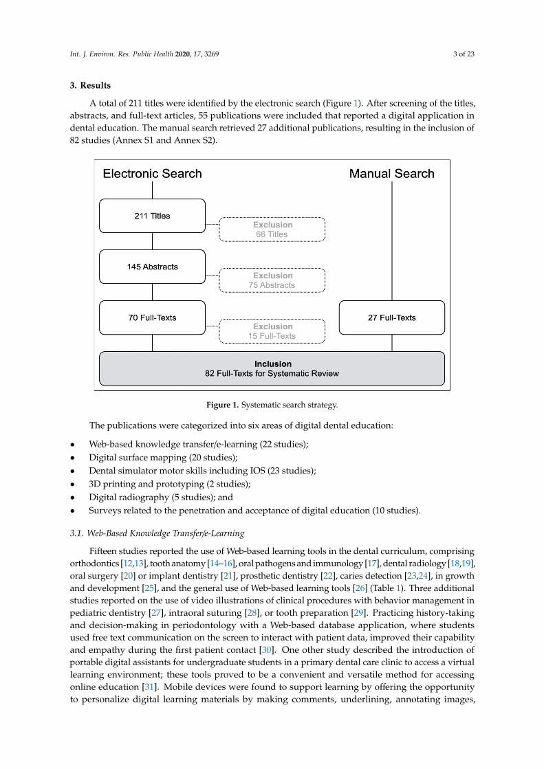
A total of 211 titles were identified by the electronic search (Figure 1). After screening of the titles,abstracts, and full-text articles, 55 publications were included that reported a digital application indental education. The manual search retrieved 27 additional publications, resulting in the inclusion of82 studies (Annex S1 and Annex S2).
Figure 1. Systematic search strategy.
The publications were categorized into six areas of digital dental education:
• Web-based knowledge transfer/e-learning (22 studies);• Digital surface mapping (20 studies);• Dental simulator motor skills including IOS (23 studies);• 3D printing and prototyping (2 studies);• Digital radiography (5 studies); and• Surveys related to the penetration and acceptance of digital education (10 studies).
3.1. Web-Based Knowledge Transfer/e-Learning
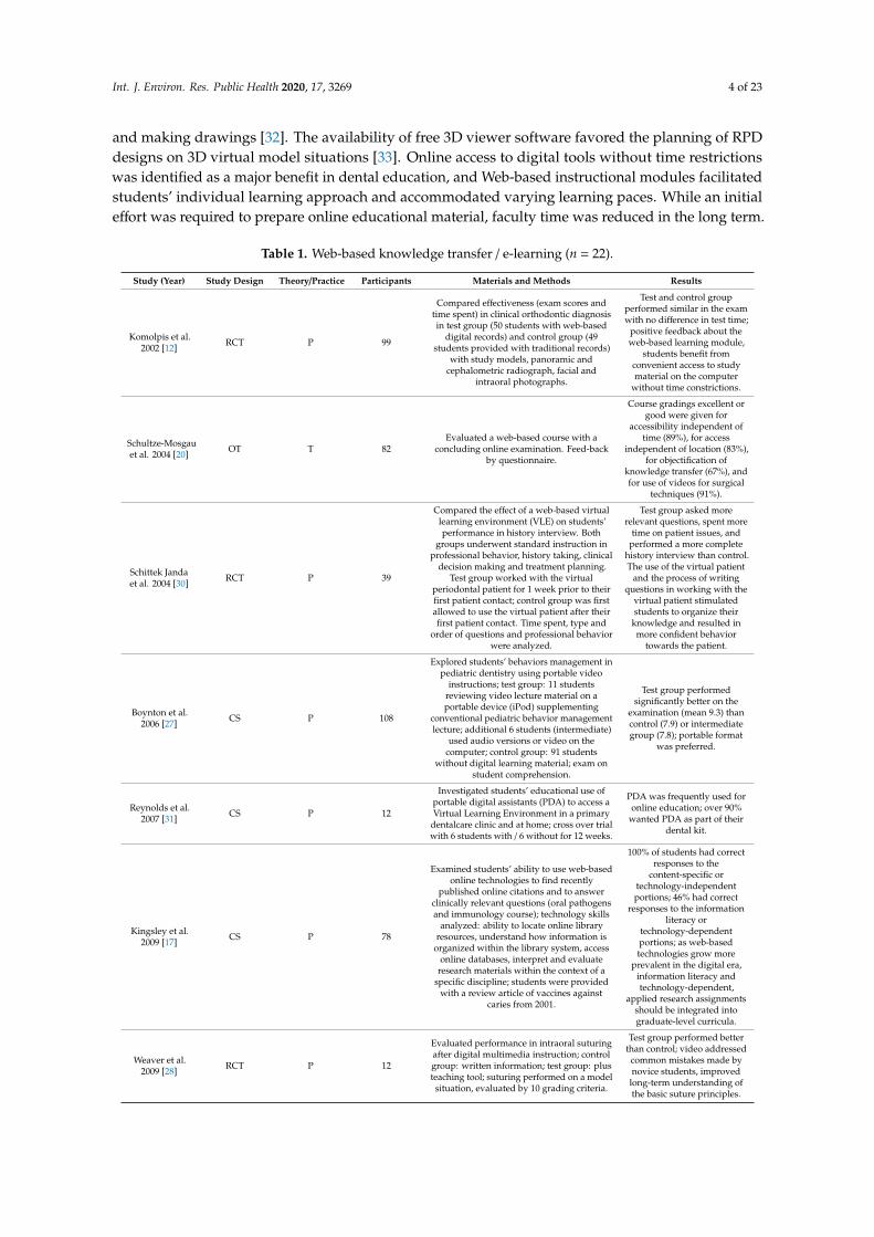
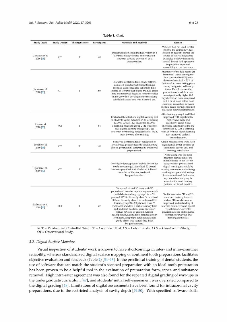
Fifteen studies reported the use of Web-based learning tools in the dental curriculum, comprisingorthodontics [12,13], tooth anatomy [14–16], oral pathogens and immunology [17], dental radiology [18,19],oral surgery [20] or implant dentistry [21], prosthetic dentistry [22], caries detection [23,24], in growthand development [25], and the general use of Web-based learning tools [26] (Table 1). Three additionalstudies reported on the use of video illustrations of clinical procedures with behavior management inpediatric dentistry [27], intraoral suturing [28], or tooth preparation [29]. Practicing history-takingand decision-making in periodontology with a Web-based database application, where studentsused free text communication on the screen to interact with patient data, improved their capabilityand empathy during the first patient contact [30]. One other study described the introduction ofportable digital assistants for undergraduate students in a primary dental care clinic to access a virtuallearning environment; these tools proved to be a convenient and versatile method for accessingonline education [31]. Mobile devices were found to support learning by offering the opportunityto personalize digital learning materials by making comments, underlining, annotating images,
Int. J. Environ. Res. Public Health 2020, 17, 3269 4 of 23
and making drawings [32]. The availability of free 3D viewer software favored the planning of RPDdesigns on 3D virtual model situations [33]. Online access to digital tools without time restrictionswas identified as a major benefit in dental education, and Web-based instructional modules facilitatedstudents’ individual learning approach and accommodated varying learning paces. While an initialeffort was required to prepare online educational material, faculty time was reduced in the long term.
Table 1. Web-based knowledge transfer / e-learning (n = 22).
Study (Year) Study Design Theory/Practice Participants Materials and Methods Results
Komolpis et al.2002 [12] RCT P 99
Compared effectiveness (exam scores andtime spent) in clinical orthodontic diagnosisin test group (50 students with web-based
digital records) and control group (49students provided with traditional records)
with study models, panoramic andcephalometric radiograph, facial and
intraoral photographs.
Test and control groupperformed similar in the examwith no difference in test time;
positive feedback about theweb-based learning module,
students benefit fromconvenient access to studymaterial on the computer
without time constrictions.
Schultze-Mosgauet al. 2004 [20] OT T 82
Evaluated a web-based course with aconcluding online examination. Feed-back
by questionnaire.
Course gradings excellent orgood were given for
accessibility independent oftime (89%), for access
independent of location (83%),for objectification of
knowledge transfer (67%), andfor use of videos for surgical
techniques (91%).
Schittek Jandaet al. 2004 [30] RCT P 39
Compared the effect of a web-based virtuallearning environment (VLE) on students’performance in history interview. Both
groups underwent standard instruction inprofessional behavior, history taking, clinical
decision making and treatment planning.Test group worked with the virtual
periodontal patient for 1 week prior to theirfirst patient contact; control group was firstallowed to use the virtual patient after theirfirst patient contact. Time spent, type and
order of questions and professional behaviorwere analyzed.
Test group asked morerelevant questions, spent more
time on patient issues, andperformed a more complete
history interview than control.The use of the virtual patient
and the process of writingquestions in working with the
virtual patient stimulatedstudents to organize their
knowledge and resulted inmore confident behavior
towards the patient.
Boynton et al.2006 [27] CS P 108
Explored students’ behaviors management inpediatric dentistry using portable video
instructions; test group: 11 studentsreviewing video lecture material on aportable device (iPod) supplementing
conventional pediatric behavior managementlecture; additional 6 students (intermediate)
used audio versions or video on thecomputer; control group: 91 students
without digital learning material; exam onstudent comprehension.
Test group performedsignificantly better on the
examination (mean 9.3) thancontrol (7.9) or intermediategroup (7.8); portable format
was preferred.
Reynolds et al.2007 [31] CS P 12
Investigated students’ educational use ofportable digital assistants (PDA) to access aVirtual Learning Environment in a primary
dentalcare clinic and at home; cross over trialwith 6 students with / 6 without for 12 weeks.
PDA was frequently used foronline education; over 90%
wanted PDA as part of theirdental kit.
Kingsley et al.2009 [17] CS P 78
Examined students’ ability to use web-basedonline technologies to find recently
published online citations and to answerclinically relevant questions (oral pathogensand immunology course); technology skills
analyzed: ability to locate online libraryresources, understand how information is
organized within the library system, accessonline databases, interpret and evaluate
research materials within the context of aspecific discipline; students were provided
with a review article of vaccines againstcaries from 2001.
100% of students had correctresponses to the
content-specific ortechnology-independentportions; 46% had correct
responses to the informationliteracy or
technology-dependentportions; as web-basedtechnologies grow more
prevalent in the digital era,information literacy andtechnology-dependent,
applied research assignmentsshould be integrated intograduate-level curricula.
Weaver et al.2009 [28] RCT P 12
Evaluated performance in intraoral suturingafter digital multimedia instruction; controlgroup: written information; test group: plusteaching tool; suturing performed on a model
situation, evaluated by 10 grading criteria.
Test group performed betterthan control; video addressed
common mistakes made bynovice students, improved
long-term understanding ofthe basic suture principles.

Int. J. Environ. Res. Public Health 2020, 17, 3269 5 of 23
Table 1. Cont.
Study (Year) Study Design Theory/Practice Participants Materials and Methods Results
Wright et al.2009 [14] OT T 235
Determined whether dental students used aninteractive DVD-tooth atlas as a study aid
and perceived the 3D interactive tooth atlasas a value-added learning experience.
14% students downloaded theDVD voluntarily prior toadding atlas-related exam
questions as incentives; afteradding incentives 43%
downloaded the material;financial concerns and overly
sophisticated content weredeemed responsible for the
low acceptance.
Curnier 2010[16] OT P 26 Assessed VR integration into teaching of
dental anatomy, feedback by questionnaire
70% of the students weresatisfied/very satisfied with ITintegration in the curriculum.
Bains et al.2010 [13] RCT T 90
Compared effectiveness and attitudes towarde-learning (EL, online tutorial without
teacher), face-to-face learning (F2FL, led byteacher) and blended learning (BL)
subdivided in BL1 (EL first then F2FL) andBL2 (F2FL first then EL) among 4th yearstudents. Groups received cephalometric
tutorial in the allocated mode, answered anMCQ (Multiple Choice Questionnaire).
F2FL and BL resulted insimilar test results; EL alone
was less effective. BL was themost and F2FL was the least
accepted method, EL wassignificantly less preferred, the
order B1 or 2 had no effect.
Mitov et al.2010 [15] CS T 36
Testing an e-learning software (morphoDent)to prepare for an anatomy exam. 3D models
with description and x-rays of permanenthuman teeth were available for viewing andinteraction on the learning platform. Practicaldental morphology exam was compared tovirtual tooth anatomy exam. Evaluation of
students’ perceptions in a questionnaire.
Similar exam scores intraditional and online exam.
Majority felt the softwarehelped them learning dental
morphology, despite ofdifficulties in operating
the program.
Vuchkova et al.2012 [19] CS P 88
Evaluated interactive digital versusconventional radiology textbook (course
radiographic anatomy), outcome wasradiographic interpretation test and
survey feedback.
95% perceived positiveenhancement of learning and
interpretation.
Smith et al.2012 [29] OT P 26
Compared the use of online video-clips withtraditional live demonstrations with
one-to-one supervision; students exam scoresbefore and after the video introduction were
compared. Feed-back by questionnaire.
76% preferred video-clips tolive demonstrations, 57%
reviewed DVD at home; 57%felt one-to-one supervisionmore effective developingtheir competence in tooth
preparation.
Qi et al. 2013[21] RCT P 95
Comparison of active versus passiveapproaches in using 3D virtual scenes in
dental implant cases. Students were exposedto educational materials about implantrestoration on three types of webpages:
traditional 2D (group 1); active-controlling3D (group 2); passive-controlling 3D (group3). After reviewing their webpages, studentswere asked to complete a posttest to assess
the relative quality of information acquisition.Before study exposure, students performed a
standardized test of spatial ability (mentalrotations test, MRT).
Posttest scores were highest ingroup 3 (passive control) and
lowest in group 2 (activecontrol). Higher MRT scoreswere associated with betterposttest performances in all
three groups. Individuals withlow spatial ability did notbenefit from 3D interactive
virtual reality, while passivecontrol produced higher
learning effects compared toactive control.
Reissmann et al.2015 [22] OT T 71
Creation of a blended learning model;e-learning modules covered fundamental
principles, additional information, andlearning tests (tests were repeated until
passed and the next video sequenceunlocked); modules comprised (i) tooth
preparation, placement of post and core, andprovisional crown; (ii) with preparation,
manufacturing and insertion of a FDP (FixedDental Prosthesis). Students rated the courseon a questionnaire, comparison to previous
courses without e-learning.
Significantly highersatisfaction among students
enrolled in the e-learningmodules compared to the
years prior to integration ofthe e-learning tests. Results
suggest that instructor-basedpractical demonstrations in
preclinical courses inprosthetic dentistry could be
successfully replaced bye-learning applications
provided that course contentis structured according to
specific predefined learninggoals and procedures.
Luz et al. 2015[24] RCT P 39
Evaluated the effect of a digital learning toolon students’ caries detection in 12 pediatric
patients (3.4 per student) using ICDAS(International Caries Detection & AssessmentSystem) (1264 dental surfaces). 2 weeks afterfirst exam students were split into 3 traininggroups: Group 1: ICDAS e-learning program;group 2: plus digital learning tool; group 3:
no learning strategy; students reassessed thesame patients 2 weeks, and results compared.
After training group 1 and 2had improved with
significantly higher sensitivity;group 2 showed significantincrease in sensitivity at theD2 and D3 thresholds as a
result of the digitallearning tool.
Int. J. Environ. Res. Public Health 2020, 17, 3269 6 of 23
Table 1. Cont.
Study (Year) Study Design Theory/Practice Participants Materials and Methods Results
Gonzales et al.2016 [18] OT T 40
Implementation social media (Twitter) in adental radiology course and evaluated
students’ use and perception by aquestionnaire.
95% (38) had not used Twitterprior to the course; 53% (21)
created an account during thecourse to view radiographicexamples and stay informed;overall Twitter had a positive
impact with improvedaccessibility to the instructor.
Jackson et al.2018 [25] OT P 80
Evaluated dental students study patternsusing self-directed web-based learning
modules with scheduled self-study timeinstead of lectures; web-based module access(date and time) was recorded for four courses
in the growth & development curriculum;scheduled access time was 8 am to 5 pm.
Frequency of module access (atleast once) varied among thefour courses (10–64%); onlythree students had > 20% of
their total accesses taking placeduring designated self-study
times. For all courses theproportion of module accesswas significantly higher 0–2
days before an exam comparedto 3–7 or >7 days before finalexam; no association between
module access during scheduledtimes and course performance.
Alves et al.2018 [23] RCT P 64
Evaluated the effect of a digital learning toolon students’ caries detection in 80 teeth using
ICDAS; Group 1 (21 students): ICDASe-learning program; group 2 (22 students):
plus digital learning tool; group 3 (21students): no training; reassessment of the 80
teeth 2 weeks after training.
After training group 1 and 2 hadimproved with significantly
higher sensitivity andspecificity; group 3 had
increased sensitivity at the D2thresholds; ICDAS e-learning
with or without digital learningtool improved occlusal
caries detection.
Botelho et al.2019 [26] OT T 40
Surveyed dental students’ perception ofcloud-based practice records (documenting
clinical progression) compared to traditionalpaper record.
Cloud based records were ratedsignificantly better in terms of
usefulness, ease of use, andlearning, satisfaction.
Pyörälä et al.2019 [32] OT T 176
Investigated perception of mobile devices forstudy use among 124 medical, 52 dental
students provided with iPads and followedfrom 1st to 5th year; feed-back
by questionnaire.
Note taking was the mostfrequent application of the
mobile device in the 1st–5thyear; students personalizeddigital learning materials by
making comments, underlining,marking images and drawings.Students retrieved their notes
anytime when studying forexaminations and treatingpatients in clinical practice.
Mahrous et al.2019 [33] RCT P 77
Compared virtual 3D casts with 2Dpaper-based exercise in planning removable
partial denture design; group 1 (n = 39)planned RPD in Kennedy class IV in virtual
3D and Kennedy class II in traditional 2Dformat, group 2 (=38) planned class IV
traditional and class II virtual; survey linesand undercut positions were drawn on
virtual 3D casts or given in writtendescriptions (2D); students planned design(with rests, clasp type, retention location,
guide plane) was scored; feed-backby questionnaire.
Similar scores for 3D and 2Dexercises; majority favoredvirtual 3D casts because of
improved understanding ofrelevant parameters and spatial
visualization. Currently,physical casts are still required
to practice surveying anddrawing on the cast.
RCT = Randomized Controlled Trial; CT = Controlled Trial; CS = Cohort Study; CCS = Case-Control-Study;OT = Observational Study.
3.2. Digital Surface Mapping
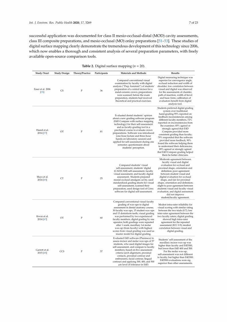
Visual inspection of students’ work is known to have shortcomings in inter- and intra-examinerreliability, whereas standardized digital surface mapping of abutment tooth preparations facilitatesobjective evaluation and feedback (Table 2) [34–46]. In the preclinical training of dental students, theuse of software that can match the student’s scanned preparation with an ideal tooth preparationhas been proven to be a helpful tool in the evaluation of preparation form, taper, and substanceremoval. High intra-rater agreement was also found for the repeated digital grading of wax-ups inthe undergraduate curriculum [47], and students’ initial self-assessment was overrated compared tothe digital grading [48]. Limitations of digital assessments have been found for intracoronal cavitypreparations, due to the restricted analysis of cavity depth [49,50]. With specified software skills,
Int. J. Environ. Res. Public Health 2020, 17, 3269 7 of 23
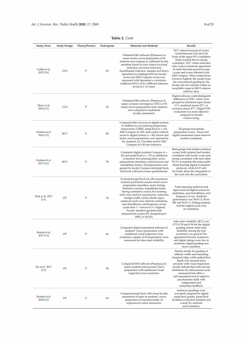
successful application was documented for class II mesio-occlusal-distal (MOD) cavity assessments,class III composite preparations, and mesio-occlusal (MO) onlay preparations [51–53]. These studies ofdigital surface mapping clearly demonstrate the tremendous development of this technology since 2006,which now enables a thorough and consistent analysis of several preparation parameters, with freelyavailable open-source comparison tools.
Table 2. Digital surface mapping (n = 20).
Study (Year) Study Design Theory/Practice Participants Materials and Methods Results
Esser et al. 2006[35] CS P 36
Compared conventional visualexamination by faculty with digital
analysis (“Prep Assistant”) of students’preparation of a central incisor for ametal-ceramic crown; preparations
were scanned; before the exampreparation, students had receivedtheoretical and practical exercises.
Digital measuring technique wassuperior for convergence angle,occlusal reduction and width of
shoulder; low correlation betweenvisual and digital was observedfor the assessments of chamfer,
path of insertion, width of beveland basic form; calibration ofevaluators benefit from digital
analysis tool.
Hamil et al.2014 [37] OT P 81
Evaluated dental students’ opinionabout a new grading software program(E4D Compare with surface mappingtechnology) for their self-assessment
and as faculty-grading tool in apreclinical course to evaluate crown
preparations. Software was introduced(one-hour lecture and three-hourhands-on laboratory session) and
applied for self-assessment during onesemester; questionnaire about
students’ perception.
Students preferred digital gradingsystem over traditional
hand-grading 95% reported onfeedback inconsistencies amongdifferent faculty members, 72%
reported on inconsistencies fromthe examiner; 85% agreed or
strongly agreed that E4DCompare provided more
consistent grading than faculty;79% responded that the software
provided more feedback, 90%found the software helping themto understand their deficiencies;89% agreed or strongly agreed
that E4D Compare grading helpedthem be better clinicians.
Mays et al.2014 [49] CT P 25
Compared students’ visualself-assessment, students’ digital
(CAD/CAM) self-assessment, facultyvisual assessment, and faculty digital
assessment. Students preparedmesial-occlusal amalgam cavity, usedstandardized grading sheets for visual
self-assessment, scanned theirpreparation, used design tool of Cerec
software for digital self-assessment.
Moderate agreement betweenfaculty visual and digital
evaluation for occlusal andproximal shape, orientation and
definition; poor agreementbetween student visual and
digital evaluation for occlusalshape, and fair for proximal
shape, orientation and definition;slight to poor agreement betweenstudents visual and faculty visualevaluation, and digital assessment
did not improvestudent/faculty agreement.
Kwon et al.2014 [47] OT P 60
Compared conventional visual facultygrading of wax-ups to digital
assessment in dental anatomy course;30 faculty wax-ups, 15 student wax-upsand 15 dentoform teeth; visual grading
was performed by two experiencedfaculty members, digital grading by oneoperator, both gradings were repeated
after 1 week; maxillary 1st molarwax-up (from faculty) with highest
scores from visual grading was used asmaster model for digital grading.
Modest intra-rater reliability forvisual scoring with similar ratingbetween the two trials (0.7); low
inter-rater agreement between thetwo faculty raters; digital grading
showed high intra-rateragreement for the repeated
assessment (ICC 0.9); modestcorrelation between visual and
digital grading.
Garrett et al.2015 [48] CCS P 57
Evaluated E4D software (Planmeca) toassess incisor and molar wax-ups of 57students, who used digital images for
self-assessment, and compare to facultymembers; based on five assessmentcriteria (arch alignment, proximal
contacts, proximal contour andembrasures, facial contour, lingual
contour) and applying 300, 400, and 500µm level of tolerance in E4D.
Students’ self-assessment of themaxillary incisor wax-up was
higher than faculty and E4D300,but lower than E4D 400 and 500.
For the molar wax-up,self-assessment was not differentto faculty, but higher than E4D300.
E4D500 evaluations were sig.superior than other assessments.
Int. J. Environ. Res. Public Health 2020, 17, 3269 8 of 23
Table 2. Cont.
Study (Year) Study Design Theory/Practice Participants Materials and Methods Results
Callan et al.2015 [34] CCS P 82
Validated E4D software (Planmeca) toassess molar crown preparation of 82
students and compare to calibrated facultymembers based on four criteria (occlusal
reduction, proximal reduction,facial/lingual reduction, margins and draw).
Agreement in rankings between facultyscores and E4D Compare scores was
measured with Spearman’s correlationcoefficient (SCC) at five different tolerance
levels (0.1–0.5 mm).
SCC values for practical examsvaried between 0.20 and 0.56.
None of the upper 95% confidencelimits reached the for strong
correlation. SCC values indicatedonly weak to moderate agreementin ranks between practical examscores and scores obtained with
E4D Compare. When ranked fromlowest to highest, the results fromthe conventional grading by the
faculty did not correlate within anacceptable range to E4D Compare
software data.
Mays et al.2016 [42] CCS P 50
Validated E4D software (Planmeca) toassess occlusal convergence (TOC) of 50molar crown preparations from students
and compared to traditionalfaculty assessment.
Digital software could distinguishdifferences in TOC, which were
grouped as minimum taper (mean11◦), moderate (mean 23◦), or
excessive (mean 47◦). Digital TOCevaluation was more objective
compared to facultyvisual scoring.
Gratton et al.2016 [45] RCT P 80
Compared effect of access to digital systemsin addition to conventional preparationinstructions; CEREC prepCheck (n = 20),
E4D Compare (n=20), and control withoutaccess to digital system (n = 40); incisor andmolar crown preparations were assessed by
the students, by 3 faculties and by E4DCompare at 0.30 mm tolerance.
All groups had similarpreparation scores. Visual and
digital assessment scores showedmodest correlation.
Gratton et al.2017 [46] RCT P 79
Compared digital systems Compare (n =42) and prepCheck (n = 37) as additional
evaluation tool assessing their crownpreparations (maxillary central incisor andmandibular molar); all preparations were
graded by faculty Compare and prepCheck;feed-back with post-course questionnaire.
Both groups had similar technicalscores; both systems had modestcorrelation with faculty scores andstrong correlation with each other.55.3% of students felt unfavorableabout learning digital evaluation
protocols, while 62.3% feltfavorable about the integration of
the tools into the curriculum.
Park et al. 2017[44] OT P 36
Evaluated prepCheck for self-assessment,students performed ceramo-metal crown
preparation (maxillary molar duringformative exercise, mandibular molar
during summative exam); five learningtools were used for assessments: reduction,
margin width, surface finish, taper,undercut; tools were rated for usefulness,
user-friendliness, and frequency of use(scale from 1 = lowest to 5 = highest).
Faculty members graded toothpreparations as pass (P), marginal-pass
(MP), or fail (F).
Tools assessing undercut andtaper received highest scores for
usefulness, user-friendliness, andfrequency of use. Students’
performance was 38.8% P, 30.6%MP and 30.6% F. Failing students
had the highest score (4.4)on usefulness.
Kateeb et al.2017 [38] OT P 96
Compared digital assessment software ofstudents’ crown preparation withtraditional visual inspection; four
examiners; sample of 20 preparations werereassessed for intra-rater reliability.
Intra-rater reliability (ICC) was0.73–0.78 and 0.99 for the digital
grading system; inter-raterreliability among the four
examiners was good (0.76);agreement between examinersand digital ratings were low tomoderate; digital grading was
more consistent.
Sly et al. 2017[50] OT P 98
Compared E4D software (Planmeca) toassess students intracoronal Class Ipreparation with traditional visual
inspection; four examiners.
Similar results for grading ofisthmus width and remaining
marginal ridge, while pulpal floordepth was assessed more
precisely with visual inspection;results indicate that software haslimitations for intracoronal cavity
assessment but offers aself-assessment tool to improve
psychomotor skills withindependent and
immediate feedback.
Kunkel et al.2018 [40] OT P 69
Compared prepCheck with visual facultyassessment of taper in students’ crown
preparation of typodont teeth, 10experienced course instructors.
Instructor gradings wereoverrated compared to digitalprepCheck grades, prepCheck
facilitates evaluation instantly andexactly by students
and examiners.

Int. J. Environ. Res. Public Health 2020, 17, 3269 9 of 23
Table 2. Cont.
Study (Year) Study Design Theory/Practice Participants Materials and Methods Results
Kozarovska &Larsson 2018
[39]RCT P 57
Evaluated a digital preparation validationtool (PVT) for students’ self-assessment ofcrown preparation (tooth 11 and 21); group
A (“prep-and-scan” self-assessed andscanned three preparations; group B
(“best-of-three”) self-assessed the threeattempts, chose the best for scanning;
questionnaire about students’ and teachers’experiences with PVT.
Group A showed an increase inagreement of self-assessment andfeedback from PVT, while group Bshowed low level agreement with
PVT. Bucco-incisal reduction,reduction of the tuberculum
surface and presence of undercutswere difficult to correctly identify
by the students. Questionnairefeedback revealed need for PVT todevelop skills, to ease assessment,while critical aspects were PVT’stime efficiency and the need for
verbal feedback. Teachersobserved the PVT as a motivationduring skills laboratory training,while verbal feedback were still
deemed necessary.
Wolgin et al.2018 [53] RCT P 47
Investigated digital self-assessment concept(prepCheck software) for students in the
phantom course preparing a three surface(MOD) class II amalgam cavity;
intervention group (IG): compared a 3Dimage of their preparation against master
preparation with PrepCheck; control group(CG): received verbal feedback from
supervisor based on pre-defined criteria.
Test and control groupsperformed similar and
self-assessment learning tool wasdeemed equivalent to
conventional supervision.
Lee et al. 2018[51] OT P 69
Compared students’ self-assessment(conventional and digital with Cerec
software) with assessment (conventionaland digital) by faculty members for class IIamalgam preparations (C2AP) and Class III
composite preparations (C3CP).
Students overestimated theirperformance (positive S-F gap) in
both the C2AP and C3CPpreparation exercises in
conventional (11% and 5%) anddigital assessments (8% and 2%);
in conventional assessments,preclinical performance wasnegatively correlated with
student-faculty gap (r = −0.47, p <0.001); particularly students in the
bottom quartile sig. improvedtheir self-assessment accuracyusing digital self-assessments
over conventional assessments.
Nagy et al.2018 [52] RCT P 36
Investigated the effect of a digital feedback(test group) for mesio-occlusal onlay
preparation by a 3D visualization of thecavity (Dental Teacher software, KaVo),
while verbal feedback from supervisor wasgiven to control group. Following
feedbacks, 2nd corrective preparationswere conducted and improvements
measured. Parameters: occlusal cavitydepth (OD), approximal depth (AD), extentof cusp reduction on the mesiobuccal cusp
(CR), width of shoulder preparationaround the mesiobuccal cusp (SW), cavity
width at two different points in the occlusalbox (OW).
Test group improved in allparameter and showed
significantly smaller deviations ofmean OD, AD and mean SW; in
control group, parameterdeviations were similar during 1st
and 2nd preparation.
Liu et al. 2018[41] RCT P 66
Evaluated the effectiveness of preclinicaltraining on ceramic crown preparationusing digital training system comparedwith traditional training method; test
group: trained with digital method withOnline Peer-Review System (OPRS) and
Real-time Dental Training and EvaluationSystem (RDTES); control group: traditionalmethod with instructor demonstration and
evaluation; central incisorcrown preparation.
Five of 15 assessed items weresignificantly better in test group;96.97% of test students agreed orstrongly agreed that using digital
training system could betterimprove the practical ability than
traditional method.
Greany et al.2019 [36] OT P 67
Compared conventional visual facultyinspection of wax-ups to digital assessment;
six examiners evaluated 67 students’wax-ups of maxillary first molar,
reevaluation after 1 week; scan with IOS,STL files imported to free available open
source data cloud comparison utility(Cloud Compare.org), digital evaluation by
two examiners.
Visual inspection had lowinter-examiner precision (ICC
0.332) and accuracy;intra-examiner precision for
reevaluation was low;inter-examiner precision of digitalexam was high (ICC 0.866) with
high accuracy.

Int. J. Environ. Res. Public Health 2020, 17, 3269 10 of 23
Table 2. Cont.
Study (Year) Study Design Theory/Practice Participants Materials and Methods Results
Miyazone et al.2019 [43] OT P 100
Compared prepCheck with visualfaculty assessment of students’ crown
preparation of typodont teeth(mandibular first molar as crown
abutment, maxillary 2nd premolar and2nd molar as FDP abutments), assessinter- and intra-grader agreement of
five experienced examiners conductingvisual and digital exam; scoring
repeated three times; parameters forcrown abutments: axial tissue removal,
margin width, undercut, occlusalreduction, cusp tips, occlusal anatomy;for FDP abutments: path of insertion.
Intra-grader agreement was betterwith prepCheck than visual
assessment for all parametersexcept cusp tip and occlusal
anatomy; inter-grader agreementfor path of insertion was
questionable with visual, butgood with digital assessment.
Inter-grader disagreement wasgreater in visual than digital
assessment. Overestimation oftooth reduction in visual gradingwas eliminated by digital analysis.
RCT = Randomized Controlled Trial; CT = Controlled Trial; CS = Cohort Study; CCS = Case-Control-Study;OT = Observational Study; ICC = Inter-Class Correlation; STL = Standard Tessellation Language.
3.3. Dental Simulator Motor Skills Including Intraoral Optical Scanning
A high level of interest and acceptance was documented among undergraduate students forsimulator training in cavity preparations [54–56], or in surgical interventions such as apicoectomies(Table 3) [57]. A trend toward improved technical skills and ergonomics was documented whensimulator training with real-time feedback was added to traditional instructions [58–60]. Training witha VR-based simulator improved students’ preparation of class I occlusal cavities [61], and of abutmentsfor porcelain-fused-to-metal crowns [62]. In evaluating the manual dexterity of students, professionals,and non-professionals, the simulator scoring algorithm showed a high reliability to differentiate betweennon-professionals and dental students or dentists [63]. Instruction time from faculty for teaching cavityand crown preparations was significantly reduced when virtual reality computer-assisted simulationsystems were used compared to contemporary non-computer-assisted simulation systems [64].Preparation performance on VR units with continuous evaluations and advice from clinical instructorsled to better preparation quality than real-time feedback from the virtual dental unit. Self-pacedlearning and the immediate software feedback were beneficial with the VR unit, and it was perceivedas adjunct, but not replacing faculty instructions [65]. Students requested software improvements withmore realistic force feedback during interaction with different tissues in the virtual oral environmentincluding the maxilla, mandible, gum, tongue, cheek, enamel, dentine, pulp, cementum, etc. [66].Recent advancements of simulators enabled variations in force feedback accounting for varyinghardness of the virtual material, cut speed gain, and push force [67].
Improved student performance in crown digitization and framework design was observed whenCAD/CAM (Computer-Aided Design/ Computer-aided manufacturing) courses were introduced indental education [68]. While students enjoyed designing a full crown using CAD as compared totraditional waxing, limits of the technology in representing anatomic contours and excursive occlusionwere identified [69]. Viewing their scanned crown preparations magnified on the screen improvedstudents’ understanding of the finishing line [70]. The application of IOS in the simulation trainingshowed that even inexperienced dental students were capable of acquiring the skills needed to usedigital tools, and students preferred IOS over conventional impressions [71,72]. Furthermore, students’work time was shorter with IOS than with conventional impression [72,73], although more teachingtime was required for digital scanning than for conventional impression techniques [74]. Applyingdigital complete denture treatment (AvaDent; AvaDent Digital Dental Solutions, Scottsdale, AZ, USA)in the student clinics resulted in restorations with superior gradings that were preferred by bothstudents and patients [75]. Using an intraoral camera increased patients’ consent for crown treatment,and was positively perceived by students and patients, while faculty members were neutral [76].

Int. J. Environ. Res. Public Health 2020, 17, 3269 11 of 23
Table 3. Dental simulator motor skills incl. IOS (n = 23).
Study (Year) Study Design Theory /Practice Participants Materials and Methods Results
Quinn et al.2003 [65] RCT P 20
Compared students’ performance inpreparing class I amalgam cavity on aVR-based training unit; test group hadvirtual real-time feedback and softwareevaluation, control group had clinical
instructor available during preparation.Anonymous scoring by 2 faculties, criteria:outline form, retention form, smoothness,
cavity depth and cavity margin angulation.Questionnaire feed-back in test group.
Similar results for retention andwall angulation, while outlineform, smoothness and cavitydepth scored better in control.
Test group assessed software assuperior for immediate feed-back,self-paced learning, consistency of
evaluation, encouragingindependent work and morethorough assessment, whileconventional training was
superior for increasing confidencein cavity preparation. VR-based
training should be used as adjunctbut not replacing conventional
training methods.
Jasinevicius etal. 2004 [64] CT P 28
Compared students’ performance inamalgam and crown preparations on
typodont teeth either with a contemporarynon-computer-assisted simulation system
(CS), or with a virtual realitycomputer-assisted simulation system (VR).
Both groups were provided withpresentations describing preparations, CSgroup received handouts, VR group had
preparation criteria available on thecomputer. Student-faculty (S-F) interaction
time was logged.
Preparation quality did not differbetween CS and VR. CS required2.8 h, VR 0.5 h S-F. CS received
five times more instructional timefrom faculty than VR.
LeBlanc et al.2004 [60] RCT P 68
Compared students’ technical skills inpreclinical operative dentistry after
standard traditional laboratory-basedinstructions (over 110 h) and additional
virtual reality simulator-enhanced training(test group with 20 students) Simulator
(DentSim, DenX) provided real-timefeedback, training conducted during 6–10 h
in 3 blocks over 8 months.
While all students improved inthe 4 tests during the year, test
students tended to better scores inthe final exam. Virtual reality
simulators can be implemented inthe traditional training of future
dentists.
Rees et al. 2007[54] CT P 16
Evaluated simulator training (DentSim,DenX) by undergraduate students for ClassI and II preparations (time, marks, numberof evaluations), students spent 6 h cuttingan unlimited number of Class I cavities andClass II cavities; feedback by questionnaire.
Class I preparations obtained amean mark of 66.8, preparation
time was 12.5 min, with 6.7evaluations; Class II had a mark of
26.5, time 18 min, with 7.0evaluations. Class II was more
difficult to cut. Studentsappreciated easy change of teeth,working at their own pace and
examine the cavity in across-section.
Welk et al. 2008[55] OT P/T 80
Evaluated students’ performance inoperative dentistry after training with
computer-assisted dental simulator(DentSim, DenX), feedback by
questionnaire.
Students indicated high interest insimulator training, high
acceptance and response toadditional elective training time inthe computer assisted simulationlab. The shift in curriculum and
instructional goals has to beoptimized continuously.
Urbankova etal. 2010 [58] RCT P 75
Evaluated adjunctive computerized dentalsimulator (CDS; DentSim) training (8 h) in
operative dentistry (Class I and IIpreparations): either before (n = 26) or after
1st exam (n = 13); control group (n = 36)with traditional preclinical dental training
alone (110 h).
CDS-trained students performedbetter than control in the 1st and2nd exam, no difference betweenpre-exam and post-exam groups.In the 3rd exam (end of the year)CDS group had higher, but not
significantly different scores thancontrol.
Pohlenz et al.2010 [57] CT P 53
Evaluated VR training (Voxel-Man) forvirtual apicoectomy; questionnaire about
simulated force feedback, spatial 3Dperception, resolution and integration of
further pathologic conditions.
92.7% recommended the virtualsimulation as additional modality
in dental education, 81.1%reported the simulated force
feedback as good or very good,86.8% evaluated 3D spatial
perception as good or very good;100% recommended integration of
further pathologies.

Int. J. Environ. Res. Public Health 2020, 17, 3269 12 of 23
Table 3. Cont.
Study (Year) Study Design Theory /Practice Participants Materials and Methods Results
Gottlieb et al.2011 [59] CT T 202
Evaluated VR simulation training(DentSim, Image Navigation Ltd.) in
operative preparations and restorations,60 h VR training, laboratory course was
reduced to 234 h (instead oftraditional 304h).
13 experienced faculties assessed 97non-VR students (1st year, control) and 105
students with 1 semester VR experience(test); survey about students’ abilities in
ergonomics, confidence level, performance,preparation, and self-assessment.
Faculty expected greaterpsychomotor skills and ability toprepare teeth in VR, abilities were
lower than anticipated butnumerically higher than innon-VR students. Faculty
members perceived students’ergonomics in the test group
better than in control.
Ben-Gal et al.2011 [56] CT P 33
Evaluated use of VR simulator (IDEADental) for dental instruction, self-practice,
and student evaluation. 21 experienceddental educators, 12 randomly selectedexperienced dental students (5th year)performed 5 drilling tasks using the
simulator, feed-back by questionnaire.
Both groups found that thesimulator could provide
significant benefits in teachingand self-learning of manual
dental skills.
Ben-Gal et al.2013 [63] CT P 106
Evaluated potential of VR trainingsimulator (IDEA Dental) to assess manualdexterity in 63 dental students, 28 dentists,14 non-dentists, performed virtual drillingtasks in different geometric shapes: time tocompletion, accuracy, number of trials tosuccessful completion, score provided by
the simulator.
Simulator scoring algorithmshowed high reliability in allparameters and was able to
differentiate betweennon-professionals and dentalstudents or non-professionals
and dentists.
Lee & Gallucci2013 [73] CT P 30
Compared digital (IOS) to conventionalimpression for single implant restorations,
evaluated efficiency, difficulty andstudents’ preference.
Mean total treatment time,preparation time and working
time were significantly longer forconventional than for IOS;
conventional impressions wereassessed as more difficult than
IOS; 60% preferred IOS, 7%conventional, 33%either techniques
Kikuchi et al.2013 [62] RCT P 43
Compared VR simulator (DentSim) trainingwith or without instructor feedback forpreparation of porcelain fused to metal
(PFM) crown preparation. 43 students (5thyear). randomly divided into: 1. VR groupwith instructor’s feedback (DSF; n = 15); 2.VR without instructor’s feedback (DS; n =15); 3. neither VR simulator training nor
faculty feedback (NDS; n = 13); preparationtime and scores of 4 crown preparations
(1week for 4 weeks).
DSF and DS had significantlyhigher total scores than NDS.
Similar results in DSF and DS, butshortened preparation time withinstructors’ feed-back (DSF) at
early stages.
Douglas et al.2014 [69] CT P 50
Compared students’ performance intraditional waxing vs. computer-aidedcrown designing (IOS with CEREC 3D,
Sirona Dental Systems), faculty grading ofocclusal contacts and anatomic form,
feed-back by questionnaire.
Similar gradings for wax design(79.1) and crown design (78.3);
more occlusal contacts with CAD;students enjoyed designing a full
contour crown using CAD andrequired less time with CAD.Students recognized limits of
CAD technology in representinganatomic contours and excursive
occlusion compared toconventional wax techniques.
Wang et al.2015 [66] CT P 20
Compared VR simulator (iDental withPhanotm Omni, SensAble Tech. Inc.) in
novice group (graduate students with lessthan 3 years clinical practice experience)
and resident group (with 3–0 years clinicalpractice); assessment of caries removal,
pulp chamber opening, time and amount ofremoved healthy/unhealthy tissue;
feed-back by a questionnaire.
No differences in time andamount of tissue removal betweengroups; residents spend slightlymore time than students; both
groups suggested improvementsin spatial registration precision,
more realistic model with materialproperties and force feedback ofdifferent tissues, improvement of
the depth of the virtual space.
Schwindling etal. 2015 [68] CT P 56
Evaluated a CAD/CAM hands-on course(test) compared to video-supported lecture
only (control); written exam about castdigitizing and zirconia crown designing.
Test group performed significantlybetter than controls (16.8/20 vs.
12.5/20 correct answers); interestof students in CAD/CAM washigher after hands-on course.
Kattadiyil et al.2015 [75] CCS P 15
Compared clinical treatment outcomes,patient satisfaction, and dental studentpreferences for digital (AvaDent, twoappointments) and conventional (five
appointments) complete dentures (CD) in15 patients, 15 dental students fabricatedtwo sets of CDs for each patient. Facultyand patient ratings, patient and studentpreferences, perceptions, treatment time
was analyzed.
Digital process was equallyeffective and more time-efficientthan conventional; faculty scoreddigital better than conventionaldentures; patients and students
preferred digital dentures.

Int. J. Environ. Res. Public Health 2020, 17, 3269 13 of 23
Table 3. Cont.
Study (Year) Study Design Theory /Practice Participants Materials and Methods Results
Zitzmann et al.2017 [72] RCT P 50
Investigated performance (time recording)and perception (questionnaire feedback) ofIOS and conventional implant impression
after video teaching.
Students rated conventionalimpressions as more difficult (VAS
46) than IOS (VAS 70), withgreater patient-friendliness of IOS
(VAS 83) compared toconventional impressions (VAS
36); 76% preferred digital, 88% feltmost effective with IOS; total
work time of all steps wassignificantly shorter with 301 sec.
for IOS and 723 sec. forconventional impressions.
Wegner et al.2017 [70] OT P 108
Evaluated students’ perception(questionnaire feedback) of IOS (Lava CosTraining, 3M Espe), scanning of 3 typodont
tooth preparations.
63.9% positive opinion, 60.2%considered scanning process as
manageable, 55.6% profited frommagnified view of their
preparation to understandchamfer finish lines.
Marti et al.2017 [74] RCT P 25
Analyzed time to instruct IOS (DS; LAVAC.O.S. digital impression system) and
conventional impression technique (CI;polyvinyl siloxane) with video lecture,
investigator led demonstration, andindependent impression exercise: time
recording and questionnaire aboutfamiliarity and student’s expectations.
Teaching DS required significantlymore time than CI for video
lecture (16 vs. 10 min),demonstration time (9 vs 5 min)
and impression time (18 vs. 9min). Initially students were more
familiar with CI (3.96) than DS(1.96) technique. After
instructions and practice, CItechnique proved significantly
easier than expected.Manageability of DS was not
influenced by the instruction andpractice experience. 96%
expressed an expectation that DSwill become their predominant
impression technique.
de Boer et al.2019 [67] RCT P 126
Investigated skill transfer between variouslevels of force feedback (FFB) usingSimodont dental trainer (Moog) forcross-figure preparations as manual
dexterity exercise. Assessment of students’satisfaction by questionnaire.
Longer practice time wascorrelated with test performance:students passing at different FFBlevels had mean of 300h, those
passing in one FFB level had 271 h,failing students had 224 h. Skilltransfer from one level of FFB to
another was feasible withsufficient training.
Schott et al.2019 [71] OT P 31
Evaluated dental students’ perception ofIOS compared to conventional alginate
impression; survey after basic training andself-practicing.
77% (24) students were overall“very” or “rather satisfied" with
the handling of IOS; 58%preferred IOS from the dentist’s
perspective, no significantdifference from the patient’s
perspective but reduced comfortrelated to the impression tray.
Murbay et al.2020 [61] RCT P 32
Incorporated VR with Moog Simodontdental trainer in preclinical training;
students performed an occlusal preparationon typodont teeth and had previousexposure to VR (group 1) or no VRexposure (group 2); assessment was
conducted (satisfactory / unsatisfactory) bymanual approach or digital (Magic 19.01
64-bit).
VR use improved preparationsignificantly with 75% (12/16)
satisfactory preparations in group1 and 44% (7/16) in group 2.
Manual and digital evaluationmethods did not differ
significantly.
Murrell et al.2019 [76] OT P 288
Evaluated completion of posterior crownplanning with or without presenting the
situation to the patient by intraoral camerause; 51 students completed 198 surveys, 35
faculty members with 64 surveys, 202patient surveys, survey was voluntary and
camera use optional.
Positive perception of intraoralcamera use by students andpatients, while faculty wasneutral; significantly higher
completion rate when intraoralcamera was used.
RCT = Randomized Controlled Trial; CT = Controlled Trial; CS = Cohort Study; CCS = Case-Control-Study;OT = Observational Study; DSF = VR group with instructor feedback; DS = VR group without instructor feedback;NDS = Neither VR simulator training nor faculty feedback; VAS = Visual Analog Scale; IDEA = International DentalEducation Association.

Int. J. Environ. Res. Public Health 2020, 17, 3269 14 of 23
3.4. 3D Rapid Prototyping
Two studies evaluated training models created by 3D rapid prototyping [77,78]. Such methodscan supplement teaching on human teeth or even replace it, and educational needs can easily beadapted to students’ skills (Table 4).
Table 4. Group 4: 3D printing and prototyping (n = 2).
Study (Year) Study Design Theory/Practice Participants Materials and Methods Results
Soares et al. 2013 [77] OT T 40
Cavity preparation was taught withconventional teaching materials with 2Dschematic illustration and photographs.New didactic material with virtual 3D
(videos of the preparations) and magnifiednylon prototyped models was introduced.
Evaluation by questionnaire.
Improvement of teachingquality when combining
3D virtual technology withreal models.
Kröger et al. 2016 [78] OT P 22
3D printed simulation models based on realpatient situations were used for hands-onpractice. Models simulated realistic toothpositions and wide variability of dental
cases and procedures. Students removed acrown from tooth 16, detected and removed
caries, did a build-up filling and crownpreparation within 3 h. Students’ feedback
on a VAS questionnaire.
Students evaluated modelsbased on real patient
situations as good trainingpossibilities. The lack ofgingiva was disturbing.
RCT = Randomized Controlled Trial; CT = Controlled Trial; CS = Cohort Study; CCS = Case-Control-Study;OT = Observational Study.
3.5. Digital Radiography
Four studies dealt with diagnosing radiographic changes [79–81] or detecting positional errors onpanoramic radiographs [82] (Table 5). Senior students showed a poor ability for approximal cariesdetection on both conventional and digital radiographs when compared to histo-pathologic analysisfrom sectioned teeth [80]. One study demonstrated that digital learning supported the developmentof students’ diagnostic skills [81]. Another study showed that the accuracy of radiographic cariesdetection was improved by a computer-assisted learning calibration program, which provided feedbackillustrating the actual tooth surface condition [79]. In one study, two digital systems for endodontic toothlength measurements were compared, and students’ positive attitudes towards digital radiographywere documented [83].
Table 5. Group 5: Digital Radiology (n = 5).
Study (Year) Study Design Theory/Practice Participants Materials and Methods Results
Mileman et al.2003 [79] RCT P 67
Investigated computer-assisted learning(CAL) calibration program to improves
dental students’ accuracy in dentin cariesdetection from bitewing radiographs;
experimental (n = 33) group: used CALwith feedback for self-calibration control
(n = 34) group.
CAL improved students’diagnostic performance; truepositive ratio (sensitivity) for
caries detection was significantlyhigher in test 76.3% than controlwith 66.9%, while false positive
ratio (specificity) was similar(28.1 and 28.7%); diagnostic oddsratio was sig. higher in test (12.4)
than in control (8.8).
Wenzel et al.2004 [83] RCT P 31
Compared 2 digital systems (RVG-ui CCDsensor, Digora PSP plate system) for
radiographic examination; after educationin digital radiography one student groupstarted with CCD, one with PSP and both
completed endodontic treatment ofsingle-rooted extracted tooth; groups
switched radiography system and treated a2nd tooth. True tooth length (TTL) and rootfilling length (RFL) were measured with the
software and compared to manualmeasurement; feed-back questionnaire
after each treatment.
Using CCD sensor required lesstime than PSP; positioning the
tooth was easier with PSP plate;positive attitudes towards digitalradiography; lengths measuredon the digital images from both
digital systems were slightlylarger than true tooth lengths with
no difference in ratio TTL/RFLbetween systems.

Int. J. Environ. Res. Public Health 2020, 17, 3269 15 of 23
Table 5. Cont.
Study (Year) Study Design Theory/Practice Participants Materials and Methods Results
Minston et al.2013 [80] CT P 20
Investigated students’ diagnosticperformance on approximal cariesdetection with analog and digital
radiographs from 46 extracted humanpremolars and molars, compared
diagnostic accuracy; teeth were sectionedand histopathologically analyzed
(gold standard)
Students ability for cariesdetection was poor, no difference
between analog anddigital radiographs.
Busanello et al.2015 [81] CCS P 62
Evaluated digital learning object toimprove skills in diagnosing radiographicdental changes (Visual Basic Applicationsoftware); test group used the digital tool,
control group: conventional imagingdiagnosis course; diagnosis test after
3 weeks.
Test group performedsignificantly better, females were
better than males.
Kratz et al.2018 [82] CT P 169
Evaluated students’ ability to identifypositional errors (tongue position, head
rotation, chin position) in panoramicradiographs of edentulous patients,
students in 2nd year (n = 84) and 3rd–4thyear (n = 85)
2nd year students identifiedsignificantly more positional
errors than 3rd and 4th students.Students were more experienced
at identifying radiographicfindings compared to
positional errors.
RCT = Randomized Controlled Trial; CT = Controlled Trial; CS = Cohort Study; CCS = Case-Control-Study;OT = Observational Study; CCD = Charged Couple Device; PSP = Photostimulable Phosphor.
3.6. Surveys Related to the Penetration and Acceptance of Digital Education
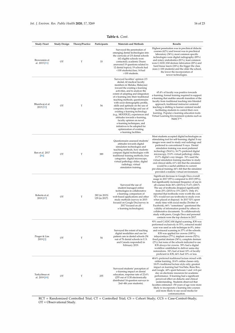
Six surveys evaluated students’ perception and acceptance of digital technologies (Table 6) [84–89].The more recent studies reflected that digital technologies have become established teaching tools,particularly in the field of digital radiography and microscopy, and the use of textbooks decreased;simulation training was preferred [86,87].
Table 6. Surveys related to digital education (n = 10).
Study (Year) Study Design Theory/Practice Participants Materials and Methods Results
Scarfe et al.1996 [88] OT T 277
Investigated the effects ofinstructions in intraoral digitalradiology on dental students’
knowledge, attitudes and beliefs;174 from a university with formal
instruction on digital dentalradiography, and 103 from a
university without instructions.
Students with instructions knewsignificantly more than students without;
93% wanted digital radiology to beincluded in the dental curriculum.
McCann et al.2010 [85] OT T 366
Surveyed student’s (dental anddental hygiene) preferences for
e-teaching and learning, using anonline questionnaire in 2008
related to computer experience,use and effectiveness of
e-resources, preferences forvarious environments, need forstandardization, and preferred
modes of communication.
64% preferred printed text over digital and74% wanted e-materials to supplement butnot replace lectures; 71% preferred buying
traditional textbooks, 11% preferredelectronic versions; among e-resources
virtual microscopy (69%), digital skull atlas(68%), and digital tooth atlas (64%) were
reported as most effective; e-materialswould enhance learning, in particular
e-lectures (59%), clinical videos (54%), andpodcasts (45%). E-resources should not
replace interactions with faculty; studentswanted lectures and clinical
procedures recorded.
Jathanna et al.2014 [84] OT T 186
Surveyed the perception of Indiandental students toward usefulness
of digital technologies inimproving dental practice,
willingness to use digital andelectronic technologies, perceived
obstacles to use digital andelectronic technologies in dentalcare setups, and their attitudestoward internet privacy issues.
Students indicated that digital technologyincreases patient satisfaction and practice
efficiency, improves record quality,doctor-doctor communication, casediagnosis and treatment planning;
obstacles to the wide adoption of thesetechnologies were cost and dentists’ lack ofknowledge and comfort with technology.
Chatham et al.2014 [90] OT T 11
Surveyed the penetration ofdigital technologies in UK dental
schools (11/16 responded).
45% did not teach digital technologies (36%because it was not part of the curriculum,
or in 95% due to the lack of technicalexpertise or support); half of those teachingdigital technologies did so with lectures or
demonstrations, the other half allowedpractical involvement.
Int. J. Environ. Res. Public Health 2020, 17, 3269 16 of 23
Table 6. Cont.
Study (Year) Study Design Theory/Practice Participants Materials and Methods Results
Brownstein etal. 2015 [91] OT T 33
Surveyed the penetration ofemerging dental technologies intothe curricula at US dental schools
(62 eligible schools werecontacted); academic Deans
answered 19 questions related to12 dental topics); 19 schools had
<100 students/class; 14 had>100 students.
Highest penetration was in preclinical didacticcourses (62%) and lowest was in preclinical
laboratory (36%); most common specifictechnologies were digital radiography (85%)and rotary endodontics (81%), least common
were CAD/CAM denture fabrication (20%) andhard tissue lasers (24%); the bigger the class
sizes (>100 students) and the older the school,the lower the incorporation of
newer technologies.
Bhardwaj et al.2015 [92] OT T 54
Surveyed faculties’ opinion (15dental, 42 medical faculty
members in Melaka, Malaysia)toward the existing e-learningactivities, and to analyze the
extent of adopting and integrationof e-learning into their traditionalteaching methods; questionnairewith socio-demographic profile,skills and aptitude on the use ofcomputer, knowledge and use of
existing e-learning technology(e.g., MOODLE), experiences and
attitudes towards e-learning,faculty opinion on novel
e-learning techniques, andinitiatives to be adopted for
optimization of existinge-learning facilities.
65.4% of faculty was positive towardse-learning; formal training required to supporte-learning that enables smooth transition of thefaculty from traditional teaching into blended
approach; traditional instructor centeredteaching is shifting to learner centered model
facilitating students to control their ownlearning. Popular e-learning education tools:
Virtual Learning Environment systems such asWebCT™.
Ren et al. 2017[86] OT T 389
Questionnaire assessed students’attitudes towards digital
simulation technologies andteaching methods, how students
compare digital technologies withtraditional training methods; four
categories: digital microscope,virtual pathology slides, digital
radiology, virtualsimulation training.
Most students accepted digital technologies asstimulating tool for self-learning; digital X-rayimages were used to study oral radiology and
preferred to conventional X-rays. Dentalsimulation training was most preferred
technology (54.6%), 16.7% preferred digitalmicroscopy, 15.0% virtual pathology slides,13.7% digital x-ray images. 76% used the
virtual simulation training machine to studyoral clinical skills; 61% felt that the simulator
would be a useful addition to currentpre-clinical training; 66% felt that the simulator
provided a realistic virtual environment.
Roberts et al.2019 [87] OT T 282 (in 2015)
129 (in 2017)
Surveyed the use ofstudent-managed online
technologies in collaborativee-learning; comparison of
web-based applications and otherstudy methods (survey in 2015
focused on Google Doc/survey in2017 focused on all
e-learning technologies).
Significant decrease in Google Docs overallusage in 2017 (95%) compared to 2015 (99%),
but significantly increased frequency of use inall courses from 36% (2015) to 71.6% (2017).The use of textbooks dropped significantly
from 25% (2015) to 15% (2017). Only 4%reported that textbooks were worth the cost.52% would not use textbooks to study evenwhen placed at disposal. In 2017 52% spent
study time with social media (Twitter orFacebook), 66% “sometimes” questioned thevalidity of information posted by others incollaborative documents. To collaborativelystudy with peers, Google Docs and personal
contacts were the top choices in 2017.
Prager & Liss2019 [2] OT T 54
Surveyed the extent of teachingdigital modalities and use for
patient care in dental schools (54out of 76 dental schools in U.S.
and Canada responded) inFebruary 2019.
93% used CAD/CAM digital scanning, IOS wasperformed exclusively in 55%, extraoral model
scan was used as sole technique in 8%, intra-and extraoral scanning in 37% of the schools.
IOS was applied for crowns (100%),inlays/onlays (77%), implant crowns (52%),
fixed partial denture (34%), complete denture(2%), but none of the schools indicated to use
IOS always for crowns. 59% had a digitalworkflow established to deliver same-day
restorations. 34% had at least 10% of facultyproficient in IOS, 66% had 10% or less.
Turkyilmaz etal. 2019 [89] OT T 255
Surveyed students’ perception ofe-learning impact on dental
education, response rate of 22.6%(255 out of 1130 electronically
distributed 14-question surveys to2nd–4th year students).
48.6% preferred traditional lecture mixed withonline learning, 18.4% online classes only,
18.0% traditional lecture style only; greatestimpact on learning had YouTube, Bone Box,
and Google. 60% spent between 1 and >4 h perday on electronic resources for academic
performance. E-learning had a significantperceived effect on didactic and clinicalunderstanding. Students observed that
faculties estimated <50 years of age were morelikely to incorporate e-learning into courses
and more likely to use social media forcommunication.
RCT = Randomized Controlled Trial; CT = Controlled Trial; CS = Cohort Study; CCS = Case-Control-Study;OT = Observational Study.

Int. J. Environ. Res. Public Health 2020, 17, 3269 17 of 23
Four surveys analyzed the penetration of and attitudes towards digital technologies at dentalschools in the UK [90], U.S. [91], North America [2], or among the faculty staff at a dental school inMalaysia [92]. According to the most recent survey, CAD/CAM technologies were taught in mostdental schools in North America (93%), while other digital modalities showed less penetration [2].
Despite a high acceptance of digital technologies in dental education by faculty [92] andstudents [86], it was concluded that e-resources should not replace interactions with faculty; studentswanted lectures and clinical procedures recorded [85].
4. Discussion
The systematic review aimed to investigate current penetration and educational qualityenhancements from digitalization in the dental curriculum. Heterogeneous study types addressingvarious fields of digital applications were found. While a meta-analysis was not feasible, a descriptiveapproach for identified publications was conducted.
Digitalization in dental education is frequently used to enhance the accessibility and exchangeof documents and to facilitate the collaboration and communication among students, teachers, andadministrative staff. Digitalization enables cloud-based records, evaluation, and feedback, as wellas the provision of e-learning modules [23]. Students today, particularly the Millennials, expectservices instantly, expect to be able to download their grades, course schedules, and other informationautomatically, and to be able to get assistance 24 h a day. In order to satisfy these expectations, it isnecessary to promote a change of mindset of the dental faculty and provide instructors with training ine-learning and e-teaching to enable theoretical and practical knowledge transfer [85]. The coronavirusdisease (Covid-19) pandemic that started in 2019 caused dental schools around the world to close, andhighlighted the need for alternative channels for education (e.g., Web-based learning platforms) [93].Scheduled webinars can provide a structure for students’ theoretical learning. Additional applicationsof digital features include educational videos illustrating clinical exams or therapeutic steps, interactivesystems, adaptive systems that monitor students’ ability and adjust teaching accordingly, onlinecollaborative tools, etc. The use of pictograms instead of scripts in educational videos facilitates alanguage-independent application in several countries.
Especially in the field of motor skills training, digital software tools can be used to evaluate themanual abilities of potential candidates for the dental curriculum, to analyze students’ preclinicalpreparations, to enable self-assessment, and to enhance the quality of education. The objective andexact nature of these digital evaluations helps to improve students’ visualization, provides immediatefeedback, and enhances instructor evaluation and student self-evaluation and self-correction [43,94].Students can learn to self-assess their work with self-reflection and faculty guidance in conjunctionwith a specially designed digital evaluation tool [48]. IOS and digital impression techniques can beincluded early in the dental curriculum to help familiarize students with ongoing development in thecomputer-assisted technologies used in oral rehabilitation [3,72].
While undergraduate students today have to be prepared for digital dentistry, they still need toacquire the knowledge of conventional treatment strategies and processes. Growing up in the digitalworld, they will easily adapt to digital features. Digital dentistry offers several options for an objectivestandardized evaluation of students’ performance, which should be used for quality enhancement.It is currently a “teaching transition time”, and new standards have to be defined for dental educationin general. Open questions remain, such as: (i) in which phase of the dental curricula should digitaltechnologies be introduced as the routine tool; (ii) which analog techniques can be omitted; and iii)which digital content should be taught in which disciplines?
Several studies indicated that personal instruction and feedback from faculty cannot be replaced bysimulator training and feedback [39,65,85]. In this context, faculty should be aware of their responsibilityin teaching young dentists, who are treating individuals with individual needs requiring empathy andan informed consent for any treatment decision. Digitalization cannot replace all educational lessons or

Int. J. Environ. Res. Public Health 2020, 17, 3269 18 of 23
courses, and the role-model function of faculty is important when supervising students during patienttreatment in the clinical courses.
It should be emphasized that there are still no uniform standards in dental education with regardto the digital tools applied. Such standards are essential to ensure uniformity in teaching, which isparticularly important for an international exchange. Society as well as dentistry is currently undergoinga digital transformation. It is necessary to clarify learning contents, to what extent conventionalworkflows should still be taught, and what can be done digitally. While digital tools and applicationsin knowledge transfer are a general challenge for undergraduate education in all disciplines, the fieldof dentistry with its high degree of practical training units is specifically demanding. Just becausetraining units are designed digitally does not mean that students learn on their own. Continuoustraining with supervision and feed-back is still the key to good dental education. In this context,digitization is certainly a great opportunity to convey the learning content with more joy and newlyawakened enthusiasm.
Following the rule, “you can only teach what you are able to perform yourself”, a highly motivatedfaculty is needed that is willing to embrace the latest digital technologies. Besides personal motivation,the financial aspect of implementing the various digital tools and applications has to be managed atdental universities. Collaborations with industry would be helpful here. This is a classic “win–winsituation”—the dental school would be equipped with the latest products and updates, and theindustry would get access to the youngest target group of potential customers. In the event of suchcollaborations, it is vital that universities maintain their objectivity by offering a variety of productsfrom diverse companies; otherwise, there is a risk of unduly influencing dental students and biasingthem towards one particular technological option. The rapid pace of change in dental technologymust also be considered. Dental technology companies are constantly introducing new productsand workflows. While this provides exciting opportunities for dental research, to test and analyzethose new developments, it complicates the implementation of digital workflows in dental educationprograms. New job descriptions are also necessary at dental schools in order to maintain the technicalinfrastructures required for these new technologies and to guarantee a smooth operation in clinicalpractice. In future, the best dental schools will be ranked according to their digital infrastructurecombined with the level of innovation of the teaching faculty.
5. Conclusions
Digital tools and applications are now widespread in routine dental care. Therefore, this trendtowards digitization and ongoing developments must be considered in dental curricula in order toprepare future dentists for their daily work-life. There is a need to establish generally accepted digitalstandards of education—at least among the different dental universities within individual countries.Digitalization offers the potential to revolutionize the entire field of dental education. More interactiveand intuitive e-learning possibilities will arise that motivate students and provide a stimulating,enjoyable, and meaningful educational experience with convenient access 24 h a day.
At present, digital dental education encompasses several areas of teaching interests, includingWeb-based knowledge transfer and specific technologies such as digital surface mapping, dentalsimulator motor skills including IOS, and digital radiography. Furthermore, it is assumed thatAR/VR-technology will play a dominant role in the future development of dental education.
Supplementary Materials: The following are available online at http://www.mdpi.com/1660-4601/17/9/3269/s1,Annex S1 and Annex S2.
Author Contributions: Conceptualization, Methodology, and Writing—Original Draft Preparation, N.U.Z. andT.J.; Writing—Review and Editing, N.U.Z., T.J., L.M., and H.O.; Supervision, N.U.Z. and T.J.; Project Administration,T.J. All authors have read and agreed to the published version of the manuscript.
Funding: This research received no external funding.
Conflicts of Interest: The authors declare no conflicts of interest.

Int. J. Environ. Res. Public Health 2020, 17, 3269 19 of 23
References
1. Fernandez, M.A.; Nimmo, A.; Behar-Horenstein, L.S. Digital Denture Fabrication in Pre- and PostdoctoralEducation: A Survey of U.S. Dental Schools. J. Prosthodont. 2016, 25, 83–90. [CrossRef] [PubMed]
2. Prager, M.C.; Liss, H. Assessment of Digital Workflow in Predoctoral Education and Patient Care in NorthAmerican Dental Schools. J. Dent. Educ. 2019. [CrossRef]
3. Joda, T.; Lenherr, P.; Dedem, P.; Kovaltschuk, I.; Bragger, U.; Zitzmann, N.U. Time efficiency, difficulty, andoperator’s preference comparing digital and conventional implant impressions: A randomized controlledtrial. Clin. Oral Implant. Res. 2017, 28, 1318–1323. [CrossRef] [PubMed]
4. Joda, T.; Ferrari, M.; Bragger, U.; Zitzmann, N.U. Patient Reported Outcome Measures (PROMs) of posteriorsingle-implant crowns using digital workflows: A randomized controlled trial with a three-year follow-up.Clin. Oral Implant. Res. 2018, 29, 954–961. [CrossRef] [PubMed]
5. Muhlemann, S.; Sandrini, G.; Ioannidis, A.; Jung, R.E.; Hammerle, C.H.F. The use of digital technologies indental practices in Switzerland: A cross-sectional survey. Swiss Dent. J. 2019, 129, 700–707.
6. Joda, T.; Zarone, F.; Ferrari, M. The complete digital workflow in fixed prosthodontics: A systematic review.BMC Oral Health 2017, 17, 124. [CrossRef]
7. Goodacre, C.J. Digital Learning Resources for Prosthodontic Education: The Perspectives of a Long-TermDental Educator Regarding 4 Key Factors. J. Prosthodont. 2018, 27, 791–797. [CrossRef]
8. De Boer, I.R.; Wesselink, P.R.; Vervoorn, J.M. The creation of virtual teeth with and without tooth pathologyfor a virtual learning environment in dental education. Eur. J. Dent. Educ. 2013, 17, 191–197. [CrossRef]
9. Joda, T.; Gallucci, G.O.; Wismeijer, D.; Zitzmann, N.U. Augmented and virtual reality in dental medicine:A systematic review. Comput. Biol. Med. 2019, 108, 93–100. [CrossRef]
10. Towers, A.; Field, J.; Stokes, C.; Maddock, S.; Martin, N. A scoping review of the use and application ofvirtual reality in pre-clinical dental education. Br. Dent. J. 2019, 226, 358–366. [CrossRef]
11. Moher, D.; Liberati, A.; Tetzlaff, J.; Altman, D.G.; Group, P. Preferred reporting items for systematic reviewsand meta-analyses: The PRISMA statement. Ann. Intern. Med. 2009, 151, 264–269. [CrossRef]
12. Komolpis, R.; Johnson, R.A. Web-based orthodontic instruction and assessment. J. Dent. Educ. 2002, 66,650–658. [PubMed]
13. Bains, M.; Reynolds, P.A.; McDonald, F.; Sherriff, M. Effectiveness and acceptability of face-to-face, blendedand e-learning: A randomised trial of orthodontic undergraduates. Eur. J. Dent. Educ. 2011, 15, 110–117.[CrossRef] [PubMed]
14. Wright, E.F.; Hendricson, W.D. Evaluation of a 3-D interactive tooth atlas by dental students in dentalanatomy and endodontics courses. J. Dent. Educ. 2010, 74, 110–122. [PubMed]
15. Mitov, G.; Dillschneider, T.; Abed, M.R.; Hohenberg, G.; Pospiech, P. Introducing and evaluating MorphoDent,a Web-based learning program in dental morphology. J. Dent. Educ. 2010, 74, 1133–1139. [PubMed]
16. Curnier, F. Teaching dentistry by means of virtual reality—The Geneva project. Int. J. Comput. Dent. 2010, 13,251–263.
17. Kingsley, K.V.; Kingsley, K. A case study for teaching information literacy skills. BMC Med. Educ. 2009, 9, 7.[CrossRef]
18. Gonzalez, S.M.; Gadbury-Amyot, C.C. Using Twitter for Teaching and Learning in an Oral and MaxillofacialRadiology Course. J. Dent. Educ. 2016, 80, 149–155.
19. Vuchkova, J.; Maybury, T.; Farah, C.S. Digital interactive learning of oral radiographic anatomy. Eur. J. Dent.Educ. 2012, 16, e79–e87. [CrossRef]
20. Schultze-Mosgau, S.; Zielinski, T.; Lochner, J. Web-based, virtual course units as a didactic concept formedical teaching. Med. Teach. 2004, 26, 336–342. [CrossRef]
21. Qi, S.; Yan, Y.; Li, R.; Hu, J. The impact of active versus passive use of 3D technology: A study of dentalstudents at Wuhan University, China. J. Dent. Educ. 2013, 77, 1536–1542. [PubMed]
22. Reissmann, D.R.; Sierwald, I.; Berger, F.; Heydecke, G. A model of blended learning in a preclinical course inprosthetic dentistry. J. Dent. Educ. 2015, 79, 157–165. [PubMed]
23. Alves, L.S.; de Oliveira, R.S.; Nora, A.D.; Cuozzo Lemos, L.F.; Rodrigues, J.A.; Zenkner, J.E.A. DentalStudents’ Performance in Detecting In Vitro Occlusal Carious Lesions Using ICDAS with E-Learning andDigital Learning Strategies. J. Dent. Educ. 2018, 82, 1077–1083. [CrossRef] [PubMed]

Int. J. Environ. Res. Public Health 2020, 17, 3269 20 of 23
24. Luz, P.B.; Stringhini, C.H.; Otto, B.R.; Port, A.L.; Zaleski, V.; Oliveira, R.S.; Pereira, J.T.; Lussi, A.; Rodrigues, J.A.Performance of undergraduate dental students on ICDAS clinical caries detection after different learningstrategies. Eur. J. Dent. Educ. 2015, 19, 235–241. [CrossRef] [PubMed]
25. Jackson, T.H.; Zhong, J.; Phillips, C.; Koroluk, L.D. Self-Directed Digital Learning: When Do Dental StudentsStudy? J. Dent. Educ. 2018, 82, 373–378. [CrossRef]
26. Botelho, J.; Machado, V.; Proenca, L.; Rua, J.; Delgado, A.; Joao Mendes, J. Cloud-based collaboration andproductivity tools to enhance self-perception and self-evaluation in senior dental students: A pilot study.Eur. J. Dent. Educ. 2019, 23, e53–e58. [CrossRef]
27. Boynton, J.R.; Johnson, L.A.; Nainar, S.M.; Hu, J.C. Portable digital video instruction in predoctoral educationof child behavior management. J. Dent. Educ. 2007, 71, 545–549.
28. Weaver, J.M.; Lu, M.; McCloskey, K.L.; Herndon, E.S.; Tanaka, W. Digital multimedia instruction enhancesteaching oral and maxillofacial suturing. J. Calif. Dent. Assoc. 2009, 37, 859–862.
29. Smith, W.; Rafeek, R.; Marchan, S.; Paryag, A. The use of video-clips as a teaching aide. Eur. J. Dent. Educ.2012, 16, 91–96. [CrossRef]
30. Schittek Janda, M.; Mattheos, N.; Nattestad, A.; Wagner, A.; Nebel, D.; Farbom, C.; Le, D.H.; Attstrom, R.Simulation of patient encounters using a virtual patient in periodontology instruction of dental students:Design, usability, and learning effect in history-taking skills. Eur. J. Dent. Educ. 2004, 8, 111–119. [CrossRef]
31. Reynolds, P.A.; Harper, J.; Dunne, S.; Cox, M.; Myint, Y.K. Portable digital assistants (PDAs) in dentistry:Part II—Pilot study of PDA use in the dental clinic. Br. Dent. J. 2007, 202, 477–483. [CrossRef] [PubMed]
32. Pyorala, E.; Maenpaa, S.; Heinonen, L.; Folger, D.; Masalin, T.; Hervonen, H. The art of note taking withmobile devices in medical education. BMC Med. Educ. 2019, 19, 96. [CrossRef] [PubMed]
33. Mahrous, A.; Schneider, G.B.; Holloway, J.A.; Dawson, D.V. Enhancing Student Learning in RemovablePartial Denture Design by Using Virtual Three-Dimensional Models Versus Traditional Two-DimensionalDrawings: A Comparative Study. J. Prosthodont. 2019, 28, 927–933. [CrossRef]
34. Callan, R.S.; Haywood, V.B.; Cooper, J.R.; Furness, A.R.; Looney, S.W. The Validity of Using E4D Compare’s“% Comparison” to Assess Crown Preparations in Preclinical Dental Education. J. Dent. Educ. 2015, 79,1445–1451. [PubMed]
35. Esser, C.; Kerschbaum, T.; Winkelmann, V.; Krage, T.; Faber, F.J. A comparison of the visual and technicalassessment of preparations made by dental students. Eur. J. Dent. Educ. 2006, 10, 157–161. [CrossRef]
36. Greany, T.J.; Yassin, A.; Lewis, K.C. Developing an All-Digital Workflow for Dental Skills Assessment: Part I,Visual Inspection Exhibits Low Precision and Accuracy. J. Dent. Educ. 2019, 83, 1304–1313. [CrossRef]
37. Hamil, L.M.; Mennito, A.S.; Renne, W.G.; Vuthiganon, J. Dental students’ opinions of preparation assessmentwith E4D compare software versus traditional methods. J. Dent. Educ. 2014, 78, 1424–1431.
38. Kateeb, E.T.; Kamal, M.S.; Kadamani, A.M.; Abu Hantash, R.O.; Abu Arqoub, M.M. Utilising an innovativedigital software to grade pre-clinical crown preparation exercise. Eur. J. Dent. Educ. 2017, 21, 220–227. [CrossRef]
39. Kozarovska, A.; Larsson, C. Implementation of a digital preparation validation tool in dental skills laboratorytraining. Eur. J. Dent. Educ. 2018, 22, 115–121. [CrossRef]
40. Kunkel, T.C.; Engelmeier, R.L.; Shah, N.H. A comparison of crown preparation grading via PrepCheck versusgrading by dental school instructors. Int. J. Comput. Dent. 2018, 21, 305–311.
41. Liu, L.; Li, J.; Yuan, S.; Wang, T.; Chu, F.; Lu, X.; Hu, J.; Wang, C.; Yan, B.; Wang, L. Evaluating the effectivenessof a preclinical practice of tooth preparation using digital training system: A randomised controlled trial.Eur. J. Dent. Educ. 2018, 22, e679–e686. [CrossRef]
42. Mays, K.A.; Crisp, H.A.; Vos, P. Utilizing CAD/CAM to Measure Total Occlusal Convergence of PreclinicalDental Students’ Crown Preparations. J. Dent. Educ. 2016, 80, 100–107.
43. Miyazono, S.; Shinozaki, Y.; Sato, H.; Isshi, K.; Yamashita, J. Use of Digital Technology to Improve Objectiveand Reliable Assessment in Dental Student Simulation Laboratories. J. Dent. Educ. 2019, 83, 1224–1232.[CrossRef] [PubMed]
44. Park, C.F.; Sheinbaum, J.M.; Tamada, Y.; Chandiramani, R.; Lian, L.; Lee, C.; Da Silva, J.; Ishikawa-Nagai, S.Dental Students’ Perceptions of Digital Assessment Software for Preclinical Tooth Preparation Exercises.J. Dent. Educ. 2017, 81, 597–603. [CrossRef] [PubMed]
45. Gratton, D.G.; Kwon, S.R.; Blanchette, D.; Aquilino, S.A. Impact of Digital Tooth Preparation EvaluationTechnology on Preclinical Dental Students’ Technical and Self-Evaluation Skills. J. Dent. Educ. 2016, 80,91–99. [PubMed]

Int. J. Environ. Res. Public Health 2020, 17, 3269 21 of 23
46. Gratton, D.G.; Kwon, S.R.; Blanchette, D.R.; Aquilino, S.A. Performance of two different digital evaluationsystems used for assessing pre-clinical dental students’ prosthodontic technical skills. Eur. J. Dent. Educ.2017, 21, 252–260. [CrossRef] [PubMed]
47. Kwon, S.R.; Restrepo-Kennedy, N.; Dawson, D.V.; Hernandez, M.; Denehy, G.; Blanchette, D.; Gratton, D.G.;Aquilino, S.A.; Armstrong, S.R. Dental anatomy grading: Comparison between conventional visual and anovel digital assessment technique. J. Dent. Educ. 2014, 78, 1655–1662.
48. Garrett, P.H.; Faraone, K.L.; Patzelt, S.B.; Keaser, M.L. Comparison of Dental Students’ Self-Directed, Faculty,and Software-Based Assessments of Dental Anatomy Wax-Ups: A Retrospective Study. J. Dent. Educ. 2015,79, 1437–1444.
49. Mays, K.A.; Levine, E. Dental students’ self-assessment of operative preparations using CAD/CAM:A preliminary analysis. J. Dent. Educ. 2014, 78, 1673–1680.
50. Sly, M.M.; Barros, J.A.; Streckfus, C.F.; Arriaga, D.M.; Patel, S.A. Grading Class I Preparations in PreclinicalDental Education: E4D Compare Software vs. the Traditional Standard. J. Dent. Educ. 2017, 81, 1457–1462.[CrossRef]
51. Lee, C.; Kobayashi, H.; Lee, S.R.; Ohyama, H. The Role of Digital 3D Scanned Models in Dental Students’Self-Assessments in Preclinical Operative Dentistry. J. Dent. Educ. 2018, 82, 399–405. [CrossRef] [PubMed]
52. Nagy, Z.A.; Simon, B.; Toth, Z.; Vag, J. Evaluating the efficiency of the Dental Teacher system as a digitalpreclinical teaching tool. Eur. J. Dent. Educ. 2018, 22, e619–e623. [CrossRef] [PubMed]
53. Wolgin, M.; Grabowski, S.; Elhadad, S.; Frank, W.; Kielbassa, A.M. Comparison of a prepCheck-supportedself-assessment concept with conventional faculty supervision in a pre-clinical simulation environment.Eur. J. Dent. Educ. 2018, 22, e522–e529. [CrossRef] [PubMed]
54. Rees, J.S.; Jenkins, S.M.; James, T.; Dummer, P.M.; Bryant, S.; Hayes, S.J.; Oliver, S.; Stone, D.; Fenton, C.An initial evaluation of virtual reality simulation in teaching pre-clinical operative dentistry in a UK setting.Eur. J. Prosthodont. Restor. Dent. 2007, 15, 89–92.
55. Welk, A.; Maggio, M.P.; Simon, J.F.; Scarbecz, M.; Harrison, J.A.; Wicks, R.A.; Gilpatrick, R.O. Computer-assistedlearning and simulation lab with 40 DentSim units. Int. J. Comput. Dent. 2008, 11, 17–40.
56. Gal, G.B.; Weiss, E.I.; Gafni, N.; Ziv, A. Preliminary assessment of faculty and student perception of a hapticvirtual reality simulator for training dental manual dexterity. J. Dent. Educ. 2011, 75, 496–504.
57. Pohlenz, P.; Grobe, A.; Petersik, A.; von Sternberg, N.; Pflesser, B.; Pommert, A.; Hohne, K.H.; Tiede, U.;Springer, I.; Heiland, M. Virtual dental surgery as a new educational tool in dental school. J. Cranio-Maxillofac.Surg. 2010, 38, 560–564. [CrossRef]
58. Urbankova, A. Impact of computerized dental simulation training on preclinical operative dentistryexamination scores. J. Dent. Educ. 2010, 74, 402–409.
59. Gottlieb, R.; Lanning, S.K.; Gunsolley, J.C.; Buchanan, J.A. Faculty impressions of dental students’ performancewith and without virtual reality simulation. J. Dent. Educ. 2011, 75, 1443–1451.
60. LeBlanc, V.R.; Urbankova, A.; Hadavi, F.; Lichtenthal, R.M. A preliminary study in using virtual reality totrain dental students. J. Dent. Educ. 2004, 68, 378–383.
61. Murbay, S.; Neelakantan, P.; Chang, J.W.W.; Yeung, S. Evaluation of the introduction of a dental virtualsimulator on the performance of undergraduate dental students in the pre-clinical operative dentistry course.Eur. J. Dent. Educ. 2019. [CrossRef] [PubMed]
62. Kikuchi, H.; Ikeda, M.; Araki, K. Evaluation of a virtual reality simulation system for porcelain fused tometal crown preparation at Tokyo Medical and Dental University. J. Dent. Educ. 2013, 77, 782–792.
63. Ben-Gal, G.; Weiss, E.I.; Gafni, N.; Ziv, A. Testing manual dexterity using a virtual reality simulator: Reliabilityand validity. Eur. J. Dent. Educ. 2013, 17, 138–142. [CrossRef] [PubMed]
64. Jasinevicius, T.R.; Landers, M.; Nelson, S.; Urbankova, A. An evaluation of two dental simulation systems:Virtual reality versus contemporary non-computer-assisted. J. Dent. Educ. 2004, 68, 1151–1162. [PubMed]
65. Quinn, F.; Keogh, P.; McDonald, A.; Hussey, D. A study comparing the effectiveness of conventional trainingand virtual reality simulation in the skills acquisition of junior dental students. Eur. J. Dent. Educ. 2003, 7,164–169. [CrossRef]
66. Wang, D.; Zhao, S.; Li, T.; Zhang, Y.; Wang, X. Preliminary evaluation of a virtual reality dental simulationsystem on drilling operation. Biomed. Mater. Eng. 2015, 26 (Suppl. 1), S747–S756. [CrossRef]
Int. J. Environ. Res. Public Health 2020, 17, 3269 22 of 23
67. de Boer, I.R.; Lagerweij, M.D.; Wesselink, P.R.; Vervoorn, J.M. The Effect of Variations in Force Feedback in aVirtual Reality Environment on the Performance and Satisfaction of Dental Students. Simul. Healthc. 2019,14, 169–174. [CrossRef]
68. Schwindling, F.S.; Deisenhofer, U.K.; Porsche, M.; Rammelsberg, P.; Kappel, S.; Stober, T. EstablishingCAD/CAM in Preclinical Dental Education: Evaluation of a Hands-On Module. J. Dent. Educ. 2015, 79,1215–1221.
69. Douglas, R.D.; Hopp, C.D.; Augustin, M.A. Dental students’ preferences and performance in crown design:Conventional wax-added versus CAD. J. Dent. Educ. 2014, 78, 1663–1672.
70. Wegner, K.; Michel, K.; Seelbach, P.H.; Wostmann, B. A questionnaire on the use of digital denture impressionsin a preclinical setting. Int. J. Comput. Dent. 2017, 20, 177–192.
71. Schott, T.C.; Arsalan, R.; Weimer, K. Students’ perspectives on the use of digital versus conventional dentalimpression techniques in orthodontics. BMC Med. Educ. 2019, 19, 81. [CrossRef] [PubMed]
72. Zitzmann, N.U.; Kovaltschuk, I.; Lenherr, P.; Dedem, P.; Joda, T. Dental Students’ Perceptions of Digital andConventional Impression Techniques: A Randomized Controlled Trial. J. Dent. Educ. 2017, 81, 1227–1232.[CrossRef] [PubMed]
73. Lee, S.J.; Gallucci, G.O. Digital vs. conventional implant impressions: Efficiency outcomes. Clin. Oral Implant.Res. 2013, 24, 111–115. [CrossRef]
74. Marti, A.M.; Harris, B.T.; Metz, M.J.; Morton, D.; Scarfe, W.C.; Metz, C.J.; Lin, W.S. Comparison of digitalscanning and polyvinyl siloxane impression techniques by dental students: Instructional efficiency andattitudes towards technology. Eur. J. Dent. Educ. 2017, 21, 200–205. [CrossRef] [PubMed]
75. Kattadiyil, M.T.; Jekki, R.; Goodacre, C.J.; Baba, N.Z. Comparison of treatment outcomes in digital andconventional complete removable dental prosthesis fabrications in a predoctoral setting. J. Prosthet. Dent.2015, 114, 818–825. [CrossRef]
76. Murrell, M.; Marchini, L.; Blanchette, D.; Ashida, S. Intraoral Camera Use in a Dental School Clinic:Evaluations by Faculty, Students, and Patients. J. Dent. Educ. 2019, 83, 1339–1344. [CrossRef]
77. Soares, P.V.; de Almeida Milito, G.; Pereira, F.A.; Reis, B.R.; Soares, C.J.; de Sousa Menezes, M.;de Freitas Santos-Filho, P.C. Rapid prototyping and 3D-virtual models for operative dentistry education inBrazil. J. Dent. Educ. 2013, 77, 358–363.
78. Kroger, E.; Dekiff, M.; Dirksen, D. 3D printed simulation models based on real patient situations for hands-onpractice. Eur. J. Dent. Educ. 2017, 21, e119–e125. [CrossRef]
79. Mileman, P.A.; van den Hout, W.B.; Sanderink, G.C. Randomized controlled trial of a computer-assistedlearning program to improve caries detection from bitewing radiographs. Dentomaxillofac. Radiol. 2003, 32,116–123. [CrossRef]
80. Minston, W.; Li, G.; Wennberg, R.; Nasstrom, K.; Shi, X.Q. Comparison of diagnostic performance onapproximal caries detection among Swedish and Chinese senior dental students using analogue and digitalradiographs. Swed. Dent. J. 2013, 37, 79–85.
81. Busanello, F.H.; da Silveira, P.F.; Liedke, G.S.; Arus, N.A.; Vizzotto, M.B.; Silveira, H.E.; Silveira, H.L.Evaluation of a digital learning object (DLO) to support the learning process in radiographic dental diagnosis.Eur. J. Dent. Educ. 2015, 19, 222–228. [CrossRef]
82. Kratz, R.J.; Nguyen, C.T.; Walton, J.N.; MacDonald, D. Dental Students’ Interpretations of Digital PanoramicRadiographs on Completely Edentate Patients. J. Dent. Educ. 2018, 82, 313–321. [CrossRef]
83. Wenzel, A.; Kirkevang, L.L. Students’ attitudes to digital radiography and measurement accuracy of twodigital systems in connection with root canal treatment. Eur. J. Dent. Educ. 2004, 8, 167–171. [CrossRef]
84. Jathanna, V.R.; Jathanna, R.V.; Jathanna, R. The awareness and attitudes of students of one Indian dentalschool toward information technology and its use to improve patient care. Educ. Health (Abingdon) 2014, 27,293–296. [CrossRef] [PubMed]
85. McCann, A.L.; Schneiderman, E.D.; Hinton, R.J. E-teaching and learning preferences of dental and dentalhygiene students. J. Dent. Educ. 2010, 74, 65–78. [PubMed]
86. Ren, Q.; Wang, Y.; Zheng, Q.; Ye, L.; Zhou, X.D.; Zhang, L.L. Survey of student attitudes towards digitalsimulation technologies at a dental school in China. Eur. J. Dent. Educ. 2017, 21, 180–186. [CrossRef] [PubMed]
87. Roberts, B.S.; Roberts, E.P.; Reynolds, S.; Stein, A.F. Dental Students’ Use of Student-Managed Google Docsand Other Technologies in Collaborative Learning. J. Dent. Educ. 2019, 83, 437–444. [CrossRef] [PubMed]

Int. J. Environ. Res. Public Health 2020, 17, 3269 23 of 23
88. Scarfe, W.C.; Potter, B.J.; Farman, A.G. Effects of instruction on the knowledge, attitudes and beliefs of dentalstudents towards digital radiography. Dentomaxillofac. Radiol. 1996, 25, 103–108. [CrossRef]
89. Turkyilmaz, I.; Hariri, N.H.; Jahangiri, L. Student’s Perception of the Impact of E-learning on DentalEducation. J. Contemp. Dent. Pract. 2019, 20, 616–621. [CrossRef]
90. Chatham, C.; Spencer, M.H.; Wood, D.J.; Johnson, A. The introduction of digital dental technology into BDScurricula. Br. Dent. J. 2014, 217, 639–642. [CrossRef]
91. Brownstein, S.A.; Murad, A.; Hunt, R.J. Implementation of new technologies in U.S. dental school curricula.J. Dent. Educ. 2015, 79, 259–264.
92. Bhardwaj, A.; Nagandla, K.; Swe, K.M.; Abas, A.B. Academic Staff Perspectives Towards Adoption of E-learningat Melaka Manipal Medical College: Has E-learning Redefined our Teaching Model? Kathmandu Univ. Med. J.(KUMJ) 2015, 13, 12–18. [CrossRef]
93. Meng, L.; Hua, F.; Bian, Z. Coronavirus Disease 2019 (COVID-19): Emerging and Future Challenges forDental and Oral Medicine. J. Dent. Res. 2020. [CrossRef]
94. Greany, T.J.; Yassin, A.; Lewis, K.C. Developing an All-Digital Workflow for Dental Skills Assessment: Part II,Surface Analysis, Benchmarking, and Grading. J. Dent. Educ. 2019, 83, 1314–1322. [CrossRef]
© 2020 by the authors. Licensee MDPI, Basel, Switzerland. This article is an open accessarticle distributed under the terms and conditions of the Creative Commons Attribution(CC BY) license (http://creativecommons.org/licenses/by/4.0/).