The Open Dentistry Journal
6
1874-2106/20 Send Orders for Reprints to [email protected] 178 DOI: 10.2174/1874210602014010178, 2020, 14, 178-183 The Open Dentistry Journal Content list available at: https://opendentistryjournal.com RESEARCH ARTICLE Is Ultrasonography Efficient for the Detection of the Zygomatic Arch, Nasal Bone and Cartilage Fractures? Amir Eskandarloo 1 , Atena karimi 2 , Abbas Shokri 1,* , Jalal Poorolajal 3 and Mohammad Hosseinipanah 4 1 Department of Oral and Maxillofacial Radiology, Dental School, Hamadan University of Medical Sciences, Hamadan, Iran 2 Department of Oral and Maxillofacial Radiology, Dental School, Kermanshah University of Medical Sciences, Kermanshah, Iran 3 Department of Epidemiology, School of Public Health, Hamadan University of Medical Sciences, Hamadan, Iran 4 Department of Anatomical Sciences, Hamadan University of Medical Sciences, Hamadan, Iran Abstract: Background: The high incidence of nasal and zygomatic arch fractures highlights the need for an accurate imaging modality for their detection. The superimposition of structures is a major problem in conventional radiography. Ultrasonography is a low-cost imaging modality with a wide range of applications, that does not employ ionizing radiation. This study aimed to assess the efficacy of ultrasonography for the detection of the zygomatic arch and nasal bone fractures. Materials and Methods: This study was conducted on 16 sheep heads. Artificial fractures were created in some parts of the zygomatic arch, dorsum and lateral wall of the nose, and nasal cartilage. All sheep heads underwent Cone-Beam Computed Tomography (CBCT) to ensure the presence of a fracture. Next, the lateral nasal and submentovertex radiographs were obtained, and ultrasonography was performed with a 12-15 MHz linear probe. Ultrasonography and radiography were repeated after 1 week to assess their reproducibility by calculating the kappa coefficient. Data were analyzed using Stata 11 software and Chi-square test. Results: The specificity and sensitivity of ultrasonography ranged from 87% to 100%, and 50% to 75%, respectively. The specificity and sensitivity of radiography ranged from 87% to 100%, and 62% to 87%, respectively. The differences between the two imaging modalities were not statistically significant (p>0.05). The kappa coefficient ranged from 46% to 100% for ultrasonography and 44% to 87% for radiography. Conclusion: Ultrasonography seemed useful for the detection of displaced bone and cartilage fractures. For non-displaced fractures, US is not recommended. Keywords: Fracture, Radiography, Sonography, Zygomatic arch, Nasal bone, Ultrasonography. Article History Received: August 28, 2019 Revised: February 23, 2020 Accepted: February 24, 2020 1. INTRODUCTION Clinicians may use several imaging modalities for accurate diagnosis and treatment planning in patients with signs and symptoms of facial bone fractures. The complexity of facial bones highlights the need for an accurate imaging modality for their radiographic examination [1]. Among the facial bones, the nasal bone is particularly vulnerable to injury and fracture due * Address correspondence to this author at the Dental Research Center, Department of Oral and Maxillofacial Surgery, Dental School, Hamadan University of Medical Sciences, Hamadan, Iran; Tel: (19) 99455-8417; E-mail: [email protected] to its prominence [ 2]. The symptoms suggesting nasal bone fracture include deformity of the nose, edema, epistaxis and periorbital bruising; while, crepitus and mobility of part of the nose strongly suggest nasal fracture [ 2]. Factors such as the direction and magnitude of force applied to the nose, the nature of trauma, the patient's age and some other patient-related factors can affect the type of damage to the nasal bone and cartilage [2]. Displacement of fracture pieces usually takes place in young individuals while the bone is often crunched in the elderly due to osteopenia. Cartilage and greenstick fractures are more common in children due to incomplete ossification of
-
Upload
khangminh22 -
Category
Documents
-
view
3 -
download
0
Transcript of The Open Dentistry Journal

1874-2106/20 Send Orders for Reprints to [email protected]
178
DOI: 10.2174/1874210602014010178, 2020, 14, 178-183
The Open Dentistry JournalContent list available at: https://opendentistryjournal.com
RESEARCH ARTICLE
Is Ultrasonography Efficient for the Detection of the Zygomatic Arch, NasalBone and Cartilage Fractures?
Amir Eskandarloo1, Atena karimi2, Abbas Shokri1,*, Jalal Poorolajal3 and Mohammad Hosseinipanah4
1 Department of Oral and Maxillofacial Radiology, Dental School, Hamadan University of Medical Sciences, Hamadan, Iran2 Department of Oral and Maxillofacial Radiology, Dental School, Kermanshah University of Medical Sciences, Kermanshah, Iran3 Department of Epidemiology, School of Public Health, Hamadan University of Medical Sciences, Hamadan, Iran4 Department of Anatomical Sciences, Hamadan University of Medical Sciences, Hamadan, Iran
Abstract:
Background:
The high incidence of nasal and zygomatic arch fractures highlights the need for an accurate imaging modality for their detection. Thesuperimposition of structures is a major problem in conventional radiography. Ultrasonography is a low-cost imaging modality with a wide rangeof applications, that does not employ ionizing radiation. This study aimed to assess the efficacy of ultrasonography for the detection of thezygomatic arch and nasal bone fractures.
Materials and Methods:
This study was conducted on 16 sheep heads. Artificial fractures were created in some parts of the zygomatic arch, dorsum and lateral wall of thenose, and nasal cartilage. All sheep heads underwent Cone-Beam Computed Tomography (CBCT) to ensure the presence of a fracture. Next, thelateral nasal and submentovertex radiographs were obtained, and ultrasonography was performed with a 12-15 MHz linear probe. Ultrasonographyand radiography were repeated after 1 week to assess their reproducibility by calculating the kappa coefficient. Data were analyzed using Stata 11software and Chi-square test.
Results:
The specificity and sensitivity of ultrasonography ranged from 87% to 100%, and 50% to 75%, respectively. The specificity and sensitivity ofradiography ranged from 87% to 100%, and 62% to 87%, respectively. The differences between the two imaging modalities were not statisticallysignificant (p>0.05). The kappa coefficient ranged from 46% to 100% for ultrasonography and 44% to 87% for radiography.
Conclusion:
Ultrasonography seemed useful for the detection of displaced bone and cartilage fractures. For non-displaced fractures, US is not recommended.
Keywords: Fracture, Radiography, Sonography, Zygomatic arch, Nasal bone, Ultrasonography.
Article History Received: August 28, 2019 Revised: February 23, 2020 Accepted: February 24, 2020
1. INTRODUCTION
Clinicians may use several imaging modalities for accuratediagnosis and treatment planning in patients with signs andsymptoms of facial bone fractures. The complexity of facialbones highlights the need for an accurate imaging modality fortheir radiographic examination [1]. Among the facial bones, thenasal bone is particularly vulnerable to injury and fracture due
* Address correspondence to this author at the Dental Research Center,Department of Oral and Maxillofacial Surgery, Dental School, HamadanUniversity of Medical Sciences, Hamadan, Iran; Tel: (19) 99455-8417;E-mail: [email protected]
to its prominence [2]. The symptoms suggesting nasal bonefracture include deformity of the nose, edema, epistaxis andperiorbital bruising; while, crepitus and mobility of part of thenose strongly suggest nasal fracture [2]. Factors such as thedirection and magnitude of force applied to the nose, the natureof trauma, the patient's age and some other patient-relatedfactors can affect the type of damage to the nasal bone andcartilage [2]. Displacement of fracture pieces usually takesplace in young individuals while the bone is often crunched inthe elderly due to osteopenia. Cartilage and greenstick fracturesare more common in children due to incomplete ossification of
Is Ultrasonography Efficient for the Detection The Open Dentistry Journal, 2020, Volume 14 179
nasal bone [2]. Thin nasal bones beneath the intercanthal lineand humpy noses are more susceptible to fracture comparedwith thick nasal bones, which are at the bottom of this line [2].Misdiagnosis of nasal fracture can cause complications such asexternal deformity, nasal obstruction, nasal septum perforationand even chronic sinusitis in the long-term. Correctmanagement of nasal fractures would minimize suchcomplications as well as the need for reconstructive surgicalprocedures such as delayed septorhinoplasty [3]. A nasalfracture is diagnosed by clinical examination and conventionalradiography. Imaging plays a critical role in correct diagnosisand management of bone fractures. In some cases, conven-tional radiography may have limitations in the diagnosis ofnasal fracture due to the high rate of false-positive results andthe inability to distinguish old fractures from new ones.Computed tomography (CT) can be used in case of severetrauma and suspected damage to the nose and its adjacentstructures to determine the extent of injury [2].
Conventional radiography is often used as the firstdiagnostic tool. However, it has some shortcomings, such asthe use of ionizing radiation and the need for specific patientpositioning during radiography; whereas, sonography does notemploy ionizing radiation and is easy to use in trauma patients[4].
Zygomatic arch fractures account for 10% to 16% ofzygomatic bone fractures [5]. In order to diagnose a zygomaticarch fracture, conventional radiographic modalities such assubmentovertex and Waters are first requested. In complexcases, CT may be required as well. At present, CT is used asthe imaging modality of choice in midfacial fractures [6]. Inthe treatment of zygomatic arch fractures, imaging is necessaryin order to determine the exact location of the fracture and forproper repositioning of fractured pieces. Submentovertexradiography is also an imaging modality of choice for thispurpose [3]. Currently, sonography is used for soft tissueevaluation and diagnosis of orbital fractures due to itsavailability, simplicity, non-invasiveness, and no use ofionizing radiation, which make it an ideal diagnostic modalityparticularly for pregnant women and children [1, 4]. Cone-beam computed tomography (CBCT) is a relatively newimaging modality, which was first used for angiography andthen for the midfacial area in 1982. It uses a cone-shaped x-raybeam three-dimensionally with a detector to create multiplanarimages, which are obtained only by full-rotation scanningaround the region of interest [7].
Nezafati et al. showed that sonography was comparable toCT and conventional radiography in sound arches, and had100% specificity and 88% sensitivity for the detection offractured arches [4]. Lee et al. compared high-resolutionsonography with CT and conventional radiography for thediagnosis of nasal fractures. The results of ultrasonography andradiography were compared with clinical and surgicaldiagnoses [8]. Thiede et al. compared sonography andconventional radiography for the diagnosis of nasal fractures.They showed that ultrasonography was more accurate thanconventional radiography in the diagnosis of lateral nasal wallfractures, while radiography was more accurate for thediagnosis of nasal dorsum fractures [9]. Since nasal bone and
zygomatic arch fractures are common facial bone fractures, thisstudy aimed to compare the diagnostic accuracy of sonographyand conventional radiography for the detection of the nasalbone and zygomatic arch fractures.
2. MATERIALS AND METHODSThis study was approved by the ethics committee of
Hamadan University of Medical Sciences (Res. Project:p/16/35/2215). This study was conducted on 16 sheep heads.Five zones, namely the nasal dorsum, lateral nasal wall, upperlateral nasal cartilage, and left and right zygomatic arches,which are more prone to fracture in accidental trauma, wereevaluated. A total of 80 zones were evaluated, out of which 40zones remained intact to serve as control sites. Fractures wereinduced using a 1-kg metal hammer. After the creation of thefractures, CBCT scans were obtained to ensure the presence offracture zones and identify the areas of unwanted fractures. AllCBCT scans were obtained by the NewTom 3G volumescanner (QR SRL, Verona, Italy) with the exposure settings of110 kVp, 2.8 mA, 3.6 s time and 12-inch field of view (Figs.1A and 2A). Next, ultrasonography (Medison, Samsung,Suwon, South Korea) was performed with a 12-15 MHz linearprobe (Figs. 1B, 2B), and submentovertex and lateral nasalradiographs were obtained by Scara II X-ray unit (Planmeca,Helsinki, Finland) with the exposure settings of 68 kVp, 10 mAand 15.3 s time (Figs. 1C, 1D, 3A and 3B).
The sheep heads were coded as follows: Firstly, the nasalcartilage zones were randomly coded in terms of presence /absence of fracture and then, other zones were coded accordingto the presence/absence of fractures in the head. Theradiologists were not aware of the sound and fractured zones(blind design). Next, the results of radiography and sonographywere examined by determining the fracture areas with directobservation as the gold standard. In order to assess thereproducibility of the results, each sheep head underwentultrasonography and radiography again after 1 week, and allimages were evaluated by two oral and maxillofacialradiologists on a 17-inch 32-bit monitor (SyncMaster 740 N,Samsung, Korea) with 1280×1024-pixel resolution in a semi-dark room. The results were recorded in a checklist. Theobservers were allowed to change the brightness and contrastof the images to obtain ideal visual conditions required fordiagnosis. Moreover, to calculate intraobserver reliability, eachobserver was requested to analyze the same images again 1week later.
The data were analyzed using Stata 11 software(StataCorp.) and the chi-square test. The level of agreementbetween the imaging modalities was determined and the intra-and inter-observer agreements for each modality werecalculated with a 95% confidence interval for the means usingkappa statistics. The sensitivity, specificity and accuracy ofimaging modalities were also calculated.
3. RESULTS
The inter-observer agreement between the first and thesecond observer was calculated to be 0.82, according to Kappastatistics for different radiographic modalities, which indicatedexcellent agreement between the observers. According to Table1, the specificity of ultrasonography ranged from 87% to

180 The Open Dentistry Journal, 2020, Volume 14 Eskandarloo et al.
100%. The sensitivity of ultrasonography ranged from 50% to75%. The specificity of radiography ranged from 87.5% to100% and its sensitivity ranged from 62% to 87%. Thedifferences between the specificity and sensitivity of the twomodalities were not significant (p>0.05).
Sensitivity (also called the true positive rate, the recall, orprobability of detection in some fields) measures the proportionof actual positives that are correctly identified as such (e.g., thepercentage of sick people who are correctly identified ashaving the condition).
As shown in Table 1, the highest correlation betweensonography and radiography was observed in the detection oflateral nasal wall fractures. Furthermore, sonography hadhigher accuracy and reproducibility for the detection of nasalcartilage fractures. The situation was reversed for the diagnosisof nasal dorsum fractures, and radiography was superior forthis purpose. The two modalities had almost similar accuracyin the detection of zygomatic fractures.
The P-values for the differences in sensitivity andspecificity of sonography and radiography for the diagnosis offractures in different areas are presented in Table 2. As shown,in all areas, no significant differences were observed betweensonography and radiology in sensitivity or specificity (p>0.05).Both modalities showed higher positive predictive values thannegative predictive values for all fractures, which means thatthe probability of subjects with a positive diagnosis of fracture(to truly have the fracture) was greater than the probability ofsubjects with a negative diagnosis of the fracture (not havingthe fracture).
4. DISCUSSION
CT, similar to other radiation-dependent techniques, hasdisadvantages such as high patient radiation dose, andincreased risk of cataract, that limit its application in somepatients and particularly in pregnant women [4]. Thus, it isimperative to find an efficient alternative to CT for specialcases/circumstances (time shortage, unavailability of CTscanner, pregnant women, etc.).
The human skull of cadaver which was the best for thisresearch was not available to us, and among animals, themonkeys have the best similarity, but because of ethicalconsideration, we could not use their head, so we used thesheep’s head because of the presence of nasal bone andcartilage and zygomatic arch which is very similar to thehuman skull. The sheeps’ heads were obtained from theindustrial slaughter house of our city.
Akizuki et al. were the first to examine the position of
zygomatic arch after surgery in three patients with zygomaticarch fractures in 1990 using ultrasonography. They introducedultrasonography as a novel technique for the diagnosis ofzygomatic arch fractures [10].
In the present study, ultrasonography was performed usinga 12-15 MHz high-frequency linear probe. Furthermore,submentovertex and lateral nasal radiographs were obtainedfrom 40 zones with fractures and 40 sound control zones.According to the kappa coefficient, the accuracy ofultrasonography for the detection of nasal cartilage fractureswas 75%, which was excellent and means that ultrasonographyhas acceptable accuracy for this purpose. In addition, itsreproducibility was 65%. The specificity and sensitivity ofradiography for the diagnosis of nasal cartilage fractures were87.5% and 62.5%, respectively. Its accuracy was 50% and itsreproducibility was 46.67%, which was in the fair-good rangeaccording to the kappa coefficient. Statistically, there was nosignificant difference between the accuracy of ultrasonographyand that of radiography (P>0.05). Ultrasonography can be thepreferred choice for the diagnosis of nasal cartilage fracturesdue to its higher accuracy and reproducibility. For the detectionof nasal dorsum fractures, there was no significant differencebetween ultrasonography and lateral nasal radiography insensitivity or specificity (p>0.05). However, the accuracy ofradiography was higher according to the kappa coefficient. Theresults of the present study are not consistent with the results ofFouad et al [11]. They showed that ultrasonography wassuperior to radiography for the diagnosis of nasal fractures. Inthe present study, although ultrasonography was performedwith a 12-15 MHz probe (high frequency is superior forrevealing the details due to higher resolution), 5 out of 8 zoneswith nasal dorsum fractures did not have displacement while inthe study by Fouad et al. [11], 93% of the zones had crepitusand displacement and thus, ultrasonography had a poorerperformance in the mentioned study. It is noteworthy that thedisplacement of bone fragments did not occur in cases in whomthe fractures were not detected by ultrasonography. Thisfinding confirms the statement by Friedrich et al., that reads,“the most important limitation of sonography is its inability todiagnose the damages without displacement”. Friedrich et al.conducted a study on 32 patients with 39 confirmedmandibular condyle fractures. They used a 7.5 MHz applicatorand reported that the fractures were diagnosed in only 67% ofthe cases. Thus, they did not recommend sonography as analternative for the diagnosis of mandibular condyle fractures[12]. Furthermore, Fouad et al. [11] reported that the onlyproblem of ultrasonography was the lack of possibility oflocalizing the fractures in the report provided to the doctor[11].
Table 1. Diagnostic accuracy of ultrasonography and radiography for the detection of fractures in different areas.
StudiedArea
Sensitivity ofRadiography
(%)
Specificity ofRadiography
(%)
Accuracy ofRadiography
(%)
SensitivityofSonography(%)
SpecificityofSonography(%)
Accuracy ofSonography
(%)
Reproducibilityof Radiography
(%)
Reproducibilityof Sonography
(%)
Correlationbetween
Radiographyand
SonographyNasal
cartilage62.50 87.50 50.00 75.50 100 75.00 46.67 65.67 46.00

Is Ultrasonography Efficient for the Detection The Open Dentistry Journal, 2020, Volume 14 181
Nasaldorsum
75.00 100 75.00 50.00 87.00 37.00 84.64 44.62 66.62
Lateralnasal wall
75.00 87.50 62.50 75.00 87.50 62.50 87.10 73.00 90.00
Zygomaticarch
87.00 100.00 87.00 62.00 87.00 50.00 62.50 87.10 61.29
Table 2. P-values for the sensitivity and specificity differences of ultrasonography and radiography for different fractures.
- Sensitivity ofRadiography (%)
Sensitivity ofSonography (%)
p-value Specificity ofRadiography (%)
Specificity ofSonography (%)
P-value -
Nasal cartilage 62.5 75.5 0.704 87.5 100 0.484Nasal dorsum 75 50.00 0.273 100 87.5 0.484
Lateral nasal wall 75 75 1 87.5 87 1Zygomatic arch 87 62 0.220 100 87 0.484
Fig. (1). Preparation of sheep head for the imaging modalities; (A) CBCT; (B) Sonography; (C) Submentovertex radiography; and (D) Lateral nasalradiography.
Fig. (2). CBCT and Sonography (A) CBCT; (B) Sonography.
(Table 1) cont.....
182 The Open Dentistry Journal, 2020, Volume 14 Eskandarloo et al.
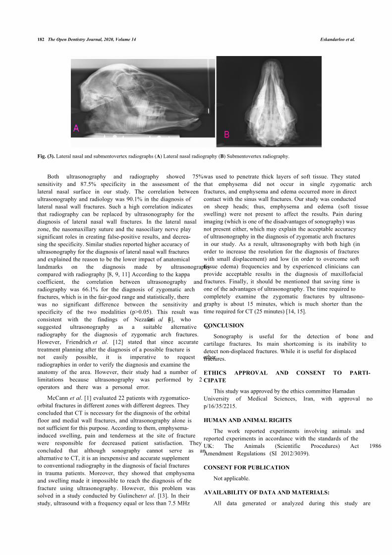
Fig. (3). Lateral nasal and submentovertex radiographs (A) Lateral nasal radiography (B) Submentovertex radiography.
Both ultrasonography and radiography showed 75%sensitivity and 87.5% specificity in the assessment of thelateral nasal surface in our study. The correlation betweenultrasonography and radiology was 90.1% in the diagnosis oflateral nasal wall fractures. Such a high correlation indicatesthat radiography can be replaced by ultrasonography for thediagnosis of lateral nasal wall fractures. In the lateral nasalzone, the nasomaxillary suture and the nasociliary nerve playsignificant roles in creating false-positive results, and decrea-sing the specificity. Similar studies reported higher accuracy ofultrasonography for the diagnosis of lateral nasal wall fracturesand explained the reason to be the lower impact of anatomicallandmarks on the diagnosis made by ultrasonographycompared with radiography [8, 9, 11] According to the kappacoefficient, the correlation between ultrasonography andradiography was 66.1% for the diagnosis of zygomatic archfractures, which is in the fair-good range and statistically, therewas no significant difference between the sensitivity andspecificity of the two modalities (p>0.05). This result wasconsistent with the findings of Nezafati et al. [4], whosuggested ultrasonography as a suitable alternative toradiography for the diagnosis of zygomatic arch fractures.However, Friendrich et al. [12] stated that since accuratetreatment planning after the diagnosis of a possible fracture isnot easily possible, it is imperative to request otherradiographies in order to verify the diagnosis and examine theanatomy of the area. However, their study had a number oflimitations because ultrasonography was performed by 2operators and there was a personal error.
McCann et al. [1] evaluated 22 patients with zygomatico-orbital fractures in different zones with different degrees. Theyconcluded that CT is necessary for the diagnosis of the orbitalfloor and medial wall fractures, and ultrasonography alone isnot sufficient for this purpose. According to them, emphysema-induced swelling, pain and tenderness at the site of fracturewere responsible for decreased patient satisfaction. Theyconcluded that although sonography cannot serve as analternative to CT, it is an inexpensive and accurate supplementto conventional radiography in the diagnosis of facial fracturesin trauma patients. Moreover, they showed that emphysemaand swelling made it impossible to reach the diagnosis of thefracture using ultrasonography. However, this problem wassolved in a study conducted by Gulincher et al. [13]. In theirstudy, ultrasound with a frequency equal or less than 7.5 MHz
was used to penetrate thick layers of soft tissue. They statedthat emphysema did not occur in single zygomatic archfractures, and emphysema and edema occurred more in directcontact with the sinus wall fractures. Our study was conductedon sheep heads; thus, emphysema and edema (soft tissueswelling) were not present to affect the results. Pain duringimaging (which is one of the disadvantages of sonography) wasnot present either, which may explain the acceptable accuracyof ultrasonography in the diagnosis of zygomatic arch fracturesin our study. As a result, ultrasonography with both high (inorder to increase the resolution for the diagnosis of fractureswith small displacement) and low (in order to overcome softtissue edema) frequencies and by experienced clinicians canprovide acceptable results in the diagnosis of maxillofacialfractures. Finally, it should be mentioned that saving time isone of the advantages of ultrasonography. The time required tocompletely examine the zygomatic fractures by ultrasono-graphy is about 15 minutes, which is much shorter than thetime required for CT (25 minutes) [14, 15].
CONCLUSION
Sonography is useful for the detection of bone andcartilage fractures. Its main shortcoming is its inability todetect non-displaced fractures. While it is useful for displacedfractures.
ETHICS APPROVAL AND CONSENT TO PARTI-CIPATE
This study was approved by the ethics committee HamadanUniversity of Medical Sciences, Iran, with approval nop/16/35/2215.
HUMAN AND ANIMAL RIGHTS
The work reported experiments involving animals andreported experiments in accordance with the standards of theUK: The Animals (Scientific Procedures) Act 1986Amendment Regulations (SI 2012/3039).
CONSENT FOR PUBLICATION
Not applicable.
AVAILABILITY OF DATA AND MATERIALS:
All data generated or analyzed during this study are

Is Ultrasonography Efficient for the Detection The Open Dentistry Journal, 2020, Volume 14 183
included in this published article.
FUNDING
None.
CONFLICT OF INTEREST
The authors declare no conflict of interest, financial orotherwise.
ACKNOWLEDGEMENTS
Declared none.
REFERENCES
McCann PJ, Brocklebank LM, Ayoub AF. Assessment of zygomatico-[1]orbital complex fractures using ultrasonography. Br J Oral MaxillofacSurg 2000; 38(5): 525-9.[http://dx.doi.org/10.1054/bjom.2000.0501] [PMID: 11010787]Flint PW, Cummings CW, Haughey B, Lund V, Niparko J, Richardson[2]M. Cummings otolaryngology head & neck surgery. Mosby 2010;3672.Baek HJ, Kim DW, Ryu JH, Lee YJ. identification of nasal bone[3]fractures on conventional radiography and facial CT: Comparison ofthe diagnostic accuracy in different imaging modalities and analysis ofinterobserver reliability. Iran J Radiol 2013; 10(3): 140-7.Nezafati S, Javadrashid R, Rad S, Akrami S. Comparison of[4]ultrasonography with submentovertex films and computed tomographyscan in the diagnosis of zygomatic arch fractures. DentomaxillofacRadiol 2010; 39(1): 11-6.[http://dx.doi.org/10.1259/dmfr/97056817] [PMID: 20089738]
Rowe N, Williams J. Fractures of the zygomatic complex and orbit.[5]Maxillofacial injuries 1985; 1: 1-43.Fonseca RJ, Barber HD, Powers MP, Frost DE. Oral and maxillofacial[6]trauma. 4th ed.. St.louis, Elsevier Health Sciences 2013. Chap 16,17.White SC, Pharoah MJ. Chap 2014; 13: 14.[7]Lee MH, Cha JG, Hong HS, et al. Comparison of high-resolution[8]ultrasonography and computed tomography in the diagnosis of nasalfractures. J Ultrasound Med 2009; 28(6): 717-23.[http://dx.doi.org/10.7863/jum.2009.28.6.717] [PMID: 19470811]Thiede O, Krömer JH, Rudack C, Stoll W, Osada N, Schmäl F.[9]Comparison of ultrasonography and conventional radiography in thediagnosis of nasal fractures. Arch Otolaryngol Head Neck Surg 2005;131(5): 434-9.[http://dx.doi.org/10.1001/archotol.131.5.434] [PMID: 15897423]Akizuki H, Yoshida H, Michi K. Ultrasonographic evaluation during[10]reduction of zygomatic arch fractures. J Craniomaxillofac Surg 1990;18(6): 263-6.[http://dx.doi.org/10.1016/S1010-5182(05)80428-7] [PMID: 2212025]Fouad Tarek, Rifaat Mohamed, Madian Yasser, Al-Hameed Azza[11]Abd. Utilization of ultrasound imaging for diagnosis of fracture nasalbone. Egypt J Ear, Nose, Throat, Allied Sci 2009; 10(1): 54-7.Friedrich RE, Plambeck K, Bartel-Friedrich S, Giese M, Schmelzle R.[12]Limitations of B-scan ultrasound for diagnosing fractures of themandibular condyle and ramus. Clin Oral Investig 2001; 5(1): 11-6.[http://dx.doi.org/10.1007/PL00010679] [PMID: 11355092]Gülicher D, Krimmel M, Reinert S. The role of intraoperative[13]ultrasonography in zygomatic complex fracture repair. Int J OralMaxillofac Surg 2006; 35(3): 224-30.[http://dx.doi.org/10.1016/j.ijom.2005.10.005] [PMID: 16364594]Heidari SF. Ultrasound applications in nasal bone fractures. Austin[14]Emerg Med 2018; 4(4): 220-2.Rajee A, Muralidhar Pai K, Smriti K, et al. Diagnostic accuracy of[15]ultrasonography in the assessment of facial fractures. PesquisaBrasileira em Odontopediatria e Clínica Integrada 2019; 19: e4832.
© 2020 Eskandarloo et al.
This is an open access article distributed under the terms of the Creative Commons Attribution 4.0 International Public License (CC-BY 4.0), a copy of which isavailable at: (https://creativecommons.org/licenses/by/4.0/legalcode). This license permits unrestricted use, distribution, and reproduction in any medium, providedthe original author and source are credited.